Axonal-SMN (a-SMN), a protein isoform of the survival motor neuron gene, is specifically involved in...
6
Axonal-SMN (a-SMN), a protein isoform of the survival motor neuron gene, is specifically involved in axonogenesis Veronica Setola*, Mineko Terao † , Denise Locatelli*, Stefania Bassanini*, Enrico Garattini † , and Giorgio Battaglia* ‡ *Molecular Neuroanatomy Laboratory, Department of Experimental Neurophysiology and Epileptology, Istituto Neurologico ‘‘C. Besta,’’ via Celoria 11, 20133 Milano, Italy; and † Molecular Biology Laboratory, Centro Catullo e Daniela Borgomainerio, Istituto di Ricerche Farmacologiche ‘‘Mario Negri,’’ via Eritrea 62, 20157 Milano, Italy Communicated by Erminio Costa, University of Illinois, Chicago, IL, December 4, 2006 (received for review December 13, 2005) Spinal muscular atrophy (SMA) is an autosomal recessive disease of childhood due to loss of the telomeric survival motor neuron gene, SMN1. The general functions of the main SMN1 protein product, full-length SMN (FL-SMN), do not explain the selective motoneu- ronal loss of SMA. We identified axonal-SMN (a-SMN), an alterna- tively spliced SMN form, preferentially encoded by the SMN1 gene in humans. The a-SMN transcript and protein are down-regulated during early development in different tissues. In the spinal cord, the a-SMN protein is selectively expressed in motor neurons and mainly localized in axons. Forced expression of a-SMN stimulates motor neuron axonogenesis in a time-dependent fashion and induces axonal-like growth in non-neuronal cells. Exons 2b and 3 are essential for the axonogenic effects. This discovery indicates an unexpected complexity of the SMN gene system and may help in understanding the pathogenesis of SMA. alternative splicing neurodegeneration spinal muscular atrophy intron retention axonal sprouting S pinal muscular atrophy (SMA) is an autosomal recessive dis- ease of childhood causing selective motor neuron death. SMA is the leading genetic cause of infant mortality, with an incidence of 1:10,000 and a carrier frequency of 1:50 (1). The telomeric survival motor neuron gene (SMN1) is the SMA disease gene, and the duplicated centromeric gene, SMN2, governs the severity of the disease (2, 3). Current knowledge suggests that SMN1 codes for a single functional protein [full-length SMN (FL-SMN)], and the major product of SMN2 is 7-SMN (lacking the C terminus), an unstable protein of minor significance (4, 5). The FL-SMN protein is expressed in the cytoplasm and nucleus of all cells (6, 7) and is involved in spliceosomal assembly and pre-mRNAs maturation (8–10). Other proposed roles include general cellular functions (11–15). How reduced FL-SMN levels lead to selective degeneration of motor neurons in SMA remains undetermined. FL-SMN may serve motor neuron-specific func- tions, perhaps interacting with partner proteins selectively ex- pressed in these cells. However, these functions and the partner proteins are still not known. Additional protein products of the SMN genes have been postulated (16–19). The identification of SMN proteins acting specifically in motor neurons would be a major step forward in understanding SMA. We identify and characterize an SMN transcript (GenBank accession no. AY876898) originating from the retention of SMN intron 3 and encoding the axonal-SMN (a-SMN) protein. In the spinal cord, a-SMN is selectively expressed in developing motor neurons and mainly localized in axons. This is confirmed by overexpression experiments in cultured cells. These also demon- strate the following: (i) a-SMN stimulates motor neuron axono- genesis in a time-dependent fashion, and (ii) a-SMN induces axonal-like growth in non-neuronal cells such as HeLa. The specific localization and function suggest a role in motor neurons. Because human a-SMN is a specific product of the SMN1 gene, its loss may be a determinant of SMA. Results Identification of an SMN Transcript in the Rat Spinal Cord. To search for new SMN transcripts, we analyzed the poly(A) RNA fraction of rat spinal cords with RACE/PCR and nested primers against exons 1 and 3. We isolated SMN cDNAs containing the entire sequence of intron 3, beside cDNAs consisting of the fully spliced form of FL-SMN (data not shown). The intron 3-containing transcript was named a-SMN. We defined the molecular structure of a-SMN mRNA in the poly(A) RNA fraction of rat spinal cord. On Northern blot analysis, an intron 3 cDNA probe revealed a specific transcript similar in size to the FL-SMN counterpart (Fig. 1a). These results were supported by RNase protection assays. An antisense RNA corresponding to a genomic sequence encompassing intron 2b, exon 3, intron 3, and exon 4 protected three fragments, originating from exons 3 and 4 of the FL-SMN mRNA, and the exon 3/intron 3/exon 4 sequence of the a-SMN mRNA. Thus, the a-SMN tran- script originates from the same genomic DNA strand encoding the FL-SMN mRNA and is also developmentally regulated (Fig. 1b). RT-PCR experiments with intron 3- and exon 1- or exon 8-primers (Fig. 1 c and d) indicated that the a-SMN transcript extends to exon 1 in the 5 direction and to exon 8 in the 3 direction. The presence of intron 3 in a-SMN was confirmed by the fact that (i) exon 3- and exon 4-primers amplified two DNA fragments corresponding to a-SMN mRNA, which retains the intron 3 sequence, and FL-SMN mRNA (Fig. 1c); and (ii) competitive PCR experiments with exon 3- and intron 3-/exon 6-primers indicated the presence of the FL-SMN and a-SMN transcripts. The latter mRNA was selectively down-regulated from embryonic day 15 to postnatal day 15 com- pared with the FL-SMN counterpart (Fig. 1e). These experiments indicate that the a-SMN mRNA accounts for only a small fraction of the total pool of SMN transcripts and is down-regulated during spinal cord development. Thus, the a-SMN transcript (Fig. 1h) is expressed mainly during the crucial phases of motoneuronal maturation, declining in adulthood. RACE-PCR and RT-PCR experiments were also conducted on the poly(A) RNA fraction of human spinal cord and the NB4 human myeloid cell line. RT-PCR experiments demonstrated the existence of the a-SMN transcript in humans, too (Fig. 1 f and g). 3-RACE-PCR experiments with intron 3-nested primers indicated that the human a-SMN mRNA is preferentially encoded by the Author contributions: V.S. and M.T. contributed equally to this work; V.S., M.T., E.G., and G.B. designed research; V.S., M.T., and D.L. performed research; S.B. contributed new reagents/analytic tools; V.S., D.L., and G.B. analyzed data; and E.G. and G.B. wrote the paper. The authors declare no conflict of interest. Abbreviations: FL-SMN, full-length SMN; IF, immunofluorescence; SMA, spinal muscular atrophy; a-SMN, axonal-SMN; WB, Western blot. Data deposition: The sequence reported in this paper has been deposited in the GenBank database (accession no. AY876898). ‡ To whom correspondence should be addressed. E-mail: [email protected]. © 2007 by The National Academy of Sciences of the USA www.pnas.orgcgidoi10.1073pnas.0610660104 PNAS February 6, 2007 vol. 104 no. 6 1959 –1964 NEUROSCIENCE
Transcript of Axonal-SMN (a-SMN), a protein isoform of the survival motor neuron gene, is specifically involved in...
Axonal-SMN (a-SMN), a protein isoform of thesurvival motor neuron gene, is specificallyinvolved in axonogenesisVeronica Setola*, Mineko Terao†, Denise Locatelli*, Stefania Bassanini*, Enrico Garattini†, and Giorgio Battaglia*‡
*Molecular Neuroanatomy Laboratory, Department of Experimental Neurophysiology and Epileptology, Istituto Neurologico ‘‘C. Besta,’’ via Celoria 11,20133 Milano, Italy; and †Molecular Biology Laboratory, Centro Catullo e Daniela Borgomainerio, Istituto di Ricerche Farmacologiche ‘‘Mario Negri,’’via Eritrea 62, 20157 Milano, Italy
Communicated by Erminio Costa, University of Illinois, Chicago, IL, December 4, 2006 (received for review December 13, 2005)
Spinal muscular atrophy (SMA) is an autosomal recessive disease ofchildhood due to loss of the telomeric survival motor neuron gene,SMN1. The general functions of the main SMN1 protein product,full-length SMN (FL-SMN), do not explain the selective motoneu-ronal loss of SMA. We identified axonal-SMN (a-SMN), an alterna-tively spliced SMN form, preferentially encoded by the SMN1 genein humans. The a-SMN transcript and protein are down-regulatedduring early development in different tissues. In the spinal cord,the a-SMN protein is selectively expressed in motor neurons andmainly localized in axons. Forced expression of a-SMN stimulatesmotor neuron axonogenesis in a time-dependent fashion andinduces axonal-like growth in non-neuronal cells. Exons 2b and 3are essential for the axonogenic effects. This discovery indicates anunexpected complexity of the SMN gene system and may help inunderstanding the pathogenesis of SMA.
alternative splicing � neurodegeneration � spinal muscular atrophy �intron retention � axonal sprouting
Spinal muscular atrophy (SMA) is an autosomal recessive dis-ease of childhood causing selective motor neuron death. SMA
is the leading genetic cause of infant mortality, with an incidence of1:10,000 and a carrier frequency of 1:50 (1). The telomeric survivalmotor neuron gene (SMN1) is the SMA disease gene, and theduplicated centromeric gene, SMN2, governs the severity of thedisease (2, 3). Current knowledge suggests that SMN1 codes for asingle functional protein [full-length SMN (FL-SMN)], and themajor product of SMN2 is �7-SMN (lacking the C terminus), anunstable protein of minor significance (4, 5).
The FL-SMN protein is expressed in the cytoplasm and nucleusof all cells (6, 7) and is involved in spliceosomal assembly andpre-mRNAs maturation (8–10). Other proposed roles includegeneral cellular functions (11–15). How reduced FL-SMN levelslead to selective degeneration of motor neurons in SMA remainsundetermined. FL-SMN may serve motor neuron-specific func-tions, perhaps interacting with partner proteins selectively ex-pressed in these cells. However, these functions and the partnerproteins are still not known. Additional protein products of theSMN genes have been postulated (16–19). The identification ofSMN proteins acting specifically in motor neurons would be a majorstep forward in understanding SMA.
We identify and characterize an SMN transcript (GenBankaccession no. AY876898) originating from the retention of SMNintron 3 and encoding the axonal-SMN (a-SMN) protein. In thespinal cord, a-SMN is selectively expressed in developing motorneurons and mainly localized in axons. This is confirmed byoverexpression experiments in cultured cells. These also demon-strate the following: (i) a-SMN stimulates motor neuron axono-genesis in a time-dependent fashion, and (ii) a-SMN inducesaxonal-like growth in non-neuronal cells such as HeLa. The specificlocalization and function suggest a role in motor neurons. Becausehuman a-SMN is a specific product of the SMN1 gene, its loss maybe a determinant of SMA.
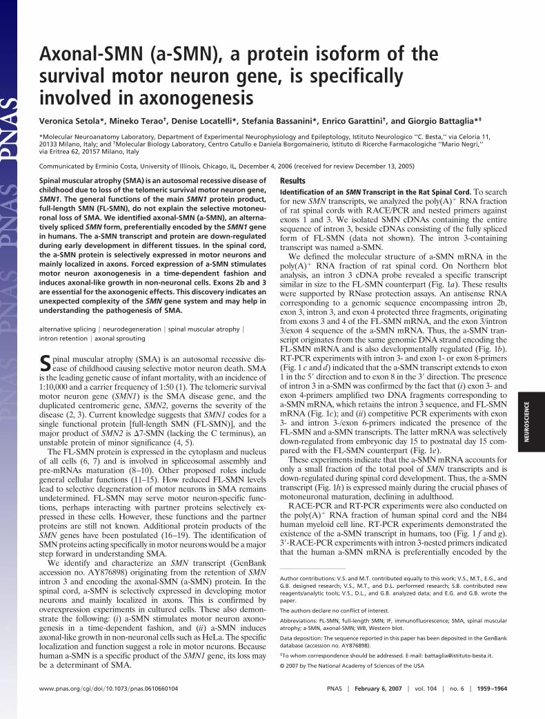
ResultsIdentification of an SMN Transcript in the Rat Spinal Cord. To searchfor new SMN transcripts, we analyzed the poly(A)� RNA fractionof rat spinal cords with RACE/PCR and nested primers againstexons 1 and 3. We isolated SMN cDNAs containing the entiresequence of intron 3, beside cDNAs consisting of the fully splicedform of FL-SMN (data not shown). The intron 3-containingtranscript was named a-SMN.
We defined the molecular structure of a-SMN mRNA in thepoly(A)� RNA fraction of rat spinal cord. On Northern blotanalysis, an intron 3 cDNA probe revealed a specific transcriptsimilar in size to the FL-SMN counterpart (Fig. 1a). These resultswere supported by RNase protection assays. An antisense RNAcorresponding to a genomic sequence encompassing intron 2b,exon 3, intron 3, and exon 4 protected three fragments, originatingfrom exons 3 and 4 of the FL-SMN mRNA, and the exon 3/intron3/exon 4 sequence of the a-SMN mRNA. Thus, the a-SMN tran-script originates from the same genomic DNA strand encoding theFL-SMN mRNA and is also developmentally regulated (Fig. 1b).RT-PCR experiments with intron 3- and exon 1- or exon 8-primers(Fig. 1 c and d) indicated that the a-SMN transcript extends to exon1 in the 5� direction and to exon 8 in the 3� direction. The presenceof intron 3 in a-SMN was confirmed by the fact that (i) exon 3- andexon 4-primers amplified two DNA fragments corresponding toa-SMN mRNA, which retains the intron 3 sequence, and FL-SMNmRNA (Fig. 1c); and (ii) competitive PCR experiments with exon3- and intron 3-/exon 6-primers indicated the presence of theFL-SMN and a-SMN transcripts. The latter mRNA was selectivelydown-regulated from embryonic day 15 to postnatal day 15 com-pared with the FL-SMN counterpart (Fig. 1e).
These experiments indicate that the a-SMN mRNA accounts foronly a small fraction of the total pool of SMN transcripts and isdown-regulated during spinal cord development. Thus, the a-SMNtranscript (Fig. 1h) is expressed mainly during the crucial phases ofmotoneuronal maturation, declining in adulthood.
RACE-PCR and RT-PCR experiments were also conducted onthe poly(A)� RNA fraction of human spinal cord and the NB4human myeloid cell line. RT-PCR experiments demonstrated theexistence of the a-SMN transcript in humans, too (Fig. 1 f and g).3�-RACE-PCR experiments with intron 3-nested primers indicatedthat the human a-SMN mRNA is preferentially encoded by the
Author contributions: V.S. and M.T. contributed equally to this work; V.S., M.T., E.G., andG.B. designed research; V.S., M.T., and D.L. performed research; S.B. contributed newreagents/analytic tools; V.S., D.L., and G.B. analyzed data; and E.G. and G.B. wrote thepaper.
The authors declare no conflict of interest.
Abbreviations: FL-SMN, full-length SMN; IF, immunofluorescence; SMA, spinal muscularatrophy; a-SMN, axonal-SMN; WB, Western blot.
Data deposition: The sequence reported in this paper has been deposited in the GenBankdatabase (accession no. AY876898).
‡To whom correspondence should be addressed. E-mail: [email protected].
© 2007 by The National Academy of Sciences of the USA
www.pnas.org�cgi�doi�10.1073�pnas.0610660104 PNAS � February 6, 2007 � vol. 104 � no. 6 � 1959–1964
NEU
ROSC
IEN
CE
human SMN1 gene. Sequence analysis of �60 clones from NB4mRNA showed that all clones came from the SMN1 gene becausethere was a C at position �6 in exon 7 and a G at position �233 inexon 8. Approximately two thirds of the clones showed exon 5skipping. These data are unlikely to be biased because primerstargeting exons 2b and 3 amplify fragments corresponding to theFL-SMN and �7-SMN mRNAs from the SMN2 gene.
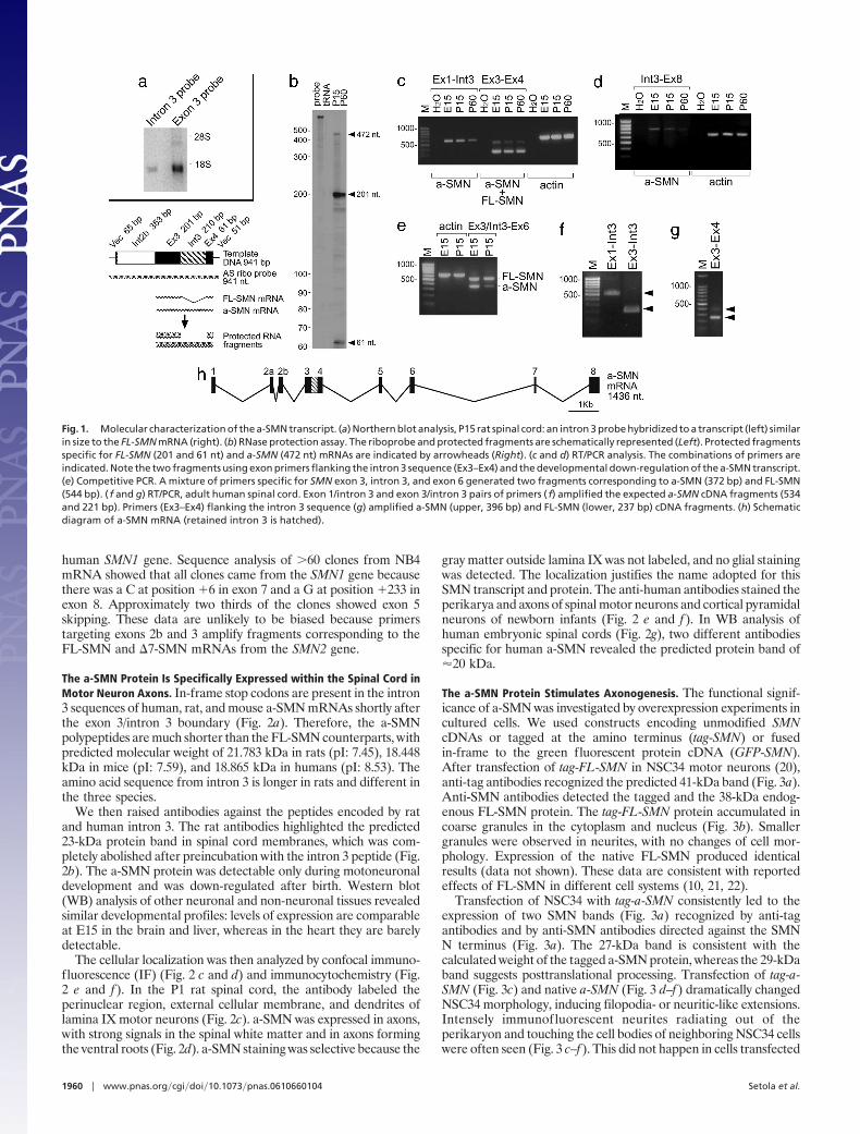
The a-SMN Protein Is Specifically Expressed within the Spinal Cord inMotor Neuron Axons. In-frame stop codons are present in the intron3 sequences of human, rat, and mouse a-SMN mRNAs shortly afterthe exon 3/intron 3 boundary (Fig. 2a). Therefore, the a-SMNpolypeptides are much shorter than the FL-SMN counterparts, withpredicted molecular weight of 21.783 kDa in rats (pI: 7.45), 18.448kDa in mice (pI: 7.59), and 18.865 kDa in humans (pI: 8.53). Theamino acid sequence from intron 3 is longer in rats and different inthe three species.
We then raised antibodies against the peptides encoded by ratand human intron 3. The rat antibodies highlighted the predicted23-kDa protein band in spinal cord membranes, which was com-pletely abolished after preincubation with the intron 3 peptide (Fig.2b). The a-SMN protein was detectable only during motoneuronaldevelopment and was down-regulated after birth. Western blot(WB) analysis of other neuronal and non-neuronal tissues revealedsimilar developmental profiles: levels of expression are comparableat E15 in the brain and liver, whereas in the heart they are barelydetectable.
The cellular localization was then analyzed by confocal immuno-fluorescence (IF) (Fig. 2 c and d) and immunocytochemistry (Fig.2 e and f). In the P1 rat spinal cord, the antibody labeled theperinuclear region, external cellular membrane, and dendrites oflamina IX motor neurons (Fig. 2c). a-SMN was expressed in axons,with strong signals in the spinal white matter and in axons formingthe ventral roots (Fig. 2d). a-SMN staining was selective because the
gray matter outside lamina IX was not labeled, and no glial stainingwas detected. The localization justifies the name adopted for thisSMN transcript and protein. The anti-human antibodies stained theperikarya and axons of spinal motor neurons and cortical pyramidalneurons of newborn infants (Fig. 2 e and f). In WB analysis ofhuman embryonic spinal cords (Fig. 2g), two different antibodiesspecific for human a-SMN revealed the predicted protein band of�20 kDa.
The a-SMN Protein Stimulates Axonogenesis. The functional signif-icance of a-SMN was investigated by overexpression experiments incultured cells. We used constructs encoding unmodified SMNcDNAs or tagged at the amino terminus (tag-SMN) or fusedin-frame to the green fluorescent protein cDNA (GFP-SMN).After transfection of tag-FL-SMN in NSC34 motor neurons (20),anti-tag antibodies recognized the predicted 41-kDa band (Fig. 3a).Anti-SMN antibodies detected the tagged and the 38-kDa endog-enous FL-SMN protein. The tag-FL-SMN protein accumulated incoarse granules in the cytoplasm and nucleus (Fig. 3b). Smallergranules were observed in neurites, with no changes of cell mor-phology. Expression of the native FL-SMN produced identicalresults (data not shown). These data are consistent with reportedeffects of FL-SMN in different cell systems (10, 21, 22).
Transfection of NSC34 with tag-a-SMN consistently led to theexpression of two SMN bands (Fig. 3a) recognized by anti-tagantibodies and by anti-SMN antibodies directed against the SMNN terminus (Fig. 3a). The 27-kDa band is consistent with thecalculated weight of the tagged a-SMN protein, whereas the 29-kDaband suggests posttranslational processing. Transfection of tag-a-SMN (Fig. 3c) and native a-SMN (Fig. 3 d–f) dramatically changedNSC34 morphology, inducing filopodia- or neuritic-like extensions.Intensely immunofluorescent neurites radiating out of theperikaryon and touching the cell bodies of neighboring NSC34 cellswere often seen (Fig. 3 c–f). This did not happen in cells transfected
Fig. 1. Molecular characterization of the a-SMN transcript. (a) Northern blot analysis, P15 rat spinal cord: an intron 3 probe hybridized to a transcript (left) similarin size to the FL-SMN mRNA (right). (b) RNase protection assay. The riboprobe and protected fragments are schematically represented (Left). Protected fragmentsspecific for FL-SMN (201 and 61 nt) and a-SMN (472 nt) mRNAs are indicated by arrowheads (Right). (c and d) RT/PCR analysis. The combinations of primers areindicated. Note the two fragments using exon primers flanking the intron 3 sequence (Ex3–Ex4) and the developmental down-regulation of the a-SMN transcript.(e) Competitive PCR. A mixture of primers specific for SMN exon 3, intron 3, and exon 6 generated two fragments corresponding to a-SMN (372 bp) and FL-SMN(544 bp). ( f and g) RT/PCR, adult human spinal cord. Exon 1/intron 3 and exon 3/intron 3 pairs of primers ( f) amplified the expected a-SMN cDNA fragments (534and 221 bp). Primers (Ex3–Ex4) flanking the intron 3 sequence (g) amplified a-SMN (upper, 396 bp) and FL-SMN (lower, 237 bp) cDNA fragments. (h) Schematicdiagram of a-SMN mRNA (retained intron 3 is hatched).
1960 � www.pnas.org�cgi�doi�10.1073�pnas.0610660104 Setola et al.
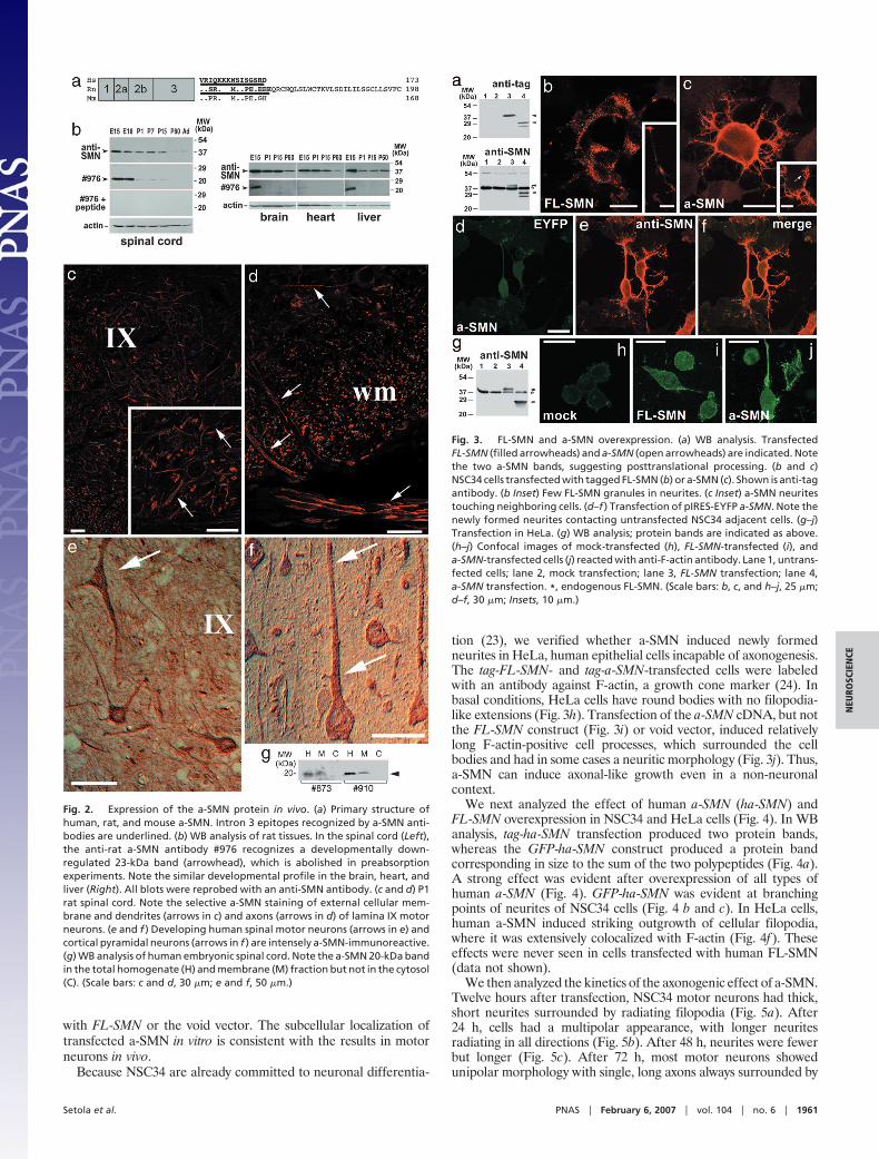
with FL-SMN or the void vector. The subcellular localization oftransfected a-SMN in vitro is consistent with the results in motorneurons in vivo.
Because NSC34 are already committed to neuronal differentia-
tion (23), we verified whether a-SMN induced newly formedneurites in HeLa, human epithelial cells incapable of axonogenesis.The tag-FL-SMN- and tag-a-SMN-transfected cells were labeledwith an antibody against F-actin, a growth cone marker (24). Inbasal conditions, HeLa cells have round bodies with no filopodia-like extensions (Fig. 3h). Transfection of the a-SMN cDNA, but notthe FL-SMN construct (Fig. 3i) or void vector, induced relativelylong F-actin-positive cell processes, which surrounded the cellbodies and had in some cases a neuritic morphology (Fig. 3j). Thus,a-SMN can induce axonal-like growth even in a non-neuronalcontext.
We next analyzed the effect of human a-SMN (ha-SMN) andFL-SMN overexpression in NSC34 and HeLa cells (Fig. 4). In WBanalysis, tag-ha-SMN transfection produced two protein bands,whereas the GFP-ha-SMN construct produced a protein bandcorresponding in size to the sum of the two polypeptides (Fig. 4a).A strong effect was evident after overexpression of all types ofhuman a-SMN (Fig. 4). GFP-ha-SMN was evident at branchingpoints of neurites of NSC34 cells (Fig. 4 b and c). In HeLa cells,human a-SMN induced striking outgrowth of cellular filopodia,where it was extensively colocalized with F-actin (Fig. 4f). Theseeffects were never seen in cells transfected with human FL-SMN(data not shown).
We then analyzed the kinetics of the axonogenic effect of a-SMN.Twelve hours after transfection, NSC34 motor neurons had thick,short neurites surrounded by radiating filopodia (Fig. 5a). After24 h, cells had a multipolar appearance, with longer neuritesradiating in all directions (Fig. 5b). After 48 h, neurites were fewerbut longer (Fig. 5c). After 72 h, most motor neurons showedunipolar morphology with single, long axons always surrounded by
Fig. 2. Expression of the a-SMN protein in vivo. (a) Primary structure ofhuman, rat, and mouse a-SMN. Intron 3 epitopes recognized by a-SMN anti-bodies are underlined. (b) WB analysis of rat tissues. In the spinal cord (Left),the anti-rat a-SMN antibody #976 recognizes a developmentally down-regulated 23-kDa band (arrowhead), which is abolished in preabsorptionexperiments. Note the similar developmental profile in the brain, heart, andliver (Right). All blots were reprobed with an anti-SMN antibody. (c and d) P1rat spinal cord. Note the selective a-SMN staining of external cellular mem-brane and dendrites (arrows in c) and axons (arrows in d) of lamina IX motorneurons. (e and f ) Developing human spinal motor neurons (arrows in e) andcortical pyramidal neurons (arrows in f ) are intensely a-SMN-immunoreactive.(g) WB analysis of human embryonic spinal cord. Note the a-SMN 20-kDa bandin the total homogenate (H) and membrane (M) fraction but not in the cytosol(C). (Scale bars: c and d, 30 �m; e and f, 50 �m.)
Fig. 3. FL-SMN and a-SMN overexpression. (a) WB analysis. TransfectedFL-SMN (filled arrowheads) and a-SMN (open arrowheads) are indicated. Notethe two a-SMN bands, suggesting posttranslational processing. (b and c)NSC34 cells transfected with tagged FL-SMN (b) or a-SMN (c). Shown is anti-tagantibody. (b Inset) Few FL-SMN granules in neurites. (c Inset) a-SMN neuritestouching neighboring cells. (d–f ) Transfection of pIRES-EYFP a-SMN. Note thenewly formed neurites contacting untransfected NSC34 adjacent cells. (g–j)Transfection in HeLa. (g) WB analysis; protein bands are indicated as above.(h–j) Confocal images of mock-transfected (h), FL-SMN-transfected (i), anda-SMN-transfected cells (j) reacted with anti-F-actin antibody. Lane 1, untrans-fected cells; lane 2, mock transfection; lane 3, FL-SMN transfection; lane 4,a-SMN transfection. *, endogenous FL-SMN. (Scale bars: b, c, and h–j, 25 �m;d–f, 30 �m; Insets, 10 �m.)
Setola et al. PNAS � February 6, 2007 � vol. 104 � no. 6 � 1961
NEU
ROSC
IEN
CE
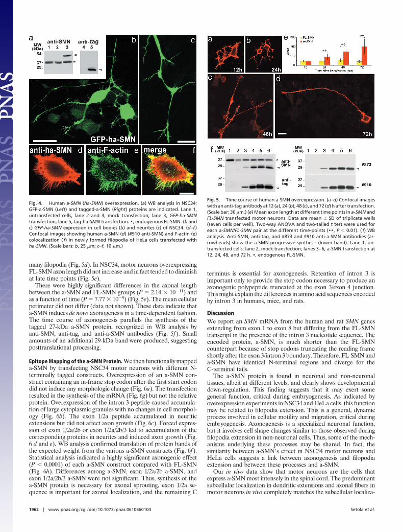
many filopodia (Fig. 5d). In NSC34, motor neurons overexpressingFL-SMN axon length did not increase and in fact tended to diminishat late time points (Fig. 5e).
There were highly significant differences in the axonal lengthbetween the a-SMN and FL-SMN groups (P � 2.14 � 10�11) andas a function of time (P � 7.77 � 10�9) (Fig. 5e). The mean cellularperimeter did not differ (data not shown). These data indicate thata-SMN induces de novo axonogenesis in a time-dependent fashion.The time course of axonogenesis parallels the synthesis of thetagged 27-kDa a-SMN protein, recognized in WB analysis byanti-SMN, anti-tag, and anti-a-SMN antibodies (Fig. 5f). Smallamounts of an additional 29-kDa band were produced, suggestingposttranslational processing.
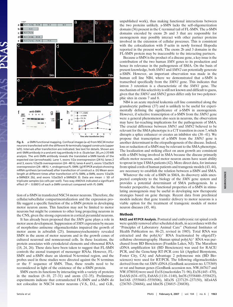
Epitope Mapping of the a-SMN Protein. We then functionally mappeda-SMN by transfecting NSC34 motor neurons with different N-terminally tagged constructs. Overexpression of an a-SMN con-struct containing an in-frame stop codon after the first start codondid not induce any morphologic change (Fig. 6a). The transfectionresulted in the synthesis of the mRNA (Fig. 6g) but not the relativeprotein. Overexpression of the intron 3 peptide caused accumula-tion of large cytoplasmic granules with no changes in cell morphol-ogy (Fig. 6b). The exon 1/2a peptide accumulated in neuriticextensions but did not affect axon growth (Fig. 6c). Forced expres-sion of exon 1/2a/2b or exon 1/2a/2b/3 led to accumulation of thecorresponding proteins in neurites and induced axon growth (Fig.6 d and e). WB analysis confirmed translation of protein bands ofthe expected weight from the various a-SMN constructs (Fig. 6f).Statistical analysis indicated a highly significant axonogenic effect(P 0.0001) of each a-SMN construct compared with FL-SMN(Fig. 6h). Differences among a-SMN, exon 1/2a/2b a-SMN, andexon 1/2a/2b/3 a-SMN were not significant. Thus, synthesis of thea-SMN protein is necessary for axonal sprouting, exon 1/2a se-quence is important for axonal localization, and the remaining C
terminus is essential for axonogenesis. Retention of intron 3 isimportant only to provide the stop codon necessary to produce anaxonogenic polypeptide truncated at the exon 3/exon 4 junction.This might explain the differences in amino acid sequences encodedby intron 3 in humans, mice, and rats.
DiscussionWe report an SMN mRNA from the human and rat SMN genesextending from exon 1 to exon 8 but differing from the FL-SMNtranscript in the presence of the intron 3 nucleotide sequence. Theencoded protein, a-SMN, is much shorter than the FL-SMNcounterpart because of stop codons truncating the reading frameshortly after the exon 3/intron 3 boundary. Therefore, FL-SMN anda-SMN have identical N-terminal regions and diverge for theC-terminal tails.
The a-SMN protein is found in neuronal and non-neuronaltissues, albeit at different levels, and clearly shows developmentaldown-regulation. This finding suggests that it may exert somegeneral function, critical during embryogenesis. As indicated byoverexpression experiments in NSC34 and HeLa cells, this functionmay be related to filopodia extension. This is a general, dynamicprocess involved in cellular motility and migration, critical duringembryogenesis. Axonogenesis is a specialized neuronal function,but it involves cell shape changes similar to those observed duringfilopodia extension in non-neuronal cells. Thus, some of the mech-anisms underlying these processes may be shared. In fact, thesimilarity between a-SMN�s effect in NSC34 motor neurons andHeLa cells suggests a link between axonogenesis and filopodiaextension and between these processes and a-SMN.
Our in vivo data show that motor neurons are the cells thatexpress a-SMN most intensely in the spinal cord. The predominantsubcellular localization in dendritic extensions and axonal fibers inmotor neurons in vivo completely matches the subcellular localiza-
Fig. 4. Human a-SMN (ha-SMN) overexpression. (a) WB analysis in NSC34;GFP-a-SMN (Left) and tagged-a-SMN (Right) proteins are indicated. Lane 1,untransfected cells; lane 2 and 4, mock transfection; lane 3, GFP-ha-SMNtransfection; lane 5, tag-ha-SMN transfection. *, endogenous FL-SMN. (b andc) GFP-ha-SMN expression in cell bodies (b) and neurites (c) of NSC34. (d–f )Confocal images showing human a-SMN (d) (#910 anti-SMN) and F-actin (e)colocalization ( f) in newly formed filopodia of HeLa cells transfected withha-SMN. (Scale bars: b, 25 �m; c–f, 10 �m.)
Fig. 5. Time course of human a-SMN overexpression. (a–d) Confocal imageswith an anti-tag antibody at 12 (a), 24 (b), 48 (c), and 72 (d) h after transfection.(Scale bar: 30 �m.) (e) Mean axon length at different time points in a-SMN andFL-SMN transfected motor neurons. Data are mean SD of triplicate wells(seven cells per well). Two-way ANOVA and two-tailed t test were used foreach a-SMN/FL-SMN pair at the different time-points (**, P 0.01). ( f) WBanalysis. Anti-SMN, anti-tag, and #873 and #910 anti-a-SMN antibodies (ar-rowheads) show the a-SMN progressive synthesis (lower band). Lane 1, un-transfected cells; lane 2, mock transfection; lanes 3–6, a-SMN transfection at12, 24, 48, and 72 h. *, endogenous FL-SMN.
1962 � www.pnas.org�cgi�doi�10.1073�pnas.0610660104 Setola et al.
tion of a-SMN in transfected NSC34 motor neurons. Therefore, thecellular/subcellular compartmentalization and the expression pro-file suggest a specific function of the a-SMN protein in developingmotor neuron axons. This function may not be limited to motorneurons but might be common to other long projecting neurons inthe CNS, given the strong expression in cortical pyramidal neurons.
It has already been proposed that the SMN gene plays a role inmotor axon development. Suppression of SMN expression by meansof morpholino antisense oligonucleotides impaired the growth ofmotor axons in zebrafish (25). Immunocytochemistry revealedSMN in the axons of motor neurons and in the growth cones ofcultured neuronal and nonneuronal cells. In these locations, theprotein associates with cytoskeletal elements and ribosomal RNA(18, 24, 26). These data have been taken to suggest that FL-SMNcontrols the axonal transport of messenger RNAs. However, FL-SMN and a-SMN share an identical N-terminal region, and theprobes used in those studies were directed against the N terminusor the 5� sequence of SMN. Thus, these results need to bereconsidered in light of the existence of the a-SMN protein.
SMN exerts its functions by interacting with a variety of proteinsin the nucleus (8–10, 27–31) and axons (32–35). Preliminaryexperiments indicate that cotransfected FL-SMN and a-SMN donot colocalize in NSC34 motor neurons (V.S., D.L., and G.B.,
unpublished work), thus making functional interactions betweenthe two proteins unlikely. a-SMN lacks the self-oligomerizationdomain (36) present in the C-terminal tail of FL-SMN. The a-SMNdomains encoded by exons 2b and 3 that are responsible foraxonogenesis may possibly interact with other partner proteinsinvolved in the extension of cellular processes. This is consistentwith the colocalization with F-actin in newly formed filopodiareported in the present work. The exons 2b and 3 domains in theFL-SMN protein may be inaccessible to the interacting partners.
Because a-SMN is the product of a disease gene, a key issue is thecontribution of the two human SMN genes to its production andhence its relevance in the pathogenesis of SMA. On the basis ofpresent knowledge, both SMN1 and SMN2 can potentially generatea-SMN. However, an important observation was made in thehuman cell line NB4, where we demonstrated that a-SMN istranscribed specifically from the SMN1 gene. This indicates thatintron 3 retention is a characteristic of the SMN1 gene. Themechanism of this selectivity is still not known and difficult to guess,given that the SMN1 and SMN2 genes differ only for two polymor-phic sites in exons 7 and 8.
NB4 is an acute myeloid leukemia cell line committed along thegranulocytic pathway (37) and is unlikely to be useful for experi-mentally defining the significance of a-SMN in axonogenesis.However, if selective transcription of a-SMN from the SMN1 genewere a general phenomenon also seen in neurons, the observationmay have far-reaching implications for the pathogenesis of SMA.The crucial difference between SMN1 and SMN2 believed to berelevant for the SMA phenotype is a C/T transition in exon 7, whichdisrupts a splice enhancer or creates an inhibitor site (38–41). Wepropose that transcription of a-SMN from the SMN1 gene isanother determinant in the etiopathogenesis of the disease. Indeed,loss or reduction of a-SMN may be relevant to the SMA phenotype.The localization and striking effect on axon growth are consistentwith a-SMN being involved in SMA because the disease primarilyaffects motor neurons, and motor neuron axons have scant abilityto sprout in type I SMA patients (42). More direct data, for instancefrom spinal cords of human patients and transgenic models (43, 44),are necessary to establish the relation between a-SMN and SMA.
Whatever the role of a-SMN in SMA, its discovery adds unex-pected complexity to the biology of the SMN gene system andprovides a potential determinant of SMA pathogenesis. In abroader perspective, the functional properties of a-SMN in stimu-lating axonogenesis may be useful in developing new therapeuticstrategies based on gene therapy. Recent data from preclinicalmodels indicate that gene transfer delivery to motor neurons is aviable option for the treatment of transgenic models of motorneuron diseases (45, 46).
MethodsRACE and RT/PCR Analysis. Postnatal and embryonic rat spinal cordswere quickly removed after scheduled death, in accordance with the‘‘Principles of Laboratory Animal Care’’ (National Institutes ofHealth Publication no. 86-23, revised in 1985). Total RNA wasextracted and the poly(A)� RNA fractionated by oligo(dT)-cellulose chromatography. Human spinal poly(A)� RNA was pur-chased from BD Biosciences (Franklin Lakes, NJ). The MarathoncDNA amplification kit (BD Biosciences) was used for RACE/PCR, and the GeneAmp RT-PCR core kit (Applied Biosystems,Foster City, CA) and Advantage 2 polymerase mix (BD Bio-sciences) were used for RT/PCR. The following oligonucleotidesderived from the rat SMN cDNA (GenBank accession no. U75369)or rat and human genes (GenBank accession nos. NW�047617 andNW�078018) were used: Ex1S (nucleotides 71–96), Ex3S (445–470),Ex4AS (634–653), Ex8AS (1118–1140), Int3S (5558400–5558425),Int3AS (5558481–5558506), hEx3S (237129–237150), hEx4AS(236783–236684), and hInt3S (236815–236838).
Fig. 6. a-SMN functional mapping. Confocal images (a–e) from NSC34 motorneurons transfected with the different N-terminally tagged constructs (upperleft). Intervals after transfection are indicated. See text for details. Shown areanti-SMN antibody in a and anti-tag antibody in b–e. (Scale bar: 30 �m.) ( f) WBanalysis. The anti-SMN antibody reveals the translated a-SMN bands of theexpected size (arrowheads). Lane 1, exons 1/2a overexpression (24 h); lanes 2and 3, exons 1/2a/2b overexpression (24–48 h); lanes 4 and 5, exons 1/2a/2b/3overexpression (24–48 h). *, endogenous FL-SMN. (g) RT/PCR analysis showingmRNA synthesis (arrowhead) after transfection of construct a. (h) Mean axonlength at different times after transfection of FL-SMN, a-SMN, exons 1/2a/2ba-SMN(EX 2b), and exons 1/2a/2b/3 a-SMN(EX 3). Data are mean SD oftriplicate samples (six cells per well). Two-way ANOVA indicated a significanteffect (P 0.0001) of each a-SMN construct compared with FL-SMN.
Setola et al. PNAS � February 6, 2007 � vol. 104 � no. 6 � 1963
NEU
ROSC
IEN
CE
Northern Blot and RNase Protection Analyses. Northern blots weredone according to standard procedures. Exon 3- and intron3-specific cDNA probes were radiolabeled by PCR with[�-32P]dCTP. For RNase protection assay, a rat genomic frag-ment containing 353 bp of intron 2b, intact exon 3 and intron 3,and 61 bp of exon 4 was amplified and cloned in pBluescriptIIKS (Stratagene, La Jolla, CA). Antisense riboprobes weresynthesized with [�-32P]UTP and hybridized to poly(A)� RNA(5 �g each) and RNase-digested by using the RPAIII kit(Ambion, Austin, TX). RNA century markers (Ambion) were[�-32P]dUTP-labeled.
Antibodies. Anti-peptide rabbit polyclonal antibodies against theC-terminal region of rat (#937 and #976) or human (#873 and#910) a-SMN were prepared by NeoMPS and used at 1:1,000dilution. Mouse anti-SMN was purchased from BD Biosciences(anti-SMN, clone 8; diluted 1:1,000 for IF, 1:25,000 for WBanalysis), mouse anti-F-actin was purchased from Chemicon (di-luted 1:1,000–1:5,000; Temecula, CA), and mouse anti-tag (anti-Xpress and anti-HisG) was purchased from Invitrogen (diluted1:1,000, Carlsbad, CA).
Western Blot Analysis. Tissues were removed from embryonic,postnatal, and adult rats. Embryonic (15 weeks) human spinal cordswere obtained through a protocol for fetal stem cell production andapproved by the ethics committee of the Neurological Institute. Alltissues were homogenized in buffer containing 20 mM Hepes (pH7.4), 1 mM DTT, 1 mM EGTA, 0.1 mM PMSF, and Complete(Roche, Indianapolis, IN). Cellular membrane and soluble frac-tions were obtained by pelleting nuclei at 1,000 � g for 10 min, thencentrifuging the supernatant at 100,000 � g for 45 min. Transfectedcell cultures were lysed in buffer containing 0.1 M Na-phosphate(pH 7.4), 0.2% Triton X-100, 0.1 mM EDTA, 0.2 mM PMSF, 1�g/ml aprotinin, and 1 �g/ml leupeptin by three freezing andthawing cycles. The cell lysates were centrifuged at 13,000 � g.Protein extracts were separated on SDS/12% PAGE, blotted tonitrocellulose membranes (1 h, 180 mA), and analyzed with ECL.
Histology, Immunocytochemistry, and Immunofluorescence Analysis.After perfusion with 4% paraformaldehyde, rat spinal cordswere dissected and postfixed overnight. Human cerebral speci-
mens were obtained from neurosurgery of non-SMA patientsand fixed by immersion in 4% paraformaldehyde. Spinal cordsand cerebral tissues were cut on a Vibratome. Paraplast-embedded spinal cord of a non-SMA 6-month-old infant wasobtained from the Neuropathology Unit of the NeurologicalInstitute and cut with a rotary microtome. Sections were ana-lyzed by immunocytochemistry and IF. Immunocytochemistryslides were analyzed with a Nikon (Florham Park, NJ) Micro-phot FXA microscope. IF images were acquired on a Bio-Rad(Hercules, CA) Radiance 2100 confocal microscope at 1,024 �1,024-pixel resolution.
Transfection. Appropriate cDNA fragments were cloned inpcDNA4/HisMaxTOPO (Invitrogen), bicistronic pIRES-EYFP(BD Biosciences), or GFP Fusion TOPO (Invitrogen) expressionvectors. Transfection was with Lipofectamine Plus (Invitrogen).Transfected cells were harvested after 6, 12, 24, 48, and 72 h andeither used for WB analysis or fixed in 4% paraformaldehyde/4%sucrose for confocal IF.
Cell Measurements and Statistical Analysis. After transfection, theperimeters, areas, and axon lengths of transfected cells werequantified at the different time points (12, 24, 48, and 72 h) by usingLaser Sharp 2000 software (Bio-Rad) on confocal images ofindividual motor neurons. For statistical analysis, triplicate exper-iments were considered. Data were analyzed by two-way ANOVAand two-tailed t test for the different constructs transfected at thedifferent time points.
We thank A. Giavazzi, A. Finardi, and B. Copes for earlier contributions;M. Di Luca and M. Salmona for insightful comments; A. Poletti forvaluable suggestions in cell cultures; M. Fratelli and A. Tedeschi forstatistical analysis; G. Ciboldi and F. Deceglie for technical support; andJ. Baggott for language assistance. This work was supported by grantsfrom the Spinal Muscular Atrophy Foundation (to G.B.), CariploFoundation Grant 2002.1836/10.4898 (to G.B.), and Italian Ministry ofHealth Fondo per gli Investimenti della Ricerca di Base GrantsRBNE01B5WW005 (to G.B.) and RBAU019CXS (to E.G.).
1. Pearn J (1980) Lancet 1:919–922.2. Lefebvre S, Burglen L, Reboullet S, Clermont O, Burlet P, Viollet L, Benichou B, Cruaud
C, Millasseau P, Zeviani M, et al. (1995) Cell 80:155–165.3. Feldkotter M, Schwarzer V, Wirth R, Wienker TF, Wirth B (2002) Am J Hum Genet
70:358–368.4. Lorson CL, Hahnen E, Androphy EJ, Wirth B (1999) Proc Natl Acad Sci USA 96:6307–6311.5. Gennarelli M, Lucarelli M, Capon F, Pizzuti A, Merlini L, Angelini C, Novelli G,
Dallapiccola B (1995) Biochem Biophys Res Commun 213:342–348.6. Liu Q, Dreyfuss G (1996) EMBO J 15:3555–3565.7. Battaglia G, Princivalle A, Forti F, Lizier C, Zeviani M (1997) Hum Mol Genet 6:1961–1971.8. Liu Q, Fischer U, Wang F, Dreyfuss G (1997) Cell 90:1013–1021.9. Fischer U, Liu Q, Dreyfuss G (1997) Cell 90:1023–1029.
10. Pellizzoni L, Kataoka N, Charroux B, Dreyfuss G (1998) Cell 95:615–624.11. Iwahashi H, Eguchi Y, Yasuhara N, Hanafusa T, Matsuzawa Y, Tsujimoto Y (1997) Nature
390:413–417.12. Strasswimmer J, Lorson CL, Breiding DE, Chen JJ, Le T, Burghes AH, Androphy EJ (1999)
Hum Mol Genet 8:1219–1226.13. Simic G, Seso-Simic D, Lucassen PJ, Islam A, Krsnik Z, Cviko A, Jelasic D, Barisic N,
Winblad B, Kostovic I, et al. (2000) J Neuropathol Exp Neurol 59:398–407.14. Williams BY, Hamilton SL, Sarkar HK (2000) FEBS Lett 470:207–210.15. Pellizzoni L, Charroux B, Rappsilber J, Mann M, Dreyfuss G (2001) J Cell Biol 152:75–85.16. Francis JW, Sandrock AW Bhide PG, Vonsattel JP, Brown RH, Jr (1998) Proc Natl Acad
Sci USA 95:6492–6497.17. Kerr DA, Nery JP, Traystman RJ, Chau BN, Hardwick JM (1998) Proc Natl Acad Sci USA
97:13312–13317.18. Pagliardini S, Giavazzi A, Setola V, Lizier C, Di Luca M, DeBiasi S, Battaglia G (2000) Hum
Mol Genet 9:47–56.19. La Bella V, Kallenbach S, Pettmann B (2004) Biochem Biophys Res Commun 324:288–293.20. Cashman NR, Durham HD, Blusztajn JK, Oda K, Tabira T, Shaw IT, Dahrouge S, Antel
JP (1992) Dev Dyn 194:209–221.21. Cisterni C, Kallenbach S, Jordier F, Bagnis C, Pettmann B (2001) Neurobiol Dis 8:240–251.22. Le TT, Pham LT, Butchbach ME, Zhang HL, Monani UR, Coovert DD, Gavrilina TO, Xing
L, Bassell GJ, Burghes AH (2005) Hum Mol Genet 14:845–857.23. Simeoni S, Mancini MA, Stenoien DL, Marcelli M, Weigel NL, Zanisi M, Martini L, Poletti
A (2000) Hum Mol Genet 9:133–144.24. Fan L, Simard LR (2002) Hum Mol Genet 11:1605–1614.25. McWhorter ML, Monani UR, Burghes AH, Beattie CE (2003) J Cell Biol 162:919–931.
26. Zhang HL, Pan F, Hong D, Shenoy SM, Singer RH, Bassell GJ (2003) J Neurosci23:6627–6637.
27. Paushkin S, Gubitz AK, Massenet S, Dreyfuss G (2002) Curr Opin Cell Biol 14:305–312.28. Young PJ, Day PM, Zhou J, Androphy EJ, Morris GE, Lorson CL (2002) J Biol Chem
277:2852–2859.29. Yong J, Wan L, Dreyfuss G (2004) Trends Cell Biol 14:226–232.30. Carissimi C, Baccon J, Straccia M, Chiarella P, Maiolica A, Sawyer A, Rappsilber J,
Pellizzoni L (2005) FEBS Lett 579:2348–2354.31. Grimmler M, Otter S, Peter C, Muller F, Chiari A, Fischer U (2005) Hum Mol Genet
14:3099–3111.32. Giesemann T, Rathke-Hartlieb S, Rothkegel M, Bartsch JW, Buchmeier S, Jockusch BM,
Jockusch H (1999) J Biol Chem 274:37908–37914.33. Rossoll W, Kroning AK, Ohndorf UM, Steegborn C, Jablonka S, Sendtner M (2002) Hum
Mol Genet 11:93–105.34. Rossoll W, Jablonka S, Andreassi C, Kroning AK, Karle K, Monani UR, Sendtner M (2003)
J Cell Biol 163:801–812.35. Sharma A, Lambrechts A, Hao le T, Le TT, Sewry CA, Ampe C, Burghes AH, Morris GE
(2005) Exp Cell Res 309:185–197.36. Lorson CL, Strasswimmer J, Yao JM, Baleja JD, Hahnen E, Wirth B, Le T, Burghes AH,
Androphy EJ (1998) Nat Genet 19:63–66.37. Pisano C, Kollar P, Gianni M, Kalac Y, Giordano V, Ferrara FF, Tancredi R, Devoto A,
Rinaldi A, Rambaldi A, et al. (2002) Blood 100:3719–3730.38. Cartegni L, Krainer AR (2002) Nat Genet 30:377–384.39. Cartegni L, Krainer AR (2003) Nat Struct Biol 10:120–125.40. Kashima T, Manley JL (2003) Nat Genet 34:460–463.41. Singh NN, Androphy EJ, Singh RN (2004) Biochem Biophys Res Commun 315:381–388.42. Monani UR, Lorson CL, Parsons DW, Prior TW, Androphy EJ, Burghes AH, McPherson
JD (1999) Hum Mol Genet 8:1177–1183.43. Hsieh-Li HM, Chang JG, Jong YJ, Wu MH, Wang NM, Tsai CH, Li H (2000) Nat Genet
24:66–70.44. Monani UR, Sendtner M, Coovert DD, Parsons DW, Andreassi C, Le TT, Jablonka S,
Schrank B, Rossol W, Prior TW, et al. (2000) Hum Mol Genet 9:333–339.45. Azzouz M, Ralph GS, Storkebaum E, Walmsley LE, Mitrophanous KA, Kingsman SM,
Carmeliet P, Mazarakis ND (2004) Nature 429:413–417.46. Azzouz M, Le T, Ralph GS, Walmsley L, Monani UR, Lee DC, Wilkes F, Mitrophanous KA,
Kingsman SM, Burghes AH, et al. (2004) J Clin Invest 114:1726–1731.
1964 � www.pnas.org�cgi�doi�10.1073�pnas.0610660104 Setola et al.