Author's personal copy Facile synthesis of highly luminescent ZnSe(S) alloyed quantum dot for...
7
This article appeared in a journal published by Elsevier. The attached copy is furnished to the author for internal non-commercial research and education use, including for instruction at the authors institution and sharing with colleagues. Other uses, including reproduction and distribution, or selling or licensing copies, or posting to personal, institutional or third party websites are prohibited. In most cases authors are permitted to post their version of the article (e.g. in Word or Tex form) to their personal website or institutional repository. Authors requiring further information regarding Elsevier’s archiving and manuscript policies are encouraged to visit: http://www.elsevier.com/copyright
Transcript of Author's personal copy Facile synthesis of highly luminescent ZnSe(S) alloyed quantum dot for...

This article appeared in a journal published by Elsevier. The attachedcopy is furnished to the author for internal non-commercial researchand education use, including for instruction at the authors institution
and sharing with colleagues.
Other uses, including reproduction and distribution, or selling orlicensing copies, or posting to personal, institutional or third party
websites are prohibited.
In most cases authors are permitted to post their version of thearticle (e.g. in Word or Tex form) to their personal website orinstitutional repository. Authors requiring further information
regarding Elsevier’s archiving and manuscript policies areencouraged to visit:
http://www.elsevier.com/copyright
Author's personal copy
Facile synthesis of highly luminescent ZnSe(S) alloyed quantum dotfor biomedical imaging
K. Saikia a, P. Deb a,*, E. Kalita b
aDepartment of Physics, Tezpur University (Central University), Tezpur 784028, IndiabDepartment of Molecular Biology and Biotechnology, Tezpur University (Central University), Tezpur 784028, India
a r t i c l e i n f o
Article history:Received 17 September 2012Received in revised form19 January 2013Accepted 21 January 2013Available online 14 February 2013
Keywords:Alloyed QDsPhotoluminescenceZnSe(S)Refluxing timeBiomedical imaging
a b s t r a c t
An alloyed complex of zinc, sulphur and selenium was synthesized in a controlled fashion using 3-mercaptopropionic acid (MPA) surfactant to obtain a new class of biocompatible, highly luminescentimaging probes. These ZnSe(S) alloyed quantum dots (QDs) exhibited a five fold enhancement in pho-toluminescence over the native ZnSe nanocrystals. The enhancement in photoluminescence was broughtabout without any post-preparative surface treatments, under ambient conditions. 3-mercaptopropionicacid (MPA) was found to optimally stabilize the ZnSe(S) alloyed system. The incorporation of S2� fromMPA after prolonged refluxing time enabled the formation of ZnSe(S) alloyed QDs with low cytotoxicity.The in vitro assays revealed the synergistic enhancement of photoluminescence with enhancement indispersibility and high biocompatibility for the ZnSe(S) system. The experimental findings give us a novelrange of blue-emitting, Zn-based high luminescence QDs for development of nano-probes for bioimagingapplications.
� 2013 Elsevier B.V. All rights reserved.
1. Introduction
QDs known to have dimensions smaller than the exciton Bohrradius, are typically synthesized with elements from the groups IIeVI, IIIeV or IVeVI, at the core [1,2]. The classical applicability ofthese materials lies in the formulation of light emitting devices [3],emitters for biological labelling [4e6], donors for efficient Forsterresonance energy transfer (FRET) [7] etc. The spotlight on QDs,developed for biological fluorescent imaging, has focused on Cd-core based QDs, owing to their excellent luminescence properties.However, the applicability of these QDs has been severely limiteddue to the leaching of the heavy metal cores leading to cellulartoxicity [8,9]. Researchers have tried to address the toxicity by us-ing inert shells, polymer coats, biocompatible coats etc. over suchQD systems, with various degrees of success. But, the prospect ofintroducing a heavy metal based probe in biological systems, stilldiscourage their widespread use for biomedical applications. Tothis end our study focuses on development of Zn-based QDs whichare intrinsically biocompatible.
ZnSe QDs have characteristic emission profile in the UV-blueregion owing to their wide band gap [10,11]. However, the
reported photoluminescence (PL) profiles of the ZnSe QD systemsare arguably less than the heavy metal based QD systems. Envel-oping the core ZnSe QDs with high band gap material such as ZnS isa conventional approach to enhance the photoluminescence [12].But, achieving a controlled epitaxial growth of ZnS shell in anaqueous route is extremely difficult. Another possible alternative inthis aspect is the development of ZnSe(S) alloyed QDs. S mediatedsurface alloying over ZnSe QDs results gradient alloyed QDs, where,surface related trap sites responsible for non-radiative emission canbe removed effectively [13]. Moreover, this gradient alloying maylead to reduction of lattice mismatch unlike in conventional coreeshell ZnSe/ZnS QDs [14]. Post-preparative illuminations [15] anduse of microwave irradiation [16] to core ZnSe QDs have been re-ported for this alloy formation, but getting good colloidal stabilityof the QDs in these methods is very difficult.
In this paper, we have developed a novel ZnSe(S) alloyed QDsystem using a facile one-pot fabrication approach to obtain highluminescence blue-emitting QDs. The synthesis route relies on agentle chemistry route approach, wherein ZnSe(S) alloyed QDs areformed in aqueous phase, eliminating the need of pyrolysingorganometallic precursors at high temperatures. The synthesisparameters were optimized to achieve stable colloidal dispersionswith high biocompatibility characteristics. As the QDs synthesizedin the current work shows high biocompatibility and fluorescencein vitro, they are likely to perform the desired objective without
* Corresponding author. Tel.: þ91 3712 27 5560; fax: þ91 3712 267005.E-mail address: [email protected] (P. Deb).
Contents lists available at SciVerse ScienceDirect
Current Applied Physics
journal homepage: www.elsevier .com/locate/cap
1567-1739/$ e see front matter � 2013 Elsevier B.V. All rights reserved.http://dx.doi.org/10.1016/j.cap.2013.01.042
Current Applied Physics 13 (2013) 925e930

Author's personal copy
posing any biological complication. Additionally, the excitonicwavelength of these QDs is l ¼ 340 nm (excitonic UV filterrange w 340e360 nm), which is not reported to cause UV-associated DNA damage to cells [17], that is the principal target ofthe UV rays in cells.
2. Experimental
2.1. Chemicals
Zinc nitrate hexahydrate (Zn (NO3)2.6H2O, 96%), sodium boro-hydride (NaBH4, 95%) and sodium hydroxide (NaOH, 97%) andthioglycolic acid (TGA, 80%) were purchased fromMerck. Seleniumpowder (w100 mesh, 99.5%), 3-Mercaptopropionic acid (MPA,99%), and mercaptosuccinic acid (MSA, 99%) were purchased fromSigma Aldrich and all chemicals were used as received. All reactionswere carried out under ambient conditions using deionised water.
2.2. Synthesis of ZnSe(S) QDs
ZnSe(S) alloyed nanoparticles were synthesized by a one potapproach in aqueous phase, so as to eliminate the phase trans-formation procedures, typical to the multi-step high temperaturepyrolysis synthesis techniques. The so prepared nanoparticles weresynthesized by modifying an aqueous synthesis route, previouslyreported by us [18]. At the outset, the Zn precursor solution wasprepared by dissolving 0.5 mM Zn(NO3)2.6H2O and 3-Mercaptopropionic acid/Thioglycolic acid/Mercaptosuccinic acidin 0.5% (v/v) hydrazine. The pH of the zinc precursor solution in thetwo-necked flask was adjusted above 9.0. Sodium hydroselenide(NaHSe), prepared by the reacting 0.5 mM selenium and 1 mMsodium borohydride (NaBH4) was slowly injected and the tem-perature raised upto 100 �C. The reactants were refluxed at 100 �Cup to 24 h, and aliquots withdrawn at periodic intervals for PLanalysis. The synthesis parameters were optimized with regard tothe precursor ratios (selenium:zinc and thiol:zinc) and differentthiol stabilizers to obtain the highest PL intensity.
2.3. Cytotoxicity experiments
The cytotoxicity assessment of the ZnSe and ZnSe(S) QDs wasperformed on mammalian leukocyte cultures. For this purpose,mammalian leukocytes were harvested from blood samples, usingHistopaque and cultured in RPMI-1640 media supplemented with10% FBS. The leukocytes were plated at a density of 1 � 104 cells/well onto a 96-well plate and cultured for at 37 �C, in 5% CO2 at-mosphere [18]. The spectroscopic analysis of the MTT test wasperformed on Thermofischer Multiskan FC Microplate Photometerat 570 nm.
3. Characterizations
The QD aliquots were diluted adding distilled (DI) water foroptical characterizations. All optical characterizations were per-formed at room temperature. UVeVisible spectra of the ZnSe(S)QDs were obtained using a Shimadzu-2450 UVevisible spectro-photometer. The photoluminescence (PL) characteristics of theliquid samples were investigated with Perkin Elmer LS 55 Fluo-rescence spectrophotometer. The quantum yield of the ZnSe QDswas estimated at room temperature using anthracene (QY ¼ 27%,emission range: 360e480 nm)1 as the reference sample [19]. X-raydiffraction (XRD) measurements were performed with Rigaku X-ray diffractometer equipped with intense Cu ka radiation. HRTEMimages were taken using JEM-2100 with an accelerating voltage200 kV. Samples for HRTEM were prepared by putting one drop ofthe ZnSe(S) QDs solution on a carbon coated copper grid andallowing the solvent to evaporate. The energy dispersive X-rayspectra (EDX) were taken using JEOL JSM 6390 LV scanning electronmicroscope. The surface compositions of the QDs were character-ized with Nicolet Fourier transforms infrared spectroscopy (FTIR).The fluorescence micrographs of ZnSe(S) QDs were taken usingLeica CTR6000 fluorescence microscope.
4. Results and discussion
4.1. Optimization of synthesis parameters for ZnSe(S) formation
4.1.1. Effect of thiol stabilizersThiol ligands used in the synthesis of QDs are important both for
stability and defect passivation mediated PL enhancement of theQDs. A comparative study on three types of thiol ligands was car-ried out to observe the formation of stable, high luminescence ZnSeQDs. As shown in the Fig. 1(a), the QDs synthesized using the MPAstabilizer showed the periodic increase in band edge emissionwithrefluxing time. In contrast, TGA showed fastest rate of enhance-ment of emission intensity independent of refluxing time, whileMSA showed an intermediate result. The position and number ofelectron pairs in the thiol ligands play an important role in theformation of stable Zn-thiol complex. In case of MPA, both thiol andcarbonyl oxygen atoms can coordinate with one Zn-site, forming astable hexagonal loop, which is the most favourable configuration[20]. This complex is decomposed slowly by hydroxyl (OH) groups,with a sustained supply of sulphur, which makes the process ofalloy formation slow and uniform. The periodic enhancement ofemission intensity can be attributed to this phenomenon. On theother hand, TGA, which has a deficiency of a carbon atom in thecarbon chain, coordinates with two Zn sites to complete the stablehexagonal structure. This complex is very prone to decompositionin the presence of hydroxyl groups, as manifested by its fastest
Fig. 1. (a) Normalized PL intensities (Iband) versus refluxing time of the ZnSe(S) QDs with different thiol stabilizers, (b) Band edge to trap emission intensity versus refluxing time ofQDs at different MPA:Zn molar ratios and (c) band edge to defect emission (Iband/Itrap) intensity versus Se2�/Zn2þ molar ratios of MPA stabilized (MPA:Zn ¼ 3.3:1) ZnSe(S) QDs.
K. Saikia et al. / Current Applied Physics 13 (2013) 925e930926

Author's personal copy
hydrolysis rate. This rapid decomposition of TGA is unfavourable forthe equilibrium growth of ZnSe(S) QDs. The defect emission of TGAcapped QDs was negligible owing to the secondary coordination ofTGAwith two other Zn sites, making the surface of QDs Zn-rich [15].MSA consists of both TGA-like and MPA-like moieties [18], showingMPA-like slow hydrolysis during the initial stages and TGA-like fasthydrolysis, in the later stages. The characteristics of the MPA sta-bilized ZnSe(S) QDs were further studied, as they provided the bestphotoluminescence yield and stability.
4.1.2. Effect of MPA to Zn molar ratioThe effect of the MPA to Zn molar ratio on the growth of the
ZnSe(S) QDs was investigated by observing the band edge to defectemission ratios (Iband/Itrap) from their PL spectra versus the reflux-ing time, as shown in the Fig. 1(b). The Iband/Itrap increased sharplywith the refluxing time for the MPA to Zn molar ratio 3.3:1. Thisresult indicates the effective incorporation of sulphur monomers inZnSe QDs leading to the formation of ZnSe(S) alloyed structures. Atthe molar ratios above and below 3.3:1, no significant changes inIband/Itrap was observed. This can be explained on the basis of therapid growth of the QDs based on the Ostwald ripening, where inpresence of low concentration of the capping agent, the internallattice defects are dispensed off, lowering the defect emission. Thered-shifting of the band edge emission peaks in the initial refluxingstages validates Ostwald ripening growth of the QDs (Fig. 2). In thelater stages, the ligand capping is eroded, owing to the thermaldecomposition of thiol ligands, lowering the band edge emission[21]. The molar excess of the ligand precursors in the reaction
mixture also showed the possibility of lowering alloy formation.When the MPA:Zn ratios were above 3.3, the defect emissionshowed the dominant effect. It is generally expected that theavailability of MPA in the system is more favourable for the surfacepassivation. In this case, the molar excess of MPA results in thedecrease of the concentration of [Zn-MPA]þ complex and an in-crease of Zn-(MPA)2 complexes, which may affect the process ofalloy formation.
4.1.3. Influence of Se to Zn ratiosTo investigate the influence of the Se/Zn ratio on the growth of
the ZnSe(S) QDs, the Se/Zn molar ratio was varied from 0.05 to 0.2keeping the other reaction parameters constant. The ratios of bandto trap emission at different Se2�/Zn2þ molar ratios, during the 18 hrefluxing intervals are shown in the Fig. 1(c). The molar ratio wasoptimized at 0.1, where the ratios above and below showed sig-nificant decrease in the Iband/Itrap values.
4.2. Optical properties of the optimized ZnSe(S) QDs
Fig. 3(a) and (b) show the temporal evolution of UVeVis ab-sorption and PL spectra of the differentially refluxed as-preparedQDs. The red shifting of the absorption peaks of the QDs in theinitial refluxing intervals indicates the growth of QDs followed bystabilization, 12 h onwards. This phenomenon is static upto 15 h(Fig. 3(a) inset), following which it is blue-shifted upto 18 h. Beyondthe 18 h interval, precipitation occurs. This substantiates the evo-lution of the ZnSe(S) from the native ZnSe QDs owing to the largerband gap energy [22]. The PL profile of the alloyed QDs (Fig. 3(b))exhibited uniform temporal enhancement in the band edge emis-sion (l w 390 nm). The highest estimated QY of 55% was obtainedfor the 18 h refluxed QDs, which is a five-fold QY enhancementcompared to the native ZnSe QDs, having negligibly small defectemission. Fig. 4 shows the comparative fluorescent micrographs ofthe ZnSe and ZnSe(S) QDs along with their respective QYs.
4.3. Microstructural study of the ZnSe(S) QDs
The microstructure of MPA stabilized ZnSe QDs was character-ized by HR-TEM and XRD measurements. The XRD pattern(Fig. 5(a)) of the 5 h and 18 h refluxed QDs exhibit (111), (220) and(311) diffraction peaks of ZnSe. In case of the 18 h refluxed QDs,there is a visible shift in the corresponding peaks of ZnSe FCC phase,towards the ZnS FCC phase. This confirms the formation of theZnSe(S) alloyed QD system [23]. The further analysis of the XRDpatterns by single line analysis method demonstrates significantincrease in lattice strain in the 18 h QDs, with negligible variation in
Fig. 2. PL spectra of MPA stabilized ZnSe(S) QDs with MPA to Zn molar ratio 2:1.
Fig. 3. (a) UVeVis absorbance and (b) PL spectra of ZnSe and ZnSe(S) QD system at different refluxing intervals.
K. Saikia et al. / Current Applied Physics 13 (2013) 925e930 927

Author's personal copy
the crystalline size. Fig. 5(b) shows the HR-TEM images of theZnSe(S) QDs having spherical particle feature with an average sizeof w5 nm and narrow size distribution. The measured interplanerspacing of the (220) plane is 1.98 �A, which is an intermediate be-tween cubic ZnSe (2.01�A) and cubic ZnS (1.91�A). The EDX analysistabulated in Table 1 reflects the enhancement of S in the ZnSe(S)alloyed QDs with the concurrent lowering of the Zn and Se atoms.
4.4. Spectroscopic analysis and mechanism of formation of ZnSe(S)
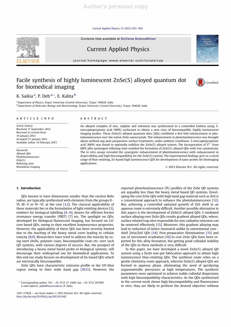
Fig. 6(a) shows the comparative FTIR spectra of MPA stabilizedZnSe and ZnSe(S) QDs. The absence of eSH stretching within2500e2600 cm�1 and the presence of the characteristic peaks of eCOOH (1569 cm�1 for asymmetric COO� and 1400 cm�1 for sym-metric COO�) confirmed the binding of MPA to Zn sites through thethiol groups [3]. The peaks at 3434 cm�1 for both the systems canbe attributed to the eOH stretching of adsorbed water on theirsurface [3]. In case of the ZnSe(S) QDs, added peaks at 1633 cm�1
and 1009 cm�1 were seen due to theeOH stretching, whichmay be
ascribed to the additionally adsorbed water molecules, leading toenhanced wettability and stability of the ZnSe(S) QDs. Fig. 6(b)shows the proposed mechanism of evolution of the ZnSe(S) QDsfrom the native ZnSe QDs.
At highly alkaline pH, MPA molecules are deprotonated formingZn-MPA complex, owing to the higher affinity of S towards Zn [11].With the refluxing at 100 �C, decomposition of the [Zn-MPA]þ
complexes provide the Zn2þ monomers to form ZnSe nanocrystalsin the presence of HSe�. The reaction rate for nucleation andgrowth of ZnSe QDs are retarded with increasing refluxing in-tervals, due to the depletion of Se precursors, as evident from theprolonged reaction time. This results in the surface passivation ofthe defect sites as well as substitution of any available Zn and Sesites at the crystal structure. This S mediated passivation of trapsites significantly contribute to the enhanced band-edge emission,leading to the higher fluorescence emission compared to nativeZnSe QDs (Fig. 3b and Fig. 6b). The quantum enhancement in the PLfor the ZnSe(S) systemmay be attributed to the cumulative effect ofe MPA mediated stabilization of surface defects, S mediated sub-stitution of lattice sites leading to the formation of higher band-gapsystem and PL enhancement resulting from the increased wateradsorption on the QD surface.
4.5. Cytotoxicity assay and cellular uptake
The heavymetal based QD systems are prone to photo-oxidationand chemical and/or biochemical oxidative processes leading to the
Fig. 4. Fluorescence micrographs of ZnSe and ZnSe(S) QDs with quantum yield bar diagram. The excitation wavelength of the laser was 360 nm.
Fig. 5. (a) XRD patterns of ZnSe (5 h refluxed) and ZnSe(S) (18 h refluxed) QD system. The standard XRD patterns of bulk, FCC ZnSe and FCC ZnS are indicated for reference. (b) HR-TEM micrographs of ZnSe(S) QDs. In the inset interplaner spacing and particle size distribution of ZnSe(S) QDs are shown.
Table 1EDX based compositional analysis of ZnSe and ZnSe(S) QD systems.
Sample Sulphur(S)atomic%
Selenium(Se)atomic%
Zinc(Zn)atomic%
S/Zn Se/Zn
ZnSe 44.10 23.87 32.12 1.37 0.74ZnSe(S) 58.15 17.21 24.64 2.35 0.69
K. Saikia et al. / Current Applied Physics 13 (2013) 925e930928
Author's personal copy
leaching of the metal ions in the immediate chemical vicinity[24,25]. Such oxidative phenomena restrict the direct usage ofthese highly efficient flurophores for niche applications like fluo-rescence imaging of biological systems. The use of surfacemodifierslike ZnS, PEG, BSA etc. are able to retard the oxidative processes byphysically obstructing the diffusion of oxygen molecules throughthe surface of such nanocrystals [9,12]. These modifications how-ever are associated with the concurrent increase in dimension ofthe system and find limited acceptability owing to the heavy metalcores. The ZnSe(S) QD system developed in the current studystrives to achieve the high luminescence property comparable toheavy-metal based systems, with the formation of a stable hydro-philic alloy. The facile aqueous synthesis route ensures the stable
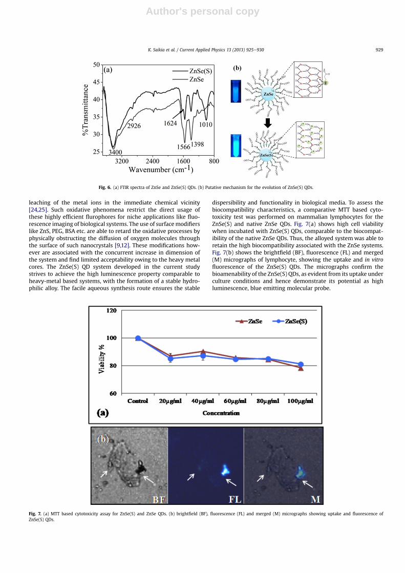
dispersibility and functionality in biological media. To assess thebiocompatibility characteristics, a comparative MTT based cyto-toxicity test was performed on mammalian lymphocytes for theZnSe(S) and native ZnSe QDs. Fig. 7(a) shows high cell viabilitywhen incubated with ZnSe(S) QDs, comparable to the biocompat-ibility of the native ZnSe QDs. Thus, the alloyed systemwas able toretain the high biocompatibility associated with the ZnSe systems.Fig. 7(b) shows the brightfield (BF), fluorescence (FL) and merged(M) micrographs of lymphocyte, showing the uptake and in vitrofluorescence of the ZnSe(S) QDs. The micrographs confirm thebioamenability of the ZnSe(S) QDs, as evident from its uptake underculture conditions and hence demonstrate its potential as highluminescence, blue emitting molecular probe.
Fig. 6. (a) FTIR spectra of ZnSe and ZnSe(S) QDs. (b) Putative mechanism for the evolution of ZnSe(S) QDs.
Fig. 7. (a) MTT based cytotoxicity assay for ZnSe(S) and ZnSe QDs. (b) brightfield (BF), fluorescence (FL) and merged (M) micrographs showing uptake and fluorescence ofZnSe(S) QDs.
K. Saikia et al. / Current Applied Physics 13 (2013) 925e930 929

Author's personal copy
5. Conclusion
In summary, a novel highly luminescent (QYw55%) ZnSe(S)bioimaging agent has been developed though a facile one-potapproach. The ZnSe(S) alloyed QDs exhibit uniform diameter ofw5 nm and were efficiently dispersible in aqueous media. Thefluorescence quantum yield of the ZnSe(S) alloyed QDs was 5 foldhigher than that of the native ZnSe QDs. Additionally, the ZnSe(S)alloyed QDs were biocompatible and could be localized withincultured leukocytes. Thus, ZnSe(S) alloyed QDs are a new range ofmaterials, which are potent alternatives to conventional Zn-basedQDs, having properties like high luminescence and biocompati-bility that may be used to develop newer molecular probes forbiomedical imaging.
Acknowledgement
The authors thank the Department of Biotechnology (DBT), Govtof India (BT/PR10874/NNT/28/136/2008) for financial support.
References
[1] R.J. Martin-Palma, M. Manso, V. Torres-Costa, Optical biosensors based onsemiconductor nanostructures, Sensor 9 (2009) 5149e5172.
[2] Z. Gyori, D. Tatrai, F. Sarlos, G. Szabo, A. Kukovecz, Z. Konya, I. Kiricsi, Laser-induced fluorescence measurements on CdSe QDs, Process. Appl. Ceram. 4(2010) 33e38.
[3] C. Wang, X. Gao, Q. Ma, X. Su, Aqueous synthesis of mercaptopropionic acidcapped Mn2þ-doped ZnSe QDs, J. Mater. Chem. 19 (2009) 7016e7022.
[4] W.W. Yu, E. Chang, R. Drezek, V.L. Colvin, Water-soluble QDs for biomedicalapplications, Biochem. Biophys. Res. Commun. 3489 (2006) 781e786.
[5] W.W.C. Chan, S. Nie, Quantum dot bioconjugates for ultrasensitive nonisotopicdetection, Science 281 (1998) 2016e2018.
[6] M. Bruchez Jr., M. Moronne, P. Gin, S. Weiss, A.P. Alivisatos, Semiconductornanocrystals as fluorescent biological labels, Science 281 (1998) 2013e2016.
[7] Q. Zeng, Y. Zhang, X. Liu, L. Tu, X. Kong, H. Zhang, Multiple homogeneousimmunoassays based on a QDsegold nanorod FRET nanoplatform, Chem.Commun. 48 (2012) 1781e1783.
[8] Y. Su, Y. He, H. Lu, L. Sai, Q. Li, W. Li, L. Wang, P. She, Q. Huang, C. Fan, Thecytotoxicity of cadmium based, aqueous phase e synthesized, QDs and itsmodulation by surface coating, Biomaterials 30 (2009) 19e25.
[9] A.M. Derfus, W.C.W. Chan, S.N. Bhatia, Probing the cytotoxicity of semi-conductor QDs, Nano Lett. 4 (2004) 11e18.
[10] C. Mehta, G.S.S. Saini, J.M. Abbas, S.K. Tripathi, Effect of deposition parameterson structural, optical and electrical properties of nanocrystalline ZnSe thinfilms, Appl. Surf. Sci. 256 (2009) 608e614.
[11] L. Huang, H. Han, One-step synthesis of water-soluble ZnSe QDs via micro-wave irradiation, Mater. Lett. 64 (2010) 1099e1101.
[12] V.V. Nikesh, S. Mahamuni, Highly photoluminescent ZnSe/ZnS quantum dots,Semicond. Sci. Technol. 16 (2001) 687e690.
[13] K. Yu, A. Hrdina, J. Ouyang, D. Kingston, X. Wu, D.M. Leek, X. Liu, C. Li, Ul-traviolet ZnSe1�xSx gradient-alloyed nanocrystals via a noninjectionapproach, ACS Appl. Mater. Interfaces 4 (2012) 4302e4311.
[14] S.K. Panda, S.G. Hickey, C. Waurisch, A. Eychmuller, Gradated alloyed CdZnSenanocrystals with high luminescence quantum yields and stability for opto-electronic and biological applications, J. Mater. Chem. 21 (2011) 11550e11555.
[15] G.Y. Lan, Y.W. Lin, Y.F. Huang, H.T. Chang, Photo-assisted synthesis of highlyfluorescent ZnSe(S) quantum dots in aqueous solution, Mater. Chem. 17(2007) 2661e2666.
[16] H. Qian, X. Qiu, L. Li, J. Ren, Microwave-Assisted aqueous synthesis: a rapidapproach to prepare highly luminescent ZnSe(S) alloyed quantum dots,J. Phys. Chem. B 110 (2006) 9034e9040.
[17] A. Besaratinia, J.I. Yoon, C. Schroeder, S.E. Bradforth, M. Cockburn, G.P. Pfeifer,Wavelength dependence of ultraviolet radiation-induced DNA damage asdetermined by laser irradiation suggests that cyclobutane pyrimidine dimersare the principal DNA lesions produced by terrestrial sunlight, FASEB J. 25(2011) 3079e3091.
[18] S. Chakraborty, M. Gogoi, E. Kalita, P. Deb, Multifunctional, high luminescent,biocompatible CdTe quantum dot fluorophores for bioimaging applications,Int. J. Nanosci. 10 (2011) 1191e1195.
[19] D.F. Faton, Reference materials for fluorescence measurement, Pure Appl.Chem. 60 (1988) 1107e1114.
[20] H. Zhang, D. Wang, H. Mohwäld, Ligand-selective aqueous synthesis of onedimensional CdTe nanostructures, Angew. Chem. Int. Ed. 45 (2006) 748e751.
[21] P. Wu, Z. Fang, X. Zhoug, Y.J. Yang, Depositing ZnS shell around ZnSe corenanocrystals in aqueous media via direct thermal treatment, Colloids Surf. A:Physicochem. Eng. Aspects 375 (2011) 109.
[22] B. Dong, L. Cao, G. Su, W. Liu, Facile synthesis of highly luminescent UV-blueemitting ZnSe/ZnS core/shell QDs by a two-step method, Chem. Commun. 46(2010) 7331e7333.
[23] F. Zan, J. Ren, Significant improvement in photoluminescence of ZnSe(S)alloyed QDs prepared in high pH solution, Luminescence 25 (2010) 378e383.
[24] Y. Shen, L. Li, Q. Lu, J. Ji, R. Fei, J. Zhang, E.S. Abdel- Halim, J.J. Zhu, Microwave-assisted synthesis of highly luminescent CdSeTe@ZnS-SiO2 QDs and theirapplication in the detection of Cu(II), Chem. Commun. 48 (2012) 2222e2224.
[25] E. Sevinc, F.S. Ertas, G. Ulusoy, C. Ozen, H.Y. Acar, Meso-2,3-dimercapto-succinic acid: from heavy metal chelation to CdS QDs, J. Mater. Chem. 22(2012) 5137e5144.
K. Saikia et al. / Current Applied Physics 13 (2013) 925e930930