Facile Synthesis and Crystal Stuructures of New Ammonium Sulfonates (CA-DNBS and TEA-TMBS)
Upload
independentCategory
view
3download
0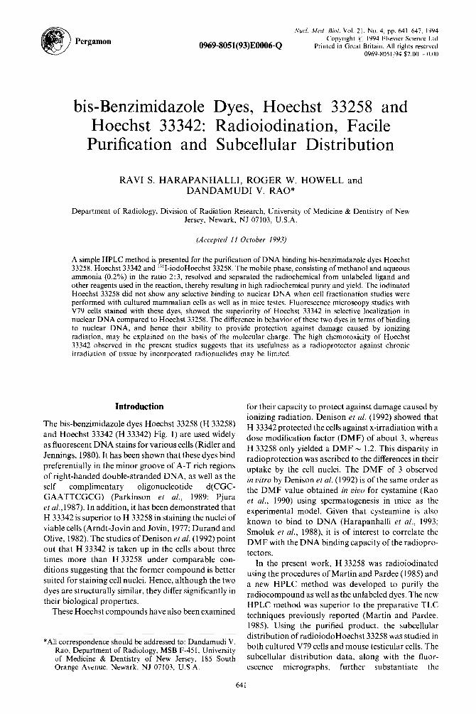
0969-8051(93)EOOO6-Q
Nucl. <Wed B;<J/. Vol. 2 I, No. 4, 64 I -647. pp. 1994 Copyright c; 1994 Elsevier Science Ltd
Printed in Great Britain. All rights reserved 0969-8051194 $7.00 + 0.00
Pergamon
bis-Benzimidazole Dyes, Hoechst 33258 and Hoechst 33342: Radioiodination, Facile
Purification and Subcellular Distribution
RAVI S. HARAPANHALLI, ROGER W. HOWELL and DANDAMUDI V. RAO*
Department of Radiology, Division of Radiation Research, University of Medicine & Dentistry of New Jersey. Newark, NJ 07103, U.S.A.
(Accepted 11 October 1993)
A simple HPLC method is presented for the purification of DNA binding bis-benzimidazole dyes Hoechst 33258, Hoechst 33342 and ‘-“I-iodoHoechst 33258. The mobile phase, consisting of methanol and aqueous ammonia (0.2%) in the ratio 2:3, resolved and separated the radiochemical from unlabeled ligand and other reagents used in the reaction, thereby resulting in high radiochemical purity and yield. The iodinated Hoechst 33258 did not show any selective binding to nuclear DNA when cell fractionation studies were performed with cultured mammalian cells as well as in mice testes. Fluorescence microscopy studies with V79 cells stained with these dyes, showed the superiority of Hoechst 33342 in selective localization in nuclear DNA compared to Hoechst 33258. The difference in behavior of these two dyes in terms of binding to nuclear DNA, and hence their ability to provide protection against damage caused by ionizing radiation, may be explained on the basis of the molecular charge. The high chemotoxicity of Hoechst 33342 observed in the present studies suggests that its usefulness as a radioprotector against chronic irradiation of tissue by incorporated radionuclides may be limited.
Introduction
The bis-benzimidazole dyes Hoechst 33258 (H 33258) and Hoechst 33342 (H 33342) Fig. 1) are used widely as fluorescent DNA stains for various cells (Ridler and Jennings, 1980). It has been shown that these dyes bind preferentially in the minor groove of A-T rich regions of right-handed double-stranded DNA, as well as the self complimentary oligonucleotide d(CGC-
GAATTCGCG) (Parkinson et al., 1989; Pjura et a1.,1987). In addition, it has been demonstrated that H 33342 is superior to H 33258 in staining the nuclei of
viable cells (Arndt-Jovin and Jovin, 1977; Durand and Olive, 1982). The studies of Denison et al. (1992) point out that H 33342 is taken up in the cells about three times more than H 33258 under comparable con-
ditions suggesting that the former compound is better suited for staining cell nuclei. Hence, although the two
dyes are structurally similar, they differ significantly in their biological properties.
These Hoechst compounds have also been examined
*All correspondence should be addressed to: Dandamudi V. Rao, Department of Radiology, MSB F-451, University of Medicine & Dentistry of New Jersey, 185 South Orange Avenue. Newark, NJ 07103, U.S.A.
for their capacity to protect against damage caused by ionizing radiation. Denison et al. (1992) showed that H 33342 protected the cells against x-irradiation with a dose modification factor (DMF) of about 3, whereas
H 33258 only yielded a DMF - 1.2. This disparity in radioprotection was ascribed to the differences in their uptake by the cell nuclei. The DMF of 3 observed in vitro by Denison et al. (1992) is of the same order as the DMF value obtained in vivo for cystamine (Rao et al., 1990) using spermatogenesis in mice as the experimental model. Given that cysteamine is also known to bind to DNA (Harapanhalli et al., 1993; Smoluk et al., 1988), it is of interest to correlate the DMF with the DNA binding capacity of the radiopro- tectors.
In the present work, H 33258 was radioiodinated
using the procedures of Martin and Pardee (1985) and a new HPLC method was developed to purify the radiocompound as well as the unlabeled dyes. The new HPLC method was superior to the preparative TLC techniques previously reported (Martin and Pardee, 1985). Using the purified product, the subcellular distribution of radioiodoHoechst 33258 was studied in both cultured V79 cells and mouse testicular cells. The subcellular distribution data, along with the fluor- escence micrographs, further substantiate the
641
642 RAW S. HARAPANHALLI et al.
superiority of H 33342 over H 33258 for radioprotec- tion. Finally, data on the chemotoxicity of H 33342 in the above models is also presented as they related to the ultimate utility of these compounds as poten- tial radioprotectors.
Experimental
Materials
H 33258, H 33342 and other chemicals were pur- chased from Aldrich Chemical Co. Lactoperoxidase (EC 1 .11.1.7) from bovine milk was obtained from Sigma Chemical Co. (product No. L-8257). A stock solution of 4 mg/mL in 80 mM sodium acetate buffer (pH 5.6), 40% glycerol, was stored at -20°C. Car- rier-free “‘1 (0.63 TBq/mg) and 13’1 (0.44 TBq/mg) were obtained from DuPont NEN (Wilmington, Del.) in the form of sodium iodide in 0.1 M NaOH. A Waters series 501 HPLC pump equipped with a model U6K universal liquid chromatography injector and a model 440 absorbance detector (operating at 254nm and 0.016AUFS) was used for purification.
Cells in culture
Chinese hamster V79-513 cells were maintained as monolayers in culture flasks (37°C and 5% CO,-95% air) containing minimum essential medium (MEM) supplemented with 10% fetal bovine serum, 50 units/ml penicillin, 50 pg/mL streptomycin and 2mM L-glutamine. All cell culture reagents were obtained from GIBCO (Grand Island, N.Y.). Cells were maintained in exponential growth phase and passaged twice a week.
Animals
Male Swiss Webster mice, 8-10 weeks old, were obtained from Taconic Farms (Germantown, N.Y.). They were maintained in the university animal care facility and given rodent diet and water ad libitum.
Iodination
Radioiodination of H 33258 with lzsI and 13’1 was carried out by the procedure of Martin and Pardee (1985) with a radiochemical yield of 70-80%. How- ever, it was not possible to iodinate H 33342 under the same conditions. Certain variations such as the enzyme concentration, pH and temperature did not result in satisfactory iodination of H 33342.
Further attempts to radioiodinate H 33342 using chloramine T and iodogen also failed. However, under these conditions H 33258 could be iodinated with a radiochemical yield of 60%.
Analytical TLC
All TLC analyses were carried out on reverse phase TLC plates (Uniplate RPSF plates from Analtech, Newark, Del.) with methanol and aqueous ammonia (2.2%) in the ratio of 2: 3 as the solvent. Aliquots of the reaction mixtures and the authentic starting material were run on TLC and examined under U.V. light. In addition the TLC plates were placed in x-ray film cassettes and the films were developed to locate the radioactive spots. Under these conditions H 33258 had an Rf of 0.62, whereas its radioiodinated form gave an R, of 0.83. TLC analyses were also carried out on silica plates in the same manner described by Martin and Pardee (1985).
PuriJication by HPLC
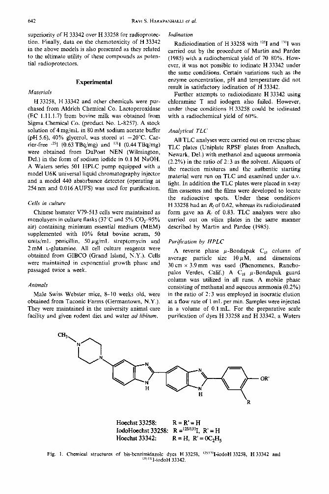
A reverse phase p-Bondapak C,, column of average particle size 10 p M, and dimensions 30 cm x 3.9 mm was used (Phenomenex, Rancho- palos Verdes, Calif.) A C,* p-Bondapak guard column was utilized in all runs. A mobile phase consisting of methanal and aqueous ammonia (0.2%) in the ratio of 2: 3 was employed in isocratic elution at a flow rate of 1 mL per min. Samples were injected in a volume of 0.1 mL. For the preparative scale purification of dyes H 33258 and H 33342, a Waters
CH3,
i
Fig. 1. Chemical structures
Hoechst 33258: R=R’=H
IodoHoechst 33258: R =125’1311, R’ = H Hoechst 33342: R=H, R’=OC,H,
of bis-benzimidazole dyes H 33258, ‘25i’3’I-iodoH 33258, H 33342 and ‘2S”311-iodoH 33342.
bis-Benzimidazole dyes 643
Radialpak C,, cartridge (8 mm x 10 cm) mounted on system (CM- 111 I, Edison, N. J.) was used for process- a Z module radial compression system was used. ing and quantitation of the fluorescence data.
Kinetics of cellular uptake and subcellular distribution
of “‘I iodoH 33258 in VI9 cells and mouse testes
The procedures of Howell et a/. (1991) were fol- lowed. In brief, exponentially growing V79 cells were removed from the flasks with trypsin and suspended
(400,000 cells/ml) in MEM modified for suspension culture (calcium-free medium) and 1 mL aliquots
were transferred to 15 mL polypropylene culture tubes. The tubes were incubated under standard conditions on a rocker roller for 3 h. Calcium-free MEM (1 mL) containing 26.2 kBq of the radioiodin-
ated dye (purified as discussed above) was added and the tubes were returned to the roller. At various times the aliquots of the radioactive cell suspension were removed and the cellular content of radioactivity was assayed. The procedure of Kassis and Adelstein (1985) was employed to determine the subcellular
distribution of radioactivity. In brief, radiolabeled cells were washed twice with cold calcium-free
salt solution (0.4 mM KH,PO,, 0.4 mM
Na,HPO, 7Hz0, 74 mM MgSO .7H,O, 5 mM KC!,
0.12 M NaCl), and suspended in cold hypotonic sucrose buffer (0.25 M, 3 mM CaCl,, 50 mM Tris, pH 7.0). After 5 min, an equal volume of sucrose buffer containing 2% Triton X-100 was added while vortexing. The nuclei were separated from the cyto- plasmic fraction by centrifugation at 200 rpm (4°C) for 15 min and washed with cold sucrose buffer. Aliquots of the nuclear and cytoplasmic fractions were assayed for radioactivity. The DNA in the remaining nuclei was precipitated with cold 6 M guanidine-HCljethanol, filtered and assayed for radioactivity. For subcellular distribution studies in mice. the procedure of Rao et al. (1988) was followed.
Results and Discussion
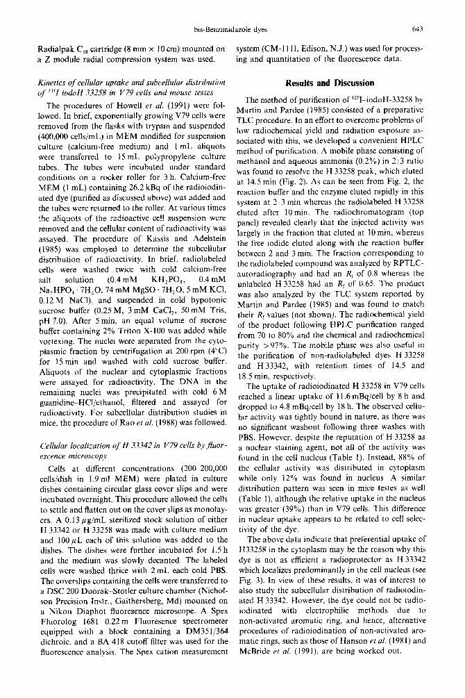
The method of purification of “‘1-iodoH-33258 by Martin and Pardee (1985) consisted of a preparative TLC procedure. In an effort to overcome problems of low radiochemical yield and radiation exposure as- sociated with this, we developed a convenient HPLC method of purification. A mobile phase consisting of
methanol and aqueous ammonia (0.2%) in 2: 3 ratio was found to resolve the H 33258 peak. which eluted at 14.5 min (Fig. 2). As can be seen from Fig. 2, the
reaction buffer and the enzyme eluted rapidly in this system at 2-3 min whereas the radiolabeled H 33258 eluted after 10min. The radiochromatogram (top panel) revealed clearly that the injected activity was
largely in the fraction that eluted at 10 min, whereas the free iodide eluted along with the reaction buffer
between 2 and 3 min. The fraction corresponding to the radiolabeled compound was analyzed by RPTLC- autoradiography and had an R, of 0.8 whereas the unlabeled H 33258 had an Rf of 0.65. The product
was also analyzed by the TLC system reported by Martin and Pardee (1985) and was found to match their Rf values (not shown). The radiochemical yield of the product following HPLC purification ranged from 70 to 80% and the chemical and radiochemical purity >97%. The mobile phase was also useful in the purification of non-radiolabeled dyes H 33258 and H 33342, with retention times of 14.5 and
18.5 min, respectively.
Cellular localization of H 33342 in V79 cells by Jruor - escence microscopy
Cells at different concentrations (200-200,000 cells/dish in 1.9 ml MEM) were plated in culture dishes containing circular glass cover slips and were
incubated overnight. This procedure allowed the cells to settle and flatten out on the cover slips as monolay- ers. A 0.13 pg/mL sterilized stock solution of either H 33342 or H 33258 was made with culture medium and IOOpL each of this solution was added to the dishes. The dishes were further incubated for 1.5 h
and the medium was slowly decanted. The labeled cells were washed thrice with 2 mL each cold PBS.
The coverslips containing the cells were transferred to a DSC 200 Duorak-Stotler culture chamber (Nichol- son Precision Instr., Gaithersberg, Md) mounted on a Nikon Diaphot fluoresence microscope. A Spex Fluorolog 168 1 0.22 m Fluoresence spectrometer equipped with a block containing a DM35!/364 dichroic, and a BA 418 cutoff filter was used for the fluorescence analysis. The Spex cation measurement
The uptake of radioiodinated H 33258 in V79 cells reached a linear uptake of 11.6 mBq/cell by 8 h and dropped to 4.8 mBq/cell by 18 h. The observed cellu- lar activity was tightly bound in nature, as there was no significant washout following three washes with
PBS. However, despite the reputation of H 33258 as a nuclear staining agent, not all of the activity was
found in the cell nucleus (Table 1). Instead, 88% of the cellular activity was distributed in cytoplasm while only 12% was found in nucleus. A similar
distribution pattern was seen in mice testes as well (Table 1), although the relative uptake in the nucleus
was greater (39%) than in V79 cells. This difference in nuclear uptake appears to be related to cell selec- tivity of the dye.
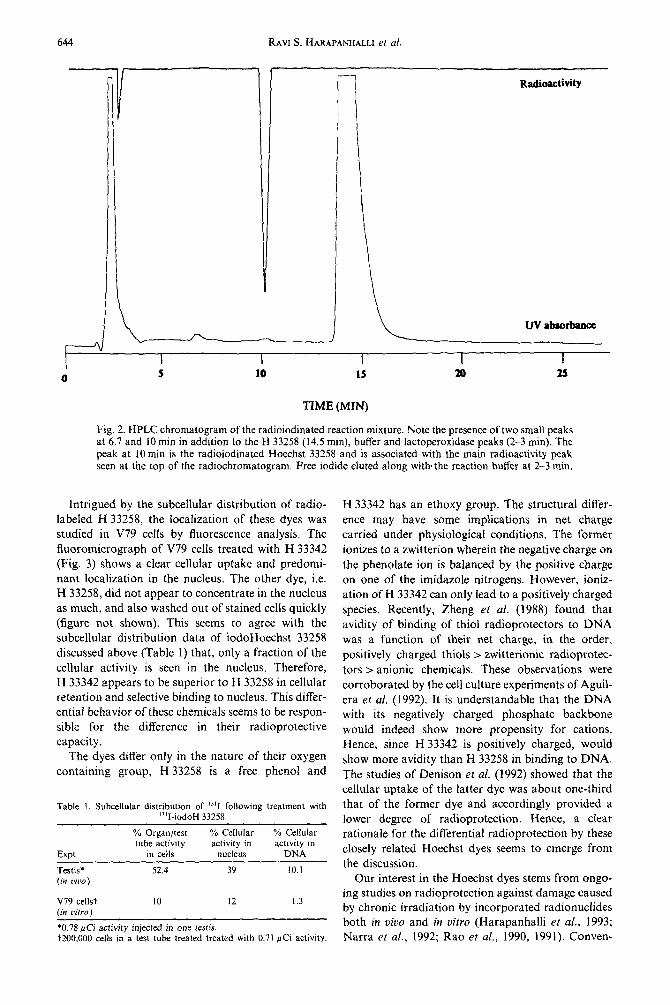
The above data indicate that preferential uptake of H33258 in the cytoplasm may be the reason why this dye is not as efficient a radioprotector as H 33342 which localizes predominantly in the cell nucleus (see Fig. 3). In view of these results, it was of interest to also study the subcellular distribution of radioiodin- ated H 33342. However, the dye could not be radio- iodinated with electrophilic methods due to
non-activated aromatic ring, and hence, alternative procedures of radioiodination of non-activated aro-
matic rings, such as those of Hanson et al. (198 1) and McBride et al. (1991), are being worked out.
644 RAW S. HARAPANHALLI et al.
F 0
1 i i
-
1 Radioactivily
UVabsorbana
I I I I I 5 10 1s 20 2s
TIME (MIN)
Fig. 2. HPLC chromatogram of the radioiodinated reaction mixture. Note the presence of two small peaks at 6.7 and 10 min in addition to the H 33258 (14.5 min), buffer and lactoperoxidase peaks (2-3 min). The peak at 10 min is the radioiodinated Hoechst 33258 and is associated with the main radioactivity peak seen at the top of the radiochromatogram. Free iodide eluted along with% the reaction buffer at 2-3 min.
Intrigued by the subcellular distribution of radio-
labeled H 33258, the localization of these dyes was studied in V79 cells by fluorescence analysis. The fluoromicrograph of V79 cells treated with H 33342 (Fig. 3) shows a clear cellular uptake and predomi- nant localization in the nucleus. The other dye, i.e. H 33258, did not appear to concentrate in the nucleus as much, and also washed out of stained cells quickly (figure not shown). This seems to agree with the subcellular distribution data of iodoHoechst 33258 discussed above (Table I) that, only a fraction of the cellular activity is seen in the nucleus. Therefore, H 33342 appears to be superior to H 33258 in cellular retention and selective binding to nucleus. This differ- ential behavior of these chemicals seems to be respon- sible for the difference in their radioprotective capacity.
The dyes differ only in the nature of their oxygen containing group, H 33258 is a free phenol and
Table I. Subcellular distribution of ‘“‘I following treatment with “‘I-iodoH 33258
Expt
Testis* (in tkw)
% Organ/test tube activity
in cells
52.4
% Cellular % Cellular activity in activity in
nucleus DNA
39 10.1
V79 cells? (in cifro)
IO I2 1.3
*0.78 pCi activity injected in one testis. t200,OOO cells in a test tube &aced treated with 0.71 PCi actiwty.
H 33342 has an ethoxy group. The structural differ- ence may have some implications in net charge carried under physiological conditions. The former ionizes to a zwitterion wherein the negative charge on the phenolate ion is balanced by the positive charge on one of the imidazole nitrogens. However, ioniz- ation of H 33342 can only lead to a positively charged species. Recently, Zheng et al. (1988) found that avidity of binding of thiol radioprotectors to DNA was a function of their net charge, in the order, positively charged thiols > zwitterionic radioprotec- tors > anionic chemicals. These observations were corroborated by the cell culture experiments of Aguil- era et al. (1992). It is understandable that the DNA with its negatively charged phosphate backbone would indeed show more propensity for cations. Hence, since H 33342 is positively charged, would show more avidity than H 33258 in binding to DNA. The studies of Denison et al. (1992) showed that the cellular uptake of the latter dye was about one-third that of the former dye and accordingly provided a lower degree of radioprotection. Hence, a clear rationale for the differential radioprotection by these closely related Hoechst dyes seems to emerge from the discussion.
Our interest in the Hoechst dyes stems from ongo- ing studies on radioprotection against damage caused by chronic irradiation by incorporated radionuclides both in uivo and in vitro (Harapanhalli et at., 1993; Narra el al., 1992; Rao et al., 1990, 1991). Conven-
bis-Benzimidazole dyes
Fig. 3. Fluoromicrograph of viable V79 cells stained with H 33342. Note the clear demarcations of cell cytoplasm and brightly lit nuclei. The dye did not localize in cytoplasm.
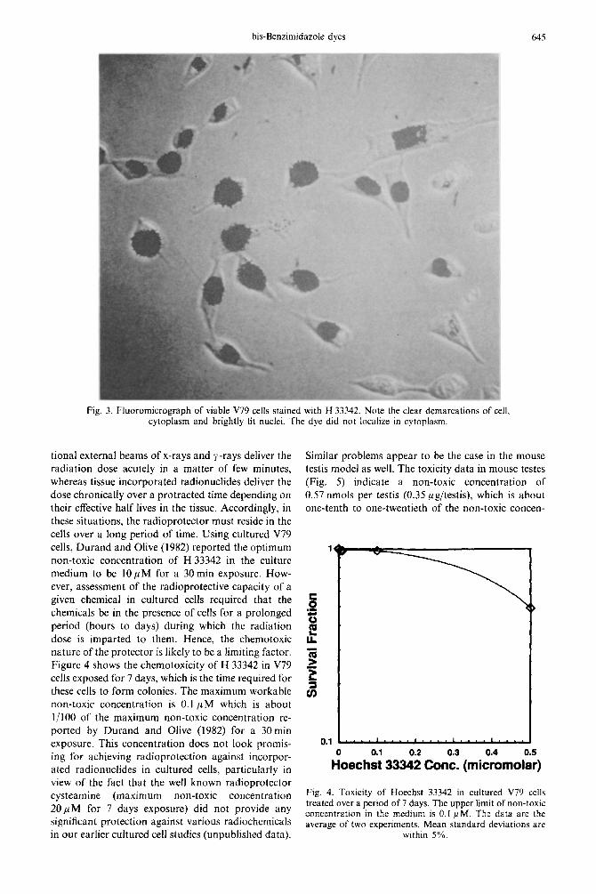
tional external beams of x-rays and y-rays deliver the radiation dose acutely in a matter of few minutes, whereas tissue incorporated radionuclides deliver the dose chronically over a protracted time depending on their effective half lives in the tissue. Accordingly, in these situations, the radioprotector must reside in the cells over a long period of time. Using cultured V79 cells, Durand and Olive (I 982) reported the optimum non-toxic concentration of H 33342 in the culture medium to be 1OpM for a 30 min exposure. How- ever, assessment of the radioprotective capacity of a given chemical in cultured cells required that the chemicals be in the presence of cells for a prolonged period (hours to days) during which the radiation dose is imparted to them. Hence, the chemotoxic nature of the protector is likely to be a limiting factor. Figure 4 shows the chemotoxicity of H 33342 in V79 cells exposed for 7 days, which is the time required for these ceils to form colonies. The maximum workable non-toxic concentration is 0.1 PM which is about l/l00 of the maximum non-toxic concentration re- ported by Durand and Olive (1982) for a 30min exposure. This concentration does not look promis- ing for achieving radioprotection against incorpor- ated radionuclides in cultured cells, particularly in view of the fact that the well known radioprotector cysteamine (maximum non-toxic concentration 20 PM for 7 days exposure) did not provide any significant protection against various radiochemicals in our earlier cultured cell studies (unpublished data).
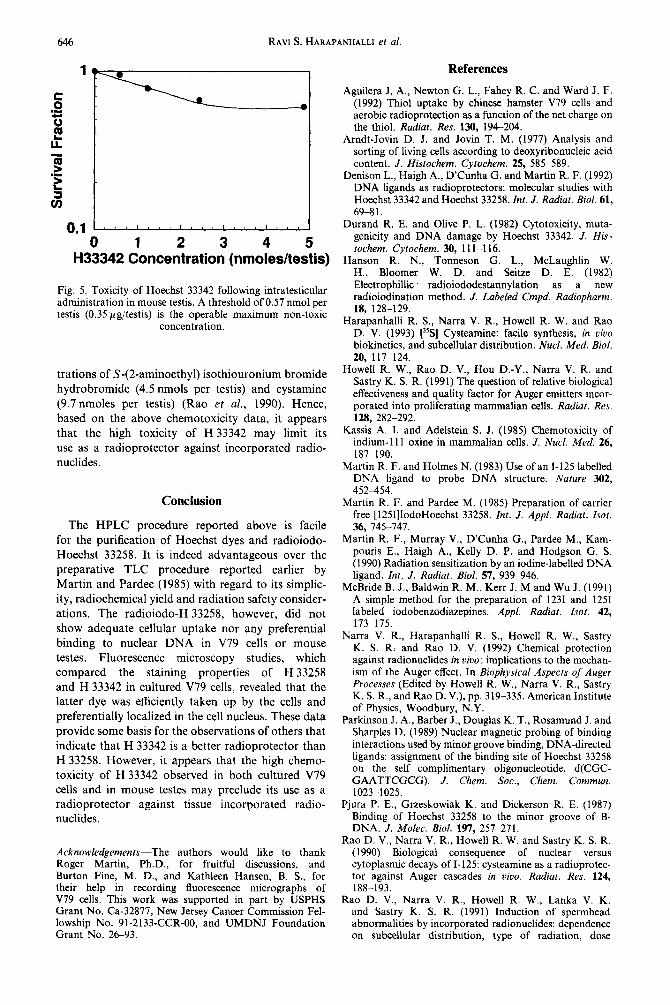
Similar problems appear to be the case in the mouse testis model as well. The toxicity data in mouse testes (Fig. 5) indicate a non-toxic concentration of 0.57 nmols per testis (0.35 pgltestis), which is about one-tenth to one-twentieth of the non-toxic concen-
3 .I
L 3
0.1 ....‘....‘,,..‘....‘...~
Fig. 4. Toxicity of Hoechst 33342 in cultured V79 cells treated over a period of 7 days. The upper limit of non-toxic concentration in the medium is 0.1 FM. The data are the average of two experiments. Mean standard deviations are
within 5%.
RAVI S. HARAPANHALLI et al.
0 1 2 3 4 5 H33342 Concentration (nmoleshestis)
Fig. 5. Toxicity of Hoechst 33342 following intratesticular administration in mouse testis. A threshold of 0.57 nmol per testis (0.35 ng/testis) is the operable maximum non-toxic
concentration.
trations of S-(2-aminoethyl) isothiouronium bromide hydrobromide (4.5 nmols per testis) and cystamine (9.7 nmoles per testis) (Rao et al., 1990). Hence, based on the above chemotoxicity data, it appears that the high toxicity of H 33342 may limit its use as a radioprotector against incorporated radio- nuclides.
Conclusion
The HPLC procedure reported above is facile for the purification of Hoechst dyes and radioiodo- Hoechst 33258. It is indeed advantageous over the preparative TLC procedure reported earlier by Martin and Pardee (1985) with regard to its simplic- ity, radiochemical yield and radiation safety consider- ations. The radioiodo-H 33258, however, did not show adequate cellular uptake nor any preferential binding to nuclear DNA in V79 cells or mouse testes. Fluorescence microscopy studies, which compared the staining properties of H 33258 and H 33342 in cultured V79 cells, revealed that the latter dye was efficiently taken up by the cells and preferentially localized in the cell nucleus. These data provide some basis for the observations of others that indicate that H 33342 is a better radioprotector than H 33258. However, it appears that the high chemo- toxicity of H 33342 observed in both cultured V79 cells and in mouse testes may preclude its use as a radioprotector against tissue incorporated radio- nuclides.
Acknowledgements-The authors would like to thank Roger Martin, Ph.D., for fruitful discussions, and Burton Fine, M. D., and Kathleen Hansen, B. S., for their help in recording fluorescence micrographs of V79 cells. This work was supported in part by USPHS Grant No. Ca-32877, New Jersey Cancer Commission Fel- lowship No. 91-2133~CCR-00, and UMDNJ Foundation Grant No. 26-93.
References
Aguilera J. A., Newton G. L., Fahey R. C. and Ward J. F. (1992) Thiol uptake by Chinese hamster V79 cells and aerobic radioprotection as a function of the net charge on the thiol. Radiat. Res. 130, 194-204.
Arndt-Jovin D. J. and Jovin T. M. (1977) Analysis and sorting of living cells according to deoxyribonucleic acid content. J. Histochem. Cytochem. 25, 585-589.
Denison L., Haigh A., D’Cunha G. and Martin R. F. (1992) DNA ligands as radioprotectors: molecular studies with Hoechst 33342 and Hoechst 33258. Int. J. Radiat. Biol. 61, 69-81.
Durand R. E. and Olive P. L. (1982) Cytotoxicity, muta- genicity and DNA damage by Hoechst 33342. J. His- tochem. Cytochem. 30, 11 l-l 16.
Hanson R. N., Tonneson G. L., McLaughlin W. H., Bloomer W. D. and Seitze D. E. (1982) Electrophillic radioiododestannylation as a new radioiodination method. J. Labeled Cmpd. Radiopharm. 18, 128-129.
Harapanhalli R. S., Narra V. R., Howell R. W. and Rao D. V. (1993) [%I Cysteamine: facile synthesis, in uiuo biokinetics, and subcellular distribution. Nucl. Med. Biol. 20, 117-124.
Howell R. W., Rao D. V., Hou D.-Y., Narra V. R. and Sastry K. S. R. (1991) The question of relative biological effectiveness and quality factor for Auger emitters incor- porated into proliferating mammalian cells. Radial. Res. 128, 282-292.
Kassis A. I. and Adelstein S. J. (1985) Chemotoxicity of indium-1 11 oxine in mammalian cells. J. Nucl. Med. 26, 1877190.
Martin R. F. and Holmes N. (1983) Use of an I-125 labelled DNA ligand to probe DNA structure. Nature 302, 452454.
Martin R. F. and Pardee M. (1985) Preparation of carrier free [125I]IodoHoechst 33258. Int. J. Appl. Radiat. Isot. 36, 745-747.
Martin R. F., Murray V., D’Cunha G., Pardee M., Kam- pouris E., Haigh A., Kelly D. P. and Hodgson G. S. (1990) Radiation sensitization by an iodine-labelled DNA ligand. Int. J. Radial. Biol. 57, 939-946.
McBride B. J., Baldwin R. M., Kerr J. M and Wu J. (1991) A simple method for the preparation of 1231 and 1251 labeled iodobenzodiazepines. Appl. Radial. Isot. 42, 173-175.
Narra V. R., Harapanhalli R. S., Howell R. W., Sastry K. S. R. and Rao D. V. (1992) Chemical protection against radionuclides in uiuo: implications to the mechan- ism of the Auger effect. In Biophysical Aspects of Auger Processes (Edited by Howell R. W., Narra V. R., Sastry K. S. R., and Rao D. V.), pp. 319-335. American Institute of Physics, Woodbury, N.Y.
Parkinson J. A., Barber J., Douglas K. T., Rosamund J. and Sharples D. (1989) Nuclear magnetic probing of binding interactions used by minor groove binding, DNA-directed ligands: assignment of the binding site of Hoechst 33258 on the self complimentary oligonucleotide, d(CGC- GAATTCGCG). J. Gem. Sot., Chem. Commun. 1023-1025.
Pjura P. E., Grzeskowiak K. and Dickerson R. E. (1987) Binding of Hoechst 33258 to the minor groove of B- DNA. J. Molec. Biol. 1!37, 257-271.
Rao D. V., Narra V. R., Howell R. W. and Sastry K. S. R. (1990) Biological consequence of nuclear versus cytoplasmic decays of I-125: cysteamine as a radioprotec- tor against Auger cascades in vivo. Radial. Res. 124, 188-193.
Rao D. V., Narra V. R., Howell R. W., Lanka V. K. and Sastry K. S. R. (1991) Induction of spermhead abnormalities by incorporated radionuclides: dependence on subcellular distribution, type of radiation, dose

bis-Benzimidazole dyes 647
rate, and presence of radioprotectors. Radiut. Res. 125, Interaction of glutathione and other low molecular 89-97. weight thiols with DNA: Evidence for counterion conden-
Rao D. V., Sastry K. S. R., Grimmond H. E., Howell R. W., sation and coion depletion near DNA. Radial. Res. 114, Govelitz G. F., Lanka V. K. and Mylavarapu V. B. (1988) 3-10. Cytotoxicity of some indium radiopharmaceuticals in Zheng S., Newton G. L., Gonick G., Fahey R. C. mouse testes. J. Nucl. Med. 29. 375-384. and Ward J. F. (1988) Radiourotection of DNA
Ridler P. J. and Jennings B. R. (J980) Int. J. Biol. Macro- by thiols: relationship between the net charge on a molec 2, 3133317. thiol and its ability to protect DNA Radiar. Res. 114,
Smoluk G. D.. Fahey R. C. and Ward J. F. (1988) 1 l-27.
Copyright © 2022 FDOKUMEN


![1-[2-(3,4-Dichlorobenzyloxy)-2-phenylethyl]-1 H -benzimidazole](https://static.fdokumen.com/doc/165x107/63152ee185333559270d05af/1-2-34-dichlorobenzyloxy-2-phenylethyl-1-h-benzimidazole.jpg)