Association of Chromosome Territories with the Nuclear Matrix
11
The Rockefeller University Press, 0021-9525/99/08/531/11 $5.00 The Journal of Cell Biology, Volume 146, Number 3, August 9, 1999 531–541 http://www.jcb.org 531 Association of Chromosome Territories with the Nuclear Matrix: Disruption of Human Chromosome Territories Correlates with the Release of a Subset of Nuclear Matrix Proteins Hong Ma, Alan J. Siegel, and Ronald Berezney Department of Biological Sciences, State University of New York at Buffalo, Buffalo, New York 14260 Abstract. To study the possible role of the nuclear ma- trix in chromosome territory organization, normal hu- man fibroblast cells are treated in situ via classic isola- tion procedures for nuclear matrix in the absence of nuclease (e.g., DNase I) digestion, followed by chromo- some painting. We report for the first time that chromo- some territories are maintained intact on the nuclear matrix. In contrast, complete extraction of the internal nuclear matrix components with RNase treatment fol- lowed by 2 M NaCl results in the disruption of higher order chromosome territory architecture. Correlative with territorial disruption is the formation of a faint DNA halo surrounding the nuclear lamina and a dis- persive effect on the characteristically discrete DNA replication sites in the nuclear interior. Identical results were obtained using eight different human chromo- some paints. Based on these findings, we developed a fractionation strategy to release the bulk of nuclear ma- trix proteins under conditions where the chromosome territories are maintained intact. A second treatment results in disruption of the chromosome territories in conjunction with the release of a small subset of acidic proteins. These proteins are distinct from the major nu- clear matrix proteins and may be involved in mediating chromosome territory organization. Key words: chromosome territory • chromosome painting • fluorescence in situ hybridization • DNA-rich nuclear matrix • nuclear matrix proteins D EVELOPMENTS in visualization approaches have stimulated a renewed interest in understanding the functional organization of the eukaryotic cell nucleus. Discrete domains of DNA replication, transcrip- tion, and RNA splicing factors have been defined by numer- ous studies (Spector, 1993; Berezney et al., 1995; Jackson and Cook, 1995; Nickerson et al., 1995; van Driel et al., 1995; Berezney and Wei, 1998; Ma et al., 1998; Wei et al., 1998, 1999). The corresponding association of these functional do- mains with the nuclear matrix has led to the view that the cell nucleus may have an underlying structural architecture as a basis for form and function (Hoffman, 1993; Spector, 1993; Baskin, 1995; Berezney et al., 1995; Nickerson et al., 1995; van Driel et al., 1995; Berezney and Wei, 1998). Progress in elucidating higher order structure in the cell nucleus is not limited to strictly functional domains, but applies to the very genome itself. It is now clear that the chromatin in the interphase cell nucleus is arranged in spa- tially separate, chromosome specific territories (Stack et al., 1977; Zorn et al., 1979; Cremer et al., 1982, 1993, 1995; Manuelidis, 1985, 1990; Schardin et al., 1985; Licher et al., 1988; Pinkel et al., 1988; Leitch et al., 1990; Schwarzacher, 1994; Zink et al., 1998). Initial results further indicate a high degree of organization of specific subchromosomal regions within individual chromosome territories (Manuel- idis, 1990; Dietzel et al., 1998). While the question remains as to what mediates organi- zation into chromosome territories, numerous studies have demonstrated that chromatin is arranged into repeat- ing loop domains of 50–200 kb in the interphase nucleus and mitotic chromosomes (Cook and Brazell, 1975; Cook et al., 1976; Paulson and Laemmli, 1977; Pardoll et al., 1980; Vogelstein et al., 1980). These chromatin loops are believed to be anchored to components of the nuclear ma- trix or chromosome scaffold by S/MARs (scaffold/matrix attachment regions) 1 , which presumably bind to specific components on the nuclear matrix/scaffold (Goldberg et al., 1983; Pienta and Coffey, 1984; Nelson and Coffey, 1987; Chai and Sandberg, 1988; Laemmli et al., 1992; Roberge This article is dedicated to Professor Donald S. Coffey on the occasion of his 40th year at The Johns Hopkins University School of Medicine. Address correspondence to Dr. Ronald Berezney, Department of Bio- logical Sciences, State University of New York at Buffalo, Buffalo, NY 14260. Tel.: (716) 645-2874. Fax: (716) 645-2975. E-mail: berezney@acsu. buffalo.edu Dr. Ma’s current address is Vollum Institute, L-474, Oregon Health Sciences University, 3181 SW Sam Jackson Park Road, Portland, OR 97201-3098. 1. Abbreviations used in this paper: BrdU, 5-bromo-2-deoxy-uridine; CTAPs, chromosome territory anchor proteins; FISH, fluorescence in situ hybridization; MARs, matrix attachment regions. on April 5, 2017 Downloaded from Published August 9, 1999
-
Upload
khangminh22 -
Category
Documents
-
view
2 -
download
0
Transcript of Association of Chromosome Territories with the Nuclear Matrix

The Rockefeller University Press, 0021-9525/99/08/531/11 $5.00The Journal of Cell Biology, Volume 146, Number 3, August 9, 1999 531–541http://www.jcb.org 531
Association of Chromosome Territories with the Nuclear Matrix: Disruption of Human Chromosome Territories Correlates with the Release of a Subset of Nuclear Matrix Proteins
Hong Ma, Alan J. Siegel, and Ronald Berezney
Department of Biological Sciences, State University of New York at Buffalo, Buffalo, New York 14260
Abstract.
To study the possible role of the nuclear ma-trix in chromosome territory organization, normal hu-man fibroblast cells are treated in situ via classic isola-tion procedures for nuclear matrix in the absence of nuclease (e.g., DNase I) digestion, followed by chromo-some painting. We report for the first time that chromo-some territories are maintained intact on the nuclear matrix. In contrast, complete extraction of the internal nuclear matrix components with RNase treatment fol-lowed by 2 M NaCl results in the disruption of higher order chromosome territory architecture. Correlative with territorial disruption is the formation of a faint DNA halo surrounding the nuclear lamina and a dis-persive effect on the characteristically discrete DNA replication sites in the nuclear interior. Identical results
were obtained using eight different human chromo-some paints. Based on these findings, we developed a fractionation strategy to release the bulk of nuclear ma-trix proteins under conditions where the chromosome territories are maintained intact. A second treatment results in disruption of the chromosome territories in conjunction with the release of a small subset of acidic proteins. These proteins are distinct from the major nu-clear matrix proteins and may be involved in mediating
chromosome territory organization.
Key words: chromosome territory • chromosome painting • fluorescence in situ hybridization • DNA-rich nuclear matrix • nuclear matrix proteins
D
EVELOPMENTS
in visualization approaches havestimulated a renewed interest in understandingthe functional organization of the eukaryotic cell
nucleus. Discrete domains of DNA replication, transcrip-
tion, and RNA splicing factors have been defined by numer-ous studies (Spector, 1993; Berezney et al., 1995; Jacksonand Cook, 1995; Nickerson et al., 1995; van Driel et al., 1995;Berezney and Wei, 1998; Ma et al., 1998; Wei et al., 1998,1999). The corresponding association of these functional do-mains with the nuclear matrix has led to the view that thecell nucleus may have an underlying structural architectureas a basis for form and function (Hoffman, 1993; Spector,1993; Baskin, 1995; Berezney et al., 1995; Nickerson et al.,1995; van Driel et al., 1995; Berezney and Wei, 1998).
Progress in elucidating higher order structure in the cellnucleus is not limited to strictly functional domains, butapplies to the very genome itself. It is now clear that the
chromatin in the interphase cell nucleus is arranged in spa-
tially separate, chromosome specific territories (Stack et al.,1977; Zorn et al., 1979; Cremer et al., 1982, 1993, 1995;Manuelidis, 1985, 1990; Schardin et al., 1985; Licher et al.,1988; Pinkel et al., 1988; Leitch et al., 1990; Schwarzacher,1994; Zink et al., 1998). Initial results further indicate ahigh degree of organization of specific subchromosomalregions within individual chromosome territories (Manuel-idis, 1990; Dietzel et al., 1998).
While the question remains as to what mediates organi-zation into chromosome territories, numerous studieshave demonstrated that chromatin is arranged into repeat-ing loop domains of 50–200 kb in the interphase nucleusand mitotic chromosomes (Cook and Brazell, 1975; Cooket al., 1976; Paulson and Laemmli, 1977; Pardoll et al.,1980; Vogelstein et al., 1980). These chromatin loops arebelieved to be anchored to components of the nuclear ma-trix or chromosome scaffold by S/MARs (scaffold/matrix
attachment regions)
1
, which presumably bind to specificcomponents on the nuclear matrix/scaffold (Goldberg et al.,1983; Pienta and Coffey, 1984; Nelson and Coffey, 1987;
Chai and Sandberg, 1988; Laemmli et al., 1992; Roberge
This article is dedicated to Professor Donald S. Coffey on the occasion ofhis 40th year at The Johns Hopkins University School of Medicine.
Address correspondence to Dr. Ronald Berezney, Department of Bio-logical Sciences, State University of New York at Buffalo, Buffalo, NY14260. Tel.: (716) 645-2874. Fax: (716) 645-2975. E-mail: [email protected]
Dr. Ma’s current address is Vollum Institute, L-474, Oregon HealthSciences University, 3181 SW Sam Jackson Park Road, Portland, OR97201-3098.
1.
Abbreviations used in this paper:
BrdU, 5-bromo-2-deoxy-uridine;CTAPs, chromosome territory anchor proteins; FISH, fluorescence in situhybridization; MARs, matrix attachment regions.
on April 5, 2017
Dow
nloaded from
Published August 9, 1999
The Journal of Cell Biology, Volume 146, 1999 532
et al., 1992; Hiraoka et al., 1993). Several MAR bindingproteins have been identified in association with the nu-clear matrix (von Kries et al., 1991; Romig et al., 1992;Dickinson et al., 1992, 1997; Tsutsui et al., 1993; Fackel-mayer et al., 1994; Dickinson and Kohwi-Shigematsu,1995; Wang et al., 1995; Liu et al., 1997; de Belle et al.,1998). Moreover, DNA topoisomerase II (Gasser andLaemmli, 1987; Adachi et al., 1989, 1991) and ScII (Saitohet al., 1994) are two major proteins of the chromosomescaffold that have been proposed to be involved in chro-matin loop organization.
Studies of chromosome architecture (Pienta and Coffey,1984; Saitoh and Laemmli, 1994) have led to the proposalthat the organization of chromatin into higher order chro-mosome bands is mediated by the corresponding clusteringof repeating loops attached to the nuclear matrix/scaffold.This higher order clustering of loop domains may also be afundamental feature of functional units of chromatin in thecell nucleus. It has long been recognized that DNA replica-tion occurs in clusters of replicons (Hand, 1978). Models ofDNA replication associated with the nuclear matrix haveproposed that the replicon clusters correspond to the 50–200-kb repeating DNA loop domains that are attached tothe nuclear matrix (Dijkwel et al., 1979; Pardoll et al., 1980;Vogelstein et al., 1980; Berezney and Buchholtz, 1981; Be-rezney, 1991, 1992; Cook, 1991). Consistent with this view,discrete replication sites corresponding to replicon clustershave been visualized in the nucleus of intact cells and arestrikingly maintained after extraction of cells for nuclearmatrix (Nakayasu and Berezney, 1989; Neri et al., 1992; Be-rezney et al., 1995). Similarly, individual transcription sitesin the cell nucleus are believed to be composed of clustersof transcriptional units attached to the nuclear matrix (Jack-son and Cook, 1995; Wei et al., 1999).
These previous findings have prompted us to investigatethe possible role of the nuclear matrix in the territorial or-ganization of human chromosomes during interphase. Wedemonstrate for the first time that chromosome territoriesare maintained intact after extraction for the nuclear ma-trix. Virtually complete extraction of the internal nuclearmatrix with RNase A and 2.0 M NaCl leads to a corre-sponding disruption of the chromosome territories. Asmall subset of nuclear matrix proteins are released in con-junction with territorial disruption and may play a role inmediating chromosomal organization.
Materials and Methods
Cell Culture and In Situ DNA-rich NuclearMatrix Extraction
Three human cell lines (WI-38, NHF-1, and HeLa S3) were used for thesestudies. WI-38 and HeLa S3 cells (American Type Culture Collection)were grown as monolayers in MEM (GIBCO BRL) plus 10% FBS (Sum-mits Biotechnology) supplemented with sodium pyruvate acid (GIBCOBRL) and MEM nonessential amino acids (GIBCO BRL). NHF-1 (nor-mal human fibroblast) cells were grown as monolayers in MEM (GIBCOBRL) plus 10% FBS (Summits Biotechnology) supplemented with so-dium pyruvate acid (GIBCO BRL) and MEM nonessential amino acids(GIBCO BRL), 1%
L
-glutamine (GIBCO BRL), and 50
m
g/ml gentami-cine (Sigma Chemical Co.).
For in situ nuclear matrix preparation, cells plated on coverslips werepermeabilized with CSK buffer (100 mM NaCl, 300 mM sucrose, 10 mMPipes, pH 6.8, 3 mM MgCl
2
, 10
m
M leupeptin, 1 mM EGTA, 1.2 mM
PMSF, 0.5% Triton X-100, with or without 4 mM vanadyl complex) fol-lowed by extraction with 0.25 M (NH
4
)
2
SO
4
,
0.65 M (NH
4
)
2
SO
4
,
or 2.0 MNaCl, with or without RNase A (200
m
g/ml) digestion for 1 h on ice (Feyet al., 1986; He et al., 1990).
Chromosome Painting and Replication Site Labeling
Human chromosome painting probes, which were directly labeled withSpectrum green or orange, were obtained from GIBCO BRL and Vysis.Experiments were performed after standard fluorescence in situ hybrid-ization (FISH) procedures, modified with instructions from GIBCO BRLand Vysis. In brief, the cells were fixed with 4% formaldehyde and dena-tured with 50% formamide/2
3
SSC by heating in an 80
8
C waterbath for30 min. 1
m
l chromosome painting probe with 7
m
l hybridization bufferand 2
m
l double distilled H
2
O was denatured by heating in an 75
8
C water-bath for 10 min, and then placed immediately on ice. The final concentra-tion of hybridization buffer was 50% formamide, 2
3
SSC, and 10% dex-tran sulfate. The probe in hybridization buffer was added to thecoverslips, placed cell-side down on a glass microscope slide, and sealedwith rubber cement. After incubation in a 37
8
C incubator overnight, thecell samples were washed three times (10 min each) in 50% formamide in2
3
SSC at 45
8
C, once for 10 min in 2
3
SSC at 45
8
C, once for 5 min in 2
3
SSC with 1% NP-40, and then air dried.DNA replication sites were labeled after a 5 min in vivo incorporation
of 5-bromo-2-deoxy-uridine (BrdU; 10
m
M) in NHF-1 cells grown on cov-erslips, according to the instructions of the BrdU labeling and detectionkit I (Boehringer Mannheim Corp.) as previously described (Ma et al.,1998) with mouse anti-BrdU mAbs and Texas red-conjugated anti-mouseIgG secondary antibodies (Boehringer Mannheim Corp.).
Immunofluorescence Labeling of Matrin 250 andLamin A/C
Double labeling of nuclear matrix associated proteins was performed inNHF-1 cells after appropriate extractions and fixation with 4% formalde-hyde. Antibody reactions were carried out successively with mAb anti-matrin 250, also known as nuclear matrix associated RNA polymerase II
0
(B3, mouse IgM, diluted 50-fold with PBS; Mortillaro et al., 1996), andrabbit antilamin A/C polyclonal antibodies (diluted 20-fold with PBS) for1 h at 37
8
C. FITC and Texas red secondary antibodies (both diluted 50-fold with PBS), respectively, were used for detection. In experiments in-volving propidium iodide staining of the DNA, the nuclear lamins weredetected with FITC-conjugated antibodies.
Electron and Confocal Microscopy
Samples from HeLa S3 cells were prepared for thin section EM (Bel-grader et al., 1991). Cells were fixed in 2.5% glutaraldehyde, 100 mM so-dium cacodylate, pH 7.4, and 3 mM MgCl
2
for at least 2 h at 4
8
C. The cellswere rinsed with cacodylate buffer without glutaraldehyde, post-fixedwith 1% OsO
4
for 30 min on ice, dehydrated (graded 30–100% ethanoland 100% acetone), and infiltrated with Epon-Araldite resin (ElectronMicroscopy Sciences). Cured blocks were thin sectioned and stained withlead citrate and uranyl acetate. Sections were examined on a HitachiH-500 electron microscope.
The confocal imaging was performed on a Bio-Rad MRC-1024 threechannel laser scanning confocal imaging system equipped with a NikonOptiphot 2 microscope, a Nikon 60
3
, 1.4 NA objective, and an argon/krypton laser (
l
5
488/565 nm). Optical sections of 512 pixels
3
512 pixels
3
8 bits/pixel were collected through the samples at 0.5
m
m intervals.
Fractionation of Nuclear Matrix Proteins, Biochemical Analysis, and Two-dimensional PAGE
For biochemical studies, HeLa S3 cells were used. Nuclei were isolatedfrom disrupted cells using the syringe technique in Belgrader et al. (1991).Our strategy of identifying proteins released in correlation with chromo-some disruption is shown in Fig. 4. It involves consecutive extraction ofisolated HeLa cell nuclei with CSK buffer, RNase A digestion, 0.65-M(NH
4
)
2
SO
4
and 2.0-M NaCl extractions. For total nuclear matrix proteins,HeLa S3 cells were permeabilized by CSK buffer, followed by DNase I di-gestion and 0.25-M (NH
4
)
2
SO
4
extraction.Protein was determined using the BCA kit (Pierce Chemical Co.).
DNA was measured after incorporation of [
3
H]thymidine (1
m
Ci/ml) intoHeLa S3 cells for 48 h of culture and RNA by alkaline hydrolysis methods
on April 5, 2017
Dow
nloaded from
Published August 9, 1999

Ma et al.
Chromosome Territories and the Nuclear Matrix
533
(Berezney and Buchholtz, 1981). The data were averaged from four indi-vidual experiments.
For two-dimensional PAGE, proteins were run on a nonequilibriumpH gradient (first dimension) and on SDS-PAGE in the second dimension(Belgrader et al., 1991). The proteins were stained with Coomassie blue(0.2%). Total nuclear matrix protein was prepared after treatment of iso-lated nuclei with DNase I and 0.25-M (NH
4
)
2
SO
4
extraction (Belgraderet al., 1991).
Results
Chromosome Territories Remain Intact After Extraction for Nuclear Matrix
Previous studies have shown that human interphase chro-mosomes are confined to discrete regions in the cell nu-cleus termed chromosome territories (Manuelidis, 1985,1990; Schardin et al., 1985; Licher et al., 1988; Pinkel et al.,1988; Leitch et al., 1990; Cremer et al., 1993; Schwarza-cher, 1994). To study the possible role of the nuclear ma-trix in chromosome territory organization, we extractednormal diploid human fibroblasts cells (NHF-1 or WI-38)grown on coverslips for DNA-rich nuclear matrix (Be-rezney and Buchholtz, 1981). In DNA-rich nuclear matrixpreparations, the nuclear DNA is not cleaved by either en-dogenous or exogenously added nucleases. The proteincomponents in DNA-rich nuclear matrix, however, arevirtually identical to DNA-depleted matrix in recovery oftotal nuclear protein and overall polypeptide compositionon SDS-PAGE (Berezney and Buchholtz, 1981). More-over, the DNA in such structures is maintained predomi-nantly inside the nuclear structures in a highly supercoiledstate (Cook and Brazell, 1975; Cook et al., 1976; Warrenand Cook, 1978; Berezney and Buchholtz, 1981).
We find that the chromosomes are maintained in theDNA-rich nuclear matrices (Fig. 1, B and E) as separateterritories that are indistinguishable from those in intactcells (Fig. 1, A and D). Similar observations were madewith all human chromosomes examined (numbers 1, 2, 4,7, 9, 11, 14, and 22) and different two paint combinations.There was no chromosome hybridization signal in DNA-depleted nuclear matrix (results not shown).
These experiments were performed using moderateionic strength for salt extraction (0.25 M (NH
4
)
2
SO
4
; Bel-grader et al., 1991). We next used a higher salt concentra-tion (2.0 M NaCl and the same ionic strength 0.65 M(NH
4
)
2
SO
4
) to extract the cells. The chromosome territo-ries remain intact after these higher salt extractions (Fig. 2,A and B), and were indistinguishable from those observedin nuclear matrix prepared with moderate salt levels (Fig.1 B) or intact cells (Fig. 1 A). Greater than 90% of the to-tal histone proteins and other soluble nuclear proteins areremoved under these salt extraction conditions (Berezneyand Buchholtz, 1981; results not shown). Chromosome ter-ritories are, therefore, maintained in association with thenuclear matrix without the bulk of the histone proteins.
Disruption of Internal Nuclear Matrix with RNase A and 2.0 M NaCl, but Not 0.65 M (NH
4
)
2
SO
4
, Results in the Corresponding Disruption ofChromosome Territories
Since the structural integrity of the nuclear matrix is de-pendent on RNA and intermolecular disulfide bonds
(Kaufman et al., 1981; Belgrader et al., 1991), salt extrac-tions were also performed after 200
m
g/ml RNase A diges-tion, in the presence of 20 mM DTT or a combination ofthese two treatments. Surprisingly, chromosome territo-ries are highly disrupted after RNase A and 2.0-M NaCltreatment (Figs. 1 C and 2 D), but extraction with similarionic strength 0.65 M (NH
4
)
2
SO
4
after RNase treatmenthad no visible effect (Fig. 2 C). Identical results were ob-tained in eight separate experiments using eight differenthuman chromosome paints (numbers 1, 2, 4, 7, 9, 11, 14,and 22) examined in three human cell lines (WI-38, NHF-1,and HeLa S3). 82% of the cells (
.
500 cells counted ineach experiment) had disrupted chromosome territorieswhen averaged among all the experiments (results notshown). In contrast,
.
90% of the cells had intact territo-ries after all other nuclear matrix preparations (Fig. 2,A–C), including the cells extracted with 20 mM DTT (re-sults not shown). This compares with intact cells, in whichnearly 100% of the chromosome territories were intacteven under conditions where a small percentage of thenuclei (
z
20%) showed visible breakage or leakage ofchromatin. We conclude that intermolecular disulfidebonds, which form between nuclear matrix proteins (Kauf-man et al., 1981; Belgrader et al., 1991), do not play a sig-nificant role in maintaining chromosome territory organi-zation. RNA and/or RNP interactions, however, are crucial.
While disrupted chromosome territories display diffusestaining inside the nucleus (Fig. 2 D), it could be arguedthat the individual chromosomes maintain a degree oftheir territorial organization despite unraveling into morediffuse structures. Double labeling experiments (e.g., chro-mosomes 1 and 11; Fig. 1 F), however, clearly show a highdegree of mixing (yellow coloration) of the two chromo-some pairs and confirm the loss of territorial arrangement.
The difference in chromosome territory organizationbetween 2.0-M NaCl extraction and its same ionic strength0.65 M (NH
4
)
2
SO
4
after RNase A digestion led us to studythe nuclear matrix organization after these salt extrac-tions. In the same preparations, matrin 250 and lamin A/Cwere used as markers for the internal nuclear matrix andthe peripheral nuclear lamina, respectively, by double im-munofluorescence labeling (see Materials and Methods).Cells extracted with 0.65 M (NH
4
)
2
SO
4
had a matrin 250staining intensity and pattern identical to intact cells (Fig.2 E; 89% of 464 cells counted). Extraction with 2.0 MNaCl, however, resulted in significant aggregation of thestructures (Fig. 2 F; 76% of 480 cells). Matrix preparationsobtained with RNase A after 0.65 M (NH
4
)
2
SO
4
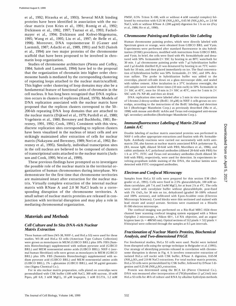
are de-pleted in matrin 250, but it is still detectable (Fig. 2 G; 81%of 447 cells). There is no detectable matrin 250 staining in-side of the nuclei after RNase A digestion and 2.0-M NaClextraction (Fig. 2 H; 80% of 367 cells). Virtually identicalresults were obtained when the internal nuclear matrixwas decorated with matrin CYP (Mortillaro and Berez-ney, 1998) instead of matrin 250 (results not shown). Incontrast, there is no detectable effect on the nuclear lam-ina structure by any of the extraction conditions (Fig. 2,E–H).
The overall nuclear morphology of the extracted cellsamples were then studied by thin sectioning EM. The re-sults are in close agreement with the double immunofluo-rescence analysis of nuclear matrix components. An elabo-
on April 5, 2017
Dow
nloaded from
Published August 9, 1999

The Journal of Cell Biology, Volume 146, 1999 534
rate structure is seen in the nuclear matrix after 0.65-M(NH
4
)
2
SO
4
extraction (Fig. 2 I). Nuclear matrix structureis significantly aggregated by 2.0-M NaCl extraction (Fig. 2J). After RNase A and 0.65-M (NH
4
)
2
SO
4
treatments, theDNA-rich nuclear matrices are depleted of internal struc-ture, although it is still detectable (Fig. 2 K).
The interiorof the nuclear matrix, however, is virtually devoid of struc-ture when extracted by 2.0 M NaCl after RNase A diges-tion (Fig. 2 L).
Extraction of internal nuclear matrix components withRNase and 0.65 M (NH
4
)
2
SO
4
had no visible effect on therelative intactness of the resulting nuclear matrix struc-tures, as evaluated by fluorescence microscopy or EM(Fig. 2). Similarly, the majority of nuclear matrix struc-tures observed after chromosome disruption (RNase and2-M NaCl extraction) were indistinguishable in terms ofoverall nuclear shape from those following other extrac-tions (Fig. 2, H and L). Significantly, chromosome territo-
ries were routinely disrupted irrespective of the degree ofintactness of overall nuclear shape (Figs. 1 C and 2 D).
DNA Halos and DNA Replication Sites after Extraction of Cells for Nuclear Matrix
When DNA-rich nuclear matrices are prepared underconditions where cleavage or nicking of DNA is avoided,the DNA remains inside the nuclear matrix structure andis highly supercoiled (Cook and Brazell, 1975; Cook et al.,1976; Berezney and Buchholtz, 1981). Progressive relax-ation of this supercoiled DNA with intercalating agents,such as ethidium bromide, leads to the corresponding for-mation of a halo of relaxed DNA loops surrounding theoverall nuclear matrix structure (Cook et al., 1976; Warrenand Cook, 1978; Vogelstein et al., 1980). Some prepara-tions of DNA-rich nuclear matrix, however, are designedfor studying DNA halos (Gerdes et al., 1994; de Belle et
Figure 1. Chromosome territories are maintained after extraction of WI-38 cells for DNA-rich nuclear matrix, but are disrupted whenRNase A digestion precedes 2.0-M NaCl extraction. A and D, intact cells; B and E, DNA-rich nuclear matrix (0.25 M (NH 4)2SO4); Cand F, DNA-rich nuclear matrix (2.0 M NaCl after RNase A digestion; nuclear matrix 2 of Fig. 4). A–C are counter-stained with propid-ium iodide. A, Chromosome 1; B, chromosome 4; C, chromosome 1; D, chromosomes 4 and 9; E, chromosomes 1 and 9; F, chromo-somes 1 and 11. Bars, 5 mm.
on April 5, 2017
Dow
nloaded from
Published August 9, 1999
Ma et al.
Chromosome Territories and the Nuclear Matrix
535
al., 1998) and include the ethanol dehydration and air dry-ing steps that are used in electron microscopic spreadingtechniques for visualizing DNA loops in halos (Paulsonand Laemmli, 1977; McCready et al., 1979).
These differences in DNA halo formation led us to ex-amine this property more closely in our preparations. Us-ing the nuclear lamina as a marker for the periphery of theoverall nuclear structure (Fig. 3 I), we determined whetheror not significant levels of DNA were detected in regionsthat extended beyond the nuclear lamina border (i.e., aDNA halo). Under conditions where chromosome territo-ries remained intact, virtually all of the DNA was detectedinside the nuclear matrix structure in a manner indistin-guishable from unextracted cells. This is demonstrated inFig. 3, D and E, for untreated cells and RNase A–0.65-M(NH
4
)
2
SO
4
extracted cells, respectively. In contrast, inDNA-rich nuclear matrix, where the chromosome territo-ries are routinely disrupted (RNase A–2-M NaCl treated),faint DNA halos were observed surrounding the nuclearlamina (Fig. 3, F–H). Although a vast majority of theDNA was still contained inside the nuclear matrix struc-
ture (Fig. 3, F–I), small amounts of both total DNA andchromosome specific territories extend beyond the borderof nuclear matrices with disrupted chromosome territories(Figs. 1 C, 2 D, and 3 G), but not in whole cells (Fig. 1 A)or other nuclear matrix preparations where the chromo-some territories remain intact (Fig. 2, A–C).
Since the formation of a faint DNA halo in these DNA-rich nuclear matrices with disrupted chromosome territo-ries might be the result of the unpacking of multiple regionsof DNA loops from within the nuclear matrix structure,we decided to examine DNA replication sites under theseconditions. Individual sites of replication in mammaliancells are discrete and contain an average of
z
1 mbp DNAper site during early S-phase (Nakayasu and Berezney,1989; Jackson and Pombo, 1998; Ma et al., 1998). Thus,each early S replication site likely contains at least severaltypically sized DNA loops (replicons) of 50–200 kbp pack-aged together (Jackson and Pombo, 1998; Ma et al., 1998).
The question we could then address is whether chromo-some territory disruption leads to a corresponding disrup-tion at the level of clusters of DNA loops that compose
Figure 2. Relationship of nuclear matrix structure to chromosome territory disruption in NHF-1 cells. Four different conditions for pre-paring DNA-rich nuclear matrix are analyzed for chromosome 1 territories by chromosome painting (A–D), with propidium iodidecounterstaining and nuclear matrix architecture as defined by immunofluorescence of nuclear matrix associated proteins (E–H), matrin250, also known as nuclear matrix associated RNA polymerase II0 large subunit (FITC as the secondary antibody) and lamin A/C(Texas red as secondary antibody), and thin section EM (I–L). The preparation conditions include: A, E, and I, 0.65 M (NH 4)2SO4 withribonucleoside vanadyl complex (4 mM); B, F, and J, 2.0 M NaCl with ribonucleoside vanadyl complex (4 mM); C, G, and K (nuclearmatrix 1 of Fig. 4), 0.65 M (NH4)2SO4 after RNase A (200 mg/ml) digestion; D, H, and L (nuclear matrix 2 of Fig. 4), 2.0 M NaCl afterRNase A (200 mg/ml) digestion. Bars, 5 mm.
on April 5, 2017
Dow
nloaded from
Published August 9, 1999

The Journal of Cell Biology, Volume 146, 1999 536
Figure 3.
DNA halos and replication sites in relationship to chromosome territory disruption in NHF-1 cells. A and D, intact cells; Band E, DNA-rich nuclear matrix 1 (RNase
1
0.65 M (NH
4
)
2
SO
4
); C, F, G, H, and I, DNA-rich nuclear matrix 2 (2.0 M NaCl followingRNase A digestion). The presence of a DNA halo was evaluated by dual staining of the nuclear periphery with nuclear lamin antibodies(green) and total DNA with propidium iodide (red). All preparations of DNA-rich nuclear matrix were devoid of DNA halos (E), aswere control cells (D), except under conditions that led to chromosome territory disruption (F, see arrow). H and I show the singlechannel images of DNA and lamin staining, respectively, that correspond to the image in F. G shows portions of a disrupted chromo-
on April 5, 2017
Dow
nloaded from
Published August 9, 1999

Ma et al.
Chromosome Territories and the Nuclear Matrix
537
each replication site. We found that replication sites char-acteristic of early S phase in unextracted cells (Fig. 3 A)are maintained in DNA-rich nuclear matrix, as long as thechromosome territories are intact (Fig. 3 B and results notshown). After territory disruption, however, the replica-tion sites were still identifiable but highly diffuse in struc-ture (Fig. 3 C). This indicates a change in the organization(relaxation) of DNA loops or their higher order packing incorrelation with territorial disruption.
A Small Subset of Acidic Nuclear Matrix Proteins Are Released in Conjunction with ChromosomeTerritory Disruption
The differential effect of 2.0-M NaCl vs. 0.65-M (NH
4
)
2
SO
4
salt extraction after RNase A treatment suggests astrategy (described in Materials and Methods and shownin Fig. 4) for identifying proteins that may play a role inchromosome territory organization. In brief, the bulk ofnuclear matrix proteins are released under conditionswhere the chromosome territories are maintained intact(0.65 M (NH
4
)
2
SO
4
after RNase A, nuclear matrix 1 ofFig. 4). Then, a second salt extraction treatment (2.0 MNaCl) results in disruption of the chromosome territories(nuclear matrix 2 of Fig. 4). Proteins released into the sec-ond extraction are candidates for chromosome territoryanchor proteins (CTAPs) and, therefore, we term thisfraction the CTAPs extract.
Under these described fractionation conditions, 4.7% ofthe total nuclear protein (
z
0.56% of total cellular protein)was released in conjunction with the chromosome terri-tory disruption. 97.3% of the total nuclear DNA, andnearly one third of the nuclear RNA, remain associatedwith the final extracted nuclear matrix pellet (Fig. 4). TheCTAPs extract was composed predominantly of protein(
.
95%) with only trace amounts of RNA (1.5% of totalnuclear RNA) and DNA (0.4% of total nuclear DNA).
Two-dimensional SDS-PAGE was then performed inan attempt to identify the specific proteins released inconjunction with chromosome territory disruption. TheCTAPs 2-M NaCl extract (Fig. 5 C) was highly depleted inthe major proteins that constitute a typical nuclear matrixfraction (Fig. 5 B; Belgrader et al., 1991; Nakayasu and Be-rezney, 1991). As anticipated, the major nuclear matrixproteins were released after the 0.65-M (NH4)2SO4 extrac-tion, along with numerous other proteins (Fig. 5 A). Over95% of the lamin proteins remain in the final pellet (nu-clear matrix 2 of Fig. 4) after extraction for CTAPs (Fig. 5D). Small amounts of lamins are occasionally found in theCTAPs extract and likely represent contamination fromthe final pellet fraction.
A relatively simple constellation of polypeptides wasfound in the CTAPs extract that migrated predominantlybetween 40 and 90 kD in the acidic region of the gel (Fig. 5C). As indicated by the corresponding boxed areas in Fig.5, these proteins were found in trace amounts or were notdetectable in the two-dimensional gel of total nuclear ma-trix proteins (Fig. 5 B), the 0.65-M (NH4)2SO4 extract (Fig.5 A), or the final pellet (Fig. 5 D). The final pellet, how-ever, contained numerous proteins in the boxed regioncorresponding to lamin B (arrow) and a cluster of pre-sumptive cytokeratin proteins that do not correspond tothose in the CTAPs extract. Taken together, our results in-dicate that the proteins released in conjunction with chro-mosome territory disruption are a minor acidic subset ofthe total nuclear matrix proteins.
some territory (chromosome 1, green) extending into the faint DNA halo (see arrows). DNA replication sites are labeled in vivo withBrdU (A, B, and C; see Materials and Methods) followed by extraction for DNA-rich nuclear matrix. Boxed inserts show replicationsites at higher magnification (see arrows for individual sites). The replication sites are identical to those of control cells (A) in all DNA-rich nuclear matrix preparations (e.g., B), except where chromosome territories are disrupted (C). Bars (A–G, 5 mm; H and I, 4 mm).
Figure 4. Protocol for releasing nuclear matrix associated pro-teins that correlate with disruption of chromosome territories.Under these described fractionation conditions, 4.7% of the totalnuclear protein (z0.56% of total cellular protein) was released inconjunction with the chromosome territory disruption. 97.3% ofthe total nuclear DNA, and near one third of the nuclear RNAare associated with the final extracted nuclear matrix pellet.*Protein subfraction that is released during chromosome terri-tory disruption. †Percent recovery of total protein, DNA, andRNA from HeLa nuclei. ‡Protein amount in this subfraction wascorrected for the added RNase A.
on April 5, 2017
Dow
nloaded from
Published August 9, 1999

The Journal of Cell Biology, Volume 146, 1999 538
DiscussionIn this paper we have examined the possible role of thenuclear matrix in the territorial organization of humanchromosomes in the interphase cell nucleus. DNA-rich nu-clear matrix are prepared by in situ extraction of humancells grown on coverslips. Previous studies have demon-strated that nuclear DNA anchored to these structures ishighly supercoiled and present predominantly inside thenuclear structures (Cook and Brazell, 1975; Cook et al.,1976; Warren and Cook, 1978; Berezney and Buchholtz,1981). Since DNA halos can be observed after relaxationof the supercoiled DNA loops (Vogelstein et al., 1980) orafter spreading techniques on electron microscopic grids(Paulson and Laemmli, 1977; McCready et al., 1979), it islikely that recent studies demonstrating DNA halos incells extracted for DNA-rich nuclear matrix are due to un-winding of the supercoiled DNA during the preparativesteps and/or spreading induced by ethanol dehydrationand air drying of the specimens (Gerdes et al., 1994; deBelle et al., 1998).
We reasoned that specific nuclear matrix proteins likelyinteract with chromatin at matrix attachment sites. More-
over, there may be higher levels of organization of theseinteractions ranging from the chromatin loop clusters tothe level of the chromosome territory. We report a strikingmaintenance of chromosome territory organization de-spite the extraction of .90% of the histones and other sol-uble nuclear proteins in these DNA-rich nuclear matrixpreparations (Berezney and Buchholtz, 1981). Our resultsfurther suggest that nuclear matrix components are in-volved not only in the anchoring of chromatin in repeatingdomains, but also in constraining the overall architectureof the chromosomes.
We next examined the role of the nuclear lamina ver-sus the internal nuclear matrix components in chromo-some territory organization. Previous studies have demon-strated that chromatin is attached to both nuclear laminaand internal nuclear matrix components (Lebkowski andLaemmli, 1982; Smith et al., 1984; Luderus et al., 1992,1994). Extraction of the bulk of the internal nuclear matrixwith RNase treatment followed by 0.65 M (NH4)2SO4 (nu-clear matrix 1, Fig. 4; Belgrader et al., 1991) had no effecton the chromosome territory organization. While thismight argue for a critical role of the nuclear lamina in ter-
Figure 5. Two-dimensional PAGE of nuclear matrix proteins released during disruption of chromosome territories. Proteins are sepa-rated on two-dimensional PAGE and stained with 0.2% Coomassie blue (see Fig. 4 for details of the fractions). A, 0.65-M (NH 4)2 SO4extract (500 mg); B, control, total HeLa nuclear matrix proteins (500 mg); C, 2.0-M NaCl extract (proteins released during chromosometerritories disruption, 100 mg); D, final pellet (500 mg). The small subset of total nuclear matrix proteins that constitutes the releasedproteins is indicated by the boxed area. Arrows indicate the positions of nuclear lamins A, B, and C. Molecular markers are indicated in kD.
on April 5, 2017
Dow
nloaded from
Published August 9, 1999

Ma et al. Chromosome Territories and the Nuclear Matrix 539
ritory organization, it was also possible that a minimal in-ternal matrix structure that resisted extraction was alsoinvolved. Indeed, examination by immunofluorescencemicroscopy and EM confirmed a minimal internal matrixstructure in these preparations. In contrast, extractionwith RNase treatment followed by 2 M NaCl (nuclear ma-trix 2, Fig. 4) resulted in a dramatic disruption of the chro-mosome territories and a corresponding complete extrac-tion of the internal matrix as evaluated by the samemicroscopic criteria.
Despite disruption of chromosome territories in con-junction with the removal of the internal matrix, the DNAremained predominantly inside the residual nuclear struc-tures. A small amount of DNA, however, extended pastthe nuclear lamina boundary to form a faint DNA halo(Fig. 3, F, H, and I). This phenomenon was also observedwith individual chromosome territories: the vast majorityof the disrupted territories remained inside the nuclearmatrix structure while a small but discrete amount ex-tended past the nuclear border in DNA loop-like fashion(Fig. 3 G). Thus, an important role in the overall anchor-ing of the chromosomal DNA is attributable to the nuclearlamina, but specific territorial arrangements requires theadditional participation of a component(s) of the internalnuclear matrix.
To study the possible relationship of chromosome terri-tories to higher order chromatin loop organization further,we examined DNA replication sites under conditions ofintact and disrupted chromosome territories. Recent stud-ies suggest that individual replication sites contain z1 mbpof DNA arranged in a cluster of repeating DNA loops orreplicons (Jackson and Pombo, 1998; Ma et al., 1998). Cor-relative with the disruption of chromosome territories, weobserved a corresponding disruption of individual replica-tion sites from discrete to markedly diffuse structures (Fig.3, A–C). These findings support the view that destabiliza-tion of DNA loop anchoring sites or their higher order ar-rangement after extraction of nuclear matrix componentsleads to a corresponding disruption of the chromosometerritories. Of course, we cannot rule out the possibilitythat these structural relationships are purely coincidentaland that other features of higher order chromatin organi-zation other than nuclear matrix association of DNA loopdomains are mediating these interactions. The overall in-tactness of the nuclear matrix shape, however, did not cor-relate with territorial disruption.
It is also possible that while RNase–2-M NaCl extrac-tion has a destabilizing effect on chromosome territories, itis actually the harsh FISH procedure that leads to disrup-tion, rather than the extractions themselves. The effect,however, is specific for RNase–2-M NaCl treatment and,in the absence of FISH, results in the virtual completeemptying of the nuclear matrix and a corresponding for-mation of a faint DNA halo and dispersion of DNA repli-cation sites. This argues for a direct dispersive effect of theextractions on higher order chromatin architecture.
Our findings also provide a potentially powerful ap-proach for elucidating the proteins and other factors thatare involved in higher order chromosome territory organi-zation. We have identified a small constellation of proteinswhose release from the nuclear matrix correlates with thedisruption of human chromosome territories. Work is in
progress to further identify these proteins and their possi-ble relationship to S/MARs binding proteins (von Krieset al., 1991; Dickinson et al., 1992; Romig et al., 1992; Tsut-sui et al., 1993; Fackelmayer et al., 1994; Dickinson andKohwi-Shigematsu, 1995; Wang et al., 1995; Dickinson et al.,1997; Liu et al., 1997; de Belle et al., 1998), proteins of thehuman SWI/SNF complex that associate with both chro-matin and nuclear matrix (Reyes et al., 1997), the mitoticscaffold associated proteins Sc II (Saitoh et al., 1994) andXCAP (Hirano and Mitchison, 1994), which are believedto play a major role in maintaining the condensed state ofchromosomes.
The nuclear matrix associations that we demonstrate inthis study further suggest that the chromosome territoriesmay be highly constrained in the cell nucleus. Initial stud-ies of territories by an in vivo labeling approach in livingmammalian cells supports this conclusion, but also demon-strates a limited degree of mobility and putative shapechanges (Zink et al., 1998). Recently, Abney et al. (1997)directly measured chromatin mobility in the nuclei ofliving cells using the FRAP (fluorescence recovery afterphotobleaching) technique. They found that the overallchromatin in the nucleus is highly immobile at the level of0.6–0.8-mm diam spots. This is a size considerably largerthan the z0.5-mm diam replication sites that have been es-timated to contain z1 mb DNA (Jackson and Pombo,1998; Ma et al., 1998). Since recent results suggest thatlarge molecules, up to 500 kD, can freely diffuse in the nu-cleus of living cells (Seksek et al., 1997), Abney et al.(1997) concluded that the chromatin in living cells is likelyconstrained by attachment to a nuclear substructure. Onthe other hand, several investigations have revealed signif-icant mobility of subchromosomal regions in the inter-phase nucleus, especially involving the centromeric andtelomeric regions of chromosomes (De Boni, 1986; Fergu-son and Ward, 1992; Funabiki et al., 1993; Janevski et al.,1995; Pluta et al., 1995; LaSalle and Lalande, 1996; Bass etal., 1997). In the interphase nucleus of living HeLa cells,however, centromeres are generally motionless (Shelby etal., 1996).
Taken together, these results suggest that chromatin inthe nucleus of living cells may be highly constrained, butthere is a degree of plasticity that allows limited motionand potentially dynamic shape changes (Shelby et al.,1997; Zink et al., 1998) while maintaining an overall highdegree of organization of the chromosome territories (Diet-zel et al., 1995, 1998; Nagele et al., 1995; Elis et al., 1996;Zink and Cremer, 1998; Zink et al., 1998). In this regard, ithas been proposed that specific positional movements ofchromosomal regions are regulated by the transcriptionalstate of the cell (Janevski et al., 1995; Park and De Boni,1998) and further postulate that this might be mediated byan underlying dynamic nuclear matrix structure (De Boni,1994). Indeed, there is growing awareness of the dynamicsof nuclear architecture (Berezney and Coffey, 1977; Be-rezney, 1979, 1984; Baskin, 1995; Berezney et al., 1995;Cremer et al., 1995; Berezney and Wei, 1998).
It is important to stress that disruption of the humanchromosome territories, while related to complete extrac-tion of the internal nuclear matrix structure, also requiresdigestion of RNA with RNase. This is consistent with theprevious findings (Fey et al., 1986; Belgrader et al., 1991)
on April 5, 2017
Dow
nloaded from
Published August 9, 1999

The Journal of Cell Biology, Volume 146, 1999 540
that treatment of HeLa matrices and their intermediatefilament-like core filaments (He et al., 1990) with RNaseA results in destabilization of internal matrix structure andfurther implies an important role of RNA and/or RNPs instabilizing chromosome territories. In this regard, the peri-chromosomal layer, which is likely involved in chromo-some organization (Hernandez-Verdun and Gautier, 1994),contains several classes of proteins and RNPs, includingnuclear matrix associated proteins (Verheijen et al., 1989;He et al., 1991).
The findings of Clemson et al. (1996) demonstrating arole of XIST RNA in maintaining the territorial organiza-tion of the inactive X chromosome may be a specializedexample of what we report for chromosome territories ingeneral. Significantly, the XIST RNA is a component ofthe underlying nuclear matrix structure (Clemson et al.,1996). Moreover, SAF-A, a presumptive MAR bindingprotein associated with the nuclear matrix, binds bothDNA (single and double strands) and RNA, and is iden-tical to hnRNP-U, which is involved in packaging ofhnRNA into RNP particles (Fackelmayer et al., 1994). Allof these studies suggest a closer interplay than generallyacknowledged between the organization of chromosomesand RNP in the cell nucleus.
We are extremely grateful to Dr. R. Summers for his valuable advice withconfocal microscopy and to Dr. J. Samarabandu, R.S. Devdhar, S. So-mananthan, and J. Stamos for their assistance with computer analysis, il-lustrations, and photography. We are very thankful to Dr. D. Kaufman forcontributing the NHF-1 cell line and Drs. N. Chaudhary and G. Blobel forthe lamin A/C and lamin B polyclonal antibodies.
The experiments were funded by the National Institutes of Healthgrant GM 23922 to R. Berezney and a grant from the Mark DiamondFund of SUNY Buffalo Graduate School to H. Ma (27-S-98).
Submitted: 3 August 1998Revised: 17 June 1999Accepted: 2 July 1999
References
Abney, J.R., B. Cutler, M.L. Fillbach, D. Axelrod, and B.A. Scalettar. 1997.Chromatin dynamics in interphase nuclei and its implications for nuclearstructure. J. Cell Biol. 137:1459–1468.
Adachi, Y., E. Kas, and U.K. Laemmli. 1989. Preferential cooperative bindingof DNA topoisomerase II to scaffold-associated regions. EMBO (Eur. Mol.Biol. Organ.) J. 13:3997–4006.
Adachi, Y., M. Luke, and U.K. Laemmli. 1991. Chromosome assembly in vitro:topoisomerase II is required for condensation. Cell. 64:137–148.
Baskin, Y. 1995. Mapping the cell’s nucleus. Science. 268:1564–1565.Bass, H.W., W.F. Marshall, J.W. Sedat, D.A. Agard, and W.Z. Cande. 1997.
Telomeres cluster de novo before the initiation of synapsis: a three-dimen-sional spatial analysis of telomere positions before and during meioticprophase. J. Cell Biol. 137:5–18.
Belgrader, P., A.J. Siegel, and R. Berezney. 1991. A comprehensive study onthe isolation and characterization of the HeLa S3 nuclear matrix. J. Cell Sci.98:281–291.
Berezney, R. 1979. Dynamic properties of the nuclear matrix. In The Cell Nu-cleus. H. Busch, editor. Academic Press, Inc., NY. 413–456.
Berezney, R. 1984. Organization and functions of the nuclear matrix. In Chro-mosomal Nonhistone Proteins. L.S. Hnilica, editor. CRC Press Inc., BocaRaton, FL. 119–180.
Berezney, R. 1991. Visualizing DNA replication sites in the cell nucleus. Sem.Cell Biol. 2:103–115.
Berezney, R. 1992. The nuclear matrix: structure, function and DNA replica-tion. Adv. Mol. Cell Biol. 4:37–73.
Berezney, R., and D.S. Coffey. 1977. Nuclear matrix. Isolation and character-ization of a framework structure from rat liver nuclei. J. Cell Biol. 73:616–637.
Berezney, R., and L.A. Buchholtz. 1981. Isolation and characterization of ratliver nuclear matrices containing high molecular weight deoxyribonucleicacid. Biochem. 20:4995–5002.
Berezney, R., and X. Wei. 1998. The new paradigm: integrating genomic func-tion and nuclear architecture. J. Cell. Biochem. Suppl. 30/31:238–242.
Berezney, R., M.J. Mortillaro, H. Ma, X. Wei, and J. Samarabandu. 1995. Thenuclear matrix: a structural milieu for genomic function. Int. Rev. Cytol.162A:1–65.
Chai, L.S., and A.A. Sandberg. 1988. Chromosomes and their relationship tonuclear components during the cell cycle in Chinese hamster cells. Cell Tis-sue Res. 251:197–204.
Clemson, C.M., J.A. McNeil, H.F. Willard, and J.B. Lawrence. 1996. XISTRNA paints the inactive X chromosome at interphase: evidence for a novelRNA involved in nuclear/chromosome structure. J. Cell Biol. 132:259–275.
Cook, P.R. 1991. The nucleoskeleton and the topology of replication. Cell. 66:627–635.
Cook, P.R., and I.A. Brazell. 1975. Supercoils in human DNA. J. Cell Sci. 19:261–279.
Cook, P.R., I.A. Brazell, and E. Jost. 1976. Characterization of nuclear struc-tures containing superhelical DNA. J. Cell Sci. 22:303–324.
Cremer, T., C. Cremer, and H. Baumann. 1982. Rabl’s model of the interphasechromosome arrangement tested in Chinese hamster’s cells by prematurechromosome condensation and laser-UV-microbeam experiments. Hum.Genet. 60:46–56.
Cremer, T., A. Kurz, R. Zirbel, S. Dietzel, B. Rink, E. Schrock, M.R. Speicher,U. Mathieu, A. Jauch, P. Emmerich, et al. 1993. Role of chromosome terri-tories in the functional compartmentalization of the cell nucleus. ColdSpring Harbor Symp. Quant. Biol. 58:777–792.
Cremer, T., S. Dietzel, R. Eils, P. Lichter, and C. Cremer. 1995. Chromosometerritories, nuclear matrix filaments and inter-chromatin channels: a topo-logical view on nuclear architecture and function. In Kew ChromosomeConference. IV. P.E. Brandham and M.D. Bennett, editors. The Royal Bo-tanical Gardens, Kew, Richmond, Surrey, UK. 63–81.
de Belle, I., S.T. Cai, and T. Kohwi-Shigematsu. 1998. The genomic sequencesbound to special at-rich sequence-binding protein 1 (Satb1) in vivo in JurkatT cells are tightly associated with the nuclear matrix at the bases of the chro-matin loops. J. Cell Biol. 141:335–348.
De Boni, U., 1994. The interphase nucleus as a dynamic structure. Int. Rev. Cy-tol. 150:149–171.
De Boni, U., and A. Mintz. 1986. Curvilinear, three-dimensional motion ofchromatin domains and nucleoli in neuronal interphase nuclei. Science. 234:863–866.
Dickinson, L.A., and T. Kohwi-Shigematsu. 1995. Nucleolin is a matrix attach-ment region DNA-binding protein that specifically recognizes a region withhigh base-unpairing potential. Mol. Cell. Biol. 15:456–465.
Dickinson, L.A., T. Joh, Y. Kohwi, and T. Kohwi-Shigematsu. 1992. A tissue-specific MAR/SAR DNA-binding protein with unusual binding site recogni-tion. Cell. 70:631–645.
Dickinson, L.A., C.D. Dickinson, and T. Kohwi-Shigematsu. 1997. An atypicalhomeodomain in SATB1 promotes specific recognition of the key structuralelement in a matrix attachment region. J. Biol. Chem. 272:11463–11470.
Dietzel, S., E. Weiland, R. Eils, C. Munkel, C. Cremer, and T. Cremer. 1995.Three-dimensional distribution of centromeric or paracentromeric hetero-chromatin of chromosomes 1, 7, 15 and 17 in human lymphocyte nuclei stud-ied with light microscopic axial tomography. Bioimaging. 3:121–133.
Dietzel, S., A. Jauch, D. Kienle, G. Qu, H. Holtgreve-Grez, R. Eils, F.C.Munkel, M. Bittner, P.S. Meltzer, J.M. Trent, et al. 1998. Separate and vari-ably shaped chromosome arm domains are disclosed by chromosome armpainting in human cell nuclei. Chromosome Res. 6:25–33.
Dijkwel, P.A., L.H. Mullenders, and F. Wanka. 1979. Analysis of the attach-ment of replicating DNA to a nuclear matrix in mammalian interphase nu-clei. Nucleic Acids Res. 6:219–230.
Elis, R.S., S. Dietzel, E. Bertin, E. Schrock, M.R. Speicher, T. Reid, M. Robert-Nicoud, C. Cremer, and T. Cremer. 1996. Three-dimensional reconstructionof painted human interphase chromosomes: active and inactive X chromo-some territories have similar volumes but differ in shape and surface struc-ture. J. Cell Biol. 135:1427–1440.
Fackelmayer, F.O., K. Dahm, A. Renz, U. Ramsperger, and A. Richter. 1994.Nucleic-acid-binding properties of hnRNP-U/SAF-A, a nuclear matrix pro-tein which binds DNA and RNA in vivo and in vitro. Eur. J. Biochem. 221:749–757.
Ferguson, M., and D.C. Ward. 1992. Cell cycle dependent chromosomal move-ment in pre-mitotic human T-lymphocyte nuclei. Chromosoma. 101:557–565.
Fey, E.G., G. Krochmalnic, and S. Penman. 1986. The nonchromatin substruc-tures of the nucleus: the ribonucleoprotein (RNP)-containing and RNP-depleted matrices analyzed by sequential fraction and resinless section elec-tron microscopy. J. Cell Biol. 102:1654–1665.
Funabiki, H., I. Hagan, S. Uzawa, and M. Yanagida. 1993. Cell cycle-dependentspecific positioning and clustering of centromeres and telomeres in fissionyeast. J. Cell Biol. 121:961–976.
Gasser, S.M., and U.K. Laemmli. 1987. A glimpse of chromosomal order.Trends Gen. 3:16–21.
Gerdes, M.G., K.C. Carter, P.T. Moen, Jr., and J.B. Lawrence. 1994. Dynamicchanges in the higher-level chromatin organization of specific sequences re-vealed by in situ hybridization to nuclear halos. J. Cell Biol. 126:289–304.
Goldberg, G.I., I. Collier, and A. Cassel. 1983. Specific DNA sequences associ-ated with the nuclear matrix in synchronized mouse 3T3 cells. Proc. Natl.Acad. Sci. USA. 80:6887–6891.
Hand, R. 1978. Eucaryotic DNA: organization of the genome for replication.Cell. 15:315–325.
on April 5, 2017
Dow
nloaded from
Published August 9, 1999
Ma et al. Chromosome Territories and the Nuclear Matrix 541
He, D., J.A. Nickerson, and S. Penman. 1990. Core filaments of the nuclear ma-trix. J. Cell Biol. 110:569–580.
He, D., T. Martin, and S. Penman. 1991. Localization of heterogeneous nuclearribonucleoprotein in the interphase nuclear matrix core filaments and onperichromosomal filaments at mitosis. Proc. Natl. Acad. Sci. USA. 88:7469–7473.
Hernandez-Verdun, D., and T. Gautier. 1994. The chromosome periphery dur-ing mitosis. Bioessays. 16:179–185.
Hirano, T., and T.J. Mitchison. 1994. A heterodimeric coiled-coil protein re-quired for mitotic chromosome condensation in vitro. Cell. 79:449–458.
Hiraoka, Y., A.F. Dernburg, S.J. Parmelee, M.C. Rykowski, D.A. Agard, andJ.W. Sedat. 1993. The onset of homologous chromosome pairing duringDrosophila melanogaster embryogenesis. J. Cell Biol. 120:591–600.
Hoffman, M. 1993. The cell’s nucleus shapes up. Science. 259:1257–1259.Jackson, D.A., and P.R. Cook. 1995. The structural basis of nuclear function.
Int. Rev. Cytol. 162A:125–149.Jackson, D.A., and A. Pombo. 1998. Replicon clusters are stable units of chro-
mosome structure: evidence that nuclear organization contributes to the effi-cient activation and propagation of S phase in human cells. J. Cell Biol. 140:1285–1295.
Janevski, J., P.C. Park, and U. De Boni. 1995. Organization of centromeric do-mains in hepatocyte nuclei: rearrangement association with de novo activa-tion of the vitellogenin gene family in Xenopus laevis. Exp. Cell Res. 217:227–239.
Kaufmann, S.H., D.S. Coffey, and J.H. Shaper. 1981. Considerations in the iso-lation of rat liver nuclear matrix, nuclear envelope, and pore complex lam-ina. Exp. Cell Res. 132:105–123.
Laemmli, U.K., E. Kas, L. Poljak, and Y. Adachi. 1992. Scaffold-associated re-gions: cis-acting determinants of chromatin structural loops and functionaldomains. Curr. Opin. Gen. Dev. 2:275–285.
LaSalle, J.M., and M. Lalande. 1996. Homologous association of oppositely im-printed chromosomal domains. Science. 272:725–728.
Lebkowski, J.S., and U.K. Laemmli. 1982. Evidence for two levels of DNAfolding in histone-depleted HeLa interphase nuclei. J. Mol. Biol. 156:309–324.
Leitch, A.R., W. Mosgoller, T. Schwarzacher, M.D. Bennett, and J.S. Heslop-Harrison 1990. Genomic in situ hybridization to sectioned nuclei showschromosome domains in grass hybrids. J. Cell Sci. 95:335–341.
Lichter, P., T. Cremer, J. Borden, L. Manuelidis, and D.C. Ward. 1988. Delin-eation of individual human chromosomes in metaphase and interphase cellsby in situ suppression hybridization using recombination DNA libraries.Hum. Genet. 80:224–234.
Liu, J., D. Bramblett, Q. Zhu, M. Lozano, R. Kobayashi, S.R. Ross, and J.P.Dudley. 1997. The matrix attachment region-binding protein SATB1 partici-pates in negative regulation of tissue-specific gene expression. Mol. Cell.Biol. 17:5275–5287.
Luderus, M.E.E., A. de Graaf, E. Mattia, J.L. den Blaauwen, M.A. Grande, L.de Jong, and R. van Driel. 1992. Binding of matrix attachment regions tolamin B1. Cell. 70:949–959.
Luderus, M.E.E., J.L. den Blaauwen, O.J. de Smit, D.A. Compton, and R. vanDriel. 1994. Binding of matrix attachment regions to lamin polymers in-volves single-stranded regions and the minor groove. Mol. Cell. Biol. 14:6297–6305.
Ma, H., J. Samarabandu, R.S. Devdhar, R. Acharya, P.-C. Cheng, C. Meng, andR. Berezney. 1998. Spatial and temporal dynamics of DNA replication sitesin mammalian cells. J. Cell Biol. 143:1415–1425.
Manuelidis, L. 1985. Individual interphase chromosome domains revealed by insitu hybridization. Hum. Genet. 71:288–293.
Manuelidis, L. 1990. A view of interphase chromosomes. Science. 250:1533–1540.
McCready, S.J., A. Akrigg, and P.R. Cook. 1979. Electron-microscopy of intactnuclear DNA from human cells. J. Cell Sci. 39:53–62.
Mortillaro, M., and R. Berezney. 1998. Matrin CyP, an SR-rich cyclophilin thatassociates with the nuclear matrix and splicing factors. J. Biol. Chem. 273:8188–8192.
Mortillaro, M., B.J. Blencowe, X. Wei, H. Nakayasu, L. Du, S.L. Warren, P.A.Sharp, and R. Berezney. 1996. A hyperphosphorylated form of the largesubunit of RNA polymerase II is associated with splicing complexes and thenuclear matrix. Proc. Natl. Acad. Sci. USA. 93:8253–8257.
Nakayasu, H., and R. Berezney. 1989. Mapping replicational sites in the eucary-otic cell nucleus. J. Cell Biol. 108:1–11.
Nakayasu, H., and R. Berezney. 1991. Nuclear matrins: identification of the ma-jor nuclear matrix proteins. Proc. Natl. Acad. Sci. USA. 88:10312–10316.
Nagele, R., T. Freeman, L. McMorrow, and H-Y. Lee. 1995. Precise spatial po-sitioning of chromosomes during premetaphase: evidence for chromosomalorder. Science. 270:1831–1834.
Nelson, W.G., and D.S. Coffey. 1987. Structural aspects of mammalian DNAreplication: topoisomerase II. NCI (Natl. Cancer Inst.) Monogr. 4:23–29.
Neri, L.M., G. Mazzotti, S. Capitani, N.M. Maraldi, C. Cinti, N. Baldini, R.Rana, and A.M. Martelli. 1992. Nuclear matrix-bound replicational sites de-tected in situ by 5-bromodeoxyuridine. Histochem. 98:19–32.
Nickerson, J.A., B.J. Blencowe, and S. Penman. 1995. The architectural organi-zation of nuclear metabolism. Int. Rev. Cytol. 162A:67–123.
Pardoll, D.M., B. Vogelstein, and D.S. Coffey. 1980. A fixed site of DNA repli-cation in eucaryotic cells. Cell. 19:527–536.
Park, P.C., and U. De Boni. 1998. A specific conformation of the territory ofchromosome 17 locates ERBB-2 sequences to a DNase-hypersensitive do-main at the nuclear periphery. Chromosoma. 107:87–95.
Paulson, J.R., and U.K. Laemmli. 1977. The structure of histone-depletedmetaphase chromosomes. Cell. 12:817–828.
Pienta, K.J., and D.S. Coffey. 1984. A structural analysis of the role of the nu-clear matrix and DNA loops in the organization of the nucleus and chromo-some. J. Cell Sci. Suppl. 1:123–135.
Pinkel, D., J. Landegent, and C. Collins. 1988. Fluorescence in situ hybridiza-tion with human chromosome-specific libraries: detection of trisomy 21 andtranslocations of chromosome 4. Proc. Natl. Acad. Sci. USA. 85:9138–9142.
Pluta, A.F., A.M. Mackay, A.M. Anisztein, I.G. Goldberg, and W.C. Earnshaw.1995. The centromere: hub of chromosomal activities. Science. 270:1519–1594.
Reyes, J.C., C. Muchardt, and M. Yaniv. 1997. Components of the human SWI/SNF complex are enriched in active chromatin and are associated with thenuclear matrix. J. Cell Biol. 137:263–274.
Roberge, M., and S.M. Gasser. 1992. DNA loops: structural and functionalproperties of scaffold-attached regions. Mol. Microbiol. 6:419–423.
Romig, H., F.O. Fackelmayer, A. Renz, U. Ramsperger, and A. Richter. 1992.Characterization of SAF-A, a novel nuclear DNA binding protein fromHeLa cells with high affinity for nuclear matrix/scaffold attachment DNA el-ements. EMBO (Eur. Mol. Biol. Organ.) J. 11:3431–3440.
Saitoh, N., I.G. Goldberg, E.R. Wood, and W.C. Earnshaw. 1994. ScII: anabundant chromosome scaffold protein is a member of a family of putativeATPases with an unusual predicted tertiary structure. J. Cell Biol. 127:303–318.
Saitoh, Y., and U.K. Laemmli. 1994. Metaphase chromosome structure: bandsarise from a differential folding path of the highly AT-rich scaffold. Cell. 76:609–622.
Schardin, M., T. Cremer, H.D. Hager, and M. Lang. 1985. Specific staining ofhuman chromosomes in Chinese hamster X man hybrid cell lines demon-strates interphase chromosome territories. Hum. Genet. 71:281–287.
Schwarzacher, T. 1994. Mapping in plants: progress and prospects. Curr. Opin.Gen. Dev. 4:868–874.
Seksek, O., J. Biwersi, and A.S. Verkman. 1997. Translational diffusion of mac-romolecule-sized solutes in cytoplasm and nucleus. J. Cell Biol. 138:131–142.
Shelby, R.D., K.M. Hahn, and K.F. Sullivan. 1996. Dynamic elastic behavior ofalpha-satellite DNA domains visualized in situ in living human cells. J. CellBiol. 135:545–557.
Shelby, R.D., O. Vafa, and K.F. Sullivan. 1997. Assembly of CENP-A into cen-tromeric chromatin requires a cooperative array of nucleosomal DNA con-tact sites. J. Cell Biol. 136:501–513.
Smith, H.C., E. Puvion, L.A. Buchholtz, and R. Berezney. 1984. Spatial distri-bution of DNA loop attachment and replicational sites in the nuclear matrix.J. Cell Biol. 99:1794–1802.
Spector, D.L. 1993. Nuclear organization of pre-mRNA processing. Curr. Opin.Cell Biol. 5:442–447.
Stack, S.M., D.B. Brown, and W.C. Dewey. 1977. Visualization of interphasechromosomes. J. Cell Biol. 135:545–557.
Tsutsui, K., K. Tsutsui, S. Okada, S. Watarai, S. Seki, T. Yasuda, and T. Shoh-mori. 1993. Identification and characterization of a nuclear scaffold proteinthat binds the matrix attachment region DNA. J. Biol. Chem. 268:12886–12894.
van Driel, R., D.G. Wansink, B. van Steensel, M.A. Grande, W. Schul, and L.de Jong. 1995. Nuclear domains and the nuclear matrix. Int. Rev. Cytol.162A:151–189.
Verheijen, R., H.J. Kuijpers, R. van Driel, J.L. Beck, J.H. van Dierendonck,G.J. Brakenhoff, and F.C. Ramaekers. 1989. Ki-67 detects a nuclear matrix-associated proliferation-related antigen. II. Localization in mitotic cells andassociation with chromosomes. J. Cell Sci. 92:531–540.
Vogelstein, B., D.M. Pardoll, and D.S. Coffey. 1980. Supercoiled loops and eu-caryotic DNA replicaton. Cell. 22:79–85.
von Kries, J.P., H. Buhrmester, and W.H. Stratling. 1991. A matrix/scaffold at-tachment region binding protein: identification, purification, and mode ofbinding. Cell. 64:123–135.
Wang, B., L.A. Dickinson, E. Koivunen, E. Ruoslahti, and T. Kohwi-Shige-matsu. 1995. A novel matrix attachment region DNA binding motif identi-fied using a random phage peptide library. J. Biol. Chem. 270:23239–23242.
Warren, A.C., and P.R. Cook. 1978. Supercoiling of DNA and nuclear confor-mation during the cell-cycle. J. Cell Sci. 30:211–226.
Wei, X., J. Samarabandu, R.S. Devdhar, A.J. Siegel, R. Acharya, and R. Be-rezney. 1998. Segregation of transcription and replication sites into higherorder domains. Science. 281:1502–1504.
Wei, X., S. Somananthan, J. Samarabandu, and R. Berezney. 1999. Three di-mensional visualization of transcription sites and their association with splic-ing factor rich nuclear speckles. J. Cell Biol. 146:543–558.
Zink, D., and T. Cremer. 1998. Cell nucleus: chromosome dynamics in nuclei ofliving cells. Curr. Biol. 8:R321–R324.
Zink, D., T. Cremer, R. Saffrich, R. Fischer, M.F. Trendelenburg, W. Ansorge,and E.H.K. Stelzer. 1998. Structure and dynamics of human interphase chro-mosome territories in vivo. Hum. Genet. 102:241–251.
Zorn, C., C. Cremer, T. Cremer, and J. Zimmer. 1979. Unscheduled DNA syn-thesis after partial UV irradiation of the cell nucleus. Exp. Cell Res. 124:111–119.
on April 5, 2017
Dow
nloaded from
Published August 9, 1999


![Albert Kahn’s Territories [Office US catalogue 2014]](https://static.fdokumen.com/doc/165x107/632397903c19cb2bd10695ea/albert-kahns-territories-office-us-catalogue-2014.jpg)