Assessment of Hearing and Management of hearing loss
76
Assessment of Hearing and Management of hearing loss DR. H. P. SINGH Additional Professor Department of ENT & Head Neck Surgery
-
Upload
khangminh22 -
Category
Documents
-
view
0 -
download
0
Transcript of Assessment of Hearing and Management of hearing loss

Assessment of Hearing and Management of hearing loss
DR. H. P. SINGHAdditional Professor
Department of ENT & Head Neck Surgery

Disclaimer
This presentation is for educational purposes only not for commercial
activity.


Principles of hearing
Air-conduction Bone-conduction

Why do we test hearing
To detect one of major hearing impairment
Quality: Sensorineural (perception) or Conductive
Quantity: how much dB loss

Types of Hearing Loss
Conductive: External or Middle ear pathology
Sensorineural: Damage at the inner ear (cochlea)
Mixed: Both cochlear damage & outer/middle ear pathology

Finger friction Watch test Speech test
Loud conversation at 12 meter Whisper at 6 meter
Tuning fork test Weber test Rinne test Bing test Schwabach’s test Gelle’s test
Audiometry Objective Subjective
Tests for detection of hearing loss

Speech test
App. 6 meter distance
Each ear must be test separately
Patient should repeat 5 words whispered by the doctor, 5 words told loudly High-frequency words (silence, similarly, sitting)
Low-frequency words (drum, button)
Results: loss of high frequencies – perception disease (i.e. presbyacusis)
low frequencies – conductive disease (i.e. otitis media)

Tuning fork tests
these allow one to distinguish (much more clearly)between conductive and sensorineural deafness
Frequencies: 128, 256, 512, 1024, 2048 & 4096 Hz

Rinne´s test
➢comparison is made between bone and airconduction
➢base of a tuning fork is placed to the mastoid area(bone), and then after the sound is no longerappreciated, the vibrating top is placed near theexternal ear canal (air)
➢Positive Rinne–healthy or perceptive disease(SNHL)
➢Negative – conductive disease
➢Rinne indicates a minimum A-B air-bonegap of 15-20 dB

Weber´s test
➢ tuning fork is placed on the patient's forehead (or in the middle line)
➢ If the sound lateralizes (is louder on one side than the other), the patient may have either an ipsilateralconductive hearing loss or a contralateral sensorineural hearing loss
➢Minimum 15-20 dB loss is needed for laterisation of the weber

Rinne´s test
Negative rinne for 256, 512 & 1024 indicates a minimum air bone gap of 15,30 & 45 dB respectively
To check the transcranial transmission Barany's noise box is used (masking)
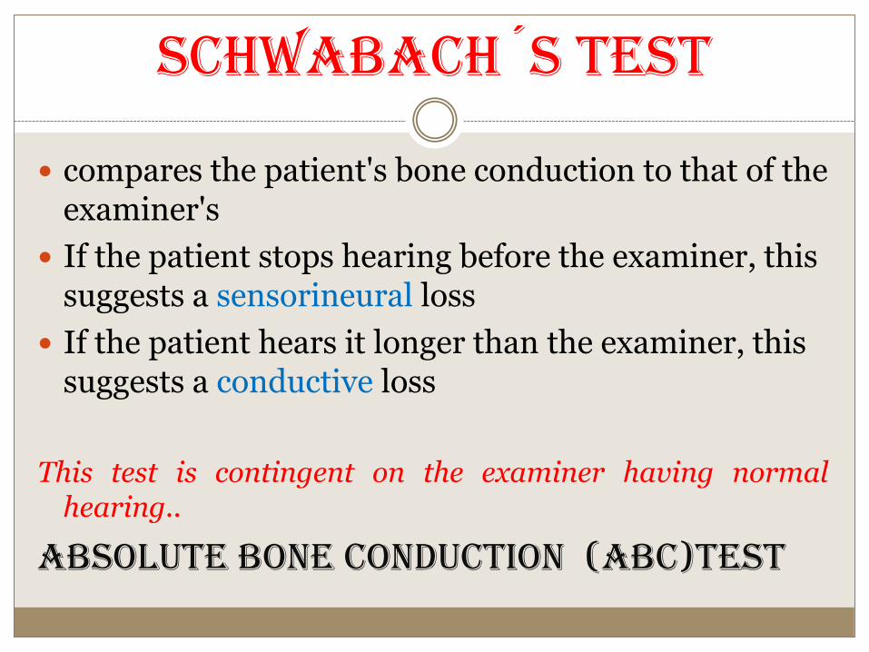
Schwabach´s test
compares the patient's bone conduction to that of the examiner's
If the patient stops hearing before the examiner, this suggests a sensorineural loss
If the patient hears it longer than the examiner, this suggests a conductive loss
This test is contingent on the examiner having normalhearing..
ABSOLUTE BONE CONDUCTION (ABC)TEST

Bing test
➢ fork is struck and placed on the patient's mastoid tip
➢examiner alternately occludes the patient's external meatus
➢patient with normal hearing or a sensorineural loss, he or she will notice a change in intensity with occlusion
➢patient with conductive hearing loss, he or she will notice no change

Gelle’s test
➢ fork is struck and placed on the patient's mastoid tip.
➢examiner increase the pressure in the patient's external meatus with the help of siegle’s speculum.
➢patient with normal hearing or a sensorineural loss, he or she will notice a change in intensity with pressure change.
➢patient with conductive hearing loss, he or she will notice no change.

Audiometry…
➢Subjective (needs patient’s response)
➢Objective (combination with EEG)

Conductive disease

Senzorineural impairment

Degrees of Hearing Loss
0 ‐ 20 dB HL: Within normal limits (WNL)
20‐40 dB HL: Mild
40‐70 dB HL: Moderate
70‐90 dB HL: Severe
> 90 dB HL: Profound

Speech Audiometry
In this test, the patient's ability to hear andunderstand speech is measured. Two parameters arestudied:
(i) speech reception threshold
(ii) Speech discrimination score

Speech reception threshold (SRT)- It is theminimum intensity at which 50% of the words arerepeated correctly by the patient. An SRT betterthan pure tone average by more than 10 dBsuggests a functional hearing loss.
Speech discrimination score- Also called speechrecognition or word recognition score. It is ameasure of patient’s ability to understand speech. Innormal persons and those with conductive hearingloss a high score of 90-100% can be obtained.

Usefulness of speech audiometry
(i) To find speech reception threshold whichcorrelates well with average of three speechfrequencies of pure tone audiogram.
(ii)To differentiate organic from non-organic(functional) hearing loss.
(iii) To find the intensity at which discriminationscore is best. This is helpful in fitting a hearing aidand setting its volume for maximum discrimination.
(iv) To differentiate a cochlear from a retrocochlearsensorineural hearing loss.

Bekesy Audiometry
It is a self-recording audiometry where various pure tone frequenciesautomatically move from low to high while the patient controls the intensitythrough a button. Two tracings, one with continuous and the other with pulsedtone are obtained. The tracings help to differentiate a cochlear fromretrocochlear and an organic from a functional hearing loss.
Various types of tracings obtained are:
Type I Continuous and pulsed tracings overlap. Seen in normal hearing orconductive hearing loss.
Type II Continuous and pulsed tracings overlap up to 1000 Hz and thencontinuous tracing falls. Seen in cochlear loss.
Type III Continuous tracing falls below pulsed tracing at 100 to 500 Hz even up to40-50 dB. Seen in retrocochlear/neural lesion.
Type IV Continuous tracing falls below pulsed lesion at frequencies up to 1000 Hzby more than 25 dB. Seen in retrocochlear/neural lesion.
Type V Continuous tracing is above pulsed one. Seen in non-organic hearing loss.
Bekesy audiometry is seldom performed these days.

Impedance Audiometry
Objective test, widely used in clinical practice.
Is particularly useful in children.
It consists of:
(a) Tympanometry
(b) Acoustic reflex measurements

Tympanometry
It is based on a simple principle, i.e. when a soundstrikes tympanic membrane, some of the soundenergy is absorbed while the rest is reflected.
A stiffer tympanic membrane would reflect more ofsound energy than a compliant one.
By changing the pressures in a sealed externalauditory canal and then measuring the reflectedsound energy, it is possible to find the compliance orstiffness of the tympano-ossicular system and thusfind the healthy or diseased status of the middle ear.

Impedence audiometer
the equipment consists of a probe which snugly fits into theexternal auditory canal and has three channels;
(i) to deliver a tone of 220 Hz,
(ii) to pick up the reflected sound through a microphone
(iii) to bring about changes in air pressure in the ear canalfrom positive to normal and then negative.
By charting the compliance of tympano-ossicular systemagainst various pressure changes, different types of graphscalled tympanograms are obtained which are diagnostic ofcertain middle ear pathologies.


Types of tympanograms
Type A-Normal tympanogram.
Type As-Compliance is lower at or near ambient
air pressure. Seen in fixation of ossicles, e.g.
otosclerosis or malleus fixation.
Type Ad- High compliance at or near ambient
pressure. Seen in ossicular discontinuity or thin
and lax tympanic membrane.
Type B- Flat or dome-shaped graph i.e. No change
in compliance with pressure changes. Seen in OME
or thick tympanic membrane.
Type C-Maximum compliance occurs with negative
pressure in excess of 100 mm of H20. Seen in
retracted tympanic membrane and may show some
fluid in middle ear.

Testing function of eustachian tube
Tympanometry has also been used to find function ofeustachian tube in cases of intact or perforatedtympanic membrane. A negative or a positivepressure (-200 or +200 mm of H2O) is created inthe middle ear and the person is asked to swallow 5times in 20 seconds. The ability to equilibrate thepressure indicates normal tubal function.
The test can also be used to find the patency of thegrommet placed in the tympanic membrane in casesof serous otitis media.

Acoustic reflex
Principle- A loud sound, 70-100 dB above thethreshold of hearing of a particular ear, causesbilateral contraction of the stapedial muscleswhich can be detected by tympanometry.
Tone can be delivered to one ear and the reflexpicked from the same or the contralateral ear.The reflex arc involved is-
Ipsilateral: CN VIII -> ventral cochlear nucleus ->CN VII nucleus -> ipsilateral stapedius muscle.
Contralateral: CN VIII -> ventral cochlearnucleus -> contralateral medial superior olivarynucleus - >contralateral CN VII nucleus ->contralateral stapedius muscle

usefulness
(i) To test the hearing in infants and young children. It is an objectivemethod.
(ii) To find malingerers. A person who feigns total deafness and does not giveany response on pure tone audiometry but shows a positive stapedial reflex is amalingerer.
(iii) To detect cochlear pathology. Presence of stapedial reflex at lowerintensities, e.g. 40 to 60 dB than the usual 70 dB indicates recruitment and thus acochlear type of hearing loss.
(iv) To detect VIIIth nerve lesion. If a sustained tone of 500 or 1000 Hz,delivered 10 dB above acoustic reflex threshold, for a period of 10 seconds, bringsthe reflex amplitude to 50%, it shows abnormal adaptation and is indicative ofVIIIth nerve lesion (stapedial reflex decay).
(v) Lesions of facial nerve. Absence of stapedial reflex when hearing is normalindicates lesion of the facial nerve, proximal to the nerve to stapedius. The reflexcan also be used to find prognosis of facial paralysis as the appearance of reflex,after it was absent, indicates return of function and a favorable prognosis.
(vi) Lesion of Brainstem. If ipsilateral reflex is present but the contralateralreflex is absent, lesion is in the area of crossed pathways in the brainstem.

Special Tests of Hearing
Recruitment
It is a phenomenon of abnormal growth of loudness. Theear which does not hear low intensity sound begins to heargreater intensity sounds as loud or even louder than normalhearing ear. Thus, a loud sound which is tolerable innormal ear may grow to abnormal levels of loudness in therecruiting ear and thus becomes intolerable.
The patients with recruitment are poor candidates forhearing aids. Recruitment is typically seen in lesions of thecochlea (e.g. Meniere's disease, presbycusis) and thus helpsto differentiate a cochlear from a retrocochlear SNHL.

Alternate binaural loudness balance test
It is used to detect recruitment inunilateral cases. A tone, say of 1000 Hz,is played alternately to the normal andthe affected ear and the intensity in theaffected ear is adjusted to match theloudness in normal ear. The test isstarted at 20 dB above the threshold ofdeaf ear and then repeated at every 20dB rise until the loudness is matched orthe limits of audiometer reached. Inconductive and neural deafness, theinitial difference is maintainedthroughout while in cochlear lesions,partial, complete or over-recruitmentmay be seen.

Short Increment Sensitivity Index (SISI)
Patients with cochlear lesions distinguish smaller changesin intensity of pure tone better than normal persons andthose with conductive or retrocochlear pathology.
SISI test is thus used to differentiate a cochlear from aretrocochlear lesion. In this test, a continuous tone ispresented 20 dB above the threshold and sustained forabout 2 minutes. Every 5 seconds, the tone is increased by 1dB and 20 such blips are presented. Patient indicates theblips heard.
In conductive deafness, SISI score is seldom more than15%; it is 70-100% in cochlear deafness; and 0-20% innerve deafness.
Threshold Tone Decay Test
It is a measure of nerve fatigue and is used to detectretrocochlear lesions. Normally, a person can hear a tonecontinuously for 60 seconds. In nerve fatigue, he stopshearing earlier.
A tone of 4000 Hz is presented at 5dB above the patient'sthreshold of hearing, continuously for a period of 60seconds. If patient stops hearing earlier, intensity isincreased by another 5 dB. The procedure is continued tillpatient can hear the tone continuously for 60 seconds, orno level exists above the threshold where tone is audible forfull 60 seconds.
The result is expressed as number of dB of decay. A decaymore than 25 dB is diagnostic of a retrocochlear lesion .

Evoked Response Audiometry
It is an objective test which measures electrical activity in
the auditory pathways in response to auditory stimuli.
It requires special equipment with an averaging computer.
There are several components of evoked electric response
but only two have gained clinical acceptance.
1. Electrocochleography (EcoG)
2. Auditory brainstem response (ABR). Also called BAER or
BAEP (brainstem auditory evoked response or potential)
or BERA (brainstem evoked response audiometry)

Electrocochleography (EcoG)
It measures electrical potentials arising in the cochlea and CN VIII inresponse to auditory stimuli within first 5 milliseconds. The response isin the form of three phenomena-
1.cochlear microphonics, 2.summating potentials and 3.action potentialof VIIIth nerve.
The recording electrode is usually a thin needle passed through thetympanic membrane onto the promontory. In adults, it can be doneunder local anesthesia but in children or anxious persons sedation orgeneral anesthesia is required. Sedation has no effect on theseresponses.
EcoG is useful (i) to find threshold of hearing in young infants andchildren to within 5-10 dB, (ii) to differentiate lesions of cochlea fromthose of the VIIIth nerve. Normally the ratio between the amplitude ofsummating potential to the action potential is less than 30%. Anincrease in this ratio is indicative of Meniere's disease.
BERA
Brainstem responses are elicited by giving auditorystimulation by clicks or tone bursts. It is a non-invasivetechnique to find the integrity of central auditory pathwaysthrough the VIIIth nerve, pons and midbrain.
In this method, electrical potentials are generated inresponse to several click stimuli or tone-bursts and pickedup from the vertex by surface electrodes. It measureshearing sensitivity in the range of 1000~000 Hz. In anormal person, 7 waves are produced in the first 10milliseconds. The first, third and fifth waves are most stableand are used in measurements. The waves are studied forabsolute latency , inter-wave latency, (usually betweenwave I and V) and the amplitude.

Wave I- Distal part of CN VIII
Wave II- Proximal part of CN VIII near the brainstem
Wave III- Cochlear nucleus
Wave IV- Superior olivary complex
Wave V- Lateral lemniscus
Waves VI and VIII- Inferior colliculusEE COLI (eight, eight, cochlear nucleus, olivary complex, lateral lemniscus, inferior colliculus)

Uses of ABR
(i) As a screening procedure for infants.
(ii) To determine the threshold of hearing in infants;also in children and adults who do not cooperate andin malingerers.
(iii) To diagnose retrocochlear pathology particularlyacoustic neuroma.
(iv) To diagnose brainstem pathology, e.g. multiplesclerosis or pontine tumours.
(vi) To monitor CN VIII intraoperatively in surgeryof acoustic neuromas to preserve the function ofcochlear nerve.

Otoacoustic Emissions (OAEs)
They are low intensity sounds produced by outer hair cellsof a normal cochlea and can be elicited by a very sensitivemicrophone placed in the external ear canal and an analysisby a computer.
Sound produced by outer hair cells travels in a reversedirection: outer hair cells -> basilar membrane ->perilymph -> oval window-> ossicles -> tympanicmembrane -> ear canal.
OAEs are present when outer hair cells are healthy and areabsent when they are damaged and thus help to test thefunction of cochlea. They do not disappear in eighth nervepathology as cochlear hair cells are normal.

Types of OAEs
Spontaneous OAEs: They are present in healthy normalhearing persons where hearing loss does not exceed 30 dB.They may be absent in 50% of normal persons.
Evoked OAEs: They are further di vided into two typesdepending on the sound stimulus used to elicit them.
(a) Transient evoked OAEs (TEOAEs)- Evoked by clicks.
(b) Distortion product OAEs (DPOAEs)- Two tones aresimultaneously presented to the cochlea to produce distortionproducts.
Uses of OAE
1. as a screening test of hearing in neonates and totest hearing in uncooperative or mentally challengedindividuals after sedation.
2. to distinguish cochlear from retrocochlear hearingloss. OAEs are absent in cochlear lesions, e.g.ototoxic sensorineural hearing loss. They detectototoxic effects earlier than pure-tone audiometry.
3. to diagnose retrocochlear pathology, especiallyauditory neuropathy.

HEARING LOSS

CONDUCTIVE HEARING LOSS ANDITS MANAGEMENT
Characteristics
1. Negative Rinne test , i.e. BC> AC.
2. Weber lateralized to poorer ear.
3. Normal absolute bone conduction.
4. Low frequencies are affected more.
5. Audiometry shows bone conduction better than airconduction with air-bone gap. Greater the air-bone gap, moreis the conductive loss.
6. Loss is not more than 60 dB.
7. Speech discrimination is good.

Causes of conductive hearing loss
Congenital causes Acquired causes

Average Hearing Loss Seen in DifferentLesions of Conductive Apparatus
1.Complete obstruction of ear canal: 30 dB
2.Perforation of tympanic membrane:(It
varies and is directly proportional to the size
of perforation)-10-40 dB
3.Ossicular interruption with intact drum: 54
dB
4.Ossicular interruption with perforation: 38
dB
5.Closure of oval window: 60 dB

Management
medical or surgical means.
1. Removal of canal obstructions, e.g. Impacted wax, foreign body,
osteoma or exostosis, keratotic mass, benign or malignant tumours,
meatal atresia
2. Removal of fluid. Myringotomy with or without grommet insertion.
3. Removal of mass from middle ear. Tympanotomy and removal of small
middle ear tumours or cholesteatoma behind intact drum.
4. Stapedectomy. as in otosclerotic fixation of stapes footplate.
S. Tympanoplasty. Repair of perforation, ossicular chain or both.
6. Hearing aid. In cases, where surgery is not possible, refused or has
failed.

SENSORINEURAL HEARING LOSS
1. A positive Rinne test, i.e. AC > BC.
2. Weber lateralized to better ear.
3. Bone conduction reduced on Schwahach and absolute boneconduction tests.
4. More often involving higher frequencies.
5. No gap between air and bone conduction curve on
Audiometry
6. Loss may exceed 60 dB.
7. Speech discrimination is poor.
8. There is difficulty in hearing in the presence of
noise.


Aetiology
1. Congenital- it is present at birth and is the result of anomalies of the inner ear or damageto the hearing apparatus by prenatal or perinatal factors.
2. Acquired
1. Infections of labyrinth; viral, bacterial or spirochaetal.
2. Trauma to labyrinth or VIIIth nerve, e.g. fractures of temporal bone or concussion oflabyrinth or ear surgery,
3. Noise-induced hearing loss
4. Ototoxic drugs
5. Presbycusis,
6. Meniere's disease,
7. Acoustic neuroma,
8. Sudden hearing loss,
9. Familial progressive SNHL,
10. Systemic disorders, e.g. Diabetes, hypothyroidism kidney disease, autoimmunedisorders, multiple sclerosis, blood dyscrasias.

SPECIFIC FORMS OF HEARING LOSS
Inflammations of Labyrinth
Ototoxicity
Noise Trauma
Sudden Hearing loss
Presbycusis
Familial Progressive Sensorineural hearing Loss

Inflammations of Labyrinth
Viral labyrinthitis.
Bacterial labyrinthitis.
Syphilitic labyrinthitis.

Ototoxicity

Noise Trauma
1. Acoustic trauma- Permanent damage to hearing
caused by a single brief exposure to very intense
sound, e.g. an explosion, gunfire or a powerful
cracker.
2. Noise-induced hearing loss (NIHL)- follows
chronic exposure to less intense sounds mainly
because of noisy occupations.

Pathology in Noise Trauma
(a)Temporary thresholdshift-The hearing is impairedimmediately after exposure tonoise but recovers after aninterval of a few minutes to afew hours.
(b)Permanent thresholdshift-The hearing impairmentis permanent and does notrecover at all.

Sudden Hearing loss
1. Ideopathic
2. Infections. Mumps, herpes zoster, meningitis, encephalitis, syphilis,otitis media.
3. Trauma. Head injury, ear operations, noise trauma, barotrauma,spontaneous rupture of cochlear membrane
4. Vascular. Haemorrhage (leukemia) , embolism or thrombosis ofLabyrinthine or cochlear artery or their vasospasm.
5. Otologic. Meniere’s disease, Cogan's syndrome, large vestibularaqueduct.
6. Toxic. Ototoxic drugs, insecticides.
7. Neoplastic. Acoustic neuroma, metastases in cerebellopontine angle,carcinomatous neuropathy
8. Miscellaneous. Multiple sclerosis, hypothyroidism, sarcoidosis.
9. Psychogenic.

Management
To be treated according to the cause.
When cause remains obscure, treatment is empirical andconsists of:
1. Bed rest.
2. Steroid therapy. Prednisolone
3. Inhalation of carbogen (5% CO2 + 95% O2).
4. Vasodilator drugs.
5. Low molecular weight dextran.
6. Hyperbaric oxygen therapy.
Prognosis: Fortunately, about half of the patients of idiopathic SNHL
recover spomaneously within 15 days. Chances of recovery are poor after 1month. Severe hearing loss and association with vertigo have poor prognosis.Younger patients below 40 and those with moderate losses have betterprognosis.

Presbycusis
SNHL associated with physiological aging process in the ear is calledpresbycusis. It usually manifests at the age of 65 years but may do soearly if there is hereditary predisposition, chronic noise exposure orgeneralized vascular disease.
1. Sensory. Higher frequencies are affected bur speech discriminationremains good.
2. Neural. This manifests With high frequency loss but speechdiscrimination is poor and out of proportion.
3. Strial or metabolic. This is characterized by atrophy of stria vascularisof cochlea. Audiogram ls flat but speech discrimination is good.
4. Cochlear conductive. This is due to stiffening of the basilar membranethus affecting its movements. Audiogram is sloping type.

Patients of presbycusis have great difficulty in hearing inthe presence of background noise though they may hearwell in quiet surroundings.
They may complain of speech being heard but notunderstood. Recruitment phenomenon is positive and allthe sounds suddenly become intolerable when volume israised.
Tinnitus is another bothersome problem and in some it isthe only complaint.
Patients of presbycusis can be helped by a hearing aid. Theyshould also have lessons in speech reading through visualcues. Cessation of smoking and drinks like tea and coffeemay help to decrease tinnitus.

Familial Progressive Sensorineural hearing Loss
It is a genetic disorder in which there is progressive degeneration of the cochlea starting in late childhood or early adult life.
Deafness is bilateral with Hat or basin-shaped audiogram bur an excellent speech discrimination.

Non-organic hearing loss (NOHL)
either due to malingering or psychogenic.
1. High index of suspicion.
2. Inconsistent results on repeat pure tone and speech audiometry tests.
3. Absence of shadow curve.
4. Inconsistency in PTA and SRT.
5. Stenger test.
6. Acoustic reflex threshold
7. EIectric response audiometry (ERA).

For Whom Hearing Aids are?
Sound‐amplifying medical devices to aid individuals with hearing loss.
Hearing aids may be useful for- Hearing loss that may or may not be medically treatable.
Any type of hearing loss, as long as the individual needs compensation for the reduction in hearing.
Selection of hearing aids should be based on the type and severity of hearing loss, listening needs, and lifestyle.

Hearing Aids: Basic Components & How They Work
Electronic components: Microphone
Amplifier circuitry
Miniature loudspeaker/receiver
Battery
How does a hearing aid work?

Hearing Aid Styles
Behind‐the‐ear (BTE) aids: A plastic case
containing most parts; resting behind the ear connected to an earmould
Easy to be cleaned and handled, relatively sturdy
"Mini" BTE (or "on‐the‐ear") aids: A very
thin tube connects the aid to the ear canal
May have an open‐fit ear tip or a regular earmould
With “open fit” – Reduced occlusion ("plugged up“) sensations, increased comfort, relatively less visible


Hearing Aid Styles
In‐the‐ear (ITE) aids: All parts contained in a shell, which fills in the ear canal
Relatively easier to handle than smaller aids such as ITC & CIC
In‐the‐canal (ITC) aids & completely‐in‐the‐canal (CIC) aids: All parts contained in tiny cases, which fits partly or completely in the ear canal
Smallest in size, which makes it difficult to handle and adjust for some users

CIC
ITC
ITE

Hearing Aid Technology: Analog vs. Digital
Analog- Converting physical sound waves into electrical waves
Making the continuous sound waves larger.
Digital- Converting sound waves to their binary format where the
sound is represented by a series of 1’s and 0’s.
Allowing manipulating sounds in relatively flexible ways to achieve more programming options.

Common Hearing Aid Features
Directional microphones: Sound from a specific direction
amplified to a greater level
May help listeners to understand speech in noisy environments
Feedback suppression: Squeals suppressed when the
hearing aid gets too close to the phone or has a loose‐fitting earmould
T‐coil (Telephone switch): Sound picked up from the
telephone when switching to the "T‐ coil" setting
Help to reduce the chance of hearing aid "whistling"
Also works well in environments (e.g., theaters, auditoriums, etc.)where there is induction loop or FM installation

Hearing Aid Fitting
Questions to consider: What styles and features would fit my daily needs?
Cost: What is the total cost of the hearing aids?
Do the benefits of newer technologies outweigh the higher costs?
Trial/adjustment period: Is there a trial or adjustment period
for me to try out the hearing aids?
What fees are non-refundable if I decide to return the hearing aids?
Care & Warranty: How should I care for my hearing aids?
What is covered during the period of warranty?
How long is the warranty? Can it be extended?

Hearing Aid Care & Maintenance
Keep hearing aids away from any moisture and heat, which may cause damage to the internal electronics.
Clean hearing aids as instructed.
Power consumption & battery safety: Turn off hearing aids
when not in use.
Keep batteries and hearing aids away from children and pets.
Visit the hearing healthcare professional on a regular basis to have hearing aids inspected.

Hearing Aid Benefits & Limitations
Benefits
Ability to hear sounds that could not be heard previously, andhelp oral communication
Ability to hear speech over the telephone
Limitations
Do not restore normal hearing
All sounds, including background noise and undesiredsounds, are made louder.
Sounds, including own voice, might seem too loud at first.
May need to be replaced every several years

COCHLEAR IMPLANT
❑Designed for those who are profoundly deaf
❑Can be used alone or with traditional hearing aid
❖TWO MAIN SYSTEMS:
External
Sound processer, microphone and transmitter
Internal
Receiver and electrode array

Future for hearing aids
BAHA(Bone Anchored Hearing Aid)- Bypass normal hearing system
Helps severe sensorineural, conductive or mixed hearing loss
Attaches to bones in middle ear
Bypasses auditory canal and middle ear

CONCLUSION
Hearing aids are smaller and more powerful
Aiming for higher sound quality in the future
Accommodate virtually every type of listening
environment
Continually adjust themselves to improve sound
quality and reduce feedback
Closer to achieving gift of hearing