Processing and Properties of Polyethylene/Montmorillonite Nanocomposites
Upload
independentCategory
view
1download
0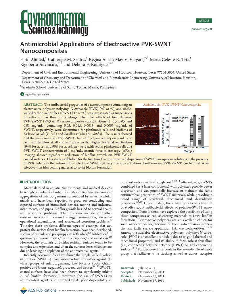
Published: November 17, 2011
r 2011 American Chemical Society 1804 dx.doi.org/10.1021/es202374e | Environ. Sci. Technol. 2012, 46, 1804–1810
ARTICLE
pubs.acs.org/est
Antimicrobial Applications of Electroactive PVK-SWNTNanocompositesFarid Ahmed,† Catherine M. Santos,† Regina Aileen May V. Vergara,‡,§ Maria Celeste R. Tria,‡
Rigoberto Advincula,‡,§ and Debora F. Rodrigues*,†
†Department of Civil and Environmental Engineering, University of Houston, Houston, Texas 77204-5003, United States‡Department of Chemistry and Department of Chemical and Biomolecular Engineering, University of Houston, Houston,Texas 77204-5003, United States§Graduate School, University of Santo Tomas, Manila, Philippines
bS Supporting Information
’ INTRODUCTION
Materials used in aquatic environments and medical deviceshave high potential for biofilm formation.1 Biofilms are complexaggregations of microorganisms surrounded by an extracellularmatrix and have been reported to grow on conducting andexposed surfaces of biomedical devices, marine and industrialinstruments, and pipes. Biofilm growth has led to several healthand economic problems. The problems include antibiotic-resistant infections, increased energy consumption, excessiveoperational expenditures, and accelerated corrosion problems.2
To solve these problems, different types of coatings, that canprotect the surface from biofilm formation, have been developed,such as polyamide and polypropylene with silver,3,4 antibiotics,4�7
quaternary ammonium salts,8 cationic peptides,9 and metal ions.10
However, the synthesis of biofilm resistant surfaces tends to becomplex and expensive, and often the surfaces loses effectivenessdue to leaching or depletion of the antimicrobial agents.5�7
Recently, several studies have shown that single-walled-carbonnanotubes (SWNTs) have antimicrobial properties against di-verse groups of microorganisms, like bacteria (both Gram-positive and Gram- negative), protozoa, and viruses.8�12 SWNT-coated surfaces have also been shown to significantly inhibitE. coli biofilm formation.7 However, the use of SWNTs asantimicrobial agent is still limited by its poor dispersibility in
most solvents as well as its high cost.5,13,14 Alternatively, SWNTscombined (as a filler component) with polymers provide betterdispersion and can potentially increase or maintain the sameantimicrobial properties of SWNT materials, while providing abroad range of structural, mechanical, and degradationproperties.1,5,15 Unfortunately, there have only been a handfulof studies about antibacterial effects of polymer-SWNT nano-composites. None of them have explored the possibility of usingthese composites as robust coating materials to resist biofilmformation. Electroactive polymers are an excellent choice forsuch nanocomposites, because of their anticorrosion proper-ties and facile surface application (via electrodeposition).16,17
Among the available electroactive polymers, polyvinyl-N-carba-zole (PVK) is an excellent candidate due to its good thermal andmechanical properties, and its ability to form robust thin films(i.e., conducting polymer network (CPN)) on any conductingsurface.18,19 Furthermore, PVK contains the aromatic N-carbazolegroup that facilitates π�π stacking as well as donor�acceptor
Received: July 10, 2011Accepted: November 17, 2011Revised: November 15, 2011
ABSTRACT:The antibacterial properties of a nanocomposite containing anelectroactive polymer, polyvinyl-N-carbazole (PVK) (97 wt %), and single-walled carbon nanotubes (SWNT) (3 wt %) was investigated as suspensionsin water and as thin film coatings. The toxic effects of four differentPVK-SWNT (97:3 wt %) nanocomposite concentrations (1, 0.5, 0.05, and0.01 mg/mL) containing 0.03, 0.015, 0.0015, and 0.0003 mg/mL ofSWNT, respectively, were determined for planktonic cells and biofilms ofEscherichia coli (E. coli) and Bacillus subtilis (B. subtilis). The results showedthat the nanocomposite PVK-SWNT had antibacterial activity on planktoniccells and biofilms at all concentration levels. Higher bacterial inactivation(94% for E. coli and 90% for B. subtilis) were achieved in planktonic cells at aPVK-SWNT concentration of 1 mg/mL. Atomic force microscopy (AFM)imaging showed significant reduction of biofilm growth on PVK-SWNTcoated surfaces. This study established for the first time that the improved dispersion of SWNTs in aqueous solutions in the presenceof PVK enhances the antimicrobial effects of SWNTs at very low concentrations. Furthermore, PVK-SWNT can be used as aneffective thin film coating material to resist biofilm formation.
1805 dx.doi.org/10.1021/es202374e |Environ. Sci. Technol. 2012, 46, 1804–1810
Environmental Science & Technology ARTICLE
interactions making it a more compatible polymer for carbon-based nanomaterials like SWNTs.20,21
In this study, we investigated the PVK-SWNT nanocompositeantibacterial properties to planktonic cells (i.e., cells in suspen-sion prior to biofilm formation) and biofilms. The bacterialtoxicity of different concentrations of PVK-SWNT dispersed inwater were investigated against Gram-positive (B. subtilis) andGram-negative (E. coli) bacteria, as well as the potential inhibi-tion properties of biofilm formation on coated surfaces with thePVK-SWNT nanocomposite. The results showed for the firsttime that, by improving dispersibility of SWNT in solution,higher bacterial toxicity of SWNT can be achieved, even inconcentrations as low as 0.0003 mg/mL of SWNT. Furthermore,PVK-SWNT coated surfaces with only 3% of SWNTs signifi-cantly inhibited biofilm formation. This result shows that coatedsurfaces for antimicrobial purposes can be made with reducedconcentrations of SWNT.
’MATERIALS AND METHODS
Single-Walled Carbon Nanotubes (SWNTs) Characteriza-tion and Preparation. Single-walled carbon nanotubes(SWNTs) were purchased from Cheap Tubes Inc. (Vermont).The characterization of these nanomaterials can be found in theSupporting Information (SI, Table S1, Table S2, and Figure S1).The SWNTs were further purified by heating at 200 �C for 6 hprior to use. The SWNT suspension (1 mg/mL) was prepared asdescribed in our previous work.22,23 Briefly, SWNTs weredispersed in DI water through 3 cycles of sonication for 1 himmediately before use for the antimicrobial assays.PVK-SWNT Nanocomposite Solutions. The poly(N-vinyl
carbazole) (PVK) was purchased from Sigma-Aldrich Chemicals(ca. MW = 25 000�50 000 g/mol). All solvents used for thePVK-SWNT preparation were purchased from Sigma Aldrichand were of analytical grade. The PVK-SWNT (97/3 wt % ratioPVK/SWNT) was prepared according to previously reportedprocedure.16 The PVK/SWNT ratio of 97/3 (wt %) was selectedon the basis of the high dispersibility and stability of SWNT forlong periods of time (several months) as described in ourprevious work.16 Briefly, a 97/3 wt/vol % ratio of PVK/SWNTwas prepared in N-cyclohexyl-2-pyrrolidone (CHP). The puri-fied SWNT was first dissolved in CHP and sonicated for 4 h.Then, in a separate vial, the PVK was dissolved in CHP andsonicated for 30 min. The PVK solution was then slowly mixedwith the SWNT solution and followed by sonication for 1 h. Afterthis, the PVK-SWNT dispersion was centrifuged (4400 rpm,1 h), and the black precipitate was removed. The remainingsolution of PVK-SWNT dispersion was then treated withmethanol (5 mL) and again centrifuged (4400 rpm) for30 min. The black precipitate was collected and redispersed inwater followed by 20 min of ultrasonication. This procedurefurnished a stable and well dispersed PVK-SWNTs solution. Forthe bacterial measurements, different PVK-SWNT concentra-tions (1.0, 0.5, 0.05, and 0.01 mg/mL) dispersed in water wereused. The SWNT concentrations (mg/mL) from the preparedPVK-SWNT (97/3 wt %) dispersions are provided (Table S3).The characterization of PVK-SWNT nanocomposite and pre-paration of PVK-SWNT nanocomposite conducting polymernetwork (CPN) films can be found in the Supporting Informa-tion; the results are presented in Figures S2 and S3.Bacterial Culture and Antimicrobial Activity of SWNT and
PVK-SWNT. Single isolated colonies of E. coli MG 1655 and
B. subtilis 102 were inoculated and incubated in 5 mL of TrypticSoya Broth (TSB) (Oxoid, England) overnight at 35 �C and200 rpm. The bacterial culture was centrifuged at 3000 rpm for10 min. The cells were washed and resuspended in phosphatebuffer solution (PBS, 0.01 M, pH = 7.4) (Fisher Scientific). Thebacterial suspension was adjusted to give an optical density (OD)of 0.5 at 600 nm, which corresponds to a concentration of 107
colony forming units (CFU)/mL. For the antimicrobial activityassay, bacterial cultures were exposed for 3 h to the differentnanomaterials. Briefly, aliquots of 180 μL of bacterial suspen-sions (107 CFU/ml) in PBS and noninoculated PBS buffer withbacteria (used as blanks) were pipetted in a 96-well flat bottomplate (Costar 3370, Corning, NY) containing triplicates of 20 μLof the following samples suspended in DI water: (1) SWNT atconcentration of 1.0 mg/mL; (2) PVK-SWNT nanocompositeat concentrations of 1.0, 0.5, 0.05, and 0.01 mg/mL; and (3)1 mg/mL of PVK. The control samples contained 20 μL of DIwater only with 180 μL of bacterial suspensions. To account forthe absorbance of SWNT and PVK-SWNT nanomaterials sus-pended in the bacterial samples, 20 μL of each concentration ofSWNT and PVK-SWNT were added to 180 μL of PBS only andlater used as blanks to subtract from the original samples. Theplates were then incubated at 37 �C at 50 rpm for 3 h. After3 h, 20 μL of the bacteria exposed to the different materials, thenegative controls, and the blank samples were transferred into 96well-plates containing 200 μL TSB. The samples were thenincubated at 37 �C at 50 rpm, and the bacterial growth wasmonitored using Synergy MX Microtiter plate reader (BioTek,VT) by measuring the OD600 every hour until the bacteriareached stationary phase (Figure 1 a and b). The results forE. coli and B. subtilis growth after exposure to the nanomaterialswere reported at their midlog phases, i.e., after 3 and 5 h, respec-tively (Figure 1c). Final OD values for each bacterial solutionexposed to the different nanomaterial samples were determinedby subtracting the OD values acquired from their respective blanks.The results are reported as average OD values with standarddeviations of the triplicate samples from all three performedexperiments. Statistical analyses (two-sided t-test, 95% confi-dence interval) were performed to determine whether the ODvalues of the samples with SWNT or PVK-SWNT were sig-nificantly different from the control. The same statistical analysiswas also performed between OD values from SWNT and PVK-SWNT samples. The antimicrobial activity was also measuredusing the “live/dead assay” as previously described.24 The experi-mental methods are presented in the Supporting Information.Biofilm Formation Assay with OD Measurement. In this
assay, we measured the total biofilm growth under exposure ofdifferent concentrations of nanomaterials as previously des-cribed.7 Briefly, biofilm growth was measured by using 96-wellflat bottom plate (Costar 3370, Corning, NY). The concentra-tions of nanomaterials used in this assay were (1) SWNT atconcentration of 1.0mg/mL; (2) PVK-SWNTnanocomposite atconcentrations of 1.0, 0.5, 0.05, and 0.01 mg/mL; and(3) 1 mg/mL of PVK. The control samples contained only DIwater instead of nanomaterials. The 96-well plate was preparedwith bacteria and nanocomposites following the same procedureas described in the Bacterial Culture and Antimicrobial Activity ofSWNT and PVK-SWNT section. For both E. coli and B. subtilis,plates were prepared in triplicate. After inoculation of bacteriawith the nanomaterials, the 96-well plates were incubated at 35 �Cfor 48 h and then stained according to crystal violet stainingmethod for biofilm quantification described elsewhere. 25 Briefly,
1806 dx.doi.org/10.1021/es202374e |Environ. Sci. Technol. 2012, 46, 1804–1810
Environmental Science & Technology ARTICLE
supernatant from the wells in the plate was poured out, and theplate was washed three times. For staining, 300 μL of 0.1% crystalviolet was added in each well and incubated for 20 min in roomtemperature. After incubation, the staining solution was pouredout and washed three times. In each well 300 μL of ethanolsolution in acetone (80% vol/vol) was added, and the plate wasread at OD540. The results are expressed as average OD valueswith standard deviations using all triplicates. Statistical analyseswere performed as described in the Bacterial Culture and Anti-microbial Activity of SWNT and PVK-SWNT section.Biofilm Formation Measurements on SWNT and PVK-
SWNT Nanocomposite Coated Surfaces. Inhibition of biofilmgrowth was determined on coated ITO (indium tin oxide) surfaces.Unmodified ITO, electrodeposited PVK-SWNT (97/3 wt % PVK/SWNT), electrodeposited PVK, and spin coated SWNT-modifiedfilms on ITOwere individually placed in a 12-well plate (FalconBD).Each well of the 12-well plate, containing TSB, were inoculatedwith 300 μL of bacterial cells at OD of 0.5 and incubated at 37 �Cfor 48 h. After incubation, the ITO surfaces were taken out andgently rinsed with sterile DI water. Biofilm fixation was doneaccording to cell fixation method previously described.26 Briefly,the ITO surfaces were incubated with 2% glutaraldehyde andsubsequently dehydrated with increasing concentrations of etha-nol (25%, 50%, 75%, 95%, and 100%). The surfaces werevacuum-dried overnight prior to AFM measurements. AFMtopography measurements were done on the ITO substratesunder ambient conditions with a PicoSPM II (PicoPlus, Molec-ular Imaging-Agilent Technologies) in the intermittent contactmode. Images obtained were processed using Gwyddion soft-ware (2.13). To determine any toxic effects of residual cleaningagent (TBAH and acetonitrile) present on ITO surfaces afterpreparation of the surfaces, a control slide was rinsed with TBAHand acetonitrile and vacuum-dried overnight. A bare glass slide(without the cleaning agents) was used as control. These glassslides were incubated with E. coli for 5 h, and then, viability assay(Live/Dead) was performed. Each slide was tested in duplicate.The results showed no visible toxicity toward E. coli. Results canbe seen in the Supporting Information (Figure S4).In addition to the AFM analysis, bacterial regrowth potential
test after attachment to the modified ITO surface was testedthrough the well established method of the plate agar test.25 Thebrief description of the methodology used is presented in theSupporting Information.
’RESULTS AND DISCUSSION
PVK-SWNTCharacterization.The dispersion of PVK-SWNT(97�3 wt %) nanocomposites were characterized using FT-IRand UV�vis. FT-IR measurements confirmed the functionalgroups present in the nanocomposite (Figure S2). As controls,IR measurements of PVK and SWNT were also acquired. Asexpected, no distinctive IR peaks were observed for the pureSWNTs. The PVK-SWNT nanocomposite showed peaks similarto those of pure PVK. In particular, the peak at 1255 cm�1, due tothe C�N stretching of vinyl carbazole, was observed in bothPVK and PVK-SWNT nanocomposite.UV�vis spectra of the PVK-SWNT dispersion were acquired
to measure interfacial interaction of SWNT and PVK. Results areshown in Figure S2b. On the basis of the results, no absorptionpeaks at the visible region were observed for pure SWNTs. Thepure PVK, however, showed two distinct peaks at 330 and343 nm, which can be attributed to the transitions of the pendant
carbazole moieties of PVK.27 Similar absorption peaks wereobserved for thePVK-SWNTnanocompositewith a slight decreasein intensity and a red-shift by ∼10 nm due to the incorporationof SWNT.Electrodeposited PVK-SWNT coated surfaces were character-
ized using XPS to determine elemental composition on thesurface. Figure S3a,b shows the narrow scans in the N1s and C1sof the electrodeposited PVK-SWNT and PVK surfaces. Toestimate the amount of SWNT after electrocrosslinking, N/Cratios of PVK-SWNT and PVK were acquired. For PVK-SWNT,a calculated N/C ratio value of 9.4 was obtained while for PVK,the N/C ratio was calculated as 9.7. Using the obtained N/Cratios, the amount of PVK and SWNT on the film was 97% and3%, respectively.UV�vis spectra after electrodeposition of the PVK-SWNT,
Figure S3c, showed the disappearance of the well-defined peaksat 342 and 352 nm that were initially found for the PVK-SWNTdispersion (Figure S2b). A new broad band centered at 450 nmwas depicted after the electrodeposition process, attributed tothe electrochemical cross-linking of the carbazole pendants inPVK.28,29 These results correlate well with our previous studieson electropolymerized PVK and carbazole-containing precur-sors. 27,29
Antibacterial Effects of Nanocomposites on PlanktonicCells. The toxic effects of PVK-SWNT, PVK, and SWNTsolutions to E. coli and B. subtilis were evaluated by OD600
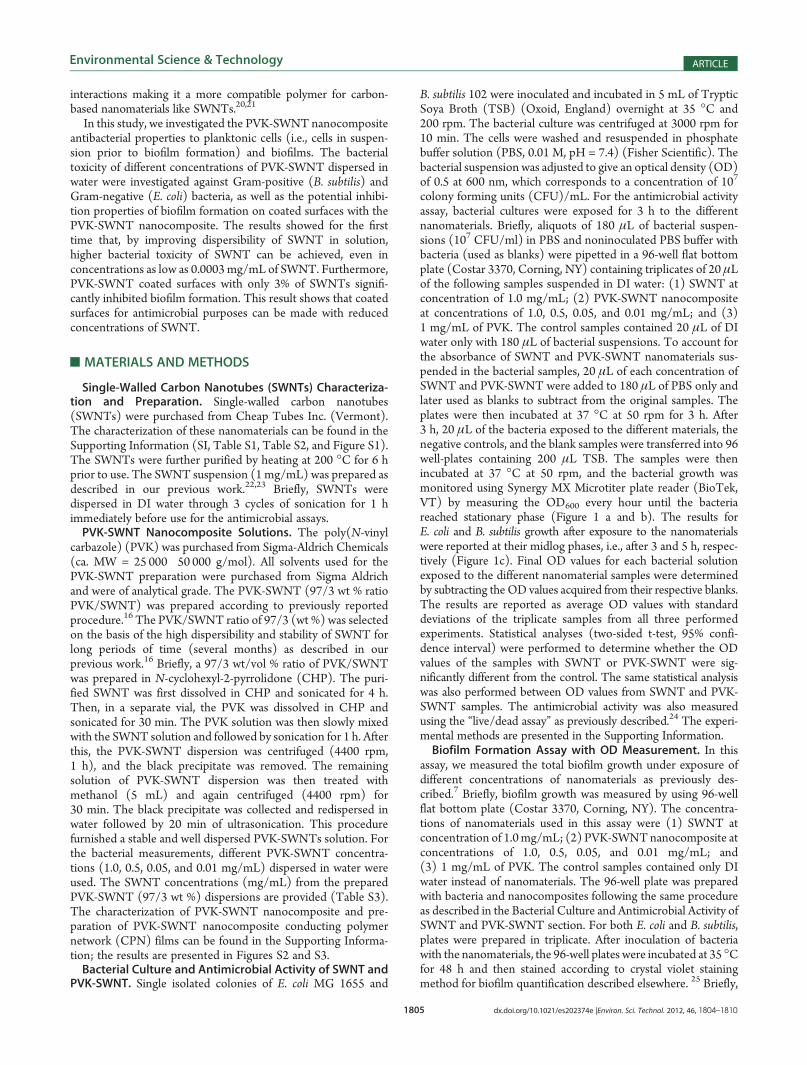
measurements of the total bacterial growth after exposure to thenanomaterials (Figure 1a,b). The results show that maximumtoxicity levels were achieved for both E. coli and B. subtilis at aconcentration of 1 mg/mL of PVK-SWNT. Furthermore, pureSWNTs (1 mg/mL) were less toxic than the PVK-SWNTcomposite (1 mg/mL), with only 0.03 mg/mL of SWNTs. Cellexposure for 3 h to SWNT (1 mg/mL) and PVK-SWNT(1 mg/mL) led to growth inhibition of ∼60% and ∼64% forE. coli, and ∼57% and ∼63% for B. subtilis, respectively(Figure 1c). Exposure to 1 mg/mL of SWNTs and PVK-SWNTnanocomposites for 1 h did not present any apparent toxic effectson B. subtilis (data not shown). From these observations, it ispossible that 1 h exposure of B. subtilis to the nanomaterial wastoo short for effective inactivation of the bacterial cells. This timedependency of SWNT toxic effects on B. subtiliswas also observedin another study.30 In this study, 1 h exposure of B. subtilis tocarbon nanotubes showed no significant growth inhibition, while4 h exposure resulted in significant growth inhibition ofB. subtilis.30
The possible reason for B. subtilis resistance to carbon nanotube atshorter exposure time can be explained by the presence of a thickerpeptidoglycan layer present in B. subtilis.30 Additionally, Figure 1calso demonstrated that the levels of tolerance of E. coli andB. subtilis to SWNTs and PVK-SWNT were not the same. Thesefindings were similar to other studies.5,31,32 The different levels oftolerance of different microorganims to carbon-based nanomater-ials are still a matter of continuing research. Several hypothesesfor the different toxicity levels consider differences in cell wallstructure, the protective effect of the outer membrane surfaceproperties, and ability to form spores and/or unique repairmechanisms of different microorganisms.30 Hence, from theseresults, it can be inferred that toxic effects of SWNTs and PVK-SWNT on both E. coli and B. subtilis are both time and concentra-tion dependent.The comparable toxicity of 100% SWNTs (1 mg/mL) with
PVK-SWNTnanocomposites (1mg/mL) containing 0.03mg/mLof SWNT can be explained by a better dispersion of the SWNTs
1807 dx.doi.org/10.1021/es202374e |Environ. Sci. Technol. 2012, 46, 1804–1810
Environmental Science & Technology ARTICLE
in aqueous solution in the presence of PVK, as previouslydemonstrated.16 This better dispersion of the SWNTs particlesin aqueous media is because of the effective π�π stacking anddonor�acceptor interactions between the carbazole group andthe SWNT. In the case of SWNT toxicity toward bacteria,dispersion is an important parameter and highly dispersedSWNTs cause greater cell contact and can potentially increasecell damage.9,13 In this study, only the dispersion effect of SWNTon PVK was investigated. Although synergistic effects of the twoconstituents in the system (i.e., morphological modifications,electronic interactions, charge transfers, or a combination ofthese effects) could also be responsible for the enhanced anti-microbial activity in the nanocomposite, they were not investi-gated in this study.The live/dead assay was performed to determine the viabi-
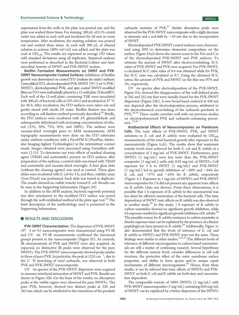
lity of the bacterial cells after interaction with nanomaterials(Figure 2). Fluorescence microscopy was used to assess the lossof bacterial viability after incubation. Figure 2 shows representa-tive fluorescence images for the bacterial solutions incubatedwith the nanocomposite PVK-SWNT and the control. Resultsshow that, in the absence of the nanomaterials, all cells were alive(Figure 2a). While cellular damage was observed in ∼94% and∼98% of the E. coli cells exposed to PVK-SWNT and SWNTs,respectively. For B. subtilis, ∼90% and ∼87% of the cells were
damaged after exposure to PVK-SWNT and SWNTs, respec-tively. The two most hypothesized mechanism of SWNT toxicityto bacteria are physical disruption of bacterial membrane andoxidative stress.5,7,24,33,34 From this study, we can say that theaddition of PVK did not prevent one of these two mechanisms tohappen since most of the cells exposed to PVK-SWNT were red-stained cells, which indicated that the PI dye could penetrateinside the damaged cells.Biofilm Growth Inhibition in the Presence of Nanocom-
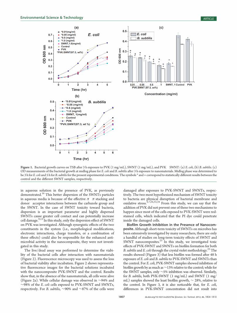
posite.Although short-term toxicity of SWNTs on microbes hasbeen extensively investigated by many researchers, there are onlya handful of studies on long-term toxicity effects of SWNT andSWNT nanocomposites.23 In this study, we investigated toxiceffects of PVK-SWNT and SWNTs on biofilm formation for bothB. subtilis and E. coli through the crystal violet methodology.25 Theresults showed (Figure 3) that less biofilm was formed after 48 hexposure of E. coli and B. subtilis to PVK-SWNT and SWNTs thanthe control. For E. coli, PVK-SWNT samples showed inhibition ofbiofilm growth by asmuch as∼13% relative to the control; while forthe SWNT samples, only ∼5% inhibition was observed. Similarly,for B. subtilis, both PVK-SWNT (1 mg/mL) and SWNT (1 mg/mL) samples showed the least biofilm growth, ∼ 28%, relative tothe control. In Figure 3, it is also noticeable that, for E. coli,differences in PVK-SWNT concentration did not result into
Figure 1. Bacterial growth curves on TSB after 3 h exposure to PVK (1 mg/mL), SWNT (1 mg/mL), and PVK�SWNT: (a) E. coli, (b) B. subtilis. (c)ODmeasurements of the bacterial growth at midlog phase for E. coli and B. subtilis after 3 h exposure to nanomaterials. Midlog phase was determined tobe 3 h forE. coli and 5 h forB. subtilis for the present experimental conditions. The symbols * and + correspond to statistically different results between thecontrol and the different SWNT samples, respectively.
1808 dx.doi.org/10.1021/es202374e |Environ. Sci. Technol. 2012, 46, 1804–1810
Environmental Science & Technology ARTICLE
different toxic effects on biofilm formation. This could be attributedto two reasons. First, based on E. coli and B. subtilis growth curves,the generation timeofE. coli in thismedia ismuch faster than that ofB. subtilis. Therefore, the E. coli cells that survived after initial contactwith SWNTs will replicate faster and potentially outnumber theavailable SWNTs that could inactivate them. Second, other studiesshowed that E. coli cells can endure and recover from membranedamages and oxidative stresses after contact with carbonnanotubes.24
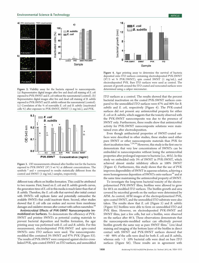
Antimicrobial Effects of PVK-SWNT Nanocomposite Im-mobilized on Surfaces. To demonstrate the efficiency of PVK-SWNT and pristine SWNTs as potential coating materials toprevent bacterial deposition and biofilm formation, the agarprinting assay was performed with E. coli and B. subtilis. For thismeasurement, electrodeposited PVK-SWNT and spin-coatedSWNTs onto ITO surfaces were used. The nanocomposite-modified film contained 3% SWNT and 97% PVK (Figure S3).The results of PVK-SWNTwere compared against electro-cross-linked PVK, spin-coated SWNT on ITO surfaces, and unmodified
ITO surfaces as a control. The results showed that the percentbacterial inactivation on the coated PVK-SWNT surfaces com-pared to the unmodified ITO surfaces were 67% and 80% for B.subtilis and E. coli, respectively (Figure 4). The PVK-coatedsurfaces did not present any antimicrobial property for eitherE. coli or B. subtilis, which suggests that the toxicity observed withthe PVK-SWNT nanocomposite was due to the presence ofSWNT only. Furthermore, these results show that antimicrobialactivity for PVK-SWNT nanocomposite solutions were main-tained even after electrodeposition.Even though antibacterial properties of SWNT-coated sur-
faces were described in other studies, these studies used eitherpure SWNT or other nanocomposite materials than PVK forshort incubation time.7,24,34 However, this study is the first one todemonstrate that very low concentrations of SWNTs can beembedded in nanocomposites without losing the antimicrobialproperties after prolonged exposure to bacteria (i.e., 48 h). In thisstudy we embedded only 3% of SWNT in PVK-SWNT, whichachieved almost similar inhibitory effects as 100% SWNT(Figure 4). Furthermore, this study shows that the use of PVKimproves dispersibility of SWNT in aqueous solution, achieving amore homogeneous deposition of SWNTs onto surfaces16 and atthe same time maintaining the antimicrobial property of SWNT.To investigate the long-term bacterial toxicity of the electro-
polymerized PVK-SWNT films, biofilms were allowed to growfor 48 h on modified ITO surfaces. The biofilm growth and areacovered by microbial growth on the surface were determined byAFM. As control, AFM images of the electropolymerized PVK,spin-coated SWNT, and the unmodified ITO substrate were alsotaken. The results show that E. coli (Figure 5) and B. subtilis(Figure S5) biofilms were able to form on unmodified ITO andPVK films. However, on electrodeposited PVK-SWNT andSWNT films, just a few cells, but not a biofilm, were observedon the surface after 48 h. These observations demonstrate thatthe nanocomposite-modified surface can effectively preventbiofilm growth the same way as pure SWNT films.7 Live/deadstaining and imaging of the bottom layer of the biofilm in directcontact with SWNT and PVK-SWNT surfaces showed that∼80�90% of the cells were dead for both E. coli and B. subtilis,whereas only ∼3�10% bacterial cells were dead on bare ITOsurfaces (Figure S6). These results are in agreement with
Figure 3. ODmeasurements obtained after biofilm test for the bacteriaexposed to PVK-SWNT (97/3 wt %) at different concentrations. Thesymbols * and + correspond to results statistically different from thecontrol and SWNT (1 mg/mL) samples, respectively.
Figure 4. Agar printing assay to determine the survival of bacteriadeposited onto ITO surfaces containing electrodeposited PVK-SWNT(97/3 wt % PVK/SWNT), spin coated SWNT (1 mg/mL), andelectrodeposited PVK. Bare ITO surfaces were used as control. Theamount of growth around the ITO coated and noncoated surfaces weredetermined using a caliper micrometer.Figure 2. Viability assay for the bacteria exposed to nanocomposite.
(a) Representative digital images after live and dead cell staining of E. coliexposed to PVK-SWNT and E. coliwithout the nanomaterial (control). (b)Representative digital images after live and dead cell staining of B. subtilisexposed to PVK-SWNT and B. subtilis without the nanomaterial (control).(c) Correlation of the % of nonviable E. coli and B. subtilis (inactivatedcells %) after exposure to PVK-SWNT, SWNT (1 mg/mL), and PVK.

1809 dx.doi.org/10.1021/es202374e |Environ. Sci. Technol. 2012, 46, 1804–1810
Environmental Science & Technology ARTICLE
previous studies where small amounts of incorporated SWNTinto polylactic-co-glycolic acid (PLGA) as polysulfonate (PSF)exhibited almost equivalent toxicity as 100 wt % SWNT coatedsurfaces.7,34 Themechanismof SWNTnanocomposites on bacterialcolonization inhibition has been suggested as the direct contact ofbacteria with SWNT ends and bundles that extend from thenanocomposite.34 It is possible that our system (PVK-SWNT films)follows a similar toxicity mechanism. It is worth mentioning that thePVK-SWNT nanocomposite can be electrodeposited onto anyconducting surface, which in terms of cost and ease of applicationis significantly better than 100% SWNT coatings.Overall, this study shows that SWNTs can be embedded into
the electroactive polymer PVK to form stable PVK-SWNTnanocomposite dispersions and films. This mixture increasedthe dispersion and effective bacterial toxicity of SWNT intoaqueous media and led to a more homogeneous coating ofPVK-SWNT on ITO surfaces via electrodeposition. In bothsuspension and coated form, PVK-SWNT exhibited strongerantibacterial effects to E. coli and B. subtilis when comparedto SWNT and PVK alone. PVK-SWNT, with only 3% SWNTs(0.03 mg/mL of SWNT), exhibited similar or stronger antibac-terial effects as compared with 100% SWNTs (1 mg/mL ofSWNTs). Our study demonstrated for the first time that, byimproving dispersibility of SWNT in solution, higher bacterialtoxicity of SWNTs can be achieved. These results also demon-strated that it is possible to obtain more economical SWNTantimicrobial coated surfaces by significantly reducing the needof higher loads of SWNTs when embedding the SWNTs in thepolymer PVK.
’ASSOCIATED CONTENT
bS Supporting Information. Supplementary data. This ma-terial is available free of charge via the Internet at http://pubs.acs.org.
’AUTHOR INFORMATION
Corresponding Author*Phone: 713-743-1495; fax: 713-743-4260; e-mail: [email protected].
’ACKNOWLEDGMENT
The authors would like to acknowledge University of HoustonNew Faculty Research Program, Proposal 102556.
’REFERENCES
(1) Venkata, K. K. U.; Venkataramana, G. Appreciating the role ofcarbon nanotube composites in preventing biofouling and promotingbiofilms on material surfaces in environmental engineering: A review.Biotechnol. Adv. 2010, 28, 802–816.
(2) Morikawa, M. Beneficial biofilm formation by industrial bacteriaBacillus subtilis and related species. J. Biosci. Bioeng. 2006, 101, 1–8.
(3) M€unstedt, H.; Kumar, C. R. Silver ion release antimicrobialpolyamide/silver composites. Biomaterials 2005, 26, 2081–2088.
(4) Radheshkumar, C.; Munstedt, H. Antimicrobial polymers frompolypropylene/silver composites-Ag release measurement by anodestripping voltammetry. React. Funct. Polym. 2006, 66, 780–788.
(5) Aslan, S.; Codruta, Z. L.; Kang, S.; Elimelech, M.; Pfefferle, L. D.;Paul, R. V. T. Antimicrobial biomaterials based on carbon nanotubesdispersed in poly(lactic-co-glycolic acid). Nanoscale 2010, 2, 1789–1794.
(6) Palmer, R. J.; White, D. C. Developmental biology of biofilms:implications for treatment and control.Trends. Microbiol. 1997, 5, 435–440.
(7) Rodrigues, D.; Elimelech, M. Toxic effects of single-walledcarbon nanotubes in the development of E. coli biofilm. Environ. Sci.Technol. 2010, 44, 4583–4589.
(8) Akasaka, T.; Watari, F. Capture of bacteria by flexible carbonnanotubes. Acta Biomater. 2009, 5, 607–612.
(9) Kang, S.; Mauter, S. M.; Elimelech, M. Physiochemical determi-nants of multiwalled carbon nanotube bacterial cytotoxicity. Environ. Sci.Technol. 2008, 42, 7528–7534.
(10) Narayan, R. J.; Berry, C. J.; Brigmon, R. L. Structural andbiological properties of carbon nanotube composite films. Mater. Sci.Eng. 2005, 123 B, 123–129.
(11) Nepal, D.; Balasubramanian, S.; Simonian, A. L.; Davis, V. A.Strong antimicrobial coatings: Single walled carbon nanotubes armoredwith biopolymers. Nano Lett. 2008, 8 (7), 1896–1902.
(12) Zhu, Y.; Ran, T.; Li, Y.; Guo, J.; Li, W. Dependence ofcytotoxicity of multi walled carbon nanotubes on the culture medium.Nanotechnology 2006, 17, 4668–4674.
(13) Arias, L. R.; Yang, L. Inactivation of bacterial pathogens bycarbon nanotubes in suspensions. Langmuir 2009, 25, 3003–3012.
(14) Venkata, K. K. U.; Shuguang, D.; Martha, C. M. Geoffrey, B. S.,Application of carbon nanotube technology for removal of contaminantsin drinking water: A review. Sci. Total Environ. 2009, 408, 1–13.
Figure 5. AFM (top) topography and (bottom) amplitude images of biofilm formation of E. coli on (a) ITO, (b) electrodeposited PVK, (c) spin coatedSWNT (1 mg/mL), and (d) electrodeposited PVK-SWNT (1 mg/mL) coated surfaces (scale: 20 μm).
1810 dx.doi.org/10.1021/es202374e |Environ. Sci. Technol. 2012, 46, 1804–1810
Environmental Science & Technology ARTICLE
(15) Li, Q.; Mahendra, S.; Lyon, D. Y.; Brunet, L.; Liga, M. V.; Li, D.;Alvarez, P. J. J. Antimicrobial nanomaterials for water disinfection andmicrobial control: Potential applications and implications. Water Res.2008, 42, 4591–4602.(16) Cui, K. M.; Tria, M. C.; Pernites, R. B.; Binag, C. A.; Advincula,
R. C. Carbon nanotube-PVK electropolymerized conjugated polymernetwork nanocomposite films. ACS Appl. Mater. Interfaces 2011, 3 (7),2300�2308.(17) Yeh, J. M.; Chang, K. C. Polymer/layered silicate nanocompo-
site anticorrosive coatings. J. Ind. Eng. Chem. (Amsterdam, Neth.) 2008,14 (3), 275–291.(18) Guimarda, N. K.; Gomezb, N.; Schmidt, C. E. Conducting
polymers in biomedical engineering. Prog. Polym. Sci. 2007, 32, 876–921.(19) Santos, C. M.; Tria, M. C.; Vergara, R. A. M. V.; Ahmed, F. A.;
Advincula, R. C.; Rodrigues, D. F. Antimicrobial graphene polymer(PVK-GO) nanocomposite films. Chem. Commun. 2011, 47 (31),8892�8894.(20) Ahujaa, T.; Ahmad, I.; Kumara, M. D. Biomolecular immobi-
lization on conducting polymers for biosensing applications. Biomateri-als 2007, 28, 791–805.(21) Frackowiaka, E. b.; Beguin, F. Interaction between electrocon-
ducting polymers and C60. J. Phys. Chem. Solids 1996, 57, 983–989.(22) Liu, S.; Wei, L.; Hao, L.; Fang, N.; Chang, M. W.; Xu, R.; Yang,
Y.; Chen, Y. Sharper and faster “nano darts” kill more bacteria: A study ofantibacterial activity of individually dispersed pristine single-walledcarbon nanotube. ACS Nano 2009, 3 (12), 3891–3902.(23) Rodrigues, D. F.; Elimelech, M. Toxic effects of single-walled
carbon nanotubes in the development of E. coli biofilm. Environ. Sci.Technol. 2010, 44 (12), 4583–4589.(24) Kang, S.; Herzberg, M.; Rodrigues, D. F.; Elimelech, M.
Antibacterial effects of carbon nanotubes: Size does matter!. Langmuir2008, 24 (13), 6409–6413.(25) Pratt, L.; Kolter, R. Genetic analysis of Escheria coli biofilm
formation: roles of flagella, motility, chemotaxis and type 1 pili. Mol.Microbiol. 1998, 30, 285–293.(26) Fox, E. N.; Demaree, R. S. J. Quick bacterial fixation technique
for scanning electron microscopy.Microsc. Res. Tech. 1999, 46, 338–339.(27) Fulghum, T. M.; Taranekar, P.; Advincula, R. C. Grafting hole-
transport precursor polymer brushes on ITO electrodes: Surface-initiated polymerization and conjugated polymer network formationof PVK. Macromolecules 2008, 41 (15), 5681–5687.(28) Ambrose, J. F.; Nelson, R. F. Anodic oxidation pathways of
carbazoles. J. Electrochem. Soc. 1968, 115 (11), 1159–1164.(29) Baba, A.; Onishi, K.; Knoll, W.; Advincula, R. C. Investigating
work function tunable hole-injection/transport layers of electrodepos-ited polycarbazole network thin films. J. Phys. Chem. B 2004, 108 (49),18949–18955.(30) Kang, S.; Mauter, M. S.; Elimelech, M. Microbial cytotoxicity of
carbon-based nanomaterials: Implications for river water andwastewatereffluent. Environ. Sci. Technol. 2009, 43 (7), 2648–2653.(31) Lyon, D.; Fortner, J. D.; Sayes, C. M.; Colvin, V. L.; Hughes,
J. B. Bacterial cell association and antimicrobial activity of a C-60 watersuspension. Environ. Toxicol. Chem. 2005, 24, 2757–2762.(32) Lyon, D. Y.; Alvarez, P. J. J. Fullerene water suspension (nC60)
exerts antibacterial effects via ROS-independent protein oxidation.Environ. Sci. Technol. 2008, 42 (21), 8127–8132.(33) Kang, S.; Pinault, M.; Pfefferle, L. D.; Elimelech, M. Single-
walled carbon nanotubes exhibit strong antimicrobial activity. Langmuir2007, 23 (17), 8670–8673.(34) Schiffman, J. D.; Elimelech, M. Antibacterial activity of electro-
spun polymer mats with incorporated narrow diameter single-walledcarbon nanotubes. ACS Appl. Mater. Interfaces 2011, 3 (2), 462–468.
Copyright © 2022 FDOKUMEN