Anti-tumour necrosis factor monoclonal antibody treatment for ocular Behçet's disease
Upload
independentCategory
view
3download
0
Anti-EMMPRIN Monoclonal Antibody as a Novel Agent for Therapyof Head and Neck Cancer
Nichole R. Dean1, J. Robert Newman1, Emily E. Helman1, Wenyue Zhang1, Seena Safavy1,D.M. Weeks1, Mark Cunningham3, Linda A. Snyder4, Yi Tang4, Li Yan4, Lacey R. McNally2,Donald J. Buchsbaum2, and Eben L. Rosenthal11 Department of Surgery, Division of Otolaryngology – Head and Neck Surgery, University ofAlabama at Birmingham, Birmingham, Alabama2 Department of Radiation Oncology, Division of Radiation Biology, University of Alabama atBirmingham, Birmingham, Alabama3 Cellular, Biology Department, Centocor4 Oncology Department, OrthoPharma, Radnor, Pennsylvania
AbstractPurpose—Extracellular matrix metalloprotease inducer (EMMPRIN) is a tumor surface proteinthat promotes growth and is overexpressed in head and neck cancer. These features make it a potentialtherapeutic target for monoclonal antibody (mAb) – based therapy. Because molecular therapy isconsidered more effective when delivered with conventional cytotoxic agents, anti-EMMPRINtherapy was assessed alone and in combination with external beam radiation.
Experimental Design—Using a murine flank model, loss of EMMPRIN function was achievedby transfection with a small interfering RNA against EMMPRIN or treatment with a chimeric anti-EMMPRIN blocking mAb. Cytokine expression was assessed for xenografts, tumor cells, fibroblasts,and endothelial cells.
Results—Animals treated with anti-EMMPRIN mAb had delayed tumor growth compared withuntreated controls, whereas treatment with combination radiation and anti-EMMPRIN mAb showedthe greatest reduction in tumor growth (P = 0.001). Radiation-treated EMMPRIN knockdownxenografts showed a reduction in tumor growth compared with untreated knockdown controls (P =0.01), whereas radiation-treated EMMPRIN – expressing xenografts did not show a delay in tumorgrowth. Immunohistochemical evaluation for Ki67 and terminal deoxynucleotidyl transferase-mediated deoxyuridine triphosphate-biotin nick end labeling (TUNEL) resulted in a reduction inproliferation (P = 0.007) and increased apoptosis in anti-EMMPRIN mAb – treated xenograftscompared with untreated controls (P = 0.087). In addition, we provide evidence that EMMPRINsuppression results in decreased interleukin 1β (IL-1β), IL-6, and IL-8 cytokine production, invitro and in vivo.
Conclusions—These data suggest that anti-EMMPRIN antibody inhibits tumor cell proliferationin vivo and may represent a novel targeted treatment option in head and neck squamous cellcarcinoma.
Requests for reprints: Eben L. Rosenthal, Division of Otolaryngology, BDB Suite 563, 1808 7th Avenue, South Birmingham, AL35294-0012. Phone: 205-934-9767; Fax: 205-934-3993; E-mail: [email protected] of Potential Conflicts of InterestNo potential conflicts of interest were disclosed.
NIH Public AccessAuthor ManuscriptClin Cancer Res. Author manuscript; available in PMC 2010 June 15.
Published in final edited form as:Clin Cancer Res. 2009 June 15; 15(12): 4058–4065. doi:10.1158/1078-0432.CCR-09-0212.
NIH
-PA Author Manuscript
NIH
-PA Author Manuscript
NIH
-PA Author Manuscript

Head and neck squamous cell carcinoma affects >40,000 people in the United States annually,with a 5-year survival between 56% (oral cavity) and 65% (larynx). Despite advances insurgical and medical therapies, the survival rate has remained unchanged for the last 30 years(1,2). Recent recognition that the tumor microenvironment may modulate tumor cell growth,invasion, and metastasis has generated enthusiasm for targeted therapeutic options (3–5).
Extracellular matrix metalloprotease inducer (EMMPRIN), also known as CD147, is amembrane-bound glycoprotein found on the surface of tumor cells (6). EMMPRIN isoverexpressed to varying degrees in most tumor types, with head and neck squamous cellcarcinoma having some of the highest levels (7). Elevated EMMPRIN expression has beenshown to correlate with lymphatic metastasis and tumor progression in tumors of the oral cavity(8) and larynx (9), as well as other non – head and neck squamous cell carcinoma tumor types(10,11). Although the exact mechanism by which EMMPRIN promotes tumor growth is unclear(12–14), it has been well established that EMM-PRIN modulates the tumor microenvironmentby stimulating matrix metalloproteinase (MMP)–1, MMP-2, and MMP-3 production fromstromal tissue (15–17). EMMPRIN-stimulated collagen degradation in vitro and tumor growthin vivo have been shown to be largely dependent on the presence of fibroblasts (14). EMMPRINhas also been shown to promote neovascularization through the expression of vascularendothelial growth factor (VEGF) in murine models of breast cancer (12,18).
Targeted therapy has gained traction for its potential to selectively inhibit neoplastic cells whileminimizing collateral damage to neighboring healthy tissue and, when combined withcytotoxic therapies, provides additional survival benefit without overlapping toxicity.EMMPRIN is expressed on the tumor surface, which makes it a novel target for monoclonalantibody (mAb) – based therapy. In this study, we evaluate EMMPRIN as a therapeutic targetthrough siRNA knockdown and the development of an anti-EMMPRIN mAb (CNTO3899).Because recent studies have shown that elevated EMMPRIN expression confers resistance toradiation therapy (19), we also evaluate anti-EMMPRIN therapy in combination with radiation(20,21).
Translational Relevance
This study evaluates extracellular matrix metalloprotease inducer (EMMPRIN) as a noveltarget for head and neck squamous cell carcinoma. Targeted therapy has become a newstrategy in the treatment of head and neck cancer, primarily through monoclonal basedtherapy. As a tumor surface protein that is highly expressed in head and neck squamous cellcarcinoma and has been shown to be associated with lymphatic metastasis, EMMPRIN mayrepresent an excellent target for future treatment of head and neck cancer. We presentpreclinical data that show that anti-EMMPRIN therapy inhibits tumor growth andproliferation and provides further growth inhibition when used in combination withradiotherapy. Furthermore, we provide evidence that EMMPRIN function is associated withcytokine production of proinflammatory and proangiogenic factors interleukin 1β (IL-1β),IL-6, IL-8, and vascular endothelial growth factor, which are elevated in head and neckcancer patients.
Materials and MethodsCell culture
FaDu (ATCC), FaDu/siE, SCC-1 (University of Michigan), and normal dermal fibroblast cellswere maintained in DMEM and supplemented with 10% fetal bovine serum and 1% penicillin-streptomycin solution. Cells were incubated at 37°C in a humidified atmosphere containing5% CO2. Human umbilical vein endothelial cells (University of Alabama at Birmingham) were
Dean et al. Page 2
Clin Cancer Res. Author manuscript; available in PMC 2010 June 15.
NIH
-PA Author Manuscript
NIH
-PA Author Manuscript
NIH
-PA Author Manuscript

maintained in M199 media with 0.1 g/L heparin, 0.1 g/L endothelial cell growth factor, and10% fetal bovine serum with antibiotics, as previously described (13). Normal dermalfibroblast isolation from primary culture and construction of the FaDu/siE cell lines by siRNAknockdown were also as described previously (13,22).
ReagentsThe anti-EMMPRIN mAb CNTO3899, which was obtained from Centocor, Inc., was derivedfrom the following procedure outlined below and then chimerized, via V-region cloning, intoa human immunoglobulin G1 isotype using standard procedures.
Generation and selection of CNTO3899BALB/c mice were immunized with a recombinant form of human EMMPRIN extracellulardomain, and hybridomas were derived from subsequent splenic fusions. From a total of 52clones identified as immunogen binders to plate-bound EMMPRIN extracellular domain, asubset was chosen for cell-based bioactivity characterization that led to the identification ofthe original CNTO3899 hybridoma. Following cloning, the binding and in vitro assays wererepeated with the human immunoglobulin G1–chimerized CNTO3899 mAb. For binding,human EMMPRIN extracellular domain (R&D Systems; ref. 23) was immobilized in an ELISAplate, and CNTO3899 mAb binding was detected using an appropriate secondary antibody.For in vitro activity, two assays were used: First, normal human lung fibroblasts (Clonetics)were stimulated, in the presence and absence of CNTO3899 mAb, with EMMPRINextracellular domain, and MMP-1 generation was monitored using the extracellular domainELISA. Secondly, normal human dermal fibroblasts (Clonetics) were cocultured, in thepresence and absence of CNTO3899 mAb, with the human melanoma G361 tumor cell line,and MMP-2 was monitored using a zymogram (Biorad; ref. 12). For in vitro testing ofEMMPRIN blockade with radiation, mouse anti–human EMMPRIN mAb (FitzgeraldIndustries International) was used.
Cell proliferation assaySCC-1 and FaDu cells (5 × 10⁴) were grown for 24 h with or without normal dermal fibroblasts(2.5 × 10⁴) and treated with anti-EMMPRIN mAb CNTO3899 at increasing concentrations (0,50, 100, and 200 μg/mL; day 0). To evaluate anti-EMMPRIN tumor specificity, normal dermalfibroblast cells were also cultured alone with CNTO3899. Two hours following antibodytreatment, cells in radiation treatment groups were exposed to 4 Gy. On day 3 cells weretrypsinized and counted with a hemacytometer.
Collagen degradation assayCollagen assays were done as previously described (13) using type I collagen (BDBiosciences). FaDu, FaDu/siE, and normal dermal fibroblast cells were plated in 25 μL ofDMEM with 0.1% bovine serum albumin in the center of each well and allowed to adhere for2 h. Additional media with or without anti-EMMPRIN mAb, CNTO3899, was added to eachwell, and cells were incubated for 4 d. At the end of treatment, cells were removed and wellswere stained with Coomassie blue (0.2%). The extent of collagenolysis was determined bydensitometry using Image J (NIH) following digital capture (Coolpix 4500, Nikon USA) andcompared with the surrounding undegraded matrix.
Preparation of cell membrane extracts and xenograft lysatesFaDu and FaDu/siE cell membrane extracts were prepared as described previously (22) andcultured alone or with normal dermal fibroblasts and human umbilical vascular endothelialcells to assess the influence of EMMPRIN on paracrine cytokine production. Serum-free mediawas collected from all samples after 18 h for ELISA analysis.
Dean et al. Page 3
Clin Cancer Res. Author manuscript; available in PMC 2010 June 15.
NIH
-PA Author Manuscript
NIH
-PA Author Manuscript
NIH
-PA Author Manuscript
SCC-1 xenografts were harvested, minced, and lysed via sonification (Branson digital sonifier)in radioimmunoprecipitation assay buffer solution to evaluate the effect of anti-EMMPRINmAb on cytokine production.
ELISA quantification of cytokine expressionOptEIA human inter-leukin 1β (IL-1β), IL-6, and IL-8 ELISA kits (II; BD Biosciences) and aQuantikine ELISA kit for human VEGF (R&D Systems) were used according tomanufacturer’s instructions for quantification of cytokine expression. Each sample wasexamined in duplicate.
Animal modelsSevere combined immunodeficient female mice with the age of 4 to 6 wk (Charles RiverLaboratories and National Cancer Institute–Frederick) were obtained and housed in accordancewith our institution’s Institutional Animal Care and Use Committee (IACUC) guidelines. Todetermine if EMMPRIN silencing provides a reduction in radiation-induced tumor growth,mice were injected with FaDu or FaDu/siE cells (2 × 10⁶) and divided into control and radiationtreatment groups (n = 8 per group). Tumors were measured biweekly using calipers toapproximate surface area, and treatment was initiated after FaDu and FaDu/siE xenograft sizewas equal to ~ 45 mm2 (on days 10 and 17, respectively). Radiation treatment was deliveredvia a 60Co therapy unit (Picker) using a lead shield to provide isolated flank xenograftirradiation (24). Animals received a total of 12 Gy of radiation divided into six 2 Gy incrementson days 10, 13, 17, 20, 24, and 27 (FaDu tumors) and on days 17, 20, 24, 27, 31, and 34 (FaDu/siE tumors).
To assess anti-EMMPRIN antibody in combination with radiation, mice were xenografted withSCC-1 (2 × 10⁶) tumor cells and divided into control, anti-EMMPRIN antibody, radiation, orcombination anti-EMMPRIN antibody and radiation groups (n = 7 per group). Tumors weremeasured trice weekly, and once average size was equal to 45 mm2, treatment began. Twogroups of mice received a total of 1.2 mg of anti-EMMPRIN mAb divided into 200 μg i.p.doses on days 13, 16, 20, 23, 27, and 30. In addition, two groups received radiation therapywith 12 Gy 60Co given over six 2 Gy fractions on days 14, 17, 21, 24, 28, and 31. Anti-EMMPRIN mAb was given 24 h before radiation for the combined treatment group. A doseof 12 Gy 60Co was chosen because previous experiments have shown inhibition of tumorgrowth in the same cell line with the use of 8 Gy in combination with chemotherapy (25).
ImmunohistochemistryTumors were harvested at the end of the study and paraffin embedded forimmunohistochemical analysis. Ki67 and TUNEL labeling and analysis were done aspreviously described (22).
Statistical analysisFaDu, FaDu/siE, and SCC-1 xenograft data was transformed to reflect tumor size as a percentchange from baseline, wherein percent change = (tumor surface area/tumor surface area at thebeginning of treatment) * 100%. A polynomial linear regression, including days after baselineand square term of days after baseline, was fitted for the tumor growth of each animal, andtime to tumor doubling and tumor tripling was calculated based on the regression model.Because a few animals did not have tumor doubling, the log-rank test was also applied tocompare doubling and tripling times between groups.
Bias between caliper, immunohistochemistry, and cytokine expression measurements wereexpressed as SE. Data analysis for cytokine expression, immunohistochemistry, collagen
Dean et al. Page 4
Clin Cancer Res. Author manuscript; available in PMC 2010 June 15.
NIH
-PA Author Manuscript
NIH
-PA Author Manuscript
NIH
-PA Author Manuscript
degradation assays, and in vitro cell growth was done using GraphPad Prism software (Graph-Pad Software, Inc.). P < 0.05 was considered significant in unpaired t test analysis.
ResultsHuman immunoglobulin G1 chimerized CNTO3899 binds to and inhibits EMMPRIN
Anti-EMMPRIN mAb, CNTO3899, was generated because of its potential use in targetedtherapy, confirmation that the cloned version of CNTO3899 mAb bound to EMMPRIN wasevaluated via a simple ELISA plate assay. Chimeric CNTO3899 mAb binds in a dose-dependent manner to immobilized human EMMPRIN and with greater affinity thancommercially available anti–human EMMPRIN mAb (RDI-CD147). No binding was observedwith the human immunoglobulin G1 isotype control (Fig. 1A). Previous studies have shownEMMPRIN stimulates MMP-1 and MMP-2 production from normal human lung fibroblasts(12). To confirm that bound chimerized CNTO3899 results in neutralization of EMMPRINactivity, normal human lung fibroblast cells were cultured in the presence of anti-EMMPRINmAb and MMP-1 levels were assessed. CNTO3899 was found to inhibit MMP-1 release in adose-dependent manner (Fig. 1B). To show that the activity of CNTO3899 extended toneutralization of cell-surface EMMPRIN, the mAb was assessed in a fibroblast/tumor cellcoculture assay. Figure 1C shows elevation in MMP-2 release above background, when normaldermal fibroblasts and G361 tumor cells are cocultured. When cells are incubated withCNTO3899 mAb, a dose-dependent decrease in MMP-2 is observed by diminishing zymogramintensity.
Anti-EMMPRIN antibody inhibits collagen degradation and cell growth in vitroTo determine the growth-suppressing effect of blocking EMMPRIN with anti-EMMPRINmAb in vitro, SCC-1, FaDu, and normal dermal fibroblast cells were incubated with increasingconcentrations of CNTO3899. Tumor cells were cocultured with normal dermal fibroblasts toevaluate the role of tumor-stromal interactions in EMMPRIN expression. Seventy-two hoursafter CNTO3899 treatment, SCC-1 cell proliferation was 15% (50 μg/mL), 30% (100 μg/mL),and 57% (200 μg/mL) less than control (Fig. 2A). Significant reductions in tumor cells wereobtained by treatment with 100 μg/mL (P = 0.02) and 200 μg/mL of CNTO3899 (P < 0.001).No reduction in tumor cell proliferation was observed in normal dermal fibroblast cells treatedwith anti-EMMPRIN antibody. However, when SCC-1 cells were cocultured with normaldermal fibroblasts, a significant reduction in tumor growth with 100 and 200 μg/mL ofCNTO3899 mAb was observed (P < 0.001). Similar results were obtained when FaDu andFaDu/normal dermal fibroblast cells were treated with anti-EMMPRIN mAb. FaDu cellsshowed a 16% (50 μg/mL), 32% (100 μg/mL), and 52% (200 μg/mL) reduction in cellproliferation following treatment with CNTO3899 compared with control (Fig. 2B). Asignificant reduction in FaDu cells was obtained with 100 (P = 0.01) and 200 μg/mL (P =0.001) of CNTO3899.
Because previous studies have shown that fibroblast-mediated collagen degradation isEMMPRIN dependent (13), we assessed FaDu and FaDu/siE cells atop type I collagen withnormal dermal fibroblasts (Fig. 2C). A significant decrease in collagenolysis was observed inEMMPRIN knockdown cells compared with FaDu controls (P = 0.01). Similarly, when FaDuand normal dermal fibroblast cells were treated with anti-EMMPRIN mAb (100 μg/mL), asignificant inhibition in collagenolysis was observed (P = 0.005; Fig. 2D).
CNTO3899 inhibits head and neck squamous cell carcinoma xenograft growth in vivoSevere combined immunodeficient mice bearing SCC-1 xenografts were divided into controland anti-EMMPRIN antibody groups (n = 7; Fig. 3A). Animals treated with CNTO3899 mAbshowed significant tumor growth delay based on tumor doubling times (34.3 ± 0.9 days) when
Dean et al. Page 5
Clin Cancer Res. Author manuscript; available in PMC 2010 June 15.
NIH
-PA Author Manuscript
NIH
-PA Author Manuscript
NIH
-PA Author Manuscript
compared with untreated controls (21.1 ± 1.0 days; P = 0.004). However, CNTO3899 treatedtumors rapidly regrew after treatment.
To evaluate the role of CNTO3899 on cytokines known to induce proliferation in head andneck squamous cell carcinoma, mice that received two doses of CNTO3899 (200 μg i.p.) orno treatment were sacrificed and SCC-1 xenografts were harvested for ELISA (Fig. 3B) andWestern blot analysis. Xenografts from mice treated with CNTO3899 showed lower levels ofall proliferative cytokines measured compared with control. Differences in cytokine expressionlevels were significant for IL-1β (P = 0.0079).
The mechanisms by which CNTO3899 acts on tumor cell growth are still relatively unknown.Based on rapid regrowth of treated xenografts, we sought to determine if CNTO3899suppressed tumor cell proliferation. To this end, xenografts were analyzed by Ki67 and TUNELassays. The percentage of cells in treated xenografts (23%) positively stained for Ki67 wassignificantly less than control (40%, Fig. 3C and D; P = 0.007), suggesting that CNTO3899treatment decreases tumor cell proliferation in vivo. Xenografts treated with CNTO3899 hada higher percentage of TUNEL positive cells (18%) compared with untreated control tumors(5%; Fig. 3C and D; P = 0.087).
Anti-EMMPRIN treatment correlates with suppression of multiple cytokinesBecause EMMPRIN has been shown to mediate its effects through tumor-stromal interactions(11,14), we investigated the autocrine and paracrine stimulation of cytokines known to beimportant in head and neck squamous cell carcinoma. To evaluate autocrine cytokineproduction, FaDu and FaDu/siE tumor cells were cultured in DMEM and serum-free mediawas collected for ELISA analysis (Fig. 4A). IL-1β (P = 0.0047) and VEGF (P = 0.0031)production was significantly higher in FaDu versus FaDu/siE cells. IL-8 secretion was greaterin FaDu cells, but not significant (P = 0.1635). The influence of EMMPRIN on paracrinecytokine production was assessed by stimulating normal dermal fibroblasts and humanumbilical vascular endothelial cells with FaDu and FaDu/siE cells membrane extracts (Fig. 4Band C). Normal dermal fibroblasts showed low baseline expression of all cytokines, whichincreased with exposure to FaDu membranes and only slightly increased in the presence ofFaDu/siE membranes (Fig. 4B). The differences between cytokine levels for FaDu and FaDu/siE membrane exposure were significant for IL-1β (P = 0.0009), IL-6 (P = 0.0003), and VEGF(P = 0.0164). Differences in IL-8 failed to reach significance (P = 0.1074). Cytokine productionincreased with EMMPRIN expression when human umbilical vascular endothelial cells werecultured with FaDu membranes (Fig. 4C). Cytokine production was significantly greater forFaDu versus FaDu/siE membrane exposure for IL-6 (P = 0.0001) and IL-8 (P = 0.0001).
Loss of EMMPRIN function and CNTO3899 augment radiation responseTo evaluate the role of EMMPRIN in combination with radiation therapy in vitro, SCC-1 andFaDu cells were plated with and without normal dermal fibroblasts in the presence of anti-EMMPRIN antibody (100 μg/mL) and exposed to 4 Gy of 60Co radiation. A significantreduction in SCC-1 cell proliferation was observed with exposure to radiation (P = 0.0003)and radiation plus anti-EMMPRIN antibody (P = 0.0002) compared with control. Thedifference between radiation– and radiation plus CNTO3899–reduced cell proliferation wassignificant (P = 0.006, Fig. 5A). Similar results were seen in FaDu cells, wherein a reductionin proliferation was seen with radiation (P = 0.0003) and combination therapy (P = 0.0002,Fig. 5B). Cell proliferation was significantly reduced for cells treated with radiation andCNTO3899 when compared with radiation alone (P = 0.009). When SCC-1 and FaDu tumorcells were combined with normal dermal fibroblasts, a reduction in cell viability was observedfor all treatment groups following treatment with anti-EMMPRIN mAb. A greater reductionin tumor cell proliferation was seen with combination treatment than with radiation alone for
Dean et al. Page 6
Clin Cancer Res. Author manuscript; available in PMC 2010 June 15.
NIH
-PA Author Manuscript
NIH
-PA Author Manuscript
NIH
-PA Author Manuscript
SCC-1/normal dermal fibroblast cells (P < 0.0001). FaDu/normal dermal fibroblast cellproliferation was not significantly reduced with combination therapy when compared withsingle-modality radiation treatment (P = 0.16). Loss of EMMPRIN expression results in tumorgrowth delay. Consistent with previous studies, we showed a reduction in tumor growth bysiRNA down-regulation of EMMPRIN expression (22). Mice were injected with FaDu andFaDu/siE (EMMPRIN silenced) cell lines, and tumors were evaluated over the course of 6weeks. Tumor doubling times were significantly different between FaDu (32 ± 2.1 days) andsiRNA knockdown (FaDu/siE) xenografts (36 ± 1 days; P = 0.008; data not shown).
To determine if silencing EMMPRIN provides greater radiation sensitivity, mice xenograftedwith FaDu or FaDu/siE tumors and assigned to radiation or control groups (n = 8 per group).Treatment mice received 2 Gy fractions of 60Co over the course of 3 weeks and were followedfor a total of 4 weeks. No significant difference was seen between tumor doubling time forFaDu control (29 ± 1.9 days) and FaDu radiation groups (29 ± 1.8 days; P = 0.61; Fig. 6A),whereas irradiated FaDu/siE xenografts showed significant growth delay (42 ± 2.1 days) incomparison with nonirradiated FaDu/siE controls (33 ± 1.9 days; P = 0.012).
Mice (n = 7) were xenografted with SCC-1 cells and divided into control, anti-EMMPRINantibody, radiation, or anti-EMMPRIN antibody and radiation groups. Animals were treatedwith 12 Gy 60Co and/or 1.2 mg of CNTO3899. Time to tumor doubling and tripling for eachgroup was assessed (Fig. 6B). Combination therapy with CNTO3899 and radiationsignificantly inhibited tumor growth (tumor doubling time, 40 ± 8.3 days) when compared withcontrol (8 ± 0.8 days; P = 0.001). Because tumors treated with single-modality therapy(radiation or CNTO3899) showed a delay in tumor reduction, time to tumor tripling was alsocompared between groups. Anti-EMMPRIN antibody treated xenografts showed inhibition oftumor growth (tumor tripling time, 27 ± 4.6 days) when compared with untreated xenografts(14 ± 1.2, P = 0.019), whereas radiation alone did not produce a significant reduction in tumorgrowth (22 ± 7.1; P = 0.510). Animals receiving radiation and anti-EMMPRIN antibodydeveloped tumor ulceration. In compliance with our institution’s IACUC guidelines, these micewere sacrificed before reaching a size that was thrice greater than that of the original tumorsize.
DiscussionThe treatment of locoregional advanced head and neck squamous cell carcinoma has evolvedover the last decade from surgery to multimodality therapy with radiation and chemotherapy.Toxicity associated with conventional chemotherapy-based regimens and radiation hasimpeded advances in improved control rates and thus survival (26). Anti–epidermal growthfactor receptor targeted mAb therapy has shown survival benefits across a range of treatmentsettings and is currently the only targeted therapy approved for head and neck squamous cellcarcinoma (20). Like epidermal growth factor receptor, EMM-PRIN is expressed in high levelsin head and neck cancer, is located on the cell surface, and is known to promote tumor growthand lymphatic metastasis. We believe these attributes make EMMPRIN a potential moleculartarget for monoclonal-based therapy and the treatment of head and neck squamous cellcarcinoma.
Assessment of a functional blocking antibody to EMMPRIN (CNTO3899) shows in vitro andin vivo antitumor activity. Tumor cells treated with anti-EMMPRIN antibody showeddecreased cell proliferation that was augmented by radiation therapy. Given the 76% reductionseen with the addition of CNTO3899 to radiotherapy, it is likely that these effects are additiverather than sensitizing. Normal dermal fibroblast cells were relatively unaffected by anti-EMMPRIN mAb treatment, which is consistent with their low exogenous EMMPRINexpression. CNTO3899 was found to reduce cell proliferation in head and neck cancer cell
Dean et al. Page 7
Clin Cancer Res. Author manuscript; available in PMC 2010 June 15.
NIH
-PA Author Manuscript
NIH
-PA Author Manuscript
NIH
-PA Author Manuscript
lines examined in vitro similar to reductions observed when the same cell lines were treatedwith anti–epidermal growth factor receptor mAb (27). The head and neck squamous cellcarcinoma cell lines SCC-1 and FaDu were chosen specifically for treatment with anti-EMMPRIN mAb in our studies because of high levels of EMMPRIN expression comparedwith other head and neck cell lines, as shown previously (28). Consistent with in vitro studies,inhibition of EMMPRIN with CNTO3899 resulted in a significant difference in tumor doublingtime in vivo (P = 0.004). Based on Ki67 immunohistochemical results, this is likely due to areduction in tumor cell proliferation. As a single-modality therapeutic agent, anti-EMMPRINmAb stabilized tumor growth for the duration of treatment. However, upon discontinuingtreatment, tumors rapidly regrew, consistent with the antiproliferative mechanism of action ofthe antibody.
When combined with radiation, the effect of anti-EMMPRIN mAb was enhanced andinhibition of growth was sustained even after cessation of treatment (P = 0.001). Further studiesin which a knockdown model of EMMPRIN expression was combined with radiation therapyconfirmed that loss of EMMPRIN function results in greater tumor growth delay in comparisonwith knockdown controls. A total dose of 12 Gy 60Co radiation given in 2 Gy fractions waschosen based on previous experiments, wherein 8 Gy showed inhibition of tumor growth whencombined with chemotherapy (25). Although a decrease in tumor growth was observed forboth SCC-1 and FaDu irradiated tumors, the reduction in tumor growth was not significantwhen compared with controls. When combined with radiation, anti-EMMPRIN mAb producedmoderate SCC-1 tumor growth suppression. Previous studies have shown that EMMPRINexpression may promote resistance to radiation therapy (19,21). Additional studies will berequired to determine if anti-EMMPRIN mAb is a radiosensitizing agent.
Although EMMPRIN induced tumor growth and metastasis has been studied in detail, themechanisms by which it exerts its effects are still not completely understood. Previous studiesshow that stimulation of matrix metalloproteases in the surrounding stroma provides afavorable microenvironment for tumor growth (13). Using a type I collagen degradation model,we confirm that loss of EMMPRIN expression through siRNA knockdown or treatment withCNTO3899 inhibits collagenolysis. These findings suggest that tumor-stromal interactionsplay a pivotal role in EMMPRIN-related tumor progression. In addition to MMP modulation,the recent discovery that EMMPRIN stimulates VEGF cytokine production suggests that itmay have a more complex mechanism of action (12,22). Cytokines IL-1β (29,30), IL-6 (30),IL-8 (31,32), and VEGF (31,32) have all been shown to be overexpressed in head and necksquamous cell carcinoma. These cytokines are believed to exert their proinflammatory andproangiogenic effects through stimulation of stromal cells, including fibroblasts andmacrophages, which is similar to the proposed mechanism of action of EMMPRIN.
Loss of EMMPRIN function seems to abrogate IL-1β and VEGF secretion in multiple celltypes in vitro, whereas production of IL-6 and IL-8 was primarily reduced in stromal elements.Analysis of whole tumor xenografts treated with CNTO3899 showed similar findings, whereinall cytokine levels decreased. Because IL-1β has been shown to modulate IL-6 and VEGFproduction, we hypothesize that EMMPRIN-mediated IL-1β secretion stimulates multipledownstream protumorigenic factors (33). The diverse local and systemic inflammatoryresponse that is observed in patients with head and neck squamous cell carcinoma is consistentwith those reported for cytokines IL-6 and VEGF. Chronic elevation of IL-6 has been shownto result in immune unresponsiveness and induction of wasting and cachexia, symptoms, whichare observed in head and neck cancer patients who have poor prognoses (32). IL-8 and VEGFstimulate neovascularization, and IL-8 promotes proliferation and chemotaxis of granulocytesand macrophages. These data suggests that anti-EMMPRIN therapy inhibits tumor growththrough cytokine and MMP suppression. Because the expression of IL-1β, IL-6, and IL-8 is
Dean et al. Page 8
Clin Cancer Res. Author manuscript; available in PMC 2010 June 15.
NIH
-PA Author Manuscript
NIH
-PA Author Manuscript
NIH
-PA Author Manuscript
known to increase with radiation therapy, treatment with anti-EMMPRIN mAb may suppressthe effects of radiation.
In conclusion, anti-EMMPRIN therapy inhibits head and neck squamous cell carcinoma tumorgrowth alone and in combination with radiotherapy in vitro and in vivo. Inhibition ofEMMPRIN, by CNTO3899 mAb and siRNA knockdown, leads to decreases in IL-1β, IL-6,IL-8, and VEGF levels. Anti-EMMPRIN therapy may provide a unique and effective optionfor the future treatment of head and neck carcinoma.
AcknowledgmentsWe thank Renee’ Desmond, D.V.M., Ph.D., for her assistance with statistical data analysis.
Grant support: National Cancer Institute (NCIK08CA102154) and the NIH (2T32 CA091078-06; E.L. Rosenthal).
References1. Leon X, Quer M, Orus C, del Prado Venegas M. Can cure be achieved in patients with head and neck
carcinomas? The problem of second neoplasm. Expert Rev Anticancer Ther 2001;1:125–33. [PubMed:12113119]
2. Casiglia J, Woo SB. A comprehensive review of oral cancer. Gen Dent 2001;49:72–82. [PubMed:12004680]
3. McCawley LJ, Crawford HC, King LE Jr, Mudgett J, Matrisian LM. A protective role for matrixmetallopro-teinase-3 in squamous cell carcinoma. Cancer Res 2004;64:6965–72. [PubMed:15466188]
4. Elenbaas B, Weinberg RA. Heterotypic signaling between epithelial tumor cells and fibroblasts incarcinoma formation. Exp Cell Res 2001;264:169–84. [PubMed: 11237532]
5. Seljelid R, Jozefowski S, Sveinbjornsson B. Tumor stroma. Anticancer Res 1999;19:4809–22.[PubMed: 10697594]
6. Biswas C, Zhang Y, DeCastro R, et al. The human tumor cell-derived collagenase stimulatory factor(renamed EMMPRIN) is a member of the immunoglobulin superfamily. Cancer Res 1995;55:434–9.[PubMed: 7812975]
7. Riethdorf S, Reimers N, Assmann V, et al. High incidence of EMMPRIN expression in human tumors.Int J Cancer 2006;119:1800–10. [PubMed: 16721788]
8. Bordador LC, Li X, Toole B, et al. Expression of emmprin by oral squamous cell carcinoma. Int JCancer 2000;85:347–52. [PubMed: 10652425]
9. Rosenthal EL, Shreenivas S, Peters GE, Grizzle WE, Desmond R, Gladson CL. Expression ofextracellular matrix metalloprotease inducer in laryngeal squamous cell carcinoma. Laryngoscope2003;113:1406–10. [PubMed: 12897567]
10. Sameshima T, Nabeshima K, Toole BP, et al. Glioma cell extracellular matrix metalloproteinaseinducer (EMMPRIN) (CD147) stimulates production of membrane-type matrix metalloproteinasesand activated gelatinase A in co-cultures with brain-derived fibroblasts. Cancer Lett 2000;157:177–84. [PubMed: 10936678]
11. Kanekura T, Chen X, Kanzaki T. Basigin (CD147) is expressed on melanoma cells and induces tumorcell invasion by stimulating production of matrix metalloproteinases by fibroblasts. Int J Cancer2002;99:520–8. [PubMed: 11992541]
12. Tang Y, Nakada MT, Kesavan P, et al. Extracellular matrix metalloproteinase inducer stimulatestumor angiogenesis by elevating vascular endothelial cell growth factor and matrixmetalloproteinases. Cancer Res 2005;65:3193–9. [PubMed: 15833850]
13. Rosenthal EL, Vidrine DM, Zhang W. Extracellular matrix metalloprotease inducer stimulatesfibroblast-mediated tumor growth in vivo. Laryngoscope 2006;116:1086–92. [PubMed: 16826041]
14. Zhang W, Matrisian LM, Holmeck K, Vick CC, Rosenthal EL. Fibroblast-derived MT1-MMPpromotes tumor progression in vitro and in vivo. BMC Cancer 2006;6:52. [PubMed: 16515711]
Dean et al. Page 9
Clin Cancer Res. Author manuscript; available in PMC 2010 June 15.
NIH
-PA Author Manuscript
NIH
-PA Author Manuscript
NIH
-PA Author Manuscript
15. Caudroy S, Polette M, Nawrocki-Raby B, et al. EMMPRIN-mediated MMP regulation in tumor andendothelialcells. Clin Exp Metastasis 2002;19:697–702. [PubMed: 12553375]
16. Braundmeier AG, Fazleabas AT, Lessey BA, Guo H, Toole BP, Nowak RA. Extracellular matrixmetalloproteinase inducer regulates metalloproteinases in human uterine endometrium. J ClinEndocrinol Metab 2006;91:2358–65. [PubMed: 16522689]
17. Dalberg K, Eriksson E, Enberg U, Kjellman M, Backdahl M. Gelatinase A, membrane type 1matrixmetalloproteinase, and extracellular matrix metalloproteinase inducer mRNA expression: correlationwith invasive growth of breast cancer. World J Surg 2000;24:334–40. [PubMed: 10658069]
18. Zucker S, Hymowitz M, Rollo EE, et al. Tumorigenic potential of extracellular matrixmetalloproteinase inducer. Am J Pathol 2001;158:1921–8. [PubMed: 11395366]
19. Ju X-Z, Yang J-M, Zhou X-Y, Li Z-T, Wu X-H. EMMPRIN expression as a prognostic factor inradiotherapy of cervical cancer. Clin Cancer Res 2008;14:494–501. [PubMed: 18223224]
20. Bonner JA, Harari PM, Giralt J, et al. Radiotherapy plus cetuximab for squamous-cell carcinoma ofthe head and neck. N Engl J Med 2006;354:567–78. [PubMed: 16467544]
21. Feng FY, Lopez CA, Normolle DP, et al. Effect of epidermal growth factor receptor inhibitor classin the treatment of head and neck cancer with concurrent radiochemotherapy in vivo. Clin CancerRes 2007;13:2512–8. [PubMed: 17438112]
22. Newman JR, Bohannon IA, Zhang W, Skipper JB, Grizzle WE, Rosenthal EL. Modulation of tumorcell growth in vivo by extracellular matrix metalloprotease inducer. Arch Otolaryngol Head NeckSurg 2008;134:1218–24. [PubMed: 19015455]
23. Tang Y, Kesavan P, Nakada MT, Yan L. Tumor-stroma interaction: positive feedback regulation ofextracellular matrix metalloproteinase inducer (EMMPRIN) expression and matrixmetalloproteinase-dependent generation of soluble EMMPRIN. Mol Cancer Res 2004;2:73–80.[PubMed: 14985463]
24. Saleh MN, Raisch KP, Stackhouse MA, et al. Combined modality therapy of A431 human epidermoidcancer using anti-EGFR antibody C225 and radiation. Cancer Biother Radiopharm 1999;14:451–63.[PubMed: 10850332]
25. Skipper JB, McNally LR, Rosenthal EL, Wang W, Buchsbaum DJ. In vivo efficacy of marimastatand chemoradiation in head and neck cancer xenografts. ORL J Otorhinolaryngol Relat Spec2009;71:1–5. [PubMed: 18931526]
26. Yang ES, Murphy BM, Chung CH, et al. Evolution of clinical trials in head and neck cancer. CritRev Oncol Hematol. 2008
27. Huang SM, Bock JM, Harari PM. Epidermal growth factor receptor blockade with C225 modulatesproliferation, apoptosis, and radiosensitivity in squamous cell carcinomas of the head and neck.Cancer Res 1999;59:1935–40. [PubMed: 10213503]
28. Rosenthal EL, Zhang W, Talbert M, Raisch KP, Peters GE. Extracellular matrix metalloproteaseinducer-expressing head and neck squamous cell carcinoma cells promote fibroblast-mediated typeI collagen degradation in vitro. Mol Cancer Res 2005;3:195 –202. [PubMed: 15831673]
29. Knerer B, Hulla W, Martinek H, Formanek M, Temmel A, Kornfehl J. IL-1 and TNF-alpha but noIL-2expression is found in squamous cell carcinomas of the head and neck by RT-PCR. ActaOtolaryngol 1996;116:132–6. [PubMed: 8820364]
30. Thomas GR, Chen Z, Leukinova E, Van Waes C, Wen J. Cytokines IL-1 alpha, IL-6, and GM-CSFconstitutively secreted by oral squamous carcinoma induce down-regulation of CD80 costimulatorymolecule expression: restoration by interferon gamma. Cancer Immunol Immunother 2004;53:33–40. [PubMed: 14551747]
31. Bancroft CC, Chen Z, Dong G, et al. Coexpression of proangiogenic factors IL-8 and VEGF by humanhead and neck squamous cell carcinoma involves coactivation by MEK-MAPK and IKK-NF-kappaBsignal pathways. Clin Cancer Res 2001;7:435–42. [PubMed: 11234901]
32. Chen Z, Malhotra PS, Thomas GR, et al. Expression of proinflammatory and proangiogenic cytokinesin patients with head and neck cancer. Clin Cancer Res 1999;5:1369–79. [PubMed: 10389921]
33. Saijo Y, Tanaka M, Miki M, et al. Proinflammatory cytokine IL-1 beta promotes tumor growth ofLewis lung carcinoma by induction of angiogenic factors: in vivo analysis of tumor-stromalinteraction. J Immunol 2002;169:469–75. [PubMed: 12077278]
Dean et al. Page 10
Clin Cancer Res. Author manuscript; available in PMC 2010 June 15.
NIH
-PA Author Manuscript
NIH
-PA Author Manuscript
NIH
-PA Author Manuscript
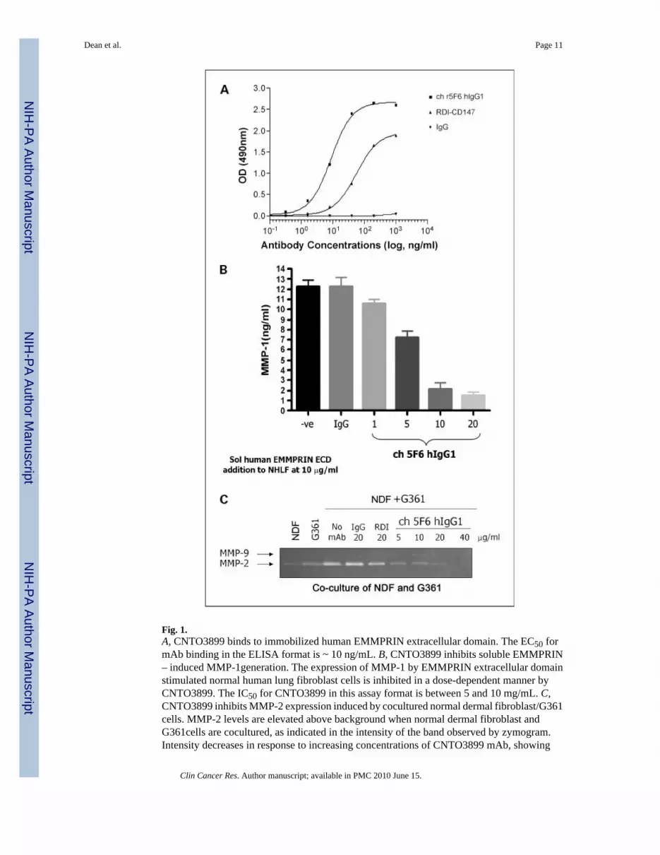
Fig. 1.A, CNTO3899 binds to immobilized human EMMPRIN extracellular domain. The EC50 formAb binding in the ELISA format is ~ 10 ng/mL. B, CNTO3899 inhibits soluble EMMPRIN– induced MMP-1generation. The expression of MMP-1 by EMMPRIN extracellular domainstimulated normal human lung fibroblast cells is inhibited in a dose-dependent manner byCNTO3899. The IC50 for CNTO3899 in this assay format is between 5 and 10 mg/mL. C,CNTO3899 inhibits MMP-2 expression induced by cocultured normal dermal fibroblast/G361cells. MMP-2 levels are elevated above background when normal dermal fibroblast andG361cells are cocultured, as indicated in the intensity of the band observed by zymogram.Intensity decreases in response to increasing concentrations of CNTO3899 mAb, showing
Dean et al. Page 11
Clin Cancer Res. Author manuscript; available in PMC 2010 June 15.
NIH
-PA Author Manuscript
NIH
-PA Author Manuscript
NIH
-PA Author Manuscript

bioactivity without the addition of exogenously added soluble EMMPRIN. No change inintensity is observed with the isotype control.
Dean et al. Page 12
Clin Cancer Res. Author manuscript; available in PMC 2010 June 15.
NIH
-PA Author Manuscript
NIH
-PA Author Manuscript
NIH
-PA Author Manuscript
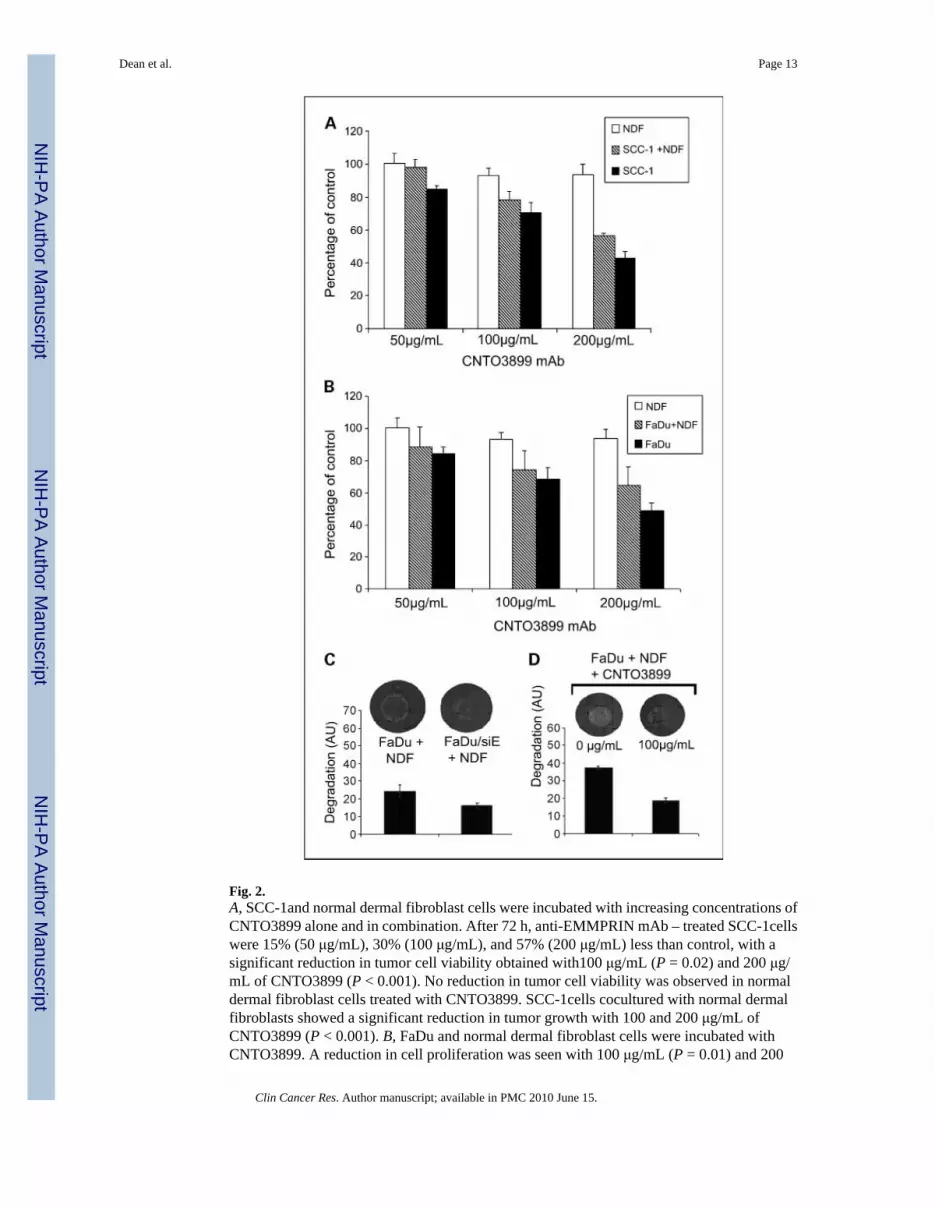
Fig. 2.A, SCC-1and normal dermal fibroblast cells were incubated with increasing concentrations ofCNTO3899 alone and in combination. After 72 h, anti-EMMPRIN mAb – treated SCC-1cellswere 15% (50 μg/mL), 30% (100 μg/mL), and 57% (200 μg/mL) less than control, with asignificant reduction in tumor cell viability obtained with100 μg/mL (P = 0.02) and 200 μg/mL of CNTO3899 (P < 0.001). No reduction in tumor cell viability was observed in normaldermal fibroblast cells treated with CNTO3899. SCC-1cells cocultured with normal dermalfibroblasts showed a significant reduction in tumor growth with 100 and 200 μg/mL ofCNTO3899 (P < 0.001). B, FaDu and normal dermal fibroblast cells were incubated withCNTO3899. A reduction in cell proliferation was seen with 100 μg/mL (P = 0.01) and 200
Dean et al. Page 13
Clin Cancer Res. Author manuscript; available in PMC 2010 June 15.
NIH
-PA Author Manuscript
NIH
-PA Author Manuscript
NIH
-PA Author Manuscript

μg/mL (P = 0.001) of CNTO3899. C, FaDu and FaDu/siE cells were plated on type I collagenwith normal dermal fibroblasts. A significant decrease in collagenolysis was observed inEMMPRIN knockdown cells compared with FaDu controls (P = 0.01). D, FaDu and normaldermal fibroblast cells (1 × 10⁵ of each) were plated on type I collagen and treated with 100μg/mL of CNTO3899. A significant inhibition in collagenolysis was observed (P = 0.005).
Dean et al. Page 14
Clin Cancer Res. Author manuscript; available in PMC 2010 June 15.
NIH
-PA Author Manuscript
NIH
-PA Author Manuscript
NIH
-PA Author Manuscript
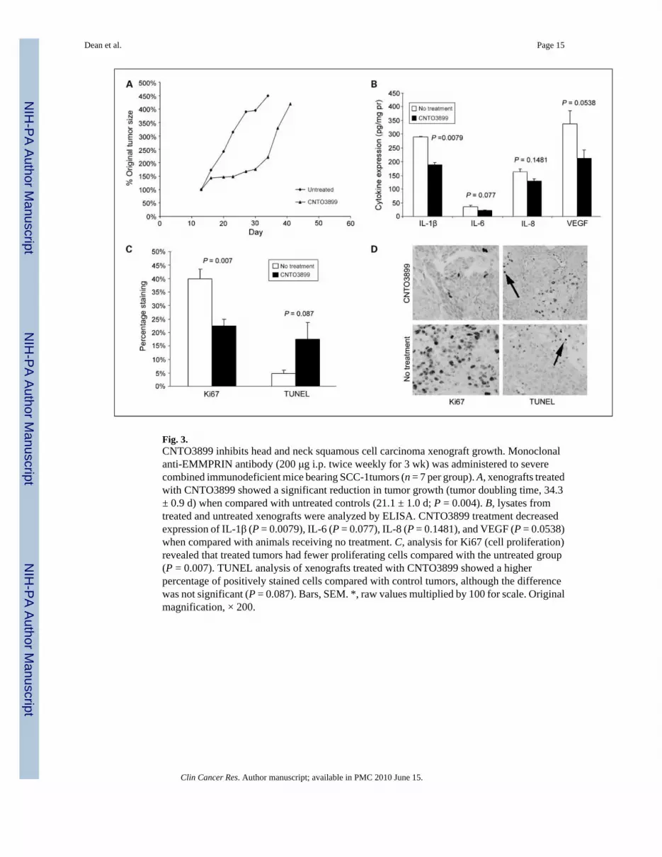
Fig. 3.CNTO3899 inhibits head and neck squamous cell carcinoma xenograft growth. Monoclonalanti-EMMPRIN antibody (200 μg i.p. twice weekly for 3 wk) was administered to severecombined immunodeficient mice bearing SCC-1tumors (n = 7 per group). A, xenografts treatedwith CNTO3899 showed a significant reduction in tumor growth (tumor doubling time, 34.3± 0.9 d) when compared with untreated controls (21.1 ± 1.0 d; P = 0.004). B, lysates fromtreated and untreated xenografts were analyzed by ELISA. CNTO3899 treatment decreasedexpression of IL-1β (P = 0.0079), IL-6 (P = 0.077), IL-8 (P = 0.1481), and VEGF (P = 0.0538)when compared with animals receiving no treatment. C, analysis for Ki67 (cell proliferation)revealed that treated tumors had fewer proliferating cells compared with the untreated group(P = 0.007). TUNEL analysis of xenografts treated with CNTO3899 showed a higherpercentage of positively stained cells compared with control tumors, although the differencewas not significant (P = 0.087). Bars, SEM. *, raw values multiplied by 100 for scale. Originalmagnification, × 200.
Dean et al. Page 15
Clin Cancer Res. Author manuscript; available in PMC 2010 June 15.
NIH
-PA Author Manuscript
NIH
-PA Author Manuscript
NIH
-PA Author Manuscript
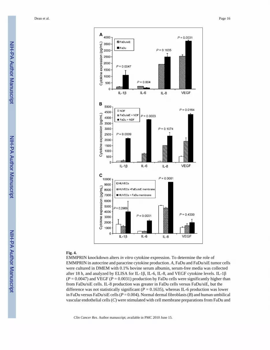
Fig. 4.EMMPRIN knockdown alters in vitro cytokine expression. To determine the role ofEMMPRIN in autocrine and paracrine cytokine production. A, FaDu and FaDu/siE tumor cellswere cultured in DMEM with 0.1% bovine serum albumin, serum-free media was collectedafter 18 h, and analyzed by ELISA for IL-1β, IL-6, IL-8, and VEGF cytokine levels. IL-1β(P = 0.0047) and VEGF (P = 0.0031) production by FaDu cells were significantly higher thanfrom FaDu/siE cells. IL-8 production was greater in FaDu cells versus FaDu/siE, but thedifference was not statistically significant (P = 0.1635), whereas IL-6 production was lowerin FaDu versus FaDu/siE cells (P = 0.004). Normal dermal fibroblasts (B) and human umbilicalvascular endothelial cells (C) were stimulated with cell membrane preparations from FaDu and
Dean et al. Page 16
Clin Cancer Res. Author manuscript; available in PMC 2010 June 15.
NIH
-PA Author Manuscript
NIH
-PA Author Manuscript
NIH
-PA Author Manuscript
FaDu/siE cells; serum-free media was collected after 18 h and analyzed for IL-1β, IL-6, IL-8,and VEGF cytokine levels. The differences between normal dermal fibroblast stimulation withFaDu and FaDu/siE were significant for IL-1β (B, P = 0.0009), IL-6 (P = 0.0003), and VEGF(P = 0.0164), but not IL-8 (P = 0.1074). C, the increases in cytokine expression after stimulationof human umbilical vascular endothelial cells with EMMPRIN-positive cell membranes weresignificant for IL-6 (P = 0.0001) and IL-8 (P = 0.0001), but not for IL-1β (P = 0.2969) or VEGF(P = 0.4300). Bars, SEM. *, raw values multiplied by 100 for scale.
Dean et al. Page 17
Clin Cancer Res. Author manuscript; available in PMC 2010 June 15.
NIH
-PA Author Manuscript
NIH
-PA Author Manuscript
NIH
-PA Author Manuscript
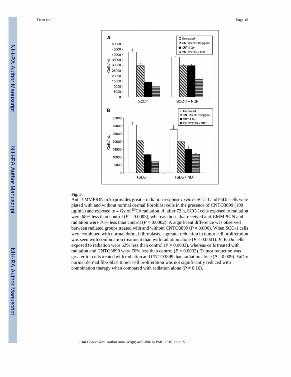
Fig. 5.Anti-EMMPRIN mAb provides greater radiation response in vitro. SCC-1 and FaDu cells wereplated with and without normal dermal fibroblast cells in the presence of CNTO3899 (100μg/mL) and exposed to 4 Gy of 60Co radiation. A, after 72 h, SCC-1cells exposed to radiationwere 68% less than control (P = 0.0003), whereas those that received anti-EMMPRIN andradiation were 76% less than control (P = 0.0002). A significant difference was observedbetween radiated groups treated with and without CNTO3899 (P = 0.006). When SCC-1 cellswere combined with normal dermal fibroblasts, a greater reduction in tumor cell proliferationwas seen with combination treatment than with radiation alone (P < 0.0001). B, FaDu cellsexposed to radiation were 62% less than control (P = 0.0003), whereas cells treated withradiation and CNTO3899 were 76% less than control (P = 0.0002). Tumor reduction wasgreater for cells treated with radiation and CNTO3899 than radiation alone (P = 0.009). FaDu/normal dermal fibroblast tumor cell proliferation was not significantly reduced withcombination therapy when compared with radiation alone (P = 0.16).
Dean et al. Page 18
Clin Cancer Res. Author manuscript; available in PMC 2010 June 15.
NIH
-PA Author Manuscript
NIH
-PA Author Manuscript
NIH
-PA Author Manuscript
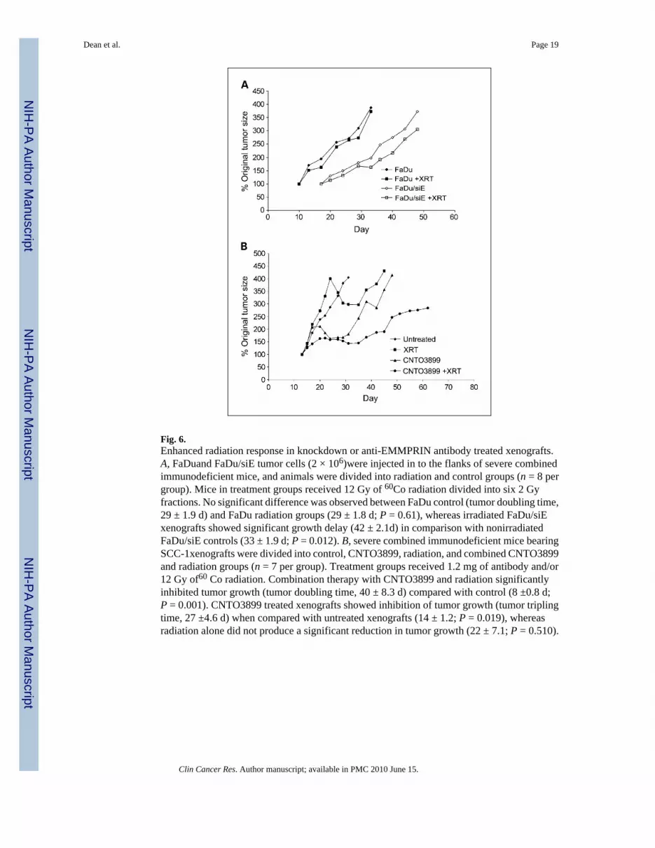
Fig. 6.Enhanced radiation response in knockdown or anti-EMMPRIN antibody treated xenografts.A, FaDuand FaDu/siE tumor cells (2 × 10⁶)were injected in to the flanks of severe combinedimmunodeficient mice, and animals were divided into radiation and control groups (n = 8 pergroup). Mice in treatment groups received 12 Gy of 60Co radiation divided into six 2 Gyfractions. No significant difference was observed between FaDu control (tumor doubling time,29 ± 1.9 d) and FaDu radiation groups (29 ± 1.8 d; P = 0.61), whereas irradiated FaDu/siExenografts showed significant growth delay (42 ± 2.1d) in comparison with nonirradiatedFaDu/siE controls (33 ± 1.9 d; P = 0.012). B, severe combined immunodeficient mice bearingSCC-1xenografts were divided into control, CNTO3899, radiation, and combined CNTO3899and radiation groups (n = 7 per group). Treatment groups received 1.2 mg of antibody and/or12 Gy of60 Co radiation. Combination therapy with CNTO3899 and radiation significantlyinhibited tumor growth (tumor doubling time, 40 ± 8.3 d) compared with control (8 ±0.8 d;P = 0.001). CNTO3899 treated xenografts showed inhibition of tumor growth (tumor triplingtime, 27 ±4.6 d) when compared with untreated xenografts (14 ± 1.2; P = 0.019), whereasradiation alone did not produce a significant reduction in tumor growth (22 ± 7.1; P = 0.510).
Dean et al. Page 19
Clin Cancer Res. Author manuscript; available in PMC 2010 June 15.
NIH
-PA Author Manuscript
NIH
-PA Author Manuscript
NIH
-PA Author Manuscript
Copyright © 2022 FDOKUMEN