Preclinical imaging of kallikrein-related peptidase 2 (hK2) in prostate cancer with a...
12
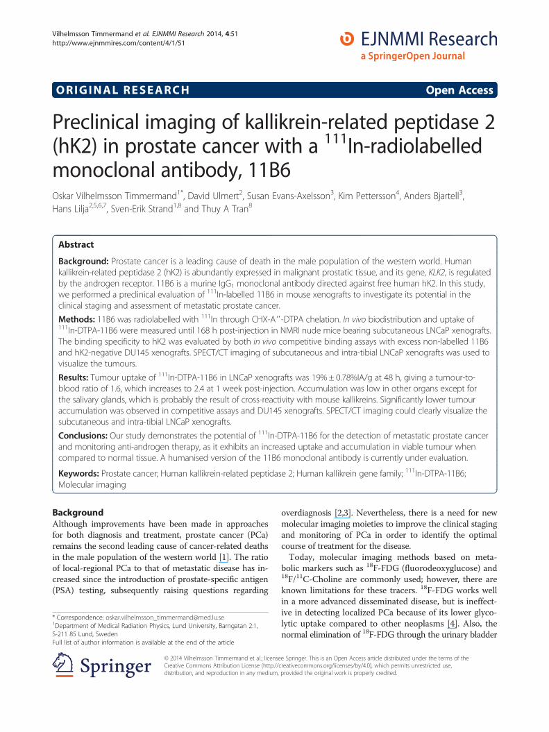
ORIGINAL RESEARCH Open Access Preclinical imaging of kallikrein-related peptidase 2 (hK2) in prostate cancer with a 111 In-radiolabelled monoclonal antibody, 11B6 Oskar Vilhelmsson Timmermand 1* , David Ulmert 2 , Susan Evans-Axelsson 3 , Kim Pettersson 4 , Anders Bjartell 3 , Hans Lilja 2,5,6,7 , Sven-Erik Strand 1,8 and Thuy A Tran 8 Abstract Background: Prostate cancer is a leading cause of death in the male population of the western world. Human kallikrein-related peptidase 2 (hK2) is abundantly expressed in malignant prostatic tissue, and its gene, KLK2, is regulated by the androgen receptor. 11B6 is a murine IgG 1 monoclonal antibody directed against free human hK2. In this study, we performed a preclinical evaluation of 111 In-labelled 11B6 in mouse xenografts to investigate its potential in the clinical staging and assessment of metastatic prostate cancer. Methods: 11B6 was radiolabelled with 111 In through CHX-A″-DTPA chelation. In vivo biodistribution and uptake of 111 In-DTPA-11B6 were measured until 168 h post-injection in NMRI nude mice bearing subcutaneous LNCaP xenografts. The binding specificity to hK2 was evaluated by both in vivo competitive binding assays with excess non-labelled 11B6 and hK2-negative DU145 xenografts. SPECT/CT imaging of subcutaneous and intra-tibial LNCaP xenografts was used to visualize the tumours. Results: Tumour uptake of 111 In-DTPA-11B6 in LNCaP xenografts was 19% ± 0.78%IA/g at 48 h, giving a tumour-to- blood ratio of 1.6, which increases to 2.4 at 1 week post-injection. Accumulation was low in other organs except for the salivary glands, which is probably the result of cross-reactivity with mouse kallikreins. Significantly lower tumour accumulation was observed in competitive assays and DU145 xenografts. SPECT/CT imaging could clearly visualize the subcutaneous and intra-tibial LNCaP xenografts. Conclusions: Our study demonstrates the potential of 111 In-DTPA-11B6 for the detection of metastatic prostate cancer and monitoring anti-androgen therapy, as it exhibits an increased uptake and accumulation in viable tumour when compared to normal tissue. A humanised version of the 11B6 monoclonal antibody is currently under evaluation. Keywords: Prostate cancer; Human kallikrein-related peptidase 2; Human kallikrein gene family; 111 In-DTPA-11B6; Molecular imaging Background Although improvements have been made in approaches for both diagnosis and treatment, prostate cancer (PCa) remains the second leading cause of cancer-related deaths in the male population of the western world [1]. The ratio of local-regional PCa to that of metastatic disease has in- creased since the introduction of prostate-specific antigen (PSA) testing, subsequently raising questions regarding overdiagnosis [2,3]. Nevertheless, there is a need for new molecular imaging moieties to improve the clinical staging and monitoring of PCa in order to identify the optimal course of treatment for the disease. Today, molecular imaging methods based on meta- bolic markers such as 18 F-FDG (fluorodeoxyglucose) and 18 F/ 11 C-Choline are commonly used; however, there are known limitations for these tracers. 18 F-FDG works well in a more advanced disseminated disease, but is ineffect- ive in detecting localized PCa because of its lower glyco- lytic uptake compared to other neoplasms [4]. Also, the normal elimination of 18 F-FDG through the urinary bladder * Correspondence: [email protected] 1 Department of Medical Radiation Physics, Lund University, Barngatan 2:1, S-211 85 Lund, Sweden Full list of author information is available at the end of the article © 2014 Vilhelmsson Timmermand et al.; licensee Springer. This is an Open Access article distributed under the terms of the Creative Commons Attribution License (http://creativecommons.org/licenses/by/4.0), which permits unrestricted use, distribution, and reproduction in any medium, provided the original work is properly credited. Vilhelmsson Timmermand et al. EJNMMI Research 2014, 4:51 http://www.ejnmmires.com/content/4/1/51
-
Upload
independent -
Category
Documents
-
view
1 -
download
0
Transcript of Preclinical imaging of kallikrein-related peptidase 2 (hK2) in prostate cancer with a...
Vilhelmsson Timmermand et al. EJNMMI Research 2014, 4:51http://www.ejnmmires.com/content/4/1/51
ORIGINAL RESEARCH Open Access
Preclinical imaging of kallikrein-related peptidase 2(hK2) in prostate cancer with a 111In-radiolabelledmonoclonal antibody, 11B6Oskar Vilhelmsson Timmermand1*, David Ulmert2, Susan Evans-Axelsson3, Kim Pettersson4, Anders Bjartell3,Hans Lilja2,5,6,7, Sven-Erik Strand1,8 and Thuy A Tran8
Abstract
Background: Prostate cancer is a leading cause of death in the male population of the western world. Humankallikrein-related peptidase 2 (hK2) is abundantly expressed in malignant prostatic tissue, and its gene, KLK2, is regulatedby the androgen receptor. 11B6 is a murine IgG1 monoclonal antibody directed against free human hK2. In this study,we performed a preclinical evaluation of 111In-labelled 11B6 in mouse xenografts to investigate its potential in theclinical staging and assessment of metastatic prostate cancer.
Methods: 11B6 was radiolabelled with 111In through CHX-A″-DTPA chelation. In vivo biodistribution and uptake of111In-DTPA-11B6 were measured until 168 h post-injection in NMRI nude mice bearing subcutaneous LNCaP xenografts.The binding specificity to hK2 was evaluated by both in vivo competitive binding assays with excess non-labelled 11B6and hK2-negative DU145 xenografts. SPECT/CT imaging of subcutaneous and intra-tibial LNCaP xenografts was used tovisualize the tumours.
Results: Tumour uptake of 111In-DTPA-11B6 in LNCaP xenografts was 19% ± 0.78%IA/g at 48 h, giving a tumour-to-blood ratio of 1.6, which increases to 2.4 at 1 week post-injection. Accumulation was low in other organs except forthe salivary glands, which is probably the result of cross-reactivity with mouse kallikreins. Significantly lower tumouraccumulation was observed in competitive assays and DU145 xenografts. SPECT/CT imaging could clearly visualize thesubcutaneous and intra-tibial LNCaP xenografts.
Conclusions: Our study demonstrates the potential of 111In-DTPA-11B6 for the detection of metastatic prostate cancerand monitoring anti-androgen therapy, as it exhibits an increased uptake and accumulation in viable tumour whencompared to normal tissue. A humanised version of the 11B6 monoclonal antibody is currently under evaluation.
Keywords: Prostate cancer; Human kallikrein-related peptidase 2; Human kallikrein gene family; 111In-DTPA-11B6;Molecular imaging
BackgroundAlthough improvements have been made in approachesfor both diagnosis and treatment, prostate cancer (PCa)remains the second leading cause of cancer-related deathsin the male population of the western world [1]. The ratioof local-regional PCa to that of metastatic disease has in-creased since the introduction of prostate-specific antigen(PSA) testing, subsequently raising questions regarding
* Correspondence: [email protected] of Medical Radiation Physics, Lund University, Barngatan 2:1,S-211 85 Lund, SwedenFull list of author information is available at the end of the article
© 2014 Vilhelmsson Timmermand et al.; licenseCreative Commons Attribution License (http://cdistribution, and reproduction in any medium,
overdiagnosis [2,3]. Nevertheless, there is a need for newmolecular imaging moieties to improve the clinical stagingand monitoring of PCa in order to identify the optimalcourse of treatment for the disease.Today, molecular imaging methods based on meta-
bolic markers such as 18F-FDG (fluorodeoxyglucose) and18F/11C-Choline are commonly used; however, there areknown limitations for these tracers. 18F-FDG works wellin a more advanced disseminated disease, but is ineffect-ive in detecting localized PCa because of its lower glyco-lytic uptake compared to other neoplasms [4]. Also, thenormal elimination of 18F-FDG through the urinary bladder
e Springer. This is an Open Access article distributed under the terms of thereativecommons.org/licenses/by/4.0), which permits unrestricted use,provided the original work is properly credited.

Vilhelmsson Timmermand et al. EJNMMI Research 2014, 4:51 Page 2 of 12http://www.ejnmmires.com/content/4/1/51
can mask the uptake in the prostate and regional lymphnodes [5]. 18F/11C-Choline is the most common radiotracerin PCa imaging today, and while they perform better than18F-FDG, they are still limited in their ability to differentiatebetween a localized PCa and benign disease [6].Molecular imaging of androgen receptor (AR) signalling
holds the promise for more accurate disease evaluationand therapeutic monitoring, with additional implicationsfor hormonal treatment and radiation therapy in PCa.PCa growth is dependent on androgens that signalthrough the AR and plays a fundamental role in cancercell proliferation, apoptosis and invasion/metastasis [7].A newly developed radiotracer, 18F-FDHT (16β-[18F]fluoro-5α-dihydrotestosterone), enables the imaging ofAR expression. Furthermore, preliminary clinical stud-ies demonstrated that 18F-FDHT might provide for theimaging of AR expression during disease progression incastration-resistant PCa [8].The commercially available ProstaScint®, 111In-capro-
mab pendetide, targets an intracellular epitope of theprostate-specific membrane antigen (PSMA) [9], anothersurrogate of AR signalling. Promising results were recentlyreported using a new antibody (J591) against an extracel-lular domain of the PSMA molecule and presents as apossibility to suppress PSMA expression following anti-androgen treatments [10]. However, the PSMA expressionin other tissues such as the human brain [11] hampers thisapproach to PCa imaging.An alternative strategy could be to target AR-dependent
antigens such as PSA and hK2 as they express almostexclusively in the prostate tissue [12,13]. Both PSA andhK2 are serine proteases, encoded by the human kallikreingenes KLK3 and KLK2 located on chromosome 19, re-spectively, and are well characterized as AR-regulatedgenes [14-16]. These kallikreins are produced by the samesecretory luminal cells in the prostate and share an 80%amino acid homology, as well as several structural simi-larities [15-17]. Importantly, the PSA and hK2 antigensare abundantly expressed in malignant prostate tissuethroughout all clinical stages. Recent publications onthe 5A10 [18] and PSA30 antibodies [19] have exploredthe concept of PSA imaging, including targeting thefree, unbound forms of prostate-specific antigen (fPSA).Results from animal studies using the PSA30 antibodyshowed a selective uptake in LNCaP tumours in vivo;however, the retention was faint and most likely due toshortcomings in the 125I-labelling method. In addition,89Zr-labelled 5A10 exhibited better imaging capabilities butwas accompanied by increased liver uptake. Nonetheless,this PET tracer was sufficient in detecting AR-dependentchanges in PSA expression levels in mouse tumour lesions,as well as in distinguishing PCa cells within bone lesions -both of which may be useful in the staging and clinicalevaluation of advanced prostate cancer [18].
Regardless of the similarities between PSA and hK2,hK2 displays properties distinct from those of PSA. hK2has been used in immunoassays to improve the accuracyof PCa screening [17,20]. In serum, most hK2 is in itsfree form, although the total levels of hK2 are in therange of 1% total PSA but similar to that of fPSA [21].As a tissue marker for PCa, the hK2-specific immuno-staining pattern differs from that of PSA, with an in-creased intensity in the PCa tumour and lymph nodemetastases compared to that observed in benign tissue[22,23]. Moreover, the very-low-to-no expression inother organs [12,13] makes hK2 a potential target candi-date for PCa imaging.Owing to these potentially advantageous characteristics
of hK2 as a PCa biomarker, we investigated the possibilityof targeting free hK2 in an androgen-dependent PCamodel using 111In-radiolabelled 11B6 - a murine IgG1
hK2-specific monoclonal antibody previously used in im-munoassays for free hK2 [24]. Here, we discuss the resultsof in vivo hK2 imaging with 111In-labelled 11B6 in AR-positive LNCaP xenografts.
MethodsAntibody conjugation and radiolabellingThe murine monoclonal antibody, 11B6, was first describedand characterized by Vaisanen et al. [24] and was providedby the University of Turku (Turku, Finland) for this study.Conjugation and radiolabelling was performed as previouslydescribed by Tolmachev et al. [25]. Briefly, 2 mg of 11B6was conjugated with the chelator CHX-A″-DTPA (B-355,Macrocyclics; Dallas, TX, USA) through the isothiocyanatefunctional group. A solution of 11B6 (4 to 5 mg/mL inPBS) was adjusted to pH 9.2 using 0.07 M sodium bor-ate buffer (Sigma Aldrich; St. Louis, MO, USA). CHX-A″-DTPA was then added to the protein solution at amolar ratio of 3:1 (chelator to antibody) and incubatedat 40°C with gentle shaking. The reaction was termi-nated after 4 h, and CHX-A″-DTPA-11B6, henceforthreferred to as DTPA-11B6, was separated from the freechelate by size-exclusion chromatography on a NAP-5 col-umn (GE Healthcare; Uppsala, Sweden) equilibrated with20 mL of 0.2 M ammonium acetate buffer (Sigma Aldrich),pH 5.5. Conjugated 11B6 was eluted with 1 mL of am-monium acetate buffer, and aliquoted samples werestored at −20°C.For radiolabelling, approximately 125 μL of DTPA-11B6
(approximately 1 μg/μL in 0.2 M ammonium acetate bufferpH 5.5) was mixed with a predetermined amount (approxi-mately 50 to 100 MBq) of 111InCl3 (Mallinckrodt Medical;Dublin, Ireland), incubated at room temperature for 1.5 to2 h and then purified on a NAP-5 column (GE Healthcare)equilibrated with PBS (Thermo Scientific; Waltham, MA,USA). Labelling efficiency and kinetics were monitored byinstant thin-layer chromatography (ITLC) (Biodex, Shirley,

Vilhelmsson Timmermand et al. EJNMMI Research 2014, 4:51 Page 3 of 12http://www.ejnmmires.com/content/4/1/51
NY, USA) eluted with 0.2 M citric acid (Sigma Aldrich).In this system, the radiolabelled conjugate remains atthe origin line, while free 111In and 111In-DTPA migratewith the solvent front. The radioactive distribution wasdetermined using a Cyclone Storage Phosphor Systemwith Optiquant quantification software (Perkin Elmer;Waltham, MA, USA).
Binding kinetics with surface plasmon resonanceThe 11B6 binding kinetics were analysed by surfaceplasmon resonance using a Biacore 2000 (Biacore AB;Uppsala, Sweden). The affinity of 11B6 to hK2 beforeand after CHX-A″-DTPA conjugation was determined.The hK2 antigen, provided by the University of Turku(Department of Biotechnology; Turku, Finland), wasproduced and purified as previously described [26].hK2 antigen (25.9 μg/mL in 10 mM sodium acetatebuffer pH 4.0 (Sigma Aldrich)) was immobilized on aCM4 research grade chip (Biacore AB) by amino couplingusing N-hydroxysuccinimide (NHS), 1-ethyl-3-(3-dimethy-laminopropyl) carbodiimide hydrochloride (EDC) and 1 Methanolamine hydrochloride-NaOH, pH 8.5, in a Biacore2000 system. Samples were flown over two flow cells, onebeing a blank reference, in five different concentrationsranging from 0.5 to 100 nM to detect eventual binding.One of the two flow cells contained immobilized hK2,while the other was served as a blank reference. The bind-ing kinetics were studied in a 3-min-long association phaseand a 15-min-long dissociation phase with a flow rateof 30 μL/min, followed by regeneration with 25 mMglycine buffer pH 2.7. Kinetic constants were calculatedusing a 1:1 Langmuir binding model with correction formass transfer. BIAEvaluation 4.1 software (Biacore AB)was used for calculations.
Stability studiesThe stability of 111In-DTPA-11B6 was assessed in tripli-cate by incubating the compound at 4°C in PBS bufferor at 37°C in murine serum collected from normalNMRI mice. For stability in PBS, 1 μL (n = 3) was takenat 1, 2, 3 and 7 days and analysed by ITLC. For stabilityin serum, 10 μL of 111In-DTPA-11B6 (corresponding to3 μg of antibody with 0.8 to 0.9 MBq 111In) was mixedwith 100 μL of mouse serum. Approximately 20 μL ofeach mixture was collected after 2, 3 and 9 days of incu-bation and analysed by SDS-PAGE on a NuPAGE 4% to12% Bis-Tris gel (Invitrogen; Carlsbad, CA, USA) inMES buffer (200 V constant, approximately 30 min).111In-DTPA and free 111In diluted in PBS were run in par-allel with the incubated sample as controls. The distribu-tion of the samples along the gel was evaluated using aCyclone Storage Phosphor System (Perkin Elmer).
Cell linesLNCaP and DU145 were purchased from AmericanType Culture Collection (ATCC; Manassas, VA, USA)and cultured in RPMI 1640 medium (Thermo Scientific)supplemented with 10% foetal bovine serum (ThermoScientific) with 100 U/mL penicillin and 100 μg/mLstreptomycin (Thermo Scientific). The cells were main-tained at 37°C in a humidified incubator at 5% CO2 andwere detached with trypsin-EDTA solution (ThermoScientific).
Animal modelsAll animal experiments were conducted in compliancewith the national legislation on laboratory animals' pro-tection and with the approval of the Ethics Committeefor Animal Research (Lund University, Sweden). Twoanimal models were used in this study, NMRI-Nu withsubcutaneous (s.c.) xenografts and SCID mice withintra-tibial xenografts. NMRI-Nu mice (6-to-8-week-old,Taconic; Ry, Denmark) were inoculated in the right flankby s.c. injection of 5 to 8 × 10⁶ cells in a 200 μL of cellsuspension of 1:1 mixture of medium with Matrigel (BDBiosciences; San Jose, CA, USA). Tumours were allowedto develop for 6 to 8 weeks. SCID mice (6-to-8-week-oldmale, Charles River; Charles River, NJ, USA) were main-tained under isoflurane anaesthesia during surgery. Forintra-tibial inoculations, the tibia was punctured using a23-gauge needle, and 1 × 10⁵ LNCaP cells were injectedinto the tibial cavity. The puncture was closed with bonewax, the incision sutured and the animals received a pallia-tive dose of Temgesic (Buprenorphine, RB Pharmaceuticals;Richmond, VA, USA) once daily for 3 days post-surgery.Intra-tibial tumours were allowed to develop for 8 to 10weeks. Additionally, a group of normal NMRI mice (n = 4)were used to study the distribution of the tracer in healthyanimals. Animals were euthanized by intraperitoneal (i.p.)injection with 20 μL per gram of body weight Ketalar-Rompun solution. (Ketalar, 10 mg/mL; Pfizer; New York,NY, USA, and Rompun, 1 mg/mL; Bayer Animal Health;Monheim, Germany).
Biodistribution studiesBiodistribution studies were conducted to evaluate the up-take of 111In-DTPA-11B6 in human prostate cancerLNCaP xenografts. Mice (n = 3 to 5 per time point) re-ceived 111In-DTPA-11B6 (0.4 to 0.6 MBq, 20 μg of mAb,in approximately 100 μL of PBS) through intravenous (i.v.)tail vein injection. Blood and organs (including tumour)were taken at 4, 24, 48, 72 and 168 h post-injection,weighed and measured in a NaI(TI) well counter (WallacWizard 1480 Wizard, Perkin Elmer). The activity injectedinto each animal was measured and used to determine thecount rate, in comparison with a standard solution of

Vilhelmsson Timmermand et al. EJNMMI Research 2014, 4:51 Page 4 of 12http://www.ejnmmires.com/content/4/1/51
111In-DTPA-11B6. Data were corrected for backgroundand physical decay.Organ-specific uptake values were calculated as percent
injected activity per gram of tissue (%IA/g) or percentinjected activity (%IA). Among the organs resected werethe lateral and ventral prostate, from now on referred toas prostate, and the submandibular glands, from now oncalled salivary glands.
In vivo binding specificityIn vivo competitive binding studies were performed toinvestigate the specificity of 111In-DTPA-11B6 to hK2. A40-fold excess of non-labelled 11B6 was i.v. injected as aco-injection or at 168, 120 and 48 h prior to an i.v. injec-tion of 111In-DTPA-11B6 in hK2-positive LNCaP xeno-grafts (n = 3 to 4 per pre-injection time point). Bloodand organs (including tumour) were taken at 48 h post-injection of 111In-DTPA-11B6, weighed and analysed asabove. The binding specificity was also evaluated bymeasuring the uptake of 111In-DTPA-11B6 in hK2-negative DU145 xenografts expressing low levels of hK2(n = 3) at 48 h post-injection, which are considered to behK2-negative when compared to other PCa tumours.
Small animal PET/SPECT/CT/MR imagingAnimals were anaesthetized with 2% to 3% isoflurane gas(Baxter; Deerfield, IL, USA) for all imaging purposes. ForSPECT/CT imaging, NMRI-nu mice with s.c. LNCaP xeno-grafts (48 h post-injection, n = 4; 72 h post-injection, n = 3;pre-dosed 11B6, n = 4; co-injection 5A10, n = 3) and SCIDmice (n = 3) with intra-tibial LNCaP xenografts were i.v.injected with approximately 8 MBq of 111In-DTPA-11B6(approximately 20 μg of mAb in 150 μL of PBS) and im-aged, for 1 h, by using a preclinical SPECT/CT scanner(NanoSPECT/CT Plus, Bioscan; Washington, DC, USA)with the NSP-106 multi-pinhole mouse collimator. SPECTdata were reconstructed using HiSPECT software (SciVis;Goettingen, Germany). CT imaging was done before eachwhole-body SPECT.Pre-dosed mice were given 0.8 mg of non-labelled mAb
48 h prior to injection of radiolabelled mAb. Co-injectionswith 111In-DTPA-11B6 and 1.5 mg of fPSA-specific 5A10were done to evaluate the possible cross-reactivity of111In-DTPA-11B6 with PSA.The legs of the SCID mice were resected after imaging,
and the radioactivity in the intra-tibial xenografted and thenon-xenografted leg were measured in the NaI(Ti) wellcounter. Radiolabelling for SPECT of intra-tibial xenograftsdemonstrated 95% radiochemical purity and was injecteddirectly without NAP-5 column purification. Verificationof the intra-tibial tumour growth was performed by MRimaging. The legs were imaged in an 11.7 T (500 MHz forprotons) vertical bore MR camera (Agilent Technologies;Palo Alto, CA, USA) equipped with Varian 88/55 micro-
imaging triple axis gradient coil (1 T/m maximum gradientstrength). Samples were placed in the centre of a Millipedeimaging probe (Agilent Technologies; Santa Clara, CA,USA), with an inner diameter of 40 mm.For PET/CT imaging, mice with LNCaP xenografts
were i.v. injected with approximately 12 MBq 18F-FDG(n = 4) or approximately 12 MBq18F-Choline (n = 4) andimaged 1 h post-injection using a Bioscan NanoPET/CTPlus preclinical scanner for approximately 15 min. BothSPECT/CT and PET/CT images were analysed usingInVivoScope 2.0 software (inviCRO; Boston, MA, USA),and ROIs were drawn using the CT image as anatomicalreference.
Autoradiography and stainingAfter SPECT imaging at 48 and 72 h, s.c. tumours wereresected and embedded in Tissue-Tek® O.C.T™ com-pound (Sakura Finetek; Alphen aan den Rijn, TheNetherlands) and frozen on dry ice. The frozen sampleswere cryosectioned with a thickness of 20 μm for auto-radiography analysis on a Cyclone Storage PhosphorSystem. The tumour sections were stained with Mayer'shematoxylin and chromotrope 2R, Ch2R (both fromHistolab; Gothenburg, Sweden), and scanned using alight-microscope slide scanner (Mirax Midi, Carl Zeiss;Oberkochen, Germany). Thresholds for the autoradio-grams were set in ImageJ v.1.47.
Statistical analysisData was analysed using the unpaired, two-tailed Student'st test (Microsoft Excel or GraphPad Prism v.4). Differencesat the 95% confidence level (P < 0.05) were considered to bestatistically significant. Figures were produced with Graph-Pad Prism v.4 (GraphPad Software). All biodistribution dataare shown as an average %IA/g of 3 to 5 animals ± SD(standard deviation) unless otherwise stated.
ResultsRadiolabelling and stabilityA radiolabelling yield of 58.6% ± 12.5% (n = 3) wasachieved. The radiochemical purity after NAP-5 purifica-tion was >99% and for labelling with higher activities forimaging a specific activity of 0.30 ± 0.03 MBq/μg (n = 3).111In-DTPA-11B6 was stable in PBS buffer at 4°C and inmouse serum at 37°C. More than 95% of the radioacti-vity was still attached to the radioconjugate after 1 weekin both conditions.
Binding kinetics with surface plasmon resonanceThe binding kinetics of 11B6 were analysed on a Biacoreinstrument. The affinity of 11B6 was high for hK2 and, al-though conjugation seemed to affect the association con-stant (ka) and the dissociation constant (kd), the calculatedbinding affinities (KD; calculated as kd/ka) were similar.

Vilhelmsson Timmermand et al. EJNMMI Research 2014, 4:51 Page 5 of 12http://www.ejnmmires.com/content/4/1/51
The KD for 11B6 and DTPA-11B6 to hK2 were estimatedto be (5.1 ± 2.2) × 10−11 M and 6.6 × 10⁻¹¹ ± 0.46 × 10⁻¹¹
M, respectively. Representative sensorgrams of the 11B6and DTPA-11B6 binding kinetics are shown in the supple-mentary data (Additional file 1: Figure S1A,B).
Figure 1 Biokinetics of 111In-DTPA-11B6. (a) Biodistribution of 111In-DTPto 5 animals per time point. (b) Biodistribution of 111In-DTPA-11B6 in normcompared to tumour-bearing mice but with a retained high uptake in theover the time of the experiment. Right, tumour-to-muscle ratio. This high, iin SPECT/CT images. (See Figure 3)
Biodistribution studiesThe biodistribution of 111In-DTPA-11B6 in NMRI micewith LNCaP xenografts revealed that the accumulation intumour tissue increased threefold from 4 h (5.9 ± 3.6%IA/g)to a maximum at 48 h (19 ± 0.78%IA/g) (Figure 1a). Blood
A-11B6 in NRMI nude mice carrying LNCaP xenografts. %IA/g ± SD, 3al NMRI mice at 48 h. An overall lower %IA/g was observed assalivary glands. (c) Left, tumour-to-blood ratio is steadily increasingncreasing ratio explains the good visualization and contrast observed
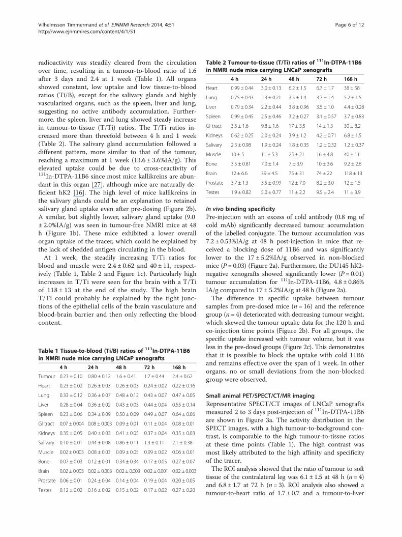
Table 2 Tumour-to-tissue (T/Ti) ratios of 111In-DTPA-11B6in NMRI nude mice carrying LNCaP xenografts
4 h 24 h 48 h 72 h 168 h
Heart 0.99 ± 0.44 3.0 ± 0.13 6.2 ± 1.5 6.7 ± 1.7 38 ± 58
Lung 0.75 ± 0.43 2.3 ± 0.21 3.5 ± 1.4 3.7 ± 1.4 5.2 ± 1.5
Liver 0.79 ± 0.34 2.2 ± 0.44 3.8 ± 0.96 3.5 ± 1.0 4.4 ± 0.28
Spleen 0.99 ± 0.45 2.5 ± 0.46 3.2 ± 0.27 3.1 ± 0.57 3.7 ± 0.83
GI tract 3.5 ± 1.6 9.8 ± 1.6 17 ± 3.5 14 ± 1.3 30 ± 8.2
Kidneys 0.62 ± 0.25 2.0 ± 0.24 3.9 ± 1.2 4.2 ± 0.71 6.8 ± 1.5
Salivary 2.3 ± 0.98 1.9 ± 0.24 1.8 ± 0.35 1.2 ± 0.32 1.2 ± 0.37
Muscle 10 ± 5 11 ± 5.3 25 ± 21 16 ± 4.8 40 ± 11
Bone 3.5 ± 0.81 7.0 ± 1.4 7 ± 3.9 10 ± 3.6 9.2 ± 2.6
Brain 12 ± 6.6 39 ± 4.5 75 ± 31 74 ± 22 118 ± 13
Prostate 3.7 ± 1.3 3.5 ± 0.99 12 ± 7.0 8.2 ± 3.0 12 ± 1.5
Testes 1.9 ± 0.82 5.0 ± 0.77 11 ± 2.2 9.5 ± 2.4 11 ± 3.9
Vilhelmsson Timmermand et al. EJNMMI Research 2014, 4:51 Page 6 of 12http://www.ejnmmires.com/content/4/1/51
radioactivity was steadily cleared from the circulationover time, resulting in a tumour-to-blood ratio of 1.6after 3 days and 2.4 at 1 week (Table 1). All organsshowed constant, low uptake and low tissue-to-bloodratios (Ti/B), except for the salivary glands and highlyvascularized organs, such as the spleen, liver and lung,suggesting no active antibody accumulation. Further-more, the spleen, liver and lung showed steady increasein tumour-to-tissue (T/Ti) ratios. The T/Ti ratios in-creased more than threefold between 4 h and 1 week(Table 2). The salivary gland accumulation followed adifferent pattern, more similar to that of the tumour,reaching a maximum at 1 week (13.6 ± 3.6%IA/g). Thiselevated uptake could be due to cross-reactivity of111In-DTPA-11B6 since most mice kallikreins are abun-dant in this organ [27], although mice are naturally de-ficient hK2 [16]. The high level of mice kallikreins inthe salivary glands could be an explanation to retainedsalivary gland uptake even after pre-dosing (Figure 2b).A similar, but slightly lower, salivary gland uptake (9.0± 2.0%IA/g) was seen in tumour-free NMRI mice at 48h (Figure 1b). These mice exhibited a lower overallorgan uptake of the tracer, which could be explained bythe lack of shedded antigen circulating in the blood.At 1 week, the steadily increasing T/Ti ratios for
blood and muscle were 2.4 ± 0.62 and 40 ± 11, respect-ively (Table 1, Table 2 and Figure 1c). Particularly highincreases in T/Ti were seen for the brain with a T/Tiof 118 ± 13 at the end of the study. The high brainT/Ti could probably be explained by the tight junc-tions of the epithelial cells of the brain vasculature andblood-brain barrier and then only reflecting the bloodcontent.
Table 1 Tissue-to-blood (Ti/B) ratios of 111In-DTPA-11B6in NMRI nude mice carrying LNCaP xenografts
4 h 24 h 48 h 72 h 168 h
Tumour 0.23 ± 0.10 0.80 ± 0.12 1.6 ± 0.41 1.7 ± 0.44 2.4 ± 0.62
Heart 0.23 ± 0.02 0.26 ± 0.03 0.26 ± 0.03 0.24 ± 0.02 0.22 ± 0.16
Lung 0.33 ± 0.12 0.36 ± 0.07 0.48 ± 0.12 0.43 ± 0.07 0.47 ± 0.05
Liver 0.28 ± 0.04 0.36 ± 0.02 0.43 ± 0.03 0.44 ± 0.04 0.55 ± 0.14
Spleen 0.23 ± 0.06 0.34 ± 0.09 0.50 ± 0.09 0.49 ± 0.07 0.64 ± 0.06
GI tract 0.07 ± 0.004 0.08 ± 0.003 0.09 ± 0.01 0.11 ± 0.04 0.08 ± 0.01
Kidneys 0.35 ± 0.05 0.40 ± 0.03 0.41 ± 0.05 0.37 ± 0.04 0.35 ± 0.03
Salivary 0.10 ± 0.01 0.44 ± 0.08 0.86 ± 0.11 1.3 ± 0.11 2.1 ± 0.38
Muscle 0.02 ± 0.003 0.08 ± 0.03 0.09 ± 0.05 0.09 ± 0.02 0.06 ± 0.01
Bone 0.07 ± 0.03 0.12 ± 0.01 0.34 ± 0.34 0.17 ± 0.05 0.27 ± 0.07
Brain 0.02 ± 0.003 0.02 ± 0.003 0.02 ± 0.003 0.02 ± 0.001 0.02 ± 0.003
Prostate 0.06 ± 0.01 0.24 ± 0.04 0.14 ± 0.04 0.19 ± 0.04 0.20 ± 0.05
Testes 0.12 ± 0.02 0.16 ± 0.02 0.15 ± 0.02 0.17 ± 0.02 0.27 ± 0.20
In vivo binding specificityPre-injection with an excess of cold antibody (0.8 mg ofcold mAb) significantly decreased tumour accumulationof the labelled conjugate. The tumour accumulation was7.2 ± 0.53%IA/g at 48 h post-injection in mice that re-ceived a blocking dose of 11B6 and was significantlylower to the 17 ± 5.2%IA/g observed in non-blockedmice (P = 0.03) (Figure 2a). Furthermore, the DU145 hK2-negative xenografts showed significantly lower (P = 0.01)tumour accumulation for 111In-DTPA-11B6, 4.8 ± 0.86%IA/g compared to 17 ± 5.2%IA/g at 48 h (Figure 2a).The difference in specific uptake between tumour
samples from pre-dosed mice (n = 16) and the referencegroup (n = 4) deteriorated with decreasing tumour weight,which skewed the tumour uptake data for the 120 h andco-injection time points (Figure 2b). For all groups, thespecific uptake increased with tumour volume, but it wasless in the pre-dosed groups (Figure 2c). This demonstratesthat it is possible to block the uptake with cold 11B6and remains effective over the span of 1 week. In otherorgans, no or small deviations from the non-blockedgroup were observed.
Small animal PET/SPECT/CT/MR imagingRepresentative SPECT/CT images of LNCaP xenograftsmeasured 2 to 3 days post-injection of 111In-DTPA-11B6are shown in Figure 3a. The activity distribution in theSPECT images, with a high tumour-to-background con-trast, is comparable to the high tumour-to-tissue ratiosat these time points (Table 1). The high contrast wasmost likely attributed to the high affinity and specificityof the tracer.The ROI analysis showed that the ratio of tumour to soft
tissue of the contralateral leg was 6.1 ± 1.5 at 48 h (n = 4)and 6.8 ± 1.7 at 72 h (n = 3). ROI analysis also showed atumour-to-heart ratio of 1.7 ± 0.7 and a tumour-to-liver

Figure 2 In vivo binding specificity of 111In-DTPA-11B6 in NRMI nude mice. (a) Dot plot of tumour accumulation in blocked LNCaP,non-blocked LNCaP xenografts (hK2+) and DU145 (hK2−). There was a significant difference in active uptake between non-blocked LNCaPxenografts and the other groups. (b) Distribution of 111In-DTPA-11B6 48 h post-injection in LNCaP xenografted NMRI mice, shown as %IA/g ± SEMwith pre-dosing of 0.8 mg of cold 11B6 antibody. (c) Dot plot with linear regression for pre-dosing and no pre-dosing as function of tumourweight. This shows that %IA increases with tumour weight for both pre-dosed and normal uptake but that the uptake increases more in tumourswith no pre-dosing. The slope calculated from linear regression was 6.1 ± 0.27%IA/g for the pre-dosed groups and 14 ± 1.2%IA/g with no pre-dosing.The R2 value was 0.97 for the pre-dosed groups and 0.99 for xenografts without pre-dosing. As %IA/g increases with smaller tumour volume this couldexplain the large difference seen in (b) between the different pre-dosing groups. By analysing %IA instead, it seems that the effect of pre-dosing isretained over the studied time interval and that 11B6 in fact has a long tumour retention.
Vilhelmsson Timmermand et al. EJNMMI Research 2014, 4:51 Page 7 of 12http://www.ejnmmires.com/content/4/1/51

Figure 3 Preclinical SPECT/CT and MR images. (a) SPECT/CT images of four NMRI nude mice with LNCaP xenografts on the right flank. Topleft, 48 h post-injection; Top right, 72 h post-injection; Bottom left, blocked mouse pre-injected with 0.8 mg of cold antibody 48 h prior to injection ofradiolabelled antibody. Bottom right, image of a mouse at 48 h co-injected with 1.5 mg of unlabelled 5A10, which did not block the tumour uptake of111In-DTPA-11B6. It should be noted that for the calculation of the T/B ratio (1.6) in the biodistribution and for the ROI analysis of the SPECT imageswith a derived T/heart ratio of 1.7, the whole tumour is taken into account. Since the colour scale in the SPECT image is scaled partially based uponhigh uptake areas of the tumour, a direct comparison between these (which are strikingly visible) and the T/B or T/heart (ROI) is not totally fair. (b)SPECT/MR images of SCID mice with intra-tibial xenograft in the left hind limb. Left, SPECT/CT side view, 48 h post-injection; middle, transverse SPECT/CT image slices of the same mouse. The accumulation of 111In-DTPA-11B6 in intra-tibial tumours is clearly shown in both images. Right, MR image ofthe same animal, where the growth of the intra-tibial tumour is well visualized (arrow).
Vilhelmsson Timmermand et al. EJNMMI Research 2014, 4:51 Page 8 of 12http://www.ejnmmires.com/content/4/1/51
ratio of 1.7 ± 0.40 at 48 h and 1.81 ± 0.10 and 1.9 ± 0.04, re-spectively, at 72 h. This is similar to the tumour-to-tissueratios derived from the biodistribution data. Interestingly,an obvious heterogeneous distribution of activity could be
seen in the SPECT images of the tumours, later confirmedin autoradiograms (Figure 4a). High accumulation of activ-ity was also detected in the salivary glands. For the blockedmice (n = 4), the ROI ratio at 48 h of the tumour to the

Figure 4 Autoradiograms and images of H- and Ch2R-stained tumour sections. (a) A heterogeneous distribution of activity with higherintensities in areas corresponding to a more intense level of staining with H and Ch2R, 48 h (left) and 72 h (right). (b) Stained sections andautoradiograms of two tumours resected at 48 h from mice pre-dosed with 0.8 mg of cold 11B6 48 h prior to injection with 111In-DTPA-11B6.(c) An autoradiogram and stained section of a submandibular gland at 48 h. This normal organ shows less heterogeneity in activity distributionand hematoxylin and Ch2R staining.
Vilhelmsson Timmermand et al. EJNMMI Research 2014, 4:51 Page 9 of 12http://www.ejnmmires.com/content/4/1/51
contralateral leg, heart and liver was 3.4 ± 0.82, 1.3 ±0.41 and 0.87 ± 0.07, respectively. The salivary gland up-take was not associated with detached indium-labelledCHX-A″-DTPA, as confirmed with SPECT/CT (Figure 5).We also checked whether 111In-DTPA-11B6 could be
used to detect PCa skeletal lesions. Osseous tumourswere established in SCID mice through intra-tibial in-jections of LNCaP cells in the left hind limb, andSPECT/CT images were performed after i.v. injectionof 111In-DTPA-11B6. The legs were resected and mea-sured in the NaI(Ti) well counter, using the right non-xenografted legs as a negative control. The data showedan at least twofold uptake of 111In-DTPA-11B6 in thetibia compared to the reference leg, some of whichmight be due to inflammation induced by the surgicalprocedure. MR imaging confirmed tumour develop-ment within the bone (Figure 3b).Although 111In-labelled 11B6 showed a good stability
in murine serum, we investigated whether the high up-take seen in the salivary glands could be a result ofindium-labelled CHX-A″-DTPA, this was not confirmed(Figure 5, left). PET/CT images of NMRI-nude micewith s.c. LNCaP showed that 18F-FDG and 18F-Cholinewere hardly able to visualize the LNCaP xenografts, ascompared to 111In-DTPA-11B6 (Figure 5, right). ROIanalysis showed that the ratio of these xenografts to thesoft tissue of the contralateral leg was 1.1 ± 0.38 for 18F-FDG (n = 4) and 1.09 ± 0.45 for 18F-Choline (n = 4).
Autoradiography and stainingActivity accumulation was confirmed to be heterogeneousin tumour sections at both 48 and 72 h. Comparative au-toradiograms between the pre-dosed (Figure 4b) and non-pre-dosed mice (Figure 4a) showed that for the former, ac-tivity was confined to areas with high hematoxylin andCh2R staining, i.e. areas of high cell density, whereas theactivity in tumours from pre-dosed animals was found inareas with low hematoxylin and Ch2R staining. This dif-ference could be due to an active uptake in the viable re-gions, blocked in the latter by pre-dosing with coldantibody. The activity distribution was more homogenousin the salivary glands of non-pre-dosed mice, indicatingan uptake in all glandular structures in this organ(Figure 4c).
DiscussionThere is a clinical need for improved methods to detectand stage prostate cancer. The imaging of androgen-regulated prostate-specific antigens overexpressed inprostate cancer, such as PSA, would be advantageous fordiagnosing and monitoring the disease. In this study, wedemonstrated the ability for a novel radiotracer targetingfree hK2, a prostate-specific kallikrein homologous toPSA, to specifically target and image hK2 expression inAR- and hK2-positive LNCaP xenografts. Significantly,111In-DTPA-11B6 exhibited strong targeting in bothsubcutaneous and intra-tibial bone xenografts, with a

Figure 5 111In-CHX-A″-DTPA SPECT/CT and 18 F-FDG and 18 F-Choline PET/CT images. SPECT/CT images (left) of non-tumour bearing NMRInude mice showing localization of 111In-CHX-A″-DTPA in the kidneys (0.5 h post-injection). PET/CT images of s.c. LNCaP xenografts 1 h after injectionwith 18 F-FDG and 18 F-Choline, respectively (middle and right). Tumours, indicated by red circles, were not well visualized with these radiotracers.
Vilhelmsson Timmermand et al. EJNMMI Research 2014, 4:51 Page 10 of 12http://www.ejnmmires.com/content/4/1/51
mean tumour uptake of 19%IA/g at 48 h in subcutane-ous tumours (Figure 1a). The elevated uptake of 111In-DTPA-11B6 in the salivary glands was an unexpected re-sult of this study; however, several glandular kallikreinsare synthesized by the salivary glands in mice [27]. Sincewe see an uptake in the salivary glands of both NMRInude and normal non-xenografted NMRI mice, it seemslikely that 111In-DTPA-11B6 cross-reacts with kallikreinsexpressed in the salivary glands of these mice.The tumour accumulation of 111In-DTPA-11B6 was
hK2-specific, as verified by the low accumulation in thenegative control, DU145 xenografts and by competitivebinding assays using excess cold non-labelled antibody.Furthermore, tumour accumulation slowly decreasedafter day 2 but was not as rapid as blood clearance atthe same time points. This gave a steadily increasing pat-tern in tumour-to-blood (T/B) ratios over time, from1.6 ± 0.41 at 48 h to 2.4 ± 0.62 at 1 week (Table 1).Though the blood clearance of 111In-11B6 is comparableto other full-sized IgGs, the high tumour uptake, an in-creasing T/B ratio and a comparatively low liver uptake,implicates the usefulness of this radiotracer for imaging.Further, most tissues seem to display blood kinetics, ex-cept for a faint active uptake by the liver, spleen andlung compared to the high accumulation rate observedin the tumour and salivary gland tissue. Tissues such as
the muscle and brain gave high T/Ti ratios of 40 ± 11and 118 ± 13, respectively, at 1 week, whereas the liverand spleen have lower T/Ti ratios of 4.4 ± 0.28 and 3.7 ±0.83, respectively. There could be some concerns raisedabout 111In-DTPA-11B6 binding to free hK2 in theblood. However, the blocking with cold non-labelledantibody does not seem to change the amount of theblood activity significantly at 48 h post-injection. Also,in the control group with DU145 xenografts, there wasno significant difference in blood activity at 48 h, but asignificant difference in tumour uptake was found. Oneinteresting observation from the pre-dosing studies wasthat the %IA as a function of the tumour size (Figure 2c)differed between the pre-dosed (all pre-dosed/co-injectedgroups) and non-blocked tumours if these were not toosmall in tumour size. This explains the large deviation forthe pre-dosing at 120 h in Figure 2b. These results alsosuggest a long tumour retention of the antibody, some-thing also seen in the T/B ratio.Despite the high sequence homology between PSA
and hK2, the 11B6 antibody did not show any cross-reactivity to PSA. This was supported by the fact thatco-injection with a high dose of PSA-specific 5A10 anti-body did not hinder the uptake of 111In-DTPA-11B6 inLNCaP xenografts (Figure 3a). This highlights the possi-bility of concurrent use. As both KLK3 and KLK2 genes

Vilhelmsson Timmermand et al. EJNMMI Research 2014, 4:51 Page 11 of 12http://www.ejnmmires.com/content/4/1/51
are regulated by AR signalling, it is reasonable totheorize that the 11B6 antibody could be used to imageAR signalling in prostate cancer in a similar fashion tothat reported with 5A10 [18].In addition, the tracer's specificity was further confirmed
by the distinct differences seen between pre-dosed andnon-pre-dosed LNCaP xenografts. This difference wasalso validated by both SPECT and in excised xenografts(Figure 3a, Figure 2). Furthermore, autoradiogramsdemonstrate that the localization of the labelled anti-body to a dense PCa tissue could be changed or blockedby pre-dosing (Figure 4a,b). Although the underlyingphysiology remains unknown, this could be due to a de-crease in an active uptake and/or the blocking of a poolof free hK2 in tumour tissue.Based on our data, we believe that radiolabelled mono-
clonal antibodies such as 111In-DTPA-11B6 have an im-mense value for use in the imaging, staging and evaluationof advanced PCa. The favourable T/Ti ratios also suggestthat 11B6 could be used as a moiety for delivering highabsorbed doses in radioimmunotherapy, whereas 111In-radiolabelled 11B6 could potentially be used for patient-individualized dose planning.
Conclusions111In-DTPA-11B6 is a new radiotracer for SPECT/CT im-aging of hK2-expressing prostate cancer. To our know-ledge, this is the first study using hK2 as a target forimmune imaging. The favourable biokinetics, high tumouraccumulation and low normal organ uptake observed with111In-DTPA-11B6 underscore its potential as a novel PCaimaging agent for the detection of metastatic PCa and formonitoring anti-androgen therapy. Because of its im-munogenicity, the murine form of 11B6 is not suitable forfuture clinical trials. Nevertheless, the therapeutic poten-tial of 11B6 in radioimmunotherapy applications is underinvestigation, as well as a humanised version of 11B6,which is currently in development.
Additional file
Additional file 1: Figure S1. Surface plasmon resonance. The bindingkinetics of 11B6 and DTPA-11B6 analysed by a Biacore 2000 Sensorgramof A. 11B6 to hK2, before conjugation and B. DTPA-11B6 to hK2, afterconjugation. ka, kd and KD for 11B6 and DTPA-11B6 are displayed belowthe graphs.
Competing interestsDU, SES and HL are shareholders of DiaProst (Lund, Sweden) who holds apatent for hK2 targeting. DU is the inventor of hK2 targeting (US Patent No.20110097276). HL holds patents for free PSA, hK2 and intact PSA assays. TTholds stock options in DiaProst. DiaProst has not financed any part of thework conducted in this study.
Authors’ contributionsOVT and TT were responsible for the study design, took part in carrying outall parts of the study, analysis and drafted the manuscript. DU contributed to
the conception of the study, took part in carrying out the intra-tibial studyand aided in manuscript revision. SES participated in the study conception,design, analysis and drafting of the manuscript. SEA and AB participated inthe biodistribution study, the in vivo binding specificity study and manuscriptrevisions. KP contributed to the study conception, production of 11B6 mAband manuscript revisions. HL contributed to the conception of the study andmanuscript revisions. All authors read and approved the final manuscript.
AcknowledgementsWe wish to thank Anna Åkesson and Susanne Strömblad at the Departmentof Medical Radiation Physics and Gustav Grafström and Adnan Bibic at LundUniversity Bioimaging Center (LBIC) for technical assistance. LBIC, LundUniversity is gratefully acknowledged for providing experimental resources.We give special thanks to Bo Jansson, Magdalena Godzwon and AnneLjungars at Bioinvent for help with the Biacore analysis. OVT was supportedby The Research School in Pharmaceutical Sciences (FLÄK, Lund University)and DU by the David H. Koch Young Investigator Award from the ProstateCancer Foundation. In addition, this study was performed with generoussupport from the Swedish Cancer Foundation, the Swedish Science Council,Mrs. Berta Kamprad's Foundation, Gunnar Nilsson's Foundation, Percy Falk'sFoundation, government funding of clinical research within the NHS(National Health Service) Lund University, Sweden (ALF), the National CancerInstitute (R01CA160816 and P50-CA92629), the Sidney Kimmel Center forProstate and Urologic Cancers, the National Institute for Health Research(NIHR) Oxford Biomedical Research Centre Program, Swedish Cancer Society(project no. 11–0624) and Fundación Federico SA.
Author details1Department of Medical Radiation Physics, Lund University, Barngatan 2:1,S-211 85 Lund, Sweden. 2Department of Surgery (Urology), MemorialSloan-Kettering Cancer Center, 1275 York Avenue, New York, NY 10065, USA.3Department of Clinical Sciences, Div. Urological Cancers, Lund University,Jan Waldenstroms gata 5, S-205 02 Malmö, Sweden. 4Department ofBiochemistry and Food Chemistry/Biotechnology, University of Turku,Tykistökatu 6A BioCity, 205 20 Turku 52, Turku, Finland. 5Department ofLaboratory Medicine and Medicine (GU Oncology), Memorial Sloan-KetteringCancer Center, 1250 York Avenue, New York, NY 10065, USA. 6NuffieldDepartment of Surgical Sciences, University of Oxford, Oxford, UK.7Department of Laboratory Medicine, University Hospital UMAS, LundUniversity, Malmö, Sweden. 8Lund University Bioimaging Center, BMC, LundUniversity, Klinikgatan 32, S-222 42 Lund, Sweden.
Received: 5 June 2014 Accepted: 7 September 2014
References1. Crawford ED: Epidemiology of prostate cancer. Urology 2003, 62:3–12.2. Welch HG, Black WC: Overdiagnosis in cancer. J Natl Cancer Inst 2010,
102:605–613.3. Schroder FH, Hugosson J, Roobol MJ, Tammela TL, Ciatto S, Nelen V, Kwiatkowski
M, Lujan M, Lilja H, Zappa M, Denis LJ, Recker F, Berenguer A, Määttänen L,Bangma CH, Aus G, Villers A, Rebillard X, van der Kwast T, Blijenberg B, MossSM, De Koning HJ, Auvinen A: Screening and prostate-cancer mortality in arandomized European study. New Engl J Med 2009, 360:1320–1328.
4. Singh G, Lakkis CL, Laucirica R, Epner DE: Regulation of prostate cancercell division by glucose. J Cell Physiol 1999, 180:431–438.
5. Pinski J, Parikh A, Bova GS, Isaacs JT: Therapeutic implications of enhancedG(0)/G(1) checkpoint control induced by coculture of prostate cancercells with osteoblasts. Cancer Res 2001, 61:6372–6376.
6. Souvatzoglou M, Weirich G, Schwarzenboeck S, Maurer T, Schuster T,Bundschuh RA, Eiber M, Herrmann K, Kuebler H, Wester HJ, Hoefler H,Gschwend J, Schwaiger M, Treiber U, Krause BJ: The sensitivity of [11C]choline PET/CT to localize prostate cancer depends on the tumorconfiguration. Clin Cancer Res 2011, 17:3751–3759.
7. Culig Z, Klocker H, Bartsch G, Hobisch A: Androgen receptors in prostatecancer. Endocr Relat Cancer 2002, 9:155–170.
8. Larson SM, Morris M, Gunther I, Beattie B, Humm JL, Akhurst TA, Finn RD,Erdi Y, Pentlow K, Dyke J, Squire O, Bornmann W, McCarthy T, Welch M,Scher H: Tumor localization of 16beta-18 F-fluoro-5alpha-dihydrotestos-terone versus 18 F-FDG in patients with progressive,metastatic prostate cancer. J Nucl Med 2004, 45:366–373.

Vilhelmsson Timmermand et al. EJNMMI Research 2014, 4:51 Page 12 of 12http://www.ejnmmires.com/content/4/1/51
9. Haseman MK, Rosenthal SA, Polascik TJ: Capromab Pendetide imaging ofprostate cancer. Cancer Biother Radio 2000, 15:131–140.
10. Evans MJ, Smith-Jones PM, Wongvipat J, Navarro V, Kim S, Bander NH,Larson SM, Sawyers CL: Noninvasive measurement of androgen receptorsignaling with a positron-emitting radiopharmaceutical that targets prostate-specific membrane antigen. Proc Natl Acad Sci U S A 2011, 108:9578–9582.
11. Luthi-Carter R, Barczak AK, Speno H, Coyle JT: Molecular characterization ofhuman brain N-acetylated alpha-linked acidic dipeptidase (NAALADase).J Pharmacol Exp Ther 1998, 286:1020–1025.
12. Olsson AY, Bjartell A, Lilja H, Lundwall A: Expression of prostate-specificantigen (PSA) and human glandular kallikrein 2 (hK2) in ileum and otherextraprostatic tissues. Int J Cancer 2005, 113:290–297.
13. Thorek DL, Evans MJ, Carlsson SV, Ulmert D, Lilja H: Prostate-specifickallikrein-related peptidases and their relation to prostate cancer biologyand detection. Established relevance and emerging roles. ThrombHaemost 2013, 110:484–492.
14. Young CY, Andrews PE, Montgomery BT, Tindall DJ: Tissue-specific andhormonal regulation of human prostate-specific glandular kallikrein.Biochemistry 1992, 31:818–824.
15. Schedlich LJ, Bennetts BH, Morris BJ: Primary structure of a humanglandular kallikrein gene. DNA 1987, 6:429–437.
16. Lawrence MG, Lai J, Clements JA: Kallikreins on steroids: structure,function, and hormonal regulation of prostate-specific antigen and theextended kallikrein locus. Endocr Rev 2010, 31:407–446.
17. Rittenhouse HG, Finlay JA, Mikolajczyk SD, Partin AW: Human kallikrein 2(hK2) and prostate-specific antigen (PSA): two closely related, butdistinct, kallikreins in the prostate. Crit Rev Clin Lab Sci 1998, 35:275–368.
18. Ulmert D, Evans MJ, Holland JP, Rice SL, Wongvipat J, Pettersson K,Abrahamsson PA, Scardino PT, Larson SM, Lilja H, Lewis JS, Sawyers CL:Imaging androgen receptor signaling with a radiotracer targeting freeprostate-specific antigen. Cancer Discov 2012, 2:320–327.
19. Evans-Axelsson S, Ulmert D, Orbom A, Peterson P, Nilsson O, Wennerberg J,Strand J, Wingardh K, Olsson T, Hagman Z, Tolmachev V, Bjartell A, Lilja H,Strand SE: Targeting free prostate-specific antigen for in vivo imaging ofprostate cancer using a monoclonal antibody specific for uniqueepitopes accessible on free prostate-specific antigen alone. CancerBiother Radiopharm 2012, 27:243–251.
20. Stephan C, Jung K, Lein M, Diamandis EP: PSA and other tissue kallikreinsfor prostate cancer detection. Eur J Cancer 2007, 43:1918–1926.
21. Steuber T, Vickers AJ, Serio AM, Vaisanen V, Haese A, Pettersson K, Eastham JA,Scardino PT, Huland H, Lilja H: Comparison of free and total forms of serumhuman kallikrein 2 and prostate-specific antigen for prediction of locallyadvanced and recurrent prostate cancer. Clin Chem 2007, 53:233–240.
22. Darson MF, Pacelli A, Roche P, Rittenhouse HG, Wolfert RL, Saeid MS, YoungCY, Klee GG, Tindall DJ, Bostwick DG: Human glandular kallikrein 2expression in prostate adenocarcinoma and lymph node metastases.Urology 1999, 53:939–944.
23. Darson MF, Pacelli A, Roche P, Rittenhouse HG, Wolfert RL, Young CY, KleeGG, Tindall DJ, Bostwick DG: Human glandular kallikrein 2 (hK2)expression in prostatic intraepithelial neoplasia and adenocarcinoma: anovel prostate cancer marker. Urology 1997, 49:857–862.
24. Vaisanen V, Eriksson S, Ivaska KK, Lilja H, Nurmi M, Pettersson K:Development of sensitive immunoassays for free and total humanglandular kallikrein 2. Clin Chem 2004, 50:1607–1617.
25. Tolmachev V, Wallberg H, Andersson K, Wennborg A, Lundqvist H, Orlova A:The influence of Bz-DOTA and CHX-A”-DTPA on the biodistribution ofABD-fused anti-HER2 affibody molecules: implications for (114 m)In-mediated targeting therapy. Eur J Nucl Med 2009, 36:1460–1468.
26. Lovgren J, Tian S, Lundwall A, Karp M, Lilja H: Production and activation ofrecombinant hK2 with propeptide mutations resulting in highexpression levels. Eur J Biochem 1999, 266:1050–1055.
27. Yousef GM, Diamandis EP: The new human tissue kallikrein gene family:structure, function, and association to disease. Endocr Rev 2001, 22:184–204.
doi:10.1186/s13550-014-0051-5Cite this article as: Vilhelmsson Timmermand et al.: Preclinical imaging ofkallikrein-related peptidase 2 (hK2) in prostate cancer with a 111In-radiolabelled monoclonal antibody, 11B6. EJNMMI Research 2014 4:51.
Submit your manuscript to a journal and benefi t from:
7 Convenient online submission
7 Rigorous peer review
7 Immediate publication on acceptance
7 Open access: articles freely available online
7 High visibility within the fi eld
7 Retaining the copyright to your article
Submit your next manuscript at 7 springeropen.com