Tissue factor-positive neutrophils bind to injured endothelial wall and initiate thrombus formation
Upload
independentCategory
view
0download
0
Human Autoantibodies and Monoclonal Antibody KHRI-3 Bind to a Phylogenetically
Conserved Inner-ear-supporting Cell Antigena MICHAEL J. DISHER,” ANNA RAMAKRISHNAN, THANKAM S . NAIR,
JOSEF M. MILLER, STEVEN A. TELIAN, H. ALEXANDER ARTS, ROBERT T. SATALOFF: RICHARD A. ALTSCHULER,
YEHOASH RAPHAEL, THOMAS E. CAREYb
Department of Otolaryngology/Head and Neck Surgery The University of Michigan
Kresge Hearing Research Institute 1301 East Ann Street
Ann Arbor; Michigan 48109-0506
INTRODUCTION
Autoimmunity as a cause of sensorineural hearing has yet to be completely confirmed or characterized. However, the evidence of antibody to inner-ear antigens in sera from patients with the clinical diagnosis of autoimmune sensorineural hearing loss (AISNHL) and experimental models strongly support this possibility.4-2’
In 1987, Harris6 demonstrated hearing loss and inner-ear lesions in guinea pigs immunized with bovine inner-ear extract. Subsequently, Orosco et al. * immunized mice and guinea pigs with chick and guinea pig inner-ear tissues and found that the animals developed transient hearing loss and serum antibodies to hair cell stereocilia. We followed these experiments with the development of monoclonal antibodies to in- ner-ear antigens by immunizing mice with chick and guinea pig inner-ear t i s ~ u e . ~ * ~ ~ ~ We characterized two classes of monoclonal antibodies (MAb) to inner-ear antigens. One class (KHRI-5 and KHRI-6) stains ~tereoci l ia .~~ The other class is represented by MAb KHRI-3, which binds to inner ear supporting cells in a characteristic punc- tate “stacked wine glass” staining pattern.22 It also identifies a 68-kD protein in Western blots of inner-ear extracts.20
In vivo studies showed that mice carrying the KHRI-3 hybridoma develop high- frequency hearing loss.2o The hearing loss is associated with high circulating KHRI- 3 antibody titers and loss of outer hair cells in the basal turn of the cochlea, the re- gion that encodes high-frequency sounds. More recent studies using in vivo infusion
“This work was supported by the Lynn and Ruth Townsend Fund, Deafness Research Foun- dation, NIH Pol-DC00078-27, NIH NIDCD R01-DC02272.
bAddress for correspondence: Thomas E. Carey, Ph.D., Cell Biology and Immunology Lab, 6020 KHRI, 1301 East Ann Street, Ann Arbor, MI 48109-0506. Phone: 313/764-4371; fax 3 131764-0014; e-mail: [email protected]
‘Present address: Ear, Nose and Throat Associates, 347 West Berry Street, Fort Wayne, IN 46802.
*resent address: 172 1 Pine Street, Philadelphia, PA 19 103.
253

254 ANNALS NEW YORK ACADEMY OF SCIENCES
of KHRI-3 antibody directly into the guinea pig cochlea have shown that KHRI-3 binds to supporting cells in viva and that animals receiving this antibody, but not an isotype-matched IgGl myeloma protein, develop hearing These experimental studies provide strong evidence for antibody-mediated disruption of hearing.
Support for antibody-mediated hearing loss in humans also has accumulated. Har- r is and his colleagues demonstrated that serum antibodies from patients with AISNHL bind to a 68-kD protein in bovine inner-ear tissue extracts.'.'"'* In a recent report, the cumulative data were summarized.16 Of 279 patients with rapidly progres- sive sensorineural hearing loss, 32% were positive for the 68-kD antigen on Western blots.16 Hughes et aLZ5 reported finding antibodies to a 68-kD antigen in 86% of pa- tients with what they termed idiopathic, progressive, bilateral sensorineural hearing loss (IPBSNHL), whereas Moscicki et a/.'* found this type of antibody activity in 42 of 72 (58%) patients. These groups also found that the presence of antibody predict- ed a clinical response to treatment with steroids.
The reactivity of human autoantibodies with heterologous tissues is based on the assumption that the autoantibodies bind to phylogenetically conserved proteins in the inner ear. Thus, human autoantibodies to inner ear have been detected using bovine inner-ear substrate^.^^'^-'^*^^ In support of this concept, Harris and Sharp' showed similar antibody binding on human, bovine, and guinea pig inner-ear substrates. Cao et aZ.'9,z' recently reported the use of guinea pig inner-ear extracts as a substrate for detecting autoantibodies from patients with AISNHL. This group found antibodies to proteins of 58 and 30 kD on Western blots, which differs from the more common finding of a 68-kD protein.
In the present study, we used guinea pig inner-ear tissue as the substrate for detec- tion of inner-ear reactive antibodies in sera from patients with AISNHL. We show that roughly one-half of patients clinically assessed as having possible AISNHL have antibodies that bind to a 68-70-kD protein in guinea pig inner-ear extracts. The same sera also stain supporting cells in the organ of Corti with a pattern like that of the KHRI-3 monoclonal antibody. This is the first demonstration that sera from humans with suddedrapidly progressive hearing loss have a reproducible binding pattern in the inner ear. In addition, we show that a 68-70-kD inner ear protein immunoprecip- itated by KHRI-3 is also reactive with human autoantibodies. Furthermore, human inner-ear-tissue, but not blood cells from the same donor, absorbs the reactivity to guinea pig inner-ear substrate, indicating that the antibodies define a phylogenetical- ly conserved protein expressed in human and guinea pig ears.
METHODS
Serum Samples
Patients with suspected autoimmune inner-ear disorders evaluated in the Depart- ment of Otolaryngology at the University of Michigan and in cooperating private otolaryngology practices in Pennsylvania, Michigan, and Indiana, were asked to par- ticipate in the study, which was approved by the University of Michigan Institutional Review Board. Serum samples and histories were obtained from patients giving writ-
DISHER el ul.: HUMAN AUTOANTIBODIES 255
ten informed consent. Sera from 91 patients thought to have autoimmune hearing loss were analyzed, and the records of the patients were subsequently reviewed and categorized. Although the criteria for patients with AISNHL was to include only pa- tients with sudden onset and rapidly progressive or fluctuating hearing loss, not all cases fit this criterion on review. Hearing loss was defined as greater than 30 dE3 at any frequency, or less than 85% speech discrimination in either one (unilateral) or both ears (bilateral). If the hearing loss developed within a 24-h period and did not progress subsequently, the patient was considered to have sudden hearing loss. Either unilateral or bilateral hearing loss with greater than lo-& progression within three months was classified as rapidly progressive. Seventeen patients that did not fit in these categories were not included in the analysis. KHRI-3 monoclonal antibody batches were prepared from hybridoma supernatant, ascites fluid, or concentrated su- pernatant from hybridoma cells grown in a b i o r e a c t ~ r . ~ ~ . ~ ~ . ~ ~
Western Blotting
All studies described in this report were approved by the University of Michigan Committee on Use and Care of Animals and NIH NIDCD PO1 DC00078 and R01 DC02272. Veterinary care and housing were provided by the Unit for Laboratory An- imal Medicine of the University of Michigan. Guinea pig inner-ear extracts were made as described previously.20*22 The organ of Corti and vestibular tissues were col- lected on ice and immediately dissolved in lysis buffer (1% NP-40 in phosphate- buffered saline pH 7.2 (PBS) containing protease inhibitors, including 1 mM PMSF, leupeptin I pg/mL, antipain 2 pg/mL, benzamidine 10 pl/mL, aprotinin 10 ku/mL, chymostatin 1 pg/rnL, pepstatin 1 pg/mL), and allowed to stand on ice for 30 min af- ter homogenizing. The homogenates and the molecular-weight standards were pre- pared for polyacrylamide gel electrophoresis (PAGE) using sample buffer containing 0.0625 M tris-HCL pH 6.8, 2% SDS, 10% glycerol, and 0.005% bromophenol blue. For reducing conditions, the sample buffer contained 5% (v/v) 2-mercaptoethanol. The samples were boiled for 5 min and separated on 3% stacking and 7% separating gels.26 For the 7 x 8-cm gels, 30-pg protein was loaded per lane and 100 pg of pro- tein per lane was loaded in the 14 x 16-cm gel. Standards (200-14.3 kD) were from Amersham (Arlington Heights, Illinois). The gels were run at constant current of 25 &gel for 90 minutes for the minigels and at constant current of 35 &gel for 4 h for the larger gels. Electrophoretic transfer to nitrocellulose paper was performed at constant voltage of 35 V overnight2’ The nitrocellulose paper was cut into strips, in- cubated with milk buffer (Blotto) (50 mM tris-HCL, pH 8.0, containing 5% nonfat dry milk, 2 mM CaCI,, and 5% Tween 20 ) to block nonspecific binding. All subse- quent incubations were done in this buffer. The strips were incubated for 2 h at room temperature with human serum diluted at 1:50, or KHRI-3 diluted to 10 pg/mL, washed three times for 10 min each, and incubated for 2 h in secondary antibody (goat antihuman IgG/IgM or antimouse IgG heavy and light chain-specific conjugat- ed to horseradish peroxidase from Jackson ImmunoResearch Laboratories Inc., West Grove, Pennsylvania) diluted at 1500. The blots were developed with 4-chloro-l- naphthol (0.5 mg/mL) in methanol-PBS, pH 7.6 ( 1 5 ) containing 0.05% H202.

256 ANNALS NEW YOFtK ACADEMY OF SCIENCES
Zmmunojluorescence
Guinea pigs were decapitated and the bullas were fixed locally with 4% paraformaldehyde for 2 h. The cochleae were dissected free of the surrounding bone, the spiral ligament and tectorial membrane were removed, and the central bony cochleae containing the organ of Corti were incubated in 3% normal goat serum for 1 h. The specimens were washed three times for five minutes in PBS, incubated in the primary antibody (serum diluted at 1:50 or KHRI-3 10 pg/mL) overnight at 4"C, washed three times for 5 min in PBS, and incubated for 45 min at room temperature in the secondary antibody (Lissamine Rhodamine LRSC anti-Human IgGAgM heavy and light chain-specific from Jackson ImmunoResearch Laboratories Inc, West Grove, or rhodamine (TRITC) conjugated to goat antimouse anti-IgG, Accurate Chemicals, Old Westbury, New York), diluted 1 :200 in PBS (PH 7.4). The cochleae were washed in PBS, and then the organ of Corti was carefully peeled off the modio- lus and mounted in GVA mounting media (Zymed Labs, San Francisco, California).
Immunoprecipitation and Western Blotting
The KHRI-3 antigen was immunoprecipitated from guinea pig cochlea and vestibular tissue extracts using KHRI-3 antibody. For each lane in an experiment, cochlear and vestibular tissue from both ears of a guinea pig were homogenized in 25-pL lysis buffer with protease inhibitors as described earlier. The lysate (25-pL) was mixed with 50 pL of BSA and 150 FL of wash buffer containing 1% Nonidet P- 40, 50 mM tris-C1 (PH 8), 150 mM NaCI, 0.1% sodium deoxycholate, 0.1% SDS, and 1 mM phenylmethylsulfonyl fluoride per sample. The samples were precleared twice with protein G-agarose for 30 minutes and incubated overnight at 4°C with 200 pL of the KHRI-3 antibody from hybridoma supernatant. Antibody-antigen com- plexes were precipitated by incubation with protein G-agarose for 2 hours at 4°C. The supernatant was removed after centrifugation at 5000 x g for 5 min. The precipi- tates were washed four times with wash buffer, centrifuged, resuspended in running buffer under reducing or nonreducing conditions, and separated by SDS-PAGE using a 7% The gel was electrophoresed at 25 mA of constant current. The proteins were transferred as described earlier and Western blotted with human sera or KHRI-3 antibody as described before.
Absorption
Two absorptions of human sera containing antibody reactive with the guinea pig inner ear were performed using part of the cochlea and labyrinth of two acoustic neu- roma patients who were undergoing ablative surgery. The tissue was saved and trans- ported to the lab together with an anticoagulated sample of each patient's whole blood. The patients gave prior written consent to use these tissues. The blood was centrifuged at 700 x g to pellet the buf@ coat and red cells. The b u m coat was care- fully removed and washed once in PBS. An aliquot of the red cell layer was also re- moved and washed in PBS. The inner-ear tissue was put into a microcentrifuge tube

DISHER et at.: HUMAN AUTOANTIBODIES 251
and washed once with PBS. The volume of the inner-ear tissue was estimated, and an equal volume of packed white cells and packed red cells were placed in similar tubes. Each sample was mixed with an equal volume of serum (diluted 150 in PBS) from an antibody-positive patient, UM-HL-37, incubated overnight at 4"C, then warmed to room temperature for 30 min. One aliquot of the same serum dilution was incubat- ed identically without any absorbing tissue as a positive control. The tubes were cen- trihged for 4 min, the sera was carehlly pipetted out, and the absorbed and sham ab- sorbed samples were incubated with segments of surface preparations of guinea pig organ of Corti prepared as described earlier for immunofluorescence staining. After three washes in PBS, the cochleae were incubated with lissamine rhodamine goat an- tihuman IgG/IgM from Jackson Immunochemicals at 1:200 for 1 h at room tempera- ture. After four 5-min washes in PBS, the surface preparations were mounted with GVA mounting media from Zymed Labs (San Francisco).
RESULTS
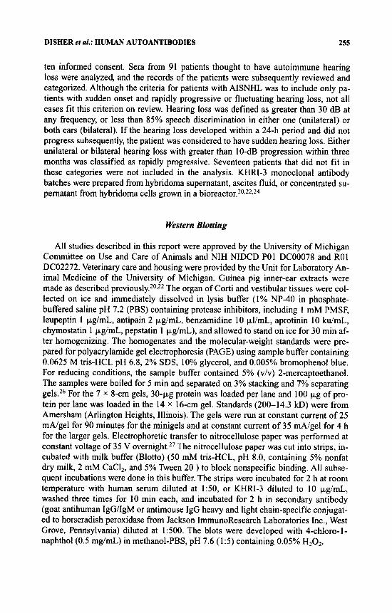
Sera from 74 patients with rapidly progressive or sudden onset hearing loss were evaluated for reactivity with guinea pig inner-ear antigens using either Western blot (73 patients) or immunofluorescence (36 patients). The breakdown of the patients by type of hearing loss is shown in TABLE 1. Fifty-four patients had rapidly progressive hearing loss. Thirty-six of these had bilateral involvement, and 18 had unilateral in- volvement. Twenty patients had sudden hearing loss; of these, only two had bilateral effects. Of the 54 patients with rapidly progressive hearing loss, tested by Western blot, 28 (52%) stained a 68-70-kD protein. Similarly, one half of the of the sudden hearing loss patients 9/19 (47%) were positive. Of the patients with rapidly progres- sive hearing loss 50% (1 8/36) with bilateral involvement and a slightly higher pro- portion, 56% (10/18), of the patients with unilateral involvement were Western blot positive. Examples of the Western blots are shown in FIGURE 1.
To determine the location of the inner-ear antigen, we tested Western blot positive sera, negative sera, normal donor sera, and a sera from a few previously untested pa- tients by immunofluorescence on surface preparations of guinea pig organ of Corti.
TABLE 1. Serological Results by Type of Hearing Loss Type of Hearing loss
Rapidly Progressive Sudden Total ~~ ~
Number of patients 54 20 74 Western blot
Number positive/number tested 28/54 9/19 31/73 Percent positive (52%) (47%) (51%)
Number positivelnumber tested 24/28 8/8 32/36 Immunofluorescence
Percent positive (86%) ( 1 OOYO) (89%)
258 ANNALS NEW YORK ACADEMY OF SCIENCES
FIGURE 1. Western blot of human sera on guinea pig inner-ear extract. UM-HL indicates University of Michigan Hearing Loss patients 03 through 12. Lane C is serum from a normal donor.
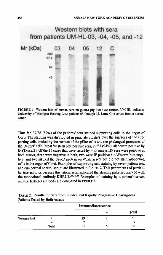
Thus far, 32/36 (89%) of the patients’ sera stained supporting cells in the organ of Corti. The staining was distributed in punctate clusters over the surfaces of the sup- porting cells, including the surface of the pillar cells and the phalangeal processes of the Deiters’ cells. Most Western blot positive sera, 29/3 1 (94%), also were positive by IF (TABLE 2). Of the 36 cases that were tested by both assays, 29 sera were positive in both assays, three were negative in both, two were IF positive but Western blot nega- tive, and two stained the 68-kD protein on Western blot but did not stain supporting cells in the organ of Corti. Examples of supporting cell staining by seven-patient sera and one normal control serum are illustrated in FIGURE 2. This pattern was of particu- lar interest to us because the patient sera replicated the staining pattern observed with the monoclonal antibody KHRI-3.20*22.24 Examples of staining by a patient’s serum and the KHRI-3 antibody are compared in FIGURE 3.
TABLE 2. Results for Sera from Sudden and Rapidly Progressive Hearing-loss Patients Tested by Both Assays
Immunofluorescence
+ - Total Western blot +
- Total
29 2 31 2 3 5
31 5 36

DISHER et at.: HUMAN AUTOANTIBODIES 259
FIGURE 2. Immunofluorescence photomicrographs of surface preparations of guinea pig or- gan of Corti immunostained with human sera. Panels (a)-(g), sera from patients UM-HL-5, UM-HL-2 I , UM-HL-25, UM-HL-26, UM-HL-37, UM-HL-53, with sudden onset or rapidly progressive hearing loss; panel (h), serum from a normal donor. Note the punctate “wine glass” staining pattern of the supporting cells in each case incubated with the patients’ sera. No such staining was observed with control sera, although the normal serum stained the stereocilia.
The similarity of staining and the prior observations that KHRI-3 can cause hear- ing 10ss20.24 suggested that the human sera might bind to the same antigen as KHRI- 3. To test this hypothesis, proteins precipitated from inner-ear extracts by KHRI-3 were subjected to SDS PAGE, Western blotted, and stained with patient sera. An ex- ample of this experiment is shown in FIGURE 4. Thus far, 414 patients’ sera that were positive for the 68-70-kD band by Western blot also stained a 70-kD band in the ma-
UM-HL-12
FIGURE 3. Confocal immunofluorescence photomicrographs of guinea pig organ of Corti sur- face preparations stained with serum from patient UM-HL-12 (left panel) and KHRI-3 mono- clonal antibody (right panel). Note the similarity of the staining pattern on the supporting cells.

260 ANNALS NEW YORK ACADEMY OF SCIENCES
FIGURE 4. Immunoprecipitation and Western blot of guinea pig inner-ear tissue. Guinea pig inner-ear extracts were immunoprecipitated with the KHRI-3 mouse monoclonal antibody, and then the precipitated proteins were separated by electrophoresis under nonreducing conditions (NR, lane 1) or reducing conditions (reduced, lanes 2-7) and transferred to a nitrocellulose membrane. The left hvo lanes were then incubated with KHRI-3 antibody and developed with antimouse immunoglobulin. Lanes 3-6 were incubated with human sera from patients UM- HL-03, -12, and -37, or with a normal serum sample and developed with antihuman im- munoglobulin. Lane 7 is the second antibody control. Note that the autoimmune human sera stain a 68-70-kD protein (bracket shows the region of the protein band) immunoprecipitated by KHRI-3 antibody. KHRI-3 antibody reacts best with nonreduced proteins, whereas the human sera react with the reduced proteins, indicating that the human sera bind to a different epitope than the murine antibody.
terial immunoprecipitated by KHRI-3. Normal sera controls were negative. The KHRI-3 antibody reacts best with nonreduced inner-ear proteins (FIG. 4, bracket on left). Under these conditions we can usually resolve a doublet of 65 and 67 kD. KHRIJ reacts poorly with reduced proteins, but faint staining of the bands that shift to 68 and 70 kD under reducing conditions can be discerned in lane 2. The human sera appear to stain most strongly the upper band detected by KHRI-3 under reducing conditions (FIG. 4, bracket on right). The heavy band at 55 kD in lane 2 is the reduced Ig heavy chain of the KHRI-3 antibody used to precipitate the inner-ear proteins. In contrast to KHRI-3, the human sera react best with reduced proteins, indicating that the human antibodies and the murine antibody may identify different conformational epitopes on the same protein.

DISHER el ul.: HUMAN AUTOANTIBODIES 261
Absorption of Autoantibody by Human Inner Ear Tissue
FIGURE 5. Immunofluorescence photomicrographs of guinea pig organ of Corti immunos- tained with an autoimmune hearing-loss serum showing the effects of absorption of the serum with: (a) no tissue; (b) human inner-ear tissue; (c) white blood cells from the inner-ear tissue donor; (d) red blood cells from the inner-ear tissue donor. Arrows indicate the location of the first row of supporting cells. Note that the staining of the supporting cells is eliminated after absorption with human inner ear, but not after absorption with blood cells from the same donor.
To determine if the antigen detected by patient sera in guinea pig inner-ear tissue is present in human inner ear, absorption analysis was performed. Inner-ear tissue re- moved from patients undergoing ablative inner-ear surgery was used as the source of inner-ear antigen, and white and red blood cells from the same donors were used as histocompatibility and blood group antigen controls. Equal volumes of either packed blood cells or inner-ear tissue were mixed with an aliquot of IF positive patient sera and then retested on guinea pig inner-ear substrate. As shown in FIGURE 5, absorption with inner ear but not blood cells removed the antibody reactivity to guinea pig inner ear. In a second experiment not shown, a similar but less complete absorption was obtained.
DISCUSSION
Although autoimmunity as one etiologic alternative for SNHL'-* has not been conclusively confirmed, there is mounting evidence to suggest that it is one of the causes of SNHL in patients who have rapidly progressive loss. Much of the experi-

262 ANNALS NEW YORK ACADEMY OF SCIENCES
mental and clinical evidence has involved the use of bovine inner-ear antigen^.'.^^-'^ We used inner-ear antigens from guinea pigs in an attempt to develop a more readily available and consistent source of antigen for In this study with human sera, the use of guinea pig tissue also allowed us to employ established immunohisto- chemistry techniques and a previously prepared monoclonal antibodyeZ2 Using West- em blots, we have been able to demonstrate that 51% of patients with sudden onset and/or rapidly progressive hearing loss have antibody that reacts strongly with guinea pig inner-ear antigens. Our findings are similar to the observations of other investiga- tors. The highest frequency of positive cases is reported by Hughes et aLZ5 who found antibodies to a 68-kD antigen in 86% of patients. Moscicki et al. found that 58% of the 72 patients they tested with bilateral rapidly progressive hearing loss had antibody to a 68-72-kD protein in bovine inner-ear extracts. Likewise, Harris and RyanI6 reported 32% of their large series of presumptive autoimmune hearing-loss cases were positive for antibody to a 68-kD protein in bovine inner-ear extract. Veld- man et ~ 1 . ~ ~ found 65-73% of patients with sudden onset or rapidly progressing hear- ing loss, respectively, had antibody to multiple bands in Western blots including 27, 45,50, and 68 kD. Cao et al. I 9 s 2 I used guinea pig inner-ear extracts similar to ours to detect human autoantibodies. These investigators did not observe a 68-70-kD band; instead, they reported bands of 58 and 30 kD. We also noted that some patients sera stain multiple bands at 200, 85, and 55 kD, but for the purposes of this study we scored as positive only those sera staining a 68-70-kD protein. The 68-70-kD band was the most consistent and usually the most strongly stained band. As we reported previously, the inner-ear extracts are very sensitive to proteolytic breakdown.23 If stringent extraction conditions using quick processing, rapid chilling on ice, and pro- tease inhibitors are not used, then high molecular-weight bands may disappear and lower molecular-weight bands may appear in their place. However, Cao et al.19*21 used protease inhibitors, so the reasons for the absence of the 68-70-kD protein in their results is not clear.
There have been a number of reports of human sera staining inner-ear tissue and human temporal bone specimens29 as summarized by S~liman.~O However, to our knowledge there have been no previous reports of consistent staining of the support- ing cells in the organ of Corti such as we observed. This may be because many of the other investigators used cross sections rather than surface preparations. In cross sec- tion, supporting cell staining, unless it is very i n t e n ~ e , ~ ~ * ~ ’ is less apparent than it is when viewed from the surface. The “wine-glass” staining pattern we observed with the human autoimmune sera on the phalangeal processes of the outer pillar cells and Deiters’ cells is like that which we previously reported with the KHRI-3 monoclonal a n t i b ~ d y . ~ ~ ~ ~ ~ . ~ ~ Furthermore, there was a strong concordance between staining of the 68-kD protein and the wine-glass staining pattern on organ of Corti supporting cells, since >90% of sera that had antibody to the 68-kD band also stained supporting cells. This is consistent with the notion that the 68-kD protein is expressed on the support- ing cells. Since the KHRI-3 antibody has been linked to hearing loss and damage to the organ of Corti in both miceZo and guinea pigs,24 we found this pattern of staining to be quite interesting. It suggested to us that the human autoantibodies might bind to the same protein as the KHRI-3 antibody and that both antibodies might have a com- mon mechanism of inducing hearing loss. To further investigate this possibility, we immunoprecipitated inner-ear proteins using the KHRI-3 antibody and then tested if

DISHER et al.: HUMAN AUTOANTIBODIES 263
the human antibodies would stain those immunoprecipitated proteins. As shown in FIGURE 4, this is the case. Our results support the concept that the human antibodies detected with guinea pig inner-ear tissue could be pathogenic in humans. However, until now there has been no evidence that antigens detected by human sera in guinea pig or bovine inner ear are also expressed in the human inner ear. Our last experiment shown in this paper demonstrates that the antigen we detect in guinea pig inner ear using human sera is also expressed specifically in human inner-ear tissue. Human in- ner-ear tissue but not lymphocytes or red blood cells from two tissue donors was ca- pable of absorbing the antibodies that bind to the guinea pig supporting cells. If the human inner ear did not contain the same antigen as the guinea pig inner ear, then the absorption would not have removed the antibody. This is compelling evidence that antibodies to inner-ear antigens that are present in the sera of a high proportion of sudden onset, rapidly progressive hearing loss patients do bind to human inner-ear antigens and may well have pathogenic effects themselves as suggested by their sim- ilarity to the pathogenic KHRIJ antibody.
The nature of the target antigen of human autoimmune sera has been suggested to be heat shock protein, specifically HSP-70.I’ We don’t know if this is correct or not, but we strongly suspect that HSP-70 is not the target of KHRI-3, because when pro- teins are precipitated from inner-ear extracts by KHRI-3 and Western blotted, nothing in the immunoprecipitate reacts with anti-HSP-70 antibodies. Thus, if KHRI-3 and human autoantibodies define the same antigen, then it seems unlikely that HSP-70 is the primary target of the human antibodies that stain proteins precipitated by KHRI- 3. This is an area of investigation in our and other laboratories and requires more work before any conclusions can be drawn.
The findings of this study support the hypothesis that autoimmune sensorineural hearing loss occurs in humans, and that its presence can be established by laboratory testing in at least some patients. Such test results may correlate with therapeutic re- sponse.I5 In light of the clinical importance of accurately and rapidly diagnosing treatable, potentially reversible causes of sensorineural hearing loss, and considering the complications associated with the use of cytotoxic medications and corticos- teroids, there is clearly a need for a readily available test to diagnose this condition.
SUMMARY
Autoimmunity is thought to be one cause of sensorineural hearing loss (SNHL). Sera from patients with rapidly progressive hearing loss have been shown to contain antibodies to a 68-kD protein in heterologous inner-ear tissue. Using guinea pig in- ner-ear tissue as the antigenic substrate and either Western blot or immunofluores- cence (IF) or both, we tested sera from 74 patients suspected to have autoimmune hearing loss for inner-ear antibodies. Sera from 73 patients were tested by Western blot, and sera from 36 were tested by IF. Thirty-seven of 73 (51%) had antibody to a 68-70-kD protein by Western blot. Sera positive by IF stained supporting cells with a staining pattern like that previously observed with the KHRI-3 monoclonal antibody. There was concordance between Western blot and IF assays. Of 36 patients tested by both assays, 2913 1 (94%) that were positive in Western blot were also positive by IF, three were negative by both tests, and two each were positive by one assay but nega-

264 ANNALS NEW YORK ACADEMY OF SCIENCES
tive by the other. Absorption of patient sera with human inner-ear tissue removed an- tibody reactivity to the guinea pig supporting cells, indicating that the antigen detect- ed by the autoantibody is also present in the human inner ear. Absorption with an equal volume of white or red blood cells from the tissue donor did not remove the an- tibody reactivity to inner ear, showing that the absorption by inner-ear tissue is spe- cific. Sera from three patients positive in both assays also stained a 68-70-kD inner- ear protein immunoprecipitated by the KHRI-3 monoclonal antibody, indicating that the monoclonal and human antibodies recognize the same antigen. The results sup- port the hypothesis that patients with autoimmune sensorineural hearing loss produce autoantibodies to an inner-ear supporting cell antigen that is phylogenetically con- served and defined by the murine monoclonal antibody KHRI-3. Since KHRI-3 can induce hearing loss after infusion into the inner ear, it is likely that autoantibodies with the same antigenic target are also pathogenic in humans.
REFERENCES
1. LEHNHARDT, E. 1958. Plotzliche Horstorungen, auf beiden Selten gleichzeitig oder
2. MCCABE, B. F. 1979. Autoimmune sensorineural hearing loss. Ann. Otol. 88: 585-859. 3. Yoo, T. J., J. M. STUART, A. H. KANG, A. S. TOWNES, K. TOMODA & S. DIXIT. 1982. Type I1
collagen autoimmunity in otosclerosis and Meniere’s disease. Science 217: 1153-1 155. 4. HARRIS, J. l? 1983. Immunology of the inner ear: Response of the inner ear to antigen chal-
lenge. Otolaryngol. HeadNeck Surg. 91: 18-23. 5 . Yoo, T. J., R. A. FLOYD, N. SUDO, T. ISHIBE, T. TAKEDA, K. TOMODA,~. YAZAWA, J. STUART, I.
S. CHOE & S. C. HA. 1983. Factors influencing collagen-induced autoimmune ear dis- ease. Am. J. Otolaryngol. 6: 209-216.
6. HARRIS, J. €? 1987. Experimental autoimmune sensorineural hearing loss. Laryngoscope
7. HARRIS, J. €? & I? A. SHARP. 1990. Inner ear autoantibodies in patients with rapidly progres- sive sensorineural hearing loss. Laryngoscope 100: 5 16-524.
8. OROZCO, C. R., J. K. NIPARKO, B. C. RICHARDSON, D. F. DOLAN, M. U. PTOK & R. A. ALTSCHULER. 1990. Experimental model of immune-mediated hearing loss using cross- species immunization. Laryngoscope 100: 941-947.
9. CRUZ, 0. L. M., A. MINITI, W. COSSERMELLI & R. M. OLIVEIRA. 1990. Autoimmune sen- sonneural hearing loss: A preliminary experimental study. Am. J. Otol. 11: 342-346.
10. MCCABE, B. F. 1991. Autoimmune inner ear disease: Results of therapy. Adv. Otorhino-
11. KUSAKARI, C., K. HOZAWA, S. KOIKE, M. KYOGUKU & T. TAKASAKA. 1992. MLR/MP- lprflpr mouse as a model of immune-induced sensorineural hearing loss. Ann. Otol. Rhi- nol. Laryngol. 101: 82-86.
12. TAGO, C. & N. YANGITA. 1992. Cochlear and renal pathology in the autoimmune strain mouse. Ann. Otol. Rhinol. Laryngol. 157(Suppl.): 87-91.
13. WONG, M. L., J. S. YOUNG, G. NILAVER, J. I. MORTON & D. R. TRUNE. 1992. Cochlear IgG in the C3Wlpr autoimmune strain mouse. Hear. Res. 59: 93-100.
14. SONE, M., H. NARIUCHI, K. SAITO & N. YANAGITA. 1994. A substrain of NZB mouse as an animal model of autoimmune inner ear disease. Hear. Res. 83: 26-36.
15. MOSCICKI, R. A., J. E. SAN MARTIN, C. H. QUINTERO, S. D. RAUCH, J. B. NADOL & K. B. BLOCH. 1994. Serum antibody to inner ear proteins in patients with progressive hearing loss: Correlation with disease activity and response to corticosteroid treatment. JAMA 272: 611-616.
nacheinander aufgetreten. Z. Laryngol. Rhinol. Otol. 37: 1.
97: 63-76.
laryngol. 46: 78-81.

DISHER et al.: HUMAN AUTOANTIBODIES 265
16. HARRIS, J. P. &A. F. RYAN. 1995. Fundamental immune mechanisms of the brain and inner ear. Otolaryngol. Head Neck Surg. 112: 639653.
17. BILLINGS, I? B., E. M. KEITHLEY & J. P. HARRIS. 1995. Evidence linking the 68 kilodalton antigen identified in progressive sensorineural hearing loss patient sera with heat shock protein 70. Ann. Otolol. Rhino]. Laryngol. 104: 18 1-188.
18. GOTTSCHLICH, S., €? B. BILLINGS, E. M. KEITHLEY, M. H. WEISMAN & J. P. HARRIS. 1995. Assessment of serum antibodies in patients with rapidly progressive sensorineural hear- ing loss and Meniere’s disease. Laryngoscope 105: 1347-1352.
19. CAO, M.-Y., M. GERSDORFF, N. DEGGOUJ, M. WARNY & J.-€? TOMASI. 1995. Detection of in- ner ear disease autoantibodies by immunoblotting. Mol. Cell Biochem. 146: 157-163.
20. NAIR, T. S., Y. RAPHAEL, D. F DOLAN, T. J. PARRETT, L. S. PERLMAN, c! R. BRAHMBHATT, Y. WANG, X. Hou, G. GHOLIZADEH, A. L. NUTTALL, R. A. ALTSCHULER & T. E. CAREY. 1995. Monoclonal antibody induced hearing loss. Hear. Res. 83: 101-1 13.
21. CAO, M. Y., N. DEGGOUJ, M. GERSWRFF & J.-P. TOMASI. 1996. Guinea pig inner ear anti- gens: Extraction and application to the study of human autoimmune inner ear disease. Laryngoscope 106: 207-212.
22. ZAJIC, G., T. S. NAIR, M. PTOK, C. VAN WAES, R. A. ALTSCHULER, J. SCHACHT & T. E. CAREY. 199 1. Monoclonal antibodies to inner ear antigens: I. Antigens expressed by sup- porting cells in guinea pig cochlea. Hear. Res. 52: 59-72.
23. PTOK, M., T. S. NAIR, R. A. ALTSCHULER, J. SCHACHT &T. E. CAREY. 1991. Monoclonal an- tibodies to inner ear antigens: 11. Antigens expressed in sensory cell stereocilia. Hear. Res. 57: 79-90.
24. NAIR, T. S., D. M. PRIESKORN, J. M. MILLER, A. MORI, J. GRAY & T. E. CAREY. 1997. In vivo binding and hearing loss after intracochlear infusion of KHRI-3 antibody. Hear. Res.
25. HUGHES, G. B., R. MOSCICKI, B. P. BARNA & J. E. SAN MARTIN. 1994. Laboratory diagnosis of immune inner ear disease. Am. J. Otology. 15: 198-202.
26. LAEMMLI, U. K. 1970. Cleavage of structural proteins during the assembly of the head of bacteriophage T4. Nature 277: 680-685.
27. TOWBIN, H., T. STAEHLIN & J. GORDON. 1979. Electrophoretic transfer of proteins from polyacrylamide gels to nitrocellulose sheets. Procedure and some applications. Proc. Natl. Acad. Sci. USA 73: 2599-2604.
28. VELDMAN, J. E., T. HANADA & F. MEEUWSEN. 1993. Diagnostic and therapeutic dilemmas in rapidly progressive sensorineural hearing loss and sudden hearing loss. A reappraisal of immune reactivity in inner ear disorders. Acta Otolaryngol. (Stockholm) 113:
29. ARNOLD, W. & C. R. PFALTZ. 1987. Critical evaluation of the immunofluorescence micro- scopic tests for identification of serum antibodies against human inner ear tissue. Acta
107: 93-10],
303-306.
Otolaryngol. (Stockholm) 103: 371-378. 30. SOLIMAN, A. M. 1992. Immune mediated inner ear disease. Am. J. Otol. 13: 575-579. 31. CHEN, H., I. THALMANN, J. C. ADAMS, K. B. AVRAHAM, N. G. COPELAND, N. A. JENKINS, D.
R. BEIER, D. €? COREY, R. THALMANN & G. M. DUYK. 1995. cDNA cloning, tissue distri- bution, and chromosomal localization of Ocp2, a gene encoding a putative transcription- associated factor predominantly expressed in the auditory organs. Genomics 27: 389-398.
Copyright © 2022 FDOKUMEN