Primary sequence determination of a monoclonal antibody against α-synuclein using a novel mass...
9
International Journal of Mass Spectrometry 312 (2012) 61–69 Contents lists available at ScienceDirect International Journal of Mass Spectrometry j our na l ho me page: www.elsevier.com/locate/ijms Primary sequence determination of a monoclonal antibody against -synuclein using a novel mass spectrometry-based approach Eric Sousa a,1 , Stephane Olland a,1 , Heather H. Shih a,1 , Kim Marquette a , Robert Martone b , Zhijian Lu a , Janet Paulsen a , Davinder Gill a , Tao He a,∗ a Global Biotherapeutics Technologies, Pfizer, 87 Cambridge Park Dr., Cambridge, MA 02140, USA b Neuroscience Research Unit, Pfizer, Groton, CT, USA a r t i c l e i n f o Article history: Received 22 February 2011 Received in revised form 2 May 2011 Accepted 9 May 2011 Available online 14 May 2011 Keywords: de novo sequencing Antibody Stable isotope labeled amino acids in cell culture (SILAC) -Synuclein Collision induced dissociation (CID) Higher energy collision dissociation (HCD) a b s t r a c t A novel mass spectrometry-based workflow for de novo sequencing of a full-length monoclonal antibody (LB509) against -synuclein is presented. This approach combines chemical modification of cysteine residues, multiple enzymatic digestion, stable isotope labeled amino acids in cell culture (SILAC), liquid chromatography coupled tandem mass spectrometry (LC/MS/MS) analysis including collision-induced dissociation (CID), higher energy collision-induced dissociation (HCD), and de novo sequencing software for data interpretation. The introduction of aminoethyl-8 (A-8) reagent which renders cysteine residues in the antibody to become substrates for trypsin digestion significantly increases the sequence coverage of the antibody, especially in the complementarity determining regions (CDRs). Incorporation of SILAC helps to distinguish minor sequence variations between original and deduced sequences. Methods in obtaining detailed sequence characterization are described, highlighting the advantage of using multiple fragmentation schemes, CID in ion trap and HCD in C-trap. Complete primary sequence for LB509 has been construed and the antibody generated exhibits specific binding to -synuclein. The development and implementation of this technology enables an alternative approach for generating therapeutic candidate and reagent antibodies. © 2011 Elsevier B.V. All rights reserved. 1. Introduction Parkinson’s disease (PD) and dementia with Lewy bodies (DLB) are neurodegenerative disorders where accumulation of patho- logic -synuclein-containing Lewy bodies in brain tissue has been shown to be one of the major morphologic and molecular events in disease progression [1]. LB509 is a mouse monoclonal anti- body raised against Lewy bodies purified from the brains of deceased patients with DLB [2]. Since its generation, LB509, an antibody that specifically recognizes -synuclein, has been widely used as a research tool to study the function of -synuclein and its pathological association with diseases such as PD and DLB [3]. Abbreviations: SILAC, stable isotope labeled amino acids in cell culture; CID, collision induced dissociation; HCD, higher-energy collisiondissociation; CDR, complementary determining region; HC, heavy chain; LC, light chain; MALDI, matrix-assisted laser desorption/ionization; LC/MS/MS, liquid chromatography cou- pled tandem mass spectrometry; RE, reverse-engineered; A-8, aminoethyl-8; IAA, iodoacetamide; ELISA, enzyme-linked immunosorbent assay. ∗ Corresponding author. Tel.: +1 617 665 8021. E-mail address: tao.he@pfizer.com (T. He). 1 Contributed equally to this work. Monoclonal antibodies have been accepted as an important therapeutic modality for a variety of human disease treatment, including cancer, immunological disorders and transplant rejection [4]. Antibody-based therapeutics offer high specificity to selected targets, owing to complementarity determining regions (CDRs) in variable region. Recombination of variable (V), diversity (D), and junctional (J) gene segment of the heavy chain (HC) and VJ gene segments of the light chain (LC) as well as somatic hypermuta- tion define the specificity of the variable region in antibodies [5]. Therefore, without cDNA information (such as loss of hybridoma or externally acquired antibodies), it is extremely challenging to obtain the complete primary sequence of monoclonal antibodies. Edman degradation [6] is a widely accepted approach to delineate unknown protein sequence. However, it is a low throughput pro- cess where large quantities of highly purified samples are needed. Mass spectrometry-based peptide and protein analysis has become an indispensable tool with the development of soft ionization techniques, namely, electrospray [7] and matrix-assisted laser des- orption/ionization (MALDI) [8,9]. One of the important factors that facilitate unknown protein identification for proteomics studies is the availability of protein database, where tandem mass spectra are searched against known theoretical sequences [10–12]. Devel- opment of stable isotope labeling reagents, such as stable-isotope labeling by amino acids in cell culture (SILAC) [13] and isotope- 1387-3806/$ – see front matter © 2011 Elsevier B.V. All rights reserved. doi:10.1016/j.ijms.2011.05.005
-
Upload
independent -
Category
Documents
-
view
1 -
download
0
Transcript of Primary sequence determination of a monoclonal antibody against α-synuclein using a novel mass...
Pu
EJa
b
a
ARRAA
KdASc�CH
1
alsibdauaD
Ccmpi
1d
International Journal of Mass Spectrometry 312 (2012) 61– 69
Contents lists available at ScienceDirect
International Journal of Mass Spectrometry
j our na l ho me page: www.elsev ier .com/ locate / i jms
rimary sequence determination of a monoclonal antibody against �-synucleinsing a novel mass spectrometry-based approach
ric Sousaa,1 , Stephane Ollanda,1 , Heather H. Shiha,1 , Kim Marquettea , Robert Martoneb , Zhijian Lua ,anet Paulsena, Davinder Gill a, Tao Hea,∗
Global Biotherapeutics Technologies, Pfizer, 87 Cambridge Park Dr., Cambridge, MA 02140, USANeuroscience Research Unit, Pfizer, Groton, CT, USA
r t i c l e i n f o
rticle history:eceived 22 February 2011eceived in revised form 2 May 2011ccepted 9 May 2011vailable online 14 May 2011
eywords:e novo sequencingntibody
a b s t r a c t
A novel mass spectrometry-based workflow for de novo sequencing of a full-length monoclonal antibody(LB509) against �-synuclein is presented. This approach combines chemical modification of cysteineresidues, multiple enzymatic digestion, stable isotope labeled amino acids in cell culture (SILAC), liquidchromatography coupled tandem mass spectrometry (LC/MS/MS) analysis including collision-induceddissociation (CID), higher energy collision-induced dissociation (HCD), and de novo sequencing softwarefor data interpretation. The introduction of aminoethyl-8 (A-8) reagent which renders cysteine residuesin the antibody to become substrates for trypsin digestion significantly increases the sequence coverageof the antibody, especially in the complementarity determining regions (CDRs). Incorporation of SILAC
table isotope labeled amino acids in cellulture (SILAC)-Synucleinollision induced dissociation (CID)igher energy collision dissociation (HCD)
helps to distinguish minor sequence variations between original and deduced sequences. Methods inobtaining detailed sequence characterization are described, highlighting the advantage of using multiplefragmentation schemes, CID in ion trap and HCD in C-trap. Complete primary sequence for LB509 has beenconstrued and the antibody generated exhibits specific binding to �-synuclein. The development andimplementation of this technology enables an alternative approach for generating therapeutic candidate
and reagent antibodies.. Introduction
Parkinson’s disease (PD) and dementia with Lewy bodies (DLB)re neurodegenerative disorders where accumulation of patho-ogic �-synuclein-containing Lewy bodies in brain tissue has beenhown to be one of the major morphologic and molecular eventsn disease progression [1]. LB509 is a mouse monoclonal anti-ody raised against Lewy bodies purified from the brains ofeceased patients with DLB [2]. Since its generation, LB509, anntibody that specifically recognizes �-synuclein, has been widelysed as a research tool to study the function of �-synuclein
nd its pathological association with diseases such as PD andLB [3].Abbreviations: SILAC, stable isotope labeled amino acids in cell culture;ID, collision induced dissociation; HCD, higher-energy collisiondissociation; CDR,omplementary determining region; HC, heavy chain; LC, light chain; MALDI,atrix-assisted laser desorption/ionization; LC/MS/MS, liquid chromatography cou-
led tandem mass spectrometry; RE, reverse-engineered; A-8, aminoethyl-8; IAA,odoacetamide; ELISA, enzyme-linked immunosorbent assay.∗ Corresponding author. Tel.: +1 617 665 8021.
E-mail address: [email protected] (T. He).1 Contributed equally to this work.
387-3806/$ – see front matter © 2011 Elsevier B.V. All rights reserved.oi:10.1016/j.ijms.2011.05.005
© 2011 Elsevier B.V. All rights reserved.
Monoclonal antibodies have been accepted as an importanttherapeutic modality for a variety of human disease treatment,including cancer, immunological disorders and transplant rejection[4]. Antibody-based therapeutics offer high specificity to selectedtargets, owing to complementarity determining regions (CDRs) invariable region. Recombination of variable (V), diversity (D), andjunctional (J) gene segment of the heavy chain (HC) and VJ genesegments of the light chain (LC) as well as somatic hypermuta-tion define the specificity of the variable region in antibodies [5].Therefore, without cDNA information (such as loss of hybridomaor externally acquired antibodies), it is extremely challenging toobtain the complete primary sequence of monoclonal antibodies.Edman degradation [6] is a widely accepted approach to delineateunknown protein sequence. However, it is a low throughput pro-cess where large quantities of highly purified samples are needed.Mass spectrometry-based peptide and protein analysis has becomean indispensable tool with the development of soft ionizationtechniques, namely, electrospray [7] and matrix-assisted laser des-orption/ionization (MALDI) [8,9]. One of the important factors thatfacilitate unknown protein identification for proteomics studies is
the availability of protein database, where tandem mass spectraare searched against known theoretical sequences [10–12]. Devel-opment of stable isotope labeling reagents, such as stable-isotopelabeling by amino acids in cell culture (SILAC) [13] and isotope-
62 E. Sousa et al. / International Journal of Mass Spectrometry 312 (2012) 61– 69
Antibody RE1-Antibody ELISA
Reduce/Alky lateDTT / IAA or A8
SILACMix w/LB509
Reduce/Alky lateDTT / IAA or A8SDS-PAGE
SDS-PAGEDigestionTry, LysC, AspN, Ch ymo
Digestion
LC-MS/MSon LTQ
LC-MS/MSon Orbitrap
gTry, LysC, AspN, Ch ymo
on OrbitrapQual Browse r, Database
Searching & PEAKSReconstruct Sequence Refine Sequence
RE2 Antibody ELISARE2-Antibody
proced
eqt
tafldoieaicmmseirasodab
pctSaWs
Fig. 1. Overall workflow for antibody reverse engineering. The
ncoded affinity tags (ICAT) [14], has enabled the emergence ofuantitative proteomics for large-scale kinetic studies of a pro-eome.
Although it is routine that hundreds if not thousands of pro-eins could be identified by mass spectrometry-based proteomicspproach [15], it is still an area of active research to develop work-ows for de novo protein sequencing when a relevant proteinatabase is not available [16]. For delineating the primary sequencef antibodies, CDR regions are extremely challenging due to thencomplete sequence coverage for CDRs in protein databases. Phamt al. presented the first de novo sequence study on a monoclonalntibody against OX40 ligand, applying both Edman sequenc-ng and mass spectrometry in combination with proteolytic andhemical digestion [17]. Recently, Perdivara et al. deduced pri-ary sequence of an antibody against �-amyloid (clone 6E10)ainly based on liquid chromatography coupled with tandem mass
pectrometry [18]. In this study, de novo tandem mass spectrom-try analysis provided majority of the variable domain sequencenformation. Additionally, advances in software development wereeported in support of de novo sequencing effort for monoclonalntibodies, which was exemplified by comparative shotgun proteinequencing (CSPS) approach [19]. However, it is still a challenge tobtain complete protein sequence of a monoclonal antibody and toemonstrate the retention of activity of a reverse-engineered (RE)ntibody, especially in distinguishing minor sequence variabilityetween original and RE antibodies.
In this study, we describe a novel workflow for de novo proteinrimary sequence analysis of monoclonal antibodies. This approachombines chemical modification of cysteine residues to becomeryptic/LysC proteolysis substrates, multiple enzymatic digestion,
ILAC for detailed sequence comparison, high resolution LC/MS/MSnalysis and de novo sequencing software for data interpretation.e applied this de novo sequencing method to deduce the primaryequence of LB509. Importantly, we showed that the reverse engi-
ure will be performed for both heavy and light chain of LB509.
neered LB509 antibody retained its specific �-synuclein bindingactivity.
2. Experimental
2.1. Materials
Mouse anti-�-synuclein monoclonal antibody (LB509, SIG-39725) was purchased from Covance (Princeton, NY). Aminoethyl-8was obtained from Pierce (Rockford, IL). Heavy Lysine and Argi-nine amino acids were purchased from Cambridge Isotope LabInc (Andover, MA). Enzymes (Trypsin, LysC, Chymotrypsin, AspN)were acquired from Roche Diagnostics (Indianapolis, IN). All otherreagents were acquired from Sigma–Aldrich (St. Louis, MO) of thehighest grade.
2.2. SDS-PAGE and in-gel digestion
The monoclonal antibody LB509 was reduced with DTT andalkylated with either A-8 or iodoacetamide (IAA) and separatedby SDS-PAGE (10% Tricine). The light and heavy chain bandswere excised and digested with different enzymes (trypsin, LysC,chymotrypsin and AspN) on a digestion workstation DigestPro(ABimed GmbH, Langenfeld, Germany). Briefly, bands were washedwith 100 mM ammonium bicarbonate after shrinking with ace-tonitrile. The enzyme was added (0.5 �g/gel band) and incubatedat 37 ◦C for 8 h. The resulting peptides were extracted with ace-tonitrile washes followed by 5% formic acid washes. In the case of
double digestion, AspN followed by trypsin, the AspN digest wascarried out first and 5% formic acid was replaced with 100 mMammonium bicarbonate. The resulting AspN peptides were furtherdigested with trypsin.
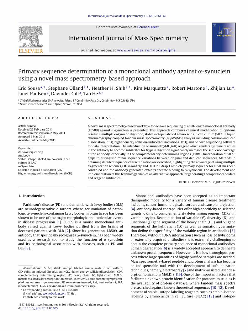
E. Sousa et al. / International Journal of Mass Spectrometry 312 (2012) 61– 69 63
Fig. 2. Use of visualization tool to aid identification of CDR-containing peptides after A-8 modification and trypsin digestion. (A) 2D map of LB509 sample alkylated with IAAa ested
o -cont
2
owrLMp0tp1(45iw1clr
nd digested with trypsin; (B) 2D map of LB509 sample alkylated with A-8 and digf highlighted peptide ion in 2B, the peptide was determined to be light chain CDR3
.3. Liquid chromatography mass spectrometry
Recovered peptide samples from in-gel digest were loadednto a Pico-frit column (New Objective, Woburn, MA) packedith reversed-phase Magic (Michrom, Auburn, CA) C18 mate-
ial (5 �m, 200 A, 75 �m × 10 cm) and coupled to an LTQ orTQ-Orbitrap XL mass spectrometer (ThermoElectron, Waltham,A). Chromatographic methods were identical for all the sam-
les analyzed. Peptides were separated at a flow rate of.2 �l/min using a 90 min linear gradient ranging from 2%o 40% B (mobile phase A: 0.1% formic acid/2% ACN; mobilehase B: 90% ACN/0.1% formic acid). Electrospray voltage was.8 kV. The instrumental method consisted of a full MS scanscan range 375–1550 m/z, with 30 K fwhm resolution @ m/z00, target value 2 × 10⁶, maximum ion injection time of00 ms) followed by data-dependent CID scans of the four most
ntense precursor ions. Peptide precursor ions were selectedith an isolation window of 2.5 Da and a target value of
× 105. Dynamic exclusion was implemented with a repeatount of 2 and exclusion duration of 75 s. The normalized col-ision energy (NCE) was set to 35% for CID and 40% for HCD,espectively.
with trypsin, the arrow highlights the additional peptide ion; (C) MS/MS spectrumaining: QQYHSYPWTFGGGTK. Insert shows MS spectrum of the peptide.
2.4. Database search, visualization software and de novosequencing
The mass spectra were searched against a public antibodydatabase using Bioworks 3.3.1 SP1 with SEQUEST seach algorithm(ThermoElectron, Waltham, MA). The mass accuracy was set to5 ppm for precursor ions and to 0.5 Da tolerances for fragment ions.The search parameters took into account two missed cleavagesfor trypsin, static modification of carboxamidomethylation at cys-teine (+57.0215 Da) or A-8 (+43.0420), and dynamic modificationfor methionine oxidation (Met +15.9949 Da). Data from A-8 andIAA modifications were displayed in Qual Browser (ThermoElec-tron, Waltham, MA). The mass spectra were also searched with denovo software Peaks (Bioinformatics Solutions Inc, Waterloo, ON)for potential matches and all hits were manually inspected, inter-preted and the light chain and heavy chain amino acid sequenceswere constructed.
2.5. Gene construction for deduced LB509
The protein sequences of LB509 heavy chain and light chainwere provided to GeneArt (Burlingame, CA) for conversion to
64 E. Sousa et al. / International Journal of Mass Spectrometry 312 (2012) 61– 69
F heavyD
Deoel
2
w(wsLvpaa1pat
ig. 3. MS/MS spectra of double digestion with AspN and trypsin for deducing
YWGQGTSVTVSSAK.
NA coding sequences with codons optimized for mammalian cellxpression. The DNA fragments encoding the heavy and light chainsf the deduced LB509 (RE-LB509) were cloned into the mammalianxpression vectors pSMED and pSMEN to express the heavy andight chains, respectively.
.6. Expression and purification of RE-LB509
The DNA constructs encoding RE-LB509 heavy and light chainsere co-transfected into HEK293 cells using Lipofectamin2000
InVitrogen, Carlsbad, CA). In 24 h post transfection, the mediaas changed to either 293 serum-free media (InVitrogen) or 293
erum-free media containing stable isotope labeled amino acidsysine and Arginine (Cambridge Isotope Lab Inc), and were har-ested in 48 h. The conditioned media expressing RE-LB509 wasurified on Protein G sepharose column to capture the RE-LB509ntibody. The antibody was subsequently eluted with 20 mM citriccid and 150 mM NaCl at pH 2.5 and immediately neutralized with
M Tris pH 8. The antibody was buffer exchanged into PBS. Theurified antibody was analyzed by size exclusion chromatographynd SDS-PAGE followed by staining with SimpleBlue according tohe manufacturer’s instructions (InVitrogen).
chain CDR3-containing peptides, (A) peptide QGFYYGYYHAM; and (B) peptide
2.7. Western blot analysis
Proteins in the conditioned media were separated onSDS/PAGE gels and subsequently transferred onto nitro-cellulose membranes. The membranes were probed withperoxidase-conjugated anti-mFc antibody. The heavychain of the antibody was detected with Western Light-ning Chemiluminescence reagents (PerkinElmer, Waltham,MA).
2.8. ˛-Synuclein binding ELISA
Full-length a-synuclein was coated on high-binding ELISAplate (CoStar, Lowell, MA) at 1 �g/ml overnight at 4 ◦C. Theplate was washed with washing buffer (PBS/0.1% Tween-20) andsubsequently blocked with assay buffer (PBS/0.1% Tween-20/1%BSA) for 1 h at 25 ◦C. Conditioned media or purified antibod-ies diluted in assay buffer were added to the blocked plate and
incubated for 1 h at 25 ◦C. After washing, the plate was incu-bated with peroxidase-conjugated anti-mFc antibody (Jackson’sLaboratories, Bar Harbor, ME) for 1hr at 25 ◦C. The plate wasthen washed and developed by incubating with tetramethylben-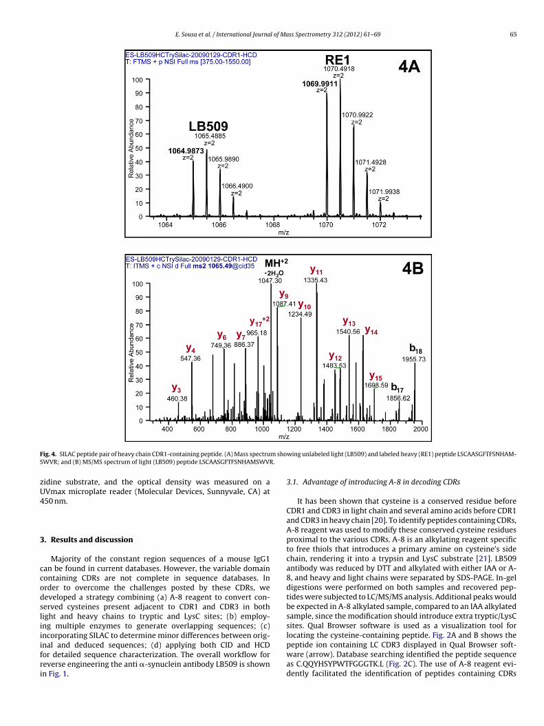
E. Sousa et al. / International Journal of Mass Spectrometry 312 (2012) 61– 69 65
F m shoS .
zU4
3
ccodsliiifri
ig. 4. SILAC peptide pair of heavy chain CDR1-containing peptide. (A) Mass spectruWVR; and (B) MS/MS spectrum of light (LB509) peptide LSCAASGFTFSNHAMSWVR
idine substrate, and the optical density was measured on aVmax microplate reader (Molecular Devices, Sunnyvale, CA) at50 nm.
. Results and discussion
Majority of the constant region sequences of a mouse IgG1an be found in current databases. However, the variable domainontaining CDRs are not complete in sequence databases. Inrder to overcome the challenges posted by these CDRs, weeveloped a strategy combining (a) A-8 reagent to convert con-erved cysteines present adjacent to CDR1 and CDR3 in bothight and heavy chains to tryptic and LysC sites; (b) employ-ng multiple enzymes to generate overlapping sequences; (c)ncorporating SILAC to determine minor differences between orig-
nal and deduced sequences; (d) applying both CID and HCDor detailed sequence characterization. The overall workflow foreverse engineering the anti �-synuclein antibody LB509 is shownn Fig. 1.wing unlabeled light (LB509) and labeled heavy (RE1) peptide LSCAASGFTFSNHAM-
3.1. Advantage of introducing A-8 in decoding CDRs
It has been shown that cysteine is a conserved residue beforeCDR1 and CDR3 in light chain and several amino acids before CDR1and CDR3 in heavy chain [20]. To identify peptides containing CDRs,A-8 reagent was used to modify these conserved cysteine residuesproximal to the various CDRs. A-8 is an alkylating reagent specificto free thiols that introduces a primary amine on cysteine’s sidechain, rendering it into a trypsin and LysC substrate [21]. LB509antibody was reduced by DTT and alkylated with either IAA or A-8, and heavy and light chains were separated by SDS-PAGE. In-geldigestions were performed on both samples and recovered pep-tides were subjected to LC/MS/MS analysis. Additional peaks wouldbe expected in A-8 alkylated sample, compared to an IAA alkylatedsample, since the modification should introduce extra tryptic/LysCsites. Qual Browser software is used as a visualization tool forlocating the cysteine-containing peptide. Fig. 2A and B shows the
peptide ion containing LC CDR3 displayed in Qual Browser soft-ware (arrow). Database searching identified the peptide sequenceas C.QQYHSYPWTFGGGTK.L (Fig. 2C). The use of A-8 reagent evi-dently facilitated the identification of peptides containing CDRs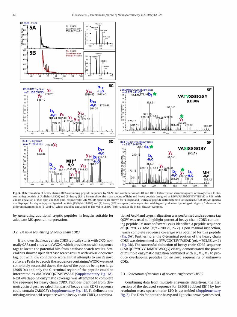
66 E. Sousa et al. / International Journal of Mass Spectrometry 312 (2012) 61– 69
Fig. 5. Determination of heavy chain CDR2-containing peptide sequence by SILAC and combination of CID and HCD. Extracted ion chromatograms of heavy chain CDR2-containing peptide of (A) light (LB509) and (B) heavy (RE1), inserts show the mass spectra of light and heavy peptide (assigned as LEWVASISSGGSYTYYPDSVK in RE1) witha own fa E1) sad ight) a
ba
3
mtetsc(iNtmcm
mass deviation of 0.35 ppm and 0.26 ppm, respectively. CID MS/MS spectra are shre displayed for chymotrypsin digested peptide, (E) light (LB509) and (F) heavy (Rifferent fragment ions (b3 and y7) which could be explained as Thr-Val in LB509 (l
y generating additional tryptic peptides in lengths suitable fordequate MS spectra interpretation.
.2. De novo sequencing of heavy chain CDR3
It is known that heavy chain CDR3 typically starts with CXX (nor-ally CAR) and ends with WGXG which provides us with sequence
ags to locate the potential hits from database search results. Sev-ral hits showed up in database search results with WGXG sequenceag, but with low confidence score. Initial attempts to use de novooftware Peaks to decode the sequences containing WGXG were notompletely successful due to the size of the peptide being too large2965 Da) and only the C-terminal region of the peptide could benterpreted as AMDYWGQGTSVTVSSAK (Supplementary Fig. 1A).ext overlapping enzymatic coverage was attempted to complete
he sequence for heavy chain CDR3. Peptides identified from chy-otrypsin digest revealed that part of heavy chain CDR3 sequence
ould contain CARQGFY (Supplementary Fig. 1B). To delineate theissing amino acid sequence within heavy chain CDR3, a combina-
or (C) light and (D) heavy peptide with matching ions labeled. HCD MS/MS spectramples (no heavy amino acid Arg or Lys due to chymotrypsin digest). *: denotes thend Ser-Ile in RE1 (heavy) samples.
tion of AspN and trypsin digestion was performed and sequence tagQGFY was used to highlight potential heavy chain CDR3 contain-ing peptide. De novo software Peaks identified a peptide sequenceof QGFYYGYYHAM (m/z = 700.29, z = 2). Upon manual inspection,nearly complete sequence coverage was obtained for this peptide(Fig. 3A). Furthermore, the C-terminal portion of the heavy chainCDR3 was determined as DYWGQGTSVTVSSAK (m/z = 793.38, z = 2)(Fig. 3B). The successful deduction of heavy chain CDR3 sequence(CAR.QGFYYGYYHAMDY.WGQG) clearly demonstrated the powerof multiple enzymatic digestion combined with LC/MS/MS to pro-duce overlapping peptides for de novo sequencing of unknownCDRs.
3.3. Generation of version 1 of reverse-engineered LB509
Combining data from multiple enzymatic digestions, the firstversion of the deduced sequence for LB509 (dubbed RE1) by lowresolution mass spectrometer LTQ is assembled (SupplementaryFig. 2). The DNA for both the heavy and light chain was synthesized,
E. Sousa et al. / International Journal of Mass Spectrometry 312 (2012) 61– 69 67
F chain
cfaF(
3
abwhaXuCit
FL
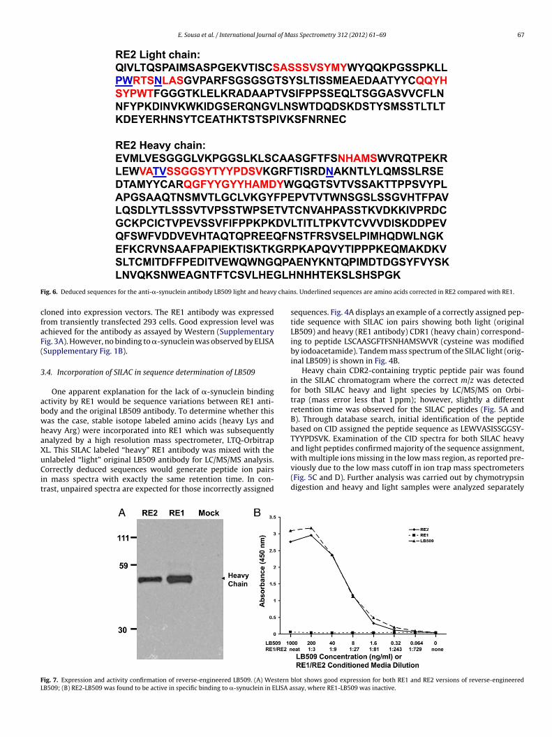
ig. 6. Deduced sequences for the anti-�-synuclein antibody LB509 light and heavy
loned into expression vectors. The RE1 antibody was expressedrom transiently transfected 293 cells. Good expression level waschieved for the antibody as assayed by Western (Supplementaryig. 3A). However, no binding to �-synuclein was observed by ELISASupplementary Fig. 1B).
.4. Incorporation of SILAC in sequence determination of LB509
One apparent explanation for the lack of �-synuclein bindingctivity by RE1 would be sequence variations between RE1 anti-ody and the original LB509 antibody. To determine whether thisas the case, stable isotope labeled amino acids (heavy Lys andeavy Arg) were incorporated into RE1 which was subsequentlynalyzed by a high resolution mass spectrometer, LTQ-OrbitrapL. This SILAC labeled “heavy” RE1 antibody was mixed with the
nlabeled “light” original LB509 antibody for LC/MS/MS analysis.orrectly deduced sequences would generate peptide ion pairsn mass spectra with exactly the same retention time. In con-rast, unpaired spectra are expected for those incorrectly assigned
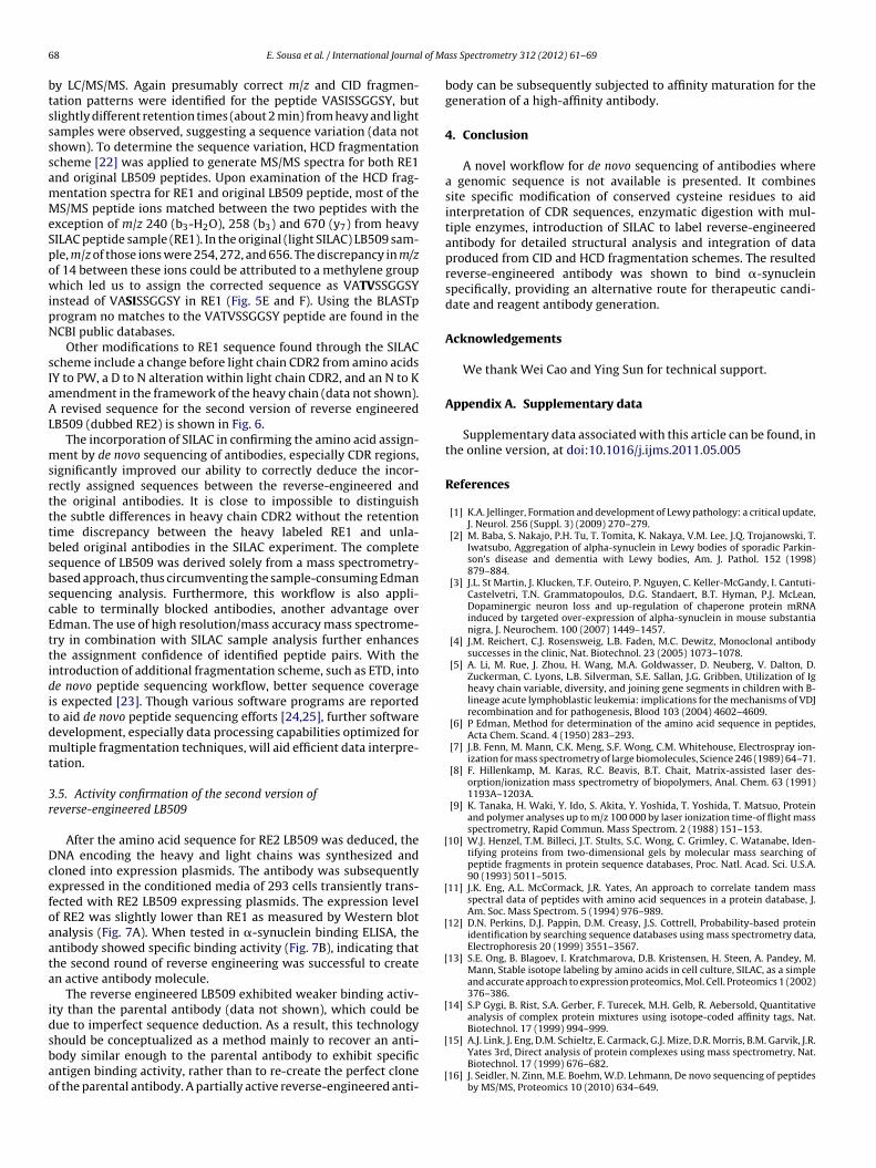
ig. 7. Expression and activity confirmation of reverse-engineered LB509. (A) Western
B509; (B) RE2-LB509 was found to be active in specific binding to �-synuclein in ELISA a
s. Underlined sequences are amino acids corrected in RE2 compared with RE1.
sequences. Fig. 4A displays an example of a correctly assigned pep-tide sequence with SILAC ion pairs showing both light (originalLB509) and heavy (RE1 antibody) CDR1 (heavy chain) correspond-ing to peptide LSCAASGFTFSNHAMSWVR (cysteine was modifiedby iodoacetamide). Tandem mass spectrum of the SILAC light (orig-inal LB509) is shown in Fig. 4B.
Heavy chain CDR2-containing tryptic peptide pair was foundin the SILAC chromatogram where the correct m/z was detectedfor both SILAC heavy and light species by LC/MS/MS on Orbi-trap (mass error less that 1 ppm); however, slightly a differentretention time was observed for the SILAC peptides (Fig. 5A andB). Through database search, initial identification of the peptidebased on CID assigned the peptide sequence as LEWVASISSGGSY-TYYPDSVK. Examination of the CID spectra for both SILAC heavyand light peptides confirmed majority of the sequence assignment,
with multiple ions missing in the low mass region, as reported pre-viously due to the low mass cutoff in ion trap mass spectrometers(Fig. 5C and D). Further analysis was carried out by chymotrypsindigestion and heavy and light samples were analyzed separatelyblot shows good expression for both RE1 and RE2 versions of reverse-engineeredssay, where RE1-LB509 was inactive.
6 l of Ma
btssssamMeSpowipN
sIaAL
msrtttbsbscEttiditdmt
3r
Dcefoaata
idsbao
[
[
[
[
[
8 E. Sousa et al. / International Journa
y LC/MS/MS. Again presumably correct m/z and CID fragmen-ation patterns were identified for the peptide VASISSGGSY, butlightly different retention times (about 2 min) from heavy and lightamples were observed, suggesting a sequence variation (data nothown). To determine the sequence variation, HCD fragmentationcheme [22] was applied to generate MS/MS spectra for both RE1nd original LB509 peptides. Upon examination of the HCD frag-entation spectra for RE1 and original LB509 peptide, most of theS/MS peptide ions matched between the two peptides with the
xception of m/z 240 (b3-H2O), 258 (b3) and 670 (y7) from heavyILAC peptide sample (RE1). In the original (light SILAC) LB509 sam-le, m/z of those ions were 254, 272, and 656. The discrepancy in m/zf 14 between these ions could be attributed to a methylene grouphich led us to assign the corrected sequence as VATVSSGGSY
nstead of VASISSGGSY in RE1 (Fig. 5E and F). Using the BLASTprogram no matches to the VATVSSGGSY peptide are found in theCBI public databases.
Other modifications to RE1 sequence found through the SILACcheme include a change before light chain CDR2 from amino acidsY to PW, a D to N alteration within light chain CDR2, and an N to Kmendment in the framework of the heavy chain (data not shown).
revised sequence for the second version of reverse engineeredB509 (dubbed RE2) is shown in Fig. 6.
The incorporation of SILAC in confirming the amino acid assign-ent by de novo sequencing of antibodies, especially CDR regions,
ignificantly improved our ability to correctly deduce the incor-ectly assigned sequences between the reverse-engineered andhe original antibodies. It is close to impossible to distinguishhe subtle differences in heavy chain CDR2 without the retentionime discrepancy between the heavy labeled RE1 and unla-eled original antibodies in the SILAC experiment. The completeequence of LB509 was derived solely from a mass spectrometry-ased approach, thus circumventing the sample-consuming Edmanequencing analysis. Furthermore, this workflow is also appli-able to terminally blocked antibodies, another advantage overdman. The use of high resolution/mass accuracy mass spectrome-ry in combination with SILAC sample analysis further enhanceshe assignment confidence of identified peptide pairs. With thentroduction of additional fragmentation scheme, such as ETD, intoe novo peptide sequencing workflow, better sequence coverages expected [23]. Though various software programs are reportedo aid de novo peptide sequencing efforts [24,25], further softwareevelopment, especially data processing capabilities optimized forultiple fragmentation techniques, will aid efficient data interpre-
ation.
.5. Activity confirmation of the second version ofeverse-engineered LB509
After the amino acid sequence for RE2 LB509 was deduced, theNA encoding the heavy and light chains was synthesized andloned into expression plasmids. The antibody was subsequentlyxpressed in the conditioned media of 293 cells transiently trans-ected with RE2 LB509 expressing plasmids. The expression levelf RE2 was slightly lower than RE1 as measured by Western blotnalysis (Fig. 7A). When tested in �-synuclein binding ELISA, thentibody showed specific binding activity (Fig. 7B), indicating thathe second round of reverse engineering was successful to createn active antibody molecule.
The reverse engineered LB509 exhibited weaker binding activ-ty than the parental antibody (data not shown), which could beue to imperfect sequence deduction. As a result, this technology
hould be conceptualized as a method mainly to recover an anti-ody similar enough to the parental antibody to exhibit specificntigen binding activity, rather than to re-create the perfect clonef the parental antibody. A partially active reverse-engineered anti-[
[
ss Spectrometry 312 (2012) 61– 69
body can be subsequently subjected to affinity maturation for thegeneration of a high-affinity antibody.
4. Conclusion
A novel workflow for de novo sequencing of antibodies wherea genomic sequence is not available is presented. It combinessite specific modification of conserved cysteine residues to aidinterpretation of CDR sequences, enzymatic digestion with mul-tiple enzymes, introduction of SILAC to label reverse-engineeredantibody for detailed structural analysis and integration of dataproduced from CID and HCD fragmentation schemes. The resultedreverse-engineered antibody was shown to bind �-synucleinspecifically, providing an alternative route for therapeutic candi-date and reagent antibody generation.
Acknowledgements
We thank Wei Cao and Ying Sun for technical support.
Appendix A. Supplementary data
Supplementary data associated with this article can be found, inthe online version, at doi:10.1016/j.ijms.2011.05.005
References
[1] K.A. Jellinger, Formation and development of Lewy pathology: a critical update,J. Neurol. 256 (Suppl. 3) (2009) 270–279.
[2] M. Baba, S. Nakajo, P.H. Tu, T. Tomita, K. Nakaya, V.M. Lee, J.Q. Trojanowski, T.Iwatsubo, Aggregation of alpha-synuclein in Lewy bodies of sporadic Parkin-son’s disease and dementia with Lewy bodies, Am. J. Pathol. 152 (1998)879–884.
[3] J.L. St Martin, J. Klucken, T.F. Outeiro, P. Nguyen, C. Keller-McGandy, I. Cantuti-Castelvetri, T.N. Grammatopoulos, D.G. Standaert, B.T. Hyman, P.J. McLean,Dopaminergic neuron loss and up-regulation of chaperone protein mRNAinduced by targeted over-expression of alpha-synuclein in mouse substantianigra, J. Neurochem. 100 (2007) 1449–1457.
[4] J.M. Reichert, C.J. Rosensweig, L.B. Faden, M.C. Dewitz, Monoclonal antibodysuccesses in the clinic, Nat. Biotechnol. 23 (2005) 1073–1078.
[5] A. Li, M. Rue, J. Zhou, H. Wang, M.A. Goldwasser, D. Neuberg, V. Dalton, D.Zuckerman, C. Lyons, L.B. Silverman, S.E. Sallan, J.G. Gribben, Utilization of Igheavy chain variable, diversity, and joining gene segments in children with B-lineage acute lymphoblastic leukemia: implications for the mechanisms of VDJrecombination and for pathogenesis, Blood 103 (2004) 4602–4609.
[6] P Edman, Method for determination of the amino acid sequence in peptides,Acta Chem. Scand. 4 (1950) 283–293.
[7] J.B. Fenn, M. Mann, C.K. Meng, S.F. Wong, C.M. Whitehouse, Electrospray ion-ization for mass spectrometry of large biomolecules, Science 246 (1989) 64–71.
[8] F. Hillenkamp, M. Karas, R.C. Beavis, B.T. Chait, Matrix-assisted laser des-orption/ionization mass spectrometry of biopolymers, Anal. Chem. 63 (1991)1193A–1203A.
[9] K. Tanaka, H. Waki, Y. Ido, S. Akita, Y. Yoshida, T. Yoshida, T. Matsuo, Proteinand polymer analyses up to m/z 100 000 by laser ionization time-of flight massspectrometry, Rapid Commun. Mass Spectrom. 2 (1988) 151–153.
10] W.J. Henzel, T.M. Billeci, J.T. Stults, S.C. Wong, C. Grimley, C. Watanabe, Iden-tifying proteins from two-dimensional gels by molecular mass searching ofpeptide fragments in protein sequence databases, Proc. Natl. Acad. Sci. U.S.A.90 (1993) 5011–5015.
11] J.K. Eng, A.L. McCormack, J.R. Yates, An approach to correlate tandem massspectral data of peptides with amino acid sequences in a protein database, J.Am. Soc. Mass Spectrom. 5 (1994) 976–989.
12] D.N. Perkins, D.J. Pappin, D.M. Creasy, J.S. Cottrell, Probability-based proteinidentification by searching sequence databases using mass spectrometry data,Electrophoresis 20 (1999) 3551–3567.
13] S.E. Ong, B. Blagoev, I. Kratchmarova, D.B. Kristensen, H. Steen, A. Pandey, M.Mann, Stable isotope labeling by amino acids in cell culture, SILAC, as a simpleand accurate approach to expression proteomics, Mol. Cell. Proteomics 1 (2002)376–386.
14] S.P Gygi, B. Rist, S.A. Gerber, F. Turecek, M.H. Gelb, R. Aebersold, Quantitativeanalysis of complex protein mixtures using isotope-coded affinity tags, Nat.Biotechnol. 17 (1999) 994–999.
15] A.J. Link, J. Eng, D.M. Schieltz, E. Carmack, G.J. Mize, D.R. Morris, B.M. Garvik, J.R.Yates 3rd, Direct analysis of protein complexes using mass spectrometry, Nat.Biotechnol. 17 (1999) 676–682.
16] J. Seidler, N. Zinn, M.E. Boehm, W.D. Lehmann, De novo sequencing of peptidesby MS/MS, Proteomics 10 (2010) 634–649.

l of Ma
[
[
[
[
[
[
[
[
E. Sousa et al. / International Journa
17] V. Pham, W.J. Henzel, D. Arnott, S. Hymowitz, W.N. Sandoval, B.-T. Truong, H.Lowman, J.R. Lill, De novo proteomic sequencing of a monoclonal antibodyraised against OX40 ligand, Anal. Biochem. 352 (2006) 77–86.
18] I. Perdivara, L. Deterding, A. Moise, K.B. Tomer, M. Przybylski, Determinationof primary structure and microheterogeneity of a beta-amyloid plaque-specific antibody using high-performance LC-tandem mass spectrometry, Anal.Bioanal. Chem. 391 (2008) 325–336.
19] N. Bandeira, V. Pham, P. Pevzner, D. Arnott, J.R. Lill, Automated de novo proteinsequencing of monoclonal antibodies, Nat. Biotechnol. 26 (2008) 1336–1338.
20] D.R. Livesay, S. Subramaniam, Conserved sequence and structure association
motifs in antibody-protein and antibody-hapten complexes, Protein Eng. Des.Sel. 17 (2004) 463–472.21] M. Thevis, R.R. Ogorzalek Loo, J.A. Loo, In-gel derivatization of proteins forcysteine-specific cleavages and their analysis by mass spectrometry, J. Pro-teome Res. 2 (2003) 163–172.
[
ss Spectrometry 312 (2012) 61– 69 69
22] J.V. Olsen, B. Macek, O. Lange, A. Makarov, S. Horning, M. Mann, Higher-energyC-trap dissociation for peptide modification analysis, Nat. Methods 4 (2007)709–712.
23] A. Bertsch, A. Leinenbach, A. Pervukhin, M. Lubeck, R. Hartmer, C. Baess-mann, Y.A. Elnakady, R. Muller, S. Bocker, C.G. Huber, O. Kohlbacher,De novo peptide sequencing by tandem MS using complementaryCID and electron transfer dissociation, Electrophoresis 30 (2009)3736–3747.
24] H. Chi, R.X. Sun, B. Yang, C.Q. Song, L.H. Wang, C. Liu, Y. Fu, Z.F.Yuan, H.P. Wang, S.M. He, M.Q. Dong, pNovo: de novo peptide sequenc-
ing and identification using HCD spectra, J. Proteome Res. 9 (2010)2713–2724.25] L. Mo, D. Dutta, Y. Wan, T. Chen, MSNovo: a dynamic programming algorithmfor de novo peptide sequencing via tandem mass spectrometry, Anal. Chem. 79(2007) 4870–4878.