Generation of stable monoclonal antibody–producing B cell receptor–positive human memory B cells...
21
Generation of stable monoclonal antibody-producing BCR + human memory B cells by genetic programming Mark J. Kwakkenbos 1,2,* , Sean A. Diehl 2,3,* , Etsuko Yasuda 1,2 , Arjen Q. Bakker 1,2 , Caroline M.M. van Geelen 1,2 , Michaël V. Lukens 4 , Grada M. van Bleek 4 , Myra N. Widjojoatmodjo 5 , Willy M.J.M. Bogers 6 , Henrik Mei 7 , Andreas Radbruch 7 , Ferenc A. Scheeren 2,8 , Hergen Spits 1,2,9 , and Tim Beaumont 1,2 1 AIMM Therapeutics, Academic Medical Center, Amsterdam, The Netherlands 2 Department of Cell Biology and Histology, Academic Medical Center, Amsterdam, The Netherlands 4 Department of Pediatrics, The Wilhelmina Children's Hospital, University Medical Center, Utrecht 5 Netherlands Vaccine Institute, Bilthoven 6 Department of Virology, Biomedical Primate Research Centre, Rijswijk, The Netherlands 7 German Rheumatism Research Center, Berlin, Germany Abstract B cell lymphoma (BCL)6 and Bcl-xL are expressed in germinal center (GC) B cells and enable them to endure the proliferative and mutagenic environment of the GC. By introducing these genes into peripheral blood memory B cells and culturing these cells with factors produced by follicular helper T cells, CD40L and IL-21, we convert them to highly proliferating, cell surface BCR positive, Ig-secreting B cells with features of GC B cells including expression of activation- induced cytidine deaminase. We generated cloned lines of B cells specific for respiratory syncytial virus and used these cells as a source of antibodies that effectively neutralized this virus in vivo. This method provides a new tool to study GC B cell biology, signal transduction through antigen- specific B cell receptors, and for the rapid generation of high affinity human monoclonal antibodies. Introduction Stable monoclonal cell lines of immortalized human B cells that express the B cell receptor (BCR) on their cell surface while secreting antibodies would be attractive tools for studying various aspects of BCR signaling but also for the generation of human monoclonal antibodies. BCR expression on polyclonal immortalized human B cells would facilitate selection of antigen–specific cells on basis of binding of antigen to the specific BCR, while production of antibodies enables selection of B cell clones on the basis of functional activities of the secreted antibodies. While naïve and memory B cells express cell surface Correspondence: Tim Beaumont: [email protected]. 3 Current address: Department of Medicine, University of Vermont, Burlington, VT, USA 8 Current address: Stanford Institute for Stem Cell Biology and Regenerative Medicine, Stanford University, Palo Alto, CA, USA 9 Current address: Department of Immunology, Genentech, South San Francisco, CA, USA. * These authors contributed equally to this work. Financial interests SAD, FAS, HS and TB are listed on patent applications describing this technology. Author contributions MJK, SAD, and TB performed experiments, analyzed data and wrote the paper. HS and TB organized research. EY, AQB and CVG performed experiments. MVL, GMvB, HM, AR and WMJMB contributed valuable reagents and developed assays. MNW performed cotton rat experiments, FAS analyzed data and provided valuable suggestions. NIH Public Access Author Manuscript Nat Med. Author manuscript; available in PMC 2010 July 01. Published in final edited form as: Nat Med. 2010 January ; 16(1): 123–128. doi:10.1038/nm.2071. NIH-PA Author Manuscript NIH-PA Author Manuscript NIH-PA Author Manuscript
-
Upload
independent -
Category
Documents
-
view
3 -
download
0
Transcript of Generation of stable monoclonal antibody–producing B cell receptor–positive human memory B cells...
Generation of stable monoclonal antibody-producing BCR+
human memory B cells by genetic programming
Mark J. Kwakkenbos1,2,*, Sean A. Diehl2,3,*, Etsuko Yasuda1,2, Arjen Q. Bakker1,2, CarolineM.M. van Geelen1,2, Michaël V. Lukens4, Grada M. van Bleek4, Myra N. Widjojoatmodjo5,Willy M.J.M. Bogers6, Henrik Mei7, Andreas Radbruch7, Ferenc A. Scheeren2,8, HergenSpits1,2,9, and Tim Beaumont1,2
1AIMM Therapeutics, Academic Medical Center, Amsterdam, The Netherlands 2Department ofCell Biology and Histology, Academic Medical Center, Amsterdam, The Netherlands 4Departmentof Pediatrics, The Wilhelmina Children's Hospital, University Medical Center, Utrecht5Netherlands Vaccine Institute, Bilthoven 6Department of Virology, Biomedical Primate ResearchCentre, Rijswijk, The Netherlands 7German Rheumatism Research Center, Berlin, Germany
AbstractB cell lymphoma (BCL)6 and Bcl-xL are expressed in germinal center (GC) B cells and enablethem to endure the proliferative and mutagenic environment of the GC. By introducing thesegenes into peripheral blood memory B cells and culturing these cells with factors produced byfollicular helper T cells, CD40L and IL-21, we convert them to highly proliferating, cell surfaceBCR positive, Ig-secreting B cells with features of GC B cells including expression of activation-induced cytidine deaminase. We generated cloned lines of B cells specific for respiratory syncytialvirus and used these cells as a source of antibodies that effectively neutralized this virus in vivo.This method provides a new tool to study GC B cell biology, signal transduction through antigen-specific B cell receptors, and for the rapid generation of high affinity human monoclonalantibodies.
IntroductionStable monoclonal cell lines of immortalized human B cells that express the B cell receptor(BCR) on their cell surface while secreting antibodies would be attractive tools for studyingvarious aspects of BCR signaling but also for the generation of human monoclonalantibodies. BCR expression on polyclonal immortalized human B cells would facilitateselection of antigen–specific cells on basis of binding of antigen to the specific BCR, whileproduction of antibodies enables selection of B cell clones on the basis of functionalactivities of the secreted antibodies. While naïve and memory B cells express cell surface
Correspondence: Tim Beaumont: [email protected] address: Department of Medicine, University of Vermont, Burlington, VT, USA8Current address: Stanford Institute for Stem Cell Biology and Regenerative Medicine, Stanford University, Palo Alto, CA, USA9Current address: Department of Immunology, Genentech, South San Francisco, CA, USA.*These authors contributed equally to this work.
Financial interestsSAD, FAS, HS and TB are listed on patent applications describing this technology.
Author contributionsMJK, SAD, and TB performed experiments, analyzed data and wrote the paper. HS and TB organized research. EY, AQB and CVGperformed experiments. MVL, GMvB, HM, AR and WMJMB contributed valuable reagents and developed assays. MNW performedcotton rat experiments, FAS analyzed data and provided valuable suggestions.
NIH Public AccessAuthor ManuscriptNat Med. Author manuscript; available in PMC 2010 July 01.
Published in final edited form as:Nat Med. 2010 January ; 16(1): 123–128. doi:10.1038/nm.2071.
NIH
-PA Author Manuscript
NIH
-PA Author Manuscript
NIH
-PA Author Manuscript

BCR, they do not secrete Ig. Cells that simultaneously express the BCR and secreteantibodies might be present in the light zone of the germinal center (GC). These cells mayrepresent plasmablasts ready to be selected into the plasma cell compartment 1.
The GC consists of two areas, the dark and the light zone, which are populated bycentroblasts and centrocytes, respectively 2. Antigen-activated naïve and memory B cells inthe GC undergo extensive proliferation, accompanied by somatic hypermutations (SHM)and class-switch recombination (CSR) of Ig genes, processes both mediated by activationinduced cytidine deaminase (AID) 2–4. Light zone GC B cells then undergo selection viaBCR–antigen interaction on follicular dendritic cells 5 and receive help from follicular Thelper cells, ultimately developing into memory or antibody-secreting plasma cells 6. Theseselected light zone GC B cells express Bcl-xL, a member of the anti apoptotic Bcl-2 proteinfamily 7 most likely to protect them against cell death.
Mature B cells can be cultured in vitro under conditions which mimic some key aspects ofthe GC reaction; that is, activation of B cells with CD40 ligand (L) and the presence ofcytokines like interleukin (IL)-4, IL-10 or IL-21 6,8,9. While B cells cultured with CD40L,IL-2 and IL-4 produce very little Ig, addition of IL-21 leads to differentiation to plasma cellsaccompanied by high Ig secretion 10,11. Although this in vitro system has proven useful tostudy some aspects of B cell differentiation, both naïve IgD+ B cells and switched IgD−
memory B cells eventually differentiate into terminally differentiated plasma cells, which isaccompanied by cell cycle arrest precluding the generation of long-term antigen-specificBCR positive cell lines.
Recent advances have provided insight into how multiple transcription factors, including B-lymphocyte-induced maturation protein 1 (BLIMP1), X-box-binding protein 1 (XBP1) and,B-cell lymphoma (BCL)6 control development of GC B cells into terminally arrested,antibody-producing plasma cells 12. The transcriptional repressor BCL6 has been shown toprevent plasma cell differentiation in GC B cells, were it facilitates expansion of B cells bydownregulating p53 and prevents premature differentiation of B cells into plasma cell bynegatively regulating BLIMP1 11,13,14,15,16.
Recently we reported that signal transducer and activator of transcription (STAT)5 isactivated in a small subset of human GC B cells. These cells expressed relatively high levelsof BCL6 and low levels of BLIMP1 17. Forced expression of constitutive active STAT5 inB cells induced expression of both BCL6 and Bcl-xL and we provided evidence that BCL6is a direct target of STAT5 in B cells. BCL6 regulates terminal differentiation facilitates invitro expansion of B cells since ectopic expression of BCL6 in B cells inhibited terminaldifferentiation in vitro 17,18. This inhibition by BCL6 facilitates in vitro expansion ofactivated B cells 17,19. Here we show that ectopically expressed BCL6 and Bcl-xLsynergize with CD40L and IL-21 to increase the proliferative lifespan of B cells in vitro.The expanded cells have features of GC B cells, express a functional BCR while secretingantibodies, providing a novel and powerful tool to generate human BCR-positive, antibody-secreting B cell lines.
ResultsTransduction of peripheral memory B cells with BCL6 and Bcl-xL
We hypothesized that BCL6 and Bcl-xL, which are expressed in GC B cells and are undercontrol of STAT5 17 synergize to increase the proliferative and survival potential of B cellsin vitro. To test this hypothesis we isolated human CD27+ memory B cells from peripheralblood and introduced BCL6 and Bcl-xL into these cells by retrovirus-mediated genetransfer. Transduced cells were cultured on irradiated CD40L expressing L cells (CD40L-L
Kwakkenbos et al. Page 2
Nat Med. Author manuscript; available in PMC 2010 July 01.
NIH
-PA Author Manuscript
NIH
-PA Author Manuscript
NIH
-PA Author Manuscript
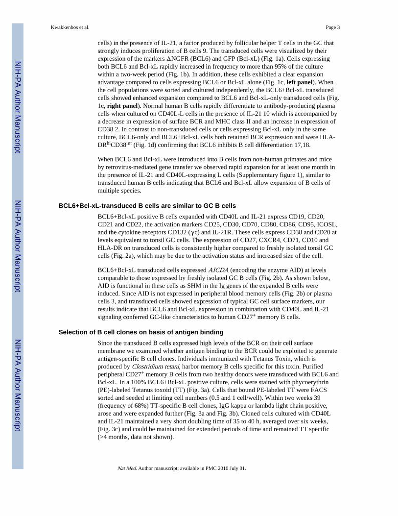
cells) in the presence of IL-21, a factor produced by follicular helper T cells in the GC thatstrongly induces proliferation of B cells 9. The transduced cells were visualized by theirexpression of the markers ΔNGFR (BCL6) and GFP (Bcl-xL) (Fig. 1a). Cells expressingboth BCL6 and Bcl-xL rapidly increased in frequency to more than 95% of the culturewithin a two-week period (Fig. 1b). In addition, these cells exhibited a clear expansionadvantage compared to cells expressing BCL6 or Bcl-xL alone (Fig. 1c, left panel). Whenthe cell populations were sorted and cultured independently, the BCL6+Bcl-xL transducedcells showed enhanced expansion compared to BCL6 and Bcl-xL-only transduced cells (Fig.1c, right panel). Normal human B cells rapidly differentiate to antibody-producing plasmacells when cultured on CD40L-L cells in the presence of IL-21 10 which is accompanied bya decrease in expression of surface BCR and MHC class II and an increase in expression ofCD38 2. In contrast to non-transduced cells or cells expressing Bcl-xL only in the sameculture, BCL6-only and BCL6+Bcl-xL cells both retained BCR expression and were HLA-DRhiCD38int (Fig. 1d) confirming that BCL6 inhibits B cell differentiation 17,18.
When BCL6 and Bcl-xL were introduced into B cells from non-human primates and miceby retrovirus-mediated gene transfer we observed rapid expansion for at least one month inthe presence of IL-21 and CD40L-expressing L cells (Supplementary figure 1), similar totransduced human B cells indicating that BCL6 and Bcl-xL allow expansion of B cells ofmultiple species.
BCL6+Bcl-xL-transduced B cells are similar to GC B cellsBCL6+Bcl-xL positive B cells expanded with CD40L and IL-21 express CD19, CD20,CD21 and CD22, the activation markers CD25, CD30, CD70, CD80, CD86, CD95, ICOSL,and the cytokine receptors CD132 (γc) and IL-21R. These cells express CD38 and CD20 atlevels equivalent to tonsil GC cells. The expression of CD27, CXCR4, CD71, CD10 andHLA-DR on transduced cells is consistently higher compared to freshly isolated tonsil GCcells (Fig. 2a), which may be due to the activation status and increased size of the cell.
BCL6+Bcl-xL transduced cells expressed AICDA (encoding the enzyme AID) at levelscomparable to those expressed by freshly isolated GC B cells (Fig. 2b). As shown below,AID is functional in these cells as SHM in the Ig genes of the expanded B cells wereinduced. Since AID is not expressed in peripheral blood memory cells (Fig. 2b) or plasmacells 3, and transduced cells showed expression of typical GC cell surface markers, ourresults indicate that BCL6 and Bcl-xL expression in combination with CD40L and IL-21signaling conferred GC-like characteristics to human CD27+ memory B cells.
Selection of B cell clones on basis of antigen bindingSince the transduced B cells expressed high levels of the BCR on their cell surfacemembrane we examined whether antigen binding to the BCR could be exploited to generateantigen-specific B cell clones. Individuals immunized with Tetanus Toxin, which isproduced by Clostridium tetani, harbor memory B cells specific for this toxin. Purifiedperipheral CD27+ memory B cells from two healthy donors were transduced with BCL6 andBcl-xL. In a 100% BCL6+Bcl-xL positive culture, cells were stained with phycoerythrin(PE)-labeled Tetanus toxoid (TT) (Fig. 3a). Cells that bound PE-labeled TT were FACSsorted and seeded at limiting cell numbers (0.5 and 1 cell/well). Within two weeks 39(frequency of 68%) TT-specific B cell clones, IgG kappa or lambda light chain positive,arose and were expanded further (Fig. 3a and Fig. 3b). Cloned cells cultured with CD40Land IL-21 maintained a very short doubling time of 35 to 40 h, averaged over six weeks,(Fig. 3c) and could be maintained for extended periods of time and remained TT specific(>4 months, data not shown).
Kwakkenbos et al. Page 3
Nat Med. Author manuscript; available in PMC 2010 July 01.
NIH
-PA Author Manuscript
NIH
-PA Author Manuscript
NIH
-PA Author Manuscript

To examine whether the BCR was functional we cultured IgG+ TT-specific and non-specificB cell lines in the absence of CD40L-L cells or IL-21 for 4 hours. The rested cells were thenstimulated with TT antigen or an IgG F(ab)2 specific antibody and analyzed forphosphorylation (p) of extracellular signal–regulated kinase (ERK) 20. Within 30 secondsafter addition of TT pERK was detected in the TT-specific cells but not in the TT-negativecells (Fig. 3d) indicating the presence of antigen specific functional BCR-coupled signaltransduction machinery in these cells. pERK was detected in both cell lines stimulated withthe IgG F(ab)2 antibody (Fig. 3d). Addition of TT or IgG F(ab)2 antibody to Indo-1-labeledB cells induced a rapid shift in the ratio bound versus unbound Indo-1 indicating that TT andIgG F(ab)2 induce Ca2+ flux (Fig. 3e). These results demonstrate that the BCR expressed onthe cell surface of BCL6+Bcl-xL transduced memory B cells is signaling competent.
Selection of B cell clones secreting neutralizing antibodiesThe IL-21 used to expand CD27+ B cells transduced with BCL6 and Bcl-xL is a stronginducer of differentiation into Ig-secreting cells 10. The transduced cells indeed secreted Ig(Fig. 4a) but the average production of IgG by BCL6+Bcl-xL transduced cells was lowerthan of plasma cells, although it was higher than that of memory B cells (SupplementaryFig. 2). Analysis of the IgG and IgM levels produced by monoclonal cell lines in a 3-dayculture revealed that all clones produced Ig. The IgM-producing clones secreted five to sixtimes more Ig than the IgG-producing clones (Fig. 4a), which has also been observed incultured non-transduced memory cells 21. Although BCL6+Bcl-xL cells cultured withCD40L and IL-21 secrete antibody, they did not downregulate the BCR as do non-transduced in vitro–generated antibody-secreting plasma cells (Supplementary Fig. 2).
Given the relatively high amounts of secreted antibodies by BCL6+Bcl-xL-transduced Bcells we examined whether we could select antigen-specific B cells on the basis of secretionof specific antibody. We chose the pathogenic respiratory syncytial virus (RSV) as theantigenic moiety. RSV is the most common cause of bronchiolitis and pneumonia amonginfants and children under 1 year of age and is a serious health problem for elderly people22. BCL6+Bcl-xL transduced memory B cells of a healthy donor were seeded at 100 cells/well and expanded with CD40L-L cells and IL-21. After 2 weeks of culture, supernatantswere harvested and screened for the presence of RSV-neutralizing antibodies in amicroneutralization experiment 23. Of 384 cultures (100 cells/well), 31 prevented RSVinfection of HEp2 cells. Four microcultures with the highest neutralizing activity weresubcloned by limiting dilution. Four monoclonal cell lines (D25, AM14, AM16, and AM23)were selected and further characterized. We observed median half maximum inhibitoryconcentrations (IC50) against the RSV-A2 virus ranging from 2.1 ng ml−1 for D25 andAM14, 4.3 ng ml−1 for AM23 to 145 ng ml−1 for AM16 (Fig. 4b). By comparison, three ofthe four Abs exhibited significantly lower IC50 values compared to palivizumab (IC50 209ng ml−1), a humanized, FDA-approved monoclonal antibody that is currently used asprophylaxis in infants at high risk 24. In addition, all RSV antibodies efficiently neutralizeda primary RSV isolate X (RSV-X) (Fig. 4c).
D25 antibody inhibits RSV infection in cotton ratsTo test the in vivo neutralizing activity of D25 in a prophylactic setting, we infected cottonrats, the standard model to test RSV infectivity and pathogenicity with the primary isolateRSV-X 25. Animals were injected i.m. with various quantities of recombinant D25 (0.6,0.15 and 0.04 mg kg−1), palivizumab (2.0 and 0.6 mg kg−1) or control IgG1 (2.0 mg kg−1)one day before intranasal inoculation with RSV-X. Animals were sacrificed 5 days postchallenge, since at this time point peak lung virus titers were observed 26. RSV-X titers inthe lung were analyzed by standard TCID50 culture assay (Fig 4d). While palivizumabcompletely prevented in vivo viral replication at 2.0 mg kg−1, D25 showed the same efficacy
Kwakkenbos et al. Page 4
Nat Med. Author manuscript; available in PMC 2010 July 01.
NIH
-PA Author Manuscript
NIH
-PA Author Manuscript
NIH
-PA Author Manuscript
already at a dose of 0.6 mg kg−1 and was partially neutralizing at a dose of 0.15 mg kg−1.These data show that also in vivo D25 is functional and potent in reducing RSV replication.
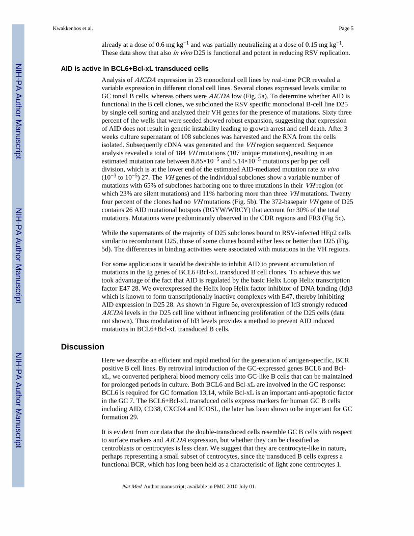
AID is active in BCL6+Bcl-xL transduced cellsAnalysis of AICDA expression in 23 monoclonal cell lines by real-time PCR revealed avariable expression in different clonal cell lines. Several clones expressed levels similar toGC tonsil B cells, whereas others were AICDA low (Fig. 5a). To determine whether AID isfunctional in the B cell clones, we subcloned the RSV specific monoclonal B-cell line D25by single cell sorting and analyzed their VH genes for the presence of mutations. Sixty threepercent of the wells that were seeded showed robust expansion, suggesting that expressionof AID does not result in genetic instability leading to growth arrest and cell death. After 3weeks culture supernatant of 108 subclones was harvested and the RNA from the cellsisolated. Subsequently cDNA was generated and the VH region sequenced. Sequenceanalysis revealed a total of 184 VH mutations (107 unique mutations), resulting in anestimated mutation rate between 8.85×10⁻⁵ and 5.14×10⁻⁵ mutations per bp per celldivision, which is at the lower end of the estimated AID-mediated mutation rate in vivo(10−3 to 10−5) 27. The VH genes of the individual subclones show a variable number ofmutations with 65% of subclones harboring one to three mutations in their VH region (ofwhich 23% are silent mutations) and 11% harboring more than three VH mutations. Twentyfour percent of the clones had no VH mutations (Fig. 5b). The 372-basepair VH gene of D25contains 26 AID mutational hotspots (RGYW/WRCY) that account for 30% of the totalmutations. Mutations were predominantly observed in the CDR regions and FR3 (Fig 5c).
While the supernatants of the majority of D25 subclones bound to RSV-infected HEp2 cellssimilar to recombinant D25, those of some clones bound either less or better than D25 (Fig.5d). The differences in binding activities were associated with mutations in the VH regions.
For some applications it would be desirable to inhibit AID to prevent accumulation ofmutations in the Ig genes of BCL6+Bcl-xL transduced B cell clones. To achieve this wetook advantage of the fact that AID is regulated by the basic Helix Loop Helix transcriptionfactor E47 28. We overexpressed the Helix loop Helix factor inhibitor of DNA binding (Id)3which is known to form transcriptionally inactive complexes with E47, thereby inhibitingAID expression in D25 28. As shown in Figure 5e, overexpression of Id3 strongly reducedAICDA levels in the D25 cell line without influencing proliferation of the D25 cells (datanot shown). Thus modulation of Id3 levels provides a method to prevent AID inducedmutations in BCL6+Bcl-xL transduced B cells.
DiscussionHere we describe an efficient and rapid method for the generation of antigen-specific, BCRpositive B cell lines. By retroviral introduction of the GC-expressed genes BCL6 and Bcl-xL, we converted peripheral blood memory cells into GC-like B cells that can be maintainedfor prolonged periods in culture. Both BCL6 and Bcl-xL are involved in the GC response:BCL6 is required for GC formation 13,14, while Bcl-xL is an important anti-apoptotic factorin the GC 7. The BCL6+Bcl-xL transduced cells express markers for human GC B cellsincluding AID, CD38, CXCR4 and ICOSL, the later has been shown to be important for GCformation 29.
It is evident from our data that the double-transduced cells resemble GC B cells with respectto surface markers and AICDA expression, but whether they can be classified ascentroblasts or centrocytes is less clear. We suggest that they are centrocyte-like in nature,perhaps representing a small subset of centrocytes, since the transduced B cells express afunctional BCR, which has long been held as a characteristic of light zone centrocytes 1.
Kwakkenbos et al. Page 5
Nat Med. Author manuscript; available in PMC 2010 July 01.
NIH
-PA Author Manuscript
NIH
-PA Author Manuscript
NIH
-PA Author Manuscript
This BCR participates in the selection by follicular dendritic cells which occurs in the lightzone 5. Moreover, expansion of these B cells is depended upon CD40 signaling and the darkzone is mostly devoid of CD154+ (CD40L) T cells, while these activated T cells are presentin the light zone. In addition, nuclear translocation of NF-kB, which is induced by CD40signaling, has only been observed in B cells in the light zone 30.
The expression of AICDA in the cell lines raised the question whether AID is functional.Although AID was expressed in all clones that were derived from BCL6+Bcl-xL transducedCD27+ peripheral blood memory cells, the level was variable. Moreover we did not observeisotype switching in these cells over time, indicating that AICDA expression by itself isinsufficient to drive this process at least in the time frame and culture conditions we applied.In contrast we did observe the occurrence of somatic hypermutations at relative lowfrequency in subclones of one RSV-specific B cell clone. Screening of the mAb containingculture supernatant of these subclones on RSV infected cells demonstrated variations inbinding capacity. This result suggested that the additional VH gene mutations of somesubclones resulted in changes in the antigen-binding affinity. These data raise the possibilitythat ongoing AID-mediated mutational activity in B cell clones can be used to select higher(or lower) affinity clones obviating the need for deliberate molecular engineering of therelevant Ig genes. It is conceivable that in some cases mutations in the Ig genes ofestablished cloned B cell lines need to be avoided. We show that following introduction ofId3 into B cells that already overexpress BCL6 and Bcl-xL AICDA levels are stronglyreduced consistent with findings published by Sayeg et al. 28. Thus overexpression of Id3should prevent ongoing AID-mediated mutational activity, if a frozen VH/VL repertoire isrequired. Despite the expression of AICDA in BCL6 and Bcl-xL-transduced IgG, IgM andIgA memory B cells we did not observe CSR in these cells (data not shown). This might berelated to the different requirements for AID-mediated SHM and CSR 31,32.
We previously showed that IL-21 was able to elicit Ig secretion from polyclonal BCL6-expressing B cells 18. In combination with the stable BCR expression, which allows forselection of antigen specific B cells by binding of fluorescent-labeled antigen, Ig secretionallows for functional screening of secreted Ig. Both methods provide a tool for rapidisolation of potential therapeutic antibodies. Indeed we were able to generate unique RSVneutralizing antibodies within 3 months by direct functional screens followingimmortalization of B cells obtained from an RSV-exposed individual.
Recently another method of generating monoclonal antibodies from human B cells wasdescribed based on Epstein-Barr virus (EBV) transformation of B cells activated with Tolllike receptor-9 antagonists 33. Both the method described by Traggiai et al. and ours utilizehuman B cells of exposed individuals to select monoclonal antibodies against a variety ofmicrobes without the need of recent vaccination of the donors. However, in contrast to Bcells transformed by EBV 34 all B cell lines obtained with BCL6+Bcl-xL geneticprogramming stably express high levels of a signaling-competent, membrane-bound BCR.This high level of cell surface BCR facilitates isolation of B cells on the basis of antigenbinding which greatly increases the frequency of antigen specific cells and abrogates theneed for extensive subcloning. Whereas the generation of antigen specific clones using theEBV method takes 3 months 33, the genetic programming approach reduces this to 1 month.The fact that rare B cells can be easily cloned compares our method favorably with arecently described single cell cloning approach used to isolate influenza-specific antibodies35. In this study, Ig DNA was isolated 7 days after influenza vaccination from a populationof CD27highCD38high B cells which contained a relatively high frequency of only severalInfluenza virus-specific B cells. The feasibility of this technique depends on a relative highfrequency of antigen-specific B cells, that can be obtained by vaccination or very recentexposure to a microbe 35,36. As discussed above expression of AID in the genetically
Kwakkenbos et al. Page 6
Nat Med. Author manuscript; available in PMC 2010 July 01.
NIH
-PA Author Manuscript
NIH
-PA Author Manuscript
NIH
-PA Author Manuscript
modified clones may be utilized to select antibodies with higher affinity providing anadvantage compared to the EBV transformation and single cell PCR methods as platforms togenerate human monoclonal antibodies.
Another advantage of the method described here is that it can be used to obtain stable linesof B cells from species other than humans. We demonstrate that BCL6+Bcl-xL-transducedB cells of mice and non-human primates proliferate for at least one month in the presence ofCD40L and IL-21. We also generated a series of cloned lines of mouse B cells (results notshown). It is therefore feasible to assume that our method will allow for expansion ofantigen-specific B cells of multiple species including rabbits and goats. Such lines cannot beobtained by using EBV, which is only capable of transforming B cells of a variety ofmonkey species such as Chimpanzee, Marmoset, Squirrel, Owl and Cebus monkeys 37.
The monoclonal cell lines that can be generated with this technology are very useful notonly for generation of monoclonal antibodies, but also allow for detailed mechanistic studiesof BCR-mediated signal transduction induced by specific antigens. They may be used tostudy signaling of BCR variants that differ in affinity for antigen. Furthermore, these celllines can be useful tools to study the effects of a variety of external signals like antigen orcytokines on the function of AID and the induction of SHM.
MethodsB cell isolation
We obtained B cells from peripheral blood (Sanquin) by Ficoll separation and CD22 MACSmicrobeads (Miltenyi Biotech). Subsequently we sorted these cells forCD19+CD3−CD27+IgM−IgA− (IgG memory cells) or CD19+CD3−CD27+IgG−IgA− (IgMmemory cells) on a FACSAria (Becton Dickinson). We obtained Tonsil B cells from routinetonsillectomies performed at the Department of Otolaryngology at the Academic MedicalCenter, Amsterdam, The Netherlands. The use of these tissues was approved by the medicalethical committees of the institution and was contingent on informed consent.
Cell cultureWe maintained B cells (2×10⁵ cells ml−1) in IMDM (Gibco) culture medium containing 8%FBS (HyClone), penicillin/streptomycin (Roche) supplemented with recombinant mouseIL-21 (25 ng ml−1, R&D systems) and co-cultured them on γ-irradiated (50Gy) mouse Lcell fibroblasts stably expressing CD40L (CD40L-L cells, 105 cells ml−1). We used rhIL-4(R&D) at 50 ng ml−1, and rhIL-2 at 100 U ml−1. We tested cells routinely by PCR for thepresence of mycoplasma and EBV (data not shown).
Retroviral transductionThe BCL6 and Id3 retroviral constructs were described previously 19,38. We cloned humanBcl-xL cDNA into the LZRS retroviral vector. We performed transfections and productionof Amphotropic and Ecotropic virus by Phoenix packaging cells as described before 17,19and co-transduced memory B cells by retroviruses after activation on CD40L-L cells in thepresence of rmIL-21 for 36 h as before 18, but then we centrifuged cells and virus at roomtemperature for 60 min at 360 × g (1800 RPM).
Flow cytometryWe analyzed stained cells on an LSRII (BD) and processed flow cytometry data withFlowJo software (Tree Star). The antibodies are described in the online supplementarymethods section.
Kwakkenbos et al. Page 7
Nat Med. Author manuscript; available in PMC 2010 July 01.
NIH
-PA Author Manuscript
NIH
-PA Author Manuscript
NIH
-PA Author Manuscript
RT-PCRWe carried out quantitative RT-PCR with a BioRad iCycler and used the 2−(ΔΔCT) methodto calculate relative mRNA expression levels normalized to ACTIN. Primers for AICDA(encoding AID) are described 39.
ELISAWe coated plates with either anti-human IgG or IgM Fc-fragment (JacksonImmunoResearch Laboratories) at 5 µg ml−1 in PBS for 1 h at 37 °C or o/n at 4 °C andwashed them in ELISA wash buffer (PBS, 0.5% Tween-20). 4% milk in PBS was used asblocking agent, before we added serial dilutions of cell culture supernatants and HRP-conjugated detection Abs (dilutions 1:2500 for HRP-conjugated IgG antibody (Jackson),dilution 1:5000 for HRP-conjugated IgM antibody (Jackson)). We used TMB substratesolution (Biosource) for development of the ELISAs.
ImmunoblottingWe lysed cells in Triton Lysis Buffer supplemented with HALT protease inhibitor (Roche)and 1 µM Na2VO4. We separated equal amounts of protein by 10% SDS-PAGE, transferredit to nitrocellulose, and probed using phospho-ERK1/2 antibody (Thr202/Tyr204) (CellSignaling Technologies) and STAT3 antibody (Santa Cruz). We used enhancedchemiluminescence (Pierce Biotechnology) and x-ray film (Hyperfilm, GE Healthcare LifeSciences) for detection.
Ca2+ fluxWe cultured transduced B cells for two days without CD40L-L cells in the presence ofIL-21, subsequently we loaded the cells with 2 µM Indo-1 AM (Sigma-Aldrich).Fluorescence ratios of Indo-1 emission at 405/485 nm were measured by flow cytometry ona FACSVantage SE (BD).
RSV culture and neutralization assayWe seeded 104 HEp2 cells in flat bottom 96 well plates (Costar). The next day we pre-incubated 100 TCID50 of RSV A2 or X virus mixed with B cell culture supernatant for 30min at 37 °C before adding it in triplicate to HEp2 cells for 1 h at 37 °C. After 2 days wefixed the cells with 80% acetone and stained them with polyclonal RSV-HRP antibody(Biodesign) and 3-amino-9-ethylcarbazole for detection and visualization of RSV plaquesby light microscopy (plaques were counted).
Cloning of D25We isolated total RNA with the RNeasy® mini kit (Qiagen), generated cDNA, performedPCR and cloned the heavy and light chain variable regions into the pCR2.1 TA cloningvector (Invitrogen). To rule out reverse transcriptase or DNA polymerase induced mutations,we performed several independent cloning experiments. To produce recombinant D25 mAbwe cloned D25 heavy and light variable regions in frame with human IgG1 and Kappaconstant regions into a pcDNA3.1 (Invitrogen) based vector and transiently transfected 293Tcells. We purified recombinant D25 from the culture supernatant with Protein A.
Cotton rat experiments7–9 week old Cotton rats (Sigmodon hispidus, Charles River) were anesthetized withisoflurane, bled and given 0.1 ml of purified mAb per 100 gram body weight (bw) byintramuscular (i.m.) injection at doses of 2.0 or 0.6 for palivizumab, 0.6, 0.15 or 0.04 forD25 and 2.0 mg kg−1 bw for the control antibody. Twenty-four hours later, animals were
Kwakkenbos et al. Page 8
Nat Med. Author manuscript; available in PMC 2010 July 01.
NIH
-PA Author Manuscript
NIH
-PA Author Manuscript
NIH
-PA Author Manuscript
anesthetized and challenged by intranasal instillation of 106 TCID50 RSV-X (100 µl). Fivedays later animals were sacrificed and their lungs were harvested. Lung virus titers weredetermined and the lowest limit of detection was 2.1 log10TCID50 g−1. The AnimalExperiment Committee of the Netherlands Vaccine Institute approved all proceduresinvolving cotton rats.
Supplementary MaterialRefer to Web version on PubMed Central for supplementary material.
AcknowledgmentsWe thank B. Hooijbrink for his excellent help with FACS sorting and maintenance of the flow cytometry facility, J.Boes for her excellent work on virus titrations at NVI, R. Molenkamp and D. Pajkrt of the Dept. of ClinicalVirology of the AMC for helpful discussions. Human Bcl-xL cDNA was kindly provided by S. Korsmeyer.Howard Hughes Medical Institute, Harvard Medical School, Boston, MA, USA. S.A.D. is supported by NIH-NIAID grant F32AI063846, M.v.L is supported by grant OZF-02-004 of the Wilhelmina Research Fund.
Abbreviations
BCR B cell receptor
RSV respiratory syncytial virus
GC germinal center
CSR class-switch recombination
Ig immunoglobulin
IL interleukin
SHM somatic hypermutation
TT tetanus toxoid
EBV Epstein-Barr virus
AID activation induced cytidine deaminase
BLIMP1 B-lymphocyte-induced maturation protein 1
XBP1 X-box-binding protein 1
BCL6 B-cell lymphoma 6
STAT5 signal transducer and activator of transcription 5
ERK extracellular signal–regulated kinase
Id3 inhibitor of DNA binding 3
References1. Close PM, Pringle JH, Ruprai AK, West KP, Lauder I. Zonal distribution of immunoglobulin-
synthesizing cells within the germinal centre: an in situ hybridization and immunohistochemicalstudy. J Pathol. 1990; 162:209–216. [PubMed: 2125071]
2. Liu YJ, Arpin C. Germinal center development. Immunol Rev. 1997; 156:111–126. [PubMed:9280752]
3. Muramatsu M, et al. Specific expression of activation-induced cytidine deaminase (AID), a novelmember of the RNA-editing deaminase family in germinal center B cells. J Biol Chem. 1999;274:18470–18476. [PubMed: 10373455]
Kwakkenbos et al. Page 9
Nat Med. Author manuscript; available in PMC 2010 July 01.
NIH
-PA Author Manuscript
NIH
-PA Author Manuscript
NIH
-PA Author Manuscript

4. Muramatsu M, et al. Class switch recombination and hypermutation require activation-inducedcytidine deaminase (AID), a potential RNA editing enzyme. Cell. 2000; 102:553–563. [PubMed:11007474]
5. Allen CD, Okada T, Tang HL, Cyster JG. Imaging of germinal center selection events duringaffinity maturation. Science. 2007; 315:528–531. [PubMed: 17185562]
6. Kim CH, et al. Subspecialization of CXCR5+ T cells: B helper activity is focused in a germinalcenter-localized subset of CXCR5+ T cells. J Exp Med. 2001; 193:1373–1381. [PubMed:11413192]
7. Tuscano JM, et al. Bcl-x rather than Bcl-2 mediates CD40-dependent centrocyte survival in thegerminal center. Blood. 1996; 88:1359–1364. [PubMed: 8695854]
8. Banchereau J, de Paoli P, Valle A, Garcia E, Rousset F. Long-term human B cell lines dependent oninterleukin-4 and antibody to CD40. Science. 1991; 251:70–72. [PubMed: 1702555]
9. Good KL, Bryant VL, Tangye SG. Kinetics of human B cell behavior and amplification ofproliferative responses following stimulation with IL-21. J Immunol. 2006; 177:5236–5247.[PubMed: 17015709]
10. Ettinger R, et al. IL-21 induces differentiation of human naive and memory B cells into antibody-secreting plasma cells. J Immunol. 2005; 175:7867–7879. [PubMed: 16339522]
11. Kuo TC, et al. Repression of BCL-6 is required for the formation of human memory B cells invitro. J Exp Med. 2007; 204:819–830. [PubMed: 17403935]
12. Shapiro-Shelef M, et al. Blimp-1 is required for the formation of immunoglobulin secreting plasmacells and pre-plasma memory B cells. Immunity. 2003; 19:607–620. [PubMed: 14563324]
13. Dent AL, Shaffer AL, Yu X, Allman D, Staudt LM. Control of inflammation, cytokine expression,and germinal center formation by BCL-6. Science. 1997; 276:589–592. [PubMed: 9110977]
14. Fukuda T, et al. Disruption of the Bcl6 gene results in an impaired germinal center formation. JExp Med. 1997; 186:439–448. [PubMed: 9236196]
15. Phan RT, Dalla-Favera R. The BCL6 proto-oncogene suppresses p53 expression in germinal-centre B cells. Nature. 2004; 432:635–639. [PubMed: 15577913]
16. Shapiro-Shelef M, Calame K. Regulation of plasma-cell development. Nat Rev Immunol. 2005;5:230–242. [PubMed: 15738953]
17. Scheeren FA, et al. STAT5 regulates the self-renewal capacity and differentiation of humanmemory B cells and controls Bcl-6 expression. Nat Immunol. 2005; 6:303–313. [PubMed:15711548]
18. Diehl SA, et al. STAT3-Mediated Up-Regulation of BLIMP1 Is Coordinated with BCL6 Down-Regulation to Control Human Plasma Cell Differentiation. J Immunol. 2008; 180:4805–4815.[PubMed: 18354204]
19. Shvarts A, et al. A senescence rescue screen identifies BCL6 as an inhibitor of anti-proliferativep19(ARF)-p53 signaling. Genes Dev. 2002; 16:681–686. [PubMed: 11914273]
20. Niiro H, Clark EA. Regulation of B-cell fate by antigen-receptor signals. Nat Rev Immunol. 2002;2:945–956. [PubMed: 12461567]
21. Bryant VL, et al. Cytokine-mediated regulation of human B cell differentiation into Ig-secretingcells: predominant role of IL-21 produced by CXCR5+ T follicular helper cells. J Immunol. 2007;179:8180–8190. [PubMed: 18056361]
22. Hall CB, et al. The burden of respiratory syncytial virus infection in young children. N Engl J Med.2009; 360:588–598. [PubMed: 19196675]
23. Johnson S, et al. A direct comparison of the activities of two humanized respiratory syncytial virusmonoclonal antibodies: MEDI-493 and RSHZl9. J Infect Dis. 1999; 180:35–40. [PubMed:10353858]
24. Sidwell RW, Barnard DL. Respiratory syncytial virus infections: recent prospects for control.Antiviral Res. 2006; 71:379–390. [PubMed: 16806515]
25. Domachowske JB, Bonville CA, Rosenberg HF. Animal models for studying respiratory syncytialvirus infection and its long term effects on lung function. Pediatr Infect Dis J. 2004; 23:S228–S234. [PubMed: 15577578]
Kwakkenbos et al. Page 10
Nat Med. Author manuscript; available in PMC 2010 July 01.
NIH
-PA Author Manuscript
NIH
-PA Author Manuscript
NIH
-PA Author Manuscript
26. Prince GA, Prieels JP, Slaoui M, Porter DD. Pulmonary lesions in primary respiratory syncytialvirus infection, reinfection, and vaccine-enhanced disease in the cotton rat (Sigmodon hispidus).Lab Invest. 1999; 79:1385–1392. [PubMed: 10576209]
27. Peled JU, et al. The biochemistry of somatic hypermutation. Annu Rev Immunol. 2008; 26:481–511. [PubMed: 18304001]
28. Sayegh CE, Quong MW, Agata Y, Murre C. E-proteins directly regulate expression of activation-induced deaminase in mature B cells. Nat Immunol. 2003; 4:586–593. [PubMed: 12717431]
29. Dong C, Temann UA, Flavell RA. Cutting edge: critical role of inducible costimulator in germinalcenter reactions. J Immunol. 2001; 166:3659–3662. [PubMed: 11238604]
30. Basso K, et al. Tracking CD40 signaling during germinal center development. Blood. 2004;104:4088–4096. [PubMed: 15331443]
31. Shinkura R, et al. Separate domains of AID are required for somatic hypermutation and class-switch recombination. Nat Immunol. 2004; 5:707–712. [PubMed: 15195091]
32. McBride KM, et al. Regulation of class switch recombination and somatic mutation by AIDphosphorylation. J Exp Med. 2008; 205:2585–2594. [PubMed: 18838546]
33. Traggiai E, et al. An efficient method to make human monoclonal antibodies from memory B cells:potent neutralization of SARS coronavirus. Nat Med. 2004; 10:871–875. [PubMed: 15247913]
34. Dykstra ML, Longnecker R, Pierce SK. Epstein-Barr virus coopts lipid rafts to block the signalingand antigen transport functions of the BCR. Immunity. 2001; 14:57–67. [PubMed: 11163230]
35. Wrammert J, et al. Rapid cloning of high-affinity human monoclonal antibodies against influenzavirus. Nature. 2008; 453:667–671. [PubMed: 18449194]
36. Zwick MB, Gach JS, Burton DR. A welcome burst of human antibodies. Nat Biotechnol. 2008;26:886–887. [PubMed: 18688242]
37. Deinhardt F, Falk LA, Wolfe LG. Transformation of nonhuman primate lymphocytes by Epstein-Barr virus. Cancer Res. 1974; 34:1241–1244. [PubMed: 4367158]
38. Jaleco AC, et al. Genetic modification of human B-cell development: B-cell development isinhibited by the dominant negative helix loop helix factor Id3. Blood. 1999; 94:2637–2646.[PubMed: 10515867]
39. Smit LA, et al. Expression of activation-induced cytidine deaminase is confined to B-cell non-Hodgkin's lymphomas of germinal-center phenotype. Cancer Res. 2003; 63:3894–3898. [PubMed:12873980]
Kwakkenbos et al. Page 11
Nat Med. Author manuscript; available in PMC 2010 July 01.
NIH
-PA Author Manuscript
NIH
-PA Author Manuscript
NIH
-PA Author Manuscript
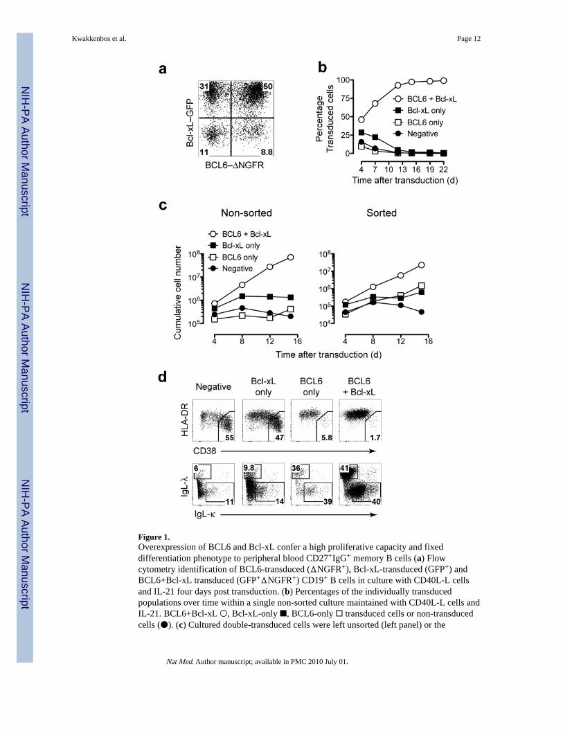
Figure 1.Overexpression of BCL6 and Bcl-xL confer a high proliferative capacity and fixeddifferentiation phenotype to peripheral blood CD27+IgG+ memory B cells (a) Flowcytometry identification of BCL6-transduced (ΔNGFR+), Bcl-xL-transduced (GFP+) andBCL6+Bcl-xL transduced (GFP+ΔNGFR+) CD19+ B cells in culture with CD40L-L cellsand IL-21 four days post transduction. (b) Percentages of the individually transducedpopulations over time within a single non-sorted culture maintained with CD40L-L cells andIL-21. BCL6+Bcl-xL ○, Bcl-xL-only ■, BCL6-only □ transduced cells or non-transducedcells (●). (c) Cultured double-transduced cells were left unsorted (left panel) or the
Kwakkenbos et al. Page 12
Nat Med. Author manuscript; available in PMC 2010 July 01.
NIH
-PA Author Manuscript
NIH
-PA Author Manuscript
NIH
-PA Author Manuscript
individual populations were sorted (right panel) and cultured separately. The cumulative cellnumber was calculated based on the original number of transduced B cells at input andillustrates the theoretical absolute cell numbers produced in culture. (d) CD19+ cells fromnon-sorted double transduced cultures were analyzed for CD38, HLA-DR, and surface Igkappa (IgL-κ) or lambda (IgL-λ) light chain expression. The gates in the upper panel showthe percentage terminally differentiated cells. Phenotype shown of cells 7 days posttransduction. Data are representative of four separate experiments.
Kwakkenbos et al. Page 13
Nat Med. Author manuscript; available in PMC 2010 July 01.
NIH
-PA Author Manuscript
NIH
-PA Author Manuscript
NIH
-PA Author Manuscript
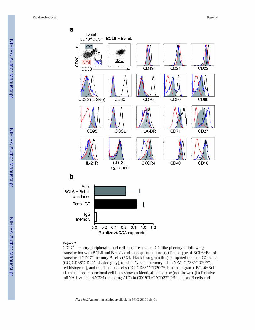
Figure 2.CD27+ memory peripheral blood cells acquire a stable GC-like phenotype followingtransduction with BCL6 and Bcl-xL and subsequent culture. (a) Phenotype of BCL6+Bcl-xLtransduced CD27+ memory B cells (6XL, black histogram line) compared to tonsil GC cells(GC, CD38+CD20+, shaded grey), tonsil naïve and memory cells (N/M, CD38−CD20low,red histogram), and tonsil plasma cells (PC, CD38++CD20low, blue histogram). BCL6+Bcl-xL transduced monoclonal cell lines show an identical phenotype (not shown). (b) RelativemRNA levels of AICDA (encoding AID) in CD19+IgG+CD27+ PB memory B cells and
Kwakkenbos et al. Page 14
Nat Med. Author manuscript; available in PMC 2010 July 01.
NIH
-PA Author Manuscript
NIH
-PA Author Manuscript
NIH
-PA Author Manuscript

CD19+CD38+CD20+IgD− tonsillar GC B cells (value set as 1) compared to BCL6+Bcl-xLtransduced bulk CD27+ memory cells using quantitative 5 RT-PCR.
Kwakkenbos et al. Page 15
Nat Med. Author manuscript; available in PMC 2010 July 01.
NIH
-PA Author Manuscript
NIH
-PA Author Manuscript
NIH
-PA Author Manuscript

Figure 3.Generation and characterization of Tetanus toxoid (TT)-specific monoclonal human B celllines. (a) Cell sorting of TT-specific B cells from bulk CD19+ BCL6+Bcl-xL transducedmemory B cell culture using TT-PE staining. (b) Monoclonal cell lines express surface IgGand are either IgL-λ or IgL-κ light chain positive. Shaded histograms are CD19+ tonsil Bcells. (c) The cumulative cell number (as in Fig. 1c) of two representative B cell clones intime. (d) TT-specific and control TT negative cell lines were incubated with TT or F(ab)2 -human IgG antibody stimulation and phospho-ERK1/2 was determined by immunoblotanalysis. STAT3 blotting was used to verify equal loading. Results are representative of two
Kwakkenbos et al. Page 16
Nat Med. Author manuscript; available in PMC 2010 July 01.
NIH
-PA Author Manuscript
NIH
-PA Author Manuscript
NIH
-PA Author Manuscript

experiments (e) Calcium flux in TT specific IgG positive B cell lines. Cells were loadedwith Indo-1 AM and were subsequently stimulated with TT (dashed black trace), IgG F(ab)2antibody (solid black trace), or control IgG (solid grey trace). The Indo-1 response of a TT-negative clone stimulated with TT antigen is shown as an antigen specificity control (dashedgrey trace). Shown is the ratio bound versus unbound Indo-1. Results are representative ofthree experiments.
Kwakkenbos et al. Page 17
Nat Med. Author manuscript; available in PMC 2010 July 01.
NIH
-PA Author Manuscript
NIH
-PA Author Manuscript
NIH
-PA Author Manuscript
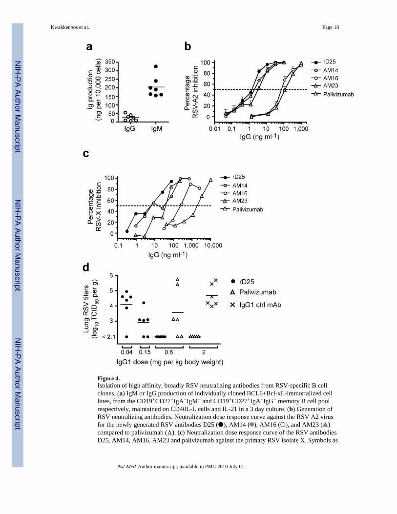
Figure 4.Isolation of high affinity, broadly RSV neutralizing antibodies from RSV-specific B cellclones. (a) IgM or IgG production of individually cloned BCL6+Bcl-xL-immortalized celllines, from the CD19+CD27+IgA−IgM− and CD19+CD27+IgA−IgG− memory B cell poolrespectively, maintained on CD40L-L cells and IL-21 in a 3 day culture. (b) Generation ofRSV neutralizing antibodies. Neutralization dose response curve against the RSV A2 virusfor the newly generated RSV antibodies D25 (●), AM14 ( ), AM16 (○), and AM23 ( )compared to palivizumab (Δ). (c) Neutralization dose response curve of the RSV antibodiesD25, AM14, AM16, AM23 and palivizumab against the primary RSV isolate X. Symbols as
Kwakkenbos et al. Page 18
Nat Med. Author manuscript; available in PMC 2010 July 01.
NIH
-PA Author Manuscript
NIH
-PA Author Manuscript
NIH
-PA Author Manuscript

in 4b (d) Effects of rD25 (●), palivizumab (Δ) and a control IgG1 (×) on RSV replication incotton rat lungs. Antibodies were administered i.m. one day before intranasal challenge withRSV X (106 TCID50/animal). Lung virus titers were calculated per gram of lung tissue;control prophylaxis reached a titer of 4.7 log TCID50 g−1, while the lower limit of detectionwas 2.1 log10TCID50 g−1.
Kwakkenbos et al. Page 19
Nat Med. Author manuscript; available in PMC 2010 July 01.
NIH
-PA Author Manuscript
NIH
-PA Author Manuscript
NIH
-PA Author Manuscript

Figure 5.Expression and activity of AID in BCL6+Bcl-xL transduced cells. (a) mRNA levels ofAICDA (encoding AID) in CD19+CD38+CD20+IgD− tonsillar GC B cells andCD19+IgG+CD27+ PB memory B cells compared to 23 BCL6+Bcl-xL transducedmonoclonal cell lines and monoclonal cell line D25 using quantitative RT-PCR. (b)Percentage of subclones with indicated number of VH mutations, as percentage of totalnumber of subclones sequenced. (c) Location of VH mutations, percentage of mutations perVH region per basepair. (d) Binding of D25-subclone Ig to RSV infected HEp2 cells. Redsquares are individual D25 subclones, black circles rD25. Blue circles indicate clones with
Kwakkenbos et al. Page 20
Nat Med. Author manuscript; available in PMC 2010 July 01.
NIH
-PA Author Manuscript
NIH
-PA Author Manuscript
NIH
-PA Author Manuscript

deviating binding activity. (e) mRNA levels of AICDA in the monoclonal cell line D25transduced with Control-YFP or Id3-YFP using quantitative RT-PCR.
Kwakkenbos et al. Page 21
Nat Med. Author manuscript; available in PMC 2010 July 01.
NIH
-PA Author Manuscript
NIH
-PA Author Manuscript
NIH
-PA Author Manuscript