
Disclosure of The Potential Phytase-Producing Maize ...
50
In: Zea mays L.: Cultivation, and Uses ISBN: 978-1-53619-181-3 Editor: Sarah Dunn © 2021 Nova Science Publishers, Inc. Chapter 1 DISCLOSURE OF THE POTENTIAL PHYTASE-PRODUCING MAIZE ENDOPHYTIC BACTERIA, AS AN INVISIBLE A VAIL FOR ZEA MAYS L. Hafsan * , Cut Muthiadin, Eka Sukmawaty, Nurhikmah and Yuniar Harviyanti Department of Biology, Universitas Islam Negeri Alauddin, South Sulawesi, Indonesia ABSTRACT Zea mays L. is a potential producer of cereal crops and the dominant primary energy source of feed for monogastric animals, such as poultry. The potential energy content of maize seeds, expressed as Metabolizable Energy (ME) is relatively high compared to other feed ingredients. However, they contain phytic acid, which acts in physiological functions * Corresponding Author’s E-mail: [email protected].
-
Upload
khangminh22 -
Category
Documents
-
view
1 -
download
0
Transcript of Disclosure of The Potential Phytase-Producing Maize ...
In: Zea mays L.: Cultivation, and Uses ISBN: 978-1-53619-181-3
Editor: Sarah Dunn © 2021 Nova Science Publishers, Inc.
Chapter 1
DISCLOSURE OF THE POTENTIAL
PHYTASE-PRODUCING MAIZE ENDOPHYTIC
BACTERIA, AS AN INVISIBLE AVAIL
FOR ZEA MAYS L.
Hafsan*, Cut Muthiadin, Eka Sukmawaty,
Nurhikmah and Yuniar Harviyanti
Department of Biology, Universitas Islam Negeri Alauddin,
South Sulawesi, Indonesia
ABSTRACT
Zea mays L. is a potential producer of cereal crops and the dominant
primary energy source of feed for monogastric animals, such as poultry.
The potential energy content of maize seeds, expressed as Metabolizable
Energy (ME) is relatively high compared to other feed ingredients.
However, they contain phytic acid, which acts in physiological functions
* Corresponding Author’s E-mail: [email protected].
Hafsan, Cut Muthiadin, Eka Sukmawaty et al. 2
(storage of main Phosphorus and cations). In Maize seeds, phosphorus is
primarily stored as phytate (60-97%), and approximately 21-25% are
found in the plant root.
This study aims to determine the potential of phytase-producing
endophytic bacteria, as an invisible avail for Zea mays L. High phytate
levels in maize seeds is a problem encountered when used as raw material
in poultry feed. The inability of poultry to produce phytase in
hydrolyzing phytate feed in the digestive tract, reduces broiler
digestibility of Phosphorus and other minerals banded by this element.
This response is due to phytic acid effect, a potent chelator categorized as
an antinutrient. Interestingly, it has the ability to bind to proteins and ions
of several essential minerals, such as calcium, iron, zinc, magnesium,
manganese, and copper, even forming complexes with digestive enzymes.
A phytate problem-solving effort required the utilization of phytase
enzyme from various sources, including those obtained from endophytic
bacteria.
The existence of phytic acid in phosphorus storage of maize plants,
allows the presence of endophytic bacteria that utilize this acid as a
metabolic substrate. Phytic acid is used as a source of Phosphorus for
metabolic needs, and also engage in mutualism interaction with maize
plants, since its life cycle does not have a detrimental impact on the host
plant. By producing extracellular enzymes, phytic acid is hydrolysed by
phytase, produced by endophytic bacteria. It is known that endophytic
bacteria play a role in increasing plant growth and yield, suppressing
contaminant pathogens, dissolving phosphates, or contributing nitrogen.
Endophytic bacteria is one of the unique groups of organisms that
have natural habitats in plant tissue, both in the root, leaves, stems, and
seeds, and are fascinating to explore. Various secondary metabolites have
been produced and studied, both as antibiotic, antiviral, anticancer,
antioxidant, anti-insecticidal, antidiabetic, and anti-immunosuppressive
compounds. The ability to produce phytase, applied in improving the
quality of poultry feed was explored. The result found and identified four
types of potential phytase-producing endophytic bacteria from the Maize
plant, namely Burkholderia strain HF.7, Enterobacter cloacae, E.
ludwigii, and Pantoea stewartii. Sequentially, each was isolated from the
roots, stems, leaves, and seeds of the maize plants.
Keywords: endophyte, phytase, phytic acid, maize, Zea mays L.,
Burkholderia
Disclosure of The Potential Phytase-Producing Maize … 3
1. INTRODUCTION
Maize (Zea mays L.) is an annual crop commonly cultivated by
residents of various communities, and is the world's largest food source
after rice and wheat. It is a cereal that has strategic economic value, and
has the opportunity to be expanded because of its position as the primary
source of carbohydrates and protein for humans. Maize is also widely used
for various purposes, such as feed ingredients for farm animals, humans,
and also an essential component in the production of other products
including ethanol fuel, adhesives (glue), cosmetics, hand soap, etc. Its plant
biomass in the form of stems and leaves is also used for green manure and
animal feed. The listed world maize consumption is found to be increasing
on a daily basis, including meeting the needs in feed production. In 2020,
its global demand is found to increase by 45% [1].
Maize containing about 72% starch, 10% protein, and 4% fat provides
relatively high metabolic energy (EM), reaching 365 Kcal/100 g [2]. This
has led to the use of seed maize as the present primary energy source in
feed, which has not been replaced. For monogastric farm animals, such as
poultry, their primary potential source of energy for metabolism are maize
seeds, compared to other feed ingredients [3, 4]. This is due to the
limitations of poultry using different crude fibre from a polygastric animal.
However, maize seeds contain phytic acid that plays a role in their
physiological functions (primary phosphorus storage and cations). During
dormancy and germination, the phytic acid in maize plays a role in
protecting oxidative damage in the storage process. Acting as the central
reserve, phytic acid is 85% of the total Phosphorus (P) in cereals and
legumes [5-7].
The high level of phytate in maize seeds is a crucial problem in its use
as the main ingredient in poultry feed. This is due to the nature of the acid
as a potent chelator categorized as anti-nutrition. Also, it has the ability to
bind proteins, ions, and some essential minerals, such as calcium, iron,
zinc, magnesium, manganese, and copper [8]. The presence of phytate
compounds reduce the digestibility of Phosphorus, protein, and other
Hafsan, Cut Muthiadin, Eka Sukmawaty et al. 4
minerals found in feeds, because they are unhydrolysable in the digestive
tract [9].
Phosphorus and calcium are crucial elements for all animals, including
poultry, needed for bone mineralization, immunity, fertility, and general
growth. It is essential to maintain the availability of Ca and P that are
digested by broiler to support metabolism [10]. The efforts to increase the
efficiency of Phosphorus and other vital minerals bind by phytic acid,
reduce its negative effect on nutrient utilization. However, the bonds are
broken by hydrolysis process [9]. Poultry of monogastric animals have
limitations in producing phytase in their digestive tract [11, 12]. To meet
the phosphorus needs of poultry, it is usually necessary to add inorganic
Phosphorus, such as dicalcium or monocalcium phosphate to the feed.
Consequently, this causes an increase in the amount of Phosphorus wasted
with faeces into the environment [13, 14], and simultaneously have
implications in ecological damage, such as leading to the occurrence of
water eutrophication [15, 16].
The existence of phytic acid as a storage form of Phosphorus in maize
plants allows the presence of endophytic bacteria. Phytic acid is used as a
source of Phosphorus for the metabolic needs of bacteria, in addition to its
symbiotic interaction with the host plant [17]. Also, it is known that the
acid life cycle does not have a detrimental impact on the host plant [18].
By producing extracellular enzymes, phytic acid is hydrolysed by the
phytase they produce [19-21]. Moreover, it is known that endophytic
bacteria play roles in increasing plant growth and yield, suppressing
pathogenic contaminants, dissolving phosphates, or contributing nitrogen
[22-24].
Endophytic bacteria are a unique group of organism having a natural
habitat in plant tissue, such as root, leaves, stems, and seeds, and are
fascinating to explore [18]. Various secondary metabolites have been
produced and researched, both as antibiotic, antiviral, anticancer,
antioxidant, anti-insecticide, antidiabetic, and anti-immunosuppressive
compounds [23-25]. The ability to produce phytase has not been widely
reported, especially their application in improving the quality of poultry
Disclosure of The Potential Phytase-Producing Maize … 5
feed. However, prospecting endophytic bacteria as a potential producer of
phytase maize crop has been explored.
Maize plant endophytic bacteria have the potential to produce phytase
through substrate induction, to be used as an additive for poultry feed. The
phytase produced supports the prospect efforts in improving poultry feed
quality, by optimizing the release of minerals and protein in the meal [12,
20, 21, 26, 27). This increases productivity because it reduces feed
production costs, and the use of inorganic Phosphorus which is relatively
expensive [10, 15, 16]. Furthermore, it also improves digestibility and
performance of broilers because it is supported by the adequacy and
absorption of feed nutrients [10, 14, 26, 28, 29], which leads to the creation
of environmentally friendly animal husbandry [13, 30].
Phytase is an enzyme of phosphomonoesterase forming a monomeric
protein [31], which hydrolyse phytate to inorganic orthophosphate, Myo-
inositol, monophosphate, free protein, and other minerals bound to the
myo-inositol group [32, 33]. The working principle of phytase in nutrient
utilization is by breaking the bonds of phytate compounds in minerals and
proteins, to be maximally utilized in the process of metabolism and
biosynthesis [19, 29, 34].
Several studies have been conducted to determine the various sources
of phytase and their effect on the availability of Phosphorus in monogastric
animal feed. [32] The results showed that the use of inorganic Phosphorus
is minimized, and it is estimated that 10 kg of calcium phosphate is
replaced with only 0.25 kg phytase [10, 28]. The use of inorganic
Phosphorus tends to be expensive, and reducing it undoubtedly decreases
feed costs [15, 35, 36]. Also, the use of inorganic Phosphorus reduces its
amount released through faeces, therefore, decreasing environmental
pollution [37].
Several other studies have shown that phytate supplementation in feed
increase the use of Phosphorus which binds to phytates. [38] It was also
observed that 500 U/kg of Natuphos® phytase enzyme supplementation in
broiler chicken feed containing low available P (0.22%), was able to
improve performance and increase the use of P, Ca, Mg, and Zn. Phytase
application also increase the bioavailability of protein and minerals
Hafsan, Cut Muthiadin, Eka Sukmawaty et al. 6
through phytate hydrolysis in the digestive tract or during the process of
making feed [39, 40]. Other studies have reported that the addition of
phytase in broiler feed improve the bioavailability of amino acids, arginine,
and other minerals [41]. In another study, the effect of phytase treatment
on ileal digestibility of amino acids was found to have a significant impact
on wheat-based feed. Individually, phytase increases the digestibility of
ileum arginine, histidine, isoleucine, leucine, lysine, methionine,
phenylalanine, threonine, aspartic acid, glutamic acid, glycine, proline, and
serine from 2.5% to 12.8% [34].
Phytase is obtained from various sources, presently, many have been
collected from plants, fungi, bacteria, and the rumen of ruminant animals
[11]. Bacteria as a source of enzymes, have more value compared to those
isolated from animals and plants. Among others, because bacterial cells are
relatively more comfortable and faster to grow, their scale of cell
production is more accessible for more excellent yield through the
regulation of growth conditions and genetic engineering. Besides, the
conditions observed during their production are not limited by the change
of seasons, as well as a more uniform quality [19, 42]. By this fact, it is
critical to focus on phytase-producing bacteria in the search for an
excellent source of enzymes. This is related to variations in the
characteristics of an enzyme produced by a different source, such as
substrate specificity, catalytic efficiency, as well as other physiological
properties. Presently, some enzyme from a strain of bacteria has been
isolated, cloned, or expressed as phytase from microbes, namely
Escherichia coli, Bacillus sp., B. amyloliquefaciens, B. licheniformis, B.
coagulans, B. stearothermophillus, Geobacillus, Lactobacillus amylovorus,
Burkholderia, Enterobacter cloacae, E. ludwigii, Pantoea stewartii,
Selenomonas ruminantium, Klebsiella pneumonia, K. oxitoca, K.
aerogenes, and K. terrigena [19, 20, 35, 43, 44-47].
Disclosure of The Potential Phytase-Producing Maize … 7
2. METHODS
2.1. Isolation, Screening, and Characterization of Phytase-
Producing Endophytic Bacteria from Maize Plants
In order to obtain and determine the characteristics of maize plant
endophytic bacteria which produces phytase. The process begins with the
isolation of bacteria from the maize plant organ including aseptic
preparation of root, stem, and seed samples. The bacterial selection was
based on isolates that had the highest phytatic index (PI), on selective
media for phytase from each of the four maize plant organs. The selected
isolates showed the highest ability to hydrolyse phytate based on PI,
namely the ratio between the diameter of the clear zone around the
growing colony [48-53]. The isolates were then characterized based on cell
and colony morphology, as well as Gram characteristics before
identification using a molecular approach.
2.1.1. Media Preparation
The media used consists of isolation, selective, and phytase production
media. The isolation medium used was Luria Bertani with a composition
per litre: 10 g peptone, 5 g yeast extract, and 10 g NaCl. The selective
medium used was Phytase Selective Media (PSM) with a composition per
litre: 15 g glucose, 5 g (NH4) 2SO4, 0.1 g NaCl, 0.5 g KCl, 0.01 g FeSO,
0.1 g MgSO4 .7H2O, 0.1 g CaCl2.2H2O, 0.01 g MnSO4, and 4 g of Na-
Phytate. The phytase production media used was Phytase Production
Media (PPM) with ingredients per litre: 15 g glucose, 5 g Na-phytate, 5 g
NH4SO4, 0.5 g KCl, 0.5 g MgSO4.7H2O, 0.1 g NaCl, 0.01 g
CaCl2.2H2O, 0.01 g FeSO4.7H2O, and 0.01 g MnSO4.H2O. This process
was carried out by dissolving all the materials that have been weighed
carefully in 1000 mL of sterile distilled water in a beaker glass, then
homogenized using a hot plate magnetic stirrer until all the ingredients
dissolve, and adjusting the pH of the media, namely pH 6. Then
sterilization was carried out by autoclaving at 121oC for 15 mins at of 2
atm.
Hafsan, Cut Muthiadin, Eka Sukmawaty et al. 8
2.1.2. Sample Preparation
The samples used were four organs from a 110-day old maize
specimen consisting of roots, stems, leaves, and seeds. Each organ was
separated, then cleaned with running water, and cut into small pieces. The
surface was sterilized by immersing in sodium hypochlorite for 2 mins,
70% ethanol for 2 mins, and 96% ethanol for 2 mins. Each sample was
rinsed with sterile distilled water twice, then crushed aseptically using a
mortal and pastle.
2.1.3. Isolation of Endophytic Bacteria from Maize Plants
This began with 10 g of samples from each prepared organ cultivated
on 90 mL LB medium. As a control for the sterility of the sample surface,
distilled water was also cultivated. When the media did not show any
bacterial growth, it was ascertained that the species obtained were
endophytic. The liquid culture was then incubated on a shaker incubator
for 1 x 24 hours at a speed of 100 rpm (rotation per minute). The culture
was serially diluted up to 10-8 dilution to avoid too dense growth on agar
plate culture. The dilution of 10-6, 10-7, and 10-8 were inoculated on solid
LB media with dispersive method, then incubated at 28ºC for 1 x 24 hours.
Colonies that grew and manifested diverse characteristics indicated
different bacteria. The various colonies are then purified by scratching on
the same media to obtain a type of bacteria that does not mix with other
species/strains. Purification was carried out by taking one loop of separate
bacterial colonies, and scratching on the media for a new sterile similar
plate to be incubated at 28ºC for 1 x 24 hours. From the growing colonies,
re-scratching was carried out on a new solid medium to obtain genuinely
pure isolates which were marked as single colonies formed at the end of
the streak. The pure isolates were stored in the medium in slant storage at
4oC.
2.1.4. Screening for Phytase-Producing Endophytic Bacteria
Each isolate was inoculated from the culture stock into the selective
media for phytase-producing bacteria, namely agar plate PSM using a
bottle and simultaneously incubated at 28ºC for 1 x 24 hours. Bacterial
Disclosure of The Potential Phytase-Producing Maize … 9
isolates that produce phytase showed a clear zone around their colonies.
The isolates with the highest PI (as superior isolates) were selected and
stored at 4oC for subsequent purposes.
2.1.5. Characterization of Bacterial Isolates
Isolates with the highest phytatic index were characterized by
macroscopic, microscopic, and Gram characteristic observations. The
macroscopic observations include remarks of size, pigmentation, shape,
elevation, surface, and colony margins. The microscopic observations were
carried out using Gram staining to observe the shape of cells and
characteristics of the isolates.
2.2. Identification of Phytase-Producing Maize
Plant Endophytic Bacteria Using a Molecular Approach
Identification of selected maize plant endophytic bacterial isolates that
produces phytase was carried out to the species level using a molecular
approach. This method was a validation of phenotypic identification that
had been carried out based on the morphological, physiological, and
biochemical characteristics of the selected isolates. This is necessary and
gives many similarities in the biochemical and physiological properties
possessed by different bacteria. The selected isolates were identified
molecularly by analysis of the 16s rRNA gene [54]. The stages of
identification of phytase-producing Maize plant endophytic bacteria using
a molecular approach were as follows:
2.2.1. Rejuvenation of Phytase Enzyme-Producing Endophytic
Bacterial Isolates
A pure culture collection of selected endophytic bacterial isolates was
purified up to eleven times on Luria Bertani media by repeating streaking
and incubating for 1 x 24 hours at 28oC. The cultures that grew in the final
purification were prepared as stock on Luria Bertani media to be slanted
for the DNA extraction stage.

Hafsan, Cut Muthiadin, Eka Sukmawaty et al. 10
2.2.2. DNA Extraction
2.2.1.1. Sample Preparation
One loop of the bacterial sample was placed in a sterile 1.5 mL
microcentrifuge tube containing 200 µL Gram (+) buffer, which had been
added with lysozyme. Then homogenize by pipetting and incubated at 28°
C for 30 mins. The tube was vortexed, then added with 20 µL proteinase K
and 200 µL Gram (-) buffer, vortexed and incubated again at 60° C for 10
mins. At every 3 mins the tube was turned back and forth to maintain
homogeneity.
2.2.1.2. Cell Lysis
First, 200 µL of BG (Buffer Geneaid) was added to the sample then
vortexed and incubated again at 50° C for 10 mins, then turning the tube at
every 3 mins.
2.2.1.3. DNA Binding
Add 200 µL of absolute ethanol 96% and vortex for 10 seconds. The
whole mixture was transferred to a spin column in a collection tube then
centrifuged at a speed of 13,100 rpm for 2 mins. The collection tube under
the spin column was discarded and replaced with new.
2.2.1.4. Washing
First, 400 µL W1 buffer (Geneaid) was added, then centrifuged at
13,100 rpm for 30 secs, after this, the liquid in the collection tube was
discarded. Adding 600 µL of wash buffer and centrifuged again, also, the
liquid in the collection tube was discarded and centrifuged again for 30
secs, then dumped the liquid in the collection tube. The collection tube that
was under the spin column was removed and replaced with new. Then, it
was again centrifuged at 13,100 rpm for 3 mins until the matrix column
was dry.
Disclosure of The Potential Phytase-Producing Maize … 11
2.2.1.5. Elution
100 µL of elution buffer (Geneaid) was added, and left standing for 3
mins then centrifuged at the same speed for 1 min. The liquid containing
DNA stored in the microcentrifuge tube was kept at 4° C to be used as a
template in the DNA amplification process with Polymerase Chain
Reaction (PCR).
2.2.3. DNA Amplification by PCR
The polymeration chain reaction stage is an enzymatic synthesis
process to multiply a specific nucleotide sequence in vitro (in a PCR tube).
The method includes three stages, namely denaturation, annealing, and
extension. This procedure is performed on DNA samples that have been
isolated and extracted at a previous step. This stage was conducted by
inserting the PCR mix into the its tube. The composition of the 25 µL PCR
mix was: 9.5 µLddh2O, 12.5 µL PCR master mix, 5 µL 63F (Forward
primer), 5 µL 1387R (reverse primer), and 2 µL of template DNA. The
total PCR mix was 25 µL for each sample, then entered in a PCR (DNA
thermal cycler) machine to amplify the DNA of the bacterial isolates. The
use of this machine began with the pre-denaturation stage at 94°C for 2
mins, denaturation at 94°C for 1 min, annealing at 58°C for 45 secs,
extension at 72° C for 90 secs and 30 cycles, followed by a final extension
at 72° C for 5 mins, and the last was held at 4°C.
2.2.4. Electrophoresis
The electrophoresis process began with the manufacture of agarose gel,
which was carried out by dissolving 2 g of agarose (2%) in 100 mL 10 X
Tris borate EDTA. Then heat it to a boil and dissolve using a hot plate and
stirrer. Then added with 1 mL ethidium bromide (0.2 mg/mL) and placed
into the printer gel that has been fitted with a comb. After the agarose
solidified, it was placed into an electrophoresis tank containing 0.5% TBE
solution. A total of 5 µL of the amplified DNA sample was added, and to
determine the size of the PCR amplification product, a 100 bp marker was
inserted in the first well, followed by the amplified DNA sample in the
second well and so on. As a ballast, 2 µL of loading dye was added for
Hafsan, Cut Muthiadin, Eka Sukmawaty et al. 12
each DNA amplified sample, then homogenized by pipetting. Furthermore,
the electrode was connected to the power supply then turn it on for 60 mins
with 100 volts. After that, the electrophoresis tool was turned off, and the
gel was taken and transferred into a gel doc tool, then the results was read
on a computer.
2.2.5. Sequencing
The PCR processed samples and the 63F primers were sent to 1st
BASE Malaysia for sequencing. The result was nucleotide sequences ±
1300 bp long. And were analyzed using the Basic Local Alignment Search
Tool (BLAST) programme from the National Center for Biotechnology
Information (NCBI) on the website (https//www.ncbi.nlm.nih.gov), to
match the species data in the gene bank. The identities used were in the 80-
100% range. Most similar Gene Bank sequences were characterized by the
same Max and Total Score, Query Coverage close to 100%, E-value close
to 0, and Max Ident close to 100%. To determine the level of kinship
between species, the sequence alignment was carried out using the Clustal
W. programme, then the construction of phylogenetic trees using the
neighbour-joining method and the Molecular Evolutionary Genetics
Analysis (MEGA) 5 programme.
2.3. Production and Optimization of Phytase Activity
of Endophytic Bacteria from Maize Plants
This stage aims to obtain phytase from selected endophytic bacteria,
and to determine the optimum temperature and pH of the resulting phytase
activity. The mechanism of bacterial phytase production was carried out by
adopting the [55, 65] following method.
2.3.1. The Decision of the Growth Curve of Bacteria Selected
Growth standard curves were made by measuring the Optical Density
(OD) value of the selected isolate cultures on the production media at each
period. A total of three loops from each pure culture of the selected
Disclosure of The Potential Phytase-Producing Maize … 13
bacterial isolates were inoculated in 50 ml of media. The suspended
bacterial isolates were incubated in a shaker incubator at room temperature
with 200 rpm agitation. The OD values were measured every 2 hours with
a spectrophotometer at a wavelength () of 600 nm to obtain a series OD
using the turbidimetric method. The growth curve was the relationship
between the OD value and the incubation time.
2.3.2. Production of Crude Phytases
Phytase production began with making a starter by inoculating three
loops of pure culture isolate, from the medium to slant into 50 mL PPM
medium, then incubated at room temperature with 100 rpm agitation until
the bacteria reached the logarithmic phase, and stored as a starter during
phytase production. Then 5 mL of the starter culture suspension was
inoculated into 1000 mL of new PPM medium, and divided into four
portions of 250 mL each, then incubated at 37oC for 1 x 24 hours with a
shaker incubator at 100 rpm. The culture of bacterial cells on the
production medium, which has been incubated produced metabolites, and
was centrifuged at 5000 rpm for 35 mins at 4oC. The supernatant was
obtained, and a crude phytase was separated from the precipitates, then
prepared for its activity measurement.
2.3.3. Determine Crude Phytase Activity
2.3.3.1. Preparation of Molybdate-Vanadate Reagent
Preparation of molybdate-vanadate reagentwas carried out by mixing
ammonium heptamolybdate solution (20 g/400 mL) and ammonium
monovanadate solution (1 g/300 mL) into 140 mL concentrated HNO3,
then diluting to one litre.
2.3.3.2. Preparation of Standard Phosphate Solution
Preparation of standard phosphate solution was carried out by
dissolving 0.3834 g KH2PO4 in 100 ml of distilled water, then diluted 100
times, fot each millilitre of a solution containing 0.03834 mg KH2PO4.
Hafsan, Cut Muthiadin, Eka Sukmawaty et al. 14
The standard series were created by taking 0, 0.25, 0.50, 0.75, 1.00,
2.00, 3.00, and 4.00 mL of the solution. Then each was added with 6.25
mL of molybdate vanadate, left to stand for 10 mins, diluted with distilled
water to 25 mL, and a series of standard phosphate solutions were
obtained.
2.3.3.3. Measurement of Phytase Activity
First, 0.15 mL of crude phytase was incubated with a substrate
containing 2 mL of Na-Phytate, 2 mL of CaCl2, and 0.6 mL of 0.1 M Tris-
HCl buffer solution pH 7, at room temperature for ± 30 mins. After this,
the reaction was stopped by adding 0.75 mL of 5% TCA, and 1.5 mL of
the molybdate-vanadate reagent. Then the absorbance was measured using
a UV-Vis spectrophotometer at a wavelength of 700 nm. The absorbance
value obtained was analyzed with the amount of phosphate content
(PO43-) formed (Unit/ml) in the crude extract solution of the enzyme using
the linear regression equation, from the standard phosphate curve. One
specific unit of enzyme phytase (FTU) is defined as the amount of enzyme
that catalyzes the formation of 1 µmol of inorganic phosphate per minute
under test conditions.
2.3.4. Optimation of Temperature and pH of Maize Plant Endophytic
Phytase Activity
The optimization of the activity of each phytase produced by the four
endophytic bacteria was carried out by measuring their activity at exposure
to temperature combined with pH treatment. The extracellular phytase was
incubated in Na-acetate buffer with temperature variations of 20, 30, 40,
50, and 60oC at various pH (2, 3, 4, 5, 6 and 7) for 10 mins. The optimum
activity was indicated by the number of enzymes that catalyze the
formation of 1 µmol of inorganic phosphate at the combination of pH and
temperature tested.
Disclosure of The Potential Phytase-Producing Maize … 15
2.4. In Vitro Hydrolysis of Phytate in Feed by Phytase from
Endophytic Bacteria of the Maize Plant
This stage aims to determine the effectiveness and optimum dose of
endophytic bacteria phytase from plant Maize in hydrolyzing feed phytate.
This stage used phytases that have been produced and optimized in the
previous step. The phytate in feeds without and with the phytase
administration at various levels were measured using modified methods
(Buddrick et al. 2014) and (Ishiguro et al. 2003).
2.4.1. Provision of Phytase in Feed
The feed of 0.5 g sterile was placed in Erlenmeyer then added with 50
mL of distilled water. Several phytases (an appropriate level of treatment)
were incubated with an incubator shaker at 100 rpm for 3 hours at 40°C,
and filtered with filter paper, then the analysis of phytate levels was carried
out. The experimental design used was a completely randomized model
with six treatments and four replications with the following details: T0 =
feed + distilled water; T1 = feed + distilled water + 500 FTU phytase, T2 =
feed + distilled water + 750 FTU phytase, T3 = feed + distilled water +
1000 FTU phytase, T4 = feed + distilled water + 1250 FTU phytase, and
T5 = feed + distilled water + 1500 FTU phytase.
2.4.2. Preparation of Ca-Phytate Standard Curve
Five test tubes each inserted with 0.0, 0.1, 0.2, 0.3, 0.4, and 0.5 mL of
1.1 mM Ca-phytate solution. Then distilled water was added for all the
tubes to have a volume of 0.5 mL. Furthermore, the solution was prepared
for absorbance measurement at = 465 nm, using 1 mL of 12H2O FeNH4
(SO4) and 0.9 mL of 0.5 M HNO3 in each tube, covered with aluminium
foil, and immersed in boiling water for 20 mins. After cooling to room
temperature, the solution was added with 5 mL of C5H11OH and 0.1 mL of
NH4SCN. Then homogenized by shaking the tube slowly. Exactly 15 mins
after the addition of the NH4SCN solution into the test tube, it was
measured for absorbance by a spectrophotometer at = 465 nm. And Amyl
alcohol was used as a blank solution. The data were then used to create a
Hafsan, Cut Muthiadin, Eka Sukmawaty et al. 16
Ca-phytate standard curve showing the relationship between the amount
and the absorbance of phytate. The equation obtained was used to calculate
the amount of phytate in the solution. The linear regression equation used
was as follows: Y = a + bx (Y = absorbance of the phytate solution, x = the
number of phytates in each phytate solution).
2.4.3. Extraction and Measurement of Phytate Levels
The feed filtrate suspension + phytase was added to 50 mL of 0.5 M
HNO3, and incubated for 3 hours on a shaker incubator at room
temperature, then filtered and the phytate content in the obtained filtrate
was analyzed. A total of 0.05 mL of filtrate was inserted into the test tube,
and preparation was made for absorbance measurements at = 465 nm, as
in the Ca-phytate standard curve. The phytate content in the dry test
material was calculated by substituting the absorbance value obtained by
the equation of the Ca-phytate standard curve.
3. RESULTS AND DISCUSSION
3.1. Isolation, Screening, and Characterization of Phytase-
Producing Endophytic Bacteria from Maize Plants
Isolation of endophytic bacteria from Zea mays L. was carried out
using liquid and solid Luria Bertani media. Cultivation using the liquid
culture method as an initiation medium was carried out using a dilution
technique. The growing liquid culture was then inoculated on the agar plate
LB medium with the pour plate method to grow bacteria from the culture
as colonies. All the colonies of growing isolates were differentiated based
on their appearance. However, it was not yet sure whether the isolates
obtained were of the same or different species. The colonies of different
isolates were then purified on the same media to obtain the types of
bacteria that did not mix with other species/strains.
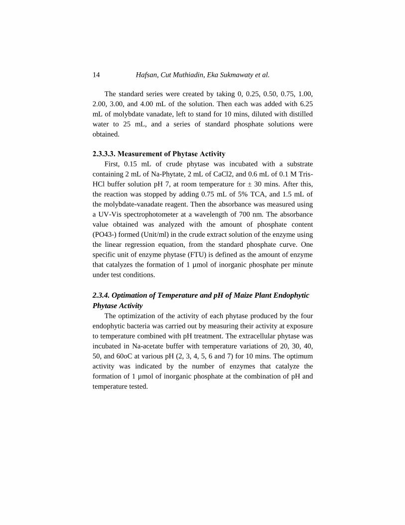
Disclosure of The Potential Phytase-Producing Maize … 17
Purification was conducted by sampling a separate loop of bacterial
colonies, and scratched on a similar solid medium which was then
incubated under the same conditions. From the growing colonies, re-
scratching was carried out on the new solid medium up to eleven times, in
order to obtain genuinely pure isolates. A total of 28 isolates of endophytic
bacteria were obtained from the roots, stems, leaves, and seeds of the
Maize plant. The obtained isolates were screened using cultivation and
selective media for phytase-producing bacteria, namely PSM by being
spotted on agar plates simultaneously. The addition of 0.4% Na-phytate to
the PSM, which was initially clear and yellowish, caused the media to
become cloudy and milky white. The bacteria that produces good phytase
showed a clear zone around the isolated colony, and this is an indication of
the enzymatic reaction of phytate hydrolysis contained in the media.
Therefore, the wider the clear zone formed the higher the phytase quantity.
Figure 1. Phytatic index (PI) of endophytic bacterial isolates from the maize plant: (a)
Isolate from roots (HF.7) with PI value of 1.38, (b) Isolate from the stem (HF.8) with
PI value of 1.31, (c) Isolate from seeds (HF.18) with PI value of 1.36, (b) Isolate from
the leaf (HF.28) with PI value of 1.23.
Hafsan, Cut Muthiadin, Eka Sukmawaty et al. 18
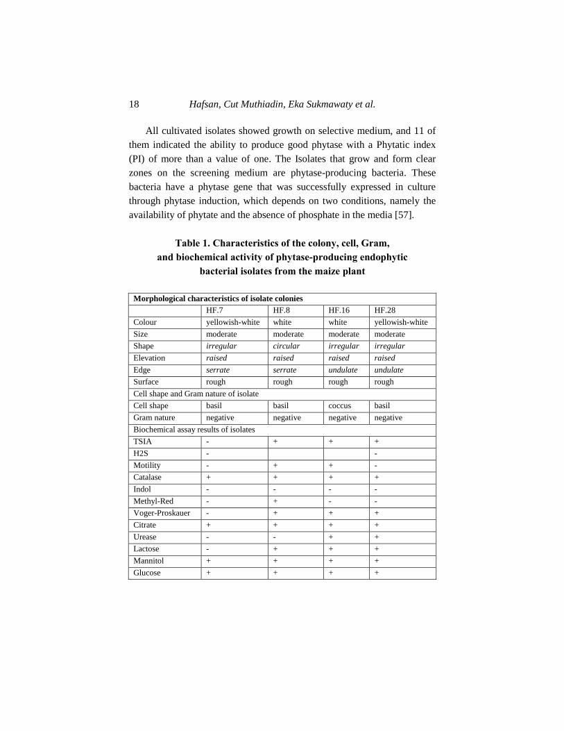
All cultivated isolates showed growth on selective medium, and 11 of
them indicated the ability to produce good phytase with a Phytatic index
(PI) of more than a value of one. The Isolates that grow and form clear
zones on the screening medium are phytase-producing bacteria. These
bacteria have a phytase gene that was successfully expressed in culture
through phytase induction, which depends on two conditions, namely the
availability of phytate and the absence of phosphate in the media [57].
Table 1. Characteristics of the colony, cell, Gram,
and biochemical activity of phytase-producing endophytic
bacterial isolates from the maize plant
Morphological characteristics of isolate colonies
HF.7 HF.8 HF.16 HF.28
Colour yellowish-white white white yellowish-white
Size moderate moderate moderate moderate
Shape irregular circular irregular irregular
Elevation raised raised raised raised
Edge serrate serrate undulate undulate
Surface rough rough rough rough
Cell shape and Gram nature of isolate
Cell shape basil basil coccus basil
Gram nature negative negative negative negative
Biochemical assay results of isolates
TSIA - + + +
H2S - -
Motility - + + -
Catalase + + + +
Indol - - - -
Methyl-Red - + - -
Voger-Proskauer - + + +
Citrate + + + +
Urease - - + +
Lactose - + + +
Mannitol + + + +
Glucose + + + +
Disclosure of The Potential Phytase-Producing Maize … 19
The characteristics of the selected isolates, as shown in Table 2, were
known through macroscopic and microscopic observations. The
macroscopic observations include remarks of size, pigmentation, shape,
elevation, surface, and colony margins. The microscopic observations
include cell shape and Gram's nature. The results of Gram staining of the
four selected phytase-producing endophytic bacteria showed Gram-
negative characteristics.
The differences in bacterial Gram nature occurred as a result of
variation in binding capacity and the dyeing process. This was caused by
differences in the structure of the cell wall between Gram-positive and
negative bacteria. Gram-positive have a thicker peptidoglycan layer than
the negative bacteria, therefore, the colour of Crystal violet adheres firmly
to it [58].
3.2. Identification of Phytase-Producing Maize Plant
Endophytic Bacteria Using a Molecular Approach
Information on the morphological characteristics of the four isolates
were different from each other, and became the basis for the identification
of a different species. The identification of selected endophytic bacteria
was carried out using a molecular approach through analysis of the 16s
rRNA gene. The 16s rRNA gene is present in the 30s ribosome subunit and
found in all prokaryotes. It has a relatively large number of nucleotides,
while some bases are sustainable and arranged as a universal primer to
amplify an organism's 16s rRNA gene. The four bacterial isolates were
determined using universal primers, namely forward 63F and reverse
1387R, for the 16S-rRNA sequence.
The 16s rRNA gene analysis for the identification of microorganisms
using a molecular approach consists of four main processes, namely the
extraction/isolation of DNA, PCR, electrophoresis, and sequencing. DNA
extraction is the process of separating it from other cell components in
order to obtain a pure isolate. The next stage was the amplification process
Hafsan, Cut Muthiadin, Eka Sukmawaty et al. 20
using PCR, which aims to multiply a DNA band in vitro. In the process,
there was a chain reaction, namely denaturation, annealing, and elongation.
The PCR process was carried out in 35 cycles for ± 2 hours. The forward
primers initiated the synthesis of DNA strands from the 5 '-------- 3' end,
while the Reverse initiated the synthesis of DNA strands from the 3 '--------
5' end. The function of the template DNA in the PCR process was a
template for the formation of the same new DNA molecule. The
chromosomal DNA profiles that have been isolated and multiplied by PCR
were analyzed using 1% agarose electrophoresis. The electropherogram
from the electrophoresis of the chromosomal DNA of the four isolates
(Figure 2), showed the presence of a single thick band produced by each
chromosomal DNA of the isolates. From the electrophoresis, results were
obtained in the extended base pair (bp), each sample with the help of
markers.
Figure 2. The electrophoregram of the 16S-rRNA gene amplification product of the
selected isolates: HF.7 = ± 1000 bp, HF.8 = ± 900 bp, HF.16 = ± 1000 bp, and HF.
28 = ± 900 bp.
The basic principle of electrophoresis technique is the separation of
charged components or DNA molecules in an electric field. The DNA
molecule is separated based on the rate of migration by the electromotive
force in the gel matrix. The DNA molecule sample is placed in a well on a
gel that is placed in a buffer solution (TBE), then an electric current flow.
Disclosure of The Potential Phytase-Producing Maize … 21
The DNA molecule move in the gel matrix towards one of the electric
poles according to the charge of the DNA molecule. The direction of the
DNA molecules movement toward the positive electrode, was due to the
negative charge on the framework of its sugar-phosphate. In order to keep
the rate of movement of the DNA molecule strictly based on size, the
substance sodium hydroxide is used to keep the DNA straight [54].
After the electrophoresis process, staining was carried out for the
separated sample molecules to be clearly observed using ethidium
bromide. And the sample molecules glowed in ultraviolet light. The bands
in the different stripes of the gel appeared after the dyeing process,
representing each lane in the movement direction of the sample from the
gel "well". The bands that were equidistant from the gel well at the end of
the electrophoresis, contain molecules that moved in the gel during the
process at the same speed, meaning that the molecules have the same size.
Markers which are molecular mixtures of different sizes were use to
determine the size of the molecules in the sample band by electrophoresis,
and the markers on the strips in the gel were parallel to the sample.
The bands in the visible marking strip were compared with the sample
bands to determine their size. The distance of the band from the gel well
was inversely proportional to the logarithm of the molecular size. Based on
the results of electrophoresis, DNA isolates of HF.8 and HF.28 showed
bands that were perforated and parallel to the markers around ± 900 bp.
This indicated that the amplified gene fragment was ± 900 bp in size. The
DNA isolates of HF.7 and HF.16 showed separated and parallel bands with
markers of around ± 1,000 bp, indicating that the amplified gene fragments
were ± 1,000 bp in size.
The 16S-rRNA gene of PCR products was sequenced in the 1st BASE
INT Malaysia sequencing. The cluster analysis on sequences was carried
out with the online BLAST program from NCBI. The results of the PCR
product sequencing of each isolate were intact DNA nucleotide base
sequences. Based on the BLAST analysis, the results of the homology of
the four selected isolates were as shown in Tables 2, 3, 4, and 5.
Hafsan, Cut Muthiadin, Eka Sukmawaty et al. 22
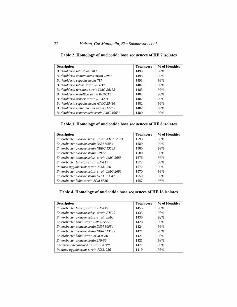
Table 2. Homology of nucleotide base sequences of HF.7 isolates
Description Total score % of Identities
Burkholderia lata strain 383
Burkholderia contaminans strain J2956
Burkholderia cepacia strain 717
Burkholderia latens strain R-5630
Burkholderia territorii strain LMG 28158
Burkholderia metallica strain R-16017
Burkholderia arboris strain R-24201
Burkholderia cepacia strain ATCC 25416
Burkholderia vietnamiensis strain TVV75
Burkholderia cenocepacia strain LMG 16656
1493
1493
1493
1487
1485
1482
1482
1482
1482
1480
99%
99%
99%
99%
99%
99%
99%
99%
99%
99%
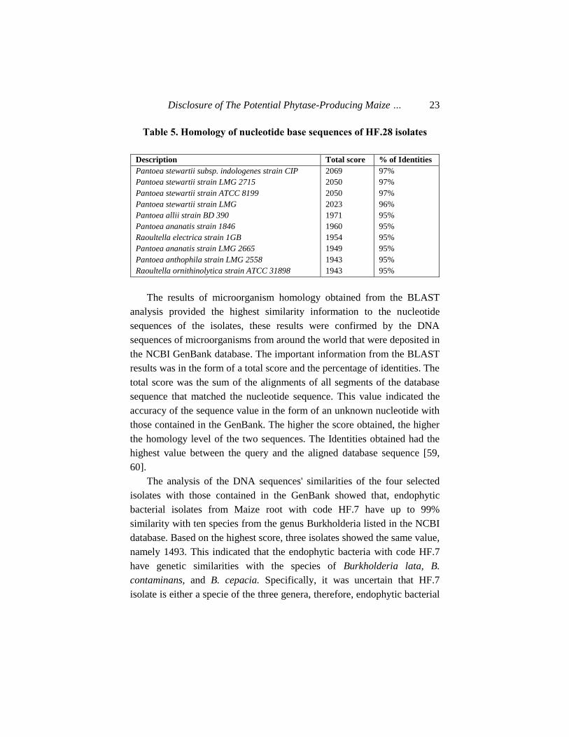
Table 3. Homology of nucleotide base sequences of HF.8 isolates
Description Total score % of Identities
Enterobacter cloacae subsp. strain ATCC 2373
Enterobacter cloacae strain DSM 30054
Enterobacter cloacae strain NBRC 13535
Enterobacter cloacae strain 279-56
Enterobacter cloacae subsp. strain LMG 2683
Enterobacter ludwigii strain EN-119
Pantoea agglomerans strain JCM1236
Enterobacter cloacae subsp. strain LMG 2683
Enterobacter cloacae strain ATCC 13047
Enterobacter kobei strain JCM 8580
1593
1580
1580
1580
1576
1572
1572
1570
1559
1557
99%
99%
99%
99%
99%
99%
99%
99%
98%
98%
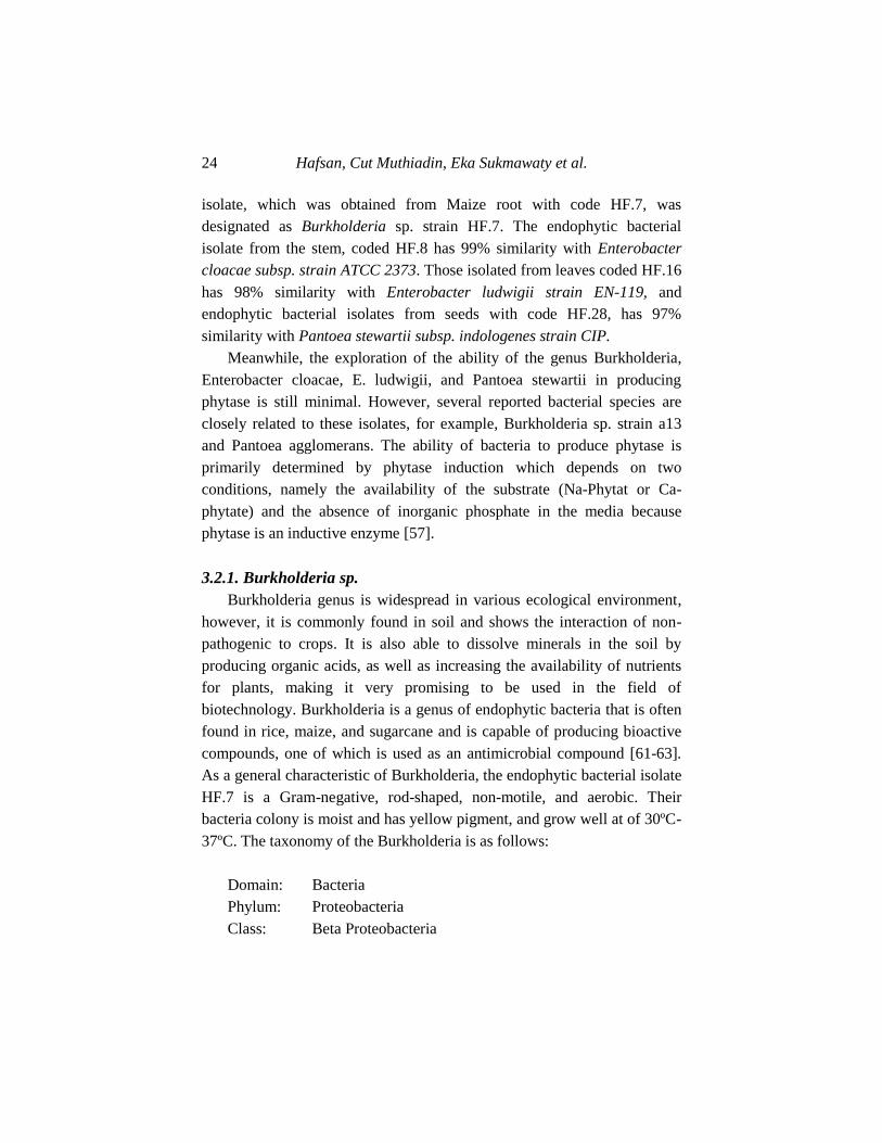
Table 4. Homology of nucleotide base sequences of HF.16 isolates
Description Total score % of Identities
Enterobacter ludwigii strain EN-119
Enterobacter cloacae subsp. strain ATCC
Enterobacter cloacae subsp. strain LMG
Enterobacter kobei strain CIP 105566
Enterobacter cloacae strain DSM 30054
Enterobacter cloacae strain NBRC 13535
Enterobacter kobei strain JCM 8580
Enterobacter cloacae strain 279-56
Leclercia adecarboxylata strain NBRC
Pantoea agglomerans strain JCM1236
1455
1435
1430
1428
1424
1421
1421
1421
1421
1419
98%
98%
98%
98%
98%
98%
98%
98%
98%
98%
Disclosure of The Potential Phytase-Producing Maize … 23
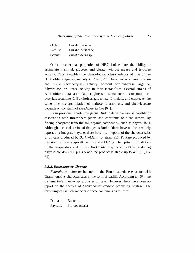
Table 5. Homology of nucleotide base sequences of HF.28 isolates
Description Total score % of Identities
Pantoea stewartii subsp. indologenes strain CIP
Pantoea stewartii strain LMG 2715
Pantoea stewartii strain ATCC 8199
Pantoea stewartii strain LMG
Pantoea allii strain BD 390
Pantoea ananatis strain 1846
Raoultella electrica strain 1GB
Pantoea ananatis strain LMG 2665
Pantoea anthophila strain LMG 2558
Raoultella ornithinolytica strain ATCC 31898
2069
2050
2050
2023
1971
1960
1954
1949
1943
1943
97%
97%
97%
96%
95%
95%
95%
95%
95%
95%
The results of microorganism homology obtained from the BLAST
analysis provided the highest similarity information to the nucleotide
sequences of the isolates, these results were confirmed by the DNA
sequences of microorganisms from around the world that were deposited in
the NCBI GenBank database. The important information from the BLAST
results was in the form of a total score and the percentage of identities. The
total score was the sum of the alignments of all segments of the database
sequence that matched the nucleotide sequence. This value indicated the
accuracy of the sequence value in the form of an unknown nucleotide with
those contained in the GenBank. The higher the score obtained, the higher
the homology level of the two sequences. The Identities obtained had the
highest value between the query and the aligned database sequence [59,
60].
The analysis of the DNA sequences' similarities of the four selected
isolates with those contained in the GenBank showed that, endophytic
bacterial isolates from Maize root with code HF.7 have up to 99%
similarity with ten species from the genus Burkholderia listed in the NCBI
database. Based on the highest score, three isolates showed the same value,
namely 1493. This indicated that the endophytic bacteria with code HF.7
have genetic similarities with the species of Burkholderia lata, B.
contaminans, and B. cepacia. Specifically, it was uncertain that HF.7
isolate is either a specie of the three genera, therefore, endophytic bacterial
Hafsan, Cut Muthiadin, Eka Sukmawaty et al. 24
isolate, which was obtained from Maize root with code HF.7, was
designated as Burkholderia sp. strain HF.7. The endophytic bacterial
isolate from the stem, coded HF.8 has 99% similarity with Enterobacter
cloacae subsp. strain ATCC 2373. Those isolated from leaves coded HF.16
has 98% similarity with Enterobacter ludwigii strain EN-119, and
endophytic bacterial isolates from seeds with code HF.28, has 97%
similarity with Pantoea stewartii subsp. indologenes strain CIP.
Meanwhile, the exploration of the ability of the genus Burkholderia,
Enterobacter cloacae, E. ludwigii, and Pantoea stewartii in producing
phytase is still minimal. However, several reported bacterial species are
closely related to these isolates, for example, Burkholderia sp. strain a13
and Pantoea agglomerans. The ability of bacteria to produce phytase is
primarily determined by phytase induction which depends on two
conditions, namely the availability of the substrate (Na-Phytat or Ca-
phytate) and the absence of inorganic phosphate in the media because
phytase is an inductive enzyme [57].
3.2.1. Burkholderia sp.
Burkholderia genus is widespread in various ecological environment,
however, it is commonly found in soil and shows the interaction of non-
pathogenic to crops. It is also able to dissolve minerals in the soil by
producing organic acids, as well as increasing the availability of nutrients
for plants, making it very promising to be used in the field of
biotechnology. Burkholderia is a genus of endophytic bacteria that is often
found in rice, maize, and sugarcane and is capable of producing bioactive
compounds, one of which is used as an antimicrobial compound [61-63].
As a general characteristic of Burkholderia, the endophytic bacterial isolate
HF.7 is a Gram-negative, rod-shaped, non-motile, and aerobic. Their
bacteria colony is moist and has yellow pigment, and grow well at of 30ºC-
37ºC. The taxonomy of the Burkholderia is as follows:
Domain: Bacteria
Phylum: Proteobacteria
Class: Beta Proteobacteria
Disclosure of The Potential Phytase-Producing Maize … 25
Order: Burkholderiales
Family: Burkholderiaceae
Genus: Burkholderia sp.
Other biochemical properties of HF.7 isolates are the ability to
assimilate mannitol, glucose, and citrate, without urease and tryptone
activity. This resembles the physiological characteristics of one of the
Burkholderia species, namely B. lata [64]. These bacteria have catalase
and lysine decarboxylase activity, without tryptophanase, arginine,
dihydrolase, or urease activity in their metabolism. Several strains of
Burkholderia lata assimilate D-glucose, D-mannose, D-mannitol, N-
acetylglucosamine, D-Burkholderiagluconate, L-malate, and citrate. At the
same time, the assimilation of maltose, L-arabinose, and phenylacetate
depends on the strain of Burkholderia lata [64].
From previous reports, the genus Burkholderia bacteria is capable of
associating with rhizosphere plants and contribute to plant growth, by
freeing phosphate from the soil organic compounds, such as phytate [61].
Although bacterial strains of the genus Burkholderia have not been widely
reported to integrate phytate, there have been reports of the characteristics
of phytase produced by Burkholderia sp. strain a13. Phytase produced by
this strain showed a specific activity of 4.1 U/mg. The optimum conditions
of the temperature and pH for Burkholderia sp. strain a13 in producing
phytase are 45-55ºC, pH 4.5 and the product is stable up to 4ºC [61, 65,
66].
3.2.2. Enterobacter Cloacae
Enterobacter cloacae belongs to the Enterobacteriaceae group with
Gram-negative characteristics in the form of bacilli. According to [67], the
bacteria Enterobacter sp. produces phytase. However, there have been no
report on the species of Enterobacter cloacae producing phytase. The
taxonomy of the Enterobacter cloacae bacteria is as follows:
Domain: Bacteria
Phylum: Proteobacteria
Hafsan, Cut Muthiadin, Eka Sukmawaty et al. 26
Class: Gammaproteobacteria
Order: Enterobacteriales
Family: Enterobacteriaceae
Genus: Enterobacter
Species: Enterobacter cloacae
Several studies reported that the endophytic bacteria Enterobacter
cloacae were shown to increase nitrogen fixation in rice plants. In addition
to producing IAA hormone and having the ability to increase nitrogen
fixation, they also produce the enzyme L-Histidine Decarboxylase (HDC)
[67, 68].
3.2.3. Enterobacter Ludwigii
Enterobacter ludwigii is an endophytic bacterium that is included in
the general characteristics of the genus enterobacter. It is a Gram-negative
bacterium, bacillus, motile, and able to ferment. The taxonomy of the
Enterobacter ludwigii bacteria is as follows:
Domain: Bacteria
Phylum: Proteobacteria
Class: Gammaproteobacteria
Order: Enterobacteriales
Family: Enterobacteriaceae
Genus: Enterobacter
Species: Enterobacter ludwigii
It was previously shown that the bacteria Enterobacter sp. produce
phytase. However, studies reporting that Enterobacter ludwigii species
produce phytase have not been found. Only a few reported on the potential
of the bacteria Enterobacter ludwigii [69-71]. Several studies have
reported the abilities possessed by Enterobacter ludwigii, including having
activity as Plant Growth Promoting Bacteria (PGPB). Those isolated from
the rhizosphere of Lolium perenne L. grass showed phosphate solvent
activity, nitrogen-fixing, and producing growth hormone IAA [72, 73].
Disclosure of The Potential Phytase-Producing Maize … 27
3.2.4. Pantoea Stewartii
Pantoea stewartii is a gram-negative bacterium and non-motile. There
is a genus of Pantoea which is pathogenic to plants, while some are
beneficial (in association). Species from Pantoea stewartii sp indologenes
are associated with sorghum grass. The classification of the Pantoea
stewartii bacteria is as follows:
Domain: Bacteria
Phylum: Proteobacteria
Class: Gammaproteobacteria
Order: Enterobacteriales
Family: Enterobacteriaceae
Genus: Enterobacter
Species: Pantoea stewartii
Several species of the genus Pantoea are reported to produce phytase,
including Pantoea agglomerans [74], which has been shown to reduce
phytate content in feed using the phytase it produced [75, 76]. [77]. Also,
previous research reported that this specie was successfully isolated from
the soil of the Republic of Tatarstan, Russia, based on the high activity of
phytate decomposers which stores 99% 16S rRNA nucleotide sequence
similar to the Pantoea sp. Moreover, [53] the ability of Pantoea stewartii
ASUIA271 to produce phytase was explored, which was triggered by the
high organic phytate content in rice husks at various experimental
temperatures.
3.3. Production and optimIzation of Phytase Activity
of Endophytic Bacteria from Maize Plants
3.3.1. Phytase Crude Extract Production
Phytase production began with the determination of the growth curves
of the four selected endophytic bacterial isolates. It was crucial to observe
the growth/survival pattern of bacteria for the optimum phase of the
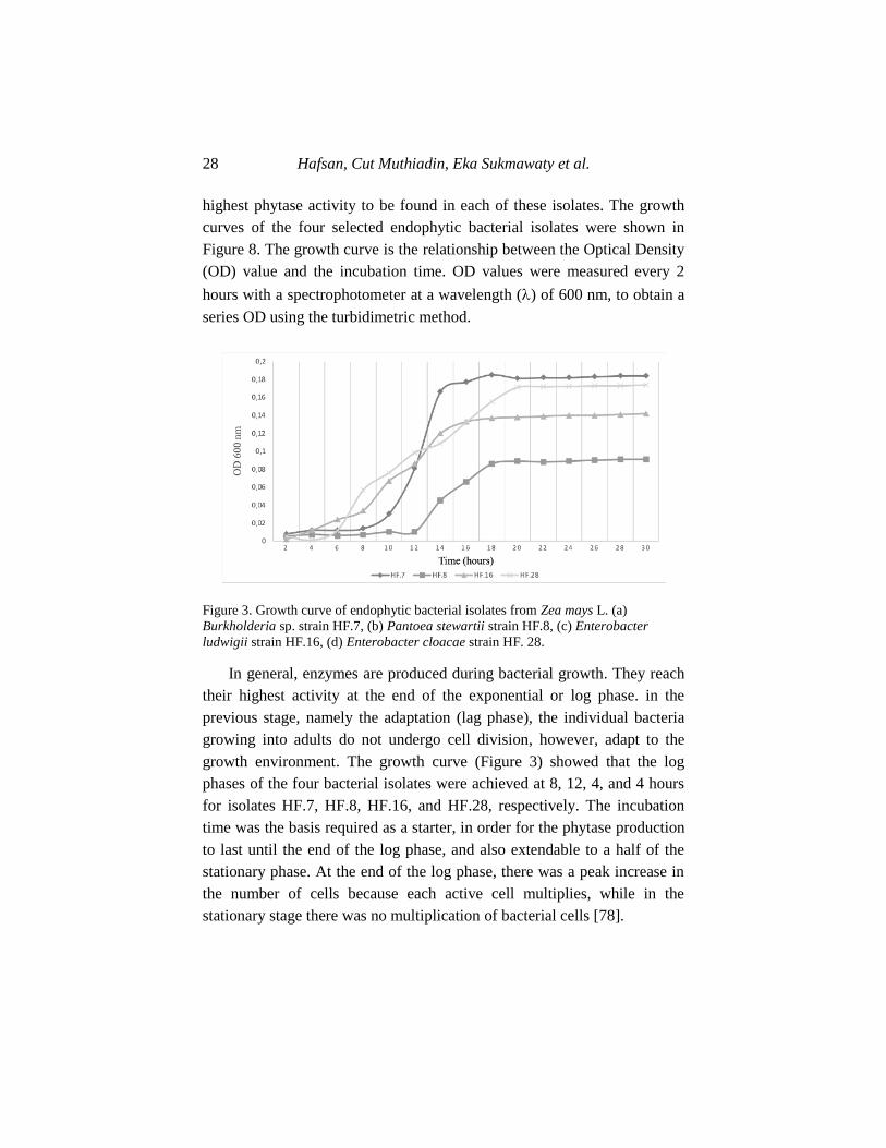
Hafsan, Cut Muthiadin, Eka Sukmawaty et al. 28
highest phytase activity to be found in each of these isolates. The growth
curves of the four selected endophytic bacterial isolates were shown in
Figure 8. The growth curve is the relationship between the Optical Density
(OD) value and the incubation time. OD values were measured every 2
hours with a spectrophotometer at a wavelength () of 600 nm, to obtain a
series OD using the turbidimetric method.
Figure 3. Growth curve of endophytic bacterial isolates from Zea mays L. (a)
Burkholderia sp. strain HF.7, (b) Pantoea stewartii strain HF.8, (c) Enterobacter
ludwigii strain HF.16, (d) Enterobacter cloacae strain HF. 28.
In general, enzymes are produced during bacterial growth. They reach
their highest activity at the end of the exponential or log phase. in the
previous stage, namely the adaptation (lag phase), the individual bacteria
growing into adults do not undergo cell division, however, adapt to the
growth environment. The growth curve (Figure 3) showed that the log
phases of the four bacterial isolates were achieved at 8, 12, 4, and 4 hours
for isolates HF.7, HF.8, HF.16, and HF.28, respectively. The incubation
time was the basis required as a starter, in order for the phytase production
to last until the end of the log phase, and also extendable to a half of the
stationary phase. At the end of the log phase, there was a peak increase in
the number of cells because each active cell multiplies, while in the
stationary stage there was no multiplication of bacterial cells [78].
Disclosure of The Potential Phytase-Producing Maize … 29
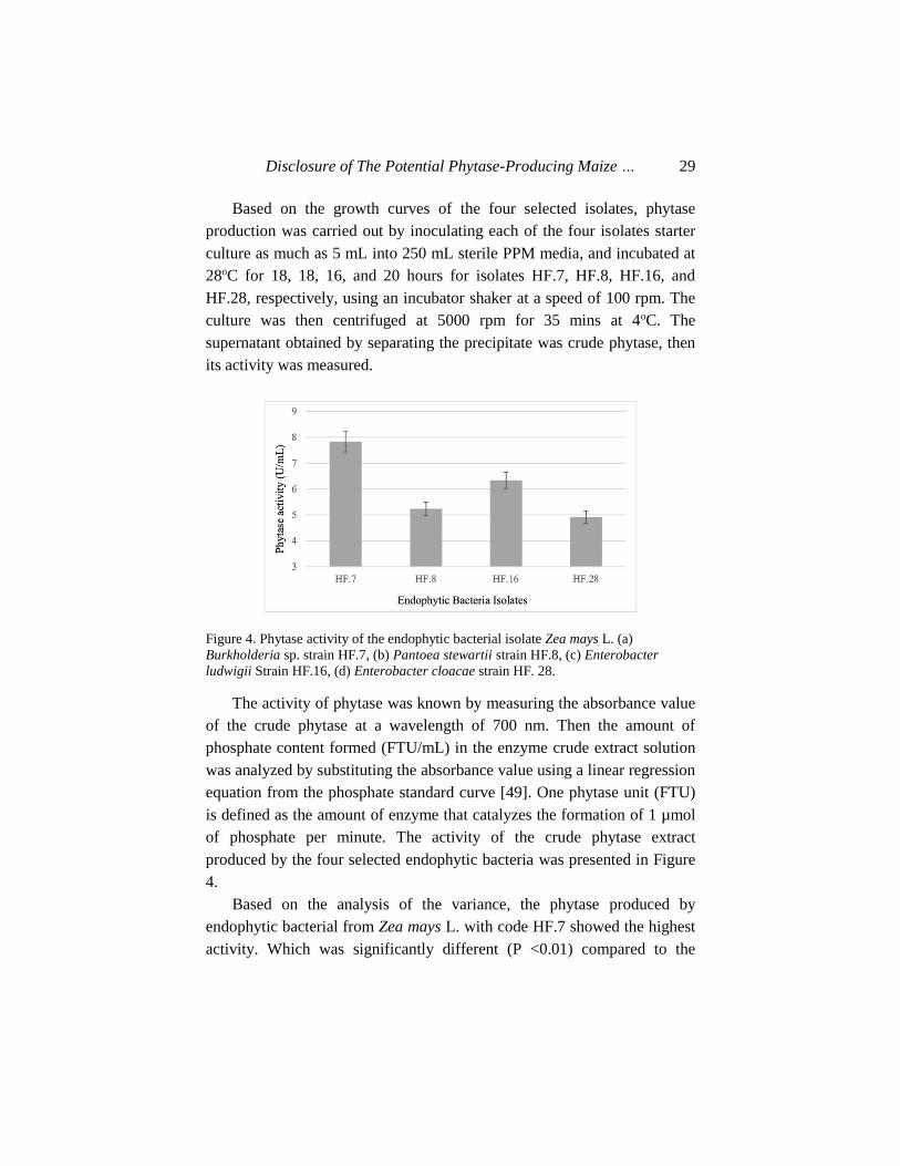
Based on the growth curves of the four selected isolates, phytase
production was carried out by inoculating each of the four isolates starter
culture as much as 5 mL into 250 mL sterile PPM media, and incubated at
28oC for 18, 18, 16, and 20 hours for isolates HF.7, HF.8, HF.16, and
HF.28, respectively, using an incubator shaker at a speed of 100 rpm. The
culture was then centrifuged at 5000 rpm for 35 mins at 4oC. The
supernatant obtained by separating the precipitate was crude phytase, then
its activity was measured.
Figure 4. Phytase activity of the endophytic bacterial isolate Zea mays L. (a)
Burkholderia sp. strain HF.7, (b) Pantoea stewartii strain HF.8, (c) Enterobacter
ludwigii Strain HF.16, (d) Enterobacter cloacae strain HF. 28.
The activity of phytase was known by measuring the absorbance value
of the crude phytase at a wavelength of 700 nm. Then the amount of
phosphate content formed (FTU/mL) in the enzyme crude extract solution
was analyzed by substituting the absorbance value using a linear regression
equation from the phosphate standard curve [49]. One phytase unit (FTU)
is defined as the amount of enzyme that catalyzes the formation of 1 µmol
of phosphate per minute. The activity of the crude phytase extract
produced by the four selected endophytic bacteria was presented in Figure
4.
Based on the analysis of the variance, the phytase produced by
endophytic bacterial from Zea mays L. with code HF.7 showed the highest
activity. Which was significantly different (P <0.01) compared to the
Hafsan, Cut Muthiadin, Eka Sukmawaty et al. 30
phytase produced by the three other endophytic bacteria isolates. An
interesting observation in the endophytic bacteria was that, the best phytase
producer was obtained from their Maize root. This was due to the phytate
content in its root area, which triggers the emergence of phytase-producing
endophytic bacteria, as a result of inositol phosphate, which is widely
distributed in Maize roots.
The phytase activity produced by the four selected endophytic
bacterial, between 4.9-7.8 FTU/mL, showed higher value compared to
those generated by Burkholderia sp. strain a13 (4.1 FTU/mL), Bacillus
cereus ASUIA 260 (1.160 FTU/mL), and Bacillus subtilis AP-17 (0.0296
FTU/mL) [57]. Similarly, the phytase produced by three Bacillus cereus
strains isolated from the volcanic ash of Mount Merapi includes 0.1071
FTU/mL, 0.1020 U/mL, and 0.0874 FTU/mL [79] [53]. It was also
reported that the phytase activity of Staphylococcus lentus ASUIA 279 was
1.913 FTU/mL. [80]. Moreover, the phytase activity of the three strains of
Bacillus cereus, isolated from water and mud samples of the Sikidang
Dieng crater were 0.32893 FTU/mL, 0.324953 FTU/mL, and 0.32182
FTU/mL.
3.3.2. The Optimization of Temperature and pH of Phytase Activity
from Endophytic Bacteria of Maize Plants
This test was conducted to determine the optimum phytase activity
produced from the four selected maize plant endophytic bacteria at the
same temperature and pH as well as the protease activity of the poultry
digestive tract in vitro. The determination of the optimum temperature and
pH was measured by observing the activity of the phytase crude extract,
exposed to varying temperatures (30, 40, 50, 60, and 70oC) for 10 mins.
The pH optimation test was carried out by incubating the crude extract of
extracellular phytase in Na-acetate buffer with varying pH (2, 3, 4, 5, 6,
and 7).
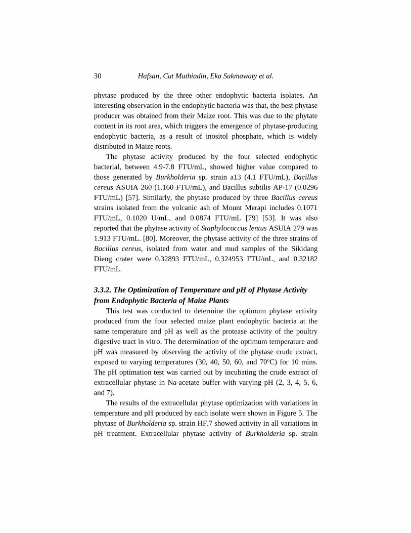
The results of the extracellular phytase optimization with variations in
temperature and pH produced by each isolate were shown in Figure 5. The
phytase of Burkholderia sp. strain HF.7 showed activity in all variations in
pH treatment. Extracellular phytase activity of Burkholderia sp. strain
Disclosure of The Potential Phytase-Producing Maize … 31
HF.7 as observed from the treatment of pH 2, showed an increase in
activity from pH 3 to 4, and at pH 5 to pH 6, and decreased activity at pH 7
[81]. The changes in activity at different pHs were caused by the
occurrence of intramolecular changes of enzymes caused by ionization to
bind and release protons (hydrogen ions) in amino, carboxyl, and other
functional groups. When the difference was too large, it results in the
denaturation of the enzyme, and its activity was lost [82]. The results of
this measurement were in line with the nature of phytase, which are
heterologous group of enzymes, hydrolyse phosphate esters, and optimal at
low pH. At neutral and alkaline pH, catalytic activity decreases. This is due
to the structural instability of the enzyme protein molecules, which causes
structural changes in these pH conditions [83, 84].
The pH condition of the digestive tract of poultry, especially chicken,
is 4.5 in the crop, 4.4 in the proventriculus, 2.6 in the gizzard, 5.7-6.0 in
the duodenum, 5.8 in the jejunum, 6.3 in the ileum, 6.3 in the colon, and
5.7 in the caeca. The extracellular phytase of the four endophytic bacteria
of the Maize plant showed stable activity at pH 4-6 [85]. Therefore, the
phytase produced by the endophytic bacteria of the maize plant is active in
the digestive tract of the poultry.
The determination of extracellular phytase activity of maize plant
endophytic bacteria against a combination of temperature and pH
variations as in Figure 5, showed that increasing temperature causes an
increase in activity until it reaches the optimum point of 40oC. The rise in
temperature decreased the phytase activity, as observed at 50oC and
continues to decline until a temperature of 70oC. At first, with increasing
temperature, the enzyme reaction speed increases due to the rise in kinetic
energy, which accelerated the vibrational, translational, and rotational
motion of the enzyme and the substrate, increasing their chance to react.
On exposure to temperatures higher than the optimum, the protein and the
substrate underwent a conformational change. This caused the reactive
group to unmatch the active side, or experience obstacles in entering the
active site of the enzyme, in order to significantly affects its catalytic
activity [83].
Hafsan, Cut Muthiadin, Eka Sukmawaty et al. 32
Figure 5. Phytase activity of maize plant endophytic bacteria at a combination of
temperature and pH variations: (a) Burkholderia sp. strain HF.7, (b) Pantoea stewartii
strain HF.8, (c) Enterobacter ludwigii Strain HF.16, (d) Enterobacter cloacae strain
HF. 28.
The optimum temperature and pH of the enzyme depends on its type
and source. Based on the activity measurement, it was known that the
optimum temperature and pH of phytase produced by Burkholderia sp.
strain HF.7 and Enterobacter cloacae strain HF.28 was 40oC and pH 6.
Pantoea stewartii strain HF.8 generated 40oC and pH 5. Enterobacter
ludwigii Strain HF.16 produced 40oC and pH 4. Temperature variations
and the optimum pH of phytase activity produced by other bacteria have
also been widely reported [86], namely those obtained from B. subtillis
(natto) N-77 were 60oC and pH 6.0-6.5, Enterobacter sp. optimum were
pH 7.5 and 50oC. Phytase is also produced by Aspergillus niger (58oC, pH
5.5), Schwanniomiyces castellii (7.7oC, pH 4.4), and Klebsiella aerogenes
(45oC, pH 7.0).
Disclosure of The Potential Phytase-Producing Maize … 33
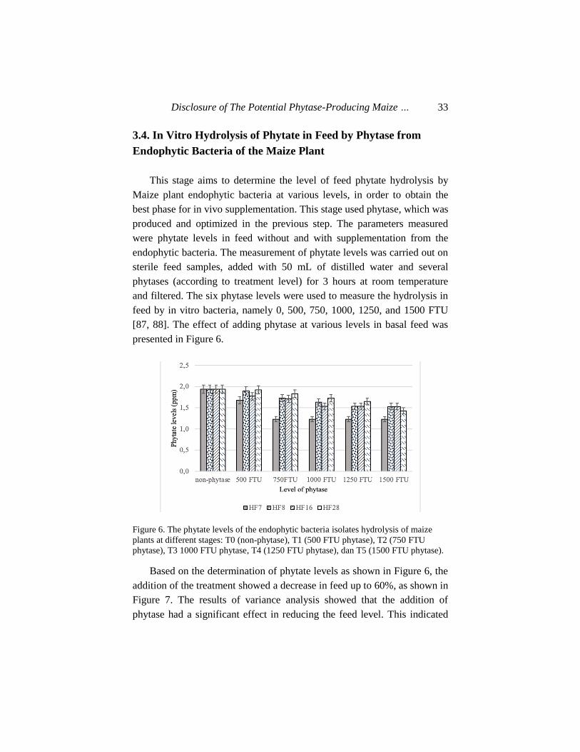
3.4. In Vitro Hydrolysis of Phytate in Feed by Phytase from
Endophytic Bacteria of the Maize Plant
This stage aims to determine the level of feed phytate hydrolysis by
Maize plant endophytic bacteria at various levels, in order to obtain the
best phase for in vivo supplementation. This stage used phytase, which was
produced and optimized in the previous step. The parameters measured
were phytate levels in feed without and with supplementation from the
endophytic bacteria. The measurement of phytate levels was carried out on
sterile feed samples, added with 50 mL of distilled water and several
phytases (according to treatment level) for 3 hours at room temperature
and filtered. The six phytase levels were used to measure the hydrolysis in
feed by in vitro bacteria, namely 0, 500, 750, 1000, 1250, and 1500 FTU
[87, 88]. The effect of adding phytase at various levels in basal feed was
presented in Figure 6.
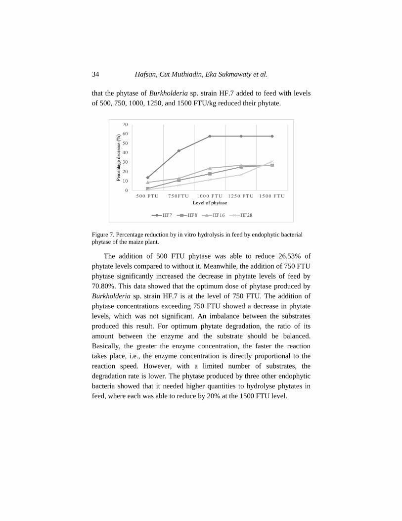
Figure 6. The phytate levels of the endophytic bacteria isolates hydrolysis of maize
plants at different stages: T0 (non-phytase), T1 (500 FTU phytase), T2 (750 FTU
phytase), T3 1000 FTU phytase, T4 (1250 FTU phytase), dan T5 (1500 FTU phytase).
Based on the determination of phytate levels as shown in Figure 6, the
addition of the treatment showed a decrease in feed up to 60%, as shown in
Figure 7. The results of variance analysis showed that the addition of
phytase had a significant effect in reducing the feed level. This indicated
Hafsan, Cut Muthiadin, Eka Sukmawaty et al. 34
that the phytase of Burkholderia sp. strain HF.7 added to feed with levels
of 500, 750, 1000, 1250, and 1500 FTU/kg reduced their phytate.
Figure 7. Percentage reduction by in vitro hydrolysis in feed by endophytic bacterial
phytase of the maize plant.
The addition of 500 FTU phytase was able to reduce 26.53% of
phytate levels compared to without it. Meanwhile, the addition of 750 FTU
phytase significantly increased the decrease in phytate levels of feed by
70.80%. This data showed that the optimum dose of phytase produced by
Burkholderia sp. strain HF.7 is at the level of 750 FTU. The addition of
phytase concentrations exceeding 750 FTU showed a decrease in phytate
levels, which was not significant. An imbalance between the substrates
produced this result. For optimum phytate degradation, the ratio of its
amount between the enzyme and the substrate should be balanced.
Basically, the greater the enzyme concentration, the faster the reaction
takes place, i.e., the enzyme concentration is directly proportional to the
reaction speed. However, with a limited number of substrates, the
degradation rate is lower. The phytase produced by three other endophytic
bacteria showed that it needed higher quantities to hydrolyse phytates in
feed, where each was able to reduce by 20% at the 1500 FTU level.
Disclosure of The Potential Phytase-Producing Maize … 35
CONCLUSION
This study shows other significant role of Zea mays L. besides being a
food source. The endophytic bacteria that are symbiotic in the maize plant
cycle, are known to play a role in increasing its growth and yield,
suppressing contaminant pathogens, dissolving phosphates, or contributing
nitrogen. The other potential endophytic bacteria producing a very
prospective enzyme, namely phytase, were applied in improving the
quality of broiler feed. This research found and identified four types of
potential phytase-producing endophytic bacteria from the Maize plant,
namely Burkholderia sp. strains HF.7, Enterobacter cloacae strains HF.8,
Enterobacter ludwigii strains HF.16, and Pantoea stewartii strains HF.28.
Sequentially, each was isolated from the roots, stems, leaves, and seeds of
maize plants. Also, each of the phytase produced by the four endophytic
bacteria hydrolysed phytate which has been tested in vitro at various dose,
temperature, and pH. Therefore, this enzyme is proven to have the
potential of improving poultry feed quality.
REFERENCES
[1] Tanumihardjo, Sherry, Laura McCulley, Rachel Roh, Santiago Lopez-
Ridaura, Natalia Palacios-Rojas, and Nilupa S. Gunaratna. 2020.
“Maize Agro-Food Systems to Ensure Food and Nutrition Security in
Reference to the Sustainable Development Goals.” Global Food
Security 25: 100-113. Accessed July 12, 2020. doi:org/10.1016/j.gfs.
2019.100327.
[2] Ranum, Peter, Juan Pablo Peña‐Rosas, and Maria Nieves
Garcia‐Casal. 2014. “Global Maize Production, Utilization, and
Consumption.” Annals of the New York Academy of Sciences 1312
(1): 105–112. Accessed September 21, 2020. doi:org/10.1111/
nyas.12396.
[3] Li, Quanfeng, Jianjun Zang, Dewen Liu, Xiangshu Piao, Changhua
Hafsan, Cut Muthiadin, Eka Sukmawaty et al. 36
Lai, and Defa Li. 2014. “Predicting Corn Digestible and
Metabolizable Energy Content from Its Chemical Composition in
Growing Pigs.” Journal of Animal Science and Biotechnology. 12
(2):186-194. Accessed July 22, 2020. doi:org/10.1186/2049-1891-5-
11.
[4] Zhao, F., H. F. Zhang, S. S. Hou, and Z. Y. Zhang. 2008. “Predicting
Metabolizable Energy of Normal Corn from Its Chemical
Composition in Adult Pekin Ducks.” Poultry Science. 87 (8): 1603-
1608. Accessed July 22, 2020. doi:org/10.3382/ps.2007-00494.
[5] Smith, K. A., C. L. Wyatt, and J. T. Lee. 2019. “Evaluation of
Increasing Levels of Phytase in Diets Containing Variable Levels of
Amino Acids on Male Broiler Performance and Processing Yields.”
Journal of Applied Poultry Research 28 (2): 253–62. Accessed July
22, 2020. doi:org/10.3382/japr/pfy065.
[6] Singh, Nand Kumar, Dharmendra Kumar Joshi, and Raj Kishor
Gupta. 2013. “Isolation of Phytase Producing Bacteria and
Optimization of Phytase Production Parameters.” Jundishapur
Journal of Microbiology 6 (5):85-97. Accessed July 22, 2020. doi:org/
10.5812/jjm.6419.
[7] Oatway, Lori, Thava Vasanthan, and James H. Helm. 2001. “Phytic
Acid.” Food Reviews International 17 (4): 419–31. Accessed July 22,
2020. doi:org/10.1081/FRI-100108531.
[8] Kies, Arie K., Leon H. De Jonge, Paul A. Kemme, and Age W.
Jongbloed. 2006. “Interaction between Protein, Phytate, and Microbial
Phytase. In Vitro Studies.” Journal of Agricultural and Food
Chemistry 54 (5): 1753–58. Accessed July 28, 2020. doi:org/
10.1021/jf0518554.
[9] Singh, P. K. 2008. “Significance of Phytic Acid and Supplemental
Phytase in Chicken Nutrition: A Review.” World’s Poultry Science
Journal 64 (4): 553–80. Accessed July 22, 2020. doi:org/10.
1017/S0043933908000202.
[10] Dersjant-Li, Y., C. Evans, and A. Kumar. 2018. “Effect of Phytase
Dose and Reduction in Dietary Calcium on Performance, Nutrient
Digestibility, Bone Ash and Mineralization in Broilers Fed Corn-
Disclosure of The Potential Phytase-Producing Maize … 37
Soybean Meal-Based Diets with Reduced Nutrient Density.” Animal
Feed Science and Technology 242 (8): 95–110. Accessed July 29,
2020. doi:org/10.1016/j.anifeedsci.2018.05.013.
[11] Kumar, Vikas, and Amit K. Sinha. 2018. “General Aspects of
Phytases.” In Enzymes in Human and Animal Nutrition, 53–72.
Elsevier. Accessed July 29, 2020. doi:org/10.1016/B978-0-12-
805419-2.00003-4.
[12] Humer, E., C. Schwarz, and K. Schedle. 2015. “Phytate in Pig and
Poultry Nutrition.” Journal of Animal Physiology and Animal
Nutrition 99 (4): 605–25. Accessed July 29, 2020. doi:org/10.
1111/jpn.12258.
[13] Bougouin, A., J. A. D. R. N. Appuhamy, E. Kebreab, J. Dijkstra, R. P.
Kwakkel, and J. France. 2014. “Effects of Phytase Supplementation
on Phosphorus Retention in Broilers and Layers: A Meta-Analysis.”
Poultry Science 93 (8): 1981–92. Accessed July 29, 2020. doi:org/10.
3382/ps.2013-03820.
[14] Catalá-Gregori, Pablo, Victoria García, Josefa Madrid, Juan Orengo,
and Fuensanta Hernández. 2007. “Response of Broilers to Feeding
Low-Calcium and Total Phosphorus Wheat-Soybean Based Diets plus
Phytase: Performance, Digestibility, Mineral Retention and
Tibiotarsus Mineralization.” Canadian Journal of Animal Science 87
(4): 563–69. Accessed July 29, 2020. doi:org/10.4141/CJAS07059.
[15] Kornegay, E. T., A. F. Harper, R. D. Jones, and L. J. Boyd. 1997.
“Environmental Nutrition: Nutrient Management Strategies to Reduce
Nutrient Excretion of Swine.” The Professional Animal Scientist 13
(3): 99–111. Accessed July 29, 2020. doi:org/10.15232/S1080-
7446(15)31861-1.
[16] Nielsen, Per H., and Henrik Wenzel. 2007. “Environmental
Assessment of Ronozyme® P5000 CT Phytase as an Alternative to
Inorganic Phosphate Supplementation to Pig Feed Used in Intensive
Pig Production.” The International Journal of Life Cycle Assessment
12 (7): 514–20. Accessed July 29, 2020. doi:org/10.1065/lca2006.
08.265.2.
[17] Jooste, Michelle, Francois Roets, Guy F. Midgley, Kenneth C.
Hafsan, Cut Muthiadin, Eka Sukmawaty et al. 38
Oberlander, and Leánne L. Dreyer. 2019. “Nitrogen-Fixing Bacteria
and Oxalis-Evidence for a Vertically Inherited Bacterial Symbiosis.”
BMC Plant Biology. Accessed July 29, 2020. doi:org/10.1186/s12870-
019-2049-7.
[18] Kandel, Shyam, Pierre Joubert, and Sharon Doty. 2017. “Bacterial
Endophyte Colonization and Distribution within Plants.” Microorga-
nisms. 21(3):116-124. Accessed September 12, 2020. doi:org/10.
3390/microorganisms5040077.
[19] Song, Hai-Yan, Aly Farag El Sheikha, and Dian-Ming Hu. 2019. “The
Positive Impacts of Microbial Phytase on Its Nutritional
Applications.” Trends in Food Science & Technology 86 (April): 553–
62. Accessed July 22, 2020. doi:org/10.1016/j.tifs.2018.12.001.
[20] Lamid, M., A. Al-Arif, O. Asmarani, and S. H. Warsito. 2018.
“Characterization of Phytase Enzymes as Feed Additive for Poultry
and Feed.” In IOP Conference Series: Earth and Environmental
Science, 137:012009. Accessed September 22, 2020. doi:org/
10.1088/1755-1315/137/1/012009.
[21] Farhadi, D., A. Karimi, Gh Sadeghi, J. Rostamzadeh, and M. R.
Bedford. 2017. “Effects of a High Dose of Microbial Phytase and
Myo-Inositol Supplementation on Growth Performance, Tibia
Mineralization, Nutrient Digestibility, Litter Moisture Content, and
Foot Problems in Broiler Chickens Fed Phosphorus-Deficient Diets.”
Poultry Science 96 (10): 3664–3675. Accessed July 22, 2020.
doi:org/10.3382/ps/pex186.
[22] Miliute, Inga, Odeta Buzaite, Danas Baniulis, and Vidmantas Stanys.
2015. “Bacterial Endophytes in Agricultural Crops and Their Role in
Stress Tolerance: A Review.” Zemdirbyste-Agriculture. 102 (4): 465-
478. Accessed September 22, 2020. doi:org/10.13080/z-a.2015.
102.060.
[23] Brader, Günter, Stéphane Compant, Birgit Mitter, Friederike Trognitz,
and Angela Sessitsch. 2014. “Metabolic Potential of Endophytic
Bacteria.” Current Opinion in Biotechnology. 27 (1): 30-37. Accessed
September 22, 2020. doi:org/10.1016/j.copbio.2013.09.012.
[24] Ryan, Robert P., Kieran Germaine, Ashley Franks, David J. Ryan, and
Disclosure of The Potential Phytase-Producing Maize … 39
David N. Dowling. 2008. “Bacterial Endophytes: Recent
Developments and Applications.” FEMS Microbiology Letters. 278
(1): 1-9. Accessed September 22, 2020. doi:org/10.1111/j.1574-
6968.2007.00918.x.
[25] Obermeier, M. M., and C. A. M. Bogot. 2019. “Prospects for
Biotechnological Exploitation of Endophytes Using Functional
Metagenomics.” In Endophyte Biotechnology: Potential for
Agriculture and Pharmacology, 164–79. Wallingford: CABI.
Accessed September 29, 2020. doi:org/10.1079/9781786399427.
0164.
[26] Woyengo, Tofuko A., Adewale Emiola, Augustine Owusu-Asiedu,
Wilhelm Guenter, Philip H. Simmins, and Charles M. Nyachoti. 2010.
“Performance and Nutrient Utilization Responses in Broilers Fed
Phytase Supplemented Mash or Pelleted Corn-Soybean Meal-Based
Diets.” The Journal of Poultry Science 47 (4): 310–15. Accessed
September 22, 2020. doi:org/10.2141/jpsa.009124.
[27] Cowieson, A. J., T. Acamovic, and M. R. Bedford. 2006. “Phytic
Acid and Phytase: Implications for Protein Utilization by Poultry.”
Poultry Science 85 (5): 878–85. Accessed Juli 24, 2020. doi:org/10.
1093/ps/85.5.878.
[28] Zeller, Ellen, Margit Schollenberger, Maren Witzig, Yauheni
Shastak, Imke Kühn, Ludwig E. Hoelzle, and Markus Rodehutscord.
2015. “Interactions between Supplemented Mineral Phosphorus and
Phytase on Phytate Hydrolysis and Inositol Phosphates in the Small
Intestine of Broilers.” Poultry Science 94 (5): 1018–29. Accessed Juli
12, 2020. doi:org/10.3382/ps/pev087.
[29] Selle, P. H., V. Ravindran, A. Caldwell, and W. L. Bryden. 2000.
“Phytate and Phytase: Consequences for Protein Utilisation.”
Nutrition Research Reviews 13 (2): 255–78. Accessed Juli 12, 2020.
doi:org/10.1079/095442200108729098.
[30] Angel, R., W. W. Saylor, A. D. Mitchell, W. Powers, and T. J.
Applegate. 2006. “Effect of Dietary Phosphorus, Phytase, and 25-
Hydroxycholecalciferol on Broiler Chicken Bone Mineralization,
Hafsan, Cut Muthiadin, Eka Sukmawaty et al. 40
Litter Phosphorus, and Processing Yields.” Poultry Science 85 (7):
1200–1211. Accessed Juli 12, 2020. doi:org/10.1093/ps/85.7.1200.
[31] Liu, Bing-Lan, Amjad Rafiq, Yew-Min Tzeng, and Abdul Rob. 1998.
“The Induction and Characterization of Phytase and Beyond.” Enzyme
and Microbial Technology 22 (5): 415–24. Accessed Juli 12, 2020.
doi:org/10.1016/S0141-0229(97)00210-X.
[32] Bhavsar, K., and J. M. Khire. 2014. “Current Research and Future
Perspectives of Phytase Bioprocessing.” RSC Adv. 4 (51): 26677–91.
Accessed September 15, 2020. doi:org/10.1039/C4RA03445G.
[33] Lei, Xin Gen, Jeremy D. Weaver, Edward Mullaney, Abul H. Ullah,
and Michael J. Azain. 2013. “Phytase, a New Life for an ‘Old’
Enzyme.” Annual Review of Animal Biosciences 1 (1): 283–309.
Accessed September 15, 2020. doi:org/10.1146/annurev-animal-
031412-103717.
[34] Selle, Peter H., and Velmurugu Ravindran. 2007. “Microbial Phytase
in Poultry Nutrition.” Animal Feed Science and Technology 135 (1–
2): 1–41. Accessed September 15, 2020. doi:org/10.1016/j.anifeedsci.
2006.06.010.
[35] Dessimoni, Gabriel Villela, Nilva Kazue Sakomura, Daniella Carolina
Zanardo Donato, Fábio Goldflus, Nayara Tavares Ferreira, and Felipe
Santos Dalólio. 2019. “Effect of Supplementation with Escherichia
Coli Phytase for Broilers on Performance, Nutrient Digestibility,
Minerals in the Tibia and Diet Cost.” Semina: Ciências Agrárias 40
(2): 767. Accessed September 15, 2020. doi:org/10.5433/1679-
0359.2019v40n2p767.
[36] Buchanan, N. P., K. G. S. Lilly, and J. S. Moritz. 2010. “The Effects
of Diet Formulation, Manufacturing Technique, and Antibiotic
Inclusion on Broiler Performance and Intestinal Morphology.”
Journal of Applied Poultry Research 19 (2): 121–31. Accessed
September 15, 2020. doi:org/10.3382/japr.2009-00071.
[37] Lim, Boon Leong, Pok Yeung, Chiwai Cheng, and Jane Emily Hill.
2007. “Distribution and Diversity of Phytate-Mineralizing Bacteria.”
The ISME Journal 1 (4): 321–30. Accessed September 15, 2020.
doi:org/10.1038/ismej.2007.40.
Disclosure of The Potential Phytase-Producing Maize … 41
[38] Viveros, A., A. Brenes, I. Arija, and C. Centeno. 2002. “Effects of
Microbial Phytase Supplementation on Mineral Utilization and Serum
Enzyme Activities in Broiler Chicks Fed Different Levels of
Phosphorus.” Poultry Science 81 (8): 1172–83. Accessed September
28, 2020. doi:org/10.1093/ps/81.8.1172.
[39] Vandenberg, G. W., S. L. Scott, P. K. Sarker, V. Dallaire, and J. de la
Noüe. 2011. “Encapsulation of Microbial Phytase: Effects on
Phosphorus Bioavailability in Rainbow Trout (Oncorhynchus
Mykiss).” Animal Feed Science and Technology 169 (4): 230–43.
Accessed September 28, 2020. doi:org/10.1016/j.anifeedsci.2011.
07.001.
[40] Cowieson, A. J., M. R. Bedford, P. H. Selle, and V. Ravindran. 2009.
“Phytate and Microbial Phytase: Implications for Endogenous
Nitrogen Losses and Nutrient Availability.” World’s Poultry Science
Journal 65 (3): 401–18. Accessed September 28, 2020. doi:
10.1017/S0043933909000294
[41] Rutherfurd, S. M., T. K. Chung, P. C. Morel, and P. J. Moughan.
2004. “Effect of Microbial Phytase on Ileal Digestibility of Phytate
Phosphorus, Total Phosphorus, and Amino Acids in a Low-
Phosphorus Diet for Broilers.” Poultry Science 83 (1): 61–68.
Accessed September 28, 2020. doi:org/10.1093/ps/83.1.61.
[42] Raveendran, Sindhu, Binod Parameswaran, Sabeela Beevi
Ummalyma, Amith Abraham, Anil Kuruvilla Mathew, Aravind
Madhavan, Sharrel Rebello, and Ashok Pandey. 2018. “Applications
of Microbial Enzymes in Food Industry.” Food Technology and
Biotechnology 56 (1): 16–30. Accessed September 28, 2020.
doi:org/10.17113/ftb.56.01.18.5491.
[43] Hafsan, Hafsan, Nurhikmah Nurhikmah, Yuniar Harviyanti, Eka
Sukmawati, Isna Rasdianah, Cut Muthiadin, Laily Agustina,
Asmuddin Natsir, and Ahyar Ahmad. 2018. “The Potential of
Endophyte Bacteria Isolated from Zea Mays L. as Phytase Producers.”
Journal of Pure and Applied Microbiology 12 (3): 1277–80. Accessed
September 28, 2020. doi:org/10.22207/JPAM.12.3.29.
[44] Zhang, Shengpeng, Shao-an Liao, Xiaoyuan Yu, Hongwu Lu, Jian-an
Hafsan, Cut Muthiadin, Eka Sukmawaty et al. 42
Xian, Hui Guo, Anli Wang, and Jian Xie. 2015. “Microbial Diversity
of Mangrove Sediment in Shenzhen Bay and Gene Cloning,
Characterization of an Isolated Phytase-Producing Strain of SPC09 B.
Cereus.” Applied Microbiology and Biotechnology 99 (12): 5339–50.
https://doi.org/10.1007/s00253-015-6405-8.
[45] Hussin, Anis Shobirin Meor, Abd-ElAziem Farouk, Ralf Greiner,
Hamzah Mohd Salleh, and Ahmad Faris Ismail. 2007. “Phytate-
Degrading Enzyme Production by Bacteria Isolated from Malaysian
Soil.” World Journal of Microbiology and Biotechnology 23 (12):
1653–60. Accessed September 28, 2020. doi:org/10.1007/s11274-
007-9412-9.
[46] Konietzny, Ursula, and Ralf Greiner. 2004. “Bacterial Phytase:
Potential Application, in Vivo Function and Regulation of Its
Synthesis.” Brazilian Journal of Microbiology 35 (1–2): 12–18.
Accessed September 28, 2020. doi:org/10.1590/S1517-838220040
00100002.
[47] Jorquera, Milko A., Stefanie Gabler, Nitza G. Inostroza, Jacquelinne
J. Acuña, Marco A. Campos, Daniel Menezes-Blackburn, and Ralf
Greiner. 2018. “Screening and Characterization of Phytases from
Bacteria Isolated from Chilean Hydrothermal Environments.”
Microbial Ecology 75 (2): 387–99. Accessed September 28, 2020.
doi:org/10.1007/s00248-017-1057-0.
[48] Aziz, G., M. Nawaz, A. A. Anjum, T. Yaqub, Mansur Ud Din Ahmed,
J. Nazir, S. U. Khan, and K. Aziz. 2015. “Isolation and
Characterization of Phytase Producing Bacterial Isolates from Soil.”
Journal of Animal and Plant Sciences. 25(3): 771-776.
[49] El-Toukhy, Nabil M. K., Amany S. Youssef, and Mariam G. M.
Mikhail. 2013. “Isolation, Purification and Characterization of
Phytase from Bacillus Subtilis MJA.” African Journal of
Biotechnology 12 (20): 2957–67. Accessed July 21, 2020. doi:org/10.
5897/AJB2013.12304.
[50] Radu, Son, and Cheah Yoke Kqueen. 2002. “Preliminary Screening of
Endophytic Fungi from Medicinal Plants in Malaysia for
Antimicrobial and Antitumor Activity.” The Malaysian Journal of
Disclosure of The Potential Phytase-Producing Maize … 43
Medical Sciences : MJMS 9 (2): 23–33. doi://www.ncbi.nlm.
nih.gov/pubmed/22844221.
[51] Bae, H. D., L. J. Yanke, K. J. Cheng, and L. B. Selinger. 1999. “A
Novel Staining Method for Detecting Phytase Activity.” Journal of
Microbiological Methods. Accessed July 21, 2020. doi:org/10.
1016/S0167-7012(99)00096-2.
[52] McInroy, John A., and Joseph W. Kloepper. 1995. “Survey of
Indigenous Bacterial Endophytes from Cotton and Sweet Corn.” Plant
and Soil 173 (2): 337–42. Accessed July 21, 2020. doi:org/10.1007/
BF00011472.
[53] Shobirin, Anis, Meor Hussin, Abd-elaziem Farouk, Abdul Manaf Ali,
and Ralf Greiner. 2010. “Production of Phytate-Degrading Enzyme
from Malaysian Soil Bacteria Using Rice Bran Containing Media.”
Journal of Agrobiotechnology 1 (1): 17–28.
[54] Magnani, G. S., C. M. Didonet, L. M. Cruz, C. F. Picheth, F. O.
Pedrosa, and E. M. Souza. 2010. “Diversity of Endophytic Bacteria in
Brazilian Sugarcane.” Genetics and Molecular Research 9 (1): 250–
58. Accessed July 21, 2020. doi:org/10.4238/vol9-1gmr703.
[55] Vohra, Ashima, and T. Satyanarayana. 2003. “Phytases: Microbial
Sources, Production, Purification, and Potential Biotechnological
Applications.” Critical Reviews in Biotechnology 23 (1): 29–60.
Accessed September 28, 2020. doi:org/10.1080/713609297.
[56] Pandey, Ashok, George Szakacs, Carlos R. Soccol, Jose A.
Rodriguez-Leon, and Vanete T. Soccol. 2001. “Production, Purifica-
tion and Properties of Microbial Phytases.” Bioresource Technology.
Accessed September 28, 2020. doi:org/10.1016/S0960-8524(00)
00139-5.
[57] Kusumadjaja, Aline Puspita, Tutuk Budiati, Sajidan Sajidan, and Ni
Nyoman Tri Puspaningsih. 2010. “Karakterisasi Ekstrak Kasar Fitase
Termofilik Dari Bakteri Kawah Ijen Banyuwangi, Isolat AP-17.”
Berkala Penelitian Hayati 16 (1): 9–14. Accessed Oktober 16, 2020.
doi:org/10.23869/bphjbr.16.1.20102. [Characterization of Thermo-
philic Phytase Crude Extract from Bacteria Ijen Banyuwangi Crater,
Isolate AP-17. Berkala Penelitian Hayati 16 (1): 9–14.].
Hafsan, Cut Muthiadin, Eka Sukmawaty et al. 44
[58] Brock, Thomas D, Michael T Madigan, and John M Martinko. 2006.
“Biology of Microorganisms, Eleventh Edition.” Biology of
Microorganisms, Eleventh Edition. Pearson; Prentice Hal.
[59] Patantis, Gintung, and Yusro Nuri Fawzya. 2009. “Teknik Identifikasi
Mikroorganisme Secara Molekuler.” Squalen Bulletin of Marine and
Fisheries Postharvest and Biotechnology 4 (2): 72. Accessed Oktober
16, 2020. doi:org/10.15578/squalen.v4i2.146.
[60] Sogandi. 2018. Biologi Molekuler: Identifikasi Bakteri Secara
Molekuler. 1st ed. Jakarta: Universitas 17 Agustus 1945 Press.
[Molecular Biology: Molecular Identification of Bacteria. 1st ed.
Jakarta: Universitas 17 Agustus 1945 Press.].
[61] Unno, Yusuke, Kenzo Okubo, Jun Wasaki, Takuro Shinano, and
Mitsuru Osaki. 2005. “Plant Growth Promotion Abilities and
Microscale Bacterial Dynamics in the Rhizosphere of Lupin Analysed
by Phytate Utilization Ability.” Environmental Microbiology 7 (3):
396–404. Accessed Oktober 16, 2020. doi:org/10.1111/j.1462-2920.
2004.00701.x.
[62] Bontemps, Cyril, Geoffrey N. Elliott, Marcelo F. Simon, Fábio B. Dos
Reis Júnior, Eduardo Gross, Rebecca C. Lawton, Nicolau Elias Neto.
2010. “Burkholderia Species Are Ancient Symbionts of Legumes.”
Molecular Ecology 19 (1): 44–52. Accessed Oktober 16, 2020.
doi:org/10.1111/j.1365-294X.2009.04458.x.
[63] Rhodes, Katherine A., and Herbert P. Schweizer. 2016. “Antibiotic
Resistance in Burkholderia Species.” Drug Resistance Updates 28
(September): 82–90. Accessed Oktober 16, 2020. doi:org/10.1016/j.
drup.2016.07.003.
[64] Vanlaere, E., John J. LiPuma, Adam Baldwin, Deborah Henry, E. De
Brandt, Eshwar Mahenthiralingam, David Speert, Chris Dowson, and
Peter Vandamme. 2008. “Burkholderia Latens Sp. Nov., Burkholderia
Diffusa Sp. Nov., Burkholderia Arboris Sp. Nov., Burkholderia
Seminalis Sp. Nov. and Burkholderia Metallica Sp. Nov., Novel
Species within the Burkholderia Cepacia Complex.” International
Journal of Systematic and Evolutionary Microbiology 58 (7): 1580–
90. Accessed Oktober 16, 2020. doi:org/10.1099/ijs.0.65634-0.
Disclosure of The Potential Phytase-Producing Maize … 45
[65] Graminho, Eduardo Rezende, Naoki Takaya, Akira Nakamura, and
Takayuki Hoshino. 2015. “Purification, Biochemical Characterization,
and Genetic Cloning of the Phytase Produced by Burkholderia Sp.
Strain A13.” Journal of General and Applied Microbiology. Accessed
Oktober 16, 2020. doi:org/10.2323/jgam.61.15.
[66] Suárez-Moreno, Zulma Rocío, Jesús Caballero-Mellado, Bruna G.
Coutinho, Lucia Mendonça-Previato, Euan K. James, and Vittorio
Venturi. 2012. “Common Features of Environmental and Potentially
Beneficial Plant-Associated Burkholderia.” Microbial Ecology 63 (2):
249–66. Accessed Oktober 16, 2020. doi:org/10.1007/s00248-011-
9929-1.
[67] Yaish, Mahmoud W., Irin Antony, and Bernard R. Glick. 2015.
“Isolation and Characterization of Endophytic Plant Growth-
Promoting Bacteria from Date Palm Tree (Phoenix Dactylifera L.)
and Their Potential Role in Salinity Tolerance.” Antonie van
Leeuwenhoek 107 (6): 1519–32. Accessed Oktober 16, 2020. doi:org/
10.1007/s10482-015-0445-z.
[68] Kryuchkova, Yelena V., Gennady L. Burygin, Natalia E. Gogoleva,
Yuri V. Gogolev, Marina P. Chernyshova, Oleg E. Makarov, Evgenii
E. Fedorov, and Olga V. Turkovskaya. 2014. “Isolation and
Characterization of a Glyphosate-Degrading Rhizosphere Strain,
Enterobacter Cloacae K7.” Microbiological Research 169 (1): 99–
105. Accessed Oktober 16, 2020. doi:org/10.1016/j.micres.2013.
03.002.
[69] Kalsi, Harpreet Kaur, Rajveer Singh, Harcharan Singh Dhaliwal, and
Vinod Kumar. 2016. “Phytases from Enterobacter and Serratia
Species with Desirable Characteristics for Food and Feed
Applications.” 3 Biotech 6 (1): 64. Accessed Oktober 16, 2020.
doi:org/10.1007/s13205-016-0378-x.
[70] Chanderman, Ashira, Adarsh Kumar Puri, Kugen Permaul, and Suren
Singh. 2016. “Production, Characteristics and Applications of Phytase
from a Rhizosphere Isolated Enterobacter Sp. ACSS.” Bioprocess and
Biosystems Engineering 39 (10): 1577–87. Accessed Oktober 16,
2020. doi:org/10.1007/s00449-016-1632-7.
Hafsan, Cut Muthiadin, Eka Sukmawaty et al. 46
[71] Yoon, Seong Jun, Yun Jaie Choi, Hae Ki Min, Kwang Keun Cho, Jin
Wook Kim, Sang Cheol Lee, and Yeon Hoo Jung. 1996. “Isolation
and Identification of Phytase-Producing Bacterium, Enterobacter Sp.
4, and Enzymatic Properties of Phytase Enzyme.” Enzyme and
Microbial Technology 18 (6): 449–54. Accessed July 16, 2020.
doi:org/10.1016/0141-0229(95)00131-X.
[72] Taghavi, Safiyh, and Daniel van der Lelie. 2013. “Genome Sequence
of the Plant Growth-Promoting Endophytic Bacterium Enterobacter
Sp. 638.” In Molecular Microbial Ecology of the Rhizosphere, 899–
908. Hoboken, NJ, USA: John Wiley & Sons, Inc. Accessed July 16,
2020. doi:org/10.1002/9781118297674.ch84.
[73] Shoebitz, Mauricio, Claudia M. Ribaudo, Martín A. Pardo, María L.
Cantore, Luigi Ciampi, and José A. Curá. 2009. “Plant Growth
Promoting Properties of a Strain of Enterobacter Ludwigii Isolated
from Lolium Perenne Rhizosphere.” Soil Biology and Biochemistry 41
(9): 1768–74. Accessed Oktober 16, 2020. doi:org/10.1016/j.soilbio.
2007.12.031.
[74] Koryagina, A. O., D. S. Bul’makova, A. D. Suleimanova, N. L.
Rudakova, A. M. Mardanova, S. Y. Smolencev, and M. R. Sharipova.
2019. “Bacterial Enzymes as Potential Feed Additives in Poultry
Farming.” Uchenye Zapiski Kazanskogo Universiteta. Seriya
Estestvennye Nauki 161 (3): 459–71. Accessed Oktober 16, 2020.
doi:org/10.26907/2542-064X.2019.3.459-471.
[75] Greiner, Ralf, and Sajidan. 2008. “Production of D-Myo-Inositol
(1,2,4,5,6) Pentakisphosphate Using Alginate-Entrapped Recombinant
Pantoea Agglomerans Glucose-1-Phosphatase.” Brazilian Archives of
Biology and Technology 51 (2): 235–46. Accessed Oktober 16, 2020.
doi:org/10.1590/S1516-89132008000200002.
[76] Greiner, Ralf. 2004. “Purification and Properties of a Phytate-
Degrading Enzyme from Pantoea Agglomerans.” Protein Journal.
Accessed Juli 22, 2020. doi:org/10.1007/s10930-004-7883-1.
[77] Suleimanova, Aliya D., Anna A. Toymentseva, Eugenia A.
Boulygina, Sergey V. Kazakov, Ayslu M. Mardanova, Nelly P.
Balaban, and Margarita R. Sharipova. 2015. “High-Quality Draft
Disclosure of The Potential Phytase-Producing Maize … 47
Genome Sequence of a New Phytase-Producing Microorganism
Pantoea Sp. 3.5.1.” Standards in Genomic Sciences 10 (1): 95.
Accessed September 16, 2020. doi:org/10.1186/s40793-015-0093-y.
[78] Hafsan, Nurjannah, Cut Muthiadin, Isna Rasdianah, Ahyar Ahmad,
Laily Agustina, and Asmuddin Natsir. 2018. “Phytate Activity of
Thermophilic Bacteria from Sulili Hot Springs In Pinrang District
South Sulawesi.” Scripta Biologica, no. 36: 1–4. Accessed July 16,
2020. doi:org/10.20884/1.sb.2018.5.3.819.
[79] Wulandari, Rita. 2011. “Analisis Gen 16S rRNA Pada Bakteri
Penghasil Enzim Fitase.” Tesis, Universitas Sebelas Maret Surakarta,
Indonesia [Analysis of 16S rRNA Gene in Phytase Enzyme Producing
Bacteria. Thesis, Sebelas Maret University Surakarta, Indonesia].
[80] Sari, Evy Novita. 2012. Identifikasi Bakteri Penghasil Fitase
Berdasarkan Gen 16s Rrna Dan Karakterisasi Fitase Dari Kawah
Sikidang Dieng. Universitas Sebelas Maret Surakarta. [Identification
of Phytase Producing Bacteria Based on 16S rRNA Genes and
Characterization of Phytase From Sikidang Dieng Crater. Sebelas
Maret University Surakarta].
[81] Hafsan, L. Agustina, A. Natsir, and A. Ahmad. 2020. “The Stability
of Phytase Activity from Burkholderia Sp. Strain HF.7.” EurAsian
Journal of BioSciences 14 (1): 973–76.
[82] Scopes, Robert K. 2002. “Enzyme Activity and Assays.” In
Encyclopedia of Life Sciences. Chichester: John Wiley & Sons, Ltd.
[83] Brown, Susan. 2008. “Enzyme Activity.” In IUPAC Compendium of
Chemical Terminology. Research Triagle Park, NC: IUPAC.
[84] Daniel, R. M., R. V. Dunn, J. L. Finney, and J. C. Smith. 2003. “The
Role of Dynamics in Enzyme Activity.” Annual Review of Biophysics
and Biomolecular Structure 32 (1): 69–92. Accessed Augustus 9,
2020.doi:org/10.1146/annurev.biophys.32.110601.142445.
[85] Gauthier, Robert. 2002. “Intestinal Health, the Key to Productivity.”
In Nutrition, 332.
[86] Shimizu, Mikio. 1992. “Purification and Characterization of Phytase
from Bacillus Suhtilis (Natto) N–77.” Bioscience, Biotechnology, and
Biochemistry 56 (8): 1266–69. Accessed Augustus 9, 2020.
Hafsan, Cut Muthiadin, Eka Sukmawaty et al. 48
doi:org/10.1271/bbb.56.1266.
[87] Buddrick, Oliver, Oliver A. H. Jones, Hugh J. Cornell, and Darryl M.
Small. 2014. “The Influence of Fermentation Processes and Cereal
Grains in Wholegrain Bread on Reducing Phytate Content.” Journal
of Cereal Science 59 (1): 3–8. Accessed Augustus 9, 2020.
doi:org/10.1016/j.jcs.2013.11.006.
[88] Ishiguro, Takahiro, Tomotada Ono, Katsuhiko Nakasato, Chigen
Tsukamoto, and Shinji Shimada. 2003. “Rapid Measurement of
Phytate in Raw Soymilk by Mid-Infrared Spectroscopy.” Bioscience,
Biotechnology, and Biochemistry 67 (4): 752–57. Accessed Augustus
9, 2020. doi:org/10.1271/bbb.67.752.
BIOGRAPHICAL SKETCH
Hafsan
Affiliation: Universitas Islam Negeri Alauddin, Indonesia
Education: Doctoral Program in agrobiotechnology Hasanuddin
University Makassar, Indonesia
Research and Professional Experience: Biotechnology,
Microbiology, Agrobiotechnology, Enzymology
Professional Appointments: Associate professor of biotechnology;
executive chairman of the quality assurance committee.
Honors:
1. The best researcher of the national development cluster in 2019;
2. Awarde satya lencana 10 years of devotion in 2020.
Disclosure of The Potential Phytase-Producing Maize … 49
Publications from the Last 3 Years:
1. Hafsan, Laily Agustina, Asamuddin Natsir, Ahyar Ahmad. 2020.
The stability of Phytase activity from Burkholderia sp. strain HF.7.
Eurasian Journal of Biosciences 14 (1), 991-994;
2. Hafsan, Fatmawati Nur, Muhammad Halifah Mustami, Khaerani
Kiramang, Rahmaniah. 2020. Functional Characteristics of
Lactobacillus Fermentum Origin of Whey (Waste Processing)
Dangke Products as Probiotic Candidate. International Journal of
Current Science and Multidisciplinary Research 3 (8), 236-241;
3. Aziz, IR, C Muthiadin, Hafsan. 2019. Biodegradasi Plastik LDPE
Hitam Dan Putih Pada Sampah TPA Antang dalam Kolom
Winogradsky. Al-Kauniyah: Jurnal Biologi 12 (2), 164-170;
4. Hafsan, M Maslan, M Masri, L Agustina, A Natsir, A Ahmad.
2019. Pengaruh Variasi Media terhadap Aktivitas Fitase
Burkholderia Sp. Strain HF. 7. Bionature 20 (1) 112-119.
5. Hafsan, S Sukmawaty, M Masri, IR Aziz, SL Wulandari. 2018.
Antioxidant Activities of Ethyl Acetic Extract of Endophytic Fungi
from Caesalpinia sappan L. and Eucheuma sp. International
Journal of Pharmaceutical Research 11 (1), 1-6;
6. Hafsan, N Nurhikmah, Y Harviyanti, E Sukmawati, I Rasdianah.
2018. The Potential of Endophyte Bacteria Isolated from Zea mays
L. as Phytase Producers. Journal of Pure and Applied
Microbiology 12 (3), 1-4.
7. Hafsan, Eka sukmawaty, Mashuri Masri, Ahyar Ahmad, Laily
Agustina and Asmuddin Natsir. 2018. Phytase Activity of Four
Endophytes Bacteria from Zea Mays L. 11th International
Conference on Chemical, Agricultural, Biological and
Environmental Sciences (CABES-2018) ISBN 978-93-84422-85-
1;
8. Hafsan, N Nurjannah, C Muthiadin, IR Azis, A Ahmad, L
Agustina. 2018. Phytate Activity of Thermophilic Bacteria from
Sulili Hot Springs in Pinrang District South Sulawesi. Scripta
Biologica 5 (3).
Hafsan, Cut Muthiadin, Eka Sukmawaty et al. 50
9. Hafsan, Y Anriana, F Nur, K Kiramang, MK Mustami. 2017.
Potency of Bacteriocin by Pediacoccus acidalacticy Indigenous
Dangke as Biopreservative for Meatballs. Proceedings 5th
National & International-HCU Thailand.
10. Arfani, N, F Nur, Hafsan, R Azrianingsih. 2017. Bacteriocin
production of Lactobacillus sp. from intestines of ducks (Anas
domesticus L.) incubated at room temperature and antibacterial
effectivity against pathogen. AIP Conference Proceedings 1844
(1), 030004;
11. Hafsan, II Irwan, L Agustina, A Natsir, A Ahmad. 2017. Isolation
and characterization of phytase-producing thermophilic bacteria
from Sulili Hot Springs in South Sulawesi. Scientific Research
Journal (SCIRJ) 5 (12), 1-4;