Angiostatin-Like Activity of a Monoclonal Antibody to the Catalytic Subunit of F1F0 ATP Synthase
10
Angiostatin-Like Activity of a Monoclonal Antibody to the Catalytic Subunit of F 1 F 0 ATP Synthase Sulene L. Chi, 1 Miriam L. Wahl, 1 Yvonne M. Mowery, 1 Siqing Shan, 2 Somnath Mukhopadhyay, 3 Susana C. Hilderbrand, 3 Daniel J. Kenan, 1 Barbara D. Lipes, 1 Carrie E. Johnson, 1 Michael F. Marusich, 1,4,5 Roderick A. Capaldi, 1,4,5 Mark W. Dewhirst, 2 and Salvatore V. Pizzo 1 Departments of 1 Pathology and 2 Radiation Oncology, Duke University Medical Center; 3 Program in Neuroscience, JL Chambers Biomedical Biotechnology Research Institute, North Carolina Central University, Durham, North Carolina; 4 Mitosciences LLC; and 5 Institute of Molecular Biology, University of Oregon, Eugene, Oregon Abstract The antiangiogenic protein angiostatin inhibits ATP synthase on the endothelial cell surface, blocking cellular proliferation. To examine the specificity of this interaction, we generated monoclonal antibodies (mAb) directed against ATP synthase. mAb directed against the B-catalytic subunit of ATP synthase (MAb3D5AB1) inhibits the activity of the F 1 domain of ATP synthase and recognizes the catalytic B-subunit of ATP synthase. We located the antibody recognition site of MAb3D5AB1 in domains containing the active site of the B- subunit. MAb3D5AB1 also binds to purified Escherichia coli F 1 with an affinity 25-fold higher than the affinity of angiostatin for this protein. MAb3D5AB1 inhibits the hydrolytic activity of F 1 ATP synthase at lower concentrations than angiostatin. Like angiostatin, MAb3D5AB1 inhibits ATP generation by ATP synthase on the endothelial cell surface in acidic conditions, the typical tumor microenvironment where cell surface ATP synthase exhibits greater activity. MAb3D5AB1 disrupts tube formation and decreases intracellular pH in endothelial cells exposed to low extracellular pH. Neither angiostatin nor MAb3D5AB1 showed an antiangiogenic effect in the corneal neovascularization assay; however, both were effective in the low-pH environment of the chicken chorioallantoic membrane assay. Thus, MAb3D5AB1 shows angiostatin-like properties superior to angiostatin and may be exploited in cancer chemotherapy. [Cancer Res 2007;67(10):4716–24] Introduction Tumor growth is dependent on new vascular growth (1–3), and angiostatin is a potent inhibitor of angiogenesis (4, 5). Angiostatin inhibits endothelial cell proliferation and migration, inducing apoptosis in vitro (4, 5). Angiostatin inhibits ATP synthase on the surface of endothelial and tumor cells (6–9). ATP synthase is a transmembrane enzyme found not only in the plasma membrane of bacteria and the inner mitochondrial membrane (10) but also on the surface of endothelial and tumor cells, where it synthesizes ATP (6, 11–13). The enzyme catalyzes ATP synthesis coupled to an electrochemical gradient and ATP hydrolysis-driven proton trans- location. The enzyme complex is composed of a soluble F 1 formed from five subunits (a 3 , h 3 , g, y, and q) and the membrane-bound F 0 forming the proton channel (Fig. 1A ; ref. 14). On the cell surface, F 1 is oriented extracellularly, localizing ATP synthesis or hydrolysis to the external cell surface (6–8, 15). Transmembrane F 0 allows protons moving through the channel to be extruded from the cell. Three a- and h-subunits alternate in a hexagonal arrangement around a central cavity containing the g-subunit. The a- and h-subunits are structurally similar, each formed from three domains. The apical NH 2 -terminal domains interact to form a h barrel (Fig. 1B and C ). The central domains contain the nucleotide- binding sites, which are catalytic in the h-subunit, but not the a- subunit (Fig. 1C ). The third and COOH-terminal domains consist of a helices, which in the h-subunit interact with the rotating g-subunit (14). Inhibition of cell surface ATP synthase by angiostatin reduces cellular proliferation, and in combination with low extracellular pH (pH e ), intracellular acidification and cytotoxicity ensue (6–8, 16– 18). Similar results were achieved with polyclonal antibodies targeting ATP synthase (6–8, 12). These studies suggest that angiostatin, or anti-ATP synthase antibodies, prevent intracellular pH (pH i ) regulation by cell surface ATP synthase under conditions of acidosis by inhibiting proton translocation across the mem- brane-embedded protein. The resultant intracellular acidosis renders the cells susceptible to cell death (9). We have identified a monoclonal antibody (mAb) directed against the catalytic h-subunit of F 1 ATP synthase that binds and potently inhibits ATP synthase. The antibody recognition site lies in a region containing the active site of the h-subunit. At low pH e , this antibody inhibits endothelial cell tube differentiation to a greater extent than angiostatin. Like angiostatin, this antibody causes deregulation of pH i in endothelial cells at low pH e , providing a potential mechanism for its effects on endothelial cells. In vivo studies show antiangiogenic activity of the antibody in the chorioallantoic membrane (CAM) assay, in which blood vessel growth occurs in an acidic environment similar to the tumor microenvironment. Conversely, the antibody does not inhibit angiogenesis in the normal physiologic pH environment of the rat corneal neovascularization assay. Whereas angiostatin is relatively unstable with a serum half-life of 15 min (8), antibodies are stable with a typical half-life of f20 days (8, 19). Humanized mAbs are well accepted as pharmaceutical agents with excellent yields and high affinity (16), raising the possibility of an angiostatin-mimetic mAb as a cancer therapeutic. Materials and Methods Materials. Recombinant Escherichia coli F 1 was cloned, expressed, and purified from plasmid pRA100 (20). Anti-ATP synthase (complex V) h- subunit mouse mAb directed against the h-catalytic subunit of ATP synthase (MAb3D5AB1), isotype IgGn, was generated by murine immunization with Requests for reprints: Salvatore V. Pizzo, Duke University Medical Center, Box 3712, Durham, NC 27710. Phone: 919-684-3529; Fax: 919-684-8689; E-mail: [email protected]. I2007 American Association for Cancer Research. doi:10.1158/0008-5472.CAN-06-1094 Cancer Res 2007; 67: (10). May 15, 2007 4716 www.aacrjournals.org Research Article Cancer Research. on September 17, 2015. © 2007 American Association for cancerres.aacrjournals.org Downloaded from
Transcript of Angiostatin-Like Activity of a Monoclonal Antibody to the Catalytic Subunit of F1F0 ATP Synthase
Angiostatin-Like Activity of a Monoclonal Antibody to the
Catalytic Subunit of F1F0 ATP Synthase
Sulene L. Chi,1Miriam L. Wahl,
1Yvonne M. Mowery,
1Siqing Shan,
2Somnath Mukhopadhyay,
3
Susana C. Hilderbrand,3Daniel J. Kenan,
1Barbara D. Lipes,
1Carrie E. Johnson,
1
Michael F. Marusich,1,4,5Roderick A. Capaldi,
1,4,5Mark W. Dewhirst,
2and Salvatore V. Pizzo
1
Departments of 1Pathology and 2Radiation Oncology, Duke University Medical Center; 3Program in Neuroscience, JL Chambers BiomedicalBiotechnology Research Institute, North Carolina Central University, Durham, North Carolina; 4Mitosciences LLC; and5Institute of Molecular Biology, University of Oregon, Eugene, Oregon
Abstract
The antiangiogenic protein angiostatin inhibits ATP synthaseon the endothelial cell surface, blocking cellular proliferation.To examine the specificity of this interaction, we generatedmonoclonal antibodies (mAb) directed against ATP synthase.mAb directed against the B-catalytic subunit of ATP synthase(MAb3D5AB1) inhibits the activity of the F1 domain of ATPsynthase and recognizes the catalytic B-subunit of ATPsynthase. We located the antibody recognition site ofMAb3D5AB1 in domains containing the active site of the B-subunit. MAb3D5AB1 also binds to purified Escherichia coli F1with an affinity 25-fold higher than the affinity of angiostatinfor this protein. MAb3D5AB1 inhibits the hydrolytic activity ofF1 ATP synthase at lower concentrations than angiostatin.Like angiostatin, MAb3D5AB1 inhibits ATP generation by ATPsynthase on the endothelial cell surface in acidic conditions,the typical tumor microenvironment where cell surface ATPsynthase exhibits greater activity. MAb3D5AB1 disrupts tubeformation and decreases intracellular pH in endothelial cellsexposed to low extracellular pH. Neither angiostatin norMAb3D5AB1 showed an antiangiogenic effect in the cornealneovascularization assay; however, both were effective in thelow-pH environment of the chicken chorioallantoic membraneassay. Thus, MAb3D5AB1 shows angiostatin-like propertiessuperior to angiostatin and may be exploited in cancerchemotherapy. [Cancer Res 2007;67(10):4716–24]
Introduction
Tumor growth is dependent on new vascular growth (1–3), andangiostatin is a potent inhibitor of angiogenesis (4, 5). Angiostatininhibits endothelial cell proliferation and migration, inducingapoptosis in vitro (4, 5). Angiostatin inhibits ATP synthase on thesurface of endothelial and tumor cells (6–9). ATP synthase is atransmembrane enzyme found not only in the plasma membraneof bacteria and the inner mitochondrial membrane (10) but also onthe surface of endothelial and tumor cells, where it synthesizes ATP(6, 11–13). The enzyme catalyzes ATP synthesis coupled to anelectrochemical gradient and ATP hydrolysis-driven proton trans-location. The enzyme complex is composed of a soluble F1 formedfrom five subunits (a3, h3, g, y, and q) and the membrane-bound F0forming the proton channel (Fig. 1A ; ref. 14). On the cell surface, F1
is oriented extracellularly, localizing ATP synthesis or hydrolysis tothe external cell surface (6–8, 15). Transmembrane F0 allowsprotons moving through the channel to be extruded from the cell.Three a- and h-subunits alternate in a hexagonal arrangementaround a central cavity containing the g-subunit. The a- andh-subunits are structurally similar, each formed from threedomains. The apical NH2-terminal domains interact to form a hbarrel (Fig. 1B and C). The central domains contain the nucleotide-binding sites, which are catalytic in the h-subunit, but not the a-subunit (Fig. 1C). The third and COOH-terminal domains consist ofa helices, which in the h-subunit interact with the rotatingg-subunit (14).
Inhibition of cell surface ATP synthase by angiostatin reducescellular proliferation, and in combination with low extracellular pH(pHe), intracellular acidification and cytotoxicity ensue (6–8, 16–18). Similar results were achieved with polyclonal antibodiestargeting ATP synthase (6–8, 12). These studies suggest thatangiostatin, or anti-ATP synthase antibodies, prevent intracellularpH (pHi) regulation by cell surface ATP synthase under conditionsof acidosis by inhibiting proton translocation across the mem-brane-embedded protein. The resultant intracellular acidosisrenders the cells susceptible to cell death (9).
We have identified a monoclonal antibody (mAb) directedagainst the catalytic h-subunit of F1 ATP synthase that binds andpotently inhibits ATP synthase. The antibody recognition site liesin a region containing the active site of the h-subunit. At low pHe,this antibody inhibits endothelial cell tube differentiation to agreater extent than angiostatin. Like angiostatin, this antibodycauses deregulation of pHi in endothelial cells at low pHe, providinga potential mechanism for its effects on endothelial cells. In vivostudies show antiangiogenic activity of the antibody in thechorioallantoic membrane (CAM) assay, in which blood vesselgrowth occurs in an acidic environment similar to the tumormicroenvironment. Conversely, the antibody does not inhibitangiogenesis in the normal physiologic pH environment of therat corneal neovascularization assay.
Whereas angiostatin is relatively unstable with a serum half-lifeof 15 min (8), antibodies are stable with a typical half-life of f20days (8, 19). Humanized mAbs are well accepted as pharmaceuticalagents with excellent yields and high affinity (16), raising thepossibility of an angiostatin-mimetic mAb as a cancer therapeutic.
Materials and Methods
Materials. Recombinant Escherichia coli F1 was cloned, expressed, and
purified from plasmid pRA100 (20). Anti-ATP synthase (complex V) h-subunit mouse mAb directed against the h-catalytic subunit of ATP synthase
(MAb3D5AB1), isotype IgGn, was generated by murine immunization with
Requests for reprints: Salvatore V. Pizzo, Duke University Medical Center, Box3712, Durham, NC 27710. Phone: 919-684-3529; Fax: 919-684-8689; E-mail:[email protected].
I2007 American Association for Cancer Research.doi:10.1158/0008-5472.CAN-06-1094
Cancer Res 2007; 67: (10). May 15, 2007 4716 www.aacrjournals.org
Research Article
Cancer Research. on September 17, 2015. © 2007 American Association forcancerres.aacrjournals.org Downloaded from
human heart mitochondria (21) with the following hybridoma partners:
immune splenocytes derived from a murine F1 hybrid of BALB/cJ and SLJ/J
mice, and P3X63-AG8.653 myelomas. Screening was accomplished byWestern blot, first to identify anti-complex VmAbs; second, sucrose gradient
material to confirm anti-complex V mAbs; third, antigen identification by
mass spectrometry; and fourth, immunocytochemical detection by the mAb
of mitochondria in cultured fibroblasts. Recombinant human angiostatinwas from Entremed. Fertilized chicken eggs were from Carolina Biological
Supply. Fisher F344 female rats (8–10 weeks old,f150 g) were from National
Cancer Institute-Frederick. Recombinant human basic fibroblast growthfactor (bFGF) was from R&D Systems. Sucrose octasulfate aluminum
complex (sucralfate) and hydron polymer (poly2-hydroxylethylmethacrylate)
were from Sigma.
Cells. Human umbilical vascular endothelial cells (HUVEC) wereobtained from umbilical cords (22) under an Institutional Review Board–
exempt protocol, as discard with no patient identifiers. HUVECs, passages 2
to 4, were grown in M199 with 20% FCS, 0.01% heparin (Sigma), 1.5%
endothelial cell growth supplement (Collaborative Biochemicals), 1%penicillin-streptomycin, and 1% L-glutamine.
ELISA binding studies. Binding studies were done with purified,
recombinant E. coli F1 adsorbed onto Immulon 4� 96-well plates (ThermoLabsystems). Plates were coated with protein (0.75 Ag/well) in 50 AL of
0.1 mol/L NaHCO3 (pH 9.6) and incubated for 20 h at 4jC. Nonspecificsites were blocked by Superblock (Pierce) incubation for 1 h at room
temperature. Increasing amounts of MAb3D5AB1 were added in a 50 ALfinal volume for 1 h at room temperature. Plates were washed and
incubated with rat anti-mouse IgG1 biotin conjugate (1:4,500 dilution;
Southern Biotech) for 1 h at room temperature. Plates were developed with
TMB substrate (Sigma; ref. 6). Control studies were done in triplicate in the
absence of ATP synthase to detect nonspecific binding. These values wereaveraged and subtracted from the averaged triplicate samples in the
presence of ATP synthase. As a positive control, plates were coated with
recombinant human h-subunit. Secondary rat anti-mouse antibody in the
absence of primary antibody served as a further negative control fornonspecific binding.
Cell surface ATP generation assay. HUVEC P4 cells (70,000 per well)
in 96-well plates were grown at 37jC, 5% CO2 overnight. Cells were placedin serum-free medium and treated with MAb3D5AB1, piceatannol (Sigma),
or medium alone for 30 min at 37jC, 5% CO2 to achieve pHe 7.2 or 17%
CO2 to achieve pHe 6.7. Piceatannol, a known ATP synthase inhibitor,
served as a positive control and to determine the component of cellsurface ATP generation attributable to ATP synthase. Cells were incubated
with ADP for 20 s, and supernatants were immediately removed and
assayed for ATP production by CellTiterGlo luminescence assay (Promega;
ref. 23).Cloning of the B-subunit of ATP synthase and its domains. HUVEC
mRNA was isolated and reverse transcribed using random primers into
single-stranded cDNA. The h-subunit (Fig. 1) was PCR amplified usingspecific primers with an NH2-terminal 6-His tag, as were domains 1, 2, and
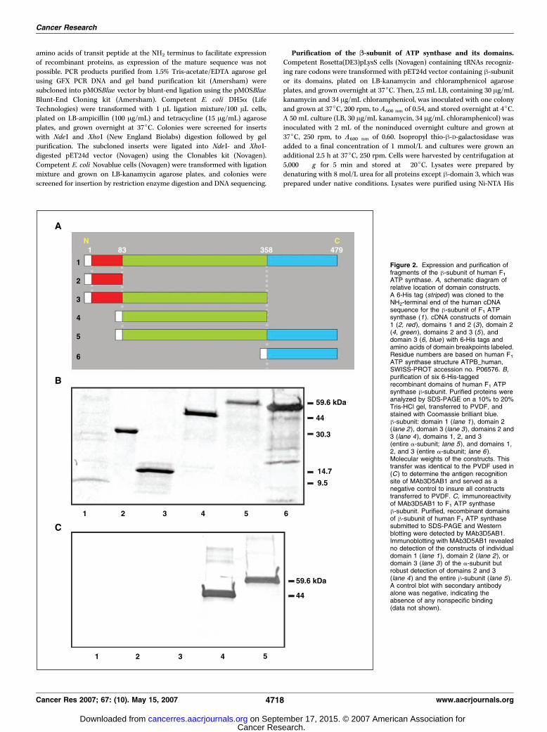
3, domains 1 to 2, and domains 2 to 3 (Fig. 2A). To generate fusion proteins
containing the NH2-terminal 6-His tag with the domains of the h-subunit,primers were designed to introduce a NdeI restriction site at the 5¶ end andan XhoI site at the 3¶ COOH-terminal end (Table 1). Domain boundaries
were determined based on structural motifs (Fig. 1C). Domain 1 included 50
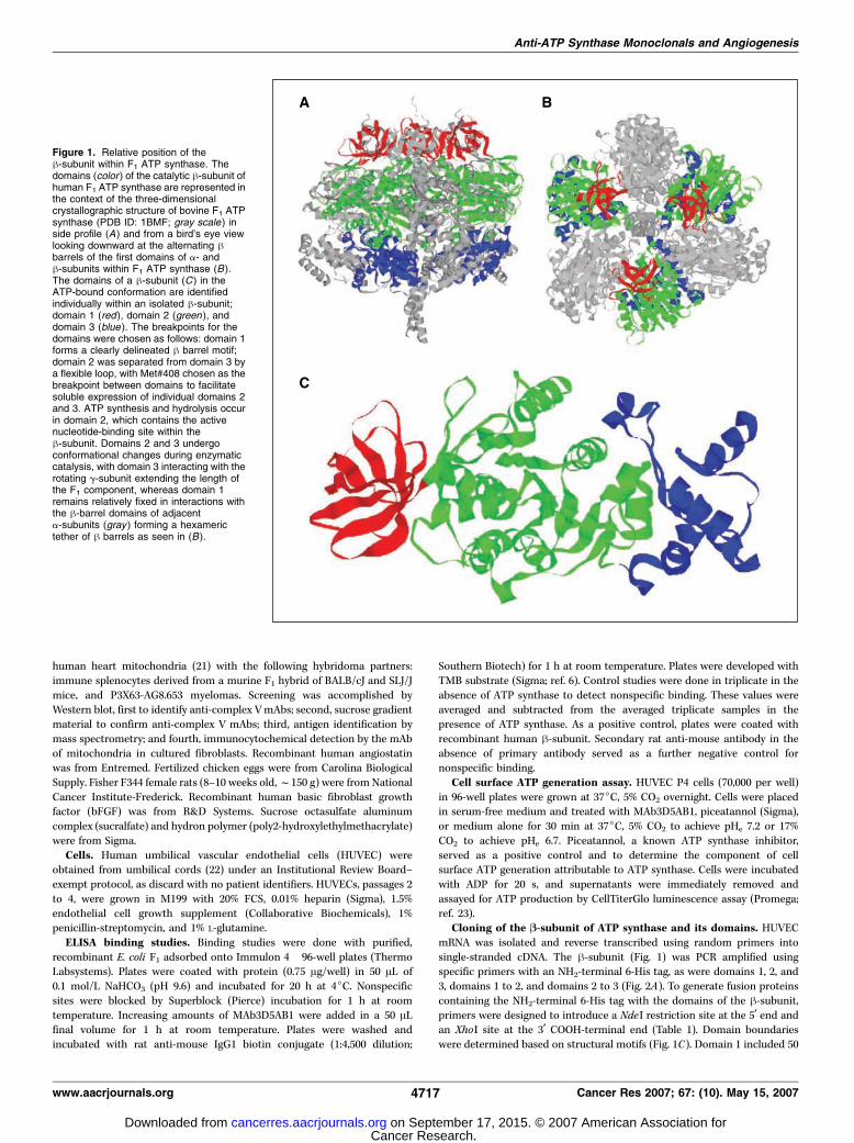
Figure 1. Relative position of theh-subunit within F1 ATP synthase. Thedomains (color ) of the catalytic h-subunit ofhuman F1 ATP synthase are represented inthe context of the three-dimensionalcrystallographic structure of bovine F1 ATPsynthase (PDB ID: 1BMF; gray scale ) inside profile (A) and from a bird’s eye viewlooking downward at the alternating hbarrels of the first domains of a- andh-subunits within F1 ATP synthase (B ).The domains of a h-subunit (C ) in theATP-bound conformation are identifiedindividually within an isolated h-subunit;domain 1 (red ), domain 2 (green ), anddomain 3 (blue ). The breakpoints for thedomains were chosen as follows: domain 1forms a clearly delineated h barrel motif;domain 2 was separated from domain 3 bya flexible loop, with Met#408 chosen as thebreakpoint between domains to facilitatesoluble expression of individual domains 2and 3. ATP synthesis and hydrolysis occurin domain 2, which contains the activenucleotide-binding site within theh-subunit. Domains 2 and 3 undergoconformational changes during enzymaticcatalysis, with domain 3 interacting with therotating g-subunit extending the length ofthe F1 component, whereas domain 1remains relatively fixed in interactions withthe h-barrel domains of adjacenta-subunits (gray ) forming a hexamerictether of h barrels as seen in (B ).
Anti-ATP Synthase Monoclonals and Angiogenesis
www.aacrjournals.org 4717 Cancer Res 2007; 67: (10). May 15, 2007
Cancer Research. on September 17, 2015. © 2007 American Association forcancerres.aacrjournals.org Downloaded from
amino acids of transit peptide at the NH2 terminus to facilitate expressionof recombinant proteins, as expression of the mature sequence was not
possible. PCR products purified from 1.5% Tris-acetate/EDTA agarose gel
using GFX PCR DNA and gel band purification kit (Amersham) were
subcloned into pMOSBlue vector by blunt-end ligation using the pMOSBlueBlunt-End Cloning kit (Amersham). Competent E. coli DH5a (Life
Technologies) were transformed with 1 AL ligation mixture/100 AL cells,
plated on LB-ampicillin (100 Ag/mL) and tetracycline (15 Ag/mL) agarose
plates, and grown overnight at 37jC. Colonies were screened for insertswith NdeI and XhoI (New England Biolabs) digestion followed by gel
purification. The subcloned inserts were ligated into NdeI- and XhoI-
digested pET24d vector (Novagen) using the Clonables kit (Novagen).
Competent E. coli Novablue cells (Novagen) were transformed with ligationmixture and grown on LB-kanamycin agarose plates, and colonies were
screened for insertion by restriction enzyme digestion and DNA sequencing.
Purification of the B-subunit of ATP synthase and its domains.Competent Rosetta(DE3)pLysS cells (Novagen) containing tRNAs recogniz-
ing rare codons were transformed with pET24d vector containing h-subunitor its domains, plated on LB-kanamycin and chloramphenicol agarose
plates, and grown overnight at 37jC. Then, 2.5 mL LB, containing 30 Ag/mLkanamycin and 34 Ag/mL chloramphenicol, was inoculated with one colony
and grown at 37jC, 200 rpm, to A600 nm of 0.54, and stored overnight at 4jC.A 50 mL culture (LB, 30 Ag/mL kanamycin, 34 Ag/mL chloramphenicol) was
inoculated with 2 mL of the noninduced overnight culture and grown at37jC, 250 rpm, to A600 nm of 0.60. Isopropyl thio-h-D-galactosidase was
added to a final concentration of 1 mmol/L and cultures were grown an
additional 2.5 h at 37jC, 250 rpm. Cells were harvested by centrifugation at
5,000 � g for 5 min and stored at �20jC. Lysates were prepared bydenaturing with 8 mol/L urea for all proteins except h-domain 3, which was
prepared under native conditions. Lysates were purified using Ni-NTA His
Figure 2. Expression and purification offragments of the h-subunit of human F1ATP synthase. A, schematic diagram ofrelative location of domain constructs.A 6-His tag (striped ) was cloned to theNH2-terminal end of the human cDNAsequence for the h-subunit of F1 ATPsynthase (1). cDNA constructs of domain1 (2, red ), domains 1 and 2 (3), domain 2(4, green ), domains 2 and 3 (5), anddomain 3 (6, blue ) with 6-His tags andamino acids of domain breakpoints labeled.Residue numbers are based on human F1ATP synthase structure ATPB_human,SWISS-PROT accession no. P06576. B,purification of six 6-His-taggedrecombinant domains of human F1 ATPsynthase h-subunit. Purified proteins wereanalyzed by SDS-PAGE on a 10% to 20%Tris-HCl gel, transferred to PVDF, andstained with Coomassie brilliant blue.h-subunit: domain 1 (lane 1), domain 2(lane 2 ), domain 3 (lane 3), domains 2 and3 (lane 4), domains 1, 2, and 3(entire a-subunit; lane 5), and domains 1,2, and 3 (entire a-subunit; lane 6 ).Molecular weights of the constructs. Thistransfer was identical to the PVDF used in(C) to determine the antigen recognitionsite of MAb3D5AB1 and served as anegative control to insure all constructstransferred to PVDF. C, immunoreactivityof MAb3D5AB1 to F1 ATP synthaseh-subunit. Purified, recombinant domainsof h-subunit of human F1 ATP synthasesubmitted to SDS-PAGE and Westernblotting were detected by MAb3D5AB1.Immunoblotting with MAb3D5AB1 revealedno detection of the constructs of individualdomain 1 (lane 1 ), domain 2 (lane 2), ordomain 3 (lane 3 ) of the a-subunit butrobust detection of domains 2 and 3(lane 4) and the entire h-subunit (lane 5).A control blot with secondary antibodyalone was negative, indicating theabsence of any nonspecific binding(data not shown).
Cancer Research
Cancer Res 2007; 67: (10). May 15, 2007 4718 www.aacrjournals.org
Cancer Research. on September 17, 2015. © 2007 American Association forcancerres.aacrjournals.org Downloaded from
Bind Resin columns (Novagen) and the resulting proteins were dialyzedagainst PBS (pH 7.0).
Western blotting. Purified, recombinant h-domain proteins (f1 Ag)under reducing conditions were separated by SDS-PAGE and transferred to
polyvinylidene difluoride (PVDF; Roche). Membranes were blocked
overnight at 4jC, incubated with MAb3D5AB1 at 2.5 mg/mL for 2 h at
room temperature, and washed. Membranes were incubated with
antimouse alkaline phosphatase conjugate (Sigma), washed, and developed
with 5-bromo-4-chloroindol-3-yl-phosphate nitroblue tetrazolium. To insure
that the secondary detection antibody did not contribute to background
detection, a membrane was developed with secondary antibody alone. To
confirm transfer of all protein domains to the membrane, a post-transfer
membrane was stained with Coomassie brilliant blue.
ATP synthase F1 activity assay. Fresh bovine heart mitochondria were
sonicated to yield submitochondrial particles. F1 was separated from
membrane-bound F0 by chloroform extraction and purified (6). Purified F1exhibits ATP hydrolytic activity alone, as ATP synthesis requires association
with F0 to couple the proton gradient across F0 with the rotational motion
of F1. Therefore, we measured F1 ATP hydrolytic activity by coupling ADP
generation to NADH oxidation via pyruvate kinase and lactate dehydroge-
nase reactions (24).
Fluorescence microscopy. HUVECs were plated in Matrigel-coated (BD
Biosciences) eight-well slides at a density of 70% confluency. Cells werewashed, fixed under nonpermeabilized conditions, and blocked (17). Cells
were incubated with a mAb to ATP synthase (1:50 in DPBS) for 1 h/37jC.Cells were washed, incubated with a Cy3 secondary antibody, and visualized
along with identification of endothelial cell nuclei as described (17). Foreach treatment, 10 fields of cells were examined.
HUVEC tube formation on Matrigel. Plates (Collaborative Biomedicals)
were coated with Matrigel at 4jC (300 AL/well at 15 mg/mL) and incubatedat 37jC/30 min. HUVECs (50,000 per well) were plated and incubated for
6 h in the presence or absence of MAb3D5AB1 (10–100 Ag/mL) in normal-
or low-pH medium at 37jC, 5% CO2 to allow tube formation. Cells were
fixed, stained, and quantified in triplicate (17, 18).pHi measurements. pHi was measured by fluorescence in HUVEC
(17, 18). Cells were incubated for 15 min with 9 Amol/L carboxy snarf-1
acetoxymethyl ester (Molecular Probes) in medium containing 10% fetalbovine serum at 37jC, 5% CO2 (25, 26). After steady-state readings were
established in medium (pH 7.4), it was removed, and 1 mL medium
(pH 6.7) with or without MAb3D5Ab1 (10 Ag/mL final concentration)
was introduced. Experiments were repeated at least twice, with three tofive measurements per field. pHi was obtained from the whole emission
spectra emitted by the cells and collected on an inverted microscope
(Nikon Diaphot, Nikon, Inc.; refs. 17, 18). Calibration procedures are as
described (27); however, 140 wavelengths (whole fluorescence spectra)were used to calculate pHi (17, 18, 25, 28).
pHe measurements. pHe measurements in the CAM assay were made
using a Sentron ISFET pH meter, designed for use with solids and viscous
materials. Corneal micropocket pH measurements were made with aFisher accumet pH meter equipped with a 21-gauge protected needle
microelectrode for pH and a flexible microreference electrode from
Microelectrodes, Inc.Rat corneal neovascularization assay. Animal experiments were done
as approved by the Duke University Institutional Animal Care and Use
Committee. The rat corneal neovascularization assay was done as described
(29). Sustained-release pellets contained equal volumes of Hydron (12% w/vin ethanol) and a solution of bFGF (100 ng), sucralfate (100 Ag), and drug or
PBS (negative control). MAb3D5AB1 was used at 650 ng/pellet and
angiostatin at 20 Ag/pellet. Vascular area density and average vessel length
were measured by an investigator blind to treatment assignment usingImageJ software (29).
Chicken CAM assay. The CAM assay was done as described (30). Test
compounds (bFGF, angiostatin F bFGF, MAb3D5AB1 F bFGF, or PBS) weredried onto a sterile quarter of a 13-mm Thermanox disc (Nunc) and placed
onto the CAM on day 10. After 70 F 4 h, CAMs were photographed and
analyzed by an investigator blind to treatment assignment.
Results
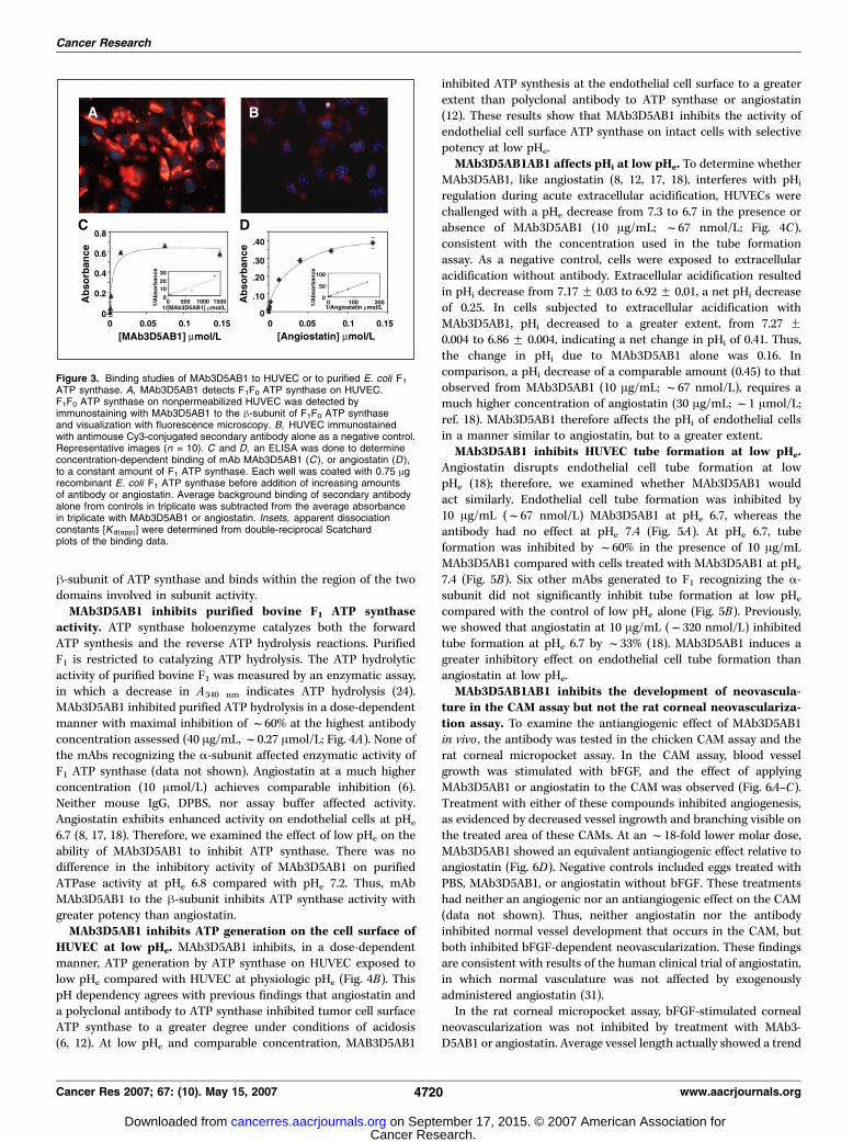
MAb3D5AB1 binding studies. We have identified ATP synthaseon the endothelial cell surface (6, 7). We now show that MAb3D5AB1targeting the human h-subunit also recognizes ATP synthase onnonpermeabilized HUVEC as visualized by fluorescence microscopy(Fig. 3A and B). MAb3D5AB1 binds to purified, recombinant E. coliF1 in a concentration-dependent and saturable manner (Fig. 3C).Human and E. coli ATP synthase exhibit 70% homology in thea-subunit (SWISS-PROT accession nos. P25705 and P00822,respectively) and 80% homology in the h-subunit (P06576 andP00824). Background binding of MAb3D5AB1 to BSA-coated wellswas comparable with the baseline, indicating that MAb3D5AB1binds specifically to F1. Apparent dissociation constants [Kd(app)]were determined from double-reciprocal Scatchard plots of thebinding data (Fig. 3C, inset). The Kd(app) for MAb3D5AB1 binding topurified E. coli F1 is 16 nmol/L compared with 405 nmol/L forangiostatin binding to E. coli F1 (6). Thus, MAb3D5AB1 targets theangiostatin receptor ATP synthase with a binding affinity 25-foldgreater than angiostatin.Mapping of the MAb3D5AB1 antibody recognition site. For
epitope mapping, a series of expression constructs of human F1domains were generated. These included the a-subunit, h-subunit,individual domains 1, 2, and 3 of the h-subunit, and a constructcomprising domains 2 and 3 (Fig. 2B). The construct spanningdomains 1 and 2 could not be expressed. By Western blot assays,MAb3D5AB1 strongly detected the h-subunit domains 2 and 3 andfull-length recombinant h-subunit (Fig. 2C). MAb3D5AB1 did notrecognize either the individual second or third domains of theh-subunit (Fig. 2C) nor the a-subunit of F1, included as a negativecontrol (data not shown). The remainder of the mAbs assessedrecognized the a-subunit but not the h-subunit of F1 (data notshown). These results show that MAb3D5AB1 targets the catalytic
Table 1. Primer sequences used for construction of 6-His-tagged domains of the h-subunit of human F1 ATPsynthase
Domain 5¶ Primer sequence 3¶ Primer sequence
1 CTTGGTCATATGCACCACCACCACCACC
ACACATCTCCTTC
GCCAAAAGC
CGGTGACTCGAGTTAGATT
GGTGCACCAG
AATCCAGTAC2 CTTCGTCATATGCACCA
CCACCACCACCACAA
AATTCCTGTTGGTCCT
GAGACTTTGGGC
CGGTGACTCGAGTTAGATAC
GAGAGGTGG
AGTCTAGAG3 CTTCGTCATATGCAC
CACCACCACCACC
ACATGGATCCCAA
CATTGTTGGCAG
CGGTGACTCGAGtcacgat
gaatgctctt
cagcca
NOTE: Sequences are based on the cDNA of human F1 ATP synthase
(protein accession no. X03559, italics). Restriction enzyme sites, in
bold, represent for 5¶ end primers, an NdeI cleavage site, and for 3¶primers, an XhoI site. Primers read from 5¶ to 3¶ with respect to cDNA.
For constructs spanning two domains, the appropriate combinations
of primers were used. For example, for domains 2 and 3, the 5¶ primer
for domain 2 was used in conjunction with the 3¶ primer for domain 3.
Anti-ATP Synthase Monoclonals and Angiogenesis
www.aacrjournals.org 4719 Cancer Res 2007; 67: (10). May 15, 2007
Cancer Research. on September 17, 2015. © 2007 American Association forcancerres.aacrjournals.org Downloaded from
h-subunit of ATP synthase and binds within the region of the twodomains involved in subunit activity.MAb3D5AB1 inhibits purified bovine F1 ATP synthase
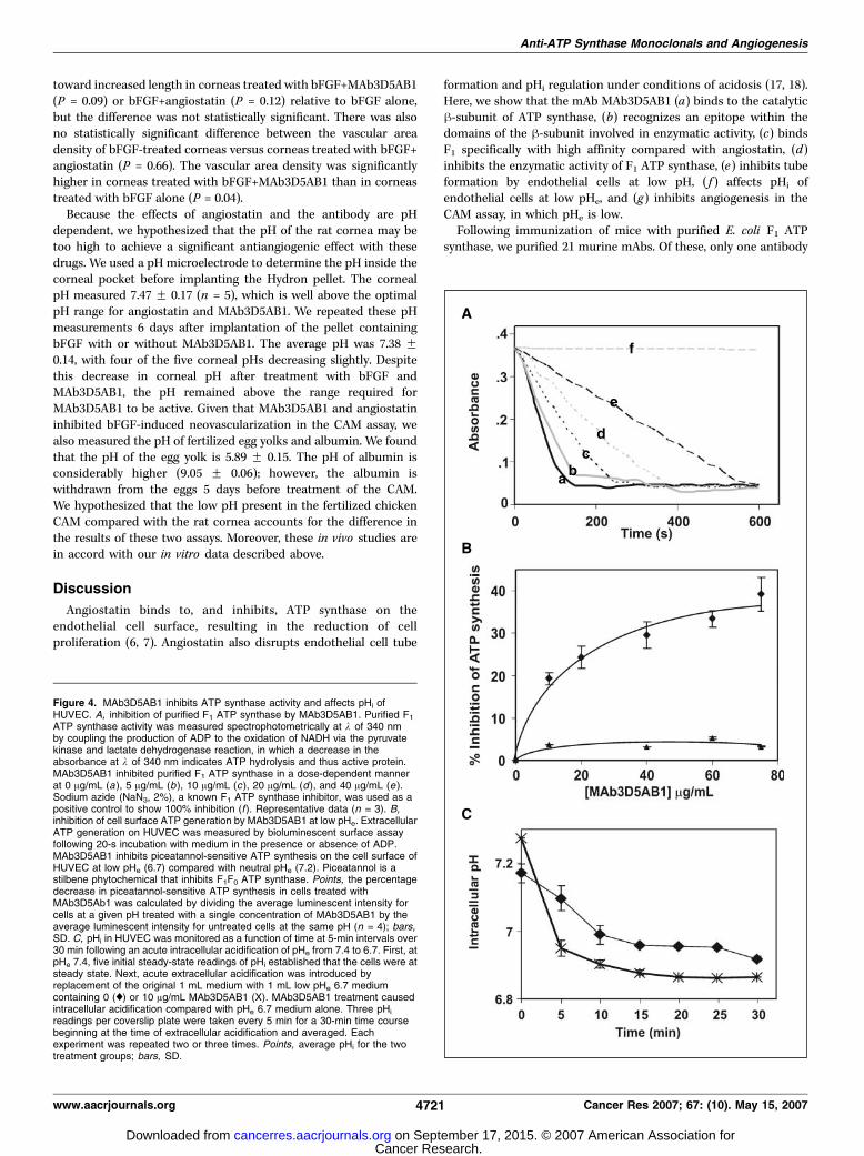
activity. ATP synthase holoenzyme catalyzes both the forwardATP synthesis and the reverse ATP hydrolysis reactions. PurifiedF1 is restricted to catalyzing ATP hydrolysis. The ATP hydrolyticactivity of purified bovine F1 was measured by an enzymatic assay,in which a decrease in A340 nm indicates ATP hydrolysis (24).MAb3D5AB1 inhibited purified ATP hydrolysis in a dose-dependentmanner with maximal inhibition of f60% at the highest antibodyconcentration assessed (40 Ag/mL, f0.27 Amol/L; Fig. 4A). None ofthe mAbs recognizing the a-subunit affected enzymatic activity ofF1 ATP synthase (data not shown). Angiostatin at a much higherconcentration (10 Amol/L) achieves comparable inhibition (6).Neither mouse IgG, DPBS, nor assay buffer affected activity.Angiostatin exhibits enhanced activity on endothelial cells at pHe
6.7 (8, 17, 18). Therefore, we examined the effect of low pHe on theability of MAb3D5AB1 to inhibit ATP synthase. There was nodifference in the inhibitory activity of MAb3D5AB1 on purifiedATPase activity at pHe 6.8 compared with pHe 7.2. Thus, mAbMAb3D5AB1 to the h-subunit inhibits ATP synthase activity withgreater potency than angiostatin.MAb3D5AB1 inhibits ATP generation on the cell surface of
HUVEC at low pHe. MAb3D5AB1 inhibits, in a dose-dependentmanner, ATP generation by ATP synthase on HUVEC exposed tolow pHe compared with HUVEC at physiologic pHe (Fig. 4B). ThispH dependency agrees with previous findings that angiostatin anda polyclonal antibody to ATP synthase inhibited tumor cell surfaceATP synthase to a greater degree under conditions of acidosis(6, 12). At low pHe and comparable concentration, MAB3D5AB1
inhibited ATP synthesis at the endothelial cell surface to a greaterextent than polyclonal antibody to ATP synthase or angiostatin(12). These results show that MAb3D5AB1 inhibits the activity ofendothelial cell surface ATP synthase on intact cells with selectivepotency at low pHe.MAb3D5AB1AB1 affects pHi at low pHe. To determine whether
MAb3D5AB1, like angiostatin (8, 12, 17, 18), interferes with pHi
regulation during acute extracellular acidification, HUVECs werechallenged with a pHe decrease from 7.3 to 6.7 in the presence orabsence of MAb3D5AB1 (10 Ag/mL; f67 nmol/L; Fig. 4C),consistent with the concentration used in the tube formationassay. As a negative control, cells were exposed to extracellularacidification without antibody. Extracellular acidification resultedin pHi decrease from 7.17 F 0.03 to 6.92 F 0.01, a net pHi decreaseof 0.25. In cells subjected to extracellular acidification withMAb3D5AB1, pHi decreased to a greater extent, from 7.27 F0.004 to 6.86 F 0.004, indicating a net change in pHi of 0.41. Thus,the change in pHi due to MAb3D5AB1 alone was 0.16. Incomparison, a pHi decrease of a comparable amount (0.45) to thatobserved from MAb3D5AB1 (10 Ag/mL; f67 nmol/L), requires amuch higher concentration of angiostatin (30 Ag/mL; f1 Amol/L;ref. 18). MAb3D5AB1 therefore affects the pHi of endothelial cellsin a manner similar to angiostatin, but to a greater extent.MAb3D5AB1 inhibits HUVEC tube formation at low pHe.
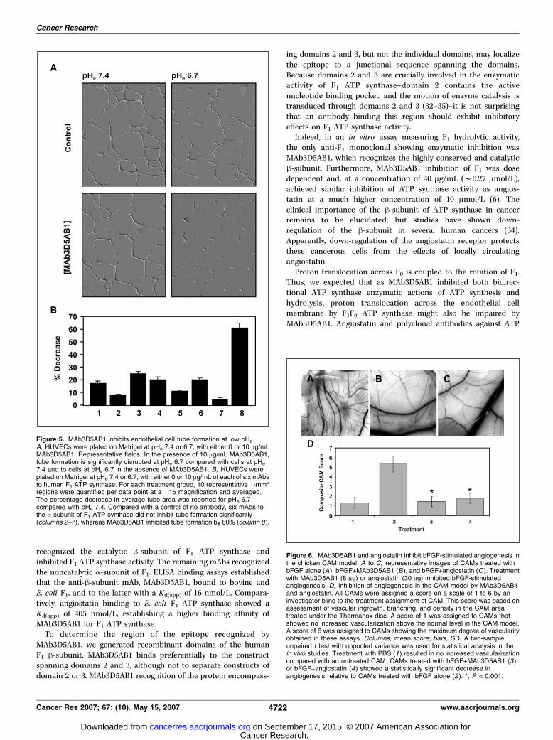
Angiostatin disrupts endothelial cell tube formation at lowpHe (18); therefore, we examined whether MAb3D5AB1 wouldact similarly. Endothelial cell tube formation was inhibited by10 Ag/mL (f67 nmol/L) MAb3D5AB1 at pHe 6.7, whereas theantibody had no effect at pHe 7.4 (Fig. 5A). At pHe 6.7, tubeformation was inhibited by f60% in the presence of 10 Ag/mLMAb3D5AB1 compared with cells treated with MAb3D5AB1 at pHe
7.4 (Fig. 5B). Six other mAbs generated to F1 recognizing the a-subunit did not significantly inhibit tube formation at low pHe
compared with the control of low pHe alone (Fig. 5B). Previously,we showed that angiostatin at 10 Ag/mL (f320 nmol/L) inhibitedtube formation at pHe 6.7 by f33% (18). MAb3D5AB1 induces agreater inhibitory effect on endothelial cell tube formation thanangiostatin at low pHe.MAb3D5AB1AB1 inhibits the development of neovascula-
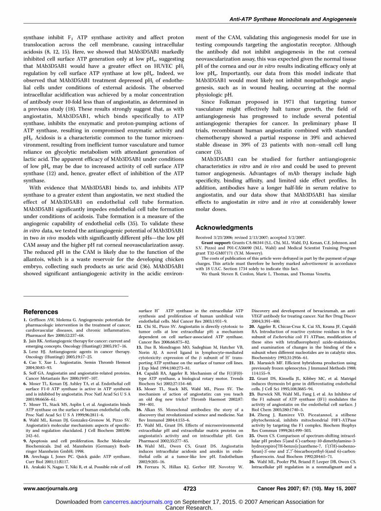
ture in the CAM assay but not the rat corneal neovasculariza-tion assay. To examine the antiangiogenic effect of MAb3D5AB1in vivo , the antibody was tested in the chicken CAM assay and therat corneal micropocket assay. In the CAM assay, blood vesselgrowth was stimulated with bFGF, and the effect of applyingMAb3D5AB1 or angiostatin to the CAM was observed (Fig. 6A–C).Treatment with either of these compounds inhibited angiogenesis,as evidenced by decreased vessel ingrowth and branching visible onthe treated area of these CAMs. At an f18-fold lower molar dose,MAb3D5AB1 showed an equivalent antiangiogenic effect relative toangiostatin (Fig. 6D). Negative controls included eggs treated withPBS, MAb3D5AB1, or angiostatin without bFGF. These treatmentshad neither an angiogenic nor an antiangiogenic effect on the CAM(data not shown). Thus, neither angiostatin nor the antibodyinhibited normal vessel development that occurs in the CAM, butboth inhibited bFGF-dependent neovascularization. These findingsare consistent with results of the human clinical trial of angiostatin,in which normal vasculature was not affected by exogenouslyadministered angiostatin (31).
In the rat corneal micropocket assay, bFGF-stimulated cornealneovascularization was not inhibited by treatment with MAb3-D5AB1 or angiostatin. Average vessel length actually showed a trend
Figure 3. Binding studies of MAb3D5AB1 to HUVEC or to purified E. coli F1ATP synthase. A, MAb3D5AB1 detects F1F0 ATP synthase on HUVEC.F1F0 ATP synthase on nonpermeabilized HUVEC was detected byimmunostaining with MAb3D5AB1 to the h-subunit of F1F0 ATP synthaseand visualization with fluorescence microscopy. B, HUVEC immunostainedwith antimouse Cy3-conjugated secondary antibody alone as a negative control.Representative images (n = 10). C and D, an ELISA was done to determineconcentration-dependent binding of mAb MAb3D5AB1 (C ), or angiostatin (D ),to a constant amount of F1 ATP synthase. Each well was coated with 0.75 Agrecombinant E. coli F1 ATP synthase before addition of increasing amountsof antibody or angiostatin. Average background binding of secondary antibodyalone from controls in triplicate was subtracted from the average absorbancein triplicate with MAb3D5AB1 or angiostatin. Insets, apparent dissociationconstants [Kd(app)] were determined from double-reciprocal Scatchardplots of the binding data.
Cancer Research
Cancer Res 2007; 67: (10). May 15, 2007 4720 www.aacrjournals.org
Cancer Research. on September 17, 2015. © 2007 American Association forcancerres.aacrjournals.org Downloaded from
toward increased length in corneas treated with bFGF+MAb3D5AB1(P = 0.09) or bFGF+angiostatin (P = 0.12) relative to bFGF alone,but the difference was not statistically significant. There was alsono statistically significant difference between the vascular areadensity of bFGF-treated corneas versus corneas treated with bFGF+angiostatin (P = 0.66). The vascular area density was significantlyhigher in corneas treated with bFGF+MAb3D5AB1 than in corneastreated with bFGF alone (P = 0.04).
Because the effects of angiostatin and the antibody are pHdependent, we hypothesized that the pH of the rat cornea may betoo high to achieve a significant antiangiogenic effect with thesedrugs. We used a pH microelectrode to determine the pH inside thecorneal pocket before implanting the Hydron pellet. The cornealpH measured 7.47 F 0.17 (n = 5), which is well above the optimalpH range for angiostatin and MAb3D5AB1. We repeated these pHmeasurements 6 days after implantation of the pellet containingbFGF with or without MAb3D5AB1. The average pH was 7.38 F0.14, with four of the five corneal pHs decreasing slightly. Despitethis decrease in corneal pH after treatment with bFGF andMAb3D5AB1, the pH remained above the range required forMAb3D5AB1 to be active. Given that MAb3D5AB1 and angiostatininhibited bFGF-induced neovascularization in the CAM assay, wealso measured the pH of fertilized egg yolks and albumin. We foundthat the pH of the egg yolk is 5.89 F 0.15. The pH of albumin isconsiderably higher (9.05 F 0.06); however, the albumin iswithdrawn from the eggs 5 days before treatment of the CAM.We hypothesized that the low pH present in the fertilized chickenCAM compared with the rat cornea accounts for the difference inthe results of these two assays. Moreover, these in vivo studies arein accord with our in vitro data described above.
Discussion
Angiostatin binds to, and inhibits, ATP synthase on theendothelial cell surface, resulting in the reduction of cellproliferation (6, 7). Angiostatin also disrupts endothelial cell tube
formation and pHi regulation under conditions of acidosis (17, 18).Here, we show that the mAb MAb3D5AB1 (a) binds to the catalytich-subunit of ATP synthase, (b) recognizes an epitope within thedomains of the h-subunit involved in enzymatic activity, (c) bindsF1 specifically with high affinity compared with angiostatin, (d)inhibits the enzymatic activity of F1 ATP synthase, (e) inhibits tubeformation by endothelial cells at low pH, ( f ) affects pHi ofendothelial cells at low pHe, and (g ) inhibits angiogenesis in theCAM assay, in which pHe is low.
Following immunization of mice with purified E. coli F1 ATPsynthase, we purified 21 murine mAbs. Of these, only one antibody
Figure 4. MAb3D5AB1 inhibits ATP synthase activity and affects pHi ofHUVEC. A, inhibition of purified F1 ATP synthase by MAb3D5AB1. Purified F1ATP synthase activity was measured spectrophotometrically at k of 340 nmby coupling the production of ADP to the oxidation of NADH via the pyruvatekinase and lactate dehydrogenase reaction, in which a decrease in theabsorbance at k of 340 nm indicates ATP hydrolysis and thus active protein.MAb3D5AB1 inhibited purified F1 ATP synthase in a dose-dependent mannerat 0 Ag/mL (a), 5 Ag/mL (b), 10 Ag/mL (c ), 20 Ag/mL (d ), and 40 Ag/mL (e ).Sodium azide (NaN3, 2%), a known F1 ATP synthase inhibitor, was used as apositive control to show 100% inhibition (f ). Representative data (n = 3). B,inhibition of cell surface ATP generation by MAb3D5AB1 at low pHe. ExtracellularATP generation on HUVEC was measured by bioluminescent surface assayfollowing 20-s incubation with medium in the presence or absence of ADP.MAb3D5AB1 inhibits piceatannol-sensitive ATP synthesis on the cell surface ofHUVEC at low pHe (6.7) compared with neutral pHe (7.2). Piceatannol is astilbene phytochemical that inhibits F1F0 ATP synthase. Points, the percentagedecrease in piceatannol-sensitive ATP synthesis in cells treated withMAb3D5Ab1 was calculated by dividing the average luminescent intensity forcells at a given pH treated with a single concentration of MAb3D5AB1 by theaverage luminescent intensity for untreated cells at the same pH (n = 4); bars,SD. C, pHi in HUVEC was monitored as a function of time at 5-min intervals over30 min following an acute intracellular acidification of pHe from 7.4 to 6.7. First, atpHe 7.4, five initial steady-state readings of pHi established that the cells were atsteady state. Next, acute extracellular acidification was introduced byreplacement of the original 1 mL medium with 1 mL low pHe 6.7 mediumcontaining 0 (x) or 10 Ag/mL MAb3D5AB1 (X). MAb3D5AB1 treatment causedintracellular acidification compared with pHe 6.7 medium alone. Three pHi
readings per coverslip plate were taken every 5 min for a 30-min time coursebeginning at the time of extracellular acidification and averaged. Eachexperiment was repeated two or three times. Points, average pHi for the twotreatment groups; bars, SD.
Anti-ATP Synthase Monoclonals and Angiogenesis
www.aacrjournals.org 4721 Cancer Res 2007; 67: (10). May 15, 2007
Cancer Research. on September 17, 2015. © 2007 American Association forcancerres.aacrjournals.org Downloaded from
recognized the catalytic h-subunit of F1 ATP synthase andinhibited F1 ATP synthase activity. The remaining mAbs recognizedthe noncatalytic a-subunit of F1. ELISA binding assays establishedthat the anti-h-subunit mAb, MAb3D5AB1, bound to bovine andE. coli F1, and to the latter with a Kd(app) of 16 nmol/L. Compara-tively, angiostatin binding to E. coli F1 ATP synthase showed aKd(app) of 405 nmol/L, establishing a higher binding affinity ofMAb3D5AB1 for F1 ATP synthase.
To determine the region of the epitope recognized byMAb3D5AB1, we generated recombinant domains of the humanF1 h-subunit. MAb3D5AB1 binds preferentially to the constructspanning domains 2 and 3, although not to separate constructs ofdomain 2 or 3. MAb3D5AB1 recognition of the protein encompass-
ing domains 2 and 3, but not the individual domains, may localizethe epitope to a junctional sequence spanning the domains.Because domains 2 and 3 are crucially involved in the enzymaticactivity of F1 ATP synthase–domain 2 contains the activenucleotide binding pocket, and the motion of enzyme catalysis istransduced through domains 2 and 3 (32–35)–it is not surprisingthat an antibody binding this region should exhibit inhibitoryeffects on F1 ATP synthase activity.
Indeed, in an in vitro assay measuring F1 hydrolytic activity,the only anti-F1 monoclonal showing enzymatic inhibition wasMAb3D5AB1, which recognizes the highly conserved and catalytich-subunit. Furthermore, MAb3D5AB1 inhibition of F1 was dosedependent and, at a concentration of 40 Ag/mL (f0.27 Amol/L),achieved similar inhibition of ATP synthase activity as angios-tatin at a much higher concentration of 10 Amol/L (6). Theclinical importance of the h-subunit of ATP synthase in cancerremains to be elucidated, but studies have shown down-regulation of the h-subunit in several human cancers (34).Apparently, down-regulation of the angiostatin receptor protectsthese cancerous cells from the effects of locally circulatingangiostatin.
Proton translocation across F0 is coupled to the rotation of F1.Thus, we expected that as MAb3D5AB1 inhibited both bidirec-tional ATP synthase enzymatic actions of ATP synthesis andhydrolysis, proton translocation across the endothelial cellmembrane by F1F0 ATP synthase might also be impaired byMAb3D5AB1. Angiostatin and polyclonal antibodies against ATP
Figure 6. MAb3D5AB1 and angiostatin inhibit bFGF-stimulated angiogenesis inthe chicken CAM model. A to C, representative images of CAMs treated withbFGF alone (A), bFGF+MAb3D5AB1 (B ), and bFGF+angiostatin (C ). Treatmentwith MAb3D5AB1 (8 Ag) or angiostatin (30 Ag) inhibited bFGF-stimulatedangiogenesis. D, inhibition of angiogenesis in the CAM model by MAb3D5AB1and angiostatin. All CAMs were assigned a score on a scale of 1 to 6 by aninvestigator blind to the treatment assignment of CAM. This score was based onassessment of vascular ingrowth, branching, and density in the CAM areatreated under the Thermanox disc. A score of 1 was assigned to CAMs thatshowed no increased vascularization above the normal level in the CAM model.A score of 6 was assigned to CAMs showing the maximum degree of vascularityobtained in these assays. Columns, mean score; bars, SD. A two-sampleunpaired t test with unpooled variance was used for statistical analysis in thein vivo studies. Treatment with PBS (1 ) resulted in no increased vascularizationcompared with an untreated CAM. CAMs treated with bFGF+MAb3D5AB1 (3)or bFGF+angiostatin (4) showed a statistically significant decrease inangiogenesis relative to CAMs treated with bFGF alone (2). *, P < 0.001.
Figure 5. MAb3D5AB1 inhibits endothelial cell tube formation at low pHe.A, HUVECs were plated on Matrigel at pHe 7.4 or 6.7, with either 0 or 10 Ag/mLMAb3D5AB1. Representative fields. In the presence of 10 Ag/mL MAb3D5AB1,tube formation is significantly disrupted at pHe 6.7 compared with cells at pHe
7.4 and to cells at pHe 6.7 in the absence of MAb3D5AB1. B, HUVECs wereplated on Matrigel at pHe 7.4 or 6.7, with either 0 or 10 Ag/mL of each of six mAbsto human F1 ATP synthase. For each treatment group, 10 representative 1-mm2
regions were quantified per data point at a �15 magnification and averaged.The percentage decrease in average tube area was reported for pHe 6.7compared with pHe 7.4. Compared with a control of no antibody, six mAbs tothe a-subunit of F1 ATP synthase did not inhibit tube formation significantly(columns 2–7), whereas MAb3D5AB1 inhibited tube formation by 60% (column 8).
Cancer Research
Cancer Res 2007; 67: (10). May 15, 2007 4722 www.aacrjournals.org
Cancer Research. on September 17, 2015. © 2007 American Association forcancerres.aacrjournals.org Downloaded from
synthase inhibit F1 ATP synthase activity and affect protontranslocation across the cell membrane, causing intracellularacidosis (8, 12, 15). Here, we showed that MAb3D5AB1 markedlyinhibited cell surface ATP generation only at low pHe, suggestingthat MAb3D5AB1 would have a greater effect on HUVEC pHi
regulation by cell surface ATP synthase at low pHe. Indeed, weobserved that MAb3D5AB1 treatment depressed pHi of endothe-lial cells under conditions of external acidosis. The observedintracellular acidification was achieved by a molar concentrationof antibody over 10-fold less than of angiostatin, as determined ina previous study (18). These results strongly suggest that, as withangiostatin, MAb3D5AB1, which binds specifically to ATPsynthase, inhibits the enzymatic and proton-pumping actions ofATP synthase, resulting in compromised enzymatic activity andpHi. Acidosis is a characteristic common to the tumor microen-vironment, resulting from inefficient tumor vasculature and tumorreliance on glycolytic metabolism with attendant generation oflactic acid. The apparent efficacy of MAb3D5AB1 under conditionsof low pHe may be due to increased activity of cell surface ATPsynthase (12) and, hence, greater effect of inhibition of the ATPsynthase.
With evidence that MAb3D5AB1 binds to, and inhibits ATPsynthase to a greater extent than angiostatin, we next studied theeffect of MAb3D5AB1 on endothelial cell tube formation.MAb3D5AB1 significantly impedes endothelial cell tube formationunder conditions of acidosis. Tube formation is a measure of theangiogenic capability of endothelial cells (35). To validate thesein vitro data, we tested the antiangiogenic potential of MAb3D5AB1in two in vivo models with significantly different pHs—the low pHCAM assay and the higher pH rat corneal neovascularization assay.The reduced pH in the CAM is likely due to the function of theallantois, which is a waste reservoir for the developing chickenembryo, collecting such products as uric acid (36). MAb3D5AB1showed significant antiangiogenic activity in the acidic environ-
ment of the CAM, validating this angiogenesis model for use intesting compounds targeting the angiostatin receptor. Althoughthe antibody did not inhibit angiogenesis in the rat cornealneovascularization assay, this was expected given the normal tissuepH of the cornea and our in vitro results indicating efficacy only atlow pHe. Importantly, our data from this model indicate thatMAb3D5AB1 would most likely not inhibit nonpathologic angio-genesis, such as in wound healing, occurring at the normalphysiologic pH.
Since Folkman proposed in 1971 that targeting tumorvasculature might effectively halt tumor growth, the field ofantiangiogenesis has progressed to include several potentialantiangiogenic therapies for cancer. In preliminary phase IItrials, recombinant human angiostatin combined with standardchemotherapy showed a partial response in 39% and achievedstable disease in 39% of 23 patients with non–small cell lungcancer (3).
MAb3D5AB1 can be studied for further antiangiogeniccharacteristics in vitro and in vivo and could be used to preventtumor angiogenesis. Advantages of mAb therapy include highspecificity, binding affinity, and limited side effect profiles. Inaddition, antibodies have a longer half-life in serum relative toangiostatin, and our data show that MAb3D5AB1 has similareffects to angiostatin in vitro and in vivo at considerably lowermolar doses.
Acknowledgments
Received 3/23/2006; revised 2/13/2007; accepted 3/2/2007.Grant support: Grants CA-86344 (S.L. Chi, M.L. Wahl, D.J. Kenan, C.E. Johnson, and
S.V. Pizzo) and P01-CA56690 (M.L. Wahl) and Medical Scientist Training Programgrant T32-GM07171 (Y.M. Mowery).
The costs of publication of this article were defrayed in part by the payment of pagecharges. This article must therefore be hereby marked advertisement in accordancewith 18 U.S.C. Section 1734 solely to indicate this fact.
We thank Steven R. Conlon, Marie L. Thomas, and Thomas Venetta.
Anti-ATP Synthase Monoclonals and Angiogenesis
www.aacrjournals.org 4723 Cancer Res 2007; 67: (10). May 15, 2007
References1. Griffioen AW, Molema G. Angiogenesis: potentials forpharmacologic intervention in the treatment of cancer,cardiovascular diseases, and chronic inflammation.Pharmacol Rev 2000;52:237–68.
2. Jain RK. Antiangiogenic therapy for cancer: current andemerging concepts. Oncology (Huntingt) 2005;19:7–16.
3. Lenz HJ. Antiangiogenic agents in cancer therapy.Oncology (Huntingt) 2005;19:17–25.
4. Cao Y, Xue L. Angiostatin. Semin Thromb Hemost2004;30:83–93.
5. Soff GA. Angiostatin and angiostatin-related proteins.Cancer Metastasis Rev 2000;19:97–107.
6. Moser TL, Kenan DJ, Ashley TA, et al. Endothelial cellsurface F1-0 ATP synthase is active in ATP synthesisand is inhibited by angiostatin. Proc Natl Acad Sci U S A2001;98:6656–61.
7. Moser TL, Stack MS, Asplin I, et al. Angiostatin bindsATP synthase on the surface of human endothelial cells.Proc Natl Acad Sci U S A 1999;96:2811–6.
8. Wahl ML, Kenan DJ, Gonzalez-Gronow M, Pizzo SV.Angiostatin’s molecular mechanism: aspects of specific-ity and regulation elucidated. J Cell Biochem 2005;96:242–61.
9. Apoptosis and cell proliferation. Roche MolecularBiochemicals. 2nd ed. Mannheim (Germany): Boeh-ringer Mannheim GmbH; 1998.
10. Arechaga I, Jones PC. Quick guide: ATP synthase.Curr Biol 2001;11:R117.
11. Arakaki N, Nagao T, Niki R, et al. Possible role of cell
surface H+ �ATP synthase in the extracellular ATPsynthesis and proliferation of human umbilical veinendothelial cells. Mol Cancer Res 2003;1:931–9.
12. Chi SL, Pizzo SV. Angiostatin is directly cytotoxic totumor cells at low extracellular pH: a mechanismdependent on cell surface-associated ATP synthase.Cancer Res 2006;66:875–82.
13. Das B, Mondragon MO, Sadeghian M, Hatcher VB,Norin AJ. A novel ligand in lymphocyte-mediatedcytotoxicity: expression of the h subunit of H+ trans-porting ATP synthase on the surface of tumor cell lines.J Exp Med 1994;180:273–81.
14. Capaldi RA, Aggeler R. Mechanism of the F(1)F(0)-type ATP synthase, a biological rotary motor. TrendsBiochem Sci 2002;27:154–60.
15. Moser TL, Stack MS, Wahl ML, Pizzo SV. Themechanism of action of angiostatin: can you teachan old dog new tricks? Thromb Haemost 2002;87:394–401.
16. Alkan SS. Monoclonal antibodies: the story of adiscovery that revolutionized science and medicine. NatRev Immunol 2004;4:153–6.
17. Wahl ML, Grant DS. Effects of microenvironmentalextracellular pH and extracellular matrix proteins onangiostatin’s activity and on intracellular pH. GenPharmacol 2002;35:277–85.
18. Wahl ML, Owen CS, Grant DS. Angiostatininduces intracellular acidosis and anoikis in endo-thelial cells at a tumor-like low pH. Endothelium2002;9:205–16.
19. Ferrara N, Hillan KJ, Gerber HP, Novotny W.
Discovery and development of bevacizumab, an anti-VEGF antibody for treating cancer. Nat Rev Drug Discov2004;3:391–400.
20. Aggeler R, Chicas-Cruz K, Cai SX, Keana JF, CapaldiRA. Introduction of reactive cysteine residues in the esubunit of Escherichia coli F1 ATPase, modification ofthese sites with tetrafluorophenyl azide-maleimides,and examination of changes in the binding of the esubunit when different nucleotides are in catalytic sites.Biochemistry 1992;31:2956–61.
21. Marusich MF. Efficient hybridoma production usingpreviously frozen splenocytes. J Immunol Methods 1988;114:155–9.
22. Grant DS, Kinsella JL, Kibbey MC, et al. Matrigelinduces thymosin b4 gene in differentiating endothelialcells. J Cell Sci 1995;108:3685–94.
23. Burwick NR, Wahl ML, Fang J, et al. An Inhibitor ofthe F1 subunit of ATP synthase (IF1) modulates theactivity of angiostatin on the endothelial cell surface. JBiol Chem 2005;280:1740–5.
24. Zheng J, Ramirez VD. Piceatannol, a stilbenephytochemical, inhibits mitochondrial F0F1-ATPaseactivity by targeting the F1 complex. Biochem BiophysRes Commun 1999;261:499–503.
25. Owen CS. Comparison of spectrum-shifting intracel-lular pH probes 5¶(and 6¶)-carboxy-10-dimethylamino-3-hydroxyspiro[7H-benzo[c]xanthene-7, 1¶(3¶H)-isobenzo-furan]-3¶-one and 2¶,7¶-biscarboxyethyl-5(and 6)-carbox-yfluorescein. Anal Biochem 1992;204:65–71.
26. Wahl ML, Pooler PM, Briand P, Leeper DB, Owen CS.Intracellular pH regulation in a nonmalignant and a
Cancer Research. on September 17, 2015. © 2007 American Association forcancerres.aacrjournals.org Downloaded from
Cancer Research
Cancer Res 2007; 67: (10). May 15, 2007 4724 www.aacrjournals.org
derived malignant human breast cell line. J Cell Physiol2000;183:373–80.
27. Grynkiewicz G, Poenie M, Tsien RY. A new genera-tion of Ca2+ indicators with greatly improved fluores-cence properties. J Biol Chem 1985;260:3440–50.
28. Popov E, Gavrilov I, Pozin E, Gabbasov Z. Multi-wavelength method for measuring concentration of freecytosolic calcium using the fluorescent probe Indo-1.Arch Biochem Biophys 1988;261:91–6.
29. White RR, Shan S, Rusconi CP, et al. Inhibition of ratcorneal angiogenesis by a nuclease-specific RNAaptamer specific for angiopoietin-2. Proc Natl Acad SciU S A 2001;100:5028–33.
30. Gho YS, Kleinman HK, Sosne G. Angiogenic activityof human soluble intercellular adhesion molecule-1.Cancer Res 1999;59:5128–32.
31. DeMoraes ED, Fogler WE, Grant DS, et al. Recombi-nant human angiostatin (rhA): A Phase I clinical trialassessing safety pharmacokinetics and pharmacody-namics [abstract #10]. Am Soc Clin Oncol 2001;20:3a.
32. Boyer PD. The ATP synthase—a splendid molecularmachine. Annu Rev Biochem 1997;66:717–49.
33. Pedersen PL, Amzel LM. ATP synthases. Structure,reaction center, mechanism, and regulation of one ofnature’s most unique machines. J Biol Chem 1993;268:9937–40.
34. Shin YK, Yoo BC, Chang HJ, et al. Down-regulation ofmitochondrial F1F0-ATP synthase in human coloncancer cells with induced 5-fluorouracil resistance.Cancer Res 2005;65:3162–70.
35. Nehls V, Drenckhahn D. A novel, microcarrier-basedin vitro assay for rapid and reliable quantification ofthree-dimensional cell migration and angiogenesis.Microvasc Res 1995;50:311–22.
36. Ribatti D, Nico B, Vacca A, Roncali L, BurriPH, Djonov V. Chorioallantoic membrane capillarybed: a useful target for studying angiogenesisand anti-angiogenesis in vivo . Anat Rec 2001;264:317–24.
Cancer Research. on September 17, 2015. © 2007 American Association forcancerres.aacrjournals.org Downloaded from
2007;67:4716-4724. Cancer Res Sulene L. Chi, Miriam L. Wahl, Yvonne M. Mowery, et al.
ATP Synthase0F1Catalytic Subunit of FAngiostatin-Like Activity of a Monoclonal Antibody to the
Updated version
http://cancerres.aacrjournals.org/content/67/10/4716
Access the most recent version of this article at:
Cited articles
http://cancerres.aacrjournals.org/content/67/10/4716.full.html#ref-list-1
This article cites 35 articles, 12 of which you can access for free at:
Citing articles
http://cancerres.aacrjournals.org/content/67/10/4716.full.html#related-urls
This article has been cited by 5 HighWire-hosted articles. Access the articles at:
E-mail alerts related to this article or journal.Sign up to receive free email-alerts
Subscriptions
Reprints and
To order reprints of this article or to subscribe to the journal, contact the AACR Publications
Permissions
To request permission to re-use all or part of this article, contact the AACR Publications
Cancer Research. on September 17, 2015. © 2007 American Association forcancerres.aacrjournals.org Downloaded from