Chemical Profiling, Toxicity and Anti-Inflammatory Activities of ...
Upload
khangminh22Category
view
3download
0
www.iajpr.com
Pag
e11
79
Indo American Journal of Pharmaceutical Research, 2018 ISSN NO: 2231-6876
ANTI-CANCER AND ANTI-INFLAMMATORY ACTIVITY OF SALVADORA PERSICA L.
Baba Fakruddin K1, Ranganayakulu G S
1, Subramanyam P
2, Muralidhara Rao D
3*
1Department of Biotechnology & Botany, Rayalaseema University, Kurnool-518007, INDIA.
2Department of Botany, Loyola Degree College (YSRR), Pulivendula-516390, INDIA.
3Department of Biotechnology, Sri Krishnadevaraya University, Ananthapuramu-515003, INDIA.
Corresponding author
Dr. D. Muralidhara Rao
Assistant Professor,
Department of Biotechnology,
Sri Krishnadevaraya University, Anantapur-51503,
Andhra Pradesh, INDIA.
9440699873
Copy right © 2018 This is an Open Access article distributed under the terms of the Indo American journal of Pharmaceutical
Research, which permits unrestricted use, distribution, and reproduction in any medium, provided the original work is properly cited.
ARTICLE INFO ABSTRACT
Article history
Received 29/05/2018
Available online
05/06/2018
Keywords
Anti-Cancer Activity,
Acute Toxicity,
Hela Cell Lines,
Salvadora Persica L.
In the present work we studied Salvadora persica L. crud extracts of ethanol, acetone and
water extracts of the leaves, stem bark and fruit peels on anti-inflammatory and anti-cancer
activity in HeLa cell lines against by trypan blue dye exclusion method. Ethanol fraction of
Salvadora persica leaf extract (300 mg/kg b.w. p.o.) has shown significantly high percentage
of anticancer property as 69.1% of dead cells. Furthermore the effect of ethanol, acetone and
water extracts of leaves, stem bark and fruit peels of Salvadora persica demonstrated that the
anti-cancer activity in EAC cell inoculated swiss albino mice. Ethanol extracts of leaf, bark
and fruit feels at 300 mg/kg b.w. p.o. dose shown significant prolongation of lifespan,
reduction in tumor volume and improvement in the hematological parameters when compared
to the other extracts of Salvodara persica. There by it can be concluded that ethanol extracts
of leaf, bark and fruit peels possesses better anti-inflammatory and anticancer activity at 300
mg/kg b.w. p.o. dose than acetone and water extracts.
Please cite this article in press as Dr. D. Muralidhara Rao et al. Anti-Cancer and Anti-Inflammatory Activity of Salvadora Persica
L. Indo American Journal of Pharmaceutical Research.2018:8(05).
www.iajpr.com
Pag
e11
80
Vol 8 Issue 05, 2018. Dr. D. Muralidhara Rao et al. ISSN NO: 2231-6876
INTRODUCTION
Now a day’s cancer is a major disease facing throughout world and it is challenging tasks to scientific community. It is
generic term for a group of over hundred diseases that can affect in any part of the body [1]. India had the highest number of the oral
and throat cancer cases in the world. Every year about 8, 50,000 new cancer cases being diagnosed and about 5, 80,000 cancer related
deaths occurred. At the same time regional, ethnic, dietary and socio-economic factors might also results in difference in the cancer
susceptibilities and the incidence [2].
Plants have a long history of use in the treatment of cancer and allied diseases. Ethno medicinal studies have offered
immense scope and opportunities for the development of new drugs. Some modern drugs have been deducted from folklore and
traditional medicines. The search for anti-cancer agents from plant sources started in the late 1950, with the discovery and
development of the vinca alkaloids (vinblastine and vincristine) and isolation of cytotoxic podophyllotoxins. As a result, the United
States National Cancer Institute (NCI) initiated an extensive plant collection program in 1960 [3].The pharmaceutical industry
continues to investigate and confirm the efficacy of many medicines used by traditional communities [4]. This led to the discovery of
many other compounds such as taxanes, camptothecins and combrestatins, paclitaxel (Taxol), vinorelbine (Navelbine), teniposide
(Vumon) and various water-soluble analogs of camptothecin (e.g., Hycamtin) which are being used in cancer treatment with varied
degrees of success. More over plant based drugs are cheap, locally available, and free from severe side effects [5]. Over 60% of
currently used anti-cancer agents are derived in one-way or another from natural sources, including plants, marine organisms and
microorganisms [6-8]. So in the present study our main objective is to screen and elucidate the anticancer properties of Salvodara
persica L. leaf, bark and fruit peels.
MATERIALS AND METHODS
Collection of plant material
The leaves, stem bark and fruit peels of Salvadora persica L. were collected from nallamalai hills, Kurnool district, Andhra
Pradesh, India. The respective materials were washed thoroughly 2-3 times with running tap water. Then the materials were air and
shadow dried and mechanically crushed into coarse powder of 40 µm sizes by using mixer grinder. Powders were stored separately in
air tight sample viols for further uses.
Preparation of extracts
Extracts were prepared in order to study their anti-inflammatory and anti-cancer activities. Ethanol, acetone and
aqueous(water) extracts of leaves, stem bark and fruit peels of Salvadora persica were prepared by soaking the material in various
solvents for 72h and after every 24h, the mixture was stirred with a sterile glass rod. After the completion of 72h, the extracts were
filtered using a Buckner funnel and Whatman No. 1filter paper and concentrated by vacuum drying.
Animals
Healthy adult Albino wistar rats weighing 150-250 g and Swiss albino mice weighing 20-25 g were obtained from the
animal facilities of the Rural college of pharmacy, Devanahalli, Karnataka were housed in well ventilated cage and animals had 12
hour day and night schedule with temperature between 23 1 oC. The animals were housed in large spacious hygienic cages during
the course of the experimental period. The animals were allowed free access to standard laboratory cube pellets and drinking water ad
libitum. The study was conducted after obtaining ethical committee clearance from the institutional animal ethical committee (No.
RCP/AEC/2010/06) of Rural College of Pharmacy, Devanahalli (Karnataka).
Determination of Acute Toxicity (LD50)
Ethanolic, acetone and aqueous extracts of leaves, stem bark and fruit peels of Salvadora persica are dissolved in suitable
solvents, to prepare a dose of 2000 mg/kg. The doses were selected according to the OECD guideline no. 425.
Procedure
The procedure was divided into two phases. Phase I (observation made on day one) and Phase II (observed the animals for
next 14 days of drug administration). Two sets of healthy female rats (each set of 3 rats) were used for this experiment. First set of
animals were divided into three groups, each of one in a group. Animals were fasted overnight with water ad libitum. Animals
received a single dose of 2000 mg/kg was selected for the test, as the test item was a source from herb. After administration of extract,
food was withheld for 3-4 hrs. If the animal dies, conduct the main test to determine the LD50. If the animal survives, dose four
additional animals sequentially so that a total of five animals are tested. However, if three animals die, the limit test is terminated and
the main test is performed. The LD50 is greater than 2000 mg/kg, if three or more animals survive. If an animal unexpectedly dies late
in the study, and there are other survivors, it is appropriate to stop dosing and observe all animals to see if other animals will also die
during a similar observation period. Late deaths should be counted the same as other deaths. The same procedure was repeated with
another set of animals to nullify the errors (OECD guide lines 2008).
www.iajpr.com
Pag
e11
81
Vol 8 Issue 05, 2018. Dr. D. Muralidhara Rao et al. ISSN NO: 2231-6876
Analgesic Activity
Acetic acid Induced Writhing in Mice
This test was conducted using the method described by [9]. Albino mice weighing 20-30 mg/kg were divided into eleven
groups of six in each group. One hour after the administration of the test drug and diclofenac (10 mg/kg i.p), the mice were given
intraperitoneal injection of 0.7%v/v acetic acid solution (volume of injection 0.1 mL/10g), the mice were placed individually into
glass beakers and 5min, were allowed to elapse. The number of writhes produced in these animals was counted for 15min. For scoring
purposes, a writhe is indicated by stretching of the abdomen with simultaneous stretching of at least one hind limb [10]. The formula
for computing present inhibition was;
Group-I: Distilled water will be supplied and served as control.
Group-II: Animals received a dose of 10 mg/kg of Diclofenac sodium i.p. and served as standard
Group-III&IV: Animals received a dose of 300 mg/kg of p.o. acetone, ethanol and water plants extracts.
Tail Flick Method
The test was conducted using the method described by [11]. Albino wistar rats weighing 150-250 mg/kg were divided into
eleven groups of six in each group. The tail flick latency was assessed by analgesiometer. A light beam is focused (exerting radiant
heat) to the proximal third of the tail. The rat tries to pull the tail away and rotates the head this reaction is known as escape reaction.
The reaction time is recorded ½, 1, 2, 3, 4, 5, 6 hours following intra peritoneal administration of the standard and oral administration
of the test compounds. The strength of the current passing through the naked nichrome wire was kept constant at 6 amperes. The
distance between the heat source and tail skin was 1.5 c.m. The site of application of the radiant heat in the tail was maintained at 2.5
c.m measured from the root of the tail. The cutoff reaction time was fixed at 10 seconds to avoid tissue damage [12].
Group-I: Distilled water will be supplied and served as control.
Group-II: Animals received a dose of 10 mg/kg of Diclofenac sodium i.p. and served as standard
Group- III &IV: Animals received a dose of 300 mg/kg of p.o. acetone, ethanol and water plants extracts.
Hot plate Method
The test was conducted using the method described by [11]. Albino mice weighing 20-30 mg/kg were divided into eleven
groups of six in each group. The temperature is controlled for 55o ± 1
0C. The animals were placed into the Perspex cylinder on the
heated surface and the time (sec) to discomfort reaction (licking paws or jumping) was recorded as response latency, period to and
30,60,90,120 and 180 min following intra peritoneal administration of the standard and oral administration of the test compounds. A
latency period of 15 sec was identified as complete analgesia and the measurement was terminated if it exceeded the latency period in
order to avoid injury [13].
Group-I: Distilled water will be supplied and served as control.
Group-II: Animals received a dose of 10 mg/kg of Pentazocine i.p. and served as standard
Group- III&IV: Animals received a dose of 300 mg/kg of p.o. acetone, ethanol and water plants extracts.
Anti-Inflammatory Activity
Acute Anti inflammatory Activity (Formalin-induced Paw Oedema in Rats)
Acute inflammation was induced by injecting formalin (0.1 mL of 1% suspension in 0.9% saline) in sub-plantar region and
paw volume was measured 0,1,2,3,4 and 5 hours, with the help of Plethysmometer. All the treatment compounds were administered
30 min, prior to formalin. Acute inflammation was induced in right hind paw. A mark was put on the leg second at the leg at the
mallaleous region to facilitate the dipping of the leg to the same level at the second and subsequent times. The initial reading was
taken at 0 hr i.e., immediately after injecting formalin and the procedure was repeated at 1,2,3,4 and 5 hours after formalin injection.
The difference between 0 hr reading and one of the subsequent readings provide the actual oedema volume at the time [14]. The mean
paw volume at different times was calculated and compared with the control and the percentage inhibition was then calculated by
using the formula.
Group-I: Distilled water will be supplied and served as control.
Group-II: Animals received a dose of 10 mg/kg of Diclofenac sodium i.p. and served as standard
Group- III&IV: Animals received a dose of 300 mg/kg of p.o. acetone, ethanol and water plants extracts.

www.iajpr.com
Pag
e11
82
Vol 8 Issue 05, 2018. Dr. D. Muralidhara Rao et al. ISSN NO: 2231-6876
Chronic Anti inflammatory Activity
Formalin Induced Paw Oedema
Albino wistar rats weighing 170-250 mg/kg were divided into eleven groups of six in each group. All these animals were
fasted for 18 hrs before the beginning of the experiment and water was given ad libitum. In animals of all the groups chronic
inflammation was produced by sub plantar injection of 20 µL of freshly prepared 2% suspension of formalin in normal saline in right
hind paw of rat was used as the oedematogenic agent. Animals were treated with drugs for 6 consecutive days [15]. The paw volume
was measured using a plethysmometer before and 6 days after formalin challenge in each group. The increase in paw volume and
percent of inhibition was calculated.
Group-I: Distilled water will be supplied and served as control.
Group-II: Animals received a dose of 100 mg/kg of Diclofenac sodium i.p. and served as standard
Group- III&IV: Animals received a dose of 300 mg/kg of p.o. acetone, ethanol and water plants extracts.
Anti Cancer Activity
Trypan Blue Exclusion Assay for Cell Viability/Cell Death The cell suspension obtained after thawing was mixed properly and taken in 5 fresh culture dishes. To all 5 culture plates 2
mL of fresh media was added. To the first dish 20 mg of the acetone extract of Salvadora persica fruit peel was added. To the second
dish 20 mg of ethanol extract of Salvadora persica fruit peel was added. The third dish was taken as control no extract was added, it
contained untreated cells. All the plates were kept for 24 hour incubation to test the effect of the sample on the cells. 10 µL of the cell
suspension was then mixed with 20 µL of Trypan Blue dye in an eppendorf tube. Meanwhile the surface of haemocytometer was
cleaned using ethanol and 10 µL of the prepared mixture was loaded such that the suspension goes in between the cover slip and the
surface of haemocytometer and the number of live cells and dead cells were counted inside L1, L2, L3, L4 chambers using a clicking
counter and the percentage viability was calculated [16].
% Cell death=
Cell line used:
In this study HeLa cell line is used which were obtained from National center for Cell Sciences, Pune, India.
Cell Culture (Media Preparation and Sterilization) 10.2 g of powdered DMEM (Dulbecco’s modified eagle’s medium) was dissolved in 1000 mL of autoclaved double distilled
water. To this 1.2 g of Sodium bicarbonate was added while the media turns pink in color. For the sterilization of the media the filter
apparatus was autoclaved and transferred to laminar air flow cabinet. The upper chamber was removed, the membrane filter was
placed and upper chamber was replaced and autoclaving was done again for the second time. Then again apparatus was transferred to
LAF cabinet and one of the lower chamber`s nozzle was attached to a vaccum pump. The media was then poured and pressure was
applied to filter the media. The filtered media was collected in the lower chamber.10% or 20% of serum was added according to
requirement before use. Later the cells were subjected to Trypinization, cryopreservation and finally thawed to collect the samples.
Statistical Analysis The results were expressed as mean ± S.E.M (n=6). The statistical analysis involving 12 groups was performed by means
analysis of variance (ANOVA) followed by Dunnett test. p value at < 0.05 was considered as statistically significant. Data were
processed with graph pad prism version 5.00 software.
RESULTS
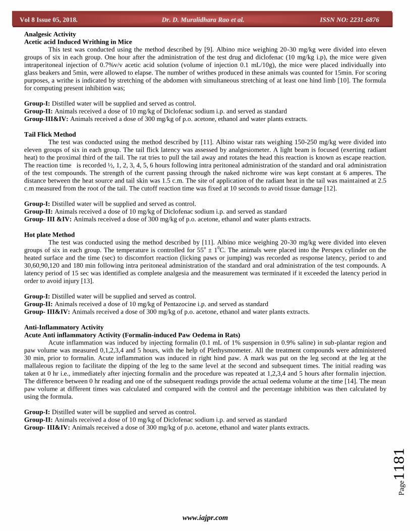
Analgesic Activity (Acetic acid Induced Writhing in Mice)
We measured control and various treated groups for their analgesic activity against acetic acid induced writhing, which is
nothing but the painful reaction. Thirty minutes after the treatment, each mouse was injected with 0.1 mL 0.7% v/v aqueous solution
of acetic acid i.p. The number of abdominal constrictions was cumulatively counted from 0 - 10 minutes. The % reduction of writhing
in standard diclofenac sodium 10 mg/kg treated group was found to be 60.02% against control. The mean response of control and
standard was 41.50 ± 1.25 and 16.59 ± 0.92 respectively. The respective test compounds SP-LF-ETH, SP-LF-ACET, SP-LF-WATE,
SP-BRK-ETH, SP-BRK-ACET and SP-BRK- WATE in its 300 mg/kg dose, showed mean writhing responses as 23.00 ± 1.06, 26.33
± 1.38, 26.17 ± 1.49, 23.83 ± 1.30, 28.33 ± 1.66 and 25.59 ± 1.43. In terms of percentage inhibition of writhing by diclofenac sodium
was 60.02% while with the test compound it was SP-LF-ETH 44.57% and SP-BRK-ETH 42.57% respectively, results were shown in
Figure 1.
www.iajpr.com
Pag
e11
83
Vol 8 Issue 05, 2018. Dr. D. Muralidhara Rao et al. ISSN NO: 2231-6876
Gro
up I
Gro
up II
Gro
up III
Gro
up IV
Gro
up V
Gro
up VI
Gro
up VII
Gro
up VIII
Gro
up IX
Gro
up X
Gro
up XI
0
10
20
30
40
50
Nu
nb
er o
f w
rit
hes
Fig. 1: Effect of Salvadora persica L. plant extracts on acetic acid induced writhing in mice (values are Mean ± SEM (n=6) one
way ANOVA followed by Dunnett’s test).
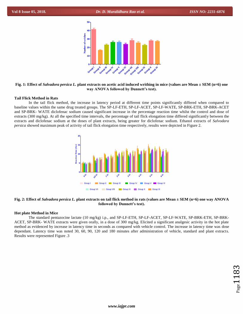
Tail Flick Method in Rats
In the tail flick method, the increase in latency period at different time points significantly differed when compared to
baseline values within the same drug treated groups. The SP-LF-ETH, SP-LF-ACET, SP-LF-WATE, SP-BRK-ETH, SP-BRK-ACET
and SP-BRK- WATE diclofenac sodium caused significant increase in the percentage reaction time whilst the control and dose of
extracts (300 mg/kg). At all the specified time intervals, the percentage of tail flick elongation time differed significantly between the
extracts and diclofenac sodium at the doses of plant extracts, being greater for diclofenac sodium. Ethanol extracts of Salvodara
persica showed maximum peak of activity of tail flick elongation time respectively, results were depicted in Figure 2.
0 hr
1/2 hr
1 hr
2 hr
3 hr
4 hr
5 hr
6 hr
0
5
10
15
20
Group I Group II Group III Group IV Group V Group VI
Group VII Group VIII Group IX Group X Group XI
Reacti
on
Tim
e (
Sec)
Fig. 2: Effect of Salvadora persica L. plant extracts on tail flick method in rats (values are Mean ± SEM (n=6) one way ANOVA
followed by Dunnett’s test).
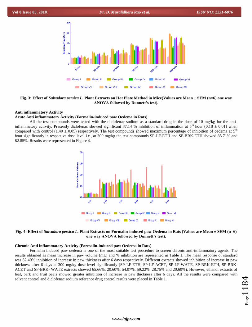
Hot plate Method in Mice
The standard pentazocine lactate (10 mg/kg) i.p., and SP-LF-ETH, SP-LF-ACET, SP-LF-WATE, SP-BRK-ETH, SP-BRK-
ACET, SP-BRK- WATE extracts were given orally, in a dose of 300 mg/kg. Elicited a significant analgesic activity in the hot plate
method as evidenced by increase in latency time in seconds as compared with vehicle control. The increase in latency time was dose
dependant. Latency time was noted 30, 60, 90, 120 and 180 minutes after administration of vehicle, standard and plant extracts.
Results were represented Figure .3
www.iajpr.com
Pag
e11
84
Vol 8 Issue 05, 2018. Dr. D. Muralidhara Rao et al. ISSN NO: 2231-6876
0 m
in
30 m
in
60 m
in
90 m
in
120
min
180
min
0
5
10
15
20
Group I Group II Group III Group IV Group V Group VI
Group VII Group VIII Group IX Group X Group XI
Rea
ctio
n T
ime
(Sec
)
Fig. 3: Effect of Salvadora persica L. Plant Extracts on Hot Plate Method in Mice(Values are Mean ± SEM (n=6) one way
ANOVA followed by Dunnett’s test).
Anti inflammatory Activity
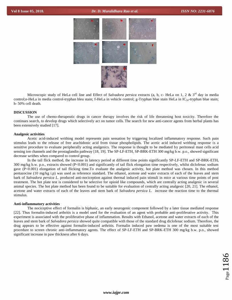
Acute Anti inflammatory Activity (Formalin-induced paw Oedema in Rats)
All the test compounds were tested with the diclofenac sodium as a standard drug in the dose of 10 mg/kg for the anti-
inflammatory activity. Presently diclofenac showed significant 87.14 % inhibition of inflammation at 5th
hour (0.18 ± 0.01) when
compared with control (1.40 ± 0.05) respectively. The test compounds showed maximum percentage of inhibition of oedema at 5th
hour significantly in respective dose level i.e., at 300 mg/kg the test compounds SP-LF-ETH and SP-BRK-ETH showed 85.71% and
82.85%. Results were represented in Figure 4.
0 hr
1 hr
2 hr
3 hr
4 h
r5 hr
0.0
0.5
1.0
1.5
2.0
Group III Group IV Group VGroup I Group II Group VI
Group VII Group VIII Group IX Group X Group XI
Paw
Oed
em
a V
olu
me
Fig. 4: Effect of Salvadora persica L. Plant Extracts on Formalin-induced paw Oedema in Rats (Values are Mean ± SEM (n=6)
one way ANOVA followed by Dunnett’s test).
Chronic Anti inflammatory Activity (Formalin-induced paw Oedema in Rats)
Formalin induced paw oedema is one of the most suitable test procedure to screen chronic anti-inflammatory agents. The
results obtained as mean increase in paw volume (mL) and % inhibition are represented in Table 1. The mean response of standard
was 82.40% inhibition of increase in paw thickness after 6 days respectively. Different extracts showed inhibition of increase in paw
thickness after 6 days at 300 mg/kg dose level significantly (SP-LF-ETH, SP-LF-ACET, SP-LF-WATE, SP-BRK-ETH, SP-BRK-
ACET and SP-BRK- WATE extracts showed 65.66%, 20.60%, 54.07%, 59.22%, 28.75% and 20.60%). However, ethanol extracts of
leaf, bark and fruit peels showed greater inhibition of increase in paw thickness after 6 days. All the results were compared with
solvent control and diclofenac sodium reference drug control results were placed in Table 1.

www.iajpr.com
Pag
e11
85
Vol 8 Issue 05, 2018. Dr. D. Muralidhara Rao et al. ISSN NO: 2231-6876
Table 1: Effect of Salvadora persica L. plant extracts on formalin-induced paw oedema (chronic) in rats (results are expressed
on mean + SEM (n=6) from four observations Paw Volume was measured after 6 days.
Groups Treatment Initial Paw
Volume
Paw Volume After 6 Days Increase in
Paw Volume
% of
Inhibition
Group-I Saline 1.28 ± 0.07 3.61 ± 0.12 2.33 ± 0.06 -
Group-II Diclofenac (100 mg/kg) 1.23 ± 0.04 1.65 ± 0.05 0.41 ± 0.07 82.40%
Group-III SP-LF-ETH (300 mg/kg) 1.26 ± 0.03 2.00 ± 0.06 0.80 ± 0.12 65.66%
Group-IV SP-LF-ACET (300 mg/kg) 1.21 ± 0.06 3.21 ± 0.24 1.85 ± 0.16 20.60%
Group-V SP-LF-WATE (300 mg/kg) 1.25 ± 0.06 2.25 ± 0.16 1.07 ± 0.14 54.07%
Group-VI SP-BRK-ETH (300 mg/kg) 1.31 ± 0.08 2.26 ± 0.10 0.95 ± 0.14 59.22%
Group-VII SP-BRK-ACET (300 mg/kg) 1.23 ± 0.06 2.90 ± 0.14 1.66 ± 0.17 28.75%
Group-VIII SP-BRK- WATE (300 mg/kg) 1.26 ± 0.06 3.11 ± 0.08 1.85 ± 0.11 20.60%
Anti-Cancer Activity (Trypan Blue Dye Exclusion Method against by HeLa cell Line)
We measured the anti cancer activity of different crude extracts of Salvodara persica (SP-LF-ETH, SP-LF-ACET, SP-LF-
WATE, SP-BRK-ETH, SP-BRK-ACET, SP-BRK- WATE, SP-FP-ETH, SP-FP-ACET and SP-FP- WATE) on % of dead cells against
HeLa cell lines against by trypan blue dye exclusion method. The percentage of dead cells was found to be 69.5%, 11.5%, 5.30%,
15.3%, 7.3%, 10.2%, 15.7%, 23.3% and 22.1% in HeLa cell line against with SP-LF-ETH, SP-LF-ACET, SP-LF-WATE, SP-BRK-
ETH, SP-BRK-ACET, SP-BRK- WATE, SP-FP-ETH, SP-FP-ACET, SP-FP- WATE 300 mg/kg b.w p.o. respectively. However the
maximum % of dead cells were found in ethanol extracts of leaf, bark and fruit peels. Results were presented in Fig 5.
Fig 5: Effect of Salvadora persica extracts on HeLa cell Line against by Trypan Blue Dye Exclusion Method.
www.iajpr.com
Pag
e11
86
Vol 8 Issue 05, 2018. Dr. D. Muralidhara Rao et al. ISSN NO: 2231-6876
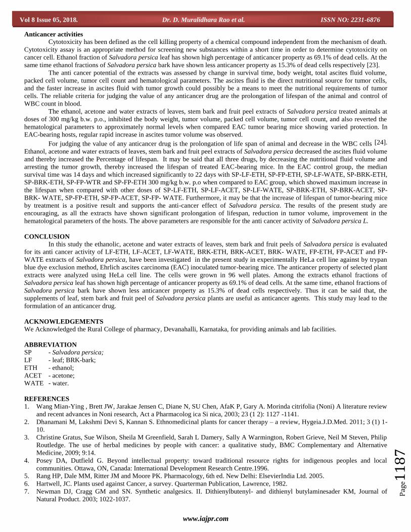
Microscopic study of HeLa cell line and Effect of Salvadora persica extracts (a, b, c- HeLa on 1, 2 & 3rd
day in media
control;e-HeLa in media control-tryphan bleu stain; f-HeLa in vehicle control; g-Tryphan blue stain HeLa in IC50-tryphan blue stain;
h- 50% cell death.
DISCUSSION The use of chemo-therapeutic drugs in cancer therapy involves the risk of life threatening host toxicity. Therefore the
continues search, to develop drugs which selectively act on tumor cells. The search for new anti-cancer agents from herbal plants has
been extensively studied [17].
Analgesic activities Acetic acid-induced writhing model represents pain sensation by triggering localized inflammatory response. Such pain
stimulus leads to the release of free arachidonic acid from tissue phospholipids. The acetic acid induced writhing response is a
sensitive procedure to evaluate peripherally acting analgesics. The response is thought to be mediated by peritoneal mast cells acid
sensing ion channels and the prostaglandin pathway [18, 19]. The SP-LF-ETH, SP-BRK-ETH 300 mg/kg b.w. p.o., showed significant
decrease writhes when compared to control group.
In the tail flick method, the increase in latency period at different time points significantly SP-LF-ETH and SP-BRK-ETH,
300 mg/kg b.w. p.o., extracts showed (P<0.001) and significantly of tail flick elongation time respectively, whilst diclofenac sodium
gave (P<0.001) elongation of tail flicking time.To evaluate the analgesic activity, hot plate method was chosen. In this method
pentazocine (10 mg/kg i.p) was used as reference standard. The ethanol, acetone and water extracts of each of the leaves and stem
bark of Salvadora persica L. produced anti-nociception against thermal induced pain stimuli in mice at various time points of post
treatment. The hot plate test is considered to be selective for opioid like compounds, which are centrally acting analgesic in several
animal species. The hot plate method has been found to be suitable for evaluation of centrally acting analgesic [20, 21]. The ethanol,
acetone and water extracts of each of the leaves and stem bark of Salvadora persica L. increase the reaction time to the thermal
stimulus.
Anti-inflammatory activities
The nociceptive effect of formalin is biphasic, an early neurogenic component followed by a later tissue mediated response
[22]. Thus formalin-induced arthritis is a model used for the evaluation of an agent with probable anti-proliferative activity. This
experiment is associated with the proliferative phase of inflammation. Results with Ethanol, acetone and water extracts of each of the
leaves and stem bark of Salvadora persica showed quite compatible with those of the standard drug diclofenac sodium. Therefore, the
drug appears to be effective against formalin-induced arthritis. Formalin induced paw oedema is one of the most suitable test
procedure to screen chronic anti-inflammatory agents. The effect of SP-LF-ETH and SP-BRK-ETH 300 mg/kg b.w. p.o., showed
significant increase in paw thickness after 6 days.
www.iajpr.com
Pag
e11
87
Vol 8 Issue 05, 2018. Dr. D. Muralidhara Rao et al. ISSN NO: 2231-6876
Anticancer activities
Cytotoxicity has been defined as the cell killing property of a chemical compound independent from the mechanism of death.
Cytotoxicity assay is an appropriate method for screening new substances within a short time in order to determine cytotoxicity on
cancer cell. Ethanol fraction of Salvadora persica leaf has shown high percentage of anticancer property as 69.1% of dead cells. At the
same time ethanol fractions of Salvadora persica bark have shown less anticancer property as 15.3% of dead cells respectively [23].
The anti cancer potential of the extracts was assessed by change in survival time, body weight, total ascites fluid volume,
packed cell volume, tumor cell count and hematological parameters. The ascites fluid is the direct nutritional source for tumor cells,
and the faster increase in ascites fluid with tumor growth could possibly be a means to meet the nutritional requirements of tumor
cells. The reliable criteria for judging the value of any anticancer drug are the prolongation of lifespan of the animal and control of
WBC count in blood.
The ethanol, acetone and water extracts of leaves, stem bark and fruit peel extracts of Salvadora persica treated animals at
doses of 300 mg/kg b.w. p.o., inhibited the body weight, tumor volume, packed cell volume, tumor cell count, and also reverted the
hematological parameters to approximately normal levels when compared EAC tumor bearing mice showing varied protection. In
EAC-bearing hosts, regular rapid increase in ascites tumor volume was observed.
For judging the value of any anticancer drug is the prolongation of life span of animal and decrease in the WBC cells [24].
Ethanol, acetone and water extracts of leaves, stem bark and fruit peel extracts of Salvadora persica decreased the ascites fluid volume
and thereby increased the Percentage of lifespan. It may be said that all three drugs, by decreasing the nutritional fluid volume and
arresting the tumor growth, thereby increased the lifespan of treated EAC-bearing mice. In the EAC control group, the median
survival time was 14 days and which increased significantly to 22 days with SP-LF-ETH, SP-FP-ETH, SP-LF-WATE, SP-BRK-ETH,
SP-BRK-ETH, SP-FP-WTR and SP-FP-ETH 300 mg/kg b.w. p.o when compared to EAC group, which showed maximum increase in
the lifespan when compared with other doses of SP-LF-ETH, SP-LF-ACET, SP-LF-WATE, SP-BRK-ETH, SP-BRK-ACET, SP-
BRK- WATE, SP-FP-ETH, SP-FP-ACET, SP-FP- WATE. Furthermore, it may be that the increase of lifespan of tumor-bearing mice
by treatment is a positive result and supports the anti-cancer effect of Salvadora persica. The results of the present study are
encouraging, as all the extracts have shown significant prolongation of lifespan, reduction in tumor volume, improvement in the
hematological parameters of the hosts. The above parameters are responsible for the anti cancer activity of Salvadora persica L.
CONCLUSION
In this study the ethanolic, acetone and water extracts of leaves, stem bark and fruit peels of Salvadora persica is evaluated
for its anti cancer activity of LF-ETH, LF-ACET, LF-WATE, BRK-ETH, BRK-ACET, BRK- WATE, FP-ETH, FP-ACET and FP-
WATE extracts of Salvadora persica, have been investigated in the present study in experimentally HeLa cell line against by trypan
blue dye exclusion method, Ehrlich ascites carcinoma (EAC) inoculated tumor-bearing mice. The anticancer property of selected plant
extracts were analyzed using HeLa cell line. The cells were grown in 96 well plates. Among the extracts ethanol fractions of
Salvadora persica leaf has shown high percentage of anticancer property as 69.1% of dead cells. At the same time, ethanol fractions of
Salvadora persica bark have shown less anticancer property as 15.3% of dead cells respectively. Thus it can be said that, the
supplements of leaf, stem bark and fruit peel of Salvadora persica plants are useful as anticancer agents. This study may lead to the
formulation of an anticancer drug.
ACKNOWLEDGEMENTS
We Acknowledged the Rural College of pharmacy, Devanahalli, Karnataka, for providing animals and lab facilities.
ABBREVIATION
SP - Salvadora persica;
LF - leaf; BRK-bark;
ETH - ethanol;
ACET - acetone;
WATE - water.
REFERENCES
1. Wang Mian-Ying , Brett JW, Jarakae Jensen C, Diane N, SU Chen, AfaK P, Gary A. Morinda citrifolia (Noni) A literature review
and recent advances in Noni research, Act a Pharmacolog ica Si nica, 2003; 23 (1 2): 1127 -1141.
2. Dhanamani M, Lakshmi Devi S, Kannan S. Ethnomedicinal plants for cancer therapy – a review, Hygeia.J.D.Med. 2011; 3 (1) 1-
10.
3. Christine Gratus, Sue Wilson, Sheila M Greenfield, Sarah L Damery, Sally A Warmington, Robert Grieve, Neil M Steven, Philip
Routledge. The use of herbal medicines by people with cancer: a qualitative study, BMC Complementary and Alternative
Medicine, 2009; 9:14.
4. Posey DA, Dutfield G. Beyond intellectual property: toward traditional resource rights for indigenous peoples and local
communities. Ottawa, ON, Canada: International Development Research Centre.1996.
5. Rang HP, Dale MM, Ritter JM and Moore PK. Pharmacology, 6th ed. New Delhi: ElsevierIndia Ltd. 2005.
6. Hartwell, JC. Plants used against Cancer, a survey. Quarterman Publication, Lawrence, 1982.
7. Newman DJ, Cragg GM and SN. Synthetic analgesics. II. Dithienylbutenyl- and dithienyl butylaminesader KM, Journal of
Natural Product. 2003; 1022-1037.
www.iajpr.com
Pag
e11
88
Vol 8 Issue 05, 2018. Dr. D. Muralidhara Rao et al. ISSN NO: 2231-6876
8. Pinney KG, Jelinek C, Edvardsen K, Chaplin DJ, Pettit GR. The discovery and development of the combretastatins. In: Cragg,
G.M., Kingston, D.G.I., Newman, D.J. (Eds.), Anticancer Agents from Natural Products. Brunner-Routledge Psychology Press,
Taylor & Francis Group, Boca Raton, FL. 2005; 23-46 (Chapter 3).
9. Cragg GM, Kingston DG, Newman DJ. Anti cancer Agents from Natural Products, Brunner- Routledge Psychology press, Taylor
& Francis Group, Boca Raton, Florida 2005.
10. Collier HOJ, Dinneen LC, Johnson CA, Schneider C. The Abdominal Constriction Response and its Suppression by Analgesic
Drugs in the Mouse. British J. Pharmacol, 1968; (32):295-310.
11. Mokarram Hossain M.D., Israt Jahan Biva. Central nervous system depressant and analgesic activity of Aphanamixis polystachya
(Wall.) parker leaf extract in mice. Afr. J. Pharm. Pharmacol. 2009; 3(5): 282-286.
12. D'Armour FE, Smith DL. A method for determining loss of pain sensation. J Pharmacol Exp Ther. 1941; 72:74.
13. Anar Patel, Timir Patel, Carol Macwan, Mayuree Patel, Khushbu Chauhan, Jatin Patel.Evaluation of Anti inflammatory and
Analgesic activity of roots of Rubia cordifolia in rats. J. Pharm. Sci. & Res. 2010; 2 (12); 809-813.
14. Eddy and Leimbach . Synthetic analgesics. II. Dithienylbutenyl- and dithienylbutylamines. J Pharmacol Exp Ther. 1953;
107(3):385-93.
15. Sunetra Patwardhan and Ghansham Sakhare. Pharmacology online. 2009; 2: 887.
16. Yogesh Baravalia, Yogeshkumar Vaghasiya and Sumitra Chanda. Iranian Journal of Pharmaceutical Research. 2012; 11(3): 851.
17. Bikash Kumar Nanda, Jyotirmoyee Jena. Der Pharmacia Lettre. 2010; 2(1): 181.
18. Sanjay P, Nirav G, Ashok S, Anand S. In-Vitro cytotoxicity activity of Solanum nigrum extract against HeLa cell line and Vero
cell line. Int J Pharm Pharm Sci. 2009; 1(1):38-46.
19. Jain S, Gill V, Vasudeva N and Singla N. Journal of Chinese international medicine. 2009c; 7(11): 1096- 1099.
20. Voilley N. Acid sensing ion channels (ASICs) New targets for analgesic- effect of non-steroid anti-inflammatory (NSAIDs)
Current drugs trends- Inflammation & Allergy. 2004; 3:71-79.
21. Hossain MM, Ali MS, Saha A and Alimuzzaman M. Antinocicceptive activity of whole plant extracts of Paederia foetida. Dhaka
Univ. J. Pharm Sci. 2006; 5: 67-69.
22. Hosseinzadeh H, Younesi H. Antinociceptive and anti-inflammatory effects of Crocus sativus L. stigma and petal extracts in
mice, BMC Pharmacol. 2002; 2:7-9.
23. Margolius, HS. Tissue kallikreins and kinins: Regulation and roles in hypertensive and diabetic diseases. Annu. Rev. Pharmacol.
Toxicol. 1989; 29:343-364.
24. Doyle A, Griffiths JB. Cell and tissue culture: Laboratory Procedures in Biotechnology. 2 ed. John Wiley & Sons Ltd.1988; 201.
25. Prasad SB and Giri A. Antitumour effect of cisplatin against murine ascites Dalton's lymphoma. Indian Journal of Experimental
Biology. 1994; (32):155-162.
26. Hogland HC (1982) SEM Oncology, 1982; (9):95-96.
54878478451180519
Copyright © 2022 FDOKUMEN