The Anti inflammatory activity of Crateva adansonii dichloromethane fraction.
Upload
khangminh22Category
view
1download
0
HAL Id: tel-01214953https://tel.archives-ouvertes.fr/tel-01214953
Submitted on 13 Oct 2015
HAL is a multi-disciplinary open accessarchive for the deposit and dissemination of sci-entific research documents, whether they are pub-lished or not. The documents may come fromteaching and research institutions in France orabroad, or from public or private research centers.
L’archive ouverte pluridisciplinaire HAL, estdestinée au dépôt et à la diffusion de documentsscientifiques de niveau recherche, publiés ou non,émanant des établissements d’enseignement et derecherche français ou étrangers, des laboratoirespublics ou privés.
Unravelling the therapeutic intervention of inflammationand cancer by Viscum album : understanding its
anti-inflammatory and immunostimulatory propertiesChaitrali Saha
To cite this version:Chaitrali Saha. Unravelling the therapeutic intervention of inflammation and cancer by Viscumalbum : understanding its anti-inflammatory and immunostimulatory properties. Biotechnology. Uni-versité de Technologie de Compiègne, 2015. English. �NNT : 2015COMP2210�. �tel-01214953�
Par Chaitrali SAHA
Thèse présentée pour l’obtention du grade de Docteur de l’UTC
Unravelling the therapeutic intervention of inflammation and cancer by Viscum album : understanding its anti-inflammatory and immunostimulatory properties
Soutenue le 09 septembre 2015 Spécialité : Biotechnologie
D2210

Université de Technologie de Compiègne
Champ disciplinaire: Biotechnologie Thèse présentée par
Chaitrali SAHA
Pour l’obtention du grade de Docteur del’UTC
Sujet de la thèse
Etude des propriétés phytothérapeutiques de Viscum album dans le traitement de l'inflammation et du cancer: Détermination de ses caracteristiques anti-inflammatoires et d'immunostimulation
Thèse dirigée par: Dr. Srinivas KAVERI and Dr. Alain FRIBOULET
Soutenue le: le 9 Septembre 2015
Le jury composé de: Prof. Bérangère BIHAN-AVALLE Présidente Prof. Kithiganahalli BALAJI Rapporteur Dr. Hicham BOUHLAL Rapporteur Dr. Pascal PONCET Examinateur Dr. Alain FRIBOULET Co-directeur de thèse Dr. Jagadeesh BAYRY Co-directeur de thèse Dr. Srinivas KAVERI Directeur de thèse

L’ intitulé de l'unité
Immunopathologie et Immunointervention Thérapeutique
L'adresse de l'unité où la thèse a été prepare
UMR S 1138 (Equipe 16)
Centre de Recherche des Cordeliers
15, rue de l’ecole de medicine
75006 Paris- France
Tel : +33 1 44 27 82 07
Fax: +33 1 44 7 81 94 www.u681.jussieu.fr
1
TABLE OF CONTENTS
Title Page No.
Acknowledgements 4
Abbreviations 7
Summary in French 10
Summary in English 12
Introduction 14
1. The Immune System
1.1. Rapid Response: Innate Immune System 14
1.2. Adaptable but Dependent Response: Adaptive Immune System 16
1.3. The Bridge between Old and New: Dendritic cells the Key Players 17
1.4. Macrophage Biology in Homeostasis and Disease: Full Spectrum
of Macrophage Activation 19
1.5. T cell Polarization and Th cell Subsets 22
1.5.1. Th1 and Th2 effector T cells: The Tip of the Iceberg 22
1.5.2. FOXP3+ Treg cells 23
1.5.3. Th17 cells 23
2. Immunologic Dysfunction 25
2.1. Cancer Despite Immunosurveillance: Means of Immunoselection and
Immunosubversion 26
3. Inflammation 29
3.1. Inflammatory Pathway 30
3.2. Inflammation and Cancer: Two Faces of Same Coin 31
3.3. Inflammation Can Cause Cancer 32
3.4. Cancer Can Cause Inflammation 32
4. Cancer and Inflammation: Friend or Foe? 33
5. Cancer Immunotherapy: Current Paradigm 34
6. Importance of cyclo-oxygenases and COX-derived Prostaglandins in Cancer and in
Inflammation 35
6.1. Cyclooxygenases: Structural and Functional Insights 36
6.2. Inhibition of the COX Pathways 38
6.3. Targeting COX-2 Expression by Natural Compounds 38
6.4. Determinants of COX-2 Expression 39
6.4.1. Transcriptional Regulation 39

2
6.4.2. Post-transcriptional Regulation 39
6.4.3. Post-translational Regulation 40
7. Phytotherapy: A Power of Nature to Cure Immuno-Inflammatory
Pathologies and Cancer 41
7.1. Conventional Oncology and Viscum album 43
7.2. Quality of Life and Viscum album 43
8. Viscum album 44
8.1. Mythological Aspect 44
8.2. Mistletoe As a Remedy 45
8.3. Preparation of Therapeutic Preparation of Viscum album 45
8.4. Chemical Compounds in Viscum album 45
8.5. Multifarious Properties of Viscum album 48
8.6. Viscum album: Clinical Evidence 49
Objectives of present study 52
1. Molecular dissection of Viscum album mediated COX-2 inhibition and better
understanding of its anti-inflammatory effect.
2. Exploring the immunomodulatory effects of Viscum album by studying differential
effect of various preparations of Viscum album on maturation and activation of human
dendritic cells and T cell response.
3. Exploring the anti-tumor response of Viscum album by understanding their effect on
the full spectrum of macrophage polarization.
Results
Article 1: Viscum album-mediated COX-2 inhibition implicates destabilization of
COX-2 mRNA 53
Article 2: Differential effect of Viscum album preparations on maturation and
activation of human dendritic cells and CD4+ T cell response 64
Article 3: Viscum album promotes anti-tumor response by modulating M1/M2
macrophage polarization switch 86
Discussion 109
Perspectives 116
References 121
Annexes 146
3
LIST OF FIGURES
Figure 1: The three sentinel cells, Dendritic, Mast, and Macrophages serves protection against
ingested pathogens
Figure 2: Dendritic cells: Bridge between old and new
Figure 3: The orchestration of macrophage activation and polarization by lymphoid cells
Figure 4: CD4+ T cell differentiation
Figure 5: The hallmarks of cancer
Figure 6: Cancer immunosurveillance and immunoediting
Figure 7: The Inflammatory Pathway
Figure 8: Steps of the inflammatory immune response
Figure 9: Types of Inflammation in Tumorigenesis and Cancer
Figure 10: Prostanoid synthesis from arachidonic acid by cyclo-oxygenases
Figure 11: Proposed functions of cyclooxygenase derived PGs
Figure 12: COX-2 Gene Expression
Figure 13: Anti-cancer effects of Phytochemicals
Figure14: Phytotherapy strategy
Figure 15: Mechanism of action of type II lectins
Figure 16: TLR signalling pathways
Figure 17: Molecular pathways of macrophage polarization
LIST OF TABLES
Table 1: List of some medicinal herbal products
Table 2: Chemical compounds identified in the European Viscum album L

4
Acknowledgements
“Take up one idea. Make that one idea your life - think of it, dream of it, live on that idea. Let
the brain, muscles, nerves, every part of your body, be full of that idea, and just leave every other
idea alone. This is the way to success.” Swami Vivekananda
First and foremost I thank the ALMIGHTY for having bestowed the blessings on me to complete
this thesis work successfully.
It is humbling experience to acknowledge those people who have, mostly out of kindness, helped
me along the journey of my PhD. I cannot claim this work to be solely mine as the successful
completion of this work had inputs from so many well-wishers.
I would like to express my sincere gratitude to Dr. Srinivas Kaveri, my Supervisor and Mentor. I
owe my heartfelt thanks to him for giving me the opportunity to accomplish my doctoral study
and providing me the resources and freedom to work while working in the lab. His scientific and
moral supports added with his vast experience have always been motivating me. His precious
suggestions and criticism also helped me a lot in the overall development of my scientific and
professional skills. His personal generosity helped making my time comfortable and enjoyable in
lab.
My deep gratitude goes to Dr. Jagadeesh Bayry. I am extremely grateful for his assistance and
valuable suggestions throughout my studies. His very close supervision and critical analysis
really helped me to achieve perfection in this work.
I extend my warm thanks with deep regards to Dr. Alain Friboulet for welcoming me in the
Universite de technologie de Compiegne and being a wonderful co-supervisor. I would like to
thank him for his kind and humble support which has helped me to come out from many difficult
situations during my studies.
I am thankful to Prof. Berangere Bihan-Avalle for kindly accepting the invitation to be the
president of the jury. I wish to convey my sincere gratitude to Dr. Hicham Bouhlal and Prof.
Kithiganahalli BALAJI for agreeing to be rapporteurs. I am grateful to Dr. Pascal Poncet for
accepting the invitation to be the examiner of my thesis.

5
I would like to specially thank Dr. Sebastein Lacroix-Desmazes for his support to my work. His
valuable suggestions have made me benefited in my research work. My special thanks go to Dr.
Jordan Dimitrov for his suggestions and motivation.
I wish to express my deepest gratitude to Mme. Veronique and Mme Sylvie Carlier for their full
hearted unwavering efforts helped me in exercising to complete all the academic and scientific
procedures.
A person who made me think about science day and night and encouraged me to be immensely
connected to science and to make me love science is Prof. Saumitra Das (Attached with Indian
Institute of Science, India) and without thanking him, this work would be virtually incomplete. I
shall always feel proud to be associated in my life with these two genius scientists, Prof. Srini
Kaveri and Prof Saumitra Das. Their enthusiasm and devotion for science is contagious.
My heartfelt thanks to all of “Team Srini Kaveri”, past and present: Cyril, Ankit, Pushpa,
Veeru, Meenu, Julie, Sandrine, Ivan, Maxime, Mathieu, Emmanuel, Mrinmoy, Varun,
Annaelle, Jules, Nimesh, Bagi, Laurent, Adeline, Isabelle, Maelle, Maya, Sambhabi, Baptiste,
Diago. My special thanks to Maxime for his unconditional and kind help throughout my stay in
lab. I extend my thanks to Justa for her help in carrying out various tasks important for lab
maintenance and assistance. I thank Mathieu and Selma for their friendly and helpful gestures. I
would always cherish the time spent with Emmanuel, Mrinmoy and Varun, especially during
the tea-break. I shall always be grateful to Julie and Emmanuel for their exemplary counsel and
soothing words of encouragement and kindness whenever and wherever I needed those most.
My heartfelt thanks to my lovely floor mates in Maison de L’Inde.
Good friends are hard to find, harder to leave, and impossible to forget. I will not leave the
opportunity to thank Upasana, Anuj, Ranjitha, Snigdhadip, Debolina, Sagar, Aswath, Sagnik,
Neeraj, who should know that their support and encouragement was worth more than I could
express in a few lines hereto.
Finally, I would like to convey my deepest gratitude, love and respect to Ma and Baba, who are
exceptionally my wonderful parents and my best friends ever. I owe all my success to my
parents; without their love and selfless efforts and above all their blessings this would have not
been materialised. Dada, I could not ask for a better brother and friend. Words are insufficient
to thank my cousins Bhadrali, Chitrali and Saitali for their unconditional love. I would like to
express my love to my most adorable nephews, Neel and Rikh, who make my heart smile, even at

6
harder times of my life. I express my sincere thanks to all my family members (especially Boudi,
Didi, Boromoa, Nama, Chotoma, Nakaka and Cotokaka,) for their blessings and kind hearted
support for my endeavour. They are the ones who have showered most affection and praise
whenever I have achieved something.
To Dadu, Mummum and Nadadu who were often in my thoughts on this journey- I missed you
all very intensively.
Not last but the least to have a few words to express my infinite gratitude to my soul-mate
Koushik. I believe that without his selfless inspiration and vigorous supports, it would have been
next to impossible for me to safe landing at the end of my this critical journey. As my life partner,
I strongly believe Koushik is only my matching husband for his value management towards me.
This thesis is dedicated to my parents and my husband for their endless love, support and
encouragement.

7
ABBREVIATIONS
AA Arachidonic acid
Ab Antibody
ADCC Antibody-dependent cell-mediated cytotoxicity
AE Adverse effect
Ag Antigen
Apaf Apoptosis-associated factor
APC Antigen presenting cell
ARE Adenylate-uridylate-rich element
Arg 1 Arginase 1
BAD Bcl-2-associated death promoter
BAX Bcl-2-associated X protein
BCL B-cell lymphoma
CAM Complementary and alternative medicine
CD Cluster of differentiation
COX Cyclo-oxygenase
COXIB COX-2 inhibitor
CRF Cancer-related fatigue
CTL Cytotoxic T lymphocyte
DC Dendritic cell
DC-SIGN Dendritic cell-specific intercellular adhesion molecule-3-
grabbing non-integrin
DNA Deoxyribonucleic acid
EGF Epidermal growth factor
ELISA Enzyme link immunosorbent assay
ERK Extracellular-signal-regulated kinase
FAP Familial adenomatous polyposis
FCS Foetal Calf Serum
FcγR Fc gamma receptor
FITC Fluorescein isothiocyanate
Foxp3 Forkhead box P3
GMCSF Granulocyte-macrophage colony-stimulating factor
HUVEC Human umbilical vein endothelial cell
IFN Interferron

8
Ig Immunoglobulin
IgE Immunoglobulin E
IL Interleukine
IRF Interferon regulatory factor
IVEC Immortalised human venous endothelial cell
JNK c-Jun N-terminal kinase
LPS Lipopolysaccharides
MAPK Mitogen-activated protein kinase
MCSF Macrophage colony-stimulating factor
MHC Major Histocompability complex
miRNA microRNA
ML Mistletoe lectin
MMP Mitochondrial membrane potential
MO Monocyte
mRNA Messenger RNA
MФ Macrophage
NEMO NF-κB essential modulator
NF-κB Nuclear factor kappa B
NK Natural killer cells
NLR Nucleotide-binding oligomerization-domain protein like
receptor
NOD Nucleotide-binding oligomerization-domain protein
NOS Nitric oxide synthase
NSAID Non-steroidal anti-inflammatory drugs
PAMP Pathogen-associated molecular patterns
PBMC Peripheral blood mononuclear cell
PBS Phosphate buffer saline
PG Prostaglandin
PRR Pathogen- recognition receptor
PTGS Prostaglandin-endoperoxide synthase
QOL Quality of life
RNA Ribonucleic acid
RNI Reactive nitrogen intermediates
RNS Reactive nitrogen species
ROI Reactive oxygen intermediates

9
ROR-γt/ RORC Retionic acid-related orphan receptor
ROS Reactive oxygen species
RIP Ribosome inactivating protein
rRNA Ribosomal ribonucleic acid
SEM Standard error of mean
STAT Signal transducer and activator of transcription
TAA Tumor-associated antigen
TAM Tumor associated macrophage
T-bet T box transcription factor
TFH Follicular helper T cell
TGF-β Transforming growth factor β
Th Helper T cells
TLR Toll-like receptor
TNF Tumor necrosis factor
Tregs Regulatory T cells
UTR Untranslated region
VA Viscum album
VEGF Vascular endothelial growth factor
VEGFA Vascular endothelial growth factor A

10
Résumé en Français
Etude des propriétés phytothérapeutiques de Viscum album dans le traitement de
l'inflammation et du cancer: Détermination de ses caractéristiques anti-inflammatoires
et d'immunostimulation
par
Chaitrali Saha
<< Biotechnologie et mise en œuvre des Fonctions Biologiques >>
Thèse est présentée à la Faculté de l’Université de Technologie de Compiègne
en vue l’obtention du grades de
Philosophiae Docteur (Ph.D.) de l’Université de Technologie de Compiègne
Les mots clés : Viscum album, lectine de gui, cyclo-oxygénases, PGE2, effet anti-
inflammatoire, effet immunomodulateur,
Les préparations de Viscum album (VA), connu sous le nom vernaculaire de gui européen,
sont fréquemment utilisées en support des traitements anticancéreux, principalement pour
améliorer la qualité de vie des malades et réduire la croissance des tumeurs. Elles sont
connues pour exercer des effets anti-tumoraux. Il existe de plus en plus de données
scientifiques faisant état de liens étroits entre cancer et inflammation. Etant donné que la
prostaglandine E2 (PGE2) induite par la cyclo-oxygénase 2 (COX-2) joue un rôle clef dans
l’inflammation, j’ai exploré la régulation du système COX-2-PGE2 par VA et ses
mécanismes sous-jacents. J’ai montré que VA exerce ses effets anti-inflammatoires en
inhibant sélectivement l’expression de COX-2 et en diminuant la production de PGE2 qui en
découle, par le biais d’une déstabilisation de l’ARNm de COX-2. En plus de leurs propriétés
cytotoxiques, il a été montré que les préparations de VA ont également des effets
immunostimulants. Les différentes préparations de VA sont hautement hétérogènes du fait de
leurs compositions biochimiques qui varient selon la récolte, l’espèce de l’arbre hôte et les
méthodes de préparation qui peuvent influer sur leur efficacité clinique. De ce fait, j’ai réalisé
une étude comparative sur cinq préparations de VA dans le but d’analyser leurs capacités de
maturation et d’activation des cellules dendritiques (DC) qui peuvent à leur tour présenter une
réponse immunitaire anti-tumorale. Les résultats ont montré que parmi les cinq préparations,
VA Qu Spez induit de manière significative l’activation des DC et la sécrétion de cytokines
pro-inflammatoires telle que l’IL-6, l’IL-8 et le TNF-α qui induisent la production d’IFN-γ,
orientant de ce fait la réponse immunitaire vers une réponse Th1. L’orchestration de la
11
fonction des cellules myelomonocytiques est un élément central à l’interface entre
inflammation et cancer. Il constitue un paradigme expliquant la plasticité et la fonction des
macrophages. Mon étude met en évidence l’influence de VA Qu Spez sur la polarisation des
macrophages qui passent d’un état alternatif (M2) à un état dit classique (ou M1). Les
macrophages M2 sont connus pour polariser les réponses immunitaires Th2, pour participer à
l’élimination des parasites, pour diminuer l’inflammation, pour promouvoir le remodelage
tissulaire et la progression des tumeurs et pour avoir des fonctions immunorégulatrices. Les
macrophages M1 sont impliqués dans la réponse Th1, favorisent la résistance aux pathogènes
intracellulaires et aux tumeurs et promeuvent des réactions de désagrégation tissulaires.
L’ensemble de ces résultats permet de comprendre les propriétés anti-inflammatoires et
immunostimulantes des préparations de VA. Des recherches complémentaires permettront
d’améliorer les stratégies d’utilisation thérapeutique de VA et son utilisation dans les soins de
support aux traitements anticancéreux.

12
Abstract in English
Unravelling the therapeutic intervention of inflammation and cancer by
Viscum album: Understanding its anti-inflammatory and immunostimulatory properties
by
Chaitrali SAHA
Thesis is presented at the Faculty of Université de Technologie de Compiègne
for obtaining the degree of
Doctor of Philosophy (Ph.D.) of the Université de Technologie de Compiègne
Key words: Viscum album, mistletoe lectin, cyclo-oxygenases, PGE2, anti-inflammatory
effect, immunomodulatory effect, complementary and alternative medicine.
Viscum album (VA) preparations, commonly known as European mistletoe, are frequently
used as complementary therapy in cancer, mainly to improve quality of life of the patients and
to reduce the tumor growth. They are known to exert anti-tumoral effects. There is increasing
evidence of the convoluted connection of cancer and inflammation. As cyclooxygenase-2
(COX-2)-induced prostaglandin E2 (PGE2) plays a key role in the inflammation, I explored
the regulation of COX-2-PGE2 axis by VA and underlying mechanisms. I found that VA
exerts anti-inflammatory effects by selectively inhibiting COX-2 expression and ensuing
PGE2 production. Inhibition of COX-2 expression implicates COX-2 mRNA destabilisation.
In addition to their cytotoxic properties, they have also been shown to have
immunostimulatory properties. Each VA preparations are highly heterogeneous because of
their chemical composition which varies depending on the time of harvest, species of host tree
and manufacturing methods, together which might influence clinical efficacy of VA.
Therefore I performed a comparative study involving five different preparations of VA
concerning maturation and activation of dendritic cells (DCs) which in turn may manifest
anti-tumoral immune response. Results showed that among all five preparations, VA Qu Spez
significantly induces DC activation, secretion of pro-inflammatory cytokines such as IL-6, Il-
8 and TNF-α, enhancing IFN-γ production hence promoting Th1 immune response. The
orchestration of myelomonocytic cell function is a key element that links inflammation and
cancer and provides a paradigm for macrophage plasticity and function. My study reveals the
effect of VA Qu Spez in switching the M2 macrophages which are known to participate in
polarizing Th2 responses, help with parasite clearance, dampen inflammation, promote tissue
remodelling and tumor progression and have immunoregulatory functions, towards classically
activated M1 macrophages which are part of a polarized Th1 response and mediate resistance

13
to intracellular pathogens and tumors and elicit tissue-disruptive reactions. These results
together should assist in understanding the anti-inflammatory and immunostimulatory
properties of VA preparations and further research is warranted to improve the therapeutic
strategies of use of VA and their role as complimentary therapy in cancer.

INTRODUCTION

14
The Immune System
The immune system is an organization of cells and molecules with specialized functions.
Immunologic defences in vertebrates comprise of two fundamentally different types of
responses to invading microbes. Natural or innate responses occur to the same extent even
after encountering the infectious agent for several times, whereas acquired or adaptive
immune responses improve upon exposure to a given infection repeatedly. Today these two
types of immune responses are well appreciated as obligatory part of immune system
mediating successful immune responses towards infection and tissue injury. The innate
immunity encompasses the elements of immune system which includes phagocytic cells such
as neutrophils, monocytes, macrophages, cells which release inflammatory mediators such as
basophils, mast cells, eosinophils and natural killer cells for immediate host defence. The
molecular components are complement, cytokines and acute phase proteins. Adaptive
immunity is triggered when B and T cell receptors encounter antigens and lead to
proliferation of these antigen-specific cells. With the help of T cells, B cells secrete antigen
specific immunoglobulins, and then activate macrophages to eliminate intracellular
pathogens. The innate response hampers normal tissues due to lack of specificity but the
process is rapid, whereas the adaptive immunity can be precise and flexible but can take
several weeks to develop and is able to combat the infections which evade the innate immune
responses (Janeway and Medzhitov 2002). Immune cells are generated from pluripotent stem
cells in the fetal liver and bone marrow and then circulate throughout the extracellular fluid in
the body. Within the bone marrow B cells mature, but for T cells they have to travel to the
thymus to mature.
Rapid response: Innate Immune System
The innate immune system is all about the immune defence which lack immunologic
memory. Thus the main characteristic of this kind of immune system is that it remains
unaltered even after several times of interactions with the antigen. It is believed that these
kinds of responses developed earlier in evolution than acquired responses (Delves and Roitt
2000). The cellular components of the innate immune response are dendritic cells, monocytes,
macrophages, granulocytes, natural killer cells, even the skin, pulmonary and the gut
epithelial cells that form the interface between an organism and its specific environment. The
non-cellular aspects of innate immune system includes complement cascade, which is
specialised to prevent the entry of pathogens through physical blockade, or destroying the
pathogens directly bringing them to the attention of phagocytes (Clark and Kupper 2005). The
immune system has evolved to recognise pathogen-associated molecular patterns (PAMP)

15
common to diverse class of pathogens. PAMPs includes lipopolysaccharides (LPS), aldehyde-
derivatized proteins, denatured DNA, mannans, teichoic acids and bacterial DNA (Medzhitov
and Janeway 2002). The PAMPS are recognised by conserved proteins pathogen- recognition
receptors (PRRs) (Janeway and Medzhitov 2002). PRR mediates many steps in inflammation
which includes phagocytosis, activation of signalling pathways in inflammation, induction of
cell death, activation of complement cascades etc. Another important pathogen recognition
receptor is Toll-like receptor (TLR), expressed on innate immune cells, on endothelial cells,
epithelial cells and fibroblasts (Janeway and Medzhitov 2002), (Schnare, Barton et al. 2001).
Phagocytes are activated when TLR interacts to their microbial ligands, leading to direct
killing of pathogens and secrete pro-inflammatory cytokines and anti-microbial peptides
(Takeda, Kaisho et al. 2003). In addition, these TLRs activate dendritic cells (DCs), thus play
important role in initiation of adaptive immunity. TLRs trigger NF-κB signalling pathway,
which masters the switch for induction of inflammation pathway (Takeda, Kaisho et al. 2003).
Other tissue factors include heat-shock proteins, cytokines, chemokines, extra-cellular matrix
components, lectins, lipids etc. lead to phagocyte and DC activation which in turn initiates
adaptive immune response. There are additional components of innate immune system; these
are anti-microbial proteins such as large proteins like lysozyme and cathepsin G, smaller
peptides like cathelicidins, defensins, and skin-antimicrobials like dermcidin and psoriasin
(Ganz 2003), (Madsen, Rasmussen et al. 1991), (Schittek, Hipfel et al. 2001). A central
feature of innate response is neutrophil recruitment and activation at the infection site to
eradicate the pathogen (Witko-Sarsat, Rieu et al. 2000). Blood-borne monocyte derived
macrophages possess receptors like mannose for carbohydrates that are not exposed on
vertebrate cells, thus become able to discriminate between “foreign” and “self”. Macrophages
and neutrophils both contain receptors for complement and antibodies, thus enhance
phagocytosis (Aderem and Underhill 1999). A key cellular component of innate immunity is
the dendritic cells, the cells which are constantly involved in the endocytosis of the
extracellular antigens (Bell, Young et al. 1999). Unlike macrophages and neutrophils,
eosinophils are the only phagocytic cells which are weak in their effect, however only on
activation they secrete reactive oxygen metabolites and cationic proteins to kill parasites
(Wardlaw, Moqbel et al. 1995). Basophils and mast cells contains high affinity IgE receptors
(FcεR) (Kinet 1999). In atopic allergies such as asthma, hay fever, eczema, an allergen bind to
IgE and cross-links to FcεR and this process further triggers specialized cells which release
inflammatory mediators such as prostaglandins, histamine and leukotrienes. Natural killer
cells (NK) remove infected cells in one of the two ways (Biron, Nguyen et al. 1999). First, the
Fc receptors link NK cells to IgG-coated target cells, and the target cells are destroyed by

16
antibody-dependent cellular cytotoxicity. Second, the killer-activating receptors of NK cells
recognize different molecules present on all nucleated cells, whereas the killer-inhibitory
receptors recognize MHC-I present on the surface of all nucleated cells. When the killer-
activating receptors are blocked, the instruction of killing by NK cells is overruled by an
inhibitory signal (Moretta, Biassoni et al. 1997), (Lanier 1998).
Adaptable but Dependent Response: Adaptive Immune System
T and B lymphocytes are the cellular components of adaptive immune system. Flexibility and
memory are the hallmarks of this acquired immune response. T and B cells involve
recombination of antigen receptor genes to create unique antigen receptors which recognize
virtually any antigen, unlike the innate cells. The memory of this system is novel as the B and
T cells are capable of retaining the encountered antigen for a long time within an organism
and provide rapid responses to reinfection. The antigen receptors on B cells are the antibodies
encoded by the heavy and light immunoglobulin (Ig) genes. Antibodies are classified based
on the isotype of their heavy chains; they are IgM, IgG, IgE. Initially B cells produce
pentameric IgM, however with the influence of T cell cytokines; B cells undergo isotype
switching and generate IgG subtypes, IgE or IgA. T cell receptors are never secreted and T
cells recognize peptides which are generated by proteolytic cleavage of antigens. Thus T cells
recognize the primary structure of a protein, whereas B cells recognize the tertiary structure.
The unique feature of T cells is that they can only recognize antigenic peptides when they are
bound to major histocompatibility complex (MHC)-I/ MHC-II proteins. Cellular cross-talk is
necessary for adaptive immune response. In response to an antigen, naïve B cells are
stimulated by CD4+ helper T cells, followed by their proliferation and differentiation. T cells
require a second signal for their proliferation and differentiation. B and T cells orchestrate the
adaptive immune response engaging them in a complex dialog. Based on the specific
functions and migration patterns T cells can be divided into distinct subsets. With the
expression of the homing receptors L-selectin and CCR7, it is observed that naïve T cells
recirculate between blood and lymph nodes primarily (Mackay, Marston et al. 1990),
(Sallusto, Lenig et al. 1999). Memory T cells can be again divided into other two subsets,
namely central memory T cells and effector memory T cells (Sallusto, Geginat et al. 2004).
Central memory cells have long lived memory and they primarily travel between blood and
lymph nodes, may also migrate to peripheral tissues (Campbell, Murphy et al. 2001). In
contrast, effector memory T cells have short life and they are aggressive in nature, migrate to
the target tissues and finally neutralize the pathogen (Sallusto, Geginat et al. 2004). CD4+ T
helper cells comprises of several subtypes, such as Th1, Th-2 and T-regulatory cells (Treg).

17
Th-1 secretes IFN-γ and TNF-β, and is able to activate macrophages and stimulate cytotoxic
T lymphocytes, thus induces cell-mediated immune response. Th2 cells secrete interleukins
such as IL-4, IL-5, IL-13 and is capable of activating B cells to generate antibodies,
specifically IgE, thus induces humoral immune response (Mosmann and Coffman 1989). The
immune response can be cellular or humoral based on the specific response towards the
pathogen. Th1 promotes cellular immunity whereas Th2 promotes humoral immunity. In
some exceptional Th2 autoimmune diseases like lupus, it is found that IFN-γ (Th1 cytokine)
induces B cell production of IgG2a antibodies (Gavalchin, Seder et al. 1987), (Snapper and
Paul 1987). Tregs are responsible for self-tolerance, but may interfere with tumor immunity
(Sakaguchi, Sakaguchi et al. 2001). T cell polarization regulates the adaptive immunity.
Figure 1: The three sentinel cells, Dendritic, Mast, and Macrophages serves protection
against ingested pathogens.
The Bridge between Old and New: Dendritic cells the Key Players
Dendritic cells are the central players of the immune system and they have the ability to
stimulate naïve T cells to respond to antigen (Banchereau and Steinman 1998).Dendritic cells
are capable of loading endocytosed antigenic peptides on both MHC class I and MHC class II
molecules, and present them to CD8 and CD4 T cells (Rescigno, Citterio et al. 1998),
(Guermonprez, Saveanu et al. 2003). They develop in the bone marrow and travel to the

18
tissues in an immature state. Dendritic cells undergo maturation when they encounter a
number of danger signals including PAMP, cytokines and tissue factors (Chain 2003). These
mature dendritic cells are extremely potent activators of T cells and their response. Dendritic
cells pass a sequence of signals to responding T cells. The initial signal is the interaction of
the T cell receptors to the specific antigen and MHC on the surface of dendritic cells and
decides the specificity of the antigen to response. The next signal is the co-signaling which is
required for T cells to decide to respond to the antigen. The co-signaling is of two types, co-
stimulation or co-inhibition, which is generally provided by the growing dendritic cells and
these cells are able to promote a Th1 type of T cells in both mice and humans (Langenkamp,
Messi et al. 2000), (Boonstra, Asselin-Paturel et al. 2003). The binding of mycoplasma
derived lipopeptide 2 to TLR-2 (TLR-6 heterodimers) induces dendritic cells to secrete IL-10
but not IL-12 and these dendritic cells encourage unpolarized T cell responses (Weigt,
Muhlradt et al. 2003). Schistosomamansoni secretes lysophosphatidylserine which triggers
TLR2 which in turn stimulates dendritic cells. These stimulated dendritic cells induce T cells
to adopt a Treg population by secreting IL-10 which is a well-known regulatory cytokine (van
der Kleij, Latz et al. 2002). The Th2-biased host response arise from the ability of parasitic
antigens to induce development of type 2 dendritic cells. A glycoprotein, Es-62, from
Acanthocheilonema, induces formation of dendritic cells of type 2 and in turn helps in the
development of Th2 T cells (Whelan, Harnett et al. 2000). An increasing number of
pathogens are being identified which induce regulatory dendritic cells and thus induce
formation of regulatory T-cell responses. Bordatella pertussis- hemagglutinin and S.mansoni-
lysophosphatidylserine efficiently generates regulatory dendritic cells hence developing
regulatory T cells (McGuirk, McCann et al. 2002), (van der Kleij, Latz et al. 2002). When T
cells are primed by activated dendritic cells situated in the gut-associated lymph nodes an up
regulation of gut-homing adhesion molecules takes place which preferentially send them back
to this tissue in future (Campbell and Butcher 2002). In summary, dendritic cells provide
three additional signals to T cells which fine tune the immune response. Innate immune
response is vital to initiate T cell response and this type of immune response has ability to
modulate tolerance of T cells to antigens. The dendritic cells which have not received any
danger signal present antigen to T cells in the absence of co-signaling molecules. This kind of
T cells can be functionally silenced and become unresponsive to antigens in future (Baxter
and Hodgkin 2002), (Chen 2004). Dendritic cells are able to form Treg response which is the
immune suppressive response. Lastly, the innate immune system can hamper tolerance by
withdrawing the action of Tregs. Thus the innate immune system can induce or suppress

19
tolerance. Dendritic cells are the key players which can link two the innate and the adaptive
immune responses.
Figure 2: Dendritic cells: Bridge between old and new. (A) Signal one determines antigen
specificity and consists of interaction of the T cell receptor (TCR) with peptides loaded onto
dendritic cell major histocompatibility (MHC) molecules. Signal two consists of co-signaling
and can be either positive, leading to cell activation (co-stimulation) or negative, leading to no
response (co-inhibition). (B) Signal three involves the polarization of CD4 T cells into Th1,
Th2, or regulatory T cells. In general, viral-associated PAMP give rise to Th1 responses, and
20
PAMP from parasitic organisms favour Th2 responses. (C) Signal four leads to spatial
imprinting of T cells, leading to the acquisition of homing receptors that induce selective
recirculation through the tissue in which antigen was first encountered. (Adapted from Clark
R, J invest Dermatol, 2005)
Macrophage Biology in Homeostasis and Disease: Full Spectrum of Macrophage
Activation
The orchestration of myelomonocytic cell function is a major step that connects inflammation
and cancer and sets up a paradigm for macrophage plasticity and function (Biswas and
Mantovani 2010). One of the hallmarks of adaptive immunity is their ability to mount an
enhanced immune response after the re-exposure of the same antigen. Similarly, sensing of
ingested microorganisms by macrophage cells results in their functional stimulation, thus in
response to phagocytes innate immune system generates a response which follows by a built-
in adaptive response (Bowdish, Loffredo et al. 2007), (Mantovani 2008). Encountering
microbial components such as lipopolysaccharide (LPS) is known to be a potent activator of
macrophages (Gordon and Taylor 2005). In response to microbe recognition, macrophages
produce huge amount of fluid-phase pattern-recognition molecules known as ante- antibodies
(Bottazzi, Doni et al. 2010). The repertoire of fluid-phase patter-recognition molecules
includes molecules which belong to ficolin family, collectin family and pentraxin family.
Pentraxin 3 is characterised to be responsible for the interaction of cellular and humoral arms
of innate immunity (Jeannin, Bottazzi et al. 2005).
Considering the Th1 and Th2 polarization, two distinct subtypes of polarized macrophages
are identified: the classically activated macrophages-M1 and the alternatively activated
macrophages-M2 (Gordon and Taylor 2005), (Mantovani, Sozzani et al. 2002). IFN-γ or LPS
polarize macrophages towards M1, whereas M2 polarization was discovered as an original
response to Th-2 cytokine IL-4 (Stein, Keshav et al. 1992). M2 macrophages are prone to
phagocytosis. They are characterized by high expression of scavenging, mannose and
galactose receptors. Through arginase pathway they produce ornithine and polyamines. M2
macrophages express high level of IL-10 and low level of IL-12 (Gordon and Taylor 2005),
(Mantovani, Sozzani et al. 2002), (Mantovani 2008). These M2 phenotypic macrophages
polarize Th2 response, suppress immune response, dampen inflammation, clear pathogens,
promote tissue remodelling, and supports tumor growth. In contrast, M1 macrophages exerts
cytotoxic effect towards ingested microorganisms and cancer cells, thus characterized as the
professional antigen presenting cells enhancing immune response and promoting tumor
regression. M1 and M2 macrophages have distinct functions and chemokine profile. M1
expresses CXCL9 and CXCL10 which are Th1 cell-attracting chemokines, whereas M2

21
expresses CCL17, CCL22, CCL24 (Martinez, Gordon et al. 2006). Th1 cells can drive M1
macrophage polarization by producing IFN-γ. The M1 cells release large amount of pro-
inflammatory cytokines such as IL-12, IL-23 and tumor necrosis factor (TNF). They are
characterized by higher expression of MHCII, co-stimulatory molecules, secretion of reactive
nitrogen intermediates, reactive oxygen species, elevated capacity of antigen presentation and
tumoricidal activity (Gordon and Taylor 2005), (Mantovani 2008). M1 macrophages through
their secreting cytokines and chemokines recruit Th1 cells, amplify them and promote a Th1
immune response. Th2 cell-derived IL-4 and IL-13 directs M2 polarization during helminth
infection (Loke, Nair et al. 2002), (Raes, Brys et al. 2005). M2 macrophages support
angiogenesis and lymph agiogenesis by releasing several pro-angiogenic factors like EGF,
VEGFA, VEGFC and IL-8 (Mantovani, Sozzani et al. 2002), (Schmidt and Carmeliet 2010),
(Lin, Li et al. 2006), (Murdoch, Muthana et al. 2008). Macrophages act as „cellular
chaperones‟ which help in the fusion of endothelial tip cells and participate in vascular
sprouting (Fantin, Vieira et al. 2010), (Schmidt and Carmeliet 2010).
Cancer serves as a major paradigm of macrophage diversity and plasticity (Mantovani,
Sozzani et al. 2002), (Biswas, Sica et al. 2008), (Lewis and Pollard 2006). Macrophages from
metastatic mouse and human tumors are of M2 phenotype (Pucci, Venneri et al. 2009), (Sica
and Bronte 2007). In the tumor microenvironment, macrophages are characterized by shoeing
low level of IL-12 and high level of IL-10, show impaired expression of nitrogen and oxygen
intermediates, low antigen presentation and tumoricidal activity, and inducing expression of
several angiogenic component (Biswas, Gangi et al. 2006), (Hagemann, Lawrence et al.
2008), (Torroella-Kouri, Silvera et al. 2009), (Sierra, Corso et al. 2008), (Loges, Schmidt et
al. 2010). Tumor cells entertain their interactions with macrophages by escaping phagocytosis
(Jaiswal, Jamieson et al. 2009) and by encouraging M2 polarization via chemokines and
cytokines such as CCL2 (Roca, Varsos et al. 2009), MSF, TNF, IL-10, TGF-β (Solinas,
Schiarea et al. 2010), (Mantovani, Allavena et al. 2008), (Hagemann, Wilson et al. 2006).
Strong genetic evidence suggests that Th2 derived IL-4 and IL-13 can activate M2 and their
protumoral function. In mammary carcinoma, Th2 derived cytokines IL-4 and IL-13 induce
polarization of tumor associated macrophages (TAMs) M2, hence promote tumor growth. In
contrast it has been shown that blocking of IL-4 or IL-4Rα diminishes lung metastasis, which
correlates with lower expression of Arg 1 and Tgfb 1 (M2 genes) but higher expression of IL-
6, NOS2, IL12a (M1 genes) by TAM‟s. The M2 pro-tumoral phenotype of TAMs in cancer is
reversible (Saccani, Schioppa et al. 2006), (Stout, Watkins et al. 2009). IFN-γ abrogates level
of TAMs in vitro (Duluc, Corvaisier et al. 2009). M1 or the classically activated macrophages
eliminate cancer cells and elicit tumor destructive properties (Schmidt and Carmeliet

22
2010).Activation of TLR9 by its ligand CpG together with IL-10, switches TAMs from M2
towards M1 phenotype (Guiducci, Vicari et al. 2005). Notch signalling in macrophages
supports anti-tumor activity by promoting M1 phenotype (Wang, He et al. 2010). TAM
infiltration is a favourable prognostic indicator of classical macrophage activation (Galon,
Costes et al. 2006). The ability of macrophages to profoundly reprogram their functions
smudges the difference between innate and adaptive response.
Figure 3: The orchestration of macrophage activation and polarization by lymphoid
cells. (a) M1-polarized macrophages and their crosstalk with Th1 and NK cells. (b) M2
polarization of macrophages driven by Th2 cells, basophils and innate lymphoid cells through
their secretion of IL-4, IL-13 or IL-33. (Adapted from Biswas, S. K., Nat Immunol, 2010)
T cell Polarization and Th cell Subsets
Human CD4+ T cells are critical regulators of immune system. CD4+ T cells are highly
heterogeneous in human adults as they are generated in response to different pathogens and
they are increasing in number of various subsets with specialized functions (Geginat, Paroni
et al. 2013). Helper T cells are defined on basis of the cytokines and/or the expression of
characteristic lineage-defining transcription factors. Five principal subsets of CD4+ T cells
have been identified so far: T helper (Th) 1, Th2 and Th17 cells which are specialized
pathogen targeting cells (Mosmann and Coffman 1989), (Romagnani 1997), (Korn, Oukka et
al. 2007), regulatory T cells (Treg) involved in self-tolerance (Sakaguchi 2005) and the
follicular helper T cells (TFH) which help in antibody production along with B cells (Crotty
2011). As naïve T cells have stem cells like properties, they can differentiate into virtually

23
any type of the above mentioned effector, memory or regulatory cells (Geginat, Paroni et al.
2014).
Th1 and Th2 effector T cells: The Tip of the Iceberg
Dendritic cells by producing IL-12, activate uncommitted naïve T cells, (Shortman and Heath
2010), (Nizzoli, Krietsch et al. 2013) which upon activation shows IFN-γ producing capacity.
These types of T cells are called as Th1 which are induced upon encounter of intracellular
pathogens like bacteria or viruses and are able to activate macrophages to eliminate
intracellular bacteria. In contrast, in presence of IL-4, naïve T cells are primed to produce
several cytokines like IL-4, IL-5, IL-10 and IL-13, but not IFN-γ. These types of T cells are
called as Th2 cells and fight against extracellular parasites like helminths, and also involved
in allergies (Robinson, Hamid et al. 1992). The capacity to produce either IFN-γ or IL-4 is
permanently imprinted by epigenetic modifications like DNA methylation and histone
acetylation (Kanno, Vahedi et al. 2012), (Allan, Zueva et al. 2012). The “master” transcription
factors T-bet and GATA-3 are involved in generating Th1 and Th2 cells and they inhibit
differentiation of alternative lineage.
FOXP3+ Treg cells
These cells are required for self-tolerance. They were first identified in mice (Sakaguchi,
Sakaguchi et al. 1995), and then in humans (Stephens, Mottet et al. 2001). Foxp3 transcription
factor is required for their production (Hori, Nomura et al. 2003), (Fontenot, Rasmussen et al.
2005). Natural/ thymic FoxP3 Tregs become regulatory upon maturation in the thymus
(Sakaguchi, Ono et al. 2006), whereas the adaptive/peripheral FoxP3 Tregs can be matured
with influence of TGF-β from mature CD4+Th cells (Fantini, Becker et al. 2006), (Tran,
Ramsey et al. 2007). In humans CD45RA+CD25+FoxP3+ cells indicate bona fide „naïve‟ thus
the thymus derived Tregs, whereas CD45RA-CD25+FoxP3+ cells represent an antigen-
experienced thymic/peripheral mixed Treg population (Hoffmann, Eder et al. 2006). Stability
of FoxP3+ cells is always debated (Hori 2011). Tissue microenvironment is responsible for
Treg functions. Tregs are capable of suppressing the Th cell lineages in mice, which are
characterized to induce several transcription factors (Sawant and Vignali 2014). STAT3
expression in Tregs is required to suppress Th17 cells (Chaudhry, Rudra et al. 2009), IRF4 to
suppress Th2 (Zheng, Chaudhry et al. 2009) and BCL-6 to suppress TFH (Linterman, Pierson
et al. 2011), (Chung, Tanaka et al. 2011). Stimulation with IL-12 insists FoxP3 Tregs to
produce T-bet and IFN-γ in turn controls Th1 responses (Kleinewietfeld and Hafler 2013),
(Sawant and Vignali 2014). These cells have cytotoxic properties in tumor-draining lymph

24
nodes in mice (Boissonnas, Scholer-Dahirel et al. 2010) and are able to inhibit anti-tumor
CTL responses (Antony, Piccirillo et al. 2005).
Th17 cells
They are characterized by an independent differentiation lineage (Park, Li et al. 2005),
(Harrington, Hatton et al. 2005), (Park, Li et al. 2005) and they express the lineage-specific
transcription factor ROR-γt in mice and RORC2 in humans (Ivanov, McKenzie et al. 2006),
(Unutmaz 2009). Th17 cells are important to eliminate extracellular bacteria and fungi as it is
reported that Th17 lacking patients suffer from unhittable infection with Candida albicans (C.
albicans) and Staphylococcus aureus (Ma, Chew et al. 2008). It was known that IL-12p40 and
IL-12Rβ1 hetero-dimerize with respective IL-12p35 and IL-12Rβ2 to induce Th1 responses,
but later it was demonstrated that they can also hetero-dimerize with respective IL-23p19 and
IL-23R to initiate Th17 cells (McKenzie, Kastelein et al. 2006). TGF-β indirectly favours
Th17 cell differentiation by inhibiting Th1 development (Santarlasci, Maggi et al. 2009). In
absence of TGF-β1 (Acosta-Rodriguez, Napolitani et al. 2007), (Cosmi, De Palma et al.
2008), (Ghoreschi, Laurence et al. 2010) or in presence of TGF-β3 in mice (Lee, Awasthi et
al. 2012), pathogenic Th17 cells are produced which secrete both IL-17 and IFN-γ both. In
this situation, Th1/Th17 cells co-express RORC2 and T-bet, are predominantly present in
autoimmune patients and are specific for both Th1 and Th17-inducing pathogens (Zielinski,
Mele et al. 2012), (Duhen and Campbell 2014). Ex-vivo isolated human Th17 cells possess
stable epigenetic marks at cytokine and transcription factor loci (Cohen, Crome et al. 2011),
suggesting in vivo generated human Th17 cells are stable. It was identified that a rare
population of human T cells simultaneously produces IL-4 and IL-17 (Cosmi, Maggi et al.
2010) and these cells were highly pro-inflammatory in allergic asthma. Th17 cells are highly
heterogeneous and characterized by their ability to produce various effector cytokines such as
Il-22 which promotes epithelial proliferation and barrier function (Zenewicz and Flavell
2008), IL-26 which is a pro-inflammatory cytokine and not expressed in mice (Dambacher,
Beigel et al. 2009), IL-21 which inhibits GM-CSF and IFN-γ and promotes IL-10 secretion in
developing Th17 cells (Peters, Lee et al. 2011).

25
Figure 4: CD4+ T cell differentiation. Naïve T cells upon activation can differentiate into
specific lineage based on the cytokine milieu in the local environment. IL-12/IFN-γ promotes
Th1; IL-4/IL-2 promotes Th2; TGFβ, IL-6, IL-21, IL-23 promotes Th17 and TGF-β/IL-2
promotes Tregs. These T cell lineage express specific set of transcription factors and
cytokines which are crucial for effector function in host defense as well as in immune-
mediated disease. (Adapted from O‟Shea and Paul, 2010, Science)
Immunologic Dysfunction
A specialized controlled protection against the invading pathogens and cancer is served by a
co-ordinated crosstalk between multi-component systems of innate and adaptive immunity.
Immune system accomplishes dual faceted mechanism to perform this regulatory function.
One hand, it serves protection to our body by fighting against infection and malignancies
while on the other it can be deceitful, in attacking self-tissues and cells to produce devastating
pathologies, and even more dangerous fatal autoimmune diseases (Matzinger 1994). Thus,
any kind of misdirected or inappropriate immune responses lead to a number of human
diseases. A hyperactive or undesirable immune response can lead to various immunological
disorders like allergy, autoimmune disease and graft rejection in transplantation while
insufficient or deprived immune response can be associated with immunodeficiency, chronic
infections or cancer. The concept of recognising and eliminating primary developing tumors
by immune system in absence of external therapeutic intervention has existed for nearly 100
years. However, it has been difficult to validate this concept. An accumulation of mutational

26
and epigenetic changes leads to cellular transformation and tumor development which pre
dominantly alters normal cell growth and survival pathways (Smyth, Dunn et al. 2006).
Cancer Despite. Immunosurveillance: Means of Immunoselection and
Immunosubversion
Numerous innate and adaptive immune effector cells and molecule exert important roles in
recognising and eliminating cancer cells and this phenomenon is known as
immunosurveillance. But in some cases cancer cells escape such immunosurveillance by two
processes, i.e. Immunoselection: outgrowth of poorly immunogenic tumor cell variants and
Immunosubversion: subversion of immune system (Zitvogel, Tesniere et al. 2006). Cancer is
a serious manifestation of misdirected immune system, which results in failure of recognising
the transformed cells and their killing by immune attack. According to Hannahan and
Weinberg, tumor is characterized by six hallmarks (Hanahan and Weinberg 2000) and
conceptual progress in the last decade has added few more emerging hallmarks (Hanahan and
Weinberg 2011). Together they are as follows: 1) sustaining proliferative signalling, 2)
evading the growth suppressors, 3) avoiding immune destruction, 4) limitless growth
potential, 5)promoting inflammation, 6) an unusual ability to invade surrounding tissues and
metastasize to distant organs 7) ability to sustain angiogenesis, 8) genome instability and
mutation, 9) resisting cell death, and 10) deregulating cellular energetic.

27
Figure 5: The hallmarks of cancer. (Adapted from Hanahan and Weinberg, Cell, 2011)
A key question in cancer immunology is whether recognition of tumor antigens by immune
system leads to activation known as surveillance or tolerance. The fundamental processes of
cancer progression are tissue invasion and metastasis, which are pro-inflammatory and thus
activates innate and adaptive anti-tumor immunity (Pardoll 2003).
Immunosurveillance actively involves lymphocytes which act as sentinels in recognizing and
eliminating continuously arising, nascent transformed cells (Shankaran, Ikeda et al. 2001).
The fundamental of immune surveillance is that tumor arises with similar frequency to
infection with pathogens and the immune system acts constantly by recognizing and
destroying these tumors based on their expression of tumor-associated antigens (TAAs)
(Pardoll 2003). Both spontaneously arising and chemically stimulated tumors show diverse
properties, with some being rejected efficiently which are known as regressor-tumors and
some progressively growing known as progessor-tumors. Cancer immunosurveillance appears
to be an important host protection process that inhibits carcinogenesis and maintains regular
cellular homeostasis. The recognitions that immune system plays a dual function in the
complex interactions between tumors and the host defined a refinement of cancer
immunesurveillance into “cancer immunoediting” (Dunn, Old et al. 2004). Gavin P Dunn
28
reported 3 Es of cancer immunoediting called elimination, equilibrium and escape. These
effector immune cells employ extremely diverse mechanisms to control tumor targets
including the induction of tumor cell death by mitochondrial and cell death receptor
pathways. Tumors not only can survive and disseminate, but also, more importantly, they can
mimic some of the signalling pathways of the immune system to propagate conditions that
favour tumor immune tolerance thereby escaping the tumor immunity.
The immune response to tumors includes NK-cell activity, CTL-mediated lysis, macrophage-
mediated tumor destruction, and ADCC mediated destruction (Smyth, Thia et al. 2000),
(Girardi, Oppenheim et al. 2001). Several cytotoxic factors, including TNF-α, TNF-β and
IFN-γ mediate tumor-cell killing (Kaplan, Shankaran et al. 1998). By modulating their tumor
antigens, reducing expression of class I MHC molecules, and by antibody mediated or
immune complex-mediated inhibition of CTL activity, tumors may evade the immune
response. Both innate and adaptive immune systems play an important role in cancer immune
editing. Recent investigate several models for the role of innate immunity in recognition and
elimination of malignant cells, where innate immune cells can sense transformed cells
through expression of molecules up-regulated during the process of malignant transformation
and tumor progression (Vesely, Kershaw et al. 2011). With respect to the self and non-self
paradigm, two types of receptors on innate immune cells namely toll-like receptors (TLR) and
the NKG2D receptor play an important role. Toll-like receptors expressed by APCs recognize
non-self-molecules, e.g., pathogen-associated molecular patterns (PAMPS) such as bacterial
cell wall structures and viral polynucleotides. However NKG2D receptor of lymphocytes
recognizes self-ligands expressed by cancer cells. The discovery of self-innate immune
receptors that are involved in activation of the innate and adaptive immune system results in
reconsideration of the framework of "evolution of an immune system to recognize foreign". T
cells, NK cells, and NKT cells express NKG2D receptors (Diefenbach, Jensen et al. 2001).
Ligands for NKG2D receptors include major histocompatibility complex (MHC) class I chain
related (MIC) A and MIC B on human cells. Ligands that are induced only in the context of
malignancy are not recognized by innate immune cells, but recognize ligands that are up-
regulated by non-malignant cells during oxidative stress, heat shock, altered cell cycle
regulation, and viral or bacterial infection (Zafirova, Wensveen et al. 2011).
Immune reactions can also potentially promote cancer development and growth. Chronic
inflammatory responses, a feature of innate immunity, can contribute to the development of
cancer. Additionally, the activation of immune cells places these cells at risk for cancer. For
example, the activation B lymphocytes require various DNA modifying activities, errors in

29
which can result in molecular lesions (oncogene mutation, chromosomal translocations) that
lead to cancer.
As there are „3E‟s suggested by Gavin P. Dunn, existing in cancer editing as follows E1:
elimination, E2: equilibrium, E3: escape, there are even „3S‟s exists suggested by Weiping
Zou, which includes 3 therapeutic strategies they are S1: subversion of tolerizing conditions
S2: supplementation of immune elements and S3: suppression of tumor angiogenesis and
growth.

30
Figure 6: Cancer immunosurveillance and immunoediting. (Adapted from Matthew D
Vesely, 2011)

31
Inflammation
Inflammation is an adaptive immune response which is triggered by noxious stimuli such as
infection and tissue injury. At a basic level, infection or tissue injury triggers acute
inflammatory response which recruits co-ordinated delivery of plasma and leukocytes.
Receptors of innate immune response such as Toll-like receptors (TLRs) and nucleotide-
binding oligomerization-domain protein (NOD) like receptors (NLRs) are involved in this
kind of response (Barton 2008). Tissue resident macrophages and mast cells recognize the
infection and lead to production of various inflammatory cytokines and mediators to elicit an
inflammatory response and then the plasma proteins and leukocytes mainly neutrophils which
are restricted to the blood vessels gain an access to the extravascular tissues at the infection
site (Medzhitov 2008). Neutrophils release toxic contents of their granules such as reactive
oxygen species (ROS) and reactive nitrogen species (RNS) cathepsin G, proteinase 3 and
elastase (Nathan 2006). The switch in lipid mediators from pro-inflammatory prostaglandins
to anti-inflammatory lipoxins is important for converting inflammation to resolution. If the
acute inflammatory response fails to destroy pathogen the inflammatory process persists.
During infection infiltration of neutrophil is replaced by macrophages and T cells. If
macrophages and T cells are not sufficient to confer the effect then a chronic inflammation
state ensues which includes formation of granulomas and tertiary lymphoid tissues (Drayton,
Liao et al. 2006). Autoimmune response can cause chronic inflammatory condition.
Inflammatory Pathway
There are two important participants in process of inflammation, inducers and mediators.
Inducers are the signals that initiate inflammation by activating specialized sensors, hence
production of mediators, whereas mediators are responsible for altering the functional states
of tissue and organs which are the effectors of inflammation.

32
Figure 7: The Inflammatory Pathway. a) Inflammatory pathway consists of inducers,
sensors, mediators and effectors. b) Further classifications of the inflammatory pathway.
(Adapted from Medzhitov R, Nature, 2008)
Inducers can be exogenous or endogenous. Exogenous inducers are classified in two groups:
microbial and non-microbial. There are two classes of microbial inducers: PAMPs and
virulence factors. Non-microbial exogenous inducers of inflammation include allergens,
irritants, toxic compounds and foreign bodies. Endogenous inducers of inflammation are the
signals which are produced by stressed, damaged, malfunctioning tissues. A plasma-derived
regulator of inflammation, the Hageman factor (factor XII) activates in contact with collagen
and other components of extracellular matrix and upon activation they act as a sensor of
vascular damage and initiates the four proteolytic cascades that generate inflammatory
mediators: the kallikrein-kinin cascade, the coagulation cascade, the fibrinolytic cascade and
the complement cascade (Majno 2004).
Mediators are produced by inducers of inflammation and they are the downstream effectors of
inflammatory pathway. These mediators are mainly involved in vasculature and in recruiting
leukocytes. They are derived from plasma proteins or they are secreted by cells. Based on
their biochemical properties inflammatory mediators can be classified into seven groups:
vasoactive amines, vasoactive peptides, fragments of complement components, lipid
mediators, cytokines, chemokines and proteolytic enzymes (Majno 2004).

33
The effectors of inflammatory response are the tissue and cells, the functional states of these
effectors are affected by the mediators of inflammatory pathways. Generally an inflammation
response is presumably engaged whenever tissue malfunctions are detected. Whatever is the
cause of inflammation, its purpose is to sequester the source of irritation, to allow the host to
adapt to the unusual environment, and ultimately, to restore the function and homeostasis to
the tissue.
Figure 8: Steps of the inflammatory immune response.
Inflammation and Cancer: Two Faces of Same Coin
The functional relationship between inflammation and cancer is nothing new. In 1863,
Virchow hypothesized the origin of cancer was at the sites of chronic inflammation (Balkwill
and Mantovani 2001). Inflammation responses orchestrate tumor development at different
stages, including initiation, promotion, malignant conversion, invasion, and metastasis.
Proliferating cells that sustain DNA damage and mutagenic assault continue to proliferate in
inflammatory condition that supports their continuous growth. In other word tumors act as
wounds that fail to heal (Dvorak 1986). Epidemiological evidence indicates a reflexive
relation between inflammation and a predisposition for the development of cancer, i.e., long-
term inflammation can cause dysplasia. Nearly 15% of the world wide cancer cases are
associated with microbial infection (Kuper, Adami et al. 2000). In the middle of 19th century
Virchow first observed that many tumors for which infection is not the predisposing factor as

34
in mammary adenocarcinoma, show a lymphophoreticular infiltrate. This type of tumors have
activated macrophages and fibroblasts, in addition to a gene expression profile with an
inflammatory signature (Rakoff-Nahoum 2006). To prevent familial adenomatous polyposis
(FAP), non-steroidal anti-inflammatory drugs (NSAIDs) are used and with the use of these
drugs role of inflammation came up (Ulrich, Bigler et al. 2006). Thus cancer and
inflammation are linked by epidemiological, histopathological, and inflammatory profiles.
Inflammation Can Cause Cancer
The chronic inflammatory states associated with infection and irritation can result in tumor
initiation. During the process of fight against microbial infections reactive oxygen
intermediates (ROI), reactive nitrogen intermediates (RNI), are produced which leads to
oxidative damage and nitration of DNA bases which in turn increases the risk of DNA
mutation (Hussain, Hofseth et al. 2003). Under physiological condition, inflammation
mediates tissue repair, but as an extension it may play a contrasting role in providing survival
and proliferative signals to the tumor initiated cells, thus promoting tumor progression. The
Wnt/β-catenin pathway plays a role in both in maintaining the steady state proliferative
compartment as well as in tumorigenesis of tissues (Beachy, Karhadkar et al. 2004). Thus, in
the presence of initiation and both tissue injury and massive cell death, inflammatory response
activates which leads to tumor promotion.
Cancer Can Cause Inflammation
Coussens and Hanahan have described that tumor growth is biphasic (Coussens, Raymond et
al. 1999). In the first phase body treats tumor as wounds. This phase is indicated as
physiological tissue repair phase. In the later phase the pro-inflammatory factors like MMPs
are under control of tumor themselves (Lin and Karin 2007). A similar transition in the
regulation of inflammatory response by early vs late tumors may be at hand in spontaneous
intestinal tumorigenesis in both mice and in humans. Playing a critical role in tumor growth,
inflammatory response can even have a role in tumor progression by mediating angiogenesis.
Cancer and Inflammation: Friend or Foe?
It is the expression of various immune mediators and modulators and the abundance and
activation of different cell types in the tumor microenvironment that decides in which
direction the balance is favored and whether tumor-promoting inflammation or antitumor
immunity will ensue (Smyth, Dunn et al. 2006). Surgery, chemotherapy and radiation are the
cancer treatments which cause local or systemic inflammation triggered by tissue injury and
cancer cell death. Surgery leads to an activation of infection-related pathways, whereas radio

35
and chemotherapy results in cancer cell death mostly through necrosis which is a pro-
inflammatory form of cell death (Vakkila and Lotze 2004). In case of more conventional
chemotherapy, therapy induced inflammation has been found to stimulate antigen
presentation by tumor-infiltrating dendritic cells and results in activation of adaptive anti-
tumor immunity by producing several cytokines (Apetoh, Ghiringhelli et al. 2007), (Zhang,
Bowerman et al. 2007). Therapy induced anti-tumor immunity is noticed in some of the drugs
like doxorubicine, etoposide but not with others (Ghiringhelli, Apetoh et al. 2009). These
drugs eliminate infiltrating immune and hematopoietic stem cells, which are essential immune
function, thus therapy induced antitumor immunity requires small doses of chemotherapy to
be effective to escape immunosuppression.
Figure 9: Types of Inflammation in Tumorigenesis and Cancer. (Adapted fromKarin M.
et al., Cell, 2010)
Cancer Immunotherapy: Current Paradigm
Since the turn of the century, scientists have tried to understand the interactions between the
immune system and cancer cells so that the anti-tumor immunity could be amplified as a
mean of cancer therapy. Tumor immunotherapy is emerging by use of several
immunomodulatory strategies such as inhibiting immune suppressors or regulatory T cells
(Tregs), paralysed APC, suppressive cytokines like TGF-β, and blocking the signalling events
which encourage the suppressive phenotype(Gajewski, Meng et al. 2006). Dendritic cells

36
vaccination can induce immunological as well as clinical responses in cancer patients (Nestle,
Banchereau et al. 2001). One of the primary goals of cancer vaccines is to target the
immunizing antigens to specific bone marrow derived APCs (Hahn and Weinberg 2002).
Recent discoveries show that tumors actively fight back by producing several
immunosuppressive factors such as IL-10 (Salazar-Onfray 1999), (Moore, de Waal Malefyt et
al. 2001), TGF-β (Gajewski, Meng et al. 2006), (Li, Wan et al. 2006), (Liu, Wong et al.
2007), and VEGF (Gabrilovich, Chen et al. 1996). Agents blocking VEGF, prostaglandins,
and estrogen which are the probable contributor of Treg differentiation and/or function in
some tumors therefore be therapeutically beneficial through Treg depletion (Curiel 2007).
Another promising immunomodulatory approach is to enhance the stimulators of the immune
system, like pro-inflammatory cytokines such as IL-2, IFN-γ, IL-12, stimulating the dendritic
cells (Liu 1998, Pardoll 1998), which can further drive the immune response towards a
specific cytotoxic T cell functioning and activation of NK cells (Rosenberg, Spiess et al.
1986), (Hahn and Weinberg 2002).
Inflammation and cancer, which share several signalling pathways and regulatory
mechanisms and the interplay between these two systems, have clearly shown the
involvement of inflammatory processes in malignant disease (Vendramini-Costa and
Carvalho 2012), (Servais and Erez 2013), (Kundu and Surh 2012), (Sethi, Shanmugam et al.
2012). This is an attractive target for an important immunotherapeutic approach for cancer
therapy, suggesting that the inflammatory cells and inflammatory mediators in the tumor
microenvironment may be targets for treatment or prevention, and therefore anti-
inflammatory drugs may be useful in cancer prevention and treatment (Balkwill and
Mantovani 2010). Considering crucial role of inflammatory mediators and the regulators of
chronic inflammation in tumor development and in generating an inflammatory tumor
microenvironment, anti-inflammatory therapeutics play a promising role in designing efficient
therapeutic strategies which can be used in the treatment of malignant diseases and vice versa.
Therefore the therapeutics with anti-tumor properties can be used in inflammatory conditions
and those with anti-inflammatory properties can be used for the treatment of cancer.
Importance of cyclo-oxygenases and COX-derived prostaglandins in Cancer and in
Inflammation
Cyclooxygenases (COXs), also known as prostaglandin-endoperoxide synthase (PTGS) are
the enzymes that regulate the biosynthesis of an important family of biological mediators
called prostanoids such as prostaglandins, prostacyclin and thrombaxane. They catalyse the
first two biochemical reactions in the conversion of arachidonic acid (AA) into prostanoids.

37
There are several reports suggesting that cyclooxygenases and cyclooxygenase-derived
prostaglandins are actively involved in cancer as well as inflammation. A range of human
tumors express high levels of cyclooxygenase-2 (COX-2) (Shono, Tofilon et al. 2001), (Wolff,
Saukkonen et al. 1998), (Joki, Heese et al. 2000). COX-2 promotes prostaglandin E2 (PGE2)
production in the tumor microenvironment and this PGE2 in turn suppresses DC
differentiation and function (Sombroek, Stam et al. 2002), (Akasaki, Liu et al. 2004). COX-2
inhibitors are able to suppress human tumor growth in mice (Leahy, Ornberg et al. 2002).
Overexpression of the pro-inflammatory mediator COX-2 is a common characteristic of
several pre-malignant and malignant cases involving organs like bladder, colon, breasts, lungs,
prostate, stomach (Zitvogel, Tesniere et al. 2006). Chemotherapeutic effect in colon cancer
has been achieved by inhibiting COX-2. COX-2 results in over production of PGE2 which is
thought to have a major role in promoting angiogenesis, through induction of VEGF (Brown
and DuBois 2005). Moreover COX-2 and prostanoids especially PGE2, suppress anti-tumor
immunity by suppressing macrophage-mediated or T cell-mediated tumor destruction by
polarizing the balance of T helper cell responses towards Th2 cell responses. In lung cancer
selective inhibition of COX-2 is able to restore the tumor-induced imbalance between IL-10
(Th2 cytokine) and IL-12 (Th1 cytokine) in mice (Stolina, Sharma et al. 2000) and even can
restore paralysed mononuclear-cell function in head and neck cancer patients (Lang, Lauffer et
al. 2003). In inflammatory setting, the inducible form of cyclooxygenase, i.e., COX-2 is
detected in a variety of cells, resulting in high amount production of pro-inflammatory and
cytotoxic PGs, playing an important role by enhancing the blood flow to the healing area of
injured tissues. Unfortunately release of PGs by inducible isoforms of COX is associated with
several diseases. Arachidonic acid is metabolized by cyclooxygenases (COX1 and COX-2) to
form eicosanoids which produce prostaglandings and these prostaglandins in turn cause
vasodilation. PGE2 is hyperalgesic and a potent inducer of fever (Higgs, Moncada et al.
1984).

38
Figure 10: Prostaglandin synthesis from arachidonic acid by cyclo-oxygenases. (Adapted
from G. Tim Bowden, Nature Reviews Cancer, 2004)
Cyclooxygenases: Structural and Functional Insights
Cyclooxygenase (COX) also known as prostaglandin H synthase is the key enzyme required
for conversion of arachidonic acid (AA) to prostaglandins (PGs). Among three isoforms of
this enzyme, cyclooxygenase-1 (COX-1) is constitutively expressed in many tissues and they
participate in tissue homeostasis, whereas cyclooxygenase 2 (COX-2) induced by pro-
inflammatory cytokines, tumor promoters, oncogenes, and growth factors (Vane, Bakhle et al.
1998), (Dubois, Abramson et al. 1998), (Crofford 1997). Recently identified another isoform
COX-3, exhibits the catalytic features of COX-1 and COX-2 (Chandrasekharan, Dai et al.
2002). Human COX-1 and COX-2 are homodimers and they are of 576 and 581 amino acids
respectively. Both the enzymes contain three high mannose oligosaccharides, one of which is
able to fold proteins. Only COX-2 contains the fourth oligosaccharide which regulates its
degradation. There is around 60% homology in the structure of COX-1 and COX-2. Each
subunit of the dimer is having three domains, residues 34-72: the epidermal growth factor
domain, residues 73-116: the membrane binding domain, and the catalytic domain. This
catalytic domain comprises the most of the protein containing the cyclooxygenase and
peroxidase function on either side of heme prosthetic group (Smith, DeWitt et al. 2000),
(Rouzer and Marnett 2003), (Mbonye, Yuan et al. 2008).

39
COX-1 is constitutively expressed in resident inflammatory cells, and studies confirm their
role in multiple inflammatory models. COX-2 plays a major role in resolution of inflammatory
response that is necessary for healing of gastric ulcers (Rouzer and Marnett 2009). Ulceration
is observed with a combination of COX-1 and COX-2 inhibitors, suggesting reduction in
global PGs is more important than inhibiting a specific COX. Studies with COX-2 knockout
mice demonstrate the homeostatic role of this enzyme. Genetic deletion of COX-2 caused a
severe dampening of development of postnatal kidney and female knockout mice are no more
fertile due to impaired ovulation and embryo implantation (Lipsky, Brooks et al. 2000),
(Langenbach, Loftin et al. 1999). It is seen that prolonged use of COX-2 selective inhibitors
results in cardiotoxicity which further confirms the homeostatic role of COX-2 (Grosser, Fries
et al. 2006), (Marnett 2009). COX-1 and COX-2 are not functionally interchangeable at the
protein level. An explanation for the difference in isoform function may be that COX-2 for
their activation requires much lower concentrations of hydroperoxide than does COX-
1(Kulmacz 2005). Another reason might be COX-2 has wider substrate specificity. Thus, it is
clear that the original „COX-2 hypothesis‟ ascribing a homeostatic function to COX-1 and a
patho-physiologic function to COX-2is oversimplified.
Figure 11: Proposed functions of cyclooxygenase derived PGs. COX-1 participates in
physiological hoemostasis, COX-2 is expressed under inflammatory conditions. COX-3 is the
splice variant of COX-1. (Adapted from Joan Claria, Current pharmaceutical Design, 2003)
40
Inhibition of the COX Pathways
COX is the main pharmacological target for nonsteroidal anti-inflammatory drugs (NSAIDs).
Vane, Ferreia et al. and Smith et al. in 1971 first reported abrogation of PGs production by
directly inhibiting COX enzymes (Vane 1971), (Ferreira, Moncada et al. 1971). At present
worldwide NSAIDs are among the most widely prescribed pharmaceutical drugs to treat pain,
fever, inflammation (Thun, Henley et al. 2002). Since COX-1 derived PGs are involved in
housekeeping functions and COX-2 derived PGs are in inflammation, NSAID gastrotoxicity
to be the consequence of inhibiting both the enzymes by NSAIDs such as aspirin,
melofenamate etc. At the site of inflammation (COX-2 activity) the dose required for
inhibiting PG biosynthesis also hamper PG production in the gastrointestinal and renal system
(COX-1 activity). A new class of compounds which selectively inhibit COX-2 without
altering COX-1 dependent PG biosynthesis is developed (Gilroy, Tomlinson et al. 1998),
(Smith, Zhang et al. 1998), (Warner, Giuliano et al. 1999). There are in-vitro evidence
proving that this new generation of anti-inflammatory drugs selectively inhibit COX-2and
showed to be as effective as standard NSAIDs in several in-vivo inflammatory models (Smith,
Zhang et al. 1998),(Warner, Giuliano et al. 1999). There are two selective COX-2 inhibitors
(COXIBS) celecoxib and refecoxib are in market for past few years, proven to provide
significant relief in osteoarthritis and rheumatoid arthritis, (Simon, Lanza et al. 1998),
(Simon, Weaver et al. 1999), (Feldman and McMahon 2000). A second generation of
selective COX-2 inhibitors, i.e., valdecoxib and etoricoxib is currently under evaluation in
rheumatoid arthritis and osteoarthritis patients.
Targeting COX-2 Expression by Natural Compounds
In autoimmune diseases, it is involved in degenerative functions whereas it correlates with
poor prognosis is cancer. Prolonged administration of COX-2 inhibitors has been ineffectual
for chemopreventive and chemotherapeutic purposes since the risks prevail over benefits. The
classical COX-2 inhibitors may cause severe side effects and efforts are underway to identify
alternative chemo preventive approaches. Thus, the current concern is that direct COX-2
enzymatic inhibition might not represent a negligible clinical strategy to target COX-2. COX-
2 is a pro-inflammatory immediate early response gene which might encode for cytokines,
chemokines and proto-oncogenes.COX-2expression is highly regulated at transcriptional and
post-transcriptional levels. As COX-2 expression implies the existence of multiple level of
modulation, thus targeting COX-2 expression may represent a promising strategy, by gaining
therapeutic benefits while avoiding the severe side effects. Thus, the complex multi-step
regulation of COX-2 gene expression offers an alternative to the challenges of COX-2
41
enzymatic inhibition. This allows the consideration of COX-2 expression as a more versatile
target to modulate the wide array of its enzymatic functions, thus potentially contributing new
perspectives in therapeutic and chemopreventive strategies. Therefore, the regulatory events
which are the determinants of COX-2 expression, in its biological functioning, can be
intervened by therapeutic approaches.
Determinants of COX-2 Expression
Transcriptional Regulation
COX-2 promoter contains a number of upstream regulatory sequences specific for binding
with a variety of transcription factors, such as NF-κB, the SP-1transcription factor (SP-1), the
cAMP responsive element binding protein (CRE), the transcription factor 4 (TCF4), the
CCAAT/enhancer-binding protein beta (c/EPB), and the activator protein 1 (AP-1). COX-1
family member lacks characteristic TATA and GC boxes in its promoter region.COX-2
promoter implicates the participation of several kinase-mediated signal transduction
mechanisms including mitogen-activated protein kinases (MAPKs), c-Jun NH2-terminal
kinase (JNK), p38 and the extracellular signal regulated protein kinases 1/2 (ERK). A wide
range of stimuli such as pathogen associated molecular patterns (PAMPs) and pro-
inflammatory cytokines may trigger these intracellular signalling pathways, and downstream
transcription of COX-2.Activation of COX-2 transcriptional cascade can be altered by
modulating the three-dimensional conformation of chromatin due to altered methylation
status, or by interfering with the binding of transcription factors, or by altering the expression
of regulatory factors required for the transactivation of COX-2. Understanding the
transcriptional regulation of COX-2 is important for the designing the therapeutics.
Post-transcriptional Regulation
Post-transcriptional regulation is the process of regulation of the stability of an mRNA.
Stability of COX-2 is regulated by two unique mechanisms, first is the presence of 3′-
untranslatedregion (3′-UTR) which contains a number of copies of the highly conserved cis-
acting consensus motif AAUAAA (AU-rich elements) or AREs and several ARE binding
proteins which modulate the physical accessibility and stability of the target mRNAs for
translation, second is the regulation by several microRNA (miRNA) such as mir-101a, mir-
26b, mir-16 and many others which regulate the stability of COX-2 transcripts, and help to
maintain the mRNA turn over and mRNA half-life differentially, depending on the cell type
and their inflammatory status. These regulatory mechanisms will maintain the mRNA
turnover and silence the COX-2 expression under normal conditions. Thus interfering with

42
the regulatory events at the post-transcriptional level may serve as potential tool to modulate
the expression of COX-2 in various pathological situations.
Post-translational Regulation
There is an interesting role of different post-translational modifications such as N-
glycosylation, S-nitrosylation, phosphorylation, and acetylation in maintaining stability and
integrity of COX-2 protein. Understanding of the role of these regulatory events is not clear
with respect to the emergence of inflammatory and pro-tumoral conditions. Thus better
understanding of the post translational steps of COX-2 might implicate potential identification
of novel chemopreventive agents acting through the modulation of the post-translational
modifications.
Figure 12: COX-2 Gene Expression. COX-2 transcripts present two alternative sites of
polyadenylation, referred as proximal and distal, leading to the formation of transcripts of 2.8
and 4.6 kb, respectively. The long transcript contains an additional sequence of >2000 nt
comprising 22 known additional AU-rich elements (AREs). The proximal 116 nt region
containing 6 AREs has, however, been characterized as a modulator of mRNA turnover and
translation. The trans-acting factors HuR and TTP compete for binding to partially
overlapping sequences, thus promoting mRNA stabilization or decay depending on their
abundance and level of activity. The two translational silencers TIA-1 and TIAR recognize
and bind sequences contained in the same regions, as do some microRNAs. All these factors
are believed to form a multimeric protein complex. (Adapted from C. Cerella et al.,
Biochemical Pharmacology, 2010)
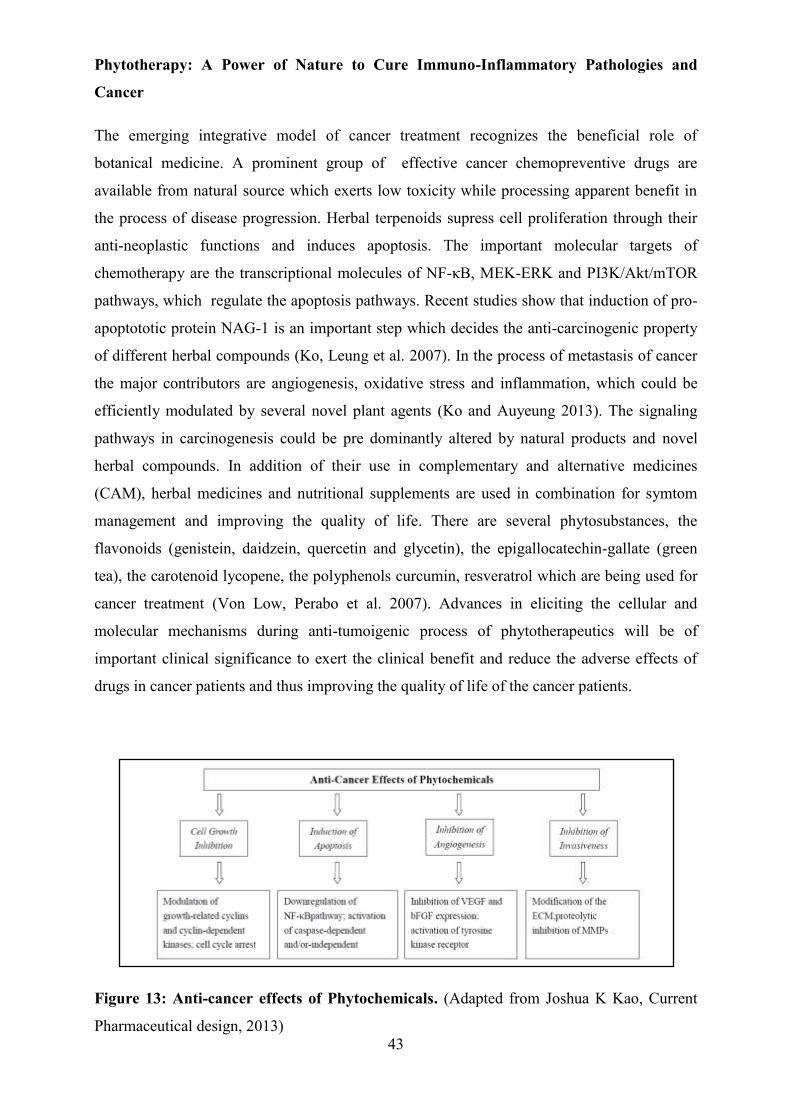
43
Phytotherapy: A Power of Nature to Cure Immuno-Inflammatory Pathologies and
Cancer
The emerging integrative model of cancer treatment recognizes the beneficial role of
botanical medicine. A prominent group of effective cancer chemopreventive drugs are
available from natural source which exerts low toxicity while processing apparent benefit in
the process of disease progression. Herbal terpenoids supress cell proliferation through their
anti-neoplastic functions and induces apoptosis. The important molecular targets of
chemotherapy are the transcriptional molecules of NF-κB, MEK-ERK and PI3K/Akt/mTOR
pathways, which regulate the apoptosis pathways. Recent studies show that induction of pro-
apoptototic protein NAG-1 is an important step which decides the anti-carcinogenic property
of different herbal compounds (Ko, Leung et al. 2007). In the process of metastasis of cancer
the major contributors are angiogenesis, oxidative stress and inflammation, which could be
efficiently modulated by several novel plant agents (Ko and Auyeung 2013). The signaling
pathways in carcinogenesis could be pre dominantly altered by natural products and novel
herbal compounds. In addition of their use in complementary and alternative medicines
(CAM), herbal medicines and nutritional supplements are used in combination for symtom
management and improving the quality of life. There are several phytosubstances, the
flavonoids (genistein, daidzein, quercetin and glycetin), the epigallocatechin-gallate (green
tea), the carotenoid lycopene, the polyphenols curcumin, resveratrol which are being used for
cancer treatment (Von Low, Perabo et al. 2007). Advances in eliciting the cellular and
molecular mechanisms during anti-tumoigenic process of phytotherapeutics will be of
important clinical significance to exert the clinical benefit and reduce the adverse effects of
drugs in cancer patients and thus improving the quality of life of the cancer patients.
Figure 13: Anti-cancer effects of Phytochemicals. (Adapted from Joshua K Kao, Current
Pharmaceutical design, 2013)

44
Table.1 List of some medicinal herbal products. (Adapted from Joshua K Kao, Current
Pharmaceutical design, 2013)

45
Figure 14: Phytotherapy strategy. a) Bioactive compounds b) Molecular targets of bioactive
compounds (Adapted from Treasure et al., Seminars in Oncology Nursing, 2005)
Conventional Oncology and Viscum album
Viscum album (Viscum album L.) is a traditional phytomedicine of Europe, commonly known
as European mistletoe, also found in Western and Southern Asia and it is the most thoroughly
studied complementary treatments in Europe. Several systemic reviews and meta-analyses
have found that viscum treatment is beneficial for cancer patients in terms of survival,
improved quality of life and reduce side effects of conventional anticancer therapies. Several
study supports that plant lectins, i.e. carbohydrate-binding proteins, exert remarkable
antitumor properties (Liu, Bian et al. 2010), (Liu, Luo et al. 2013), (Zhang, Chen et al. 2012).
Schink et al., in a randomised clinical trial, demonstrated that perioperative infusion of
Iscador® can inhibit NK cell activity associated with major surgery, which favours
haematogenic tumor cell dissemination (Schink, Troger et al. 2007).
Quality of life and Viscum album
Quality of life and the safety of adjuvant mistletoe therapy in gastric cancer patients were
tested in a randomised clinical trial (Kim, Yook et al. 2012). The additional mistletoe therapy
was found to be safe and associated with improved quality of life in these gastric cancer
patients. In a study of 70 cancer patients with different digestive tract cancers, use of
mistletoe drug- Isorel® showed improved immune competence and an improved overall health
a b

46
status of cancer patients undergoing conventional anti-tumor therapies (Enesel, Acalovschi et
al. 2005), (Galun 2015).
Viscum album
Viscum album has been used in European countries as sole intervention or as complement to
the conventional cancer therapies for more than eighty years (Bock, Friedel et al. 2004),
(Kroz, Kienle et al. 2014). It is a semiparasitic shrub that grows on other trees. Mistletoelectin
belong to the families Loranthaceae and Viscaceae which are both are taxonomically related
(Lyu and Park 2006). The pharmacologically applicable components of European mistletoe
are lectins, polysaccharides, alkaloids, lipids, triterpenes, peptides, vesicles, flavonoids,
visalbcBA (chitin binding agglutinin) (Khwaja, Dias et al. 1986), (Hajto, Hostanska et al.
1989), (Mueller and Anderer 1990), (Park, Hyun et al. 1998). Mistletoe lectin share hetero-
dimeric glycoproteins comprising two polypeptide chains: a carbohydrate-binding B-chain
which has a capability of binding to cell surface glycol conjugates which permits protein to
enter the cell and the catalytic A-chain which acts as ribosome-inactivating substance and
hence hinders protein synthesis intracellularly by removing an adenine residue from the
28SrRNA of 60S subunit of the ribosome (Peumans, Verhaert et al. 1996). The extract acts
not only as immunostimulatory drug but also have cytotoxic properties and DNA-stabilising
properties (Bussing, Regnery et al. 1995). It also stimulates the immune system in vivo and in
vitro by activating monocytes, macrophages, T cells, dendritic cells, granulocytes, NK cells
further induces several cytokines like IL-1, IL-2, IL-4, IL-5, IL-6, IL-8, IL10, IL-12, GMCSF,
IFN-γ and TNF-α (Thies, Nugel et al. 2005).
Mythological Aspect
The European mistletoe is a pharmaceutical plant and a symbol in mythology. Depending on
host specificity, there are three host races existing in Europe, they are 1. V. Album album-
grows on a wide variety of deciduous trees. 2. V. a. abietis (fir). 3. V. a. austriacum (Pine).
European mistletoe, i.e., is the first plant which was termed as “mistletoe”. Theopharastos
(371-286 BC) recognised that mistletoe is spread to the trees by birds and never touched the
earth. During the dead winters in Europe when branches of Oak trees were bare, mistletoe is
still green even considering the fact of not having roots on earth, made the Celtic Druids
believe mistletoe as sacred as ever-lasting life. According to G. P. Secundus (23-79 AC) this
plant was considered to be an antidote for poisons and the plant became a miracle because of
its ability to cure each illness. In ancient times this mythical plant was used in combination

47
with aromatic compounds and the surmise was the plant served protection against bad dreams,
lighting etc (Büssing 2003).
Mistletoe as a Remedy
Diosorides, the Greek author and physician (15-85 AC) reported that during 460-377 BC
spleen related diseases were treated using Oak tree mistletoe. During 23-79 AC, Plinius
explained the beneficial role of mistletoe in the treatment of infertility, ulcers, epilepsy.
Platonist around 150 AC described the utilization of mistletoe to treat tumors. During 1731
mistletoe was used for various treatments including labour pain, guts and deworming
children. Mistletoe treatment seemed to be beneficial for mumps, leprosy, and hepatitis.
During 18th Century this remedy was used for Oedema and heart weakness. In spite of having
a strong historical background of mistletoe, in the 19th Century scientific community rejected
mistletoe remedy. However, the interest was re awakened in the 20th century when Ganltier
(1907, 1910) demonstrated oral/subcutaneous administration of fresh mistletoe (L. Extract) to
cure blood pressure related issues both in animal and human. In 1920, the founder of
anthroposophy, Rudolf Steiner, introduced L. as an anti- cancer remedy (Büssing 2003).
Preparation of therapeutic preparation of Viscum album
Iscador® is the commercial preparation of VA preparations. It is prepared as an aqueous
extract of the whole mistletoe plant with a formulated fermentation with the bacterium
Lactobacillus plantarum. The product is then mixed and filtered to remove the bacteria before
being standardized and packaged in ampules for injection. Fermentation of the mistletoe
extract alters its medicinal activity to a significant degree and this change is thought to be
related to the degradation of the most toxic lectins. It is believed that the efficacy of mistletoe
extracts like Iscador are due to a synergy between both its components that are medicinally
active when isolated, and those components like polysaccharides that are medicinally
inactive, but can interact with the more active constituents to form complexes.
Chemical Compounds in Viscum album
Viscotoxins
Viscotoxin shows the structural characteristics of plant α- and β-thionins, which are the
cysteine rich residues and highly basic in nature. Seven different isoforms have been
identified so far, they are viscotoxin A1, A2, A3, B, B2, C1 and 1-PS. These isoforms are
consist of 46 amino acids. , among which 32 have identical amino acids and all isoforms
contain 3 disulphide bridges at highly conserved positions which are Cys3/Cys40,

48
Cys3/Cys32 and Cys16/Cys26). They are compact in structure and highly stable under several
denaturing conditions. The phosphate binding site of viscotoxins interferes with cell
membrane and dampens their integrity, which is the main reason of their cytotoxic effects,
primarily necrosis and later in apoptosis (Schaller, Urech et al. 1998).
Lectins
Three different mistletoe lectins (ML) were identified in Viscum album with differential
sugar-binding specificities, they are galactose-specific MLI: 115 kDa, diner, galactose- and
N-acetyl-D-galactosamine-specific MLII: 60 kDa and N-acetyl-D-galactosamine-specific
MLIII: 60 kDa. They belong to type 2 ribosome inactivating proteins that consist of a lectin
subunit B and a toxophoricA chain, which is RNA N-glycosidase. In eukaryotic cells, MLs
block protein synthesis by hydrolysing 28S rRNA. They induce apoptotic cell death
(Krauspenhaar, Eschenburg et al. 1999), (Wacker, Stoeva et al. 2004).
Polysaccharides
Mistletoe stems and leaves show differences due to the structural difference in the
polysaccharides. From these parts of the plant, a highly methylated galacturonan, a pectin (42
kD) and arabinogalactan (110 kD) were isolated. The high-molecular weight arabinogalactan
of viscum stimulates CD4+ Th cells proliferation (Stein, Edlund et al. 1999). Stimulation of
NK cells by Iscador® is due to its rhamnogalacturonan (Mueller and Anderer 1990).
Liposoluble compounds
Viscum album is rich in triterpenes, includingoleanolic acid, β-amyrinacetate, β-amyrin, the
lupanesleupeol, lupeol acetate, betulinic acid and urosolic acid (Jager, Trojan et al. 2009),
(Urech 2015).
Flavonoids, Phenylpropanoids and phenolic Acids
Viscum album contains phenolic compounds such as flavonoids, phenolic acids,
phenylpropanoids. Evidence of their pharmacological roles in pathologies is of great interest.
Lectin-induced apoptosis in cancer cells through increased oxidative stress is due to certain
flavonoids. They exert cytotoxic, anti-angiogenic, detoxicative, anti-inflammatory and anti-
hormonal effects on cancer cells (Schramm 2015), (Urech 2015).

49
Table 2. Chemical compounds identified in the European Viscum album L. (Adapted from
Konrad Urech, Transl Res Biomed, 2015)
Class of chemical compounds Compounds Proteins Thionins Viscotoxins A1, A2, A3, B, B2, C1, 1-PS
Lectins Mistletoe lectin I (MLI), II (MLII), III (MLIII) Chitin-binding mistletoe lectin 1 (cbML1), 2 (cbML2), 3 (cbML3)
Peptides Oligopeptides Glutathione
Amino acids Arginine, cystein, γ-aminobutyric acid
Amines Acetylcholine, choline, tyramine, histamine
Proteoglycans Arabinogalactan-proteins
Polysaccharides Methylated poly-1→α4 galacturonic acid, arabinogalactan, rhamnogalacturonan
Fatty acids
Oleic acid, palmitic acid, linoleic acid, linolenic acid, arachidic acid, cerotic acid, stearic acid, lignoceric acid
Phenolic compounds
Flavonoids 2′-hydroxy-4′,6′-dimethoxychaIcone-glucosid, 2′-hydroxy-4′,6′-trimethoxychaIcone-glucosid, 2′-hydroxy-4′,6′ dimethoxy- chalcone-4- O-[apiosyl(1→2)] glucoside, (2R)-5,7-dimethoxyflavanone-4′-O-gIucosid, (2S)-3′,5,7-trimethoxy-flavanone-4′-O- gIucoside, homoeriodictyol-7-Oglucoside, rhamnazin-3,4′-di-O-glucoside, 5,7-dimethoxy-4′-hydroxyflavon After acid hydrolysis: homoeriodyctiol, sakuranetin, rhamnazin, isorhamnetin, quercetin, 6 different quercetin methylesters, kaemferol, naringenin
Phenylpropanoids
Syringenin-4′-O-glucoside (syringin), syringenin-4′-O-apiosyl-1 (Pfeil) 2 glucoside (syringoid), syringaresinol-4,4′-O-glucoside, eleutheroside E, syringaresinol- mono-O-glucoside, sinapic acid, cinnamic acid, rosmarinic acid, caffeic acid, ferulic acid, chlorogenic acid, isochlorogenic acid, syringic acid, p- and m-coumaric acid
Other phenolic acids
Gallic acid (3,4,5-trihydroxybenzoic acid), digallic acid, para-OH benzoic acid, syringic acid (methylated trihydroxybenzoic acid), salicylic acid, protocatechuic acid (3,4-dihydroxybenzoic acid), vanillic acid (methylated dihydroxybenzoic acid), gentisic acid (2,5 dihydroxybenzoic acid), salicylic acid (2 hydroxybenzoic acid), ellagic acid
Terpenoids
Triterpenoids
Oleanolic acid, betulinic acid, ursolic acid, β-amyrin, β-amyrin acetate, lupeol, lupeol acetate
Tetraterpenoids Carotin
Phytosterols β-sitosterol, stigmasterol
Inorganic substances
Manganese, potassium, calcium (calcium oxalate)
Various compounds
Ascorbic acid 7-iso-jasmonic acid, and its precursor 12-oxophytodienoic acid

50
Figure 15: Mechanism of action of type II lectins.
Multifarious Properties of Viscum album
Cytotoxicity
Mistletoe lectin can induce apoptosis depending on the apoptosis-associated factor-1 (Apaf-1)
pathway by mitochondrial membrane potential (MMP) breakdown and stimulating caspase-3
(Lyu, Choi et al. 2002), (Liu, Luo et al. 2013). JNK can be stimulated by ML-I and leads to
translocation of the pro-apoptotic proteins Bax and Bad. Mistletoes induce apoptosis in
human peripheral blood lymphocytes, murine lymphocytes, mononuclear leukemia cells
MOLT4 and human monocytic THP1 cells (Janssen, Scheffler et al. 1993), (Mockel, Schwarz
et al. 1997), (Kim, So et al. 2000). ML-I downregulates Bcl-2 and upregulates TNF-α and
hence provoke apoptosis. We have demonstrated that VA Qu Frf, induces significant cell
toxicity in vitro in the human T cell lines CEM and in monocytic cell lines HL-60 and MM-6
(Duong Van Huyen, Sooryanarayana et al. 2001).
Anti-inflammatory
Diarylheptanoids and flavonoids of mistletoe inhibit LPS-stimulated production of pro-
inflammatory cytokines such as TNF-α, IL-6, IL-12p40 in bone marrow-derived dendritic
cells (Nhiem, Kiem et al. 2013). Our group have found that VA Qu Spez impedes cytokine-
induced PGE2, by selectively inhibiting COX-2 which is transcriptionally activated in
response to various pro-inflammatory cytokines (Hegde, Maddur et al. 2011). Further, we
have dissected the molecular events of COX-2 regulation recently we observed significant
reduction of COX-2 mRNA half-life without influencing its protein stability, clearly
suggesting that it induces destabilisation of COX-2 mRNA (Saha, Hegde et al. 2015).

51
Anti-angiogenic:
Tumor growth can be impedimented by inhibiting angiogenesis. Treating B16L6 melanoma
cells with viscum suppressed tumor growth and resulted in DNA fragmentation, nuclear
morphological changes, suggesting that it inhibits tumor growth and metastasis by elevating
apoptosis and blocking angiogenesis (Park, Lyu et al. 2001). Our group has shown that VA
QU Frf induces apoptosis of endothelial cells in human umbilical vein endothelial cells
(HUVEC) and in immortalised human venous endothelial cell line (IVEC). In an in vivo
system it was clearly demonstrated that VA Qu Spez dramatically impedes the vessel score in
mice, diminishing angiogenic growth (Elluru, Duong Van Huyen et al. 2009).
Immunomodulatory
Viscum album, significantly enhanced IFN-γ; strongly supports the fact that is an immune
modulator (Lyu and Park 2007). Our group has reported that in a murine melanoma model,
QU Frf mistletoe preparation significantly inhibited tumor growth and up-regulated IL-12
secretion, which was confirmed by abrogation of IL-12 expression in IL-12 knockout mice
(Duong Van Huyen, Delignat et al. 2006). Qu Spez amplifies the expression of several
antigen presenting and co-stimulatory molecules on human dendritic cells and additionally
induces secretion of pro-inflammatory cytokines such as IL-6 and IL-8 and stimulates
proliferation of CD4+ Tcells (Elluru, Duong van Huyen et al. 2008).
Viscum album: Clinical Evidence
Rudolf Steiner, the father of anthroposophy, in 1961 had first propounded the use of mistletoe
extracts for cancer treatment. Anthrosopical medicine professes that mistletoe is effective for
cancer patients as it delays disease progression, protracts survival time, modulates immune
function and overall improves general well-being. Only three among several preparations of
mistletoe is tested clinically, they are Iscador®, Eurixor®, and Helixor® (Kleijnen and
Knipschild 1994). There are several reports on the use of mistletoe in clinics and their
efficacy. However, the proof of results is controversial because of the loopholes in the
methodology used to evaluate the effectiveness of complementary medicine.
H.S.LIN (2004) group had taken an attempt to gather information on the efficacy, safety and
side effects of standardized mistletoe extract by following “Good Clinical Practice”. This
study enrolled 233 patients with 71 ovarian, 68 breast and 94 non-small lung cancer. Among
these 233 cancer patients 224 patients were included in the final analysis out of which 115
patients were treated with mistletoe. All patients underwent the conventional chemotherapies.
52
According to the “Intension to treat principle” analysis, complementarily mistletoe treated
patient group had shown a significant improve in the quality of life (QOL) compared to the
control group. Concerning the side effects such as fatigue, nausea, anorexia and insomnia, the
adverse effects (AES) recorded were fewer than the control group. Most of these effects were
self-limiting and harmless, proving the safety of mistletoe use (Piao, Wang et al. 2004).
In 2004, another clinical study reported by U. Mengs, recruiting 272 patients, showed
PS76A2, an aqueous mistletoe extract at a dose of 15 ng ML/0.5ml/twice weekly is safe for
treatment and improved QOL effectively in breast cancer patients who were receiving
adjuvant cyclophosphamide-methotrexate-fluorouracil (CMF). An increase of T helper
lymphocytes (CD4+) as well as the ratio between CD4+/CD8+supported the data on QOL
(Semiglasov, Stepula et al. 2004).
Schiernoz team (2008) investigated the safety and efficacy of standardised mistletoe extract
HELIXOR® complimentary treatment of breast cancer patients. The study was carried out
during a defined after care period of 5 years of the patients through a comparative
epidemiological cohort study by random selection of 53 hospitals in Germany including 681
patients. Firstly data showed 56.3% study group versus 70% control group
complaints/therapy-related symptoms and secondly disease-related signs was lessened with
mistletoe treated patients harmonized to a significantly improved QOL. The adverse drug
reactions were self-limiting (Beuth, Schneider et al. 2008).
Physicians often noticed that mistletoe intake have favourable impact on cancer-related
fatigue (CRF). In one of the clinical study, a 36-year old Swedish woman with 10-year history
of reappearance of breast cancer was suffering from CRF and when the lady was treated with
mistletoe beside the conventional therapy, she improved her QOL by reducing fatigue level
(Wode, Schneider et al. 2009).
Preclinical and clinical studies exploring the effect of extract recommended strongest
beneficial role of VAE in terms of QOL and tolerance to the conventional anti-cancer
treatments in gynaecological and breast cancer. VAE was able to elevate survival rate and
abrogate tumor especially in mice. There was a strong cytotoxic effects observed on cancer
cells in vitro with VAE treatment (Kienle, Glockmann et al. 2009).
A study investigated Post relapse (12 month) disease-free survival rate in Osteosarcoma
patients who has a high chance of second relapsing receiving either Etoposide or Viscum
album. Twenty patients were matriculated in the study. In Etoposide group the median

53
PRDFS was 4 months, whereas it was prolonged to 39 months in viscum group. Thereby,
viscum therapy remains superior to Etoposide treatment (Longhi, Reif et al. 2014).
Mansky et al (2003) conducted a phase I, 2 stage dose-escalation study (Case Study: 02-074)
with the intension to test European mistletoe (EurixorTM) used with an approved
antimetabolite chemotherapeutic agent gemcitabine in solid tumor patients. Combination of
1380 mg/m2 gemitabine and 250 mg mistletoe was the maximum tolerated dose. Out of 44
patients, 33 patients completed minimum 3 cycles of therapy. 6% (n=2) showed partial
response, 42% (n=14) developed stable disease, 43% (n=14) improved upon treatment. 37%
patients developed non neutropenic fever, whereas control group was associated with 41%
fever. Mistletoe/gemcitabine cocktail and gemcitabine alone showed similar hematologic
toxicity profile and febrile reaction. There was an ascending trend of ANC with mistletoe
treatment (Mansky, Grem et al. 2003), (Mansky, Wallerstedt et al. 2013).
European mistletoe extracts (L.) are the most commonly prescribed cancer treatments in
Germany per se in 2010 (Kroz, Kienle et al. 2014). In–vitro and in-vivo studies have
identified their immunomodulatory and cytostatic effects (Lyu and Park 2006). Today the
therapeutic goal is to improve health related QOL and that is acknowledged as an end point in
clinical trials. Pharmacological actions of mistletoe lectin are well documented, however,
clinical trials evidence was rare and the existing proofs have been criticised. Wide variety of
commercial availability of mistletoe impedes the comparative assessment of the benefits of
use of the extract in cancer therapy. Difference in the extraction process and manufacturing
method, result in the variation of pharmacological or clinical effects of mistletoe (Kleijnen
and Knipschild 1994). Consequently complementary treatment with standardised mistletoe
extract in cancer can be regarded as safe.

OBJECTIVES

54
In view of the critical link between inflammation and cancer which share several signalling
events, regulatory mechanisms, it is necessary to unravel the molecular and cellular
mechanisms of underlying anti-inflammatory and immunomodulatory effect of Viscum
album, which can provide a better understanding of its immunotherapeutic strategies to
develop integrative medicinal approaches to inflammatory pathologies and cancer. Therefore
my study addresses the anti-inflammatory and immunomodulatory properties of viscum and
the mode of action which in turn can strengthen the beneficial application of viscum in
complementary therapy to improve the survival and quality of life of cancer patients.
Following are the objectives of my study.
Objective 1: Molecular dissection of Viscum album mediated COX-2 inhibition and
better understanding of its anti-inflammatory effect.
Objective 2: Exploring the immunomodulatory effects of Viscum album by studying
differential effect of various preparations of Viscum album on maturation and activation
of human dendritic cells and T cell response.
Objective 3: Exploring the anti-tumor response of Viscum album by understanding their
effect on the full spectrum of macrophage polarization.

RESULTS

55
a111
RESEARCH ARTICLE
Viscum album-Mediated COX-2 Inhibition Implicates Destabilization of COX-2 mRNA Chaitrali Saha1,2,3‡, Pushpa Hegde1,2,3‡, Alain Friboulet2, Jagadeesh Bayry1,3,4,5, Srinivas V. Kaveri1,3,4,5*
1 Institut National de la Santé et de la Recherche Médicale, Unité 1138, Paris, France, 2 Université de Technologie de Compiègne, UMR CNRS 6022, Compiègne, France, 3 Centre de Recherche des Cordeliers, Equipe-Immunopathology and therapeutic immunointervention, Paris, France, 4 Sorbonne Universités, UPMC Univ Paris 06, UMR_S 1138, Paris, France, 5 Université Paris Descartes, Sorbonne Paris Cité, UMR_S 1138, Paris, France
11 ‡ These authors contributed equally to this work.
Abstract
OPEN ACCESS
Citation: Saha C, Hegde P, Friboulet A, Bayry J, Kaveri SV (2015) Viscum album-Mediated COX-2 Inhibition Implicates Destabilization of COX-2 mRNA. PLoS ONE 10(2): e0114965. doi:10.1371/journal. pone.0114965
Academic Editor: Masaru Katoh, National Cancer Center, JAPAN
Received: July 25, 2014
Accepted: November 17, 2014
Published: February 9, 2015
Copyright: © 2015 Saha et al. This is an open access article distributed under the terms of the Creative Commons Attribution License, which permits unrestricted use, distribution, and reproduction in any medium, provided the original author and source are credited.
Data Availability Statement: All relevant data are within the paper.
Funding: This work is supported by Institut National de la Santé et de la Recherche Médicale (INSERM), Centre National de la Recherche Scientifique (CNRS), Université Pierre et Marie Curie and Université Paris Descartes, Regional Program Bio- Asie 2010 by the French Ministry of Foreign and European Affairs and Institut Hiscia, Arlesheim, Switzerland. The funders had no role in study design, data collection and analysis, decision to publish, or preparation of the manuscript.
Extensive use of Viscum album (VA) preparations in the complementary therapy of cancer and in several other human pathologies has led to an increasing number of cellular and mo- lecular approaches to explore the mechanisms of action of VA. We have recently demon- strated that, VA preparations exert a potent anti-inflammatory effect by selectively down- regulating the COX-2-mediated cytokine-induced secretion of prostaglandin E2 (PGE2), one of the important molecular signatures of inflammatory reactions. In this study, we ob- served a significant down-regulation of COX-2 protein expression in VA-treated A549 cells however COX-2 mRNA levels were unaltered. Therefore, we hypothesized that VA induces destabilisation of COX-2 mRNA, thereby depleting the available functional COX-2 mRNA for the protein synthesis and for the subsequent secretion of PGE2. To address this ques- tion, we analyzed the molecular degradation of COX-2 protein and its corresponding mRNA in A549 cell line. Using cyclohexamide pulse chase experiment, we demonstrate that, COX- 2 protein degradation is not affected by the treatment with VA whereas experiments on tran- scriptional blockade with actinomycin D, revealed a marked reduction in the half life of COX-2 mRNA due to its rapid degradation in the cells treated with VA compared to that in IL-1β-stimulated cells. These results thus demonstrate that VA-mediated inhibition of PGE2 implicates destabilization of COX-2 mRNA. Introduction
Cyclo-oxygenase-2 (COX-2) is an early response protein, up-regulated during many pathologi- cal conditions and human malignancies. It is over expressed in most of the cells upon stimula- tion with diverse pro-inflammatory stimuli such as pro-inflammatory cytokines, chemokines, infectious agents, bacterial lipopolysaccharide etc. COX-2 is a critical enzyme required for the biosynthesis of prostaglandin E2, one of the important molecular mediators of inflammation [1]. Two other COX isoenzymes, COX-1 and COX-3, catalyze the same kind of reaction. COX-

Viscum album-Mediated COX-2 Inhibition
56
Competing Interests: The authors have read the journal’s policy and the authors of this manuscript have the following competing interests: Part of the research was supported by Institut Hiscia, Arlesheim, Switzerland. Jagadeesh Bayry and Srinivas Kaveri are currently academic editors for Plos one. This does not alter the authors’ adherence to PLOS ONE Editorial policies and criteria.
1 is an important cyclo-oxygenase family member, and is constitutively expressed in cells and tissues, while precise functions are not known for COX-3, which is expressed only in some spe- cific compartments including brain and spinal cord [2, 3]. The pattern of expression of COX-1 versus COX-2 further regulates their differential functions. COX-1 is constitutively and stably expressed at low levels in many tissues. This ensures a constant production of prostaglandins, which are essentially required for the maintenance of important physiological functions, such as platelet aggregation, normal renal functions and gastric mucosal protection. In contrast, COX-2 is mostly quiescent but the expression can be induced in response to diverse pro-in- flammatory and pathogenic stimuli. When stimulated, its expression is high and transient which leads to a burst of prostaglandin production in a regulated time-limited manner [4]. Thus, depending on the COX isoform, the production of the same precursor PGH2 from ara- chidonic acid differs with respect to the amount and timing of production. This can be differ- entially decoded by the cells, thereby leading to the activation of various intracellular pathways involving specific classes of prostaglandins and therefore, different responses [5].
Since COX-2 expression is up-regulated during several pathological conditions and human malignancies, strategies controlling the expression and activity of COX-2 have been developed as potent anti-tumor and anti-inflammatory treatments [6–10]. In line with the therapeutic benefit of non steroid anti-inflammatory drugs (NSAID), which are synthetically designed mainly to inhibit the enzymatic activity of COX-2, a diverse spectrum of therapeutics of natural origin such as phytotherapeutics have been characterized to evaluate their potential to inhibit the COX-2 functioning thereby down-regulating the pathological level of prostaglandins. Due to the structural homology between COX-1 and COX-2, most of the NSAID inhibit both the enzymes and thus resulting in several severe side effects due to the inhibition of physiological prostaglandins. Therefore, selective inhibitors of COX-2 are of great interest. Although, a promising class of synthetic COX-2 selective inhibitors called COXIBS have been developed, their therapeutic efficacy is compromised due to various side effects [11, 12]. Interestingly, sev- eral phytotherapeutics have been shown to exert therapeutic benefit via selective inhibition of COX-2. These natural molecules have been shown to interfere with the expression and regula- tory mechanisms of COX-2 to inhibit its functioning [13, 14].
Viscum album (VA) preparations commonly called as mistletoe extracts, are extensively used as complementary therapeutics in cancer and also in the treatment of several inflammato- ry pathologies [15–19]. Despite their therapeutic application for several years, the underlying mechanisms are not yet clearly understood. Several lines of evidence have revealed that these preparations exert anti-tumor activities, which involve the cytotoxic properties, induction of apoptosis, inhibition of angiogenesis and several other immunomodulatory and anti-inflam- matory mechanisms [20–30]. These properties collectively define the mechanistic basis for the therapeutic benefit of VA preparations. Recently we have shown that, VA preparations exert a potent anti-inflammatory effect by selectively down-regulating the COX-2-mediated cytokine- induced secretion of prostaglandin E2 (PGE2), one of the important molecular signatures of in- flammatory reactions [31]. However, the molecular mechanisms associated with the Viscum- mediated COX-2 inhibition are not clear. VA preparations are shown to inhibit the COX-2 protein expression without modulating its expression at mRNA level suggesting a possible ef- fect of VA on post-transcriptional events of COX-2 regulation. Several molecules and phy- totherapeutics are known to interfere with the post-transcriptional and post-translation regulation of COX-2 in order to inhibit the COX-2 expression and subsequent reduction of PGE2 [32–34]. Therefore in the current study, we investigated the post-transcriptional and post-translational regulation of COX-2 by analyzing the stability of COX-2 protein and mRNA, which can explain in part, the molecular mechanisms of Viscum-mediated COX- 2 inhibition.

Viscum album-Mediated COX-2 Inhibition
57
Materials and Methods Viscum album preparations
VA Qu Spez was a kind gift from Weleda AG (Arlesheim, Switzerland). VA Qu Spez is a thera- peutic preparation of Viscum album that grows on oak trees and is obtained as an isotonic solu- tion of 10mg/ml formulated in 0.9% NaCl. It is free from endotoxins and contains the standardized levels of mistletoe lectins.
Culture of A549 cells
Human lung adenocarcinoma cell line A549 was a kind gift from Dr. Maria Castedo-Delrieu, Institute Gustave Roussy, Villejuif, France. A549 cells were grown in 75 cm2 culture flasks in Dulbecco’s modified Eagle’s medium (DMEM) F-12 (GIBCO, Life Technologies, Grand Island, NY, USA) supplemented with 10% fetal calf serum (FCS) and 50 U/ml penicillin and 50 μg/ml of streptomycin (GIBCO). Cells are incubated at 37°C with 5% CO2 in humidified atmosphere to obtain the cells of about 80–90% confluence and used for all experiments.
Co- and post- treatment of VA Qu Spez and induction of COX-2
Cells grown in complete medium (DMEM with 10% FCS) were harvested by trypsinisation using 0.5% trypsin (Biological Industries, Kibbutz Beit Haemek, Israel) and were seeded in 12-well culture plates (0.5×10⁶/ml cells per well). Wells containing the adherent A549 were then replenished with the complete medium containing recombinant human IL-1β (10 ng/ml) (Immuno Tools, Friesoythe, Germany). In one set of experiment VA Qu Spez is added at the time of addition of IL-1 β (co-treatment) and in another set, we add VA Qu Spez 14 hours after adding IL-1β (post-treatment) and both the sets were incubated until 18 hours at 37° C and 5% CO2. After 18 hours of incubation cells were harvested by trypsinization and used for the anal- ysis of COX-1/COX-2 protein by flow cytometry.
Analysis of the degradation profile of COX-2 protein by cyclohexamide pulse chase experiment A549 cells with an appropriate confluency were treated with IL-1β for 18 hours in the presence or absence of VA Qu Spez. To block the protein synthesis 10 μg/ml of cyclohexamide (Sigma- Aldrich, Lyon, France) was added after 90 minutes of addition of IL-1β and then cells were har- vested at different time intervals as indicated to achieve a clear pattern of COX-2 degradation. At each time point, expression of remaining COX-2 protein was analyzed by intracellular label- ling, by flow cytometry and further validated by western blotting.
Analysis of COX-2 mRNA half-life by actinomycin D pulse chase experiment A549 cells with an appropriate confluency were treated with IL-1β for 4 hours in the presence or absence of VA Qu Spez. After 4 hours, 10 μg/ml of actinomycin D (Sigma-Aldrich) was added to the cells and cells were harvested by trypsinisation at different time intervals as indi- cated. Expression of remaining COX-2 mRNA was analyzed by RT-PCR.
Statistical analysis
Densitometric analysis of the immunoblots was performed using BIO-1D analysis software. Values are expressed as arbitrary units. All the observations are expressed as Mean ±SEM and

Viscum album-Mediated COX-2 Inhibition
58
analyzed using two-way ANOVA. Graph-Pad Prism 5.0 is used for all the statistical analysis. P values less than 0.05 were considered to be statistically significant.
Results Co-treatment of A549 cell with IL-1β and Viscum album inhibits the cytokine-induced COX-2 expression Following our observation of the inhibition of cytokine-induced COX-2 expression, we investi- gated the appropriate window of efficient inhibition by VA. Human lung adenocarcinoma (A549) cells were stimulated with IL-1β for 18 hours in the presence or absence of VA Qu Spez. VA was added to the cells either along with the cytokine (co-treatment) or after 14 hours of IL-1β induction. Flow cytometric analysis of intracellular COX-2 expression demonstrated that VA significantly inhibits cytokine-induced COX-2 expression as measured by mean fluo- rescent intensity (MFI) only when it is added as a co-treatment with IL-1β but not when it was added after 14 hours (Fig. 1A and 1B). This suggests that, VA-mediated COX-2 inhibition oc- curs at the early phases of inflammatory process and opens other exploratory avenues to un- derstand the regulatory mechanisms of COX-2 inhibition mediated by VA at the early phase of inflammation.
Inhibition of COX-2 protein expression by Viscum album is independent of modulation of stability of COX-2 protein In order to address the effect of VA on the molecular stability of COX-2, which could be a po- tential contributing factor for the observed reduction in COX-2 protein expression, we ana- lyzed the stability of COX-2 protein. A549 cells were stimulated with a pro-inflammatory cytokine IL-1β in the presence and absence of VA Qu Spez. At 18 hours, we observed a signifi- cant reduction in COX-2 protein level treated with VA Qu Spez. Further, cells were harvested at different time intervals after blocking the protein synthesis by treating the cells with cyclo- hexamide and analyzed for COX-2. Flow cytometric analysis of COX-2 protein has revealed that, there is no significant difference in the protein degradation profile of COX-2 in VA-treat- ed and untreated cells after 90 minutes of blocking the protein synthesis (Fig. 2A and Fig. 2B). Further, western blot analysis of COX-2 protein expression at different time intervals showed that despite the clear inhibition in the protein expression after 18 hours of exposure to cytokine followed by VA treatment (Fig. 3A), upon blocking the protein synthesis, there is no remark- able difference in the COX-2 degradation profile in cells treated with cytokine irrespective of VA treatment (Fig. 3B, 3C and 3D). Fig. 3B indicates the level of COX-2 expression immediate- ly after 90 minutes of cyclohexamide addition (0 hour). Figs. 3C and 3D indicate the level of COX-2 expression upon blocking the protein synthesis after 5 and 11 hours respectively. These results may indicate that the regulation of COX-2 by VA may occur in an early phase of COX- 2 expression but not at the later stages of protein expression and stabilization.
Viscum album increases the COX-2mRNA degradation
Due to the indication of effect of VA in the early stages of COX-2 expression, but not at the level of its mRNA expression, we analyzed the mRNA stability of COX-2 modulated by VA. A549 cells were stimulated with IL-1β in the presence and absence of VA Qu Spez for 4 hours. After 4 hours, cells were treated with actinomycin D and harvested at different time intervals. Total cellular RNA was isolated and used for RT-PCR for the estimation of COX-2 mRNA. Treatment with IL-1β is known to induce the expression of COX-2 mRNA by transcriptional activation and also by increasing the stability of COX-2 mRNA. RT-PCR analysis of COX-2
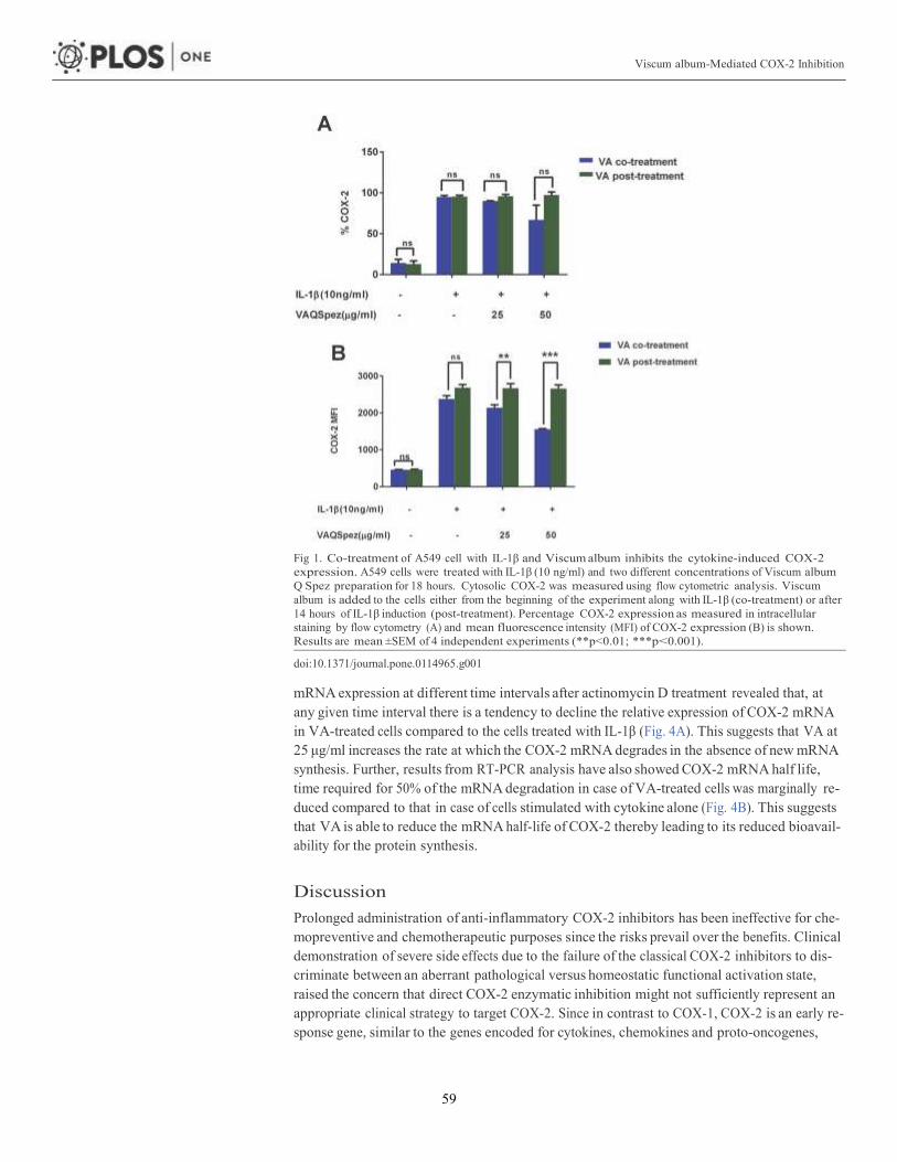
Viscum album-Mediated COX-2 Inhibition
59
Fig 1. Co-treatment of A549 cell with IL-1β and Viscum album inhibits the cytokine-induced COX-2 expression. A549 cells were treated with IL-1β (10 ng/ml) and two different concentrations of Viscum album Q Spez preparation for 18 hours. Cytosolic COX-2 was measured using flow cytometric analysis. Viscum album is added to the cells either from the beginning of the experiment along with IL-1β (co-treatment) or after 14 hours of IL-1β induction (post-treatment). Percentage COX-2 expression as measured in intracellular staining by flow cytometry (A) and mean fluorescence intensity (MFI) of COX-2 expression (B) is shown. Results are mean ±SEM of 4 independent experiments (**p<0.01; ***p<0.001).
doi:10.1371/journal.pone.0114965.g001
mRNA expression at different time intervals after actinomycin D treatment revealed that, at any given time interval there is a tendency to decline the relative expression of COX-2 mRNA in VA-treated cells compared to the cells treated with IL-1β (Fig. 4A). This suggests that VA at 25 μg/ml increases the rate at which the COX-2 mRNA degrades in the absence of new mRNA synthesis. Further, results from RT-PCR analysis have also showed COX-2 mRNA half life, time required for 50% of the mRNA degradation in case of VA-treated cells was marginally re- duced compared to that in case of cells stimulated with cytokine alone (Fig. 4B). This suggests that VA is able to reduce the mRNA half-life of COX-2 thereby leading to its reduced bioavail- ability for the protein synthesis.
Discussion
Prolonged administration of anti-inflammatory COX-2 inhibitors has been ineffective for che- mopreventive and chemotherapeutic purposes since the risks prevail over the benefits. Clinical demonstration of severe side effects due to the failure of the classical COX-2 inhibitors to dis- criminate between an aberrant pathological versus homeostatic functional activation state, raised the concern that direct COX-2 enzymatic inhibition might not sufficiently represent an appropriate clinical strategy to target COX-2. Since in contrast to COX-1, COX-2 is an early re- sponse gene, similar to the genes encoded for cytokines, chemokines and proto-oncogenes,
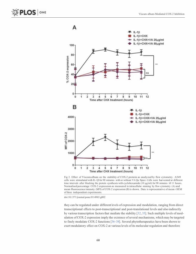
Viscum album-Mediated COX-2 Inhibition
60
Fig 2. Effect of Viscum album on the stability of COX-2 protein as analyzed by flow cytometry. A549 cells were stimulated with IL-1β for 90 minutes with or without VA Qu Spez. Cells were harvested at different time intervals after blocking the protein synthesis with cyclohexamide (10 μg/ml) for 90 minutes till 11 hours. Normalised percentage COX-2 expression as measured in intracellular staining by flow cytometry (A) and mean fluorescence intensity (MFI) of COX-2 expression (B) is shown. Data is representative of mean ±SEM of three independent experiments.
doi:10.1371/journal.pone.0114965.g002
they can be regulated under different levels of expression and modulation, ranging from direct transcriptional effects to post-transcriptional and post-translational levels and also indirectly by various transcription factors that mediate the stability [32, 35]. Such multiple levels of mod- ulation of COX-2 expression imply the existence of several mechanisms, which may be targeted to finely modulate COX-2 functions [36–38]. Several phytotherapeutics have been shown to exert modulatory effect on COX-2 at various levels of its molecular regulation and therefore

Viscum album-Mediated COX-2 Inhibition
61
Fig 3. Effect of Viscum album on the stability of COX-2 protein as determined by western blot. Confluent A549 cells were treated with IL-1β in the presence and absence of VA Qu Spez in dose dependent concentrations in μg/ml. Cells were harvested at different time intervals after blocking the protein synthesis with cyclohexamide (10 μg/ml) for 90 minutes till 11 hours. COX-2 expression was measured by western blot using the cytosolic extracts. (A), inhibition of COX2 protein synthesis by VA at 18 hours. (B) (C) (D) are the representative western blots after 90 minutes, 5 hours and 11 hours respectively showing level of COX-2 expression after cyclohexamide treatment with or without Viscum album. β-actin was used as an internal control. All blots are representative of three independent experiments and the densitometry values for each band are mentioned below the representative blots.
doi:10.1371/journal.pone.0114965.g003
have been considered as an effective alternative strategy to control the pathogenic expression of COX-2 [33, 39, 40]. Given that VA preparations exert a potent anti-inflammatory effect by selective down regulation of COX-2, it is extremely interesting to dissect the COX-2 inhibition mediated by VA in different regulatory mechanisms at molecular level.
Co-treatment of VA along with cytokine stimulation, marginally decreases COX-2 expres- sion indicated by the percentage-positive COX-2 expression in Fig. 1A. However, VA signifi- cantly inhibits intensity of expression of COX-2 as analyzed by MFI. The fact that VA treatment at the later phases of cytokine induction does not inhibit COX-2 suggests that, inhi- bition of COX-2 by VA occurs in the early phase of COX-2 regulation but not at the later phases (Fig. 1). Since we observed an inhibition of COX-2 protein expression by VA but not of mRNA, we analyzed the protein stability of COX-2 in the presence of VA by cyclohexamide pulse chase experiments. Flow cytometric analysis of COX-2 expression after 90 minutes of blocking the protein synthesis with cyclohexamide showed that, there is no significant differ- ence in the COX-2 degradation profile of cells simulated with IL-1β with or without treatment with VA (Fig. 2A and 2B). Western blot analysis of COX-2 protein after 5 and 11 hours of cyclohexamide blockade showed no significant difference in the degradation pattern of COX-2 in cytokine stimulated cells with or without VA treatment (Fig. 3C and 3D). Similar results at

Viscum album-Mediated COX-2 Inhibition
62
Fig 4. Increase in the COX-2 mRNA degradation by Viscum album treatment. A549 cells were stimulated with a pro-inflammatory cytokine IL-1β in the presence and absence of VA Qu Spez for 4 hours. After 4 hours of IL-1β stimulation cells are blocked with actinomycin D (10 μg/ml). Cells were harvested at different time intervals after adding actinomycin D and total cellular RNA was isolated and used for RT-PCR for the estimation of COX-2 mRNA. Relative expression of remaining COX-2 mRNA at each time point, in VA treated and untreated cells (A) and the time required for 50% of the mRNA degradation as COX-2 mRNA half life (B). Data is obtained from three independent experiments.
doi:10.1371/journal.pone.0114965.g004
different time points were observed (data not shown). Therefore, it is clear that COX-2 protein degradation is not affected by VA. Further, reduced level of COX-2 expression at 0 hour in this experiment (Fig. 3B) also suggests that, there may be modulation by VA of the COX-2 expres- sion before the addition of inhibitor of protein synthesis. Inhibition of COX-2 protein expres- sion by VA (Fig. 3A) without modulating its stability (Fig. 3B, 3C and 3D) strongly indicates that, there is a possible modulation by VA at an early stage than when the proteins were ex- pressed. However VA did not modulate COX-2 mRNA expression and therefore we analyzed the mRNA stability of COX-2 by actinomycin D pulse chase experiment. mRNA degradation

Viscum album-Mediated COX-2 Inhibition
63
profile of COX-2 obtained by analyzing the COX-2 mRNA at different time intervals after blocking the transcription using actinomycin D showed that the rate of degradation of COX-2 mRNA is higher in cells treated with VA compared to those treated with cytokine alone (Fig. 4A). This reduction in the mRNA half-life of COX-2 in the cells treated with VA (Fig. 4B) suggests that, VA induces destabilization of COX-2 mRNA, thereby diminishing the available functional mRNA for the protein synthesis and for the subsequent secretion of PGE2.
Although this study postulates destabilization of COX-2 mRNA by VA preparations as a possible mechanism for VA-mediated COX-2 inhibition, further molecular dissection is neces- sary in order to clearly understand the regulatory events of COX-2 regulation, contributing fac- tors and their modulation by VA preparations.
Conclusion
Increasing body of evidence for anti-inflammatory activity of plant-derived molecules by mod- ulating the COX-2 functions has evolved as a potent alternative strategy for the conception of novel therapeutic molecules in the treatment of various inflammatory pathologies and in vari- ous malignancies. In view of the therapeutic benefit of VA preparations in diverse pathological situations including inflammatory and cancer conditions, dissecting their molecular mecha- nisms would contribute enormously to the understanding of role of phytotherapy-based treat- ment strategies either in complementary or alternative medicine or in other combinational therapies.
Author Contributions
Conceived and designed the experiments: CS PH AF JB SVK. Performed the experiments: CS PH. Analyzed the data: CS PH AF JB SVK. Contributed reagents/materials/analysis tools: AF JB SVK. Wrote the paper: PH CS AF JB SVK.
References 1. Rouzer CA, Marnett LJ (2009) Cyclooxygenases: structural and functional insights. J Lipid Res 50
Suppl: S29–34. doi: 10.1194/jlr.R800042-JLR200 PMID: 18952571 2. Chandrasekharan NV, Dai H, Roos KL, Evanson NK, Tomsik J, et al. (2002) COX-3, a cyclooxygen-
ase-1 variant inhibited by acetaminophen and other analgesic/antipyretic drugs: cloning, structure, and expression. Proc Natl Acad Sci U S A 99: 13926–13931. doi: 10.1073/pnas.162468699 PMID: 12242329
3. Kis B, Snipes JA, Isse T, Nagy K, Busija DW (2003) Putative cyclooxygenase-3 expression in rat brain cells. J Cereb Blood Flow Metab 23: 1287–1292. doi: 10.1097/01.WCB.0000090681.07515.81 PMID: 14600435
4. Crofford LJ (1997) COX-1 and COX-2 tissue expression: implications and predictions. J Rheumatol Suppl 49: 15–19. PMID: 9249646
5. Kam PC, See AU (2000) Cyclo-oxygenase isoenzymes: physiological and pharmacological role. An- aesthesia 55: 442–449. doi: 10.1046/j.1365-2044.2000.01271.x PMID: 10792135
6. Martel-Pelletier J, Pelletier JP, Fahmi H (2003) Cyclooxygenase-2 and prostaglandins in articular tis- sues. Semin Arthritis Rheum 33: 155–167. doi: 10.1016/S0049-0172(03)00134-3 PMID: 14671726
7. Zhang L, Bertucci AM, Smith KA, Xu L, Datta SK (2007) Hyperexpression of cyclooxygenase 2 in the lupus immune system and effect of cyclooxygenase 2 inhibitor diet therapy in a murine model of sys- temic lupus erythematosus. Arthritis Rheum 56: 4132–4141. doi: 10.1002/art.23054 PMID: 18050205
8. Giulietti A, van Etten E, Overbergh L, Stoffels K, Bouillon R, et al. (2007) Monocytes from type 2 diabetic patients have a pro-inflammatory profile. 1,25-Dihydroxyvitamin D(3) works as anti-inflammatory. Dia- betes Res Clin Pract 77: 47–57. doi: 10.1016/j.diabres.2006.10.007 PMID: 17112620
9. Zhao X, Goswami M, Pokhriyal N, Ma H, Du H, et al. (2008) Cyclooxygenase-2 expression during im- mortalization and breast cancer progression. Cancer Res 68: 467–475. doi: 10.1158/0008-5472.CAN- 07-0782 PMID: 18199541

Viscum album-Mediated COX-2 Inhibition
64
10. Cai Y, Lee YF, Li G, Liu S, Bao BY, et al. (2008) A new prostate cancer therapeutic approach: combina- tion of androgen ablation with COX-2 inhibitor. Int J Cancer 123: 195–201. doi: 10.1002/ijc.23481 PMID: 18386814
11. Davenport HW (1967) Salicylate damage to the gastric mucosal barrier. N Engl J Med 276: 1307–1312. doi: 10.1056/NEJM196706082762308 PMID: 6024894
12. Marnett LJ (2009) The COXIB experience: a look in the rearview mirror. Annu Rev Pharmacol Toxicol 49: 265–290. doi: 10.1146/annurev.pharmtox.011008.145638 PMID: 18851701
13. Chrubasik S, Kunzel O, Model A, Conradt C, Black A (2001) Treatment of low back pain with a herbal or synthetic anti-rheumatic: a randomized controlled study. Willow bark extract for low back pain. Rheu- matology (Oxford) 40: 1388–1393.
14. Cravotto G, Boffa L, Genzini L, Garella D (2010) Phytotherapeutics: an evaluation of the potential of 1000 plants. J Clin Pharm Ther 35: 11–48. doi: 10.1111/j.1365-2710.2009.01096.x PMID: 20175810
15. Bock PR, Friedel WE, Hanisch J, Karasmann M, Schneider B (2004) [Efficacy and safety of long-term complementary treatment with standardized European mistletoe extract (Viscum album L.) in addition to the conventional adjuvant oncologic therapy in patients with primary non-metastasized mammary carcinoma. Results of a multi-center, comparative, epidemiological cohort study in Germany and Swit- zerland]. Arzneimittelforschung 54: 456–466. PMID: 15460213
16. Klopp R, Schmidt W, Werner E, Werner M, Niemer W, et al. (2005) Influence of complementary Viscum album (Iscador) administration on microcirculation and immune system of ear, nose and throat carcino- ma patients treated with radiation and chemotherapy. Anticancer Res 25: 601–610. PMID: 15816634
17. Christen-Clottu O, Klocke P, Burger D, Straub R, Gerber V (2010) Treatment of clinically diagnosed equine sarcoid with a mistletoe extract (Viscum album austriacus). J Vet Intern Med 24: 1483–1489. doi: 10.1111/j.1939-1676.2010.0597.x PMID: 21039860
18. Kienle GS, Kiene H (2010) Review article: Influence of Viscum album L (European mistletoe) extracts on quality of life in cancer patients: a systematic review of controlled clinical studies. Integr Cancer Ther 9: 142–157. doi: 10.1177/1534735410369673 PMID: 20483874
19. Tusenius KJ, Spoek AM, van Hattum J (2005) Exploratory study on the effects of treatment with two mistletoe preparations on chronic hepatitis C. Arzneimittelforschung 55: 749–753. PMID: 16430029
20. Bussing A, Bischof M, Hatzmann W, Bartsch F, Soto-Vera D, et al. (2005) Prevention of surgery- induced suppression of granulocyte function by intravenous application of a fermented extract from Vis- cum album L. in breast cancer patients. Anticancer Res 25: 4753–4757. PMID: 16334172
21. Bussing A, Schietzel M (1999) Apoptosis-inducing properties of Viscum album L. extracts from different host trees, correlate with their content of toxic mistletoe lectins. Anticancer Res 19: 23–28. PMID: 10226520
22. Duong Van Huyen JP, Bayry J, Delignat S, Gaston AT, Michel O, et al. (2002) Induction of apoptosis of endothelial cells by Viscum album: a role for anti-tumoral properties of mistletoe lectins. Mol Med 8: 600–606. PMID: 12477970
23. Duong Van Huyen JP, Delignat S, Bayry J, Kazatchkine MD, Bruneval P, et al. (2006) Interleukin-12 is associated with the in vivo anti-tumor effect of mistletoe extracts in B16 mouse melanoma. Cancer Lett 243: 32–37. doi: 10.1016/j.canlet.2005.11.016 PMID: 16412563
24. Duong Van Huyen JP, Delignat S, Kazatchkine MD, Kaveri SV (2003) Comparative study of the sensi- tivity of lymphoblastoid and transformed monocytic cell lines to the cytotoxic effects of Viscum album extracts of different origin. Chemotherapy 49: 298–302. doi: 10.1159/000074530 PMID: 14671430
25. Duong Van HuyenSooryanarayana JP, Delignat S, Bloch MF, Kazatchkine MD, et al. (2001) Variable sensitivity of lymphoblastoid cells to apoptosis induced by Viscum album Qu FrF, a therapeutic prepa- ration of mistletoe lectin. Chemotherapy 47: 366–376. doi: 10.1159/000048545
26. Elluru S, Duong Van Huyen JP, Delignat S, Prost F, Bayry J, et al. (2006) Molecular mechanisms un- derlying the immunomodulatory effects of mistletoe (Viscum album L.) extracts Iscador. Arzneimittel- forschung 56: 461–466. PMID: 16927527
27. Elluru SR, Duong van Huyen JP, Delignat S, Kazatchkine MD, Friboulet A, et al. (2008) Induction of maturation and activation of human dendritic cells: a mechanism underlying the beneficial effect of Vis- cum album as complimentary therapy in cancer. BMC Cancer 8: 161. doi: 10.1186/1471-2407-8-161 PMID: 18533025
28. Elluru SR, Duong Van Huyen JP, Delignat S, Prost F, Heudes D, et al. (2009) Antiangiogenic properties of viscum album extracts are associated with endothelial cytotoxicity. Anticancer Res 29: 2945–2950. PMID: 19661299
29. Hostanska K, Hajto T, Spagnoli GC, Fischer J, Lentzen H, et al. (1995) A plant lectin derived from Vis- cum album induces cytokine gene expression and protein production in cultures of human peripheral blood mononuclear cells. Nat Immun 14: 295–304. PMID: 8933823

Viscum album-Mediated COX-2 Inhibition
65
30. Lavastre V, Cavalli H, Ratthe C, Girard D (2004) Anti-inflammatory effect of Viscum album agglutinin-I (VAA-I): induction of apoptosis in activated neutrophils and inhibition of lipopolysaccharide-induced neutrophilic inflammation in vivo. Clin Exp Immunol 137: 272–278. doi: 10.1111/j.1365-2249.2004. 02545.x PMID: 15270843
31. Hegde P, Maddur MS, Friboulet A, Bayry J, Kaveri SV (2011) Viscum album exerts anti-inflammatory effect by selectively inhibiting cytokine-induced expression of cyclooxygenase-2. PLoS One 6: e26312. doi: 10.1371/journal.pone.0026312 PMID: 22028854
32. Ristimaki A, Garfinkel S, Wessendorf J, Maciag T, Hla T (1994) Induction of cyclooxygenase-2 by inter- leukin-1 alpha. Evidence for post-transcriptional regulation. J Biol Chem 269: 11769–11775.
33. Cerella C, Sobolewski C, Dicato M, Diederich M (2010) Targeting COX-2 expression by natural com- pounds: a promising alternative strategy to synthetic COX-2 inhibitors for cancer chemoprevention and therapy. Biochem Pharmacol 80: 1801–1815. doi: 10.1016/j.bcp.2010.06.050 PMID: 20615394
34. Tong X, Van Dross RT, Abu-Yousif A, Morrison AR, Pelling JC (2007) Apigenin prevents UVB-induced cyclooxygenase 2 expression: coupled mRNA stabilization and translational inhibition. Mol Cell Biol 27: 283–296. doi: 10.1128/MCB.01282-06 PMID: 17074806
35. Tamura M, Sebastian S, Yang S, Gurates B, Fang Z, et al. (2002) Interleukin-1beta elevates cyclooxy- genase-2 protein level and enzyme activity via increasing its mRNA stability in human endometrial stro- mal cells: an effect mediated by extracellularly regulated kinases 1 and 2. J Clin Endocrinol Metab 87: 3263–3273. doi: 10.1210/jcem.87.7.8594 PMID: 12107235
36. Tetsuka T, Baier LD, Morrison AR (1996) Antioxidants inhibit interleukin-1-induced cyclooxygenase and nitric-oxide synthase expression in rat mesangial cells. Evidence for post-transcriptional regulation. J Biol Chem 271: 11689–11693.
37. Chun KS, Surh YJ (2004) Signal transduction pathways regulating cyclooxygenase-2 expression: po- tential molecular targets for chemoprevention. Biochem Pharmacol 68: 1089–1100. doi: 10.1016/j.bcp. 2004.05.031 PMID: 15313405
38. Surh YJ, Kundu JK (2005) Signal transduction network leading to COX-2 induction: a road map in search of cancer chemopreventives. Arch Pharm Res 28: 1–15. doi: 10.1007/BF02975128 PMID: 15742801
39. Kundu JK, Na HK, Chun KS, Kim YK, Lee SJ, et al. (2003) Inhibition of phorbol ester-induced COX-2 expression by epigallocatechin gallate in mouse skin and cultured human mammary epithelial cells. J Nutr 133: 3805S–3810S. PMID: 14608118
40. Shrotriya S, Kundu JK, Na HK, Surh YJ (2010) Diallyl trisulfide inhibits phorbol ester-induced tumor pro- motion, activation of AP-1, and expression of COX-2 in mouse skin by blocking JNK and Akt signaling. Cancer Res 70: 1932–1940. doi: 10.1158/0008-5472.CAN-09-3501 PMID: 20179211

66
Differential effect of Viscum album preparations on the maturation and activation of
human dendritic cells and CD4+ T cell response
Chaitrali Sahaa,b,c, Alain Fribouletb, Jagadeesh Bayrya,c,d,e, Srini V Kaveria,c,d,e
aINSERM Unité 1138, Paris, F-75006, France. bUniversité de Technologie de Compiègne, UMR CNRS 6022, Compiègne cCentre de Recherche des Cordeliers, Equipe-Immunopathology and therapeutic
immunointervention, Paris, F-75006, France. dSorbonne Universités, UPMC Univ Paris 06, UMR S 1138, Paris, F-75006, France. eUniversité Paris Descartes, UMR S 1138, Paris, F-75006, France.
Correspondence to: Srin V Kaveri, INSERM U 1138, 15 rue de l’Ecole de Médicine, Paris,
F-75006, France. Tel: +33 1 44 27 82 01; Fax: +33 1 44 27 81 94; E-mail:

67
Abstract
Extracts of Viscum album (VA), the semi-parasitic plant, are frequently used in
complementary cancer treatment. Various reports show that VA modulates the immune
system and exerts immune-adjuvant activities that might influence the tumor regression.
Currently, several preparations of VA are available for the therapy. Each VA preparation is
heterogeneous because of its chemical composition, which varies depending on the time of
harvest, species of host tree and preparation methods; together these factors influence
considerably the clinical efficacy of VA. An insight into the mechanism of action of different
VA preparations is therefore necessary, that will contribute further guidelines for the utility of
VA preparations in cancer treatment. In the present study we performed a comparative study
involving five different preparations of VA aimed at their effect on maturation and activation
of human dendritic cells (DCs) in view of the critical role they play in anti-tumoral immune
response. Among five preparations tested, VA Qu Spez, a fermented extract with a high level
of lectins, significantly induced DC activation, secretion of pro-inflammatory cytokines such
as IL-6, IL-8, TNF-α and also enhanced the production of CD4+ Th1 cytokine IFN-γ. These
results are of significant relevance for adopting appropriate therapeutic regimen along with
other conventional treatments.
Key words: Viscum album; dendritic cells; cytokines; CD4+ T cells; IFN-γ

68
1. Introduction
Viscum album (VA), European mistletoe lectin, has been used successfully in several cancer
therapies as a complementary therapy in addition to the conventional mainstream anti-tumor
treatments [1]. VA belongs to the family of type II ribosome-inactivating proteins and consists
of two subunits, A chain (29 KDa) and B chain (34 KDa). A-chain is responsible for ribosome
inactivation whereas the B-chain helps to bind to the terminal galactoside residues on cell
membrane thus enabling the entry of the protein into the cell [2,3] VA preparation is a
heterogeneous mixture of several bio active molecules, but the major components of this
extract are lectin and viscotoxin.
Mistletoe preparations are grown on different host trees (table 1). The difference in their
biological activities majorly depends on the host trees they are derived from, the time they are
harvested, and the extraction method [4,5]. Various cancer cells respond to mistletoe treatment
in different manner. Lectin of this preparation is the major contributor, which is primarily
responsible for the cytotoxicity of each preparation. Thus often, it is difficult to prescribe the
most suitable mistletoe preparation for a specific cancer condition, as the specific response to
certain preparation is not well identified. Therefore the cytotoxic effect of mistletoe is not only
cell type-dependent but also dependent on the composition of each extracts. The production
process considerably differs for different preparations and it is unlikely that, the effect of one
particular preparation will be similar for all other extracts [6]. Attempts to distinguish the
extracts using microarray analysis have revealed variations in their ability to activate several
immunoregulatory genes [7].
Dendritic cells (DCs) are the versatile controllers of the immune system. They are the
professional antigen presenting (APCs) and sensing cells, which are involved in initiating and
modulating the immune response and bridge innate and adaptive immunity. Thus DCs are the
potential targets for therapeutic intervention in immune-mediated conditions [8]. Immature
DCs expressing low MHC II on their surface are specialized to internalize and process
antigens. Upon interacting with specific stimuli, the DCs undergo maturation and induce T
cell immunity. The mature DCs express high level of MHC II, T cell adhesive and co
stimulatory molecules [9]. Under inflammatory condition, DCs receive danger signals from
pathogen-associated molecules and lead to the development of effector T cells. However, in
the absence of such danger signals at a steady state situation, presentation of self-antigens by

69
DCs results in eradication of responsive T cells or production of regulatory T cells promoting
tolerance [8,10]. Whereas DCs are capable of recognizing and presenting antigens to CD4+ T
cells on MHC II, their ability to present the exogenous antigens to CD8+ T cells on MHC I is
highly regulated and this cross-presentation is important for tumor regression for generation of
cytotoxic T lymphocytes [11,12].
When the host anti-tumor immune response is compromised, tumors can evade
immunosureveillance. DCs are the central players to induce anti-tumor immune responses and
requisite function of these cells is crucial for the success of the cancer immunotherapy [13].
DCs are immature and functionally defective in cancer patients and tumor‐bearing animals.
Functional impairment of DCs is the repercussion of the insufficient danger signal in tumor
environment [14]. The major reason for functional inability of DC could be direct contact of
immature DCs to tumor cells that hamper maturation process of DC and antigen presentation
to T cells. [15-18].
There is strong consistent evidence of relevant therapeutic efficacy of mistletoe in the field of
cancer to improve survival of patients, damage recovery caused by the conventional cancer
therapies and to improve the quality of life of the patients [19,20] Further, VA-educated DCs
favour them towards maturation and activation [21]. Clinical outcome of one preparation is
not necessarily similar for other preparations because of the variation in the composition [22].
Thus the effects of VA have to be ideally authenticated by means of controlled trials for every
single VA preparation. In the current study, we thus set out to investigate the ‘potent’ VA
preparation among several commercially available preparations from immunomodulatory
angle by exploring their differential effect on DC activation.
2. Materials and Methods
2.1. VA preparations
Five clinically validated preparations of VA namely VA Qu Spez, VA Qu Frf, VA M Spez,
VA P and VA A were a kind gift from Weleda AG (Arlesheim, Switzerland). These are the
extracts of VA that grow on different trees like oak, apple, pine and abies. All these
preparations are free from endotoxins. VA preparations are formulated in 0.9% sodium
chloride isotonic solution as 5 mg/ml vials. During the manufacturing process, VA
preparations are prepared by standardizing the levels of mistletoe lectins and viscotoxins and
the method of preparing these standard extracts are different, thus they are either fermented or
unfermented (see: Table1).

70
Table1. Composition of VA preparations
Preparation
Concentration
Host trees Lectin Content
(ng/ml)
Viscotoxin content
(µg/ml)
Method of
preparation
VA Qu Spez
10 mg
Quercus (Oak) 785±10% 5±5% Fermented
VA Qu Frf
10 mg
Quercus (Oak) 2391±10% 19±5% Unfermented
VA M Spez
10 mg
Malus (Apple) 548±10% 4±5% Fermented
VA P
10 mg
Pinus (Pine) 28±10% 6±5% Fermented
VA A
10 mg
Abies (Fir) 23±10% 19±5% Fermented
2.2. Differentiation of human monocyte-derived DCs
Peripheral blood mononuclear cells (PBMC) were isolated from buffy coats of healthy donors
obtained from Centre Necker-Cabanel (EFS, Paris). Circulating monocytes were isolated
using CD14 beads (MiltenyiBiotec, France) and were cultured for 5 days in RPMI 1640
containing 10% FCS, rhIL-4 (500 IU/106 cells) and rhGM-CSF (1000 IU/106 cells) to obtain
immature DCs as previously described [23].
2.3. Viscum album treatment of DCs
Immature DCs were washed and cultured in respective cytokines and treated with VA Qu
Spez, VA Qu Frf, VA M Spez, VA P and VA A at four different concentrations: 5, 10, 15 and
20 µg/ml for 48 hours. Cell culture supernatants were collected for analysing cytokines and
the phenotype of cells was analyzed by flow cytometry.
2.4. DC: CD4+ T cell co-cultures
CD4+ T cells were obtained from PBMC using CD4 microbeads (MiltenyBiotec). DCs
following treatment with VA were washed extensively and seeded with 1×10⁵ responder
allogenic CD4+ T cells at DC:T cell ratio of 1:10. Post 5th day, cell culture supernatants were
collected for analysing cytokines and cells were analyzed for either intracellular T-cell
cytokines/transcription factor.

71
2.5. Flow cytometry
FITC-conjugated monoclonal antibodies (MAbs) to CD1a, CD86, HLA-DR, and CD25; PE-
conjugated MAbs to CD83, (BD Biosciences, France), CD40 (Beckman Coulter, France) and
Alexa Flour®700-conjugated MAbs to CD4 (eBioscience, France) were used for the surface
phenotype analysis.
For intra-cellular staining, FITC-conjugated MAbs to IFN-γ (eBioscience), PE-conjugated
MAbs to IL-17A and IL-4 (eBioscience) and APC–conjugated MAbs to Foxp3 (eBioscience)
were used. Live-dead cells were differentiated by PO-Fixable Viable Dye (eBioscience).
For surface staining, following Fc receptor blockade, antibodies against surface molecules
were added at pre-determined concentration and incubated at 4°C for 30 min. Cells were
acquired on LSRII and processed with FACS DIVA software (BD Biosciences) and analysed
by Flowjo. The data are presented as % positive cells for indicated markers or mean
fluorescence intensities (MFI) of their expression.
For intra-cellular staining, cells were stimulated with phorbolmyristate acetate (50 ng/ml;
Sigma-Aldrich, France) and ionomycin (500 ng/ml; Sigma-Aldrich) at 37°C for 5-6 hours in
the presence of golgistop (BD Biosciences) during the last 2 hours. Cells were fixed and
permeabilized using Foxp3 Fixation/Permeabilization kit (eBioscience) and incubated at 4° C
with anti-Foxp3.
2.6. Cytokine assay
IL-6, IL-8, IL-10, TNF-α, IL-4, IL-13, IFN-γ and IL-17 in cell-free culture supernatants were
quantified by Ready-SET-Go enzyme-linked immunosorbent assay (ELISA) kits
(eBioscience, France).
2.7. Statistical analysis
The significant difference between samples were determined by One way ANOVA Tukey’s
Multiple Comparison Test using Prism 5 software (GraphPad Software, Inc, La Jolla, Calif).
Values of P<0.05 were considered statistically correlated (*P<0.05, **P<0.01, ***P<0.001,
****P<0.0001).
3. Results
3.1. Comparison of the effect of different VA preparations on the maturation of DCs
Aim of this study was to investigate the differential effect of five VA preparations on
immature human DCs. Immature DCs of 5 day old culture were either untreated or treated
with five VA preparations and each at four different concentrations: 5 µg/ml, 10 µg/ml, 15

72
µg/ml, 20 µg/ml for 48 hours. Post two days, cells were analyzed for the expression of several
surface molecules (Fig. 1A-F). VA Qu Frf, VA M Spez, VA P, VA A were not able to
enhance the expression of antigen presenting molecule HLA-DR and co-stimulatory
molecules such as CD80, CD86, CD83 and CD40. Interestingly, VA Qu Spez was able to
enhance the expression of both antigen presenting molecules and the co-stimulatory
molecules. These results suggest that among all the preparations tested; only VA Qu Spez is
able to induce maturation of DCs.
3.2. Comparison of the differential effect of VA preparations on the secretion of DC cytokines
It is well reported that DC-derived cytokines are crucial for priming T-cell response. We
analyzed the differential effect of five VA preparations on the level of secreted cytokines such
as IL-6, IL-8, IL-10 and TNF-α. As shown in Fig.1, VA Qu Spez was capable of activating
DCs, in turn suggesting the possible effect of VA Qu Spez on the secretion of cytokines.
Compared to untreated DCs, VA Qu Spez-treated DCs showed significantly increased
secretion of IL-6, IL-8 and TNF-α (Fig. 2A, B, D). The untreated or control DCs secreted
4.773 ± 5.17 pg/ml of IL-6 and was significantly enhanced to 156.957 ±105.15 pg/ml by VA
Qu Spez. In case of IL-8, the control DCs secreted 102.29 ± 78.54 pg/ml, whereas VA Qu
Spez at highest concentration, induced 612.13 ±20.47 pg/ml. TNF-α secretion by untreated
DCs was 3.22 pg/ml, and with VA Qu Spez-treatment, this cytokine was increased to 135.7 ±
37.9 pg/ml. Apart from VA Qu Spez, even VA Qu Frf and VA M Spez showed moderate
enhancement of above pro-inflammatory cytokines, but VA P and VA A were unable to
modulate DC cytokines. These results showed that VA Qu Spez is the most potent preparation
capable of enhancing IL-6, IL-8 and TNF-α. IL-10 level was unaltered with all five VA
treatments even at highest dose (Fig. 2C). Together, our data suggest that VA Qu Spez
significantly induces several pro-inflammatory cytokine secretions without modulating
immune-suppressive cytokine IL-10.
3.3. Comparison of the effect of VA preparations on the CD4+ T cell response
One of the key functions of APC is to promote T cell response. DCs primed with various
preparations of VA were co-cultured with allogenic total CD4+ T cells at a ratio of 1:10 and
Th1, Th2, Th17 and T regulatory cells (Treg) responses were measured by flow cytometric
analysis of intracellular IFN-γ (Th1), IL-4 (Th2), IL-17 (Th17), FOXP3 (Treg). Although VA
Qu Spez induced maturation of DC, this effect was not associated with the modulation of
frequency of various T cell subsets (Fig. 3A-D). However, analysis of T cell cytokines in DC-
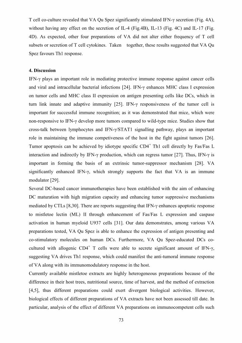
73
T cell co-culture revealed that VA Qu Spez significantly stimulated IFN-γ secretion (Fig. 4A),
without having any effect on the secretion of IL-4 (Fig.4B), IL-13 (Fig. 4C) and IL-17 (Fig.
4D). As expected, other four preparations of VA did not alter either frequency of T cell
subsets or secretion of T cell cytokines. Taken together, these results suggested that VA Qu
Spez favours Th1 response.
4. Discussion
IFN-γ plays an important role in mediating protective immune response against cancer cells
and viral and intracellular bacterial infections [24]. IFN-γ enhances MHC class I expression
on tumor cells and MHC class II expression on antigen presenting cells like DCs, which in
turn link innate and adaptive immunity [25]. IFN-γ responsiveness of the tumor cell is
important for successful immune recognition; as it was demonstrated that mice, which were
non-responsive to IFN-γ develop more tumors compared to wild-type mice. Studies show that
cross-talk between lymphocytes and IFN-γ/STAT1 signalling pathway, plays an important
role in maintaining the immune competiveness of the host in the fight against tumors [26].
Tumor apoptosis can be achieved by idiotype specific CD4+ Th1 cell directly by Fas/Fas L
interaction and indirectly by IFN-γ production, which can regress tumor [27]. Thus, IFN-γ is
important in forming the basis of an extrinsic tumor-suppressor mechanism [28]. VA
significantly enhanced IFN-γ, which strongly supports the fact that VA is an immune
modulator [29].
Several DC-based cancer immunotherapies have been established with the aim of enhancing
DC maturation with high migration capacity and enhancing tumor suppressive mechanisms
mediated by CTLs [8,30]. There are reports suggesting that IFN- enhances apoptotic response
to mistletoe lectin (ML) II through enhancement of Fas/Fas L expression and caspase
activation in human myeloid U937 cells [31]. Our data demonstrates, among various VA
preparations tested, VA Qu Spez is able to enhance the expression of antigen presenting and
co-stimulatory molecules on human DCs. Furthermore, VA Qu Spez-educated DCs co-
cultured with allogenic CD4+ T cells were able to secrete significant amount of IFN-γ,
suggesting VA drives Th1 response, which could manifest the anti-tumoral immune response
of VA along with its immunomodulatory response in the host.
Currently available mistletoe extracts are highly heterogeneous preparations because of the
difference in their host trees, nutritional source, time of harvest, and the method of extraction
[4,5], thus different preparations could exert divergent biological activities. However,
biological effects of different preparations of VA extracts have not been assessed till date. In
particular, analysis of the effect of different VA preparations on immunocompetent cells such

74
as DCs and its correlation with the lectin content has not been reported. The present
comparative study of different VA preparations represents an important aspect in the field of
phytotherapy to be addressed in order to understand the underlying mechanism of action of
VA.
MLs are the active components of mistletoe extracts and have several functions. MLs are
responsible for stimulating effector cells of the innate and adaptive immune system such as
DCs, macrophages, natural killer cells, and B and T lymphocytes. This function of MLs might
represent one of the mechanisms responsible for the anti-tumoral effect of mistletoe extracts.
It is known that ML-I B-chain (MLB) causes Ca2+ influx in Jurkat cells mediated by its
interaction with surface glycoprotein receptors which participates in early activation of T-cells
[32]. Chemical labelling of the lectin revealed that it binds to surface of peripheral and
intratumoral monocytes and this lectin component of ML-I plays a major role in
immunomodulation [33]. Depending on the concentration used for treatment, mistletoe
extracts also induce cell death in tumor cells and exert direct necrotic effects or apoptosis [34].
Cytotoxicity of mistletoe is attributed majorly to its lectin contents [35, 36]. Several studies
have clearly demonstrated that lectin internalization is required for ML-I mediated apoptosis,
independent of surface receptor-mediated pathway [37].
VA Qu Frf, an unfermented preparation containing highest concentration of lectin and
viscotoxin, was unable to activate DCs. In parallel, other VA preparations, which are
fermented and contain low lectin, were unable to stimulate DCs. Whereas VA Qu Spez, a
fermented preparation which contains second highest concentration of lectin, (785 +/- 10%
ng/ml) efficiently activated DCs and promoted Th1 response. These results suggest that, in
addition to the lectin content, the methodology of preparation, i.e., fermented vs unfermented
is crucial in conferring stimulatory properties to VA and indicates that it might be possible that
fermentation could have an effect on the lectin structure.
A recent study shows that the 3D structure of ML-A chain shares structural homology with
shiga toxin from Shigella dysenteriae. As Mistletoe plant produces a bacterial toxin, it has
strong immune stimulatory capacity [38]. It is also demonstrated that the Korean mistletoe
lectin (KML) is a TLR-4 ligand [39]. Since Korean ML and European ML have 84 % of
sequence identity [40], it is presumable that european ML could mimic TLR-4 ligand [38]. It
is known that TLRs are potent activators of DCs, which is a pre-requisite to initiate a full-
blown T cell response against cancer. Several clinical studies have provided evidence in
support of the beneficial effects of mistletoe in cancer patients and mistletoe thus remains as
one of the remedies most often used as a complementary therapy.

75
Taken together, our study delineates the differential effects of various VA preparations and
reveals VA Qu Spez to be the potent preparation in activating DCs and promoting Th1
response. Thus, this study along with other reports provides a rational for the use of VA Qu
Spez as an immune modulator [41-43] and further strengthens the beneficial effect of VA
preparations as complimentary therapy in cancer.
Acknowledgements
Supported by Institut National de la Santé et de la Recherche Médicale, Centre National de la
Recherche Scientifique, Regional Program Bio-Asie 2010 by the French Ministry of Foreign
and European Affairs and Institut Hiscia, Arlesheim, Switzerland.

76
References:
[1] S.Y. Lyu, J.H. Choi, H.J. Lee, et al., Korean mistletoe lectin promotes proliferation
and invasion of trophoblast cells through regulation of Akt signaling, Reprod. Toxicol.
39 (2013) 33-39.
[2] Y. Endo, K. Tsurugi, H. Franz, The site of action of the A-chain of mistletoe lectin I
on eukaryotic ribosomes. The RNA N-glycosidase activity of the protein, FEBS. Lett.
231 (1988) 378-380.
[3] F. Stirpe, L. Barbieri, M.G. Battelli, et al., Ribosome-inactivating proteins from plants:
present status and future prospects, Biotechnology 10 (1992) 405-412.
[4] F. Knopfl-Sidler, A. Viviani, L. Rist, et al., Human cancer cells exhibit in vitro
individual receptiveness towards different mistletoe extracts, Pharmazie 60 (2005)
448-454.
[5] H. Hulsen, C. Doser, F. Mechelke, Differences in the in vitro effectiveness of
preparations produced from mistletoes of various host trees, Arzneimittelforschung 36
(1986) 433-436.
[6] J. Kleijnen, P. Knipschild, Mistletoe treatment for cancer review of controlled trials in
humans, Phytomedicine 1 (1994) 255-260.
[7] J. Eggenschwiler, L. von Balthazar, B. Stritt, et al., Mistletoe lectin is not the only
cytotoxic component in fermented preparations of Viscum album from white fir
(Abies pectinata), BMC Complement. Altern. Med. 7 (2007) 14.
[8] R.M. Steinman, J. Banchereau, Taking dendritic cells into medicine, Nature 449
(2007) 419-426.
[9] P. Pierre, S.J. Turley, E. Gatti, et al., Developmental regulation of MHC class II
transport in mouse dendritic cells, Nature 388 (1997) 787-792.
[10] R.M. Steinman, Decisions about dendritic cells: past, present, and future, Annu. Rev.
Immunol. 30 (2012)1-22.
[11] C. Kurts, B.W. Robinson, P.A. Knolle, Cross-priming in health and disease, Nat. Rev.
Immunol. 10 (2010) 403-414.
[12] C. Dresch, Y. Leverrier, J. Marvel, et al., Development of antigen cross-presentation
capacity in dendritic cells, Trends. Immunol. 33 (2012) 381-388.

77
[13] B. Almand, J.R. Resser, B. Lindman, et al., Clinical significance of defective dendritic
cell differentiation in cancer, Clin. Cancer Res. 6 (2000) 1755-1766.
[14] A.P. Vicari, C. Caux, G. Trinchieri, Tumour escape from immune surveillance
through dendritic cell inactivation, Semin. Cancer Biol. 12 (2002) 33-42.
[15] D.I. Gabrilovich, J. Corak, I.F. Ciernik, et al., Decreased antigen presentation by
dendritic cells in patients with breast cancer, Clin. Cancer Res. 3 (1997) 483-490.
[16] D.I. Gabrilovich, H.L. Chen, K.R. Girgis, et al., Production of vascular endothelial
growth factor by human tumors inhibits the functional maturation of dendritic cells,
Nat. Med. 2 (1996) 1096-1103.
[17] C. Menetrier-Caux, G. Montmain, M.C. Dieu, et al., Inhibition of the differentiation
of dendritic cells from CD34(+) progenitors by tumor cells: role of interleukin-6 and
macrophage colony-stimulating factor, Blood 92 (1998) 4778-4791.
[18] H. Saito, S. Tsujitani, M. Ikeguchi, et al., Relationship between the expression of
vascular endothelial growth factor and the density of dendritic cells in gastric
adenocarcinoma tissue, Br. J. Cancer 78 (1998) 1573-1577.
[19] M. Augustin, P.R. Bock, J. Hanisch, et al., Safety and efficacy of the long-term
adjuvant treatment of primary intermediate- to high-risk malignant melanoma
(UICC/AJCC stage II and III) with a standardized fermented European mistletoe
(Viscum album L.) extract. Results from a multicenter, comparative, epidemiological
cohort study in Germany and Switzerland, Arzneimittelforschung 55 (2005) 38-49.
[20] J. Melzer, F. Iten, K. Hostanska, et al., Efficacy and safety of mistletoe preparations
(Viscum album) for patients with cancer diseases. A systematic review, Forsch
Komplementmed 16 (2009) 217-226.
[21] S.R. Elluru, J.P. Duong van Huyen, S. Delignat, et al., Induction of maturation and
activation of human dendritic cells: a mechanism underlying the beneficial effect of
Viscum album as complimentary therapy in cancer, BMC Cancer 8 (2008) 161.
[22] G. Ribereau-Gayon, M.L. Jung, D. Di Scala, et al., Comparison of the effects of f
ermented and unfermented mistletoe preparations on cultured tumor cells, Oncology
43 Suppl 1 (1986) 35-41.

78
[23] M.S. Maddur, M. Sharma, P. Hegde, et al., Human B cells induce dendritic cell
maturation and favour Th2 polarization by inducing OX-40 ligand, Nat. Commun. 5
(2014) 4092.
[24] J. Zhu, W.E. Paul, CD4 T cells: fates, functions, and faults, Blood 112 (2008)1557–
1569.
[25] E. Vivier, E. Tomasello, M. Baratin, et al., Functions of natural killer cells, Nat.
Immunol. 9 (2008) 503-510.
[26] V. Shankaran, H. Ikeda, A.T. Bruce, et al., IFNgamma and lymphocytes prevent
primary tumour development and shape tumour immunogenicity, Nature 410 (2001)
1107-1111.
[27] S. Hong, J. Qian, J. Yang, et al., Roles of idiotype-specific t cells in myeloma cell
growth and survival: Th1 and CTL cells are tumoricidal while Th2 cells promote
tumor growth, Cancer Res. 68 (2008) 8456-8464.
[28] D.H. Kaplan, V. Shankaran, A.S. Dighe, et al., Demonstration of an interferon -
dependent tumor surveillance system in immunocompetent mice, Proc. Natl. Acad.
Sci. U. S. A. 95 (1998) 7556-7561.
[29] S.Y. Lyu, W.B. Park, Effects of Korean mistletoe lectin (Viscum album coloratum) on
proliferation and cytokine expression in human peripheral blood mononuclear cells
and T-lymphocytes, Arch. Pharm. Res. 30 (2007) 1252-1264.
[30] K.J. Radford, K.M.Tullett, M.H.Lahoud, Dendritic cells and cancer immunotherapy,
Curr. Opin. Immunol. 27 (2014)26-32.
[31] M.S. Kim, J. Lee, H.S. So, et al., -interferon (IFN-) augments apoptotic response to
mistletoe lectin-II via upregulation of Fas/Fas L expression and caspase activation in
human myeloid U937 cells, Immunopharmacol. Immunotoxicol. 23 (2001) 55-66.
[32] H. Walzel, M. Blach, P. Neels, et al., The B-chain of mistletoe lectin I efficiently
stimulates calcium signaling in human Jurkat T-cells, Immunol. Lett. 78 (2001) 57-66.
[33] K. Kayser, H.J. Gabius, S. Gabius, [Biotinylated ligands for receptor localization. An
alternative for immunohistochemistry], Zentralbl. Pathol. 137 (1991) 473-478.

79
[34] O. Podlech, P.N. Harter, M. Mittelbronn, et al., Fermented mistletoe extract as a
multimodal antitumoral agent in gliomas, Evid. Based Complement Alternat. Med.
2012 (2012) 501796.
[35] A. Engert, F. Burrows, W. Jung, et al., Evaluation of ricin A chain-containing
immunotoxins directed against the CD30 antigen as potential reagents for the
treatment of Hodgkin's disease, Cancer Res. 50 (1990) 84-88.
[36] A. Stettin, J.L. Schultze, E. Stechemesser, et al., Anti-mistletoe lectin antibodies are
produced in patients during therapy with an aqueous mistletoe extract derived from
Viscum album L. and neutralize lectin-induced cytotoxicity in vitro, Klin.
Wochenschr. 68 (1990) 896-900.
[37] V. Lavastre, M. Pelletier, R. Saller, et al., Mechanisms involved in spontaneous and
Viscum album agglutinin-I-induced human neutrophil apoptosis: Viscum album
agglutinin-I accelerates the loss of antiapoptotic Mcl-1 expression and the degradation
of cytoskeletal paxillin and vimentin proteins via caspases, J. Immunol. 168 (2002)
1419-1427.
[38] C. Maletzki, M. Linnebacher, R. Savai, et al., Mistletoe lectin has a shiga toxin-like
structure and should be combined with other Toll-like receptor ligands in cancer
therapy, Cancer Immunol. Immunother. 62 (2013) 1283-1292.
[39] H.J. Park, J.H. Hong, H.J. Kwon, et al., TLR4-mediated activation of mouse
macrophages by Korean mistletoe lectin-C (KML-C), Biochem. Biophys. Res.
Commun. 396 (2010) 721-725.
[40] R.A. Abagyan, S. Batalov, Do aligned sequences share the same fold?, J. Mol. Biol.
273 (1997) 355-368.
[41] J.P. Duong Van Huyen, J. Bayry , S. Delignat et al., Induction of apoptosis of
endothelial cells by Viscum album: a role for anti-tumoral properties of mistletoe
lectins, Mol. Med. 8 (2002)600-606.
[42] J.P. Duong Van Huyen, S. Delignat, J. Bayry et al., Interleukin-12 is associated with
the in vivo anti-tumor effect of mistletoe extracts in B16 mouse melanoma, Cancer
Lett. 243(2006)32-37.

80
[43] C.I. Delebinski, M.Twardziok, S. Kleinsimon et al., A natural combination extract of
viscum album L. containing both triterpene acids and lectins is highly effective
against AML in vivo, PLoS One 10 (2015) e0133892.

81
Figure Legend
Fig 1. Comparison of the differential effects of VA preparations on phenotype of human DCs.
Immature DCs were treated with medium alone (control) or with 5 preparations of VA at
various concentrations for 48 hours. Expressions (mean ±SEM, ≥ 5 independent donors) (A)
CD1a, (B) CD83, (C) HLA-DR, (D) CD40, (E, F) CD86 on DCs were analyzed by flow
cytometry. The data are presented either as % positive cells or MFI of indicated markers. X-
axis denotes concentrations of VA preparations. *p <0.05, ***p <0.001.
Fig 2. Comparison of the effect of VA preparations on the secretion of DC cytokines.
Immature DCs were untreated (control) or treated with 5 preparations of VA at various
concentrations for 48 hours. The secretion (pg/ml, mean ± SEM, four independent donors) of
(A) IL-6, (B) IL-8, (C) IL-10 and (D) TNF-α in cell-free supernatants were measured. **p<
0.01, ***p < 0.001, ****p <0.0001.
Fig 3. Differential effect of VA preparations on T cell response. DCs treated with medium
alone (control) or with 5 preparations of VA for 48 hours. These DCs were co-cultured with
allogenic CD4+ T cells at 1:10 ratio. After 5 days of co-culture, the cells were analyzed. A-D
indicates percentage (mean ± SEM, six independent donors) of IFN-γ+ Th1, IL-4+ Th2, IL-17+
Th17 and CD25+Foxp3+ Treg cells. ns, non-significance.
Fig 4. Effect of VA preparations on T cell cytokine secretion. Immature DCs were treated
with medium alone (control) or with 5 preparations of VA for 48 hours. These DCs were co-
cultured with allogenic CD4+ T cells for 5 days. Amount of secretion (pg/ml, mean ± SEM,
six independent donors) of (A) IFN-γ, (B) IL-4, (C) IL-13 and (D) IL-17 in the cell-free
supernatants from the above co-cultures. *p< 0.05.

82
Figure1.

83
Figure2.
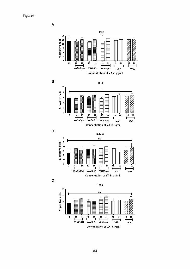
84
Figure3.
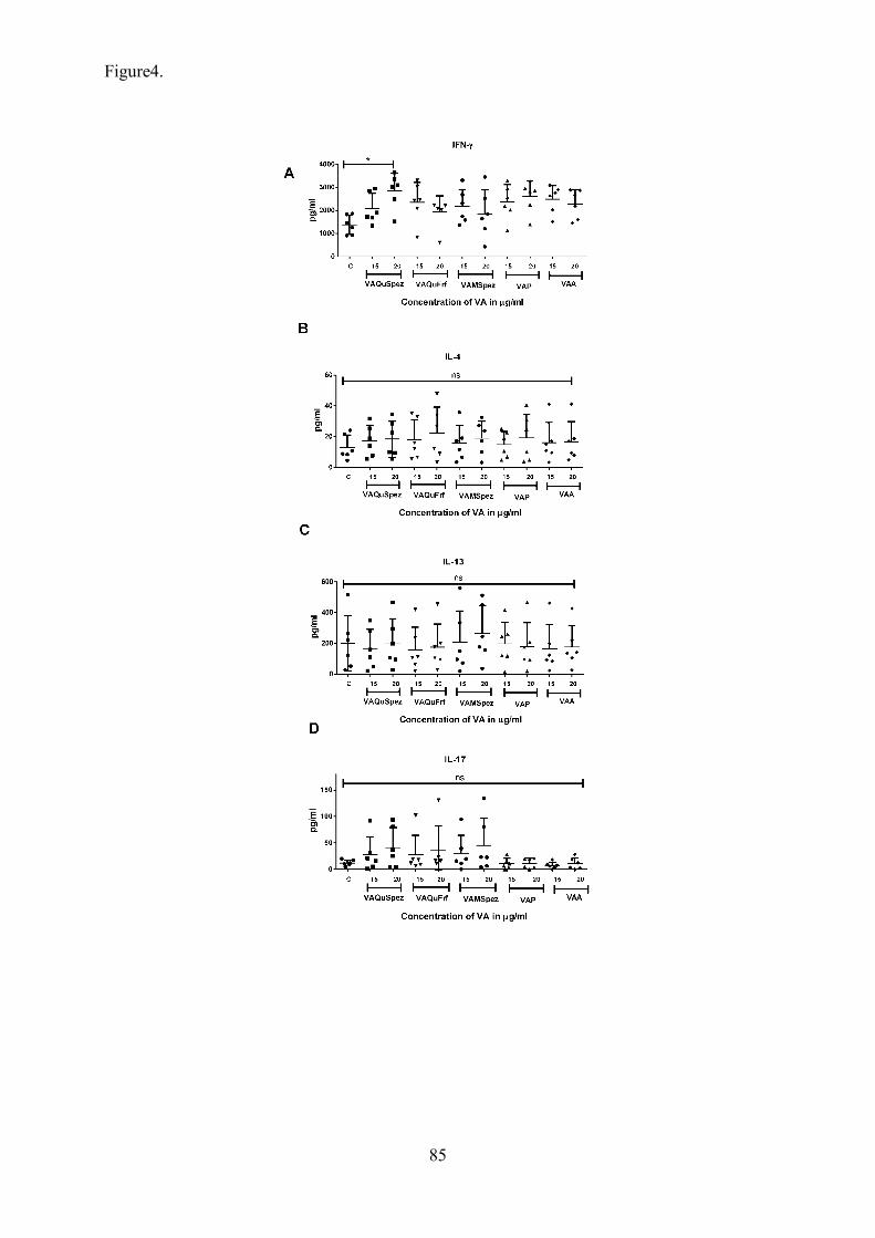
85
Figure4.

86
Viscum album promotes anti-tumor response by modulating M1/M2 macrophage
polarization switch
Chaitrali Saha1,2, 4,*, Alain Friboulet2, Jagadeesh Bayry1,2,3, Srini V Kaveri1,2,3
1Institut National de la Santé et de la Recherche Médicale, Unité 1138, 15 rue de l’Ecole de
Médicine, Paris, F-75006, France.
2 Centre de Recherche des Cordeliers, Equipe 16- Immunopathology and therapeutic
immunointervention, Université Pierre et Marie Curie – Paris 6, UMR S 1138, Paris, F-75006,
France.
3 Université Paris Descartes, UMR S 1138, Paris, F-75006, France.
4Université de Technologie de Compiègne, UMR CNRS 6022, Compiègne
Correspondence to: Srini V Kaveri, INSERM U 1138, Centre de Recherche des Cordeliers,
15 rue de l’Ecole de Médicine, Paris, F-75006, France. Tel: 00 33 1 44 27 82 01; Fax: 00 33 1
44 27 81 94; E-mail: [email protected]

87
Abstract
Wide use of Viscum album (VA) preparations in complementary and alternative therapies for
cancer has revealed their beneficial effects on the survival and the quality of life of cancer
patients. The underlying molecular and cellular mechanism of their therapeutic efficacy
encompasses cytotoxic properties, inhibition of angiogenesis and importantly several
immunomodulatory functions. Tumor progression depends on their ability to re-educate the
polarization state of tumor associated macrophages from pro-inflammatory M1 to anti-
inflammatory M2. The orchestration of monocyte-macrophage function is a key element that
links inflammation and cancer and provides a paradigm for macrophage diversity and
plasticity. Thus, we have investigated whether VA can modulate macrophage polarization,
which in turn can be associated with their anti-tumor property. Our data demonstrates VA
mediates the switch of M2 macrophage polarization towards M1 macrophages. Therefore, the
immunomodulatory activity of VA is dependent on the polarization state of the responding
macrophages, and their ability to drive M1 macrophages over M2 macrophages might be
another angle underlying their therapeutic benefit in cancer.
Key words: Viscum album, myelomonocytic cells, macrophage cells, Immune dysregulation,
pro-inflammatory cytokines

88
Introduction:
A constant influx of myelomonocytic cells is an important characteristic of tumor to support
stroma remodelling and angiogenesis for their continuous growth. In early 1980s an
interesting population called natural suppressors was identified which are known to be
associated with immune suppression and tumor development [1]. Another fascinating
observation was in tumor microenvironment, number of myeloid cells are elevated which in
turn alter anti-tumor immune reactivity [2], [3]. Immune dysregulation and immune
suppression are the common features in cancer bearing patients in which even tumor-specific
T cell death can occur [4]. At tumor site, accumulation of immunosuppressive
myelomonocytic cells can take place [5], [6]. Recently these immune suppressor cells are
called as myeloid-derived suppressor cells (MDSCs), which are deficient of expressing
mature myeloid cell markers [7]. In 1980s, Alberto Mantovani originally described that [8], in
the tumor site, circulating monocytes are recruited and with the help of a tumor-derived
chemotactic factor CCL2, these cells differentiate into tumor associated macrophages (TAMs)
[9]. These tumor-derived chemokines along with recruiting monocytes in the tumor site play
an important role in tumor progression by encouraging inflammation and angiogenesis thus
inducing neoplastic growth [10]. TAMs are the second population of myelomonocytic
immune suppressors which shows a negative effect on anti-tumor immune responses, they are
believed to be derived from or related MDSCs [4]. Cancer-related inflammation is associated
with mononuclear phagocytes [11], [12] and cancer is a major paradigm of macrophage (M
diversity and plasticity [13], [14], [15].
Considering T helper type 1 (Th1) and T helper type 2 (Th2), polarization, two distinct types
of macrophages have been recognized: classically activated macrophages-M1 and
alternatively activated macrophages-M2 [16], [13]. Th1 cells can drive M1 macrophages by
producing IFN-γ and even bacterial moieties such as LPS can polarize M1 macrophages.
These cells are characterized by their ability to secrete pro-inflammatory cytokines such as
IL-12, IL-23 and tumor necrosis factor (TNF), reactive oxygen and nitrogen species, antigen
presentation and elevated expression of major histocompatibility complex II, and tumoricidal
activity [17]. Through expression of several cytokines and chemokines such as IL-12, CXCL-
9 and CXCL-10, they recruit Th1 cells thus promoting Th1 response [9]. They confer
resistance to intracellular pathogens and tumors leading to tumor-disruptive response [16],
[18]. M1 macrophages show a phenotype of high IL-12 and low IL-10 expression. In contrast,
Th2 cytokine IL-4 triggers M2 macrophage polarization [19]. These cells are involved in
amplifying Th2 responses. These cells are more phagocytic in nature and exert high

89
expression of scavenging, galactose and mannose receptors, through arginase pathway they
produce polyamines and ornithine, and even show a characteristic of expressing low IL-12
and high IL-10 [16], [13], [18]. M2 macrophage expressing chemokines are CCL17, CCL22
and CCL24 [20]. As M2 macrophages drive Th2 immune response in turn they actively
participate in encapsulation and clearance of parasites hence in tumor progression and tissue
remodelling and importantly exert an immune regulatory property [17].
Viscum album (VA) commonly known as European mistletoe is extensively used in
complementary and alternative medicine in cancer and also in the treatment of several
inflammatory diseases [21]. Several systemic reviews and meta-analyses supports a beneficial
effect of mistletoe treatment on cancer patient survival [22], [23] and due to their ability to
improve quality of life of cancer patients [22], [24], [25] and minimising the side effects of
conventional anti-cancer therapies [24]. Viscum preparations consist of multiple components
at variable concentrations, depending on the host tree they are harvested from, the time at
which they were harvested and the method of preparation. Mistletoe lectins (MLs) I-III,
especially ML-I and viscotoxins are the major bioactive components of this preparation which
are majorly involved to exert their anti-cancer property [26]. This preparation also composed
of several other biologically active components such as flavonoids, peptides, several enzymes,
polysaccharides, phenols, triterpenes, lipids, phytosterols, amino acids, thiols, cyclitoles,
phenylpropanes and minerals [27]. ML-I belongs to a class of ribosome inactivating protein
(RIP), such as highly toxic ricin and abirin [28]. It consists of two subunits, the α and the β-
chain connected by a disulphide bridge [29]. The β-chain binds to sugar components
expressed on the surface of cells which enables their uptake into the cells. This characteristic
of viscum is believed to be the reason for their specific anti-cancer effects. The α-chain exerts
RNA-glycoside hydrolase activity, resulting in inactivation of ribosomes hence their ability to
induce apoptosis [30]. Increasing evidence has revealed that VA exerts anti-tumor activities
including cytotoxic properties [31], [32], induction of apoptosis [33], inhibition of
angiogenesis [34], and immunomodulatory properties and anti-inflammatory properties [35],
[36].
There is increasing evidence supporting the fact that in cancer, the M2-like pro-tumoral
phenotype of TAMs is reversible [37], [38]. Guiducci et al. reported that CpG activated TLR9
and IL-10 Ab can switch M2 TAMs to M1 phenotype [39]. Notch signalling can enhance
anti-tumor activity by encouraging classically activated macrophages, which are known to kill
cancer cells [40]. Re-directing macrophage polarization can be achieved by genetic blocking
of molecular determinants of macrophage polarization such as STAT3, STAT6, NF-kB p50
90
which might lead to anti-tumor activity [37], [41], [42]. Thus it is confirmed that in early
stages of cancer T cell response decides classical macrophage polarization. Therefore, in view
of the anti-tumoral and immunomodulatory effect of viscum, we were interested to investigate
whether VA can modulate macrophage polarization, which in part explains the mechanisms
underlying the beneficial effect of VA in cancer therapy.
Results
VA Qu Spez induced switch of macrophage polarization in a M1/M2 mixed phenotype
We first investigated whether VA modulates polarization of M1 and M2 macrophages. M1
macrophages are known to be pro-inflammatory in nature, thus we were initially interested to
study the effect of VA Qu Spez on polarization of M1 macrophages. The cells were cultured
either with GMCSF, LPS and IFN-γ to obtain M1 macrophages or with M-CSF, IL-13 and
IL-4 for M2 macrophage polarization. In both the culture conditions we obtained a mixed
M1/M2 phenotype population of macrophages. And in both the conditions, interestingly VA
Qu Spez was able to significantly favour M1 phenotype over M2 phenotype. VA induced M1
phenotype is showed by elevated expression of M1 surface markers such as CD80 (Fig. 2B
and 2C) and CCR7 (Fig. 2D). Along the same line VA provoked a dramatic polarization
switch in M2 macrophages, as it reduced the expression of M2 specific marker CD206 (Fig.
1C). Altogether these results demonstrate the ability of VA Qu Spez to promote M2 to M1
polarization switch in human macrophages.
Significant induction of IL-12 expression by VA Qu Spez in a M1/M2 mixed phenotype
indicating the effect of VA in M2 to M1 polarization switch
M1 macrophages are characterized by high expression of IL-12 and low IL-10 expression, but
in contrast, M2 macrophages exert a phenotype of low IL-12 and high IL-10 expression.
Previous results indicated that VA is able to favour M1 macrophage polarization over M2.
Thus we were interested to explore whether VA can modulate the expression of IL-12 and IL-
10, which in turn can strengthen our previous observation. Interestingly in both the culture
condition of M1 and M2, VA Qu Spez significantly induced IL-12 expression (Fig. 3A and
3D), confirming that VA skew them towards the acquisition of the phenotypic characteristics
of M1 macrophages.
VA Qu Spez significantly drives M1 polarization in distinct M2 macrophage population
To determine the extent of the relevance of these findings, we next determined whether VA
Qu Spez-mediated polarization switch in a mixed M1/M2 phenotype, holds true even in a
91
distinct M1 and M2 scenario. Thus we validated polarization state of distinct M1 and M2
macrophages. Macrophages showing significant induced expression of CD80 and CCR7,
confirmed specific M1 population (Fig. 4A), whereas elevated expression of CD206 and
CD209 validated specific M2 polarized macrophages (Fig. 5A). Although there was no
modulation of polarization observed with VA Qu Spez treatment in both M1 and in M2 cells
(Fig4. and Fig5. B-E), but surprisingly induced CD80 expression by VA treatment compared
to the untreated cells in M2 macrophages, once again strongly indicates the role of VA in
switching M2 to M1 phenotype.
VA Qu Spez drives M2 to M1 switch by inhibiting expression of IL-10 in M2
macrophages
Regarding previously reported immunomodulatory and anti-inflammatory properties of VA
Qu Spez and the prognostic impact of alternatively polarized macrophages, we were
interested in the effect of VA Qu Spez on polarization of macrophages. Macrophages were
polarized in-vitro as described above to obtain distinct M1 and M2 phenotype. There was no
significant alteration in the IL12 and IL-10 profile (Fig. 6A, 6b and 6D) after treatment with
VA compared to the control untreated cells. But interestingly VA significantly abrogated the
expression of IL-10 in M2 macrophages. Therefore all results collectively suggest that, VA
Qu Spez alters the macrophage polarization, i.e., drives alternatively activated macrophage
switch towards classically activated macrophages, which in turn can be beneficial to limit
tumor growth.
Discussion
Mechanisms of immune surveillance are able to control the growth of new tumors or affect
the progression of existing tumors is well known [43]. Immune dysregulation and immune
suppression in cancer patients is a composite event which in turn leads to abnormal
myelopoesis and recruitment of several immunosuppressive myelomonocytic cells at tumor
vicinity [5], [6]. This immune dysfunction even can cause death of tumor-speecific T cells or
lymphocyte dysfunction. These immunosuppressive myelomonocytic cells are involved in
process of angiogenesis and stroma remodelling needed for tumor progression. In the tumor
site, tumor-derived factors are involved in functional differentiation of myelomonocytic cells
majorly the macrophages to sustain myelopoiesis [4]. Regulation of Monocyte-macrophage
function at equilibrium is essential for dealing with pathogens, tissue damage and repair. The
orchestration of myelomonocytic cell function links inflammation and cancer. Macrophages
show considerable functional plasticity and exert an immediate response to alter according to

92
the change in the microenvironment [44]. Cancer serves a paradigm of macrophage plasticity
and diversity [13], [14], [15]. Based on Th1 and Th2 polarization, macrophages are classified
in two distinct states: the classically activated macrophages or M1 and the alternatively
activated macrophages or M2 [17]. Myelomonocytic cells are involved in carcinogenesis at
every possible stages of tumor progression [11], [12], [45]. These cells influence the
following steps: suppressing adaptive immune response [46], supporting angiogenic network
and lymphangiogenesis [47], [48], [49], promoting genetic instability, senescence regulation
[50], promoting invasion and tumor metastasis [48]. But on the other hand tumor cells are
capable of escaping phagocytosis by macrophage cells [51], and majorly by recruiting M2
macrophages in the tumor site through chemokines and cytokines such as MCSF, IL-10,
TGF-β etc. [52], [11].
Several study showed that Viscum album exerts anti-tumor properties. VA induces cell
toxicity and inhibits proliferation of a variety of cell types [31], VA Qu Frf mistletoe extract
induces apoptosis in a variety of transformed cells in a Fas independent way [32]. Kaveri
group had demonstrated anti- tumoral property of that VA and reported that VA is able to
induce apoptosis of endothelial cells and can inhibit angiogenesis which is essential for tumor
metastasis [33], [34]. Apart from its anti-tumor properties VA also employs a novel anti-
inflammatory effect by inhibiting cytokine induced prostaglandin E2 via selective inhibition
of Cyclooxygenase-2 [53], [54]. In addition to its ani-tumor properties, viscum preparations
have immunomodulatory effects. VA facilitates tumor elimination in experimental models
[55]. VA lectin induces gene expression of IL-1α, IL-6, TNF-α, IFN-γ and GM-CSF from
PBMC [56]. VA preparations stimulate the maturation and activation of human DCs, which in
turn might facilitate anti-tumor immune responses [36]. Triterpenes in VALE is shown to
have immunomodulatory effects on tumor cell co-cultured macrophages in vitro by
modulating monocyte chemotactic transmigration [57]. Mistletoe exerts a beneficial effect on
cancer patient survival [22], [23] and their ability to improve quality of life of cancer patients
[24], [25] and minimising the side effects of conventional anti-cancer therapies [24] is
demonstrated by several systemic reviews and clinical studies.
In the present in vitro study, we demonstrate that VA Qu Spez drives M2 switch towards M1,
in a mixed M1/M2 population as well as in distinct M1 and M2 phenotype. VA- mediated
modulation of this switch is demonstrated by induced expression of M1 surface markers such
as CD80 and CCR7, but in parallel, reduced expression of M2 surface markers such as
CD206 and CD209, with VA treatment in both the culture conditions. In addition, VA
provoked polarization of IL-12hi and IL-10lo characterised M1cells over IL12lo and IL-10hi M2

93
macrophages, suggesting that VA is able to play M2 to M1switch. There are several reports
suggest that M2-like pro tumoral phenotype of TAMs in cancer is reversible [38] [58]. Tumor
associated macrophages are the main source of IL-10 which is an imuunomodulating cytokine
which can turn down innate and adaptive immune response and they are responsible to
maintain M2 state of macrophages [39]. Strategies to overcome tumor escape mechanisms by
changing the immunosuppressive situation are the key to effective immunotherapy. Our
results show that VA down modulates IL-10 expression in M2 macrophages will probably be
one of the reasons for VA to exert immunomodulatory effect. IL-12 links innate and adaptive
immunity by promoting cytotoxic T lymphocytes (CTL). Reduced production of CTL and
abrogation of tumor rejection was observed in IL-12 knockout mice [39]. In this study we
show that VA Qu Spez was able to significantly induce IL-12 expression in M1/M2 mixed
phenotype which strengthens the underlying therapeutic efficacy of VA as an
immunomodulatory compound. The ability of viscum to manipulate M2 macrophages
towards M1 phenotype by increased expression of M1 markers such as CD80, CCR7 and IL-
12, and reduced expression of M2 markers such as CD206, CD209 and IL-10, indirectly
suggests recruitment of Th1 response and a possibility of relief of tumor immunosuppression,
which may represent potential effective anti-tumor therapeutic benefit of VA.
Conclusion
A better understanding of the interplay between myelomonocytic cells and neoplastic cells
may confer novel targets for therapeutic intervention and enhancing the anti-tumor response.
Balance in monocyte- macrophage function is important for developing immune response
which is resistance to pathogens, tissue damage and repair. Viscum album is known to
improve quality of life of cancer patients and their survival. Our results demonstrate the
ability of VA Qu Spez to switch immunoregulatory M2 macrophage cells towards M1, thus
indicating the ability of VA to drive Th1 response. This study provides another new angle to
understand VA-associated immunomodulation which could be critical in understanding their
role as complimentary therapy in cancer.
Methods
VA preparation
VA Qu Spez was a kind gift from Weleda AG (Arlesheim, Switzerland). This is the extract of
Viscum album growing on different oak trees (Quercus). The VA preparations are therapeutic
preparations that are free from endotoxins. VA preparations are formulated in sodium
chloride (NaCl 0.9%) isotonic solution as 5 mg/ml vials. VA preparations are prepared by
94
standardizing the levels of Mistletoe lectins and viscotoxins. VA Qu Spez is a fermented
preparation, composed of 785 +/- 10% ng/ml of lectin and 5 +/- 5% µg/ml of viscotoxin.
Isolation of circulating human monocytes, their differentiation to macrophages containing
a mixed M1/M2 population
Peripheral blood mononuclear cells (PBMC) were isolated from buffy coats of healthy donors
purchased from Centre Necker-Cabanel (Etablissement Franc¸ ais du Sang, Paris, France).
Circulating monocytes were isolated using CD14 beads (MiltenyiBiotec, Paris,France), and
subsequently cultured for 5 days in medium (RPMI 1640, GIBCO/Invitrogen) with 10% FCS,
50 U/ml penicillin, 50 µg/ml streptomycin, and either with 1000 IU/106 cells recombinant
human GM-CSF (MiltenyiBiotec) to generate M1 macrophage or 2000 IU/106recombinant
M-CSF (Immunotools) to generate M2 macrophage. Post 5 days macrophage polarization was
obtained by removing the culture media and culturing cells for additional 48 hours in the
similar medium condition with 200 ng ml-1 LPS (from Escherichia coli, sigma-Aldrich) plus
40 ng ml-1 IFN-γ (Immunotools) for M1 polarization or 500 IU ml-1 IL-4 (MiltenyiBiotec)
plus 200 ng ml-1 IL-13 (Immunotools) for M2 polarization and at the same time cells are co-
treated with VA Qu Spez for the subsequent hours.
Isolation of circulating human monocytes and their polarization to distinct M1 and M2
macrophages
Human peripheral blood mononuclear cells were isolated using density gradient
centrifugation. Monocytes were isolated using anti-CD14 microbeads (MiltenyiBiotec, Paris,
France). Macrophages were obtained by culturing monocytes for 6 days in RPMI 1640
supplemented with 20% FCS plus 50 U/ml penicillin and 50 µg/ml streptomycin and 100 ng
ml-1macrophage colony-stimulating factor at a density of 1.5 ×10⁵ per cm2. Macrophage
polarization was obtained by removing the culture media and culturing cells for an additional
72 hours in RPMI 1640 supplemented with 5% FCS and 100 ng ml-1 LPS plus 20 ng ml-1
IFN-γ (for M1 polarization) or 20 ng ml-1 IL4 plus 20 ng ml-1 IL-13 (for M2 polarization).
Post 72 hours M1 and M2 macrophages were treated with VA Qu Spez for 48 hours.
Viscum album treatment
In case of the first protocol, where we obtained a mixed population of M1 and M2, cells were
treated with VA Qu Spez for 48 hours at two different concentrations, i.e., 15 µg/ml and 20
µg/ml or they were untreated (control cells). The phenotype of cells was analyzed by flow
cytometry. When we obtained distinct M1 and M2 phenotypic cells, they were either

95
untreated (control cells) or treated with VA Qu Spez at much lower concentration, i.e., 0.5
µg/ml and 1 µg/ml respectively as these concentrations were optimum to the cells. Further
cell phenotype was analyzed by flow cytometry.
Flow cytometry
To investigate the effect of VA on macrophages, 0.5 × 10⁶ macrophages were either untreated
or treated with VA preparations for 48 hrs. To analyse M1 macrophage phenotype we used
the following fluorochrome-conjugated antibodies such as: PE-conjugated-CD80 (BD
Biosciences) and APC-conjugated-CCR7 (eBioscience) and to analyze M2 macrophage
phenotype, we used PE-conjugated-CD206 (BD Biosciences) and APC-conjugated-CD209
(BD Biosciences).
For determining intracellular M1/M2 expression we used APC-conjugated IL-12
(eBioscience) and PE-conjugated IL-10 (BD Biosciences).
For surface staining cells were suspended in 10% FCS/PBS and antibodies against surface
molecules were added at pre-determined concentration and incubated at 4° C for 30 min. BD
Fix buffer was used to fix the cells and washed before analysis. The data are presented as %
positive cells for indicated molecules or mean fluorescence intensities (MFI) of their
expression.
Macrophage cells were stimulated with Phytohaemagglutinin (PHA)-L (10 µg ml-1, Sigma-
Aldrich) at 37°C for 18 hours and for extra 2 hours with golgistop (BD Biosciences). Cells
were fixed and permeabilized using Foxp3 Fixation/Permeabilization kit (eBioscience) and
incubated at 4°C with fluorescence-conjugated mAbs. Cells were acquired on LSRII, 5000
events were recorded and analyzed for each sample. Data was analyzed by BD FACSDIVA
software (BD Biosciences, France).
Statistical analysis
Levels of significance for comparison between samples were determined by One way
ANOVA Tukey’s Multiple Comparison Test. Values of P <.05 were considered statistically
correlated (*P <0 .05, *** p< 0.0001). All statistical analyses were performed using Prism 5
software (GraphPad Software, Inc, La Jolla, Calif).

96
References
1. Maier, T., J.H. Holda, and H.N. Claman, Natural suppressor cells. Prog Clin Biol Res,
1989. 288: p. 235-44.
2. Seung, L.P., et al., Synergy between T-cell immunity and inhibition of paracrine
stimulation causes tumor rejection. Proc Natl Acad Sci U S A, 1995. 92(14): p. 6254-
8.
3. Pekarek, L.A., et al., Inhibition of tumor growth by elimination of granulocytes. J Exp
Med, 1995. 181(1): p. 435-40.
4. Sica, A. and V. Bronte, Altered macrophage differentiation and immune dysfunction
in tumor development. J Clin Invest, 2007. 117(5): p. 1155-66.
5. Rabinovich, G.A., D. Gabrilovich, and E.M. Sotomayor, Immunosuppressive
strategies that are mediated by tumor cells. Annu Rev Immunol, 2007. 25: p. 267-96.
6. Serafini, P., I. Borrello, and V. Bronte, Myeloid suppressor cells in cancer:
recruitment, phenotype, properties, and mechanisms of immune suppression. Semin
Cancer Biol, 2006. 16(1): p. 53-65.
7. Gabrilovich, D.I., et al., The terminology issue for myeloid-derived suppressor cells.
Cancer Res, 2007. 67(1): p. 425; author reply 426.
8. Mantovani, A., et al., The origin and function of tumor-associated macrophages.
Immunol Today, 1992. 13(7): p. 265-70.
9. Mantovani, A., et al., The chemokine system in diverse forms of macrophage
activation and polarization. Trends Immunol, 2004. 25(12): p. 677-86.
10. Balkwill, F., Chemokine biology in cancer. Semin Immunol, 2003. 15(1): p. 49-55.
11. Mantovani, A., et al., Cancer-related inflammation. Nature, 2008. 454(7203): p. 436-
44.
12. Pollard, J.W., Trophic macrophages in development and disease. Nat Rev Immunol,
2009. 9(4): p. 259-70.
13. Mantovani, A., et al., Macrophage polarization: tumor-associated macrophages as a
paradigm for polarized M2 mononuclear phagocytes. Trends Immunol, 2002. 23(11):
p. 549-55.
14. Biswas, S.K., A. Sica, and C.E. Lewis, Plasticity of macrophage function during
tumor progression: regulation by distinct molecular mechanisms. J Immunol, 2008.
180(4): p. 2011-7.
15. Lewis, C.E. and J.W. Pollard, Distinct role of macrophages in different tumor
microenvironments. Cancer Res, 2006. 66(2): p. 605-12.

97
16. Gordon, S. and P.R. Taylor, Monocyte and macrophage heterogeneity. Nat Rev
Immunol, 2005. 5(12): p. 953-64.
17. Biswas, S.K. and A. Mantovani, Macrophage plasticity and interaction with
lymphocyte subsets: cancer as a paradigm. Nat Immunol, 2010. 11(10): p. 889-96.
18. Mantovani, A., From phagocyte diversity and activation to probiotics: back to
Metchnikoff. Eur J Immunol, 2008. 38(12): p. 3269-73.
19. Stein, M., et al., Interleukin 4 potently enhances murine macrophage mannose
receptor activity: a marker of alternative immunologic macrophage activation. J Exp
Med, 1992. 176(1): p. 287-92.
20. Martinez, F.O., et al., Transcriptional profiling of the human monocyte-to-
macrophage differentiation and polarization: new molecules and patterns of gene
expression. J Immunol, 2006. 177(10): p. 7303-11.
21. Bock, P.R., et al., [Efficacy and safety of long-term complementary treatment with
standardized European mistletoe extract (Viscum album L.) in addition to the
conventional adjuvant oncologic therapy in patients with primary non-metastasized
mammary carcinoma. Results of a multi-center, comparative, epidemiological cohort
study in Germany and Switzerland]. Arzneimittelforschung, 2004. 54(8): p. 456-66.
22. Kienle, G.S., et al., Mistletoe in cancer - a systematic review on controlled clinical
trials. Eur J Med Res, 2003. 8(3): p. 109-19.
23. Ostermann, T., C. Raak, and A. Bussing, Survival of cancer patients treated with
mistletoe extract (Iscador): a systematic literature review. BMC Cancer, 2009. 9: p.
451.
24. Kienle, G.S. and H. Kiene, Review article: Influence of Viscum album L (European
mistletoe) extracts on quality of life in cancer patients: a systematic review of
controlled clinical studies. Integr Cancer Ther, 2010. 9(2): p. 142-57.
25. Melzer, J., et al., Efficacy and safety of mistletoe preparations (Viscum album) for
patients with cancer diseases. A systematic review. Forsch Komplementmed, 2009.
16(4): p. 217-26.
26. Hajto, T., K. Hostanska, and R. Saller, [Mistletoe therapy from the pharmacologic
perspective]. Forsch Komplementarmed, 1999. 6(4): p. 186-94.
27. Urech, K., G. Schaller, and C. Jaggy, Viscotoxins, mistletoe lectins and their isoforms
in mistletoe (Viscum album L.) extracts Iscador. Arzneimittelforschung, 2006. 56(6A):
p. 428-34.
28. Barbieri, L., M.G. Battelli, and F. Stirpe, Ribosome-inactivating proteins from plants.
Biochim Biophys Acta, 1993. 1154(3-4): p. 237-82.

98
29. Eschenburg, S., et al., Primary structure and molecular modeling of mistletoe lectin I
from Viscum album. Biochem Biophys Res Commun, 1998. 247(2): p. 367-72.
30. Puri, M., et al., Ribosome-inactivating proteins: current status and biomedical
applications. Drug Discov Today, 2012. 17(13-14): p. 774-83.
31. Duong Van Huyen, J.P., et al., Comparative study of the sensitivity of lymphoblastoid
and transformed monocytic cell lines to the cytotoxic effects of Viscum album extracts
of different origin. Chemotherapy, 2003. 49(6): p. 298-302.
32. Duong Van Huyen, J.P., et al., Variable sensitivity of lymphoblastoid cells to
apoptosis induced by Viscum album Qu FrF, a therapeutic preparation of mistletoe
lectin. Chemotherapy, 2001. 47(5): p. 366-76.
33. Duong Van Huyen, J.P., et al., Induction of apoptosis of endothelial cells by Viscum
album: a role for anti-tumoral properties of mistletoe lectins. Mol Med, 2002. 8(10):
p. 600-6.
34. Elluru, S.R., et al., Antiangiogenic properties of viscum album extracts are associated
with endothelial cytotoxicity. Anticancer Res, 2009. 29(8): p. 2945-50.
35. Elluru, S., et al., Molecular mechanisms underlying the immunomodulatory effects of
mistletoe (Viscum album L.) extracts Iscador. Arzneimittelforschung, 2006. 56(6A): p.
461-6.
36. Elluru, S.R., et al., Induction of maturation and activation of human dendritic cells: a
mechanism underlying the beneficial effect of Viscum album as complimentary
therapy in cancer. BMC Cancer, 2008. 8: p. 161.
37. Saccani, A., et al., p50 nuclear factor-kappaB overexpression in tumor-associated
macrophages inhibits M1 inflammatory responses and antitumor resistance. Cancer
Res, 2006. 66(23): p. 11432-40.
38. Stout, R.D., S.K. Watkins, and J. Suttles, Functional plasticity of macrophages: in situ
reprogramming of tumor-associated macrophages. J Leukoc Biol, 2009. 86(5): p.
1105-9.
39. Guiducci, C., et al., Redirecting in vivo elicited tumor infiltrating macrophages and
dendritic cells towards tumor rejection. Cancer Res, 2005. 65(8): p. 3437-46.
40. Wang, Y.C., et al., Notch signaling determines the M1 versus M2 polarization of
macrophages in antitumor immune responses. Cancer Res, 2010. 70(12): p. 4840-9.
41. Ostrand-Rosenberg, S., Immune surveillance: a balance between protumor and
antitumor immunity. Curr Opin Genet Dev, 2008. 18(1): p. 11-8.
42. Kortylewski, M., et al., Inhibiting Stat3 signaling in the hematopoietic system elicits
multicomponent antitumor immunity. Nat Med, 2005. 11(12): p. 1314-21.

99
43. Dunn, G.P., L.J. Old, and R.D. Schreiber, The immunobiology of cancer
immunosurveillance and immunoediting. Immunity, 2004. 21(2): p. 137-48.
44. Stout, R.D., et al., Macrophages sequentially change their functional phenotype in
response to changes in microenvironmental influences. J Immunol, 2005. 175(1): p.
342-9.
45. Sierra, J.R., et al., Tumor angiogenesis and progression are enhanced by Sema4D
produced by tumor-associated macrophages. J Exp Med, 2008. 205(7): p. 1673-85.
46. Kuang, D.M., et al., Activated monocytes in peritumoral stroma of hepatocellular
carcinoma foster immune privilege and disease progression through PD-L1. J Exp
Med, 2009. 206(6): p. 1327-37.
47. Murdoch, C., et al., The role of myeloid cells in the promotion of tumour angiogenesis.
Nat Rev Cancer, 2008. 8(8): p. 618-31.
48. Lin, E.Y., et al., Macrophages regulate the angiogenic switch in a mouse model of
breast cancer. Cancer Res, 2006. 66(23): p. 11238-46.
49. Clear, A.J., et al., Increased angiogenic sprouting in poor prognosis FL is associated
with elevated numbers of CD163+ macrophages within the immediate sprouting
microenvironment. Blood, 2010. 115(24): p. 5053-6.
50. Reimann, M., et al., Tumor stroma-derived TGF-beta limits myc-driven
lymphomagenesis via Suv39h1-dependent senescence. Cancer Cell, 2010. 17(3): p.
262-72.
51. Lin, E.Y., et al., Colony-stimulating factor 1 promotes progression of mammary
tumors to malignancy. J Exp Med, 2001. 193(6): p. 727-40.
52. Solinas, G., et al., Tumor-conditioned macrophages secrete migration-stimulating
factor: a new marker for M2-polarization, influencing tumor cell motility. J Immunol,
2010. 185(1): p. 642-52.
53. Hegde, P., et al., Viscum album exerts anti-inflammatory effect by selectively
inhibiting cytokine-induced expression of cyclooxygenase-2. PLoS One, 2011. 6(10):
p. e26312.
54. Saha, C., et al., Viscum album-mediated COX-2 inhibition implicates destabilization of
COX-2 mRNA. PLoS One, 2015. 10(2): p. e0114965.
55. Duong Van Huyen, J.P., et al., Interleukin-12 is associated with the in vivo anti-tumor
effect of mistletoe extracts in B16 mouse melanoma. Cancer Lett, 2006. 243(1): p. 32-
7.
100
56. Hostanska, K., et al., A plant lectin derived from Viscum album induces cytokine gene
expression and protein production in cultures of human peripheral blood
mononuclear cells. Nat Immun, 1995. 14(5-6): p. 295-304.
57. Estko, M., et al., Tumour cell derived effects on monocyte/macrophage polarization
and function and modulatory potential of Viscum album lipophilic extract in vitro.
BMC Complement Altern Med, 2015. 15(1): p. 130.
58. Moore, K.W., et al., Interleukin-10 and the interleukin-10 receptor. Annu Rev
Immunol, 2001. 19: p. 683-765.
101
Figure Legends
Figure.1. VA Qu Spez favours M1 MФ by reducing M2 surface marker CD206 in a
mixed M1/M2 MФ population of cells. The bar graphs show expression of several MФ
surface markers on M1 and M2 mixed human macrophage cell population treated or untreated
with VA Qu Spez in a dose dependent manner. In all the panels X axis indicates concentration
of VA Qu Spez, i.e., 15 µg/ml and 20 µg/ml respectively. Untreated macrophage cells are
considered as control cells. The expressions of all the markers are analyzed by flow cytometry
(BD LSRII). A) % positive cells expressing CD80: M1marker. B) Mean fluorescence
intensity (MFI) of CD80 expressing macrophage cells. C) Mean fluorescence intensity (MFI)
of the M2 surface marker CD206. Data are mean ± SD, representative of 3 independent
experiments done and the statistical significance (*, p <0.05) as analyzed by One-way
ANOVA Tukey’s multiple comparison Test.
Figure.2. VA Qu Spez efficiently drives M1 phenotypic cells in a M1/M2 MФ mixed
population by significantly upregulating M1 MФ surface markers such as CD80 and
CCR7. Human monocyte- derived macrophage cells were treated either with LPS and IFN-γ
for M1 polarization or with IL-4 and IL-13 for M2 polarization. These M1/M2 mixed
phenotypic macrophage cells were treated with VA Qu Spez for 48 hours and analysed for
surface marker expression by flow cytometry. A) Mean fluorescence intensity (MFI) of
CD206. B) % of macrophage cells positive for CD80. C) Mean fluorescence intensity (MFI)
of CD80. D) % positive cells expressing CCR7 (M1 phenotype marker). E) Mean
fluorescence intensity (MFI) of CCR7. Data are presented as mean ± SEM from minimum 3
independent donors. Statistical significance (*, p< 0.05) as analyzed by One-way ANOVA
Tukey’s multiple comparison Test.
Figure.3. VA Qu Spez promotes M1 cells in a M1/M2 MФ mixed population by
significant induction of IL-12. Flow cytometric analysis of intracellular IL-12 and IL-10 in
M1/M2 macrophage population after 5 days of culture for macrophage polarization. Cells
were stimulated with PHA for 18 hours and extra 2 hours of golgi stop stimulation. A-D)
Percentage of IL-12 and IL-10 cells (mean ± SEMs, n=3 donors) in M1/M2 polarized
macrophages. *, p< 0.05, ***, p< 0.0001, One-way ANOVA Tukey’s multiple comparison
Test.
Figure.4. VA Qu Spez slightly induces M1 marker CCR7 expression in M1 MФ cells. 6
days old monocyte-derived macrophages were cultured for 72 hours with LPS and IFN-γ to

102
obtain M1 macrophages. These M1 cells were treated with or without VA Qu Spez for
additional 48 hours and then analysed for surface marker expression by flow cytometry. A)
Validation of M1 macrophage polarization indicated by significant % positive expression of
M1 surface markers such as CD80 and CCR7. B) Percentage of cells positive for CD80. C)
Mean fluorescence intensity (MFI) of CD80 D) % positive cells expressing CCR7. E) Mean
fluorescence intensity (MFI) of CCR7 expression. Results are presented as mean ± SEM from
minimum 3 healthy donors.
Figure.5. Indication of M2 MФ switch to M1 MФ by VA Qu Spez by significant
induction of M1 marker CD80. Post 6 days monocyte-derived macrophages were cultured
for 72 hours with IL-13 and IL-4 to obtain M2 macrophages. M2 cells were treated with or
without VA Qu Spez for 48 hours. The expressions of all the surface markers are analyzed by
BD LSRII flow cytometry. A) Conformation of M2 phenotypic expression indicated by
significant expression of the mannose-receptor (C-type lectin) CD206 and CD209 (DC-
SIGN). B) Percentage of CD206 positive cells. C) MFI of CD206. C) % positive cells
expressing CD209. D) MFI OF CD209. E) % positive cells for M1 surface marker CD80.
Data from minimum 3 donors is presented (mean ± SEM). *Statistical significance as
determined by One-way ANOVA Tukey’s multiple comparison Test, where p< 0.05.
Figure.6. VA Qu Spez favours M2 MФ switch to M1 MФ by reducing IL-10 expression
in M2 MФ population. Flow cytometric analysis of intracellular IL-12 and IL-10 in the M1
and M2 macrophage cells after 9 days culture either in presence of LPS and IFN-γ for M1
macrophage polarization or in the presence of IL-13 and IL-4 for M2 macrophage
polarization. In all the panels X axis indicates concentration of VA Qu Spez, i.e., 0.5 µg/ml
and 1 µg/ml respectively. These were the optimum concentration to be tolerated by the
distinct M1 and M2 macrophage cells in this particular culture condition. Results are
representative of minimum 3 healthy donors. A) % positive cells for IL-12 in M1 macrophage
cells. B) Percentage of IL-10 cells in M1 macrophage cells. C) Percentage of IL-10 in M2
macrophages. D) % positive cells for IL-12 in M2 macrophage cells. Statistical significance
(*, p< 0.05) as analyzed by One-way ANOVA Tukey’s Multiple comparison Test.


104

105

106

107

108

DISCUSSION &
PERSPECTIVES

109
The immune system can discriminate between a range of stimuli, in one hand it allows some
to provoke immune responses, which leads to immunity, and on the other hand it can prevent
others from doing so, which leads to tolerance. To this end, the immune system is highly
organized and orchestrated at molecular, cellular and systemic levels. In the context of tumour
immunology, tumour immunity or immune tolerance to tumour means the success
or failure of the immune system to eliminate a tumour. When the immune tolerance is
interrupted, an exacerbated activity of the immune system towards self-tissues leads to
autoimmune disease characterized by inflammation. Inflammation is a physiopathological
symptom of infection, autoimmunity or cancer. Thus, dysfunctional immune system serves as
an ideal target for several therapeutic interventions. Therapeutic intervention using natural
compounds has enormously contributed to the field of complementary and alternative
medicines (CAM). A prominent group of effective cancer chemo preventive drugs has been
derived from natural products having low toxicity while possessing apparent benefit in the
disease process. Our research group is extremely interested in elucidating the
immunomodulatory effects of Viscum album, a heterogeneous plant preparation, extensively
used in the complementary and alternative medicine for cancer for several years. Several
preclinical and clinical data suggest that Viscum album improves the survival and quality of
life of cancer patients. In addition, VA preparations have been implicated as conventional
phytotherapeutics in the treatment of several conditions associated with allergic reactions,
nervous system abnormalities and immune-inflammatory diseases. Long-term side effects of
nonsteroidal anti-inflammatory drugs (NSAID) in various pathological conditions and the
increasing body of evidence for anti-inflammatory activity of plant-derived molecules
together encourage the conception of phytotherapeutics as potent alternatives to classical anti-
inflammatory drugs.
Despite the use of viscum in complementary medicine and their therapeutic benefit, being
observed for several decades, the underlying molecular mechanisms have not been clearly
demonstrated. Therefore, my study is focused towards understanding the molecular
mechanisms of these therapeutic preparations, in the context of inflammation and majorly
immunomodulation.
Viscum album-Mediated COX-2 Inhibition Implicates Destabilization of COX-2 mRNA
Owing to the intricate association of inflammation and cancer and in view of the fact that
several anti-tumor phytotherapeutics also exert a potent anti-inflammatory effect, our group
have recently demonstrated that, VA Qu Spez exerts a potent anti-inflammatory effect by
selectively downregulating the COX-2-mediated cytokine-induced secretion of prostaglandin

110
E2 (PGE2), one of the important molecular signatures of inflammatory reactions. Therefore,
we hypothesized that VA induces destabilisation of COX-2 mRNA, thereby depleting the
available functional COX-2 mRNA for the protein synthesis and for the subsequent secretion
of PGE2.
Clinical demonstration of severe side effects due to the failure of the classical COX-2
inhibitors to discriminate between an aberrant pathological versus homeostatic functional
activation state, raised the concern that direct COX-2 enzymatic inhibition might not
sufficiently represent an appropriate clinical strategy to target COX-2. COX-2 is an early
response gene, similar to the genes encoded for cytokines, chemokines and proto-oncogenes,
they can be regulated under different levels of expression and modulation, ranging from direct
transcriptional effects to post-transcriptional and post-translational levels and also indirectly
by various transcription factors that mediate the stability. Such multiple levels of modulation
of COX-2 expression imply the existence of several mechanisms, which may be targeted to
finely modulate COX-2 functions. Several phytotherapeutics have been shown to exert
modulatory effect on COX-2 at various levels of its molecular regulation and therefore have
been considered as an effective alternative strategy to control the pathogenic expression of
COX-2.
Thus, I analyzed the protein stability of COX-2 in the presence of VA by cyclohexamide
pulse chase experiments. Flow cytometric analysis of COX-2 expression after 90 minutes of
blocking the protein synthesis with cyclohexamide showed that, there is no significant
difference in the COX-2 degradation profile of cells simulated with IL-1β with or without
treatment with VA. Western blot analysis of COX-2 protein after 5 and 11 hours of
cyclohexamide blockade showed no significant difference in the degradation pattern of COX-
2 in cytokine stimulated cells with or without VA treatment. Similar results at different time
points were observed. Therefore, it is clear that COX-2 protein degradation is not affected by
VA. Further, reduced level of COX-2 expression at 0 hour in this experiment also suggests
that, there may be modulation by VA of the COX-2 expression before the addition of inhibitor
of protein synthesis. Inhibition of COX-2 protein expression by VA without modulating its
stability strongly indicates that, there is a possible modulation by VA at an early stage than
when the proteins were expressed. However VA did not modulate COX-2 mRNA expression
and therefore, I analyzed the mRNA stability of COX-2 by actinomycin D pulse chase
experiment. mRNA degradation profile of COX-2 obtained by analyzing the COX-2 mRNA
at different time intervals after blocking the transcription using actinomycin D showed that
the rate of degradation of COX-2 mRNA is higher in cells treated with VA compared to those
treated with cytokine alone. This reduction in the mRNA half-life of COX-2 in the cells

111
treated with VA suggests that, VA induces destabilization of COX-2 mRNA, thereby
diminishing the available functional mRNA for the protein synthesis. Although this study
postulates destabilization of COX-2 mRNA by VA preparations as a possible mechanism for
VA-mediated COX-2 inhibition, further molecular dissection is necessary in order to clearly
understand the regulatory events of COX-2 regulation, contributing factors and their
modulation by VA preparations.
Differential effect of various preparations of Viscum album on maturation and
activation of human dendritic cells and T cell response
In addition to the anti-inflammatory and cytotoxic properties, VA preparations have
immunostimulatory effects. However, to mount an effective anti-tumoral immune response,
an induced expression of co-stimulatory molecules on the DCs, the sentinels of the immune
system, which bridges the innate immune system with adaptive immune system, accompanied
by an enhanced secretion of pro-inflammatory cytokines that culminates in T cell proliferation
is required. There are several reports, suggesting that in breast cancer patients DCs found to
exert deficiencies in expressing several co-stimulatory molecules and impaired generation of
inflammatory cytokines. Our group had demonstrated that mistletoe extracts induce
maturation and activation of DCs accompanied by the induction of inflammatory cytokines
and stimulation of tumor-specific T cells. It is a well-documented fact that mistletoe extracts
are heterogeneous preparations and thus exert different response. The difference in the
biological activities of different Viscum album majorly depends on the host trees or the
nutritional source, the time they are harvested or seasonal variation, and the method of
extraction. Therefore, I was interested to perform a comparative study of different
preparations of Viscum album which could be an important contribution to the field to
understand the underlying mechanism of action of each VA preparations associated with its
anti-tumor effect. In my study, I included five different preparations of VA - VA Qu Spez,
VA Qu Frf, VA M Spez, VAP and VAA respectively. I demonstrated the differential effect of
these preparations on maturation and activation of dendritic cells (DCs) which in turn may
manifest anti-tumoral immune response. We found among all five preparations, VA Qu Spez
significantly induces DC activation and secretion of pro-inflammatory cytokines such as IL-6,
Il-8 and TNF-α, enhancing IFN-γ production hence promoting Th1 immune response. Flow
cytometric data revealed that VA Qu Frf, VA M Spez, VAP and VAA were not able to
enhance the expression of antigen presenting molecule HLADR and co-stimulatory molecules
such as CD80, CD86, CD83, CD40. But interestingly, VA Qu Spez was able to enhance the
expression of the co-stimulatory as well as the antigen presenting molecules. Further, I
analyzed the differential effect of five VA preparations on secretory cytokines such as IL-6,
112
IL-8, IL-10 and TNF-α. Results showed apart from VA Qu Spez, VA Qu Frf and VA M Spez
showed moderate enhancement of these pro-inflammatory cytokines, but VAP and VAA were
unable to modulate their secretion level not even at considerable level. VA Qu Spez is
efficiently capable of enhancing IL-6, IL-8 and TNF-α, without having any effect on immune-
suppressive cytokine IL-10. One of the key functions of APC is to promote polarized T cell
response. Thus, next explored the effect of VA Qu Spez on T cell responses. Five
preparations of VA primed DCs were co-cultured with allogenic total CD4+T cells at a ratio
of 1:10 and Th1, Th2,Th17 and Treg response was measured by flowcytometric analysis of
intracellular IL-17 (Th17), IFN-γ (Th1), IL-4 (Th2), FOXP3 (Treg). Previous data suggested
that VA Qu Spez is capable of activating DCs differentially, but these activated DCs were
unable to promote any of the T cell response analyzed by flow cytometry. Further, I sustained
the observation by analyzing the quantity of IFN-γ, IL-4, IL-13 and Il-17 in culture
supernatants by ELISA. Data revealed that VA Qu Spez significantly stimulates IFN-γ
secretion without having any effect on modulation of IL-4, IL-13, and IL-17, suggesting VA
Qu Spez favours Th1 response.
Cytotoxicity of mistletoe is majorly attributed to its lectins. As mistletoe lectins bind to sugar
residues, it is meaningful to speculate that glycoproteins from serum competes with cell
surface glycoproteins for binding of lectin, thus abrogating the amount of lectins engulfed by
cells. Cytotoxic glycoproteins, the lectins, are one of the active components of mistletoe
extracts which is responsible for stimulating effector cells of the innate and adaptive immune
system such as dendritic cells, macrophages, natural killer cells, and B and T lymphocytes,
that might be one of the mechanisms responsible for the anti-tumoral effect of mistletoe
extracts. Several studies clearly demonstrated lectin internalization is required for ML-I
mediated apoptosis independent of surface receptor- mediated pathway. Among all five
preparations, VA Qu Spez contains the second highest amount of lectin, i.e., 785 +/- 10%
ng/ml, this could one of the possible reasons to account for it to be the most effective
preparation in terms of DC activation and conferring T cell response.
For therapeutic intervention at least three approaches can be considered to induce tumor
rejection by cytotoxic T lymphocytes (CTLs). First is encouraging antigen presenting ability
of DCs, second is promoting protective T cell response and last but not the least defeating
immunosuppressants in tumor vicinity. Several DC-based cancer immunotherapy has been
established with the aim of enhancing DC maturation with elevated expression of maturation-
molecules, high migration capacity, enhancing CTLs. Tumor apoptosis can be achieved by
Idiotype specific CD4+ Th1 cell by FasLFas interaction directly and indirectly by IFN-γ
production which can regress tumor. Our data demonstrates that VA Qu Spez induces IFN-γ

113
production thus favouring Th1 type immune response which indirectly could be one of the
mechanisms strengthening the beneficial effect of VA preparations as complimentary therapy
in cancer.
Viscum album promotes anti-tumor response by modulating M1/M2 macrophage
polarization switch
Mechanisms of immune surveillance are able to control the growth of new tumors or affect
the progression of existing tumors is well known. Immune dysregulation and immune
suppression in cancer patients are the composite events which in turn leads to abnormal
myelopoesis and recruitment of several immunosuppressive myelomonocytic cells at tumor
vicinity. This immune dysfunction even can cause death of tumor-speecific T cells or
lymphocyte dysfunction. These immunosuppressive myelomonocytic cells are involved in
process of angiogenesis and stroma remodelling needed for tumor progression. In a tumor
site, tumor-derived factors are involved in functional differentiation of myelomonocytic cells
majorly the macrophages to sustain myelopoiesis. The orchestration of myelomonocytic cell
function links inflammation and cancer. Macrophages show considerable functional plasticity
and exert an immediate respond to alter according to the change in the microenvironment.
Cancer serves a paradigm of macrophage plasticity and diversity. Alberto Mantovani
described that, in the tumor site, circulating monocytes are recruited and these cells
differentiate into tumor associated macrophages (TAMs). These TAMs play an important role
in tumor progression by encouraging inflammation and angiogenesis thus inducing neoplastic
growth. Based on Th1 and Th2 polarization, macrophages are classified in two distinct states:
the classically activated macrophages or M1 and the alternatively activated macrophages or
M2. M1 macrophages recruit Th1 cells thus promoting Th1 response and they confer
resistance to intracellular pathogens and tumors leading to tumor-disruptive response. In
contrast, as M2 macrophages drive Th2 immune response in turn they actively participate in
encapsulation and clearance of parasites hence in tumor progression and tissue remodelling
and importantly exert an immune regulatory property. Therefore, in view of the anti-tumoral
and immunomodulatory effect of viscum, I was interested to investigate whether VA can
modulate macrophage polarization, which in part explains the mechanisms underlying the
beneficial effect of VA in cancer therapy. In the present in vitro study, I demonstrated that
VA Qu Spez drives M2 switch towards M1, in a mixed M1/M2 population as well as in
distinct M1 and M2 phenotype. VA- mediated modulation of this switch is demonstrated by
induced expression of M1 surface markers such as CD80 and CCR7, but in parallel, reduced
expression of M2 surface markers such as CD206 and CD209, with VA treatment. In
addition, VA provoked polarization of IL-12hi and IL-10lo characterised M1cells over IL12lo

114
and IL-10hi M2 macrophages, suggesting that VA is able to play M2 to M1 switch. There are
several reports suggesting, M2-like pro-tumoral phenotype of TAMs in cancer is reversible.
Tumor associated macrophages are the main source of IL-10 which is an imuunomodulating
molecule which can turn down innate and adaptive immune response and they are responsible
to maintain M2 state of macrophages. Strategies to overcome tumor escape mechanisms by
changing the immunosuppressive situation are the key to effective immunotherapy. My
results show that VA down modulates IL-10 expression in M2 macrophages will probably be
one of the reasons for VA to exert immunomodulatory effect. IL-12 links innate and adaptive
immunity by promoting cytotoxic T lymphocytes (CTL). Reduced production of CTL and
abrogation of tumor rejection was observed in IL-12 knockout mice. In this study we show
that VA Qu Spez was able to significantly induce IL-12 expression in M1/M2 mixed
phenotype which strengthens the underlying therapeutic efficacy of VA as an
immunomodulatory compound. The ability of viscum to manipulate M2 macrophages
towards M1 phenotype by increased expression of M1 markers such as CD80, CCR7 and IL-
12, and reduced expression of M2 markers such as CD206, CD209 and IL-10, indirectly
suggests recruitment of Th1 response, which may represent potential effective anti-tumor
therapeutic benefit of VA.

115
Altogether, the aim of my thesis to understand the anti-inflammatory and
immunomodulatory effect of Viscum album, I found that VA exerts anti-inflammatory
effects by selectively inhibiting COX-2 expression and ensuing PGE2 production,
implicating viscum mediated COX-2 mRNA destabilisation. An in vitro comparative
study involving five different preparations of VA concerning maturation and activation
of dendritic cells (DCs) which in turn may manifest anti-tumoral immune response,
showed that among all five preparations, VA Qu Spez significantly induces DC
activation, secretion of pro-inflammatory cytokines such as IL-6, Il-8 and TNF-α,
enhancing IFN-γ production hence promoting Th1 immune response. In view of the key
role of myelomonocytic cells that links inflammation and cancer and provides a
paradigm for macrophage plasticity and function, my study revealed an interesting
effect of VA Qu Spez in switching the M2 macrophages which are known to participate
in polarizing Th2 responses, help with parasite clearance, dampen inflammation,
promote tissue remodelling and tumor progression and have immunoregulatory
functions, towards classically activated M1 macrophages which are part of a polarized
Th1 response and mediate resistance to intracellular pathogens and tumors and elicit
tissue-disruptive reactions.

116
Perspectives
Although viscum is known for several years for their traditional and medicinal history and
known to exert several therapeutic benefits, an extensive future investigation is required with
respect to its mechanism of action at systemic, cellular and molecular level.
Thus, I propose some of the future direction of this study to extend my thesis studies which
broadly include:
TLR4-mediated activation of immune cells: To explore the downstream cell
signalling target of viscum
To decipher the downstream target of Viscum album associated with viscum-
mediated M2 to M1 polarization switch: better understanding of therapeutic
benefit of viscum
To explore the clinical relevance of immunomodulatory effect of Visum album:
angle of viscum mediated DC activation and macrophage polarization switch
TLR4-mediated activation of immune cells: To explore the downstream cell signalling
target of viscum
The main immunostimulatory component of Viscum album preparation, mistletoe lectin, has
been recently shown to be a pattern recognition receptor ligand, which binds to an important
class of pathogen-sensing receptors. Pattern recognition receptor ligands are key players of
cancer immunotherapy, which are responsible for activating dendritic cells, leading to T cell
response against cancer cells. Recent study revealed a structural similarity between bacterial
origin mistletoe lectin and shiga toxin from Shigella dysenteriae, which describes
immunogenicity of mistletoe lectin (Maletzki, Linnebacher et al. 2013). Another study
suggests Korean mistletoe lectin is a Toll-like receptor (TLR) 4 ligand (Park, Hong et al.
2010). As European mistletoe and Korean mistletoe have 84% of sequence resemblance and
similar 3D structures, suggesting European mistletoe presumably is a TLR-4 ligand (Abagyan
and Batalov 1997). The TLR family is one of the best identified PRR families responsible for
sensing invading pathogens outside of the cells and intracellular endosomes and lysosomes
(Akira, Uematsu et al. 2006). TLRs can recognise PAMPs and this can lead to transcriptional
upregulation of distinct genes, which depends on the TLRs and the cell types (Maletzki,

117
Linnebacher et al. 2013). Based on the selection of the distinct adaptor molecules, TLR
signalling is divided into two broad pathways, MyD88 and TRIF. TLR4 together with
myeloid differentiation factor 2 (MD2) recognize lipopolysaccharide (LPS) on the cell
surface. TLR4 triggers both MyD88-dependent and TRIF-dependent signalling. TLR4 is
involved in severity of inflammation induced by nonmicrobial agents. MyD88 interacts with
IL-1R-associated kinase (IRAK)-4, which activates other IRAK family members such as
IRAK-1 and IRAK-2. The IRAKs further dissociate from MyD88 and interact with TNFR-
associated factor 6 (TRAF6), which acts as an E3 ubiquitin protein ligase. Downstream of this
pathway, TGF-β-activated kinase 1 (TAK1) along with TAK1-binding protein1 (TAB1),
TAB2, and TAB3 is activated and phosphorylates IκB kinase (IKK)-β and MAP kinase kinase
6. IKK-α, IKK-β, and NF-κB essential modulator (NEMO), together known as the IKK
complex, phosphorylates an NF-κB inhibitory protein IκBa. This phosphorylated IκB
undergoes degradation by ubiquitin-proteosome system, which in turn helps NF-κB to
translocate to the nucleus and activates the expression of several pro-inflammatory cytokine
genes (Maletzki, Linnebacher et al. 2013). TLR signalling induces expression of noncoding
RNAs, which can lead to production of microRNAs (Guttman, Amit et al. 2009), (Taganov,
Boldin et al. 2006). TLR4 signalling is negatively modulated by miR-21 by the tumor
suppressor PDCD4, which is required for NF-κB activation (Sheedy, Palsson-McDermott et
al. 2010). In view of mistletoe being a TLR4 ligand and active participation of TLR4 in
triggering the intracellular signalling cascades, leading to transcriptional expression of
inflammatory mediators that coordinate the elimination of pathogens and infected cells, it
would be interesting to unravel the underlying molecular mechanism of viscum which can
support its immunomodulatory effect. Though the wide use of viscum as successful adjuvant
therapy is known, but their mechanism of action is yet to be explored extensively. Therefore
investigating the target of viscum in these downstream signalling pathways and identifying
the microRNA target of viscum, which underlies its therapeutic benefit could be an interesting
angle to explore to have a better understanding of the specific target of viscum, which in turn
might help in the adjuvant therapy of viscum for the cancer patients.

118
Figure 16: TLR signalling pathways. (Adapted from Shizuo Aira, Cell, 2010)
To decipher the downstream target of Viscum album associated with viscum-mediated
M2 to M1 polarization switch: better understanding of therapeutic benefit of viscum
LPS and IFN-γ are the major stimuli for M1 polarization, which signal trough TLR4, IFN-α,
or IFN-β receptor (IFNAR) and IFN-γ receptor (IFNGR) pathways, inducing activation of the
transcription factors such as NF-κB (p65 and p50), AP-1, IRF3 and STAT1, leading to the
transcription of M1 genes. STAT6, a master regulator of M2 macrophage polarization,
induces the expression of transcription factor PPAR-γ. Histone demethylase JMJD3 regulates
transcription of several M2-asscociated genes, such as Arg1, Ym1, and Fizz at an epigenetic
level. IL-4 induces upregulation of JMJD3, which in contrast inhibits M1 transcription.
JMJD3 regulates M2 polarization by inducing transcription factor IRF4 expression, which is
known to be a negative regulator of TLR4 signalling by binding to MyD88. The binding of
immune complexes to activatory FcγR on macrophages triggers a tyrosine kinase dependent
pathway, which inhibits TLR4 through upregulation of IL-10. Prostaglandin E2 in produced
when the inhibitory receptor FcγRIIb on macrophages is ligated, which inhibits TLR4
triggered inflammatory cytokines expression (Biswas and Mantovani 2010). Our results
demonstrated Viscum album directs the M2 polarization switch towards M1; suggesting
VA
VA

119
viscum is capable of promoting Th1 response efficiently. Thus, it would be of great interest to
dissect the molecular mechanism of action of viscum on this macrophage switch axis. As I
described earlier that, there are reports suggesting viscum indeed is a TLR4 ligand and
considering the fact of the negative regulation of TLR4 signalling by several M2 specific
genes, it would be enthralling to investigate the direct inhibitory effect of viscum on any of
these M2 associated downstream signalling cascade.
Figure 17: Molecular pathways of macrophage polarization. (Adapted from Subhra K
Biswas, nature Immunology, 2010)
To explore the clinical relevance of immunomodulatory effect of Visum album: angle of
viscum mediated DC activation and macrophage polarization switch
Results presented in my thesis reveal mechanism of action of Viscum album in an in vitro
system. This complete study can be extended to validate the similar observations in cancer
patients following viscum therapy and other experimental models of cancer which in turn can
strengthen the study.
We are very interested in generating ideas and developing possible preclinical models in
collaboration with the scientists in Arlesheim to investigate the effect of viscum treatment on
cancer-related fatigue, especially determining the mode of action.
Cancer fatigue is one of the main symptoms that significantly affect the quality of life of
patients, which on the other hand is beneficially affected by complementary viscum treatment.
Therefore, it is interesting to explore the underlying therapeutic benefit of viscum in the
VA
VA

120
context of cancer related fatigue considering better understanding of their effective role as an
anti-tumor, anti-inflammatory, anti-angiogenic and importantly immunomodulatory
compound.

REFERENCES

121
Abagyan, R. A. and S. Batalov (1997). "Do
aligned sequences share the same
fold?" J Mol Biol 273(1): 355-368.
Acosta-Rodriguez, E. V., G. Napolitani, A.
Lanzavecchia and F. Sallusto (2007).
"Interleukins 1beta and 6 but not
transforming growth factor-beta are
essential for the differentiation of
interleukin 17-producing human T
helper cells." Nat Immunol8(9): 942-
949.
Aderem, A. and D. M. Underhill (1999).
"Mechanisms of phagocytosis in
macrophages." Annu Rev
Immunol17: 593-623.
Akasaki, Y., G. Liu, N. H. Chung, M.
Ehtesham, K. L. Black and J. S. Yu
(2004). "Induction of a CD4+ T
regulatory type 1 response by
cyclooxygenase-2-overexpressing
glioma." J Immunol173(7): 4352-
4359.
Akira, S., S. Uematsu and O. Takeuchi
(2006). "Pathogen recognition and
innate immunity." Cell 124(4): 783-
801.
Allan, R. S., E. Zueva, F. Cammas, H. A.
Schreiber, V. Masson, G. T. Belz, D.
Roche, C. Maison, J. P. Quivy, G.
Almouzni and S. Amigorena (2012).
"An epigenetic silencing pathway
controlling T helper 2 cell lineage
commitment." Nature487(7406):
249-253.
Antony, P. A., C. A. Piccirillo, A.
Akpinarli, S. E. Finkelstein, P. J.
Speiss, D. R. Surman, D. C. Palmer,
C. C. Chan, C. A. Klebanoff, W. W.
Overwijk, S. A. Rosenberg and N. P.
Restifo (2005). "CD8+ T cell
immunity against a tumor/self-
antigen is augmented by CD4+ T
helper cells and hindered by
naturally occurring T regulatory
cells." J Immunol174(5): 2591-2601.
Apetoh, L., F. Ghiringhelli, A. Tesniere, A.
Criollo, C. Ortiz, R. Lidereau, C.
Mariette, N. Chaput, J. P. Mira, S.
Delaloge, F. Andre, T. Tursz, G.
Kroemer and L. Zitvogel (2007).
"The interaction between HMGB1
and TLR4 dictates the outcome of
anticancer chemotherapy and
radiotherapy." Immunol Rev220: 47-
59.
Balkwill, F. and A. Mantovani (2001).
"Inflammation and cancer: back to
Virchow?" Lancet357(9255): 539-
545.
Balkwill, F. and A. Mantovani (2010).
"Cancer and inflammation:
implications for pharmacology and
therapeutics." Clin Pharmacol
Ther87(4): 401-406.
Banchereau, J. and R. M. Steinman (1998).
"Dendritic cells and the control of
immunity." Nature392(6673): 245-
252.

122
Barton, G. M. (2008). "A calculated
response: control of inflammation by
the innate immune system." J Clin
Invest118(2): 413-420.
Baxter, A. G. and P. D. Hodgkin (2002).
"Activation rules: the two-signal
theories of immune activation." Nat
Rev Immunol2(6): 439-446.
Beachy, P. A., S. S. Karhadkar and D. M.
Berman (2004). "Tissue repair and
stem cell renewal in carcinogenesis."
Nature432(7015): 324-331.
Bell, D., J. W. Young and J. Banchereau
(1999). "Dendritic cells." Adv
Immunol72: 255-324.
Beuth, J., B. Schneider and J. M.
Schierholz (2008). "Impact of
complementary treatment of breast
cancer patients with standardized
mistletoe extract during aftercare: a
controlled multicenter comparative
epidemiological cohort study."
Anticancer Res28(1B): 523-527.
Biron, C. A., K. B. Nguyen, G. C. Pien, L.
P. Cousens and T. P. Salazar-Mather
(1999). "Natural killer cells in
antiviral defense: function and
regulation by innate cytokines."
Annu Rev Immunol17: 189-220.
Biswas, S. K., L. Gangi, S. Paul, T.
Schioppa, A. Saccani, M. Sironi, B.
Bottazzi, A. Doni, B. Vincenzo, F.
Pasqualini, L. Vago, M. Nebuloni, A.
Mantovani and A. Sica (2006). "A
distinct and unique transcriptional
program expressed by tumor-
associated macrophages (defective
NF-kappaB and enhanced IRF-
3/STAT1 activation)." Blood107(5):
2112-2122.
Biswas, S. K. and A. Mantovani (2010).
"Macrophage plasticity and
interaction with lymphocyte subsets:
cancer as a paradigm." Nat
Immunol11(10): 889-896.
Biswas, S. K., A. Sica and C. E. Lewis
(2008). "Plasticity of macrophage
function during tumor progression:
regulation by distinct molecular
mechanisms." J Immunol180(4):
2011-2017.
Bock, P. R., W. E. Friedel, J. Hanisch, M.
Karasmann and B. Schneider (2004).
"[Efficacy and safety of long-term
complementary treatment with
standardized European mistletoe
extract (Viscum album L.) in
addition to the conventional adjuvant
oncologic therapy in patients with
primary non-metastasized mammary
carcinoma. Results of a multi-center,
comparative, epidemiological cohort
study in Germany and Switzerland]."
Arzneimittelforschung54(8): 456-
466.
Boissonnas, A., A. Scholer-Dahirel, V.
Simon-Blancal, L. Pace, F. Valet, A.
Kissenpfennig, T. Sparwasser, B.
Malissen, L. Fetler and S. Amigorena
(2010). "Foxp3+ T cells induce

123
perforin-dependent dendritic cell
death in tumor-draining lymph
nodes." Immunity32(2): 266-278.
Boonstra, A., C. Asselin-Paturel, M.
Gilliet, C. Crain, G. Trinchieri, Y. J.
Liu and A. O'Garra (2003).
"Flexibility of mouse classical and
plasmacytoid-derived dendritic cells
in directing T helper type 1 and 2 cell
development: dependency on antigen
dose and differential toll-like
receptor ligation." J Exp Med197(1):
101-109.
Bottazzi, B., A. Doni, C. Garlanda and A.
Mantovani (2010). "An integrated
view of humoral innate immunity:
pentraxins as a paradigm." Annu Rev
Immunol28: 157-183.
Bowdish, D. M., M. S. Loffredo, S.
Mukhopadhyay, A. Mantovani and
S. Gordon (2007). "Macrophage
receptors implicated in the
"adaptive" form of innate immunity."
Microbes Infect9(14-15): 1680-1687.
Brown, J. R. and R. N. DuBois (2005).
"COX-2: a molecular target for
colorectal cancer prevention." J Clin
Oncol23(12): 2840-2855.
Büssing, A. (2003). Mistletoe: the genus
Viscum.
Bussing, A., A. Regnery and K. Schweizer
(1995). "Effects of Viscum album L.
on cyclophosphamide-treated
peripheral blood mononuclear cells
in vitro: sister chromatid exchanges
and activation/proliferation marker
expression." Cancer Lett94(2): 199-
205.
Campbell, D. J. and E. C. Butcher (2002).
"Rapid acquisition of tissue-specific
homing phenotypes by CD4(+) T
cells activated in cutaneous or
mucosal lymphoid tissues." J Exp
Med195(1): 135-141.
Campbell, J. J., K. E. Murphy, E. J.
Kunkel, C. E. Brightling, D. Soler, Z.
Shen, J. Boisvert, H. B. Greenberg,
M. A. Vierra, S. B. Goodman, M. C.
Genovese, A. J. Wardlaw, E. C.
Butcher and L. Wu (2001). "CCR7
expression and memory T cell
diversity in humans." J
Immunol166(2): 877-884.
Chain, B. M. (2003). "Current issues in
antigen presentation--focus on the
dendritic cell." Immunol Lett89(2-3):
237-241.
Chandrasekharan, N. V., H. Dai, K. L.
Roos, N. K. Evanson, J. Tomsik, T.
S. Elton and D. L. Simmons (2002).
"COX-3, a cyclooxygenase-1 variant
inhibited by acetaminophen and
other analgesic/antipyretic drugs:
cloning, structure, and expression."
Proc Natl Acad Sci U S A99(21):
13926-13931.
Chaudhry, A., D. Rudra, P. Treuting, R. M.
Samstein, Y. Liang, A. Kas and A.
Y. Rudensky (2009). "CD4+
regulatory T cells control TH17

124
responses in a Stat3-dependent
manner." Science326(5955): 986-
991.
Chen, L. (2004). "Co-inhibitory molecules
of the B7-CD28 family in the control
of T-cell immunity." Nat Rev
Immunol4(5): 336-347.
Chung, Y., S. Tanaka, F. Chu, R. I.
Nurieva, G. J. Martinez, S. Rawal, Y.
H. Wang, H. Lim, J. M. Reynolds, X.
H. Zhou, H. M. Fan, Z. M. Liu, S. S.
Neelapu and C. Dong (2011).
"Follicular regulatory T cells
expressing Foxp3 and Bcl-6 suppress
germinal center reactions ." Nat
Med17(8): 983-988.
Clark, R. and T. Kupper (2005). "Old
meets new: the interaction between
innate and adaptive immunity." J
Invest Dermatol125(4): 629-637.
Cohen, C. J., S. Q. Crome, K. G.
MacDonald, E. L. Dai, D. L. Mager
and M. K. Levings (2011). "Human
Th1 and Th17 cells exhibit
epigenetic stability at signature
cytokine and transcription factor
loci." J Immunol187(11): 5615-5626.
Cosmi, L., R. De Palma, V. Santarlasci, L.
Maggi, M. Capone, F. Frosali, G.
Rodolico, V. Querci, G. Abbate, R.
Angeli, L. Berrino, M. Fambrini, M.
Caproni, F. Tonelli, E. Lazzeri, P.
Parronchi, F. Liotta, E. Maggi, S.
Romagnani and F. Annunziato
(2008). "Human interleukin 17-
producing cells originate from a
CD161+CD4+ T cell precursor." J
Exp Med205(8): 1903-1916.
Cosmi, L., L. Maggi, V. Santarlasci, M.
Capone, E. Cardilicchia, F. Frosali,
V. Querci, R. Angeli, A. Matucci, M.
Fambrini, F. Liotta, P. Parronchi, E.
Maggi, S. Romagnani and F.
Annunziato (2010). "Identification of
a novel subset of human circulating
memory CD4(+) T cells that produce
both IL-17A and IL-4." J Allergy
Clin Immunol125(1): 222-230 e221-
224.
Coussens, L. M., W. W. Raymond, G.
Bergers, M. Laig-Webster, O.
Behrendtsen, Z. Werb, G. H.
Caughey and D. Hanahan (1999).
"Inflammatory mast cells up-regulate
angiogenesis during squamous
epithelial carcinogenesis." Genes
Dev13(11): 1382-1397.
Crofford, L. J. (1997). "COX-1 and COX-2
tissue expression: implications and
predictions." J Rheumatol Suppl49:
15-19.
Crotty, S. (2011). "Follicular helper CD4 T
cells (TFH)." Annu Rev Immunol29:
621-663.
Curiel, T. J. (2007). "Tregs and rethinking
cancer immunotherapy." J Clin
Invest117(5): 1167-1174.
Dambacher, J., F. Beigel, K. Zitzmann, E.
N. De Toni, B. Goke, H. M.
Diepolder, C. J. Auernhammer and

125
S. Brand (2009). "The role of the
novel Th17 cytokine IL-26 in
intestinal inflammation." Gut58(9):
1207-1217.
Delves, P. J. and I. M. Roitt (2000). "The
immune system. First of two parts."
N Engl J Med343(1): 37-49.
Diefenbach, A., E. R. Jensen, A. M.
Jamieson and D. H. Raulet (2001).
"Rae1 and H60 ligands of the
NKG2D receptor stimulate tumour
immunity." Nature413(6852): 165-
171.
Drayton, D. L., S. Liao, R. H. Mounzer
and N. H. Ruddle (2006). "Lymphoid
organ development: from ontogeny
to neogenesis." Nat Immunol7(4):
344-353.
Dubois, R. N., S. B. Abramson, L.
Crofford, R. A. Gupta, L. S. Simon,
L. B. Van De Putte and P. E. Lipsky
(1998). "Cyclooxygenase in biology
and disease." FASEB J12(12): 1063-
1073.
Duhen, T. and D. J. Campbell (2014). "IL-
1beta promotes the differentiation of
polyfunctional human
CCR6+CXCR3+ Th1/17 cells that
are specific for pathogenic and
commensal microbes." J
Immunol193(1): 120-129.
Duluc, D., M. Corvaisier, S. Blanchard, L.
Catala, P. Descamps, E. Gamelin, S.
Ponsoda, Y. Delneste, M. Hebbar
and P. Jeannin (2009). "Interferon-
gamma reverses the
immunosuppressive and protumoral
properties and prevents the
generation of human tumor-
associated macrophages." Int J
Cancer125(2): 367-373.
Dunn, G. P., L. J. Old and R. D. Schreiber
(2004). "The three Es of cancer
immunoediting." Annu Rev
Immunol22: 329-360.
Duong Van Huyen, J. P., S. Delignat, J.
Bayry, M. D. Kazatchkine, P.
Bruneval, A. Nicoletti and S. V.
Kaveri (2006). "Interleukin-12 is
associated with the in vivo anti-
tumor effect of mistletoe extracts in
B16 mouse melanoma." Cancer
Lett243(1): 32-37.
Duong Van Huyen, J. P., Sooryanarayana,
S. Delignat, M. F. Bloch, M. D.
Kazatchkine and S. V. Kaveri
(2001). "Variable sensitivity of
lymphoblastoid cells to apoptosis
induced by Viscum album Qu FrF, a
therapeutic preparation of mistletoe
lectin." Chemotherapy47(5): 366-
376.
Dvorak, H. F. (1986). "Tumors: wounds
that do not heal. Similarities between
tumor stroma generation and wound
healing." N Engl J Med315(26):
1650-1659.
Elluru, S. R., J. P. Duong van Huyen, S.
Delignat, M. D. Kazatchkine, A.
Friboulet, S. V. Kaveri and J. Bayry
126
(2008). "Induction of maturation and
activation of human dendritic cells: a
mechanism underlying the beneficial
effect of Viscum album as
complimentary therapy in cancer."
BMC Cancer8: 161.
Elluru, S. R., J. P. Duong Van Huyen, S.
Delignat, F. Prost, D. Heudes, M. D.
Kazatchkine, A. Friboulet and S. V.
Kaveri (2009). "Antiangiogenic
properties of viscum album extracts
are associated with endothelial
cytotoxicity." Anticancer Res29(8):
2945-2950.
Enesel, M. B., I. Acalovschi, V. Grosu, A.
Sbarcea, C. Rusu, A. Dobre, T.
Weiss and N. Zarkovic (2005).
"Perioperative application of the
Viscum album extract Isorel in
digestive tract cancer patients."
Anticancer Res25(6C): 4583-4590.
Fantin, A., J. M. Vieira, G. Gestri, L.
Denti, Q. Schwarz, S. Prykhozhij, F.
Peri, S. W. Wilson and C. Ruhrberg
(2010). "Tissue macrophages act as
cellular chaperones for vascular
anastomosis downstream of VEGF-
mediated endothelial tip cell
induction." Blood116(5): 829-840.
Fantini, M. C., C. Becker, I. Tubbe, A.
Nikolaev, H. A. Lehr, P. Galle and
M. F. Neurath (2006). "Transforming
growth factor beta induced FoxP3+
regulatory T cells suppress Th1
mediated experimental colitis."
Gut55(5): 671-680.
Feldman, M. and A. T. McMahon (2000).
"Do cyclooxygenase-2 inhibitors
provide benefits similar to those of
traditional nonsteroidal anti-
inflammatory drugs, with less
gastrointestinal toxicity?" Ann Intern
Med132(2): 134-143.
Ferreira, S. H., S. Moncada and J. R. Vane
(1971). "Indomethacin and aspirin
abolish prostaglandin release from
the spleen." Nat New Biol231(25):
237-239.
Fontenot, J. D., J. P. Rasmussen, L. M.
Williams, J. L. Dooley, A. G. Farr
and A. Y. Rudensky (2005).
"Regulatory T cell lineage
specification by the forkhead
transcription factor foxp3."
Immunity22(3): 329-341.
Gabrilovich, D. I., H. L. Chen, K. R.
Girgis, H. T. Cunningham, G. M.
Meny, S. Nadaf, D. Kavanaugh and
D. P. Carbone (1996). "Production of
vascular endothelial growth factor by
human tumors inhibits the functional
maturation of dendritic cells." Nat
Med2(10): 1096-1103.
Gajewski, T. F., Y. Meng and H. Harlin
(2006). "Immune suppression in the
tumor microenvironment." J
Immunother29(3): 233-240.
Galon, J., A. Costes, F. Sanchez-Cabo, A.
Kirilovsky, B. Mlecnik, C. Lagorce-

127
Pages, M. Tosolini, M. Camus, A.
Berger, P. Wind, F. Zinzindohoue, P.
Bruneval, P. H. Cugnenc, Z.
Trajanoski, W. H. Fridman and F.
Pages (2006). "Type, density, and
location of immune cells within
human colorectal tumors predict
clinical outcome."
Science313(5795): 1960-1964.
Galun, D. (2015). Cancer Surgery and
Supportive Mistletoe Therapy: From
Scepticism to Randomised Clinical
Trials. Translational Research in
Biomedicine.
Ganz, T. (2003). "Defensins: antimicrobial
peptides of innate immunity." Nat
Rev Immunol3(9): 710-720.
Gavalchin, J., R. A. Seder and S. K. Datta
(1987). "The NZB X SWR model of
lupus nephritis. I. Cross-reactive
idiotypes of monoclonal anti-DNA
antibodies in relation to antigenic
specificity, charge, and allotype.
Identification of interconnected
idiotype families inherited from the
normal SWR and the autoimmune
NZB parents." J Immunol138(1):
128-137.
Geginat, J., M. Paroni, F. Facciotti, P.
Gruarin, I. Kastirr, F. Caprioli, M.
Pagani and S. Abrignani (2013).
"The CD4-centered universe of
human T cell subsets." Semin
Immunol25(4): 252-262.
Geginat, J., M. Paroni, S. Maglie, J. S.
Alfen, I. Kastirr, P. Gruarin, M. De
Simone, M. Pagani and S. Abrignani
(2014). "Plasticity of human CD4 T
cell subsets." Front Immunol5: 630.
Ghiringhelli, F., L. Apetoh, A. Tesniere, L.
Aymeric, Y. Ma, C. Ortiz, K.
Vermaelen, T. Panaretakis, G.
Mignot, E. Ullrich, J. L. Perfettini, F.
Schlemmer, E. Tasdemir, M. Uhl, P.
Genin, A. Civas, B. Ryffel, J.
Kanellopoulos, J. Tschopp, F. Andre,
R. Lidereau, N. M. McLaughlin, N.
M. Haynes, M. J. Smyth, G. Kroemer
and L. Zitvogel (2009). "Activation
of the NLRP3 inflammasome in
dendritic cells induces IL-1beta-
dependent adaptive immunity against
tumors." Nat Med15(10): 1170-1178.
Ghoreschi, K., A. Laurence, X. P. Yang, C.
M. Tato, M. J. McGeachy, J. E.
Konkel, H. L. Ramos, L. Wei, T. S.
Davidson, N. Bouladoux, J. R.
Grainger, Q. Chen, Y. Kanno, W. T.
Watford, H. W. Sun, G. Eberl, E. M.
Shevach, Y. Belkaid, D. J. Cua, W.
Chen and J. J. O'Shea (2010).
"Generation of pathogenic T(H)17
cells in the absence of TGF-beta
signalling." Nature467(7318): 967-
971.
Gilroy, D. W., A. Tomlinson and D. A.
Willoughby (1998). "Differential
effects of inhibitors of
cyclooxygenase (cyclooxygenase 1

128
and cyclooxygenase 2) in acute
inflammation." Eur J
Pharmacol355(2-3): 211-217.
Girardi, M., D. E. Oppenheim, C. R.
Steele, J. M. Lewis, E. Glusac, R.
Filler, P. Hobby, B. Sutton, R. E.
Tigelaar and A. C. Hayday (2001).
"Regulation of cutaneous malignancy
by gammadelta T cells."
Science294(5542): 605-609.
Gordon, S. and P. R. Taylor (2005).
"Monocyte and macrophage
heterogeneity." Nat Rev
Immunol5(12): 953-964.
Grosser, T., S. Fries and G. A. FitzGerald
(2006). "Biological basis for the
cardiovascular consequences of
COX-2 inhibition: therapeutic
challenges and opportunities." J Clin
Invest116(1): 4-15.
Guermonprez, P., L. Saveanu, M.
Kleijmeer, J. Davoust, P. Van Endert
and S. Amigorena (2003). "ER-
phagosome fusion defines an MHC
class I cross-presentation
compartment in dendritic cells."
Nature425(6956): 397-402.
Guiducci, C., A. P. Vicari, S. Sangaletti, G.
Trinchieri and M. P. Colombo
(2005). "Redirecting in vivo elicited
tumor infiltrating macrophages and
dendritic cells towards tumor
rejection." Cancer Res65(8): 3437-
3446.
Guttman, M., I. Amit, M. Garber, C. French,
M. F. Lin, D. Feldser, M. Huarte, O.
Zuk, B. W. Carey, J. P. Cassady, M.
N. Cabili, R. Jaenisch, T. S.
Mikkelsen, T. Jacks, N. Hacohen, B.
E. Bernstein, M. Kellis, A. Regev, J.
L. Rinn and E. S. Lander (2009).
"Chromatin signature reveals over a
thousand highly conserved large non-
coding RNAs in mammals." Nature
458(7235): 223-227.
Hagemann, T., T. Lawrence, I. McNeish,
K. A. Charles, H. Kulbe, R. G.
Thompson, S. C. Robinson and F. R.
Balkwill (2008). ""Re-educating"
tumor-associated macrophages by
targeting NF-kappaB." J Exp
Med205(6): 1261-1268.
Hagemann, T., J. Wilson, F. Burke, H.
Kulbe, N. F. Li, A. Pluddemann, K.
Charles, S. Gordon and F. R.
Balkwill (2006). "Ovarian cancer
cells polarize macrophages toward a
tumor-associated phenotype." J
Immunol176(8): 5023-5032.
Hahn, W. C. and R. A. Weinberg (2002).
"Modelling the molecular circuitry of
cancer." Nat Rev Cancer2(5): 331-
341.
Hajto, T., K. Hostanska and H. J. Gabius
(1989). "Modulatory potency of the
beta-galactoside-specific lectin from
mistletoe extract (Iscador) on the
host defense system in vivo in rabbits

129
and patients." Cancer Res49(17):
4803-4808.
Hanahan, D. and R. A. Weinberg (2000).
"The hallmarks of cancer."
Cell100(1): 57-70.
Hanahan, D. and R. A. Weinberg (2011).
"Hallmarks of cancer: the next
generation." Cell144(5): 646-674.
Harrington, L. E., R. D. Hatton, P. R.
Mangan, H. Turner, T. L. Murphy,
K. M. Murphy and C. T. Weaver
(2005). "Interleukin 17-producing
CD4+ effector T cells develop via a
lineage distinct from the T helper
type 1 and 2 lineages." Nat
Immunol6(11): 1123-1132.
Hegde, P., M. S. Maddur, A. Friboulet, J.
Bayry and S. V. Kaveri (2011).
"Viscum album exerts anti-
inflammatory effect by selectively
inhibiting cytokine-induced
expression of cyclooxygenase-2."
PLoS One6(10): e26312.
Higgs, G. A., S. Moncada and J. R. Vane
(1984). "Eicosanoids in
inflammation." Ann Clin Res16(5-6):
287-299.
Hoffmann, P., R. Eder, T. J. Boeld, K.
Doser, B. Piseshka, R. Andreesen
and M. Edinger (2006). "Only the
CD45RA+ subpopulation of
CD4+CD25high T cells gives rise to
homogeneous regulatory T-cell lines
upon in vitro expansion."
Blood108(13): 4260-4267.
Hori, S. (2011). "Regulatory T cell
plasticity: beyond the controversies."
Trends Immunol32(7): 295-300.
Hori, S., T. Nomura and S. Sakaguchi
(2003). "Control of regulatory T cell
development by the transcription
factor Foxp3." Science299(5609):
1057-1061.
Hussain, S. P., L. J. Hofseth and C. C.
Harris (2003). "Radical causes of
cancer." Nat Rev Cancer3(4): 276-
285.
Ivanov, II, B. S. McKenzie, L. Zhou, C. E.
Tadokoro, A. Lepelley, J. J. Lafaille,
D. J. Cua and D. R. Littman (2006).
"The orphan nuclear receptor
RORgammat directs the
differentiation program of
proinflammatory IL-17+ T helper
cells." Cell126(6): 1121-1133.
Jager, S., H. Trojan, T. Kopp, M. N.
Laszczyk and A. Scheffler (2009).
"Pentacyclic triterpene distribution in
various plants - rich sources for a
new group of multi-potent plant
extracts." Molecules14(6): 2016-
2031.
Jaiswal, S., C. H. Jamieson, W. W. Pang,
C. Y. Park, M. P. Chao, R. Majeti, D.
Traver, N. van Rooijen and I. L.
Weissman (2009). "CD47 is
upregulated on circulating
hematopoietic stem cells and
leukemia cells to avoid
phagocytosis." Cell138(2): 271-285.

130
Janeway, C. A., Jr. and R. Medzhitov
(2002). "Innate immune
recognition." Annu Rev Immunol20:
197-216.
Janssen, O., A. Scheffler and D. Kabelitz
(1993). "In vitro effects of mistletoe
extracts and mistletoe lectins.
Cytotoxicity towards tumor cells due
to the induction of programmed cell
death
(apoptosis)."Arzneimittelforschung4
3(11): 1221-1227.
Jeannin, P., B. Bottazzi, M. Sironi, A.
Doni, M. Rusnati, M. Presta, V.
Maina, G. Magistrelli, J. F. Haeuw,
G. Hoeffel, N. Thieblemont, N.
Corvaia, C. Garlanda, Y. Delneste
and A. Mantovani (2005).
"Complexity and complementarity of
outer membrane protein A
recognition by cellular and humoral
innate immunity receptors."
Immunity22(5): 551-560.
Joki, T., O. Heese, D. C. Nikas, L. Bello, J.
Zhang, S. K. Kraeft, N. T. Seyfried,
T. Abe, L. B. Chen, R. S. Carroll and
P. M. Black (2000). "Expression of
cyclooxygenase 2 (COX-2) in human
glioma and in vitro inhibition by a
specific COX-2 inhibitor, NS-398."
Cancer Res60(17): 4926-4931.
Kanno, Y., G. Vahedi, K. Hirahara, K.
Singleton and J. J. O'Shea (2012).
"Transcriptional and epigenetic
control of T helper cell specification:
molecular mechanisms underlying
commitment and plasticity." Annu
Rev Immunol30: 707-731.
Kaplan, D. H., V. Shankaran, A. S. Dighe,
E. Stockert, M. Aguet, L. J. Old and
R. D. Schreiber (1998).
"Demonstration of an interferon
gamma-dependent tumor
surveillance system in
immunocompetent mice." Proc Natl
Acad Sci U S A95(13): 7556-7561.
Khwaja, T. A., C. B. Dias and S. Pentecost
(1986). "Recent studies on the
anticancer activities of mistletoe
(Viscum album) and its alkaloids."
Oncology43 Suppl 1: 42-50.
Kienle, G. S., A. Glockmann, M. Schink
and H. Kiene (2009). "Viscum album
L. extracts in breast and
gynaecological cancers: a systematic
review of clinical and preclinical
research." J Exp Clin Cancer Res28:
79.
Kim, K. C., J. H. Yook, J. Eisenbraun, B.
S. Kim and R. Huber (2012).
"Quality of life, immunomodulation
and safety of adjuvant mistletoe
treatment in patients with gastric
carcinoma - a randomized, controlled
pilot study." BMC Complement
Altern Med12: 172.
Kim, M. S., H. S. So, K. M. Lee, J. S.
Park, J. H. Lee, S. K. Moon, D. G.
Ryu, S. Y. Chung, B. H. Jung, Y. K.
Kim, G. Moon and R. Park (2000).

131
"Activation of caspase cascades in
Korean mistletoe (Viscum album
var. coloratum) lectin-II-induced
apoptosis of human myeloleukemic
U937 cells." Gen Pharmacol34(5):
349-355.
Kinet, J. P. (1999). "The high-affinity IgE
receptor (Fc epsilon RI): from
physiology to pathology." Annu Rev
Immunol17: 931-972.
Kleijnen, J. and P. Knipschild (1994).
"Mistletoe treatment for cancer
review of controlled trials in
humans." Phytomedicine1(3): 255-
260.
Kleinewietfeld, M. and D. A. Hafler
(2013). "The plasticity of human
Treg and Th17 cells and its role in
autoimmunity." Semin
Immunol25(4): 305-312.
Ko, J. K. and K. K. Auyeung (2013).
"Target-oriented mechanisms of
novel herbal therapeutics in the
chemotherapy of gastrointestinal
cancer and inflammation." Curr
Pharm Des19(1): 48-66.
Ko, J. K., W. C. Leung, W. K. Ho and P.
Chiu (2007). "Herbal diterpenoids
induce growth arrest and apoptosis in
colon cancer cells with increased
expression of the nonsteroidal anti-
inflammatory drug-activated gene."
Eur J Pharmacol559(1): 1-13.
Korn, T., M. Oukka, V. Kuchroo and E.
Bettelli (2007). "Th17 cells: effector
T cells with inflammatory
properties." Semin Immunol19(6):
362-371.
Krauspenhaar, R., S. Eschenburg, M.
Perbandt, V. Kornilov, N. Konareva,
I. Mikailova, S. Stoeva, R. Wacker,
T. Maier, T. Singh, A. Mikhailov, W.
Voelter and C. Betzel (1999).
"Crystal structure of mistletoe lectin
I from Viscum album." Biochem
Biophys Res Commun257(2): 418-
424.
Kroz, M., G. S. Kienle, G. Feder, S. Kaveri
and S. Rosenzweig (2014).
"Mistletoe: from basic research to
clinical outcomes in cancer and other
indications." Evid Based
Complement Alternat Med2014:
987527.
Kulmacz, R. J. (2005). "Regulation of
cyclooxygenase catalysis by
hydroperoxides." Biochem Biophys
Res Commun338(1): 25-33.
Kundu, J. K. and Y. J. Surh (2012).
"Emerging avenues linking
inflammation and cancer." Free
Radic Biol Med52(9): 2013-2037.
Kuper, H., H. O. Adami and D.
Trichopoulos (2000). "Infections as a
major preventable cause of human
cancer." J Intern Med248(3): 171-
183.
Lang, S., L. Lauffer, C. Clausen, I. Lohr,
B. Schmitt, D. Holzel, B.
Wollenberg, O. Gires, E.

132
Kastenbauer and R. Zeidler (2003).
"Impaired monocyte function in
cancer patients: restoration with a
cyclooxygenase-2 inhibitor." FASEB
J17(2): 286-288.
Langenbach, R., C. Loftin, C. Lee and H.
Tiano (1999). "Cyclooxygenase
knockout mice: models for
elucidating isoform-specific
functions." Biochem
Pharmacol58(8): 1237-1246.
Langenkamp, A., M. Messi, A.
Lanzavecchia and F. Sallusto (2000).
"Kinetics of dendritic cell activation:
impact on priming of TH1, TH2 and
nonpolarized T cells." Nat
Immunol1(4): 311-316.
Lanier, L. L. (1998). "NK cell receptors."
Annu Rev Immunol16: 359-393.
Leahy, K. M., R. L. Ornberg, Y. Wang, B.
S. Zweifel, A. T. Koki and J. L.
Masferrer (2002). "Cyclooxygenase-
2 inhibition by celecoxib reduces
proliferation and induces apoptosis in
angiogenic endothelial cells in vivo."
Cancer Res62(3): 625-631.
Lee, Y., A. Awasthi, N. Yosef, F. J.
Quintana, S. Xiao, A. Peters, C. Wu,
M. Kleinewietfeld, S. Kunder, D. A.
Hafler, R. A. Sobel, A. Regev and V.
K. Kuchroo (2012). "Induction and
molecular signature of pathogenic
TH17 cells." Nat Immunol13(10):
991-999.
Lewis, C. E. and J. W. Pollard (2006).
"Distinct role of macrophages in
different tumor microenvironments."
Cancer Res66(2): 605-612.
Li, M. O., Y. Y. Wan, S. Sanjabi, A. K.
Robertson and R. A. Flavell (2006).
"Transforming growth factor-beta
regulation of immune responses."
Annu Rev Immunol24: 99-146.
Lin, E. Y., J. F. Li, L. Gnatovskiy, Y.
Deng, L. Zhu, D. A. Grzesik, H.
Qian, X. N. Xue and J. W. Pollard
(2006). "Macrophages regulate the
angiogenic switch in a mouse model
of breast cancer." Cancer Res66(23):
11238-11246.
Lin, W. W. and M. Karin (2007). "A
cytokine-mediated link between
innate immunity, inflammation, and
cancer." J Clin Invest117(5): 1175-
1183.
Linterman, M. A., W. Pierson, S. K. Lee,
A. Kallies, S. Kawamoto, T. F.
Rayner, M. Srivastava, D. P.
Divekar, L. Beaton, J. J. Hogan, S.
Fagarasan, A. Liston, K. G. Smith
and C. G. Vinuesa (2011). "Foxp3+
follicular regulatory T cells control
the germinal center response." Nat
Med17(8): 975-982.
Lipsky, P. E., P. Brooks, L. J. Crofford, R.
DuBois, D. Graham, L. S. Simon, L.
B. van de Putte and S. B. Abramson
(2000). "Unresolved issues in the
role of cyclooxygenase-2 in normal
133
physiologic processes and disease."
Arch Intern Med160(7): 913-920.
Liu, B., H. J. Bian and J. K. Bao (2010).
"Plant lectins: potential
antineoplastic drugs from bench to
clinic." Cancer Lett287(1): 1-12.
Liu, M. (1998). "Transfected human
dendritic cells as cancer vaccines."
Nat Biotechnol16(4): 335-336.
Liu, V. C., L. Y. Wong, T. Jang, A. H.
Shah, I. Park, X. Yang, Q. Zhang, S.
Lonning, B. A. Teicher and C. Lee
(2007). "Tumor evasion of the
immune system by converting
CD4+CD25- T cells into
CD4+CD25+ T regulatory cells: role
of tumor-derived TGF-beta." J
Immunol178(5): 2883-2892.
Liu, Z., Y. Luo, T. T. Zhou and W. Z.
Zhang (2013). "Could plant lectins
become promising anti-tumour drugs
for causing autophagic cell death?"
Cell Prolif46(5): 509-515.
Loges, S., T. Schmidt, M. Tjwa, K. van
Geyte, D. Lievens, E. Lutgens, D.
Vanhoutte, D. Borgel, S. Plaisance,
M. Hoylaerts, A. Luttun, M.
Dewerchin, B. Jonckx and P.
Carmeliet (2010). "Malignant cells
fuel tumor growth by educating
infiltrating leukocytes to produce the
mitogen Gas6." Blood115(11): 2264-
2273.
Loke, P., M. G. Nair, J. Parkinson, D.
Guiliano, M. Blaxter and J. E. Allen
(2002). "IL-4 dependent
alternatively-activated macrophages
have a distinctive in vivo gene
expression phenotype." BMC
Immunol3: 7.
Longhi, A., M. Reif, E. Mariani and S.
Ferrari (2014). "A Randomized
Study on Postrelapse Disease-Free
Survival with Adjuvant Mistletoe
versus Oral Etoposide in
Osteosarcoma Patients." Evid Based
Complement Alternat Med2014:
210198.
Lyu, S. Y., S. H. Choi and W. B. Park
(2002). "Korean mistletoe lectin-
induced apoptosis in
hepatocarcinoma cells is associated
with inhibition of telomerase via
mitochondrial controlled pathway
independent of p53." Arch Pharm
Res25(1): 93-101.
Lyu, S. Y. and W. B. Park (2006).
"Mistletoe lectin (Viscum album
coloratum) modulates proliferation
and cytokine expressions in murine
splenocytes." J Biochem Mol
Biol39(6): 662-670.
Lyu, S. Y. and W. B. Park (2007). "Effects
of Korean mistletoe lectin (Viscum
album coloratum) on proliferation
and cytokine expression in human
peripheral blood mononuclear cells
and T-lymphocytes." Arch Pharm
Res30(10): 1252-1264.

134
Ma, C. S., G. Y. Chew, N. Simpson, A.
Priyadarshi, M. Wong, B.
Grimbacher, D. A. Fulcher, S. G.
Tangye and M. C. Cook (2008).
"Deficiency of Th17 cells in hyper
IgE syndrome due to mutations in
STAT3." J Exp Med205(7): 1551-
1557.
Mackay, C. R., W. L. Marston and L.
Dudler (1990). "Naive and memory
T cells show distinct pathways of
lymphocyte recirculation." J Exp
Med171(3): 801-817.
Madsen, P., H. H. Rasmussen, H. Leffers,
B. Honore, K. Dejgaard, E. Olsen, J.
Kiil, E. Walbum, A. H. Andersen, B.
Basse and et al. (1991). "Molecular
cloning, occurrence, and expression
of a novel partially secreted protein
"psoriasin" that is highly up-
regulated in psoriatic skin." J Invest
Dermatol97(4): 701-712.
Majno, G. J., I. (2004). Cells, Tissues, and
Disease Principles of General
Pathology.
Maletzki, C., M. Linnebacher, R. Savai and
U. Hobohm (2013). "Mistletoe lectin
has a shiga toxin-like structure and
should be combined with other Toll-
like receptor ligands in cancer
therapy." Cancer Immunol
Immunother 62(8): 1283-1292.
Mansky, P. J., J. Grem, D. B. Wallerstedt,
B. P. Monahan and M. R. Blackman
(2003). "Mistletoe and gemcitabine
in patients with advanced cancer: a
model for the phase I study of
botanicals and botanical-drug
interactions in cancer therapy."
Integr Cancer Ther2(4): 345-352.
Mansky, P. J., D. B. Wallerstedt, T. S.
Sannes, J. Stagl, L. L. Johnson, M.
R. Blackman, J. L. Grem, S. M.
Swain and B. P. Monahan (2013).
"NCCAM/NCI Phase 1 Study of
Mistletoe Extract and Gemcitabine in
Patients with Advanced Solid
Tumors." Evid Based Complement
Alternat Med2013: 964592.
Mantovani, A. (2008). "From phagocyte
diversity and activation to probiotics:
back to Metchnikoff." Eur J
Immunol38(12): 3269-3273.
Mantovani, A., P. Allavena, A. Sica and F.
Balkwill (2008). "Cancer-related
inflammation." Nature454(7203):
436-444.
Mantovani, A., S. Sozzani, M. Locati, P.
Allavena and A. Sica (2002).
"Macrophage polarization: tumor-
associated macrophages as a
paradigm for polarized M2
mononuclear phagocytes." Trends
Immunol23(11): 549-555.
Marnett, L. J. (2009). "The COXIB
experience: a look in the rearview
mirror." Annu Rev Pharmacol
Toxicol49: 265-290.
Martinez, F. O., S. Gordon, M. Locati and
A. Mantovani (2006).

135
"Transcriptional profiling of the
human monocyte-to-macrophage
differentiation and polarization: new
molecules and patterns of gene
expression." J Immunol177(10):
7303-7311.
Matzinger, P. (1994). "Tolerance, danger,
and the extended family." Annu Rev
Immunol12: 991-1045.
Mbonye, U. R., C. Yuan, C. E. Harris, R.
S. Sidhu, I. Song, T. Arakawa and
W. L. Smith (2008). "Two distinct
pathways for cyclooxygenase-2
protein degradation." J Biol
Chem283(13): 8611-8623.
McGuirk, P., C. McCann and K. H. Mills
(2002). "Pathogen-specific T
regulatory 1 cells induced in the
respiratory tract by a bacterial
molecule that stimulates interleukin
10 production by dendritic cells: a
novel strategy for evasion of
protective T helper type 1 responses
by Bordetella pertussis." J Exp
Med195(2): 221-231.
McKenzie, B. S., R. A. Kastelein and D. J.
Cua (2006). "Understanding the IL-
23-IL-17 immune pathway." Trends
Immunol27(1): 17-23.
Medzhitov, R. (2008). "Origin and
physiological roles of inflammation."
Nature454(7203): 428-435.
Medzhitov, R. and C. A. Janeway, Jr.
(2002). "Decoding the patterns of
self and nonself by the innate
immune system." Science296(5566):
298-300.
Mockel, B., T. Schwarz, H. Zinke, J. Eck,
M. Langer and H. Lentzen (1997).
"Effects of mistletoe lectin I on
human blood cell lines and
peripheral blood cells. Cytotoxicity,
apoptosis and induction of
cytokines."
Arzneimittelforschung47(10): 1145-
1151.
Moore, K. W., R. de Waal Malefyt, R. L.
Coffman and A. O'Garra (2001).
"Interleukin-10 and the interleukin-
10 receptor." Annu Rev Immunol19:
683-765.
Moretta, A., R. Biassoni, C. Bottino, D.
Pende, M. Vitale, A. Poggi, M. C.
Mingari and L. Moretta (1997).
"Major histocompatibility complex
class I-specific receptors on human
natural killer and T lymphocytes."
Immunol Rev155: 105-117.
Mosmann, T. R. and R. L. Coffman
(1989). "TH1 and TH2 cells:
different patterns of lymphokine
secretion lead to different functional
properties." Annu Rev Immunol7:
145-173.
Mueller, E. A. and F. A. Anderer (1990).
"Chemical specificity of effector
cell/tumor cell bridging by a Viscum
album rhamnogalacturonan
enhancing cytotoxicity of human NK

136
cells." Immunopharmacology19(1):
69-77.
Mueller, E. A. and F. A. Anderer (1990).
"A Viscum album oligosaccharide
activating human natural cytotoxicity
is an interferon gamma inducer."
Cancer Immunol Immunother32(4):
221-227.
Murdoch, C., M. Muthana, S. B. Coffelt
and C. E. Lewis (2008). "The role of
myeloid cells in the promotion of
tumour angiogenesis." Nat Rev
Cancer8(8): 618-631.
Nathan, C. (2006). "Neutrophils and
immunity: challenges and
opportunities." Nat Rev
Immunol6(3): 173-182.
Nestle, F. O., J. Banchereau and D. Hart
(2001). "Dendritic cells: On the
move from bench to bedside." Nat
Med7(7): 761-765.
Nhiem, N. X., P. V. Kiem, C. V. Minh, N.
Kim, S. Park, H. Y. Lee, E. S. Kim,
Y. H. Kim, S. Kim, Y. S. Koh and S.
H. Kim (2013). "Diarylheptanoids
and flavonoids from viscum album
inhibit LPS-stimulated production of
pro-inflammatory cytokines in bone
marrow-derived dendritic cells." J
Nat Prod76(4): 495-502.
Nizzoli, G., J. Krietsch, A. Weick, S.
Steinfelder, F. Facciotti, P. Gruarin,
A. Bianco, B. Steckel, M. Moro, M.
Crosti, C. Romagnani, K. Stolzel, S.
Torretta, L. Pignataro, C.
Scheibenbogen, P. Neddermann, R.
De Francesco, S. Abrignani and J.
Geginat (2013). "Human CD1c+
dendritic cells secrete high levels of
IL-12 and potently prime cytotoxic
T-cell responses." Blood122(6): 932-
942.
Pardoll, D. (2003). "Does the immune
system see tumors as foreign or
self?" Annu Rev Immunol21: 807-
839.
Pardoll, D. M. (1998). "Cancer vaccines."
Nat Med4(5 Suppl): 525-531.
Park, H., Z. Li, X. O. Yang, S. H. Chang,
R. Nurieva, Y. H. Wang, Y. Wang,
L. Hood, Z. Zhu, Q. Tian and C.
Dong (2005). "A distinct lineage of
CD4 T cells regulates tissue
inflammation by producing
interleukin 17." Nat Immunol6(11):
1133-1141.
Park, H. J., J. H. Hong, H. J. Kwon, Y.
Kim, K. H. Lee, J. B. Kim and S. K.
Song(2010). "TLR4-mediated
activation of mouse macrophages by
Korean mistletoe lectin-C (KML-
C)." Biochem Biophys Res Commun
396(3): 721-725.
Park, J. H., C. K. Hyun and H. K. Shin
(1998). "Cytotoxicity of heat-treated
Korean mistletoe." Cancer
Lett126(1): 43-48.
Park, W. B., S. Y. Lyu, J. H. Kim, S. H.
Choi, H. K. Chung, S. H. Ahn, S. Y.
Hong, T. J. Yoon and M. J. Choi

137
(2001). "Inhibition of tumor growth
and metastasis by Korean mistletoe
lectin is associated with apoptosis
and antiangiogenesis." Cancer
Biother Radiopharm16(5): 439-447.
Peters, A., Y. Lee and V. K. Kuchroo
(2011). "The many faces of Th17
cells." Curr Opin Immunol23(6):
702-706.
Peumans, W. J., P. Verhaert, U. Pfuller and
E. J. Van Damme (1996). "Isolation
and partial characterization of a
small chitin-binding lectin from
mistletoe (Viscum album)." FEBS
Lett396(2-3): 261-265.
Piao, B. K., Y. X. Wang, G. R. Xie, U.
Mansmann, H. Matthes, J. Beuth and
H. S. Lin (2004). "Impact of
complementary mistletoe extract
treatment on quality of life in breast,
ovarian and non-small cell lung
cancer patients. A prospective
randomized controlled clinical trial."
Anticancer Res24(1): 303-309.
Pucci, F., M. A. Venneri, D. Biziato, A.
Nonis, D. Moi, A. Sica, C. Di Serio,
L. Naldini and M. De Palma (2009).
"A distinguishing gene signature
shared by tumor-infiltrating Tie2-
expressing monocytes, blood
"resident" monocytes, and embryonic
macrophages suggests common
functions and developmental
relationships." Blood114(4): 901-
914.
Raes, G., L. Brys, B. K. Dahal, J. Brandt,
J. Grooten, F. Brombacher, G.
Vanham, W. Noel, P. Bogaert, T.
Boonefaes, A. Kindt, R. Van den
Bergh, P. J. Leenen, P. De Baetselier
and G. H. Ghassabeh (2005).
"Macrophage galactose-type C-type
lectins as novel markers for
alternatively activated macrophages
elicited by parasitic infections and
allergic airway inflammation." J
Leukoc Biol77(3): 321-327.
Rakoff-Nahoum, S. (2006). "Why cancer
and inflammation?" Yale J Biol
Med79(3-4): 123-130.
Rescigno, M., S. Citterio, C. Thery, M.
Rittig, D. Medaglini, G. Pozzi, S.
Amigorena and P. Ricciardi-
Castagnoli (1998). "Bacteria-induced
neo-biosynthesis, stabilization, and
surface expression of functional class
I molecules in mouse dendritic
cells." Proc Natl Acad Sci U S
A95(9): 5229-5234.
Robinson, D. S., Q. Hamid, S. Ying, A.
Tsicopoulos, J. Barkans, A. M.
Bentley, C. Corrigan, S. R. Durham
and A. B. Kay (1992). "Predominant
TH2-like bronchoalveolar T-
lymphocyte population in atopic
asthma." N Engl J Med326(5): 298-
304.
Roca, H., Z. S. Varsos, S. Sud, M. J. Craig,
C. Ying and K. J. Pienta (2009).
"CCL2 and interleukin-6 promote

138
survival of human CD11b+
peripheral blood mononuclear cells
and induce M2-type macrophage
polarization." J Biol Chem284(49):
34342-34354.
Romagnani, S. (1997). "The Th1/Th2
paradigm." Immunol Today18(6):
263-266.
Rosenberg, S. A., P. Spiess and R.
Lafreniere (1986). "A new approach
to the adoptive immunotherapy of
cancer with tumor-infiltrating
lymphocytes." Science233(4770):
1318-1321.
Rouzer, C. A. and L. J. Marnett (2003).
"Mechanism of free radical
oxygenation of polyunsaturated fatty
acids by cyclooxygenases." Chem
Rev103(6): 2239-2304.
Rouzer, C. A. and L. J. Marnett (2009).
"Cyclooxygenases: structural and
functional insights." J Lipid Res50
Suppl: S29-34.
Saccani, A., T. Schioppa, C. Porta, S. K.
Biswas, M. Nebuloni, L. Vago, B.
Bottazzi, M. P. Colombo, A.
Mantovani and A. Sica (2006). "p50
nuclear factor-kappaB
overexpression in tumor-associated
macrophages inhibits M1
inflammatory responses and
antitumor resistance." Cancer
Res66(23): 11432-11440.
Saha, C., P. Hegde, A. Friboulet, J. Bayry
and S. V. Kaveri (2015). "Viscum
album-mediated COX-2 inhibition
implicates destabilization of COX-2
mRNA." PLoS One10(2): e0114965.
Sakaguchi, S. (2005). "Naturally arising
Foxp3-expressing CD25+CD4+
regulatory T cells in immunological
tolerance to self and non-self." Nat
Immunol6(4): 345-352.
Sakaguchi, S., M. Ono, R. Setoguchi, H.
Yagi, S. Hori, Z. Fehervari, J.
Shimizu, T. Takahashi and T.
Nomura (2006). "Foxp3+ CD25+
CD4+ natural regulatory T cells in
dominant self-tolerance and
autoimmune disease." Immunol
Rev212: 8-27.
Sakaguchi, S., N. Sakaguchi, M. Asano,
M. Itoh and M. Toda (1995).
"Immunologic self-tolerance
maintained by activated T cells
expressing IL-2 receptor alpha-
chains (CD25). Breakdown of a
single mechanism of self-tolerance
causes various autoimmune
diseases." J Immunol155(3): 1151-
1164.
Sakaguchi, S., N. Sakaguchi, J. Shimizu, S.
Yamazaki, T. Sakihama, M. Itoh, Y.
Kuniyasu, T. Nomura, M. Toda and
T. Takahashi (2001). "Immunologic
tolerance maintained by CD25+
CD4+ regulatory T cells: their
common role in controlling
autoimmunity, tumor immunity, and

139
transplantation tolerance." Immunol
Rev182: 18-32.
Salazar-Onfray, F. (1999). "Interleukin-10:
a cytokine used by tumors to escape
immunosurveillance." Med
Oncol16(2): 86-94.
Sallusto, F., J. Geginat and A.
Lanzavecchia (2004). "Central
memory and effector memory T cell
subsets: function, generation, and
maintenance." Annu Rev
Immunol22: 745-763.
Sallusto, F., D. Lenig, R. Forster, M. Lipp
and A. Lanzavecchia (1999). "Two
subsets of memory T lymphocytes
with distinct homing potentials and
effector functions."
Nature401(6754): 708-712.
Santarlasci, V., L. Maggi, M. Capone, F.
Frosali, V. Querci, R. De Palma, F.
Liotta, L. Cosmi, E. Maggi, S.
Romagnani and F. Annunziato
(2009). "TGF-beta indirectly favors
the development of human Th17
cells by inhibiting Th1 cells." Eur J
Immunol39(1): 207-215.
Sawant, D. V. and D. A. Vignali (2014).
"Once a Treg, always a Treg?"
Immunol Rev259(1): 173-191.
Schaller, G., K. Urech, G. Grazi and M.
Giannattasio (1998). "Viscotoxin
composition of the three European
subspecies of Viscum album." Planta
Med64(7): 677-678.
Schink, M., W. Troger, A. Dabidian, A.
Goyert, H. Scheuerecker, J. Meyer, I.
U. Fischer and F. Glaser (2007).
"Mistletoe extract reduces the
surgical suppression of natural killer
cell activity in cancer patients. a
randomized phase III trial." Forsch
Komplementmed14(1): 9-17.
Schittek, B., R. Hipfel, B. Sauer, J. Bauer,
H. Kalbacher, S. Stevanovic, M.
Schirle, K. Schroeder, N. Blin, F.
Meier, G. Rassner and C. Garbe
(2001). "Dermcidin: a novel human
antibiotic peptide secreted by sweat
glands." Nat Immunol2(12): 1133-
1137.
Schmidt, T. and P. Carmeliet (2010).
"Blood-vessel formation: Bridges
that guide and unite."
Nature465(7299): 697-699.
Schnare, M., G. M. Barton, A. C. Holt, K.
Takeda, S. Akira and R. Medzhitov
(2001). "Toll-like receptors control
activation of adaptive immune
responses." Nat Immunol2(10): 947-
950.
Schramm, H. M. (2015). The Anti-Cancer
Activity of Mistletoe Preparations, as
Related to Their Polyphenolic
profiles. Translational Research in
Biomedicine.
Semiglasov, V. F., V. V. Stepula, A.
Dudov, W. Lehmacher and U. Mengs
(2004). "The standardised mistletoe
extract PS76A2 improves QoL in

140
patients with breast cancer receiving
adjuvant CMF chemotherapy: a
randomised, placebo-controlled,
double-blind, multicentre clinical
trial." Anticancer Res24(2C): 1293-
1302.
Servais, C. and N. Erez (2013). "From
sentinel cells to inflammatory
culprits: cancer-associated fibroblasts
in tumour-related inflammation." J
Pathol229(2): 198-207.
Sethi, G., M. K. Shanmugam, L.
Ramachandran, A. P. Kumar and V.
Tergaonkar (2012). "Multifaceted
link between cancer and
inflammation." Biosci Rep32(1): 1-
15.
Shankaran, V., H. Ikeda, A. T. Bruce, J. M.
White, P. E. Swanson, L. J. Old and
R. D. Schreiber (2001). "IFNgamma
and lymphocytes prevent primary
tumour development and shape
tumour immunogenicity."
Nature410(6832): 1107-1111.
Sheedy, F. J., E. Palsson-McDermott, E. J.
Hennessy, C. Martin, J. J. O'Leary,
Q. Ruan, D. S. Johnson, Y. Chen and
L. A. O'Neill (2010). "Negative
regulation of TLR4 via targeting of
the proinflammatory tumor
suppressor PDCD4 by the
microRNA miR-21." Nat Immunol
11(2): 141-147.
Shono, T., P. J. Tofilon, J. M. Bruner, O.
Owolabi and F. F. Lang (2001).
"Cyclooxygenase-2 expression in
human gliomas: prognostic
significance and molecular
correlations." Cancer Res61(11):
4375-4381.
Shortman, K. and W. R. Heath (2010).
"The CD8+ dendritic cell subset."
Immunol Rev234(1): 18-31.
Sica, A. and V. Bronte (2007). "Altered
macrophage differentiation and
immune dysfunction in tumor
development." J Clin Invest117(5):
1155-1166.
Sierra, J. R., S. Corso, L. Caione, V.
Cepero, P. Conrotto, A. Cignetti, W.
Piacibello, A. Kumanogoh, H.
Kikutani, P. M. Comoglio, L.
Tamagnone and S. Giordano (2008).
"Tumor angiogenesis and
progression are enhanced by
Sema4D produced by tumor-
associated macrophages." J Exp
Med205(7): 1673-1685.
Simon, L. S., F. L. Lanza, P. E. Lipsky, R.
C. Hubbard, S. Talwalker, B. D.
Schwartz, P. C. Isakson and G. S.
Geis (1998). "Preliminary study of
the safety and efficacy of SC-58635,
a novel cyclooxygenase 2 inhibitor:
efficacy and safety in two placebo-
controlled trials in osteoarthritis and
rheumatoid arthritis, and studies of
gastrointestinal and platelet effects."
Arthritis Rheum41(9): 1591-1602.
141
Simon, L. S., A. L. Weaver, D. Y. Graham,
A. J. Kivitz, P. E. Lipsky, R. C.
Hubbard, P. C. Isakson, K. M.
Verburg, S. S. Yu, W. W. Zhao and
G. S. Geis (1999). "Anti-
inflammatory and upper
gastrointestinal effects of celecoxib
in rheumatoid arthritis: a randomized
controlled trial." JAMA282(20):
1921-1928.
Smith, C. J., Y. Zhang, C. M. Koboldt, J.
Muhammad, B. S. Zweifel, A.
Shaffer, J. J. Talley, J. L. Masferrer,
K. Seibert and P. C. Isakson (1998).
"Pharmacological analysis of
cyclooxygenase-1 in inflammation."
Proc Natl Acad Sci U S A95(22):
13313-13318.
Smith, W. L., D. L. DeWitt and R. M.
Garavito (2000). "Cyclooxygenases:
structural, cellular, and molecular
biology." Annu Rev Biochem69:
145-182.
Smyth, M. J., G. P. Dunn and R. D.
Schreiber (2006). "Cancer
immunosurveillance and
immunoediting: the roles of
immunity in suppressing tumor
development and shaping tumor
immunogenicity." Adv Immunol90:
1-50.
Smyth, M. J., K. Y. Thia, S. E. Street, E.
Cretney, J. A. Trapani, M.
Taniguchi, T. Kawano, S. B. Pelikan,
N. Y. Crowe and D. I. Godfrey
(2000). "Differential tumor
surveillance by natural killer (NK)
and NKT cells." J Exp Med191(4):
661-668.
Snapper, C. M. and W. E. Paul (1987).
"Interferon-gamma and B cell
stimulatory factor-1 reciprocally
regulate Ig isotype production."
Science236(4804): 944-947.
Solinas, G., S. Schiarea, M. Liguori, M.
Fabbri, S. Pesce, L. Zammataro, F.
Pasqualini, M. Nebuloni, C.
Chiabrando, A. Mantovani and P.
Allavena (2010). "Tumor-
conditioned macrophages secrete
migration-stimulating factor: a new
marker for M2-polarization,
influencing tumor cell motility." J
Immunol185(1): 642-652.
Sombroek, C. C., A. G. Stam, A. J.
Masterson, S. M. Lougheed, M. J.
Schakel, C. J. Meijer, H. M. Pinedo,
A. J. van den Eertwegh, R. J.
Scheper and T. D. de Gruijl (2002).
"Prostanoids play a major role in the
primary tumor-induced inhibition of
dendritic cell differentiation." J
Immunol168(9): 4333-4343.
Stein, G. M., U. Edlund, U. Pfuller, A.
Bussing and M. Schietzel (1999).
"Influence of polysaccharides from
Viscum album L. on human
lymphocytes, monocytes and
granulocytes in vitro." Anticancer
Res19(5B): 3907-3914.

142
Stein, M., S. Keshav, N. Harris and S.
Gordon (1992). "Interleukin 4
potently enhances murine
macrophage mannose receptor
activity: a marker of alternative
immunologic macrophage
activation." J Exp Med176(1): 287-
292.
Stephens, L. A., C. Mottet, D. Mason and
F. Powrie (2001). "Human
CD4(+)CD25(+) thymocytes and
peripheral T cells have immune
suppressive activity in vitro." Eur J
Immunol31(4): 1247-1254.
Stolina, M., S. Sharma, Y. Lin, M.
Dohadwala, B. Gardner, J. Luo, L.
Zhu, M. Kronenberg, P. W. Miller, J.
Portanova, J. C. Lee and S. M.
Dubinett (2000). "Specific inhibition
of cyclooxygenase 2 restores
antitumor reactivity by altering the
balance of IL-10 and IL-12
synthesis." J Immunol164(1): 361-
370.
Stout, R. D., S. K. Watkins and J. Suttles
(2009). "Functional plasticity of
macrophages: in situ reprogramming
of tumor-associated macrophages." J
Leukoc Biol86(5): 1105-1109.
Taganov, K. D., M. P. Boldin, K. J. Chang
and D. Baltimore (2006). "NF-
kappaB-dependent induction of
microRNA miR-146, an inhibitor
targeted to signaling proteins of
innate immune responses." Proc Natl
Acad Sci U S A 103(33): 12481-
12486.
Takeda, K., T. Kaisho and S. Akira (2003).
"Toll-like receptors." Annu Rev
Immunol21: 335-376.
Thies, A., D. Nugel, U. Pfuller, I. Moll and
U. Schumacher (2005). "Influence of
mistletoe lectins and cytokines
induced by them on cell proliferation
of human melanoma cells in vitro."
Toxicology207(1): 105-116.
Thun, M. J., S. J. Henley and C. Patrono
(2002). "Nonsteroidal anti-
inflammatory drugs as anticancer
agents: mechanistic, pharmacologic,
and clinical issues." J Natl Cancer
Inst94(4): 252-266.
Torroella-Kouri, M., R. Silvera, D.
Rodriguez, R. Caso, A. Shatry, S.
Opiela, D. Ilkovitch, R. A.
Schwendener, V. Iragavarapu-
Charyulu, Y. Cardentey, N. Strbo
and D. M. Lopez (2009).
"Identification of a subpopulation of
macrophages in mammary tumor-
bearing mice that are neither M1 nor
M2 and are less differentiated."
Cancer Res69(11): 4800-4809.
Tran, D. Q., H. Ramsey and E. M. Shevach
(2007). "Induction of FOXP3
expression in naive human
CD4+FOXP3 T cells by T-cell
receptor stimulation is transforming
growth factor-beta dependent but
does not confer a regulatory

143
phenotype." Blood110(8): 2983-
2990.
Ulrich, C. M., J. Bigler and J. D. Potter
(2006). "Non-steroidal anti-
inflammatory drugs for cancer
prevention: promise, perils and
pharmacogenetics." Nat Rev
Cancer6(2): 130-140.
Unutmaz, D. (2009). "RORC2: the master
of human Th17 cell programming."
Eur J Immunol39(6): 1452-1455.
Urech, K. (2015). Chemical constituents of
Viscum album L.: Implications for
the pharmaceutical Preparation of
Mistletoe. Translation Research in
Biomedicine.
Vakkila, J. and M. T. Lotze (2004).
"Inflammation and necrosis promote
tumour growth." Nat Rev
Immunol4(8): 641-648.
van der Kleij, D., E. Latz, J. F. Brouwers,
Y. C. Kruize, M. Schmitz, E. A.
Kurt-Jones, T. Espevik, E. C. de
Jong, M. L. Kapsenberg, D. T.
Golenbock, A. G. Tielens and M.
Yazdanbakhsh (2002). "A novel
host-parasite lipid cross-talk.
Schistosomal lyso-
phosphatidylserine activates toll-like
receptor 2 and affects immune
polarization." J Biol Chem277(50):
48122-48129.
Vane, J. R. (1971). "Inhibition of
prostaglandin synthesis as a
mechanism of action for aspirin-like
drugs." Nat New Biol231(25): 232-
235.
Vane, J. R., Y. S. Bakhle and R. M.
Botting (1998). "Cyclooxygenases 1
and 2." Annu Rev Pharmacol
Toxicol38: 97-120.
Vendramini-Costa, D. B. and J. E.
Carvalho (2012). "Molecular link
mechanisms between inflammation
and cancer." Curr Pharm Des18(26):
3831-3852.
Vesely, M. D., M. H. Kershaw, R. D.
Schreiber and M. J. Smyth (2011).
"Natural innate and adaptive
immunity to cancer." Annu Rev
Immunol29: 235-271.
Von Low, E. C., F. G. Perabo, R. Siener
and S. C. Muller (2007). "Review.
Facts and fiction of phytotherapy for
prostate cancer: a critical assessment
of preclinical and clinical data." In
Vivo21(2): 189-204.
Wacker, R., S. Stoeva, K. Pfuller, U.
Pfuller and W. Voelter (2004).
"Complete structure determination of
the A chain of mistletoe lectin III
from Viscum album L. ssp. album." J
Pept Sci10(3): 138-148.
Wang, Y. C., F. He, F. Feng, X. W. Liu, G.
Y. Dong, H. Y. Qin, X. B. Hu, M. H.
Zheng, L. Liang, L. Feng, Y. M.
Liang and H. Han (2010). "Notch
signaling determines the M1 versus
M2 polarization of macrophages in

144
antitumor immune responses."
Cancer Res70(12): 4840-4849.
Wardlaw, A. J., R. Moqbel and A. B. Kay
(1995). "Eosinophils: biology and
role in disease." Adv Immunol60:
151-266.
Warner, T. D., F. Giuliano, I. Vojnovic, A.
Bukasa, J. A. Mitchell and J. R. Vane
(1999). "Nonsteroid drug
selectivities for cyclo-oxygenase-1
rather than cyclo-oxygenase-2 are
associated with human
gastrointestinal toxicity: a full in
vitro analysis." Proc Natl Acad Sci U
S A96(13): 7563-7568.
Weigt, H., P. F. Muhlradt, A.
Emmendorffer, N. Krug and A.
Braun (2003). "Synthetic
mycoplasma-derived lipopeptide
MALP-2 induces maturation and
function of dendritic cells."
Immunobiology207(3): 223-233.
Whelan, M., M. M. Harnett, K. M.
Houston, V. Patel, W. Harnett and K.
P. Rigley (2000). "A filarial
nematode-secreted product signals
dendritic cells to acquire a phenotype
that drives development of Th2
cells." J Immunol164(12): 6453-
6460.
Witko-Sarsat, V., P. Rieu, B. Descamps-
Latscha, P. Lesavre and L.
Halbwachs-Mecarelli (2000).
"Neutrophils: molecules, functions
and pathophysiological aspects." Lab
Invest80(5): 617-653.
Wode, K., T. Schneider, I. Lundberg and
G. S. Kienle (2009). "Mistletoe
treatment in cancer-related fatigue: a
case report." Cases J2(1): 77.
Wolff, H., K. Saukkonen, S. Anttila, A.
Karjalainen, H. Vainio and A.
Ristimaki (1998). "Expression of
cyclooxygenase-2 in human lung
carcinoma." Cancer Res58(22):
4997-5001.
Zafirova, B., F. M. Wensveen, M. Gulin
and B. Polic (2011). "Regulation of
immune cell function and
differentiation by the NKG2D
receptor." Cell Mol Life Sci68(21):
3519-3529.
Zenewicz, L. A. and R. A. Flavell (2008).
"IL-22 and inflammation: leukin'
through a glass onion." Eur J
Immunol38(12): 3265-3268.
Zhang, B., N. A. Bowerman, J. K. Salama,
H. Schmidt, M. T. Spiotto, A.
Schietinger, P. Yu, Y. X. Fu, R. R.
Weichselbaum, D. A. Rowley, D. M.
Kranz and H. Schreiber (2007).
"Induced sensitization of tumor
stroma leads to eradication of
established cancer by T cells." J Exp
Med204(1): 49-55.
Zhang, X., L. X. Chen, L. Ouyang, Y.
Cheng and B. Liu (2012). "Plant
natural compounds: targeting
pathways of autophagy as anti-cancer

145
therapeutic agents." Cell Prolif45(5):
466-476.
Zheng, Y., A. Chaudhry, A. Kas, P.
deRoos, J. M. Kim, T. T. Chu, L.
Corcoran, P. Treuting, U. Klein and
A. Y. Rudensky (2009). "Regulatory
T-cell suppressor program co-opts
transcription factor IRF4 to control
T(H)2 responses." Nature458(7236):
351-356.
Zielinski, C. E., F. Mele, D.
Aschenbrenner, D. Jarrossay, F.
Ronchi, M. Gattorno, S. Monticelli,
A. Lanzavecchia and F. Sallusto
(2012). "Pathogen-induced human
TH17 cells produce IFN-gamma or
IL-10 and are regulated by IL-1beta."
Nature484(7395): 514-518.
Zitvogel, L., A. Tesniere and G. Kroemer
(2006). "Cancer despite
immunosurveillance:
immunoselection and
immunosubversion." Nat Rev
Immunol6(10): 715-727.

ANNEXES

146
List of Publications
1. Saha C, Hegde P, Friboulet A, Bayry J, Kaveri SV. Viscum album-mediated COX-2 inhibition implicates destabilization of COX-2 mRNA. PLoS One 2015,10:e0114965.
2. Stephen-Victor E, Saha C, Sharma M, Holla S, Balaji KN, Kaveri SV, et al. Inhibition of programmed death 1 ligand 1 on dendritic cells enhances Mycobacterium-mediated interferon gamma (IFN-gamma) production without modulating the frequencies of IFN-gamma-producing CD4+ T cells. J Infect Dis 2015,211:1027-1029.
3. Elluru SR, Saha C, Hegde P, Friboulet A, Bayry J and Kaveri SV. 2015. Dissecting the anti-inflammatory effects of Viscum album: Inhibition of cytokine-induced expression of cyclooxygenase-2 and secretion of prostaglandin E2. In Mistletoe: From Mythology to Evidence-Based-Medicine (Edited by Zaenker KS, and Kaveri SV). Translational Research in Biomedicine (Chan SHH, Series Editor) vol 4. Karger Publisher, Basel. Page: 67-73.
4. Kaveri SV, Lecerf M, Saha C, Kazatchkine MD, Lacroix-Desmazes S, Bayry J. Intravenous immunoglobulin and immune response. Clin Exp Immunol 2014,178 Suppl 1:94-96.
5. Sharma M, Schoindre Y, Hegde P, Saha C, Maddur MS, Stephen-Victor E, et al. Intravenous immunoglobulin-induced IL-33 is insufficient to mediate basophil expansion in autoimmune patients. Sci Rep 2014,4:5672.
6. Sharma M, Saha C, Schoindre Y, Gilardin L, Benveniste O, Kaveri SV, et al. Interferon-alpha inhibition by intravenous immunoglobulin is independent of modulation of the plasmacytoid dendritic cell population in the circulation: Arthritis Rheumatol 2014,66:2308-2309.
7. Othy S, Topcu S, Saha C, Kothapalli P, Lacroix-Desmazes S, Kasermann F, et al. Sialylation may be dispensable for reciprocal modulation of helper T cells by intravenous immunoglobulin. Eur J Immunol 2014,44:2059-2063..
8. Saha C, Friboulet A, Bayry J, Kaveri SV. Differential effect of Viscum album preparations on maturation and activation of human dendritic cells and T cell response. (Manuscript No. BBRC-15-5516)
9. Saha C, Friboulet A, Bayry J, Kaveri SV. Viscum album promotes anti-tumor response by modulating M1/M2 macrophage polarization switch. (Under communication)

CORRESPONDENCE • JID 2015:211 (15 March) • 1027 147
Dow
nloaded from http://jid.oxfordjournals.org/ at IN
SERM
on June 21, 2015
Correspondence 1 5 M A R C H
Inhibition of Programmed Death 1 Ligand 1 on Dendritic Cells Enhances Mycobacterium-Mediated Interferon γ (IFN-γ) Production Without Modulating the Frequencies of IFN-γ– Producing CD4+ T Cells
TO THE EDITOR—Mycobacterium tubercu- losis, the causative agent of tuberculosis, uses several strategies to evade the im- mune system, which include inhibition of phagosomal maturation and antigen presentation, blockade of apoptosis and autophagy of infected cells, suppression of T-helper type 1 (Th1) and interferon γ (IFN-γ) responses, and expansion of CD4+CD25+FoxP3+ regulatory T cells (Tregs) [1–3]. Recently Singh et al re- ported that M. tuberculosis exploits the programmed death 1 (PD-1) pathway to inhibit IFN-γ responses [4]. Conversely, blockade of the PD-1 pathway either by blocking PD-1 on CD3+ T cells or block- ing PD-1 ligand 1 (PD-L1) on monocytes in vitro rescued IFN-γ–producing T cells from undergoing apoptosis. However, 2 issues remain unanswered: (1) the specific role of PD-L1 on CD4+ T cells and (2) the contribution of PD-L1 on dendritic cells (DCs), the professional an- tigen-presenting cells, in polarizing My- cobacterium-mediated IFN-γ responses from naive CD4+ T cells.
Human CD4+ T cells, when activated, were reported to express PD-L1 [5]. Therefore, it is likely that interaction of PD-L1–expressing CD4+ T cells with PD-1–positive T cells might modulate IFN-γ responses. We found that Mycobac- terium induced only a marginal increase in PD-L1 expression on CD4+ T cells (Figure 1A). Our results thus indicate that the relatively high expression of PD-L1 on CD3+ T cells (up to 25%)
observed by Singh et al [4] upon stimula- tion with mycobacterial antigens might re- flect modulation of PD-L1 expression on CD8+ T cells, rather than CD4+ T cells. PD-L2 expression, however, remained negative on these activated CD4+ T cells (data not shown). In accordance with data on low-level expression of PD-L1 on CD4+ T cells, blockade of this molecule by using monoclonal antibodies (mAbs) did not significantly modulate either the fre- quency of IFN-γ+CD4+ T cells (Figure 1B and 1C) or the quantities of IFN-γ sec- reted from these cells (Figure 1D). Thus, our results suggest that PD-L1 on CD4+
T cells plays only a marginal role in mediating impaired IFN-γ responses by Mycobacterium.
Dendritic cells (DCs) are sentinels of the immune system that orchestrate primary immune responses to Mycobacterium by polarizing distinct CD4+ T-cell responses from naive T cells [1]. Therefore, we next examined the role of PD-L1 on DCs in regulating IFN-γ polarizing respon- ses from naive CD4+ T cells. DCs were generated from circulating monocytes as previously described [6]. Similar to the results obtained with monocytes [4], stimulation of DCs with gamma-irradiated M. tuberculosis H37Rv or bacillus Calm- ette–Guérin induced significant upregu- lation of PD-L1 (Figure 1E and 1F ). Live Mycobacterium bacilli were more efficient in inducing PD-L1 than killed bacilli, implying that, in addition to cell-wall path- ogen-associated molecular patterns, secre- tory antigens and signals associated with replication of bacteria provide stimuli for the induction of PD-L1. However, we could not detect PD-L2 on DCs (data not shown).
Analysis of polarization of T-cell re- sponses from naive CD4+ T cells revealed
that so-called Mycobacterium-educated DCs significantly enhanced the frequency of IFN-γ+ Th1 cells (Figure 1G and 1H). However, it was not associated with the increased quantities of IFN-γ secretion from these CD4+ T cells (Figure 1I), pos- sibly because of negative signaling by PD- L1 on DCs. Therefore, we attempted to confirm this proposition by blocking PD-L1 on DCs. We confirm that blocking mAbs to PD-L1 were functional, as these antibodies quenched even the basal ex- pression of PD-L1 (Figure 1E ). Further, in contrast to the results obtained with monocytes [4], blocking PD-L1 on DCs did not significantly alter the fre- quency of IFNγ+CD4+ T cells (Figure 1G and 1H). However, PD-L1 blockade led to significant increase in the quantity of IFN-γ produced by CD4+ T cells (Figure 1I).
It should be noted that to analyze the expression of surface molecules and in- tracellular T-cell cytokines, Singh et al stimulated peripheral blood mononuclear cells with M. tuberculosis antigens for 48 hours in the presence of brefeldin A, a Golgi transport blocker [4]. For blocking experiments involving PD ligands or re- ceptors, monocyte–T-cell cultures were treated with brefeldin A for 72 hours [4]. However, because brefeldin A is highly toxic to cells if they are treated for longer periods, short-period treatment (dura- tion, typically 4–6 hours) is recommend- ed. Hence, we suggest that the results reported by Singh et al on Mycobacteri- um-mediated IFN-γ responses need to be judged with caution because of the possible toxic effects of brefeldin A.
Together, these results provide insight on how PD-L1 on innate cells regula- tes IFN-γ responses to Mycobacterium. However, the functional repercussion of
CORRESPONDENCE • JID 2015:211 (15 March) • 1027

CORRESPONDENCE • JID 2015:211 (15 March) • 1027 148
Dow
nloaded from http://jid.oxfordjournals.org/ at IN
SERM
on June 21, 2015
Figure 1. Inhibition of programmed death 1 ligand 1 (PD-L1) on dendritic cells (DCs) enhances Mycobacterium-mediated interferon γ (IFN-γ) production without modulating the frequencies of IFN-γ–producing CD4+ T cells. A, The expression of PD-L1 on activated CD4+ T cells. CD4+ T cells were isolated from buffy coat specimens from healthy donors by using CD4 microbeads (Miltenyi Biotec, France). Permission from the ethics committee was obtained for the use of buffy coats (protocol 12/EFS/079). CD4+ T cells were cultured in 96-well plates at a concentration of 0.1 × 10⁶ cells/well in 200 µL. Cells were stimulated with anti-CD3 and anti-CD28 monoclonal antibodies (mAbs; 1 µg/mL, both from R&D systems, France) alone (Ctr) or with either gamma-irradiated Mycobacterium tuberculosis H37Rv (20 µg/mL) or bacillus Calmette–Guérin (multiplicity of infection, 1:10). The expression of PD-L1 was analyzed by flow cytometry (LSR II, BD Biosciences, France) after 5-day culture by using fluorochrome-conjugated mAbs to PD-L1 (BD Biosciences). To block PD-L1 on CD4+ T cells, blocking mAbs to PD-L1 (10 µg/mL, eBioscience, France) were added 18 hours after Mycobacterium stimulation. The quenching effect of anti–PD-L1 blocking mAbs was analyzed by flow cytometry. Results are mean (± standard error of the mean [SEM]) for 4 independent donors. B–D, The role of PD-L1 on CD4+ T cells in modulating Mycobacterium-mediated IFN-γ responses. The CD4+ T cells were cultured and stimulated as described panel A. After 5 days, cell-free supernatants were collected, and T cells were activated with phorbol myristate acetate (50 ng/mL) and ionomycin (500 ng/mL, Sigma-Aldrich, France), along with GolgiStop (BD Biosciences), for 4 hours. IFN- γ+CD4+ T cells were analyzed by flow cytometry. Surface staining was done with fluorochrome-conjugated CD4 mAb (BD Biosciences) and fixable viability dye (eBioscience) to gate and analyze viable CD4+ T cells. Further, cells were fixed, permeabilized (Fix/Perm; eBioscience), and incubated at 4°C with fluorochrome- conjugated mAbs to IFN-γ (BD Biosciences). B, A representative dot plot showing the frequency of CD4+IFN-γ+ T cells. C, The results from 6 independent donors are expressed using a box and whisker plot, in which boxes represent the interquartile range of data between the 25th and 75th percentiles, whiskers represent the upper and lower limits of the data, and the dividing line in the box represents the median. D, The quantity of IFN-γ (n = 6) in the culture supernatants described above was determined by enzyme-linked immunosorbent assay (ELISA; eBioscience). The results are expressed using a box and whisker plot and the dividing line in the box represents the median. E and F, The expression of PD-L1 on DCs following stimulation with Mycobacterium. Immature DCs (0.5 × 10⁶ cells/mL) derived from peripheral blood monocytes (isolated using CD14 microbeads; Miltenyi Biotec) from healthy donors were cultured in the presence of the cytokines gran- ulocyte-macrophage colony-stimulating factor (1000 IU/106 cells) and interleukin 4 (500 IU/106 cells; both from Miltenyi Biotec) alone (DC-Ctr) or in the presence of cytokines plus gamma-irradiated M. tuberculosis or bacillus Calmette–Guérin for 48 hours. The expression of PD-L1 was analyzed by flow cytometry. Rep- resentative histograms (E ) and mean values (± SEM; F ) for 4 independent donors are shown. Following Mycobacterium stimulation, DCs were incubated with anti–PD-L1 blocking mAbs for 3 hours, and the quenching effect of blocking mAbs was determined by flow cytometry (E and F ). G–I, Inhibition of PD-L1 on DCs enhances Mycobacterium-mediated IFN-γ production without modulating the frequency of IFN-γ–producing CD4+ T cells. DCs were stimulated Mycobacterium and washed extensively. Following incubation with or without anti–PD-L1 mAbs, DCs were cocultured with autologous CD45RA+CD25− naive CD4+ T cells (0.1 × 10⁶ cells/well/200 µL) at 1:20 ratios for 5 days. The frequency of CD4+IFN-γ+ cells was analyzed by flow cytometry. Representative dot blots (G) and pooled data for 6 independent donors are expressed using a box and whisker plot (H). I, The quantity of IFN-γ (n = 6) in the supernatants of DC–CD4+ T-cell cocultures as determined by ELISA. The results are expressed using a box and whisker plot and the dividing line in the box represents the median. *P < .05, **P < .01, and ***P < .001, by 1-way analysis of variance. Abbreviations: BCG, bacillus Calmette–Guérin; ns, not significant.

CORRESPONDENCE • JID 2015:211 (15 March) • 1027 149
Dow
nloaded from http://jid.oxfordjournals.org/ at IN
SERM
on June 21, 2015
PD-L1 blockade might depend on the type of innate cells (monocytes vs DCs) and T cells (memory vs naive T-cell polarization). Previous reports have also implicated PD-L1 in the Mycobacterium- mediated expansion of Tregs, the im- mune suppressor [7, 8]. These data thus provide a rationale for targeting the PD- 1–PD-L1 pathway to combat tuberculo- sis [9, 10].
Notes
Acknowledgments. We thank the Biode- fense and Emerging Infections Research Re- sources Repository, National Institute of Allergy and Infectious Diseases, National Institutes of Health, for kindly supplying the gamma-irradiat- ed whole cells of M. tuberculosis strain H37Rv, NR-14819.
Financial support. This work was supported by the Institut National de la Santé et de la Re- cherche Médicale, Centre National de la Recherche Scientifique, Université Pierre et Marie Curie, Université Paris Descartes (to S. V. K. and J. B.); the Indo-French Center for Promotion of Ad- vanced Research ( grant 4803-1 to J. B. and K. N. B.); the Indo-French Center for Promotion of Advanced Research ( fellowship to E. S.-V.); and the Indian Institute of Science ( fellowship to S. H.).
Potential conflicts of interest. All authors: No reported conflicts.
All authors have submitted the ICMJE Form for Disclosure of Potential Conflicts of Interest. Conflicts that the editors consider relevant to the content of the manuscript have been disclosed.
Emmanuel Stephen-Victor,1,2,3 Chaitrali Saha,1,2,4
Meenu Sharma,1,2 Sahana Holla,5
Kithiganahalli N. Balaji,5 Srini V. Kaveri,1,2,3,6,7
and Jagadeesh Bayry1,2,3,6,7
1Institut National de la Santé et de la Recherche Médicale, Unité 1138, and 2Centre de Recherche des
Cordeliers, Equipe 16–Immunopathology and Therapeutic Immunointervention, and 3Sorbonne
Universités, UPMC Univ Paris 06, UMR S 1138, and 4Université de Technologie de Compiègne, France;
2. Shafiani S, Tucker-Heard G, Kariyone A, Ta-
katsu K, Urdahl KB. Pathogen- specific regu- latory T cells delay the arrival of effector T cells in the lung during early tuberculosis. J Exp Med 2010; 207:1409–20.
3. Holla S, Kurowska-Stolarska M, Bayry J, Ba- laji KN. Selective inhibition of IFNG-induced autophagy by Mir155- and Mir31-responsive WNT5A and SHH signaling. Autophagy 2014; 10:311–30.
4. Singh A, Mohan A, Dey AB, Mitra DK. In- hibiting the programmed death 1 pathway rescues Mycobacterium tuberculosis-specific interferon gamma-producing T cells from apoptosis in patients with pulmonary tuber- culosis. J Infect Dis 2013; 208:603–15.
5. Butte MJ, Keir ME, Phamduy TB, Sharpe AH, Freeman GJ. Programmed death-1 li- gand 1 interacts specifically with the B7-1 costimulatory molecule to inhibit T cell re- sponses. Immunity 2007; 27:111–22.
6. Maddur MS, Sharma M, Hegde P, et al. Human B cells induce dendritic cell matura- tion and favour Th2 polarization by inducing OX-40 ligand. Nat Commun 2014; 5:4092. doi:10.1038/ncomms5092.
7. Periasamy S, Dhiman R, Barnes PF, et al. Programmed death 1 and cytokine inducible SH2-containing protein dependent expan- sion of regulatory T cells upon stimulation With Mycobacterium tuberculosis. J Infect Dis 2011; 203:1256–63.
8. Trinath J, Maddur MS, Kaveri SV, Balaji KN, Bayry J. Mycobacterium tuberculosis pro- motes regulatory T-cell expansion via in- duction of programmed death-1 ligand 1 (PD-L1, CD274) on dendritic cells. J Infect Dis 2012; 205:694–6.
9. Ha SJ, Mueller SN, Wherry EJ, et al. Enhanc- ing therapeutic vaccination by blocking PD-1-mediated inhibitory signals during chronic infection. J Exp Med 2008; 205: 543–55.
10. Sable SB. Programmed death 1 lives up to its reputation in active tuberculosis. J Infect Dis 2013; 208:541–3.
Received 28 July 2014; accepted 18 September 2014; elec-
tronically published 25 September 2014. Correspondence: Jagadeesh Bayry, DVM, PhD, Institut
National de la Santé et de la Recherche Médicale Unité 1138, Centre de Recherche des Cordeliers, 15 rue de l’Ecole de Méd- icine, Paris, F-75006, France ([email protected]).
®5Department of Microbiology and Cell Biology, Indian The Journal of Infectious Diseases 2015;211:1027–9
Institute of Science, Bangalore, and 6International Associated Laboratory IMPACT (INSERM, France-ICMR, India), National Institute of Immunohaematology, Mumbai, India; and
7Université Paris Descartes, UMR S 1138, France
References
1. Nunes-Alves C, Booty MG, Carpenter SM, Jayaraman P, Rothchild AC, Behar SM. In search of a new paradigm for protective im- munity to TB. Nat Rev Microbiol 2014; 12:289–99.
© The Author 2014. Published by Oxford University Press on behalf of the Infectious Diseases Society of America. All rights reserved. For Permissions, please e-mail: journals. [email protected]. DOI: 10.1093/infdis/jiu532

150
bs_bs_banner
Clinical and Experimental Immunology MECHANISMS OF A CTION doi:10.1111/cei.12526
Intravenous immunoglobulin and immune response
S. V. Kaveri,*†‡ M. Lecerf,*†‡
C. Saha,*§ M. D. Kazatchkine,¶
S. Lacroix-Desmazes*†‡ and J. Bayry*†‡ *Unité 1138, Institut National de la Santé et de la
Recherche Médicale, †Centre de Recherche des
Cordeliers, Equipe-Immunopathology and
Therapeutic Immunointervention, Sorbonne
Universités , UPMC Univ Paris 06, UMR_S 1138, ‡Université Paris Descartes, Sorbonne Paris Cité,
UMR_S 1138, Paris, §Université de Technologie
de Compiègne, Compiègne, France, and ¶UN
Secretary-General special Envoy on HIV/AIDS in
Eastern Europe and Central Asia, Geneva,
Switzerland
Correspondence: S. V. Kaveri.
E-mail: [email protected]
Intravenous immunoglobulin (IVIg) is a pooled prepara-
tion of polyspecific, polyreactive immunoglobulin (Ig)G
molecules from several thousand healthy donors. The
immunoglobulin (Ig) molecule has Fab region, which is
involved in antigen-binding and the Fc portion, which is
involved in effector function. As IVIg is prepared from mul-
tiple donors, it contains numerous antibodies directed
against a wide range of antigens; consequently,
the variable regions on the Ig Fab fragments in IVIg
preparations are diverse. The variable region can bind to
non-self-antigens (foreign antigens), self-antigens and anti-
idiotypic antibodies. IVIg contains a broad spectrum of
antibody specificities against bacterial, viral, parasitic and
mycoplasma antigens, that are capable of both opsonization
and neutralization of microbes and toxins.
In addition to its initial use as replacement therapy in
primary and secondary immunodeficiencies, IVIg is widely
indicated in a large spectrum of autoimmune and inflam-
matory diseases. One of the first proposed mechanisms of
action of IVIg was via Fcγ receptor blockade [1]. This study
demonstrated that infusion of Fcγ fragments in idiopathic
thrombocytopenic purpura/immune thrombocytopenia
(ITP) patients increased platelet count, mediated by the
blockade of Fcγ receptors [1]. It has since been demon-
strated in vivo that Fcγ fragments, particularly if sialylated,
can exert anti-inflammatory effects [2]. This suggests that
the clinical benefits of IVIg may be mediated via an Fc
pathway; indeed, to date there have been no studies that
demonstrate the clinical benefit of Fab fragments alone.
However, non-Fc mechanisms have been proposed that
provide an insight into the possible molecular mechanisms
of action of IVIg, although these do not exclude the poten-
tial co-operation of Fab and Fc portion of IgG to elicit the
effects of IVIg.
One mechanism of action was proposed by Sultan et al.,
who found that anti-idiotypic antibodies in IVIg were effec-
tive in the treatment of autoimmune haemophilia [3]. This
led to the study and characterization of anti-idiotypic anti-
bodies in IVIg which neutralize pathogenic autoantibodies
[4,5]. IVIg was found to contain anti-idiotypes against anti-
factor VIII, anti-neutrophil cytoplasmic antibody, anti-
DNA, anti-thyroglobulin, anti-acetylcholine receptors and
anti-neuroblastoma antigens. Furthermore, anti-idiotypic
antibodies have been found to play a role in transplantation
due to the anti-human leucocyte antigen antibodies.
In addition to ITP and haemophilia, IVIg has been found
to be effective in several inflammatory and autoimmune
diseases. Therefore, anti-inflammatory effects of IVIg were
studied and were shown to be mediated in part through
anti-complement effects. Dermatomyosis is a condition
mediated by C5b/C9 membranolytic attack complexes
(MACs) in intramuscular capillaries. The formation of
MAC occurs when C3 is hydrolyzed into C3b, which leads
to the activation of C5b and the formation of MACs. In a
study conducted by Basta et al. [6], IVIg was found to form
complexes with C3, preventing MAC formation and deposi-
tion in patients with dermatomyosis. This suggests that the
clinical benefit of IVIg can be attributed to complement
94 © 2014 British Society for Immunology, Clinical and Experimental Immunology, 178: 94–96

151
IVIg and immune response scavenging, demonstrating an additional distinct mecha- nism of action that may be mediated by F(ab′)2 and whole IVIg, but not Fcγ fragments alone.
Anaphylatoxins are complement peptides that are pro- duced when the complement system is activated. A study by Basta et al. implicates F(ab′)2 in the neutralization of anaphylatoxins, such as C3a and C5a [7]. IVIg is able to suppress C3a- and C5a-induced release of thromboxane B2 and histamine, which have proinflammatory properties. Moreover, circulatory collapse caused by C5a was prevented in pigs pretreated with F(ab′)2 IVIg. The neutralization of C3a and C5a were observed in cells treated with F(ab′)2 IVIg and whole IVIg and not Fcγ IVIg fragments, suggesting that F(ab′)2 and not Fcγ are implicated in this process.
IVIg has also been found to be beneficial in a murine model of brain ischaemia and stroke, via a complement scavenging mechanism. Administration of IVIg, either prior to an ischaemic event or during reperfusion, led to a two- to three-fold improvement in functional outcomes in ischae- mia and reperfusion. C3 levels were higher in injured com- pared to non-injured brain regions. Furthermore, compared with wild-type mice, C5-deficient mice were protected from ischaemia and reperfusion. IVIg decreased C3 and caspase 3 activation, suggesting that IVIg inhibits complement- mediated cell damage via scavenging of complement pro- teins to elicit beneficial effects [8]. In addition to a role in scavenging complement in inflammatory and immune dis- eases, IVIg has also been shown to alter the cytokine network and mediate the balance between T helper (Th) types. Th cells can be classified into several subsets, such as Th1, Th2, Th17 and regulatory T cells, which produce dis- tinct cytokines. Th1 cells produce cytokines such as inter- feron (IFN)-γ and tumour necrosis factor (TNF)-α, Th2 cells produce IL-4, IL-5, IL-13 and IL-10, Th17 cells produce IL-17, IL-21 and IL-22, and regulatory T cells which are immunosuppressor cells produce TGF-β and IL-10. In a study conducted by Ruiz de Souza et al., periph- eral blood monocytes treated with IVIg induced an up-regulation of anti-inflammatory cytokine IL-1 receptor antagonist and down-regulation of several proinflam- matory cytokines [9]. By the early 2000s there was an increasing focus on the role of dendritic cells and their effect on T cell polarization. Mature dendritic cells can stimulate naive T helper cells (Th0) and polarize them into distinct subsets. Our study demonstrated that both the F(ab′)2 and Fc fragments of IVIg are capable of inhibiting the differentiation and maturation of dendritic cells, sug- gesting that IVIg is capable of inducing tolerogenic dendritic cell phenotypes [10].
As a consequence of IVIg-induced tolerogenic dendritic cells, regulatory T cells are up-regulated. Using a murine model of autoimmune encephalomyelitis (EAE), prophylac- tic IVIg was found to increase CD4+CD25+forkhead box protein 3 (FoxP3+) regulatory T cells [11]. This proliferation of regulatory T cells has also been observed in humans
following high-dose IVIg treatment in patients with auto- immune rheumatic disease [12].
We recently reported that, in EAE mice, IVIg inhibits the differentiation of CD4+ T cells to Th1 and Th17 cells [13]. The down-regulation of Th1 and Th17 cells was observed with a concomitant up-regulation of regulatory T cells, demonstrating the reciprocal regulation mechanism of IVIg. Furthermore, the reciprocal regulation was suggested to be F(ab′)2-dependent due to the comparable inhibition of Th1 and Th17 cells observed in mice treated with F(ab′)2
fragments or IVIg [13]. IVIg-induced expansion of regulatory T cells may be due
to several mechanisms. Mazer et al. propose that IVIg renders dendritic cells tolerogenic via its interaction with dendritic cell immunoreceptor (DCIR) [14]. This leads to increased levels of FoxP3+ regulatory T cells which can attenuate autoimmune disease severity. Another mechanism of action for regulatory T cell expansion is provided recently by our group. Our report suggests that IVIg- induced expansion of regulatory T cells is due to the induc- tion of cyclo-oxygenase 2-dependent prostaglandin E2
production in dendritic cells [15]. Inhibition of cyclo- oxygenase 2 enzymatic activity significantly reduced IVIg- mediated regulatory T cell expansion both in vitro and in vivo in EAE mice. This mechanism was dependent on Fab fragments of IVIg but not Fc.
Immunomodulatory mechanisms of IVIg in autoim- mune conditions are not fully understood, although several mutually non-exclusive effects have been proposed. Individually, each of these mechanisms may participate to a certain extent in the overall effect of IVIg. While some of the effects may rely upon the binding of the Fc moiety of IgG to Fcγ receptors on target cells, others may be primarily dependent on the range of variable regions of IgG. Acknowledgements This study was supported by Institut National de la Santé et de la Recherche Médicale, Université Pierre et Marie Curie, Université Paris Descartes and Centre National de la Recherche Scientifique. The authors would like to thank Meridian HealthComms Ltd for providing medical writing services. Disclosure The authors have received research and travel grants from CSL Behring. References
1 Debre M, Bonnet MC, Fridman WH et al. Infusion of Fc gamma fragments for treatment of children with acute immune thrombocytopenic purpura. Lancet 1993; 342:945–9.
© 2014 British Society for Immunology, Clinical and Experimental Immunology, 178: 94–96 95

152
S. V. Kaveri et al. 2 Samuelsson A, Towers TL, Ravetch JV. Anti-inflammatory activity
of IVIG mediated through the inhibitory Fc receptor. Science 2001; 291:484–6.
3 Sultan Y, Kazatchkine MD, Maisonneuve P, Nydegger UE. Anti- idiotypic suppression of autoantibodies to factor VIII (antihae- mophilic factor) by high-dose intravenous gammaglobulin. Lancet 1984; 2:765–8.
4 Dietrich G, Kazatchkine MD. Normal immunoglobulin G (IgG) for therapeutic use (intravenous Ig) contain antiidiotypic specifi- cities against an immunodominant, disease-associated, cross- reactive idiotype of human anti-thyroglobulin autoantibodies. J Clin Invest 1990; 85:620–5.
5 Kazatchkine MD, Dietrich G, Hurez V et al. V region-mediated selection of autoreactive repertoires by intravenous immuno- globulin (i.v.Ig). Immunol Rev 1994; 139:79–107.
6 Basta M, Dalakas MC. High-dose intravenous immunoglobulin exerts its beneficial effect in patients with dermatomyositis by blocking endomysial deposition of activated complement frag- ments. J Clin Invest 1994; 94:1729–35.
7 Basta M, Van Goor F, Luccioli S et al. F(ab′)2-mediated neutralization of C3a and C5a anaphylatoxins: a novel effector function of immunoglobulins. Nat Med 2003; 9: 431–8.
8 Arumugam TV, Tang SC, Lathia JD et al. Intravenous immuno- globulin (IVIG) protects the brain against experimental stroke by preventing complement-mediated neuronal cell death. Proc Natl Acad Sci USA 2007; 104:14104–9.
9 Ruiz de Souza V, Carreno MP, Kaveri SV et al. Selective induction of interleukin-1 receptor antagonist and interleukin-8 in human monocytes by normal polyspecific IgG (intravenous immuno- globulin). Eur J Immunol 1995; 25:1267–73.
10 Bayry J, Lacroix-Desmazes S, Carbonneil C et al. Inhibition of maturation and function of dendritic cells by intravenous immu- noglobulin. Blood 2003; 101:758–65.
11 Ephrem A, Chamat S, Miquel C et al. Expansion of CD4+CD25+ regulatory T cells by intravenous immunoglobulin: a critical factor in controlling experimental autoimmune encephalomyeli- tis. Blood 2008; 111:715–22.
12 Bayry J, Mouthon L, Kaveri SV. Intravenous immunoglobulin expands regulatory T cells in autoimmune rheumatic disease. J Rheumatol 2012; 39:450–1.
13 Othy S, Hegde P, Topcu S et al. Intravenous gammaglobulin inhibits encephalitogenic potential of pathogenic T cells and inter- feres with their trafficking to the central nervous system, implicat- ing sphingosine-1 phosphate receptor 1-mammalian target of rapamycin axis. J Immunol 2013; 190:4535–41.
14 Massoud AH, Yona M, Xue D et al. Dendritic cell immunoreceptor: a novel receptor for intravenous immunoglobu- lin mediates induction of regulatory T cells. J Allergy Clin Immunol 2014; 133:853–63.
15 Trinath J, Hegde P, Sharma M et al. Intravenous immunoglobulin expands regulatory T cells via induction of cyclooxygenase-2- dependent prostaglandin E2 in human dendritic cells. Blood 2013; 122:1419–27.
96 © 2014 British Society for Immunology, Clinical and Experimental Immunology, 178: 94–96

153
ARTHRITIS & RHEUMATOLOGY Vol. 66, No. 8, August 2014, pp 2308–2312 © 2014, American College of Rheumatology
LETTERS
DOI 10.1002/art.38683
Interferon-a inhibition by intravenous immunoglobulin is independent of modulation of the plasmacytoid dendritic cell population in the circulation: comment on the article by Wiedeman et al
To the Editor: High-dose intravenous immunoglobulin (IVIG) is used
in the therapy of various rheumatic diseases, and the beneficial effects of IVIG in these autoimmune and inflammatory con- ditions are mediated through several mutually nonexclusive mechanisms (1–3). Recent data reported by Wiedeman et al (4) suggest that one such action of IVIG comprises inhibition of interferon-a (IFNa) production by two distinct mechanisms. The first mechanism described by Wiedeman and colleagues involved Fc-mediated inhibition of immune complex binding to Fcμ receptor IIa on plasmacytoid dendritic cells (PDCs). The second mechanism involved F(ab')2 fragment–dependent inhibition of IFNa production when PDCs were stimulated with Toll-like receptor 7 (TLR-7) and TLR-9 agonists. Those authors also reported that the inhibitory effect of IVIG on IFNa production by TLR-stimulated PDCs required monocyte-derived prostaglandin E2 (PGE2) (4). These data, along with findings described in a previous report on the inhibitory effect of IVIG on IFNa-mediated differentiation of monocyte-derived DCs (5), suggest that IVIG affects IFNa- mediated inflammatory pathways. The inhibitory effect of IVIG on IFNa production reported by Wiedeman et al also raises another possibility, that this inhibition might be due to a reduction in the number of PDCs, the principal producers of IFNa.
prednisone. PDCs in whole blood were analyzed by flow cytometry using surface expression of HLA–DR and CD123 (Figure 1A).
Before IVIG therapy, the mean ± SD percentage of circulating PDCs among total blood leukocytes in the myositis patients was 0.104 ± 0.132%. After IVIG therapy, we observed a marginal increase in PDCs in 4 of the patients, probably indicating the inhibitory effects of IVIG on the migration of PDCs toward inflamed tissue. However, overall, IVIG therapy did not lead to significant alterations in circulating PDC
PDCs and type I IFN are implicated in the pathogen- esis of various rheumatic diseases, including systemic lupus erythematosus, myositis, rheumatoid arthritis, and psoriasis (6,7). Aberrant activation of PDCs and their migration to inflamed tissue, and high levels of type I IFN, are hallmarks of these diseases. Ablation of PDCs in vivo was found Heparinized blood samples were obtained from 9 patients with myositis (7 female and 2 male; ages 27–70 years), before and 48–72 hours after initiation of IVIG (1 gm/kg). All patients provided written informed consent for participation in the study, and ethics committee permission was received prior to study initiation. The specific diagnoses of the patients were as follows: polymyositis (n = 3), dermatomyositis (n = 1), anti–signal recognition particle–associated necrotizing myop- athy (n = 2), anti–3-hydroxy-3-methylglutaryl-coenzyme A reductase–associated necrotizing myopathy (n = 2), and anti- Mi2–associated unclassified myositis (n = 1). Additional treat- ments patients were receiving included methotrexate and
to inhibit autoimmunity via expansion of myeloid-derived suppressor cells (8). In addition, antiinflammatory agents, such as cortico- steroids in high doses (1 gm/day), are known to reduce the number of circulating PDCs (9). We therefore investigated whether the inhibitory effects of IVIG on IFNa production reported by Wiedeman et al also implicate modulation of the circulating PDC population in vivo in patients with rheumatic disease.
Figure 1. Effect of intravenous immunoglobulin (IVIG) on circulat- ing plasmacytoid dendritic cells (PDCs) from patients with myositis. Heparinized blood samples were obtained 48–72 hours after initiation of high-dose IVIG therapy. Red blood cells were separated from nucleated cells using HetaSep (StemCell Technologies) (1 part Heta- Sep, 5 parts blood). A, Representative dot plots showing the percent- age of PDCs with positive gating for HLA–DR and CD123. B, Changes in the percentage of HLA–DR+CD123+ PDCs in the circulation of myositis patients (n = 9) following IVIG therapy. Each symbol represents an individual patient. PDCs were analyzed by flow cytom- etry (LSR II; BD Biosciences) using fluorescence-conjugated mono- clonal antibodies to HLA–DR (BD Biosciences) and CD123 (eBiosci- ence). Statistical significance was assessed by Student’s paired 2-tailed t-test. NS = not significant.
2308

154
LETTERS 2309
numbers, which were a mean ± SD of 0.197 ± 0.242% of total leukocyte numbers after treatment (P = 0.249) (Figure 1B). These data, along with those reported by Wiedeman et al (4), thus suggest that although IVIG inhibits IFNa production from PDCs via monocyte-derived PGE2, this reduction in IFNa production is not due to an alteration in the number of circulating PDCs in vivo. Importantly, it has been shown that IVIG could also induce cyclooxygenase 2–dependent PGE2 from human DCs (10), which would lead to an expansion of CD4+CD25+FoxP3+ Treg cells.
Supported by Institut National de la Sante et de la Recherche Medicale (INSERM), Centre National de la Recherche Scientifique (CNRS), Universite Pierre et Marie Curie, and Universite Paris Descartes.
Meenu Sharma, PhD Chaitrali Saha, MSc Institut National de la Sante et de la
Recherche Medicale Unite 1138 Paris, France
and Universit´e de Technologie de Compi`egne Compi`egne, France
Yoland Schoindre, MD D´epartement de M´edecine Interne et Immunologie Clinique Hopital Pitie-Salpetriere, AP-HP
and Universit´e Pierre et Marie Curie Paris, France Laurent Gilardin, MD Institut National de la Sante et de la
Recherche Medicale Unite 1138 and Universit´e Pierre et Marie Curie
Paris, France Olivier Benveniste, MD, PhD D´epartement de M´edecine Interne et Immunologie Clinique Hopital Pitie-Salpetriere, AP-HP Universite Pierre et Marie Curie
and Institut National de la Sante et de la Recherche Medicale Unite 974
Paris, France Srini V. Kaveri, DVM, PhD Jagadeesh Bayry, DVM, PhD Institut National de la Sant´e et de la
Recherche Medicale Unite 1138 Universite Pierre et Marie Curie and Universite Paris Descartes
Paris, France and International Associated Laboratory IMPACT National Institute of Immunohaematology Mumbai, India
1. Bayry J, Negi VS, Kaveri SV. Intravenous immunoglobulin ther-
apy in rheumatic diseases. Nat Rev Rheumatol 2011;7:349–59. 2. Seite JF, Shoenfeld Y, Youinou P, Hillion S. What is the contents
of the magic draft IVIg? Autoimmun Rev 2008;7:435-9. 3. Schwab I, Nimmerjahn F. Intravenous immunoglobulin therapy:
how does IgG modulate the immune system? Nat Rev Immunol 2013;13:176–89.
4. Wiedeman AE, Santer DM, Yan W, Miescher S, Kasermann F, Elkon KB. Contrasting mechanisms of interferon-a inhibition by intravenous immunoglobulin after induction by immune com- plexes versus Toll-like receptor agonists. Arthritis Rheum 2013; 65:2713–23.
5. Bayry J, Lacroix-Desmazes S, Delignat S, Mouthon L, Weill B, Kazatchkine MD, et al. Intravenous immunoglobulin abrogates
dendritic cell differentiation induced by interferon-a present in serum from patients with systemic lupus erythematosus. Arthritis Rheum 2003;48:3497–502.
6. Eloranta ML, Alm GV, Ronnblom L. Pathways: plasmacytoid dendritic cells and their role in autoimmune rheumatic diseases. Arthritis Rheum 2013;65:853–63.
7. Bilgic H, Ytterberg SR, Amin S, McNallan KT, Wilson JC, Koeuth T, et al. Interleukin-6 and type I interferon–regulated genes and chemokines mark disease activity in dermatomyositis. Arthritis Rheum 2009;60:3436–46.
8. Ioannou M, Alissafi T, Boon L, Boumpas D, Verginis P. In vivo ablation of plasmacytoid dendritic cells inhibits autoimmunity through expansion of myeloid-derived suppressor cells. J Immunol 2013;190:2631–40.
9. Suda T, Chida K, Matsuda H, Hashizume H, Ide K, Yokomura K, et al. High-dose intravenous glucocorticoid therapy abrogates circulating dendritic cells. J Allergy Clin Immunol 2003;112: 1237–9.
10. Trinath J, Hegde P, Sharma M, Maddur MS, Rabin M, Vallat JM, et al. Intravenous immunoglobulin expands regulatory T cells via induction of cyclooxygenase-2-dependent prostaglandin E2 in human dendritic cells. Blood 2013;122:1419–27.
DOI 10.1002/art.38684
Reply
To the Editor: Sharma et al present the results of their studies on the
effect of IVIG on circulating PDCs in patients with myositis. Based on our reported finding that IVIG inhibits PDC pro- duction of IFNa in vitro, they offer two possible hypotheses on how IVIG may affect PDCs in vivo. One possibility is that IVIG treatment would simply reduce PDC numbers. Alterna- tively, IVIG inhibition of PDCs may reduce their activation and subsequent migration to inflamed tissue, thus resulting in increased numbers of PDCs in the periphery. By comparing the percentage of PDCs in peripheral blood before, and then 2–3 days after, high-dose IVIG therapy, they found that the peripheral PDCs were slightly, but not statistically significantly, increased. These results indicate that IVIG does not induce cell death of PDCs.
We find these results of interest as they demonstrate what we would expect to see in vivo based on our observation that IVIG alters the functional properties of PDCs. We also considered death of PDCs as a potential mechanism by which IVIG could inhibit IFNa production. However, we found that IVIG treatment of lupus immune complex–stimulated PDCs did not increase cell death after 22 hours of culture (Figures 1A and B). As reported in our article, we had shown that in response to TLR ligand stimulation of IFNa, the sialylated subset of IVIG (sialic acid–specific Sambucus nigra agglutinin positive) was a more potent inhibitor. Even so, treatment with this IVIG subset did not result in increased PDC death in vitro (Figure 1C). These results are consistent with the maintenance of PDC numbers after IVIG treatment in vivo observed by Sharma et al.
While the number of peripheral PDCs is unaltered with IVIG treatment, it would be of great interest to determine whether high-dose IVIG regulates IFNa production in vivo. Increased serum IFNa has been linked to both myositis and systemic lupus erythematosus, and implicated in their patho- genesis (1,2). It would be relatively straightforward to test

155
OPEN
SUBJECT AREAS:
AUTOIMMUNITY
DENDRITIC CELLS
IMMUNOTHERAPY
Received
25 April 2014
Accepted 26 June 2014
Published 11 July 2014
Correspondence and
requests for materials
should be addressed to
S.V.K. (srini.kaveri@
crc.jussieu.fr) or J.B.
(jagadeesh.bayry@
crc.jussieu.fr)
Intravenous immunoglobulin-induced IL-33 is insufficient to mediate basophil expansion in autoimmune patients Meenu Sharma1,2, Yoland Schoindre3,4, Pushpa Hegde1, Chaitrali Saha1,2, Mohan S. Maddur1,5, Emmanuel Stephen-Victor1,5, Laurent Gilardin1,5, Maxime Lecerf1,5,6, Patrick Bruneval7, Luc Mouthon8,9, Olivier Benveniste3,4,10, Srini V. Kaveri1,5,6,11 & Jagadeesh Bayry1,5,6,11
1Institut National de la Sante et de la Recherche Me´dicale Unite 1138, Paris, F-75006, France, 2Universite de Technologie de
Compie`gne, Compie`gne, F-60205, France, 3De´partement de Me´decine Interne et Immunologie Clinique, Centre de re fe rence
maladies neuro-musculaires, Hoˆpital Pitie -Salpeˆtrie re, AP-HP, Paris F-75013, France, 4Universite Pierre et Marie Curie -Paris 6, F-
75013, France, 5Centre de Recherche des Cordeliers, Equipe - Immunopathology and therapeutic immunointervention, Universite
Pierre et Marie Curie - Paris 6, UMR S 1138, 15 rue de l’Ecole de Me´dicine, Paris, F-75006, France, 6Universite Paris Descartes,
UMR S 1138 Paris, F-75006, France, 7Service d’anatomie pathologique, Hoˆpital Europe´en Georges Pompidou, Paris, F-75015,
France, 8Institut Cochin, Institut National de la Sante et de la Recherche Me´dicale Unite 1016, CNRS UMR 8104, Universite Paris
Descartes, F-75014, France, 9Po le de Me´decine Interne, Centre de Re fe rence pour les maladies syste´miques autoimmunes rares;
Assistance Publique-Hoˆpitaux de Paris (AP-HP); Paris, France, 10Institut National de la Sante et de la Recherche Me´dicale Unite 974,
Paris, F-75013, France, 11International Associated Laboratory IMPACT (Institut National de la Sante et de la Recherche Me´dicale, France - Indian council of Medical Research, India), National Institute of Immunohaematology, Mumbai, 400012, India.
Intravenous immunoglobulin (IVIg) is used in the therapy of various autoimmune and inflammatory diseases. Recent studies in experimental models propose that anti-inflammatory effects of IVIg are mainly mediated by a2,6-sialylated Fc fragments. These reports further suggest that a2,6-sialylated Fc fragments interact with DC-SIGN1 cells to release IL-33 that subsequently expands IL-4-producing basophils. However, translational insights on these observations are lacking. Here we show that IVIg therapy in rheumatic patients leads to significant raise in plasma IL-33. However, IL-33 was not contributed by human DC-SIGN1 dendritic cells and splenocytes. As IL-33 has been shown to expand basophils, we analyzed the proportion of circulating basophils in these patients following IVIg therapy. In contrast to mice data, IVIg therapy led to basophil expansion only in two patients who also showed increased plasma levels of IL-33. Importantly, the fold-changes in IL-33 and basophils were not correlated and we could hardly detect IL-4 in the plasma following IVIg therapy. Thus, our results indicate that IVIg-induced IL-33 is insufficient to mediate basophil expansion in autoimmune patients. Hence, IL-33 and basophil-mediated anti-inflammatory mechanism proposed for IVIg might not be pertinent in humans.
ntravenous immunoglobulin (IVIg) is a therapeutic preparation of normal pooled immunoglobulin G (IgG) obtained from the plasma of several thousand healthy donors. High-dose IVIg (1–2 g/kg) is widely used in the
treatment of various autoimmune and inflammatory diseases including Kawasaki disease, idiopathic thrombocytopenic purpura, Guillain-Barre´ syndrome, chronic inflammatory demyelinating polyneuropathy, myasthenia gravis, autoimmune blistering diseases, inflammatory myopathies, graft versus host disease and
others1–4. The cellular and molecular mechanisms of action of IVIg in these diverse diseases remain incompletely understood. However, available evidence both from experimental and clinical studies provide an indicator that IVIg could benefit these diverse diseases via several mutually non-exclusive mechanisms2,5–10. These mechanisms include inhibition of activation and functions of innate immune cells such as dendritic cells (DCs), monocytes, macrophages and neutrophils; inhibition of pathogenic effector T cells such as Th1 and Th17 cells; expansion of regulatory T cells (Tregs); modulation of B cell responses; and inhibition of complement pathways. In addition, IVIg has been shown to inhibit inflammatory cytokines and to augment anti-inflammatory molecules such as IL- 10 and IL-1 receptor antagonist11–21.
IgGs are glycoproteins and contain fragment antigen-binding (Fab) regions that recognize antigens, and fragment crystallizable (Fc) regions that exert effector functions upon binding to Fcc receptors. The Fc fragments are glycosylated at Asn297 and recent studies in animal models advocate that anti-inflammatory effects of IVIg

www.nature.com/scientificreports
156
are mediated by a small fraction of antibodies that contain terminal a2,6-sialylated glycans at Asn297. It was proposed that a2,6-sialy- lated Fc fragments interact with dendritic cell-specific intercellular adhesion molecule-3-grabbing non-integrin-positive (DC-SIGN1) innate cells to release IL-33, which subsequently expands IL-4-pro- ducing basophils22. However, translational insights on these observa- tions are lacking. Therefore, we investigated whether high-dose IVIg therapy induces IL-33 production in autoimmune patients, which in turn would mediate basophil expansion and IL-4 responses.
Results IVIg therapy induces IL-33 in autoimmune patients. Previous work on the role of IL-33 in IVIg-mediated anti-inflammatory effects was performed in K/BxN serum-induced murine arthritis model. It should be noted that IVIg is not recommended for rheumatoid arthritis due to its inefficacy to relieve inflammation4. Therefore, K/BxN serum-induced murine arthritis model might not provide factual image of the mechanisms of IVIg in autoimmune patients. Earlier studies have indicated that IVIg therapy benefits patients with inflammatory myopathies1,4. Therefore, by using heparinized blood samples of these patients (cohort 1 patients), we first investigated the repercussion of IVIg therapy on the induction of IL-33. We found that, out of nine patients, six had minimal level of plasma IL-33 prior to IVIg therapy. The pre-IVIg plasma level of IL- 33 was in the range of 150.75 6 79.52 pg/ml (n 5 9) (Fig. 1a). Following IVIg therapy, with an exception of one patient, all remaining patients had significant raise in plasma IL-33 and was in the range of 492.23 6 130.30 pg/ml (n 5 9) (Fig. 1a). However, the increase in IL-33 following IVIg therapy was heterogeneous and was varying from 1.2 to 911-fold.
To confirm these results, we analyzed the plasma samples from another cohort of patients with inflammatory myopathies (n 5 4) or anti-neutrophil cytoplasmic antibody-associated vasculitis (n 5 3) (cohort 2 patients). Importantly, these patients also showed signifi- cant increase in plasma IL-33 following IVIg therapy (Fig. 1b) thus confirming the results obtained with cohort 1 patients. The pre-IVIg plasma level of IL-33 was 80.43 6 24.93 pg/ml (n 5 7) that increased to 291.58 6 34.40 pg/ml following IVIg therapy. Together, these results indicate that irrespective of pathologies, IVIg therapy in patients leads to increased plasma level of IL-33.
IVIg-induced IL-33 is not associated with an expansion of baso- phils. Basophils play a crucial role in the induction of Th2 responses23,24. Recent data from K/BxN serum-induced murine arthritis model suggest that IVIg-induced IL-33 promotes basophil expansion22. Therefore, we investigated changes in the circulating basophils following IVIg therapy in cohort 1 patients. Basophils were identified based on the expression of FceRI and CD203c (Fig. 2a)25. In contrast to the results from murine model, we found that IVIg therapy leads to basophil expansion only in two patients who also showed increased plasma level of IL-33 (Fig. 2b). In other patients, basophils were either declined or unaltered. The changes in the proportion of basophils in the circulation following IVIg therapy were not statistically significant. Importantly, the fold-changes in IL- 33 and basophils were not correlated (Fig. 2c). Also contrary to previous report22, we could hardly detect IL-4 in the plasma of patients following IVIg therapy. Thus, these results demonstrate that IVIg therapy in patients does not lead to an expansion of basophils. Of note, a recent data from murine models of collagen antibody-induced arthritis and K/BxN serum transfer arthritis also reveal that therapeutic effect of IVIg is independent of sialylation and basophils26.
DC-SIGN-positive human innate cells do not produce IL-33 upon IVIg exposure. DC-SIGN1 innate cells (or SIGN-R11 cells in the murine spleen) were proposed to produce IL-33 upon interaction
Figure 1 | Consequence of IVIg therapy in autoimmune patients on the plasma level of IL-33. (a) Heparinized blood samples were obtained from nine patients with inflammatory myopathies (Cohort 1 patients) before (Pre-IVIg) and 2-3 days after initiation of IVIg therapy (Post-IVIg). IL-33 (pg/ml) in the plasma was measured by ELISA. Each symbol in the graph represents individual patient. (b) IL-33 in the plasma of four inflammatory myopathies and three anti-neutrophil cytoplasmic antibody-associated vasculitis patients (Cohort 2 patients) before and post-IVIg therapy. The statistical significance as determined by two-tailed Student-t-test is indicated, where *,P , 0.05; **, P , 0.01.
with a2,6-sialylated Fc fragments of IVIg22. By generating humanized DC-SIGN-transgenic mice, the authors found that these transgenic mice express DC-SIGN on DCs, macrophages and monocytes in the blood, bone marrow and spleen. Importantly, higher percentage of monocytes in these transgenic mice expressed DC-SIGN22.
We analyzed the expression of DC-SIGN in human myeloid cells. Contrary to humanized DC-SIGN-transgenic mice, circulating human monocytes did not express DC-SIGN whereas its expression on macrophages was restricted to M2 type macrophages wherein up to 28% cells were positive for DC-SIGN. We could observe high expression of DC-SIGN (<100%) only in monocyte-derived DCs (Mo-DCs) (Fig. 3a). In the human spleen, up to 24% splenocytes were positive for DC-SIGN (Fig. 3b).
Therefore, we explored if Mo-DCs secrete IL-33 upon IVIg treat- ment. In contrast to proposition by Ravetch and colleagues, we could
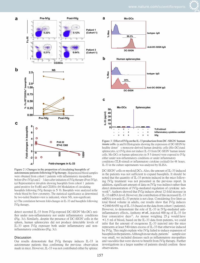
www.nature.com/scientificreports
157
Figure 2 | Changes in the proportion of circulating basophils of autoimmune patients following IVIg therapy. Heparinized blood samples were obtained from cohort 1 patients with inflammatory myopathies before (Pre-IVIg) and 2–3 days after initiation of IVIg therapy (Post-IVIg). (a) Representative dot-plots showing basophils from cohort 1 patients gated positive for FceRI and CD203c (b) Modulation of circulating basophils following IVIg therapy (n 5 9). Basophils were analyzed in the whole blood by flow cytometry. The statistical significance as determined by two-tailed Student-t-test is indicated, where NS, non-significant.
(c) The correlation between fold-changes in IL-33 and basophils following IVIg therapy.
detect secreted IL-33 from IVIg-exposed DC-SIGN1 Mo-DCs nei- ther under non-inflammatory nor under inflammatory conditions (Fig. 3c). Similarly, despite the presence of DC-SIGN1 cells in the spleen, human splenocytes did not produce detectable levels of IL-33 upon IVIg exposure both under inflammatory and non- inflammatory conditions (Fig. 3c).
Discussion Our results demonstrate that IVIg therapy induces IL-33 in autoimmune patients thus confirming the previous observation made in mice. However, IL-33 was not contributed either by splenic
Figure 3 | Effect of IVIg on the IL-33 production from DC-SIGN1 human innate cells. (a and b) Histograms showing the expression of DC-SIGN by healthy donor’s monocyte-derived human dendritic cells (Mo-DCs) and splenocytes. (c) IVIg does not induce IL-33 from DC-SIGN1 human innate cells. Mo-DCs or human splenocytes (n 5 5 donors) were exposed to IVIg either under non-inflammatory conditions or under inflammatory conditions (TLR-stimuli or inflammatory cytokine cocktail) for 48 hours. IL-33 in the culture supernatants was analyzed by ELISA.
DC-SIGN1 cells or myeloid DCs. Also, the amount of IL-33 induced in the patients was not sufficient to expand basophils. It should be noted that the quantity of IL-33 protein induced in the mice follow- ing IVIg treatment was not presented in the previous report. In addition, significant amount of data on IVIg was indirect rather than direct demonstration of IVIg-mediated regulation of cytokine net- work22. Authors showed that IVIg induces about 12-fold increase in IL-33 mRNA level. However, the contribution of this increased IL-33 mRNA towards IL-33 protein is not clear. Considering five liters as total blood volume in adults, our results show that IVIg induces <2460 6 650 ng of IL-33 (based on the data from cohort 1 patients). However, to demonstrate the role of IL-33 in IVIg-mediated anti- inflammatory effects, Anthony et al., injected 400 ng of IL-33 for four consecutive days22. As mouse weighing 25 g would have <1.5 ml of blood, based on the IL-33 data from patients, we could infer that the amount of exogenous IL-33 injected into the mice represents at least 540-times excess of IL-33 that otherwise induced by IVIg. This might explain why IVIg failed to induce expansion of basophils in the patients. Although in our study, patients’ sample size was small, we included diseases such as inflammatory myopathies and vasculitis that were shown to benefit from IVIg therapy. Further investigations in a larger number of patients should confirm these observations.

www.nature.com/scientificreports
158
Table 1 | Summary of data for autoimmune rheumatic patients
Cohort 1 patients
Number Disease Age (years) Sex IVIg Additional treatments
1 Polymyositis 59 F CLAIRYGH 1 g/kg Methylprednisolone 2 Anti-SRP associated necrotizing myopathy 27 F CLAIRYGH 1 g/kg Prednisone, Methotrexate 3 Anti-HMGCR associated necrotizing myopathy 62 F CLAIRYGH 0.5 g/kg Prednisone, Methotrexate 4 Anti-HMGCR associated necrotizing myopathy 61 F CLAIRYGH 1 g/kg Prednisone, Methotrexate 5 Dermatomyositis 52 F CLAIRYGH 1 g/kg Prednisone, Methotrexate 6 Polymyositis associated with mixed connective 41 F CLAIRYGH 1 g/kg Prednisone, Methotrexate
tissue disease and Sjogren’s syndrome 7 Anti-SRP associated necrotizing myopathy 40 M CLAIRYGH 1 g/kg Prednisone, Methotrexate 8 Anti-Mi2 associated unclassified myositis 30 M CLAIRYGH 1 g/kg Prednisone, Methotrexate 9 Polymyositis and probable associated Sjogren’s 70 F CLAIRYGH 1 g/kg Prednisone, Methotrexate
syndrome Cohort 2 patients
Number Disease Age (years) Sex IVIg Additional treatments
1 Dermatomyositis 22 F TEGELINEH 1g/kg Prednisone, Mycophenolate mofetil 2 Polymyositis 42 M TEGELINEH 1g/kg Prednisone, Methotrexate 3 Dermatomyositis 35 M TEGELINEH 1g/kg Prednisone 4 Polymyositis 46 F TEGELINEH 1g/kg Prednisone, ciclosporin 5 Microscopic polyangiitis 61 F TEGELINEH 1g/kg Prednisone 6 Wegener’s granulomatosis 62 M TEGELINEH 1g/kg None 7 Microscopic polyangiitis 61 M TEGELINEH 1g/kg Prednisone, Mycophenolate mofetil
SRP, Signal Recognition Particle; HMGCR, 3-hydroxy-3-methylglutaryl-CoA reductase.
The role of Fc-sialylation, DC-SIGN and Fcc receptor IIB (FccRIIB) in the anti-inflammatory effects of IVIg has been debated recently by several groups27. Mice and humans show wide variations in the expression pattern of FccRs, and the phenotype and anatom- ical distribution of innate cells. Unlike mice, human innate cells express both activating FccRIIA and inhibitory FccRIIB. Therefore, the proposition that IVIg enhances FccRIIB on effector macrophages of mice without having corresponding data on FccRIIA might provide a biased picture on the mechanisms of IVIg. Gene array analysis could not confirm IVIg-mediated up-regu- lation of FccRIIB in the patients with Kawasaki disease28. In line with this report, another recent study failed to demonstrate enhanced expression of FccRIIB on monocytes following IVIg therapy in chil- dren with immune thrombocytopenia29. Also, FccR polymorphisms did not predict response to IVIg in myasthenia gravis30. Although DC-SIGN promoter 2336 A/G (rs4804803) polymorphism was associated with susceptibility of Kawasaki disease, this variant was found to be not associated with the occurrence of IVIg resistance31. Of note, treatment response in Kawasaki disease is apparently assoc- iated with sialylation levels of endogenous IgG but not therapeutic IVIg32. All these data thus questions the relevance of DC-SIGN- FccRIIB pathway of anti-inflammatory mechanisms of IVIg in humans.
Several recent studies have challenged the concept of a2,6- sialylated Fc fragments-mediated anti-inflammatory mechanism of IVIg both in experimental models and in humans. IVIg could inhibit human Th17 cell differentiation and expansion independent of anti- gen presenting cells and hence independent of interaction of DC- SIGN and a2,6-sialylated Fc fragments13–15. Also, F(ab’)2 fragments of IVIg exerted similar effects thus pointing towards dispensability of a2,6-sialylated Fc fragments in mediating the suppression of Th17 cells. We have demonstrated that DC-SIGN and a2,6-sialylated Fc fragment interaction is dispensable for the anti-inflammatory activ- ity of IVIg on human DCs33. F(ab’)2 fragments but not Fc fragments of IVIg were shown to mediate Treg expansion by inducing cycloox- ygenase-2-mediated prostaglandin E2 secretion in human myeloid DCs and was dependent in part on DC-SIGN19. Similarly, sialylation-
enriched F(ab’)2 fragments could inhibit interferon-a production from toll-like receptor (TLR)7 and TLR9 stimulated human plasma- cytoid DCs, although sialic acid itself was not required34.
In the previous reports, Ravetch and colleagues enriched sialic acid-containing IgG-Fc by using sialic acid-specific lectin Sambu- cus nigra agglutinin-based affinity fractionation22,35–37. However, by using same fractionation method, Guhr et al., showed that IVIg fractions depleted for the sialylated antibody fraction exert benefits in a murine model of passive-immune thrombocytopenia similar to that of intact IVIg. However, sialic acid-enriched IVIg fraction failed to enhance platelets count in this model38. Similar sialic-acid inde- pendent anti-inflammatory mechanisms were also reported in mur- ine herpes simplex virus encephalitis model39. Further, Ka¨sermann and colleagues showed that lectin fractionation of IVIg results in increased sialylation of Fab fragments but not Fc fragments. By using human whole blood stimulation assay either with lipopolysaccharide or phytohaemagglutinin, they further showed that anti-inflammat- ory effects of IVIg is associated with F(ab’)2 fraction of IVIg40. In animal models of immune thrombocytopenia and multiple sclerosis, the beneficial effects of IVIg were independent of Fc or F(ab’)2 - sialylation and FccRIIB41–44. Based on these results, it was suggested that genetic background of the mice and dose of IVIg are the critical factors that determine the role of FccRIIB in IVIg-mediated bene- ficial effects. In line with these observations, two studies have failed to demonstrate the direct interaction between sialylated IgG Fc frag- ment and DC-SIGN45,46. These data thus point out that a2,6- sialylated Fc fragment-DC-SIGN-FccRIIB mechanism merely repre- sents one of the several anti-inflammatory mechanisms of IVIg that were reported. Therefore, this anti-inflammatory pathway of IVIg might be operational in certain pathologies and experimental models and might not be considered as a universal mechanism.
It was proposed that in humanized DC-SIGN-transgenic mice, DC-SIGN1 innate cells such as monocytes, macrophages and DCs produce IL-33 upon interaction with a2,6-sialylated Fc fragments of IVIg22. Recent reports show that IL-33 is an important player for the promotion of Th2 responses and activated DCs are one of the sources of this cytokine47,48. We found that unlike monocytes from huma-

www.nature.com/scientificreports
159
nized DC-SIGN-transgenic mice that were highly positive for DC- SIGN, human monocytes hardly express DC-SIGN. Further, human Mo-DCs despite expressing DC-SIGN, failed to produce IL-33 when exposed to IVIg either under non-inflammatory or inflammatory conditions. In wild type mice, it was suggested that a2,6-sialylated Fc fragments bind to SIGN-R1 expressed on splenic marginal zone macrophages35. Marginal zone macrophages are absent in human spleen and data from humans show that spleen is dispensable for the anti-inflammatory effects of IVIg. In line with this concept, by using a passive model of induced immune thrombocytopenia, it was shown that IVIg is fully functional in splenectomized mice although this report supported the sialic acid and SIGN-R1-dependent mechanisms of IVIg49. We found that despite the presence of DC- SIGN1 innate cells in the human spleen, IVIg could not induce IL-33 from the splenocytes. All these data indicate that spleen and DC- SIGN1 cells are dispensable for IVIg-mediated IL-33 induction in humans. Thus, the source of IL-33 in humans following IVIg therapy remains elusive. As IVIg is known to cause apoptosis of cells, we suggest that secondary necrosis of late stage apoptotic cells could release IL-3350–52. This process might depend on the signals provided by anti-Fas IgG or anti-Siglec IgG in the IVIg preparations rather than the repercussion of interaction of a2,6-sialylated Fc fragments with DC-SIGN53,54. In addition, IL-33 is also constitutively expressed in the nucleus of endothelial cells and epithelial cells in vivo55.
Methods Patients. All experiments were performed in accordance with relevant guidelines and regulations. We obtained heparinized blood samples of nine patients (cohort 1 patients) with inflammatory myopathies (Table 1). Patients were aged 49.1 6 15.2 years and include two men. Blood samples were obtained before and 2–3 days
following initiation of IVIg therapy (CLAIRYGH, Laboratoire Français du Fractionnement et des Biotechnologies, France). Informed consent was obtained from all the patients. The study was approved by CPP-Ile-de-France VI, Groupe Hospitalier Pitie -Salpe trie re, Paris. In addition, we also analyzed plasma samples of seven rheumatic patients (cohort 2 patients) before and 2–3 days post-IVIg therapy
(TEGELINEH, Laboratoire Français du Fractionnement et des Biotechnologies). The patients were aged 47 6 5.8 years (four men) and include inflammatory myopathies and anti-neutrophil cytoplasmic antibody-associated vasculitis (Table 1).
Analysis of basophils. Red blood cells (RBCs) from heparinized blood samples of cohort 1 patients were depleted by using HetaSepTM (Stemcell Technologies Sarl, France) and nucleated cell suspension was obtained. Basophils were analyzed in RBC- depleted cell suspension by flow cytometry (LSR II, BD Biosciences, France) using fluorochrome-conjugated monoclonal antibodies to FceRI (Miltenyi Biotec, France) and CD203c (eBioscience, France). Data were analyzed by FACSDivaTM software (BD Biosciences).
Generation of monocyte-derived DCs. Buffy coats from the healthy donors were purchased from Centre Necker-Cabanel, Etablissement Français du Sang (EFS), Paris, France. Institut National de la Sante et de la Recherche Me´dicale-EFS ethical committee permission (Nu12/EFS/079) was obtained for the use of buffy coats of healthy donors. Peripheral blood mononuclear cells (PBMCs) were purified from the buffy coats by density gradient centrifugation using Ficoll-paque PREMIUM (GE healthcare, France). CD141 monocytes were isolated from PBMCs by using CD14 microbeads (Miltenyi Biotec). Purified monocytes were then cultured for 6 days in RPMI-1640 medium plus 10% fetal calf serum (FCS) containing cytokines GM-CSF (1000 IU/106 cells) and IL-4 (500 IU/106 cells) (both from Miltenyi Biotec) to obtain DCs56. The purity of DCs was .98%. DC-SIGN expression on Mo-DCs was examined by flow cytometry using fluorochrome-conjugated monoclonal antibodies (BD Biosciences) and data were analyzed by FACSDivaTM and FlowJo softwares (Tree Star, USA).
Isolation of human splenocytes. The remnant human spleen sections from individuals submitted for pathological diagnosis were obtained from service d’anatomie pathologique, Hoˆpital Europe´en Georges Pompidou, Paris, France. Only healthy spleen tissues were used for the research purpose. Since the study did not require additional sampling, an approval from an ethics committee was not required under French law according to the article L.1121-1 of the public health code. The article states that: The research organized and performed on human beings in the development of biological knowledge and medical research are permitted under the conditions laid down in this book and are hereinafter referred to by the term ‘‘biomedical research’’. The article further states that it does not imply under conditions: For research in which all actions are performed and products used in the usual way, without any additional or unusual diagnostic procedure or surveillance.
The spleen sections were collected in RPMI 1640 medium supplemented with 100 IU/ml penicillin, 100 mg/ml streptomycin, and 10% FCS. Single-cell suspension
of splenocytes was obtained by mechanical disaggregation of spleen tissue pieces by using gentleMACS dissociator (Miltenyi Biotec) followed by filtration through 70-mm nylon membrane filter (BD Biosciences). Splenocytes were then subjected to Ficoll- Paque PREMIUM density gradient centrifugation to obtain mononuclear cells. DC- SIGN expression on the splenocytes was investigated by flow cytometry using fluorochrome-conjugated monoclonal antibodies and data were analyzed by FACSDivaTM and FlowJo softwares.
Stimulation of cells. Mo-DCs (0.5 3 106/ml) were cultured in RPMI 1640-10% FCS containing GM-CSF and IL-4 in a 12-well plate. The cells were then exposed to IVIg (25 mg/ml) for 48 hours to analyze the effect of IVIg on IL-33 production under non- inflammatory conditions. In parallel, Mo-DCs were stimulated with either TLR4 ligand lipopolysaccharide (100 ng/ml/0.5 3 106 cells) (Sigma-Aldrich, France) or inflammatory cytokine cocktail (10 ng/ml each of IL-1b, IL-6 and TNF-a, all from ImmunoTools, Germany)57. After four hours, IVIg was added and cultures were maintained for 48 hours to analyze the effect of IVIg on IL-33 production under inflammatory conditions.
Similarly, splenocytes (0.5 3 106/ml) were cultured in RPMI 1640-10% FCS for 48 hours either alone or with IVIg. In addition, splenocytes were also stimulated with inflammatory cytokine cocktail and IVIg was added to the cultures after four hours. The cultures were maintained for 48 hours.
Quantification of cytokines. IL-33 in the plasma samples of the patients and in cell- free culture supernatants was quantified by ELISA (R&D systems, France). IL-4 in the plasma was also measured by ELISA (R&D systems).
Statistical analysis. Data was analyzed using Prism 5 software (GraphPad software). Two-tailed Student’s t-test was used to determine the statistical significance of the data. Values of P , 0.05 were considered as statistically significant.
1. Dalakas, M. C. Intravenous immunoglobulin in autoimmune neuromuscular diseases. JAMA 291, 2367–2375 (2004).
2. Kazatchkine, M. D. & Kaveri, S. V. Immunomodulation of autoimmune and inflammatory diseases with intravenous immune globulin. N. Engl. J. Med. 345, 747–755 (2001).
3. Arnson, Y., Shoenfeld, Y. & Amital, H. Intravenous immunoglobulin therapy for autoimmune diseases. Autoimmunity 42, 553–560 (2009).
4. Bayry, J., Negi, V. S. & Kaveri, S. V. Intravenous immunoglobulin therapy in rheumatic diseases. Nat. Rev. Rheumatol. 7, 349–359 (2011).
5. Schwab, I. & Nimmerjahn F. Intravenous immunoglobulin therapy: how does IgG modulate the immune system? Nat. Rev. Immunol. 13, 176–189 (2013).
6. Seite, J. F., Shoenfeld, Y., Youinou, P. & Hillion, S. What is the contents of the magic draft IVIg? Autoimmun. Rev. 7, 435–439 (2008).
7. Tha-In, T., Bayry, J., Metselaar, H. J., Kaveri, S. V. & Kwekkeboom, J. Modulation of the cellular immune system by intravenous immunoglobulin. Trends Immunol. 29, 608–615 (2008).
8. Ballow, M. The IgG molecule as a biological immune response modifier: Mechanisms of action of intravenous immune serum globulin in autoimmune and inflammatory disorders. J. Allergy Clin. Immunol. 127, 315–323 (2011).
9. Jordan, S. C., Toyoda, M. & Vo, A. A. Intravenous immunoglobulin a natural regulator of immunity and inflammation. Transplantation 88, 1–6 (2009).
10. Semple, J. W. et al. Intravenous immunoglobulin prevents murine antibody- mediated acute lung injury at the level of neutrophil reactive oxygen species (ROS) production. PLoS One 7, e31357 (2012).
11. Bayry, J. et al. Intravenous immunoglobulin abrogates dendritic cell differentiation induced by interferon-alpha present in serum from patients with systemic lupus erythematosus. Arthritis Rheum. 48, 3497–3502 (2003).
12. Aubin, E., Lemieux, R. & Bazin, R. Indirect inhibition of in vivo and in vitro T-cell responses by intravenous immunoglobulins due to impaired antigen presentation. Blood 115, 1727–1734 (2010).
13. Maddur, M. S. et al. Inhibition of differentiation, amplification, and function of human TH17 cells by intravenous immunoglobulin. J. Allergy Clin. Immunol. 127, 823–830 e821–827 (2011).
14. Maddur, M. S., Kaveri, S. V. & Bayry, J. Comparison of different IVIg preparations on IL-17 production by human Th17 cells. Autoimmun. Rev. 10, 809–810 (2011).
15. Maddur, M. S. et al. Inhibitory effect of IVIG on IL-17 production by Th17 cells is independent of anti-IL-17 antibodies in the immunoglobulin preparations. J. Clin. Immunol. 33, S62–S66 (2013).
16. Kessel, A. et al. Intravenous immunoglobulin therapy affects T regulatory cells by increasing their suppressive function. J. Immunol. 179, 5571–5575 (2007).
17. De Groot, A. S. et al. Activation of natural regulatory T cells by IgG Fc- derived peptide ‘‘Tregitopes’’. Blood 112, 3303–3311 (2008).
18. Massoud, A. H. et al. Intravenous immunoglobulin attenuates airway inflammation through induction of forkhead box protein 3-positive regulatory T cells. J. Allergy Clin. Immunol. 129, 1656–1665 e1653 (2012).
19. Trinath, J. et al. Intravenous immunoglobulin expands regulatory T cells via induction of cyclooxygenase-2-dependent prostaglandin E2 in human dendritic cells. Blood 122, 1419–1427 (2013).
20. Seite, J. F., Goutsmedt, C., Youinou, P., Pers, J. O. & Hillion, S. Intravenous immunoglobulin induces a functional silencing program similar to anergy in human B cells. J. Allergy Clin. Immunol. 133, 181–188 e181–189 (2014).

www.nature.com/scientificreports
160
21. Massoud, A. H. et al. Dendritic cell immunoreceptor: a novel receptor for intravenous immunoglobulin mediates induction of regulatory T cells. J. Allergy Clin. Immunol. 133, 853–863 e855 (2014).
22. Anthony, R. M., Kobayashi, T., Wermeling, F. & Ravetch, J. V. Intravenous gammaglobulin suppresses inflammation through a novel T(H)2 pathway. Nature 475, 110–113 (2011).
23. Voehringer, D. Protective and pathological roles of mast cells and basophils. Nat. Rev. Immunol. 13, 362–375 (2013).
24. Otsuka, A. et al. Basophils are required for the induction of Th2 immunity to haptens and peptide antigens. Nat. Commun. 4, 1739, doi:10.1038/ncomms2740 (2013).
25. Sharma, M. et al. Circulating human basophils lack the features of professional antigen presenting cells. Sci. Rep. 3, 1188, doi: 10.1038/srep01188 (2013).
26. Campbell, I. K. et al. Therapeutic effect of IVIG on inflammatory arthritis in mice is dependent on the Fc portion and independent of sialylation or basophils. J. Immunol. 192, 5031–5038 (2014).
27. von Gunten, S. et al. IVIG pluripotency and the concept of Fc-sialylation: challenges to the scientist. Nat. Rev. Immunol. 14, 349 (2014).
28. Abe, J. et al. Gene expression profiling of the effect of high-dose intravenous Ig in patients with Kawasaki disease. J. Immunol. 174, 5837–5845 (2005).
29. Shimomura, M. et al. Intravenous immunoglobulin does not increase FccRIIB expression levels on monocytes in children with immune thrombocytopenia. Clin. Exp. Immunol. 169, 33–37 (2012).
30. Barnett, C. et al. Fcc receptor polymorphisms do not predict response to intravenous immunoglobulin in myasthenia gravis. J. Clin. Neuromuscul. Dis. 14, 1–6 (2012).
31. Yu, H. R. et al. DC-SIGN (CD209) promoter -336 A/G (rs4804803) polymorphism associated with susceptibility of Kawasaki disease. Sci. World J. 2012, 634835 (2012).
32. Ogata, S. et al. Treatment response in kawasaki disease is associated with sialylation levels of endogenous but not therapeutic intravenous immunoglobulin G. PLoS One 8, e81448 (2013).
33. Bayry, J., Bansal, K., Kazatchkine, M. D. & Kaveri, S. V. DC-SIGN and a2,6- sialylated IgG Fc interaction is dispensable for the anti-inflammatory activity of IVIg on human dendritic cells. Proc. Natl. Acad. Sci. U S A. 106, E24 (2009).
34. Wiedeman, A. E. et al. Contrasting mechanisms of interferon-alpha inhibition by intravenous immunoglobulin after induction by immune complexes versus Toll- like receptor agonists. Arthritis Rheum. 65, 2713–2723 (2013).
35. Anthony, R. M., Wermeling, F., Karlsson, M. C. & Ravetch, J. V. Identification of a receptor required for the anti-inflammatory activity of IVIG. Proc. Natl. Acad. Sci. U S A. 105, 19571–19578 (2008).
36. Anthony, R. M. et al. Recapitulation of IVIG anti-inflammatory activity with a recombinant IgG Fc. Science 320, 373–376 (2008).
37. Kaneko, Y., Nimmerjahn, F. & Ravetch, J. V. Anti-inflammatory activity of immunoglobulin G resulting from Fc sialylation. Science 313, 670–673 (2006).
38. Guhr, T. et al. Enrichment of sialylated IgG by lectin fractionation does not enhance the efficacy of immunoglobulin G in a murine model of immune thrombocytopenia. PLoS One 6, e21246 (2011).
39. Ramakrishna, C., Newo, A. N., Shen, Y. W. & Cantin, E. Passively administered pooled human immunoglobulins exert IL-10 dependent anti- inflammatory effects that protect against fatal HSV encephalitis. PLoS Pathog. 7, e1002071 (2011).
40. Kasermann, F. et al. Analysis and functional consequences of increased Fab- sialylation of intravenous immunoglobulin (IVIG) after lectin fractionation. PLoS One 7, e37243 (2012).
41. Leontyev, D. et al. Sialylation-independent mechanism involved in the amelioration of murine immune thrombocytopenia using intravenous gammaglobulin. Transfusion 52, 1799–1805 (2012).
42. Leontyev, D., Katsman, Y. & Branch, D. R. Mouse background and IVIG dosage are critical in establishing the role of inhibitory Fcgamma receptor for the amelioration of experimental ITP. Blood 119, 5261–5264 (2012).
43. Othy, S. et al. Intravenous gammaglobulin inhibits encephalitogenic potential of pathogenic T cells and interferes with their trafficking to the central nervous system, implicating sphingosine-1 phosphate receptor 1-mammalian target of rapamycin axis. J. Immunol. 190, 4535–4541 (2013).
44. Othy, S. et al. Sialylation may be dispensable for reciprocal modulation of helper T cells by intravenous immunoglobulin. Eur. J. Immunol. (epub ahead of print) doi:10.1002/eji.201444440 (2014).
45. Crispin, M., Yu, X. & Bowden, T. A. Crystal structure of sialylated IgG Fc: implications for the mechanism of intravenous immunoglobulin therapy. Proc. Natl. Acad. Sci. U S A. 110, E3544–3546 (2013).
46. Yu, X., Vasiljevic, S., Mitchell, D. A., Crispin, M. & Scanlan, C. N. Dissecting the molecular mechanism of IVIg therapy: the interaction between serum IgG and DC-SIGN is independent of antibody glycoform or Fc domain. J. Mol. Biol. 425, 1253–1258 (2013).
47. Grotenboer, N. S., Ketelaar, M. E., Koppelman, G. H. & Nawijn, M. C. Decoding asthma: translating genetic variation in IL33 and IL1RL1 into disease pathophysiology. J. Allergy Clin. Immunol. 131, 856–865 (2013).
48. Williams, J. W. et al. Transcription factor IRF4 drives dendritic cells to promote Th2 differentiation. Nat. Commun. 4, 2990, doi:10.1038/ncomms3990 (2013).
49. Schwab, I., Biburger, M., Kronke, G., Schett, G. & Nimmerjahn, F. IVIg- mediated amelioration of ITP in mice is dependent on sialic acid and SIGNR1. Eur. J. Immunol. 42, 826–830 (2012).
50. Bonilla, W. V. et al. The alarmin interleukin-33 drives protective antiviral CD81 T cell responses. Science 335, 984–989 (2012).
51. Cayrol, C. & Girard, J. P. The IL-1-like cytokine IL-33 is inactivated after maturation by caspase-1. Proc. Natl. Acad. Sci. U S A. 106, 9021–9026 (2009).
52. Haraldsen, G., Balogh, J., Pollheimer, J., Sponheim, J. & Kuchler, A. M. Interleukin-33 - cytokine of dual function or novel alarmin? Trends Immunol. 30, 227–233 (2009).
53. Prasad, N. K. et al. Therapeutic preparations of normal polyspecific IgG (IVIg) induce apoptosis in human lymphocytes and monocytes: a novel mechanism of action of IVIg involving the Fas apoptotic pathway. J. Immunol. 161, 3781–3790 (1998).
54. von Gunten, S. et al. Immunologic and functional evidence for anti-Siglec-9 autoantibodies in intravenous immunoglobulin preparations. Blood 108, 4255–4259 (2006).
55. Moussion, C., Ortega, N. & Girard, J. P. The IL-1-like cytokine IL-33 is constitutively expressed in the nucleus of endothelial cells and epithelial cells in vivo: a novel ‘alarmin’? PLoS One 3, e3331 (2008).
56. Maddur, M. S. et al. Human B cells induce dendritic cell maturation and favour Th2 polarization by inducing OX-40 ligand. Nat. Commun. 5, 4092, doi:10.1038/ ncomms5092 (2014).
57. Banchereau, J. et al. Immunobiology of dendritic cells. Annu. Rev. Immunol. 18, 767–811 (2000).
Acknowledgments This study was supported by Institut National de la Sante et de la Recherche Medicale
(INSERM), Centre National de la Recherche Scientifique (CNRS), Universite Pierre et
Marie Curie, Universite Paris Descartes and European Community’s Seventh Framework Programme [FP7/2007–2013] under grant agreement HEALTH-2010.2.4.5-2 ALLFUN. We also thank Laboratoire Français du Fractionnement et des Biotechnologies, France for the support.
Author contributions J.B. designed the research, M.S., C.S., P.H., M.S.M., E.S-V., L.G. & M.L. performed the experiments, M.S., P.H., M.S.M., S.V.K. & J.B. analyzed the data, Y.S., L.M. & O.B. provided blood samples of the patients, P.B. provided the spleen tissues, J.B. wrote the paper and all authors reviewed and approved the manuscript.
Additional information Competing financial interests: The authors declare no competing financial interests.
How to cite this article: Sharma, M. et al. Intravenous immunoglobulin-induced IL-33 is insufficient to mediate basophil expansion in autoimmune patients. Sci. Rep. 4, 5672; DOI:f10.1038/srep05672 (2014).
This work is licensed under a Creative Commons Attribution-NonCommercial- NoDerivs 4.0 International License. The images or other third party material in this article are included in the article’s Creative Commons license, unless indicated otherwise in the credit line; if the material is not included under the Creative Commons license, users will need to obtain permission from the license holder in order to reproduce the material. To view a copy of this license, visit http:// creativecommons.org/licenses/by-nc-nd/4.0/

WILEY-VCH
Sialylation may be dispensable for reciprocal modulation of helper T cells by intravenous immunoglobulin
Shivashankar Othy1,2, Selma Top¸cu1, Chaitrali Saha1,3,
Prathap Kothapalli1, Sebastien Lacroix-Desmazes1,2,4,5,
Fabian Ka¨sermann6, Sylvia Miescher6, Jagadeesh Bayry1,2,4,5
and Srini V. Kaveri1,2,4,5
1 Institut National de la Sante et de la Recherche Medicale Unite 1138, Paris, France 2 Centre de Recherche des Cordeliers, Equipe 16-Immunopathology and therapeutic
immunointervention, Universit´e Pierre et Marie Curie – Paris, Paris, France 3 Universite de Technologie de Compiegne, Compiegne, France 4 Universite Paris Descartes, Paris, France 5 International Associated Laboratory IMPACT (Institut National de la Sant e et de la Recherche
M´edicale, France–Indian Council of Medical Research), National Institute of
Immunohematology, Mumbai, India 6 Research and Development, CSL Behring AG, Bern, Switzerland
Several mechanisms account for the beneficial effect of intravenous immunoglobulin
(IVIg) in autoimmune and inflammatory diseases. These mechanisms include effects
on the cellular compartment and on the humoral compartment. Thus, IVIg impacts on
dendritic cells, macrophages, neutrophils, basophils, NK cells, and B and T lymphocytes.
Several studies have emphasized that the antiinflammatory effect of IVIg is dependent
on α2,6-sialylation of the N-linked glycan on asparagine-297 of the Fc portion of IgG.
However, recent reports have questioned the necessity of sialylated Fc and the role of
FcγRIIB in IVIg-mediated antiinflammatory effects. In view of the critical role played by
Th17 cells in several autoimmune pathologies and the increasing use of IVIg in several
of these conditions, by using neuraminidase-treated, desialylated IVIg, we addressed
whether the α2,6-sialylation of IgG is essential for the beneficial effect of IVIg in experi-
mental autoimmune encephalomyelitis (EAE), a Th17-driven condition, and for the recip-
rocal modulation of helper T-cell subsets. We observed no difference in the ability of IVIg
to ameliorate EAE irrespective of its sialylation. Our findings thus show that sialylation
of IVIg is not necessary for IVIg-mediated amelioration of EAE or for downregulation of
Th17 cells and upregulation of regulatory T cells.
Keywords: Experimental autoimmune encephalomyelitis . Intravenous immunoglobulin . Reg-
ulatory T cells . Sialylation . Th1 . Th17
Additional supporting information may be found in the online version of this article at the
publisher’s web-site
Correspondence: Dr. Srini V. Kaveri or Jagadeesh Bayry
ail: [email protected]; [email protected]
161

WILEY-VCH
Introduction
Intravenous immunoglobulin (IVIg) is a therapeutic preparation
of polyspecific human gammaglobulin (IgG) derived from the
pooled plasma of thousands of healthy donors [1–3]. IVIg is pri-
marily used as an IgG replacement therapy in immune-deficient
patients. Paradoxically, high doses of IVIg are also used to treat a
number of autoimmune and inflammatory pathologies including
immune thrombocytopenia, Guillain-Barr´e syndrome, Kawasaki
disease, and chronic inflammatory demyelinating polyneuropa-
thy [1, 3]. The beneficial effect of IVIg in two opposing clini-
cal scenarios (immunodeficiency and autoimmune pathology) is
intriguing; notably the role of IVIg therapy in the latter con-
dition is under intense investigation. IVIg is known to func-
tion by several mutually nonexclusive mechanisms modulating
both molecular and cellular networks of the immune system
[4]. Molecular pathways modulated by IVIg include cytokines,
autoantibodies, complement proteins and Fc receptors; cellu-
lar targets include antigen-presenting cells (DCs), macrophages,
neutrophils, basophils, natural killer cells, B and T lymphocytes
[5–8].
Several previous reports have uncovered a surprising role of
IgG glycosylation that accounts for the therapeutic efficacy of
IVIg [9–12]; it has been demonstrated that the antiinflamma-
tory activity of IVIg is dependent exclusively on α2,6-sialylation of
N-linked glycan on asparagine-297 in the Fc portion of IgG [9].
Using a K/BxN model of arthritis, it was shown that the sialylated
Fc fraction of IVIg upregulates FcγRIIB (an inhibitory Fc recep-
tor) on the effector macrophages, thus increasing their activa-
tion threshold and circumventing the joint damage [9]. However,
recent studies have questioned the requirement of sialylated Fc
and the role of FcγRIIB in models of immune thrombocytopenia
and in IVIg-mediated inhibition of innate immune cell functions
[13–19].
Interleukin-17 secreting helper T (Th17) cells have emerged
as key pathogenic players in rheumatoid arthritis, antineu-
trophil cytoplasmic antibody-associated vasculitis, asthma, allergic
contact dermatitis, systemic lupus erythematosus, chronic inflam-
matory bowel disease, and MS [20]. In these conditions, Th17
cells coordinate with IFN-γ-secreting Th1 cells and attract other
effector cells to the sites of inflammation. Interestingly, regulatory
T (Treg) cells expressing the transcription factor Foxp3 are impli-
cated in the suppression of autoreactive T cells, including Th17
cells and preventing Th17-dependent autoimmune conditions
[20].
In view of the importance of the Th17 cells in auto-
immune and inflammatory conditions and the proposed role
of sialylation of IVIg, in this study, we examined the role of
sialylation of IVIg in a Th17 cell mediated autoimmune model,
the experimental autoimmune encephalomyelitis (EAE), a
classical murine model of MS proven to be mediated by Th17
cells [21]. Our results indicate that the reciprocal regulation
of CD4+ T cells by IVIg in EAE appears to be independent of
sialylation.
Figure 1. SDS-PAGE and lectin blot profiles of native and desialylated
IVIg (NA-IVIg). One microgram of IgG was loaded and run on NuPage
10% BisTris gels under nonreducing conditions. The gels were stained
with colloidal Coomassie (left) or blotted onto nitrocellulose, probed
with biotin-SNA and AP-streptavidin, and visualized with chromogenic
AP conjugate substrate (right). The molecular weight markers are also
indicated. Data shown are representative of more than five indepen-
dent desialylation and purification processes.
Results and discussion
Considering the crucial role played by Th17 cells in many autoim-
mune pathologies and increasing use of IVIg in several of these
conditions, it is important to understand the role of sialylation of
IVIg in a Th17 cell mediated autoimmune model. EAE is classical
murine model of MS proven to be mediated by Th17 cells [21].
Using myelin oligodendrocyte glycoprotein (MOG35–55)-induced
EAE, we have recently demonstrated that IVIg delays the onset and
decreases severity of disease by inhibiting Th17 and Th1 cells, and
concomitantly expanding Foxp3+ Treg cells [22]. To our surprise,
this reciprocal regulation was independent of inhibitory FcγRIIB.
Here, we addressed whether the α2,6-sialylation on IgG is required
for the beneficial effect of IVIg in EAE and for the reciprocal mod-
ulation of helper T-cell subsets by using neuraminidase-treated,
desialylated IVIg.
Desialylation and integrity of the neuraminidase-treated IVIg
(NA-IVIg) was confirmed by SDS-PAGE, and lectin blottings
(Fig. 1). Reverse phase high performance liquid chromatography
(RP-HPLC) revealed that NA-IVIg contained less than 0.1 mg of
sialic acid per gram of IgG as compared native, untreated IVIg
(0.67 mg/g of IgG) (data not shown) [14].
EAE was induced in 10-week-old female C57BL/6J mice.
NA-IVIg delayed the onset and decreased severity of the disease
similar to that of IVIg (p < 0.001 for both IVIg and NA-IVIg)
(Fig. 2). We observed no difference in the ability to ameliorate
EAE between IVIg and desialylated IVIg (Mean maximal score
(MMS) ± SD for control = 3.28 ± 0.58; IVIg = 2.04 ± 0.98;
162

WILEY-VCH
Figure 2. Neuraminidase-treated desialylated IVIg (NA-IVIg) delays the
onset of EAE similar to native IVIg. EAE was induced in 10-week-old
female C57BL/6J mice. Native IVIg (open triangles) or NA-IVIg (filled
circles) was given at 0.8 g/kg from day 0 to 18. Control mice received
not been completely elucidated. It has been proposed that anti-
inflammatory activity of IVIg is dependent exclusively on sialyla-
tion of the Fc portion of IgG. In the present study, we demonstrate
that the protection against EAE by IVIg and reciprocal regula-
tion of proinflammatory encephalitogenic Th17 and Th1 cells and
Foxp3+ Treg cells are not dependent on sialylation of IgG. In con-
clusion, these data along with those reported in murine immune
thrombocytopenia further question the role of Fc sialylation in the
mechanism of IVIg effect and highlight the importance of multi-
pronged antiinflammatory action of IVIg.
Desialylation of IVIg with neuraminidase and
confirmation by lectin blotting and HPLC
§R
an equal volume of PBS (open circles). Daily clinical scores of control, IVIg (Hizentra ) was desialylated by enzymatic digestion as
IVIg and NA-IVIg groups are shown as mean + SEM of n = 11–18 mice
pooled from two independent experiments. *** p < 0.001, using two-way
ANOVA with Bonferroni’s post t test.
NA-IVIg = 2.30 ± 0.60, p = 0.7124 between IVIg and NA-IVIg
group). Hence, sialylation of IgG is not required for beneficial
effect of IVIg in EAE.
Mechanistically, IVIg delays the onset of EAE by inhibiting dif-
ferentiation of na ıve CD4+ T cells into encephalitogenic Th17 and
Th1 cells, and expanding Foxp3+ Treg cells. We explored whether
desialylated IVIg also exerted similar mechanisms. Consistent with
our published results on IVIg [22], NA-IVIg also decreased Th17
cells (fivefold change, p = 0.015) and Th1 cells (sevenfold change,
p = 0.015) in draining lymph nodes (Fig. 3A and C). Accordingly,
this inhibition was associated with an increase in the number of
Foxp3+ Treg cells in the spleen (1.5 fold change, p = 0.015; Fig. 1B
and D). Therefore, reciprocal regulation of CD4+ T cells by IVIg
in EAE appears to be independent of sialylation. Recent studies
from our laboratory showed that F(abr)2 fragments of IVIg could
inhibit expansion and activation of human Th17 cells [23] and
could induce Treg-cell expansion by inducing COX-2-dependent
prostaglandin E2 in DCs [18]. Further, in vitro studies showed that
the observed antiinflammatory effects of IVIG are Fab-mediated
and not sialic acid-dependent. It was proposed that the skewed
antibody repertoire in sialylated IVIg prepared by lectin chro-
matography is responsible for this effect [19]. Together, these
data raise intriguing questions on the role of sialylation of Fc-
fragment of IgG for the beneficial effect of IVIg and the reciprocal
modulation of helper T-cell subsets by IVIg in murine model of
MS.
Concluding remarks
The mechanisms underlying the potent antiinflammatory effect of
IVIg in a number of autoimmune and inflammatory diseases have
reported previously [14]. Briefly, seven units of recombinant neu-
raminidase (New England BioLabs, USA) were incubated with
each mg IVIg for 48 h at 37°C. Following this, concentration and
buffer exchange to PBS was performed by tangential flow filtration
using a 100 kD millipore filter unit (minimate TFF system, Pall).
To confirm desialylation by lectin blotting, one microgram IgG
was loaded and ran on a NuPage 10% Bis-Tris gels under non-
reducing conditions (Invitrogen, USA). The gels were stained
with colloidal Coomassie (Gelcode, Thermo Scientific, USA) or
blotted onto nitrocellulose. The blots were blocked with Carbo-
Free blocking solution (Vector Laboratories, USA), probed with
biotin-SNA (2 g/L, Vector) and AP-streptavidin (1.5 g/L, Invit-
rogen) and visualized with chromogenic AP conjugate sub-
strate (BioRad, Switzerland). Alternatively for HPLC, sialic
acid was released by acidic hydrolysis of neuraminic acid
in 0.25 M NaHSO4 followed by derivatization of the gly-
can with the fluorophore1,2-diamino-4,5-methylenedioxybenzene
dihydrochloride (DMB). Quantification of the derivatized sialic
acid was performed by RP-HPLC using N-acetyl neuraminic acid
(Neu5Ac; Fluka, Switzerland) as a standard and expressed as
Neu5Ac per IgG (weight/weight) [14].
Induction EAE and treatment with IVIg or NA-IVIg
Animal experiments were performed as previously described
[18, 22] according to the Charles Darwin ethical committee guid-
ance (UPMC Paris). A 10-week-old C57BL/6J mice (Janvier Labo-
ratories, France) were immunized subcutaneously with 200 μL
of emulsion (50 μL per site) containing 200 μg of MOG35–55
peptide (MEVGWYRSPFSRVVHLYRNGK, PolyPeptide laboratory
Strasbourg, France) emulsified in complete Freund’s adjuvant
(Sigma–Aldrich) containing 880 μg of nonviable Mycobacterium
tuberculosis antigen H37RA (Difco Laboratories). Mice received
300 ng of pertussis toxin after 2 and 48 h intravenously. Develop-
ment of EAE was assessed daily according to the following criteria;
163

WILEY-VCH
0-No signs, 1-tail paresis, 2-hindlimb paresis, 3-hind limb paral-
ysis, 4-tetraplegia, 5-moribound. IVIg (Hizentra§R 20% w/v, CSL
Behring) or desialylated (NA-IVIg) was given daily at 0.8 g/kg i.p
from the day of the immunization until the peak of the disease
(day 16–18). Control mice received equal volume PBS.
Isolation of cells from draining lymph nodes and
spleen, and flow cytometry
Nine days after EAE induction, mice were sacrificed under
Xylazinane/Ketamine anesthesia to obtain draining inguinal
lymph nodes and spleen. Single cell suspension was prepared
by mechanical disaggregation and passing through 70 μm nylon
membrane filter. Red blood cells were removed using ACK lysis
buffer. A total of 1.5 × 10⁶ cells were stimulated with 25 ng
of phorbol 12-myristate 13-acetate (PMA) and 1 μg ionomycin
(Sigma) in the presence of Monensin (Golgistop§R BD biosciences)
in 10% FCS/RPMI for 4 h at 37°C. Cells were labeled with anti-
mouse CD4-Pacific Blue (Clone RM4–5, BD biosciences) after
blocking the Fc-receptors with anti-mouse CD16/32 antibody (BD
Fc Block). Intracellular staining was performed using FoxP3 stain-
ing buffer set (eBioscience) with the following antibodies: IL-17A-
Figure 3. Neuraminidase-treated desia-
lylated IVIg reciprocally modulates
helper T lymphocytes, as does native
IVIg. Nine days after EAE induction, mice
were sacrificed. Spleen and draining
lymph nodes (DLN) were collected and
analyzed for helper T-cell subsets by
flow cytometry. Representative dot plots
showing CD4+ T cells from (A, C) the
DLN gated for IL-17 and IFN-γ; and
(B, D) the spleen gated for Foxp3 in
control, desialylated IVIg (NA-IVIg) and
native IVIg-treated mice are shown.
Value in each quadrant denotes percent-
age of cells positive for IL-17, IFN-γ, and
Foxp3 among the CD4+ population. Each
symbol represents an individual mouse
and data are shown as mean ± SEM from
4–6 mice pooled from two experiments
(right panels, A–D). *p < 0.05, **p < 0.01,
Mann–Whitney test.
A488 (clone 11B11, BD), IFN-γ-allophycocyanin (Clone XMG1.2,
BD), Foxp3-PE (clone FJK16s, eBioscience). BD LSR II with FACS
Diva software was used to acquire and analyze the data.
Statistical analysis
Two-way analysis of variance (ANOVA) with Bonferroni’s post test
was used to compare daily clinical score. Mann–Whitney’s U test
was used to compare parameters between control and NA-IVIg
group. Values of p obtained are indicated in Figure legends.
Acknowledgments: Supported by Institut National de la Sant e et
de la Recherche M´edicale (INSERM), Centre National de la
Recherche Scientifique (CNRS), Universit´e Pierre et Marie Curie
and Universit´e Paris Descartes. Monika Ru¨egsegger and Andreas
Hofmann (CSL Behring, Bern, Switzerland), for excellent technical
assistance. O.S received fellowship for PhD from Erasmus Mundus
Program.
164

WILEY-VCH
Conflict of interest: F.K. and S.M. are full time employees of CSL
Behring AG, Bern, Switzerland; otherwise the authors declare no
financial conflict of interest.
References
1 Zandman-Goddard, G., Krauthammer, A., Levy, Y., Langevitz, P. and
Shoenfeld, Y., Long-term therapy with intravenous immunoglobulin
is beneficial in patients with autoimmune diseases. Clin. Rev. Allergy
Immunol. 2012. 42: 247–255.
2 Kaveri, S. V., Intravenous immunoglobulin: exploiting the potential of
natural antibodies. Autoimmun. Rev. 2012. 11: 792–794.
3 Kazatchkine, M. D. and Kaveri, S. V., Immunomodulation of autoimmune
and inflammatory diseases with intravenous immune globulin. N. Engl.
J. Med. 2001. 345: 747–755.
4 Tha-In, T., Bayry, J., Metselaar, H. J., Kaveri, S. V. and Kwekkeboom, J.,
Modulation of the cellular immune system by intravenous immunoglob-
ulin. Trends Immunol. 2008. 29: 608–615.
5 Seite, J. F., Shoenfeld, Y., Youinou, P. and Hillion, S., What is the contents
of the magic draft IVIg? Autoimmun. Rev. 2008. 7: 435–439.
6 Cousens, L. P., Tassone, R., Mazer, B. D., Ramachandiran, V., Scott, D.
W. and De Groot, A. S., Tregitope update: mechanism of action parallels
IVIg. Autoimmun. Rev. 2013. 12: 436–443.
7 Casulli, S., Topcu, S., Fattoum, L., von Gunten, S., Simon, H. U.,
Teillaud, J. L., Bayry, J. et al., A differential concentration-dependent
effect of IVIg on neutrophil functions: relevance for anti-microbial and
anti-inflammatory mechanisms. PLoS One 2011. 6: e26469.
8 Araujo, L. M., Chauvineau, A., Zhu, R., Diem, S., Bourgeois, E. A.,
Levescot, A., Huerre, M. et al., Cutting edge: intravenous Ig inhibits
invariant NKT cell-mediated allergic airway inflammation through
FcgammaRIIIA-dependent mechanisms. J. Immunol. 2011. 186: 3289–3293.
9 Anthony, R. M., Kobayashi, T., Wermeling, F. and Ravetch, J. V., Intra-
venous gammaglobulin suppresses inflammation through a novel T(H)2
pathway. Nature 2011. 475: 110–113.
10 Schwab, I., Biburger, M., Kro nke, G., Schett, G. and Nimmerjahn, F., IVIg-
mediated amelioration of ITP in mice is dependent on sialic acid and
SIGNR1. Eur. J. Immunol. 2012 42: 826–830.
11 Schwab, I., Mihai, S., Seeling, M., Kasperkiewicz, M., Ludwig, R.
and Nimmerjahn, F., Broad requirement for terminal sialic acid
residues and FcγRIIB for the preventive and therapeutic activity of
intravenous immunoglobulins in vivo. Eur. J. Immunol. 2014. DOI:
10.1002/eji.201344230. [Epub ahead of print].
12 Schwab, I., Seeling, M., Biburger, M., Aschermann, S., Nitschke, L. and
Nimmerjahn, F., B cells and CD22 are dispensable for the immediate
antiinflammatory activity of intravenous immunoglobulins in vivo. Eur.
J. Immunol. 2012. 42: 3302–3309.
13 Guhr, T., Bloem, J., Derksen, N. I., Wuhrer, M., Koenderman, A. H.,
Aalberse, R. C. and Rispens, T., Enrichment of sialylated IgG by lectin
fractionation does not enhance the efficacy of immunoglobulin G in a
murine model of immune thrombocytopenia. PLoS One 2011. 6: e21246.
14 Ka sermann, F., Boerema, D. J., Ruegsegger, M., Hofmann, A., Wymann,
S., Zuercher, A. W. and Miescher, S., Analysis and functional conse-
quences of increased Fab-sialylation of intravenous immunoglobulin
(IVIG) after lectin fractionation. PLoS One 2012. 7: e37243.
15 Leontyev, D., Katsman, Y., Ma, X. Z., Miescher, S., Ka sermann, F. and
Branch, D. R., Sialylation-independent mechanism involved in the ame-
lioration of murine immune thrombocytopenia using intravenous gam-
maglobulin. Transfusion 2012. 52: 1799–1805.
16 Leontyev, D., Katsman, Y. and Branch, D. R., Mouse background and IVIG
dosage are critical in establishing the role of inhibitory Fcgamma receptor
for the amelioration of experimental ITP. Blood 2012. 119: 5261–5264.
17 Bayry, J., Bansal, K., Kazatchkine, M. D. and Kaveri, S. V., DC-SIGN
and alpha2,6-sialylated IgG Fc interaction is dispensable for the anti-
inflammatory activity of IVIg on human dendritic cells. Proc. Natl. Acad.
Sci. U.S.A. 2009. 106: E24.
18 Trinath, J., Hegde, P., Sharma, M., Maddur, M. S., Rabin, M., Vallat, J.
M., Magy, L. et al., Intravenous immunoglobulin expands regulatory T
cells via induction of cyclooxygenase-2-dependent prostaglandin E2 in
human dendritic cells. Blood 2013. 122: 1419–1427.
19 Wiedeman, A. E., Santer, D. M., Yan, W., Miescher, S., Ka sermann, F.
and Elkon, K. B., Contrasting mechanisms of interferon-alpha inhibition
by intravenous immunoglobulin after induction by immune complexes
versus Toll-like receptor agonists. Arthritis Rheum. 2013. 65: 2713–2723.
20 Korn, T., Bettelli, E., Oukka, M. and Kuchroo VK., IL-17 and Th17 Cells.
Annu. Rev. Immunol. 2009. 27: 485–517.
21 Gold, R., Linington, C. and Lassmann, H., Understanding pathogenesis
and therapy of multiple sclerosis via animal models: 70 years of merits
and culprits in experimental autoimmune encephalomyelitis research.
Brain 2006. 129: 1953–1971.
22 Othy, S., Hegde, P., Topcu, S., Sharma, M., Maddur, M. S., Lacroix-
Desmazes, S., Bayry, J. et al., Intravenous gammaglobulin inhibits
encephalitogenic potential of pathogenic T cells and interferes with
their trafficking to the central nervous system, implicating sphingosine-
1 phosphate receptor 1-mammalian target of rapamycin axis. J. Immunol.
2013. 190: 4535–4541.
23 Maddur, M. S., Vani, J., Hegde, P., Lacroix-Desmazes, S., Kaveri, S. V.
and Bayry, J., Inhibition of differentiation, amplification, and function
of human TH17 cells by intravenous immunoglobulin. J. Allergy Clin.
Immunol. 2011. 127:823–830.e1–7.
Abbreviations: IVIg: intravenous immunoglobulin · NA-IVIg:
neuraminidase-treated IgG
Full correspondence: Dr. Srini V. Kaveri or Jagadeesh Bayry, INSERM
Unite 1138, Centre de Recherche des Cordeliers, Equipe 16
Immunopathology and therapeutc immunointervention, 15, Rue de
l’Ecole de Me decine, 75006 Paris, France
Fax: +33-144278194
E-mail: [email protected] or [email protected]
Received: 3/1/2014
Revised: 24/2/2014
Accepted: 26/3/2014
Accepted article online: 3/4/2014
165

1
1


167
167


169
169


171
171
Copyright © 2022 FDOKUMEN