Antinociceptive, Anti-Inflammatory and Antipyretic Effects of Lapidin, a Bicyclic Sesquiterpene
1.0 Introduction
Research on analgesic and anti-inflammatory drugs has
gained great attention for the past ten years (Farouk
et al., 2008). However, the number of new drugs remains
low. Most of analgesic and anti- inflammatory compounds
available on market have adverse effects, including
life-threatening, bleeding or perforation of gastro
duodenal tract (Buttgereit et al., 2001) which
therefore, constitute an obstacle for their
prescription. Consequently there results the need to
search for more active compounds with less adverse
effects.
Free radicals and reactive oxygen species are by
products of numerous physiological and biochemical
processes. Natural antioxidants and anti-inflammatory
effects have been found in a number of food and
agricultural products like seed, roots, stem back ETC.
Besides the traditional resources used for
antioxidants, many plant species have been investigated
in the search for natural antioxidants and anti
inflammatory effect.
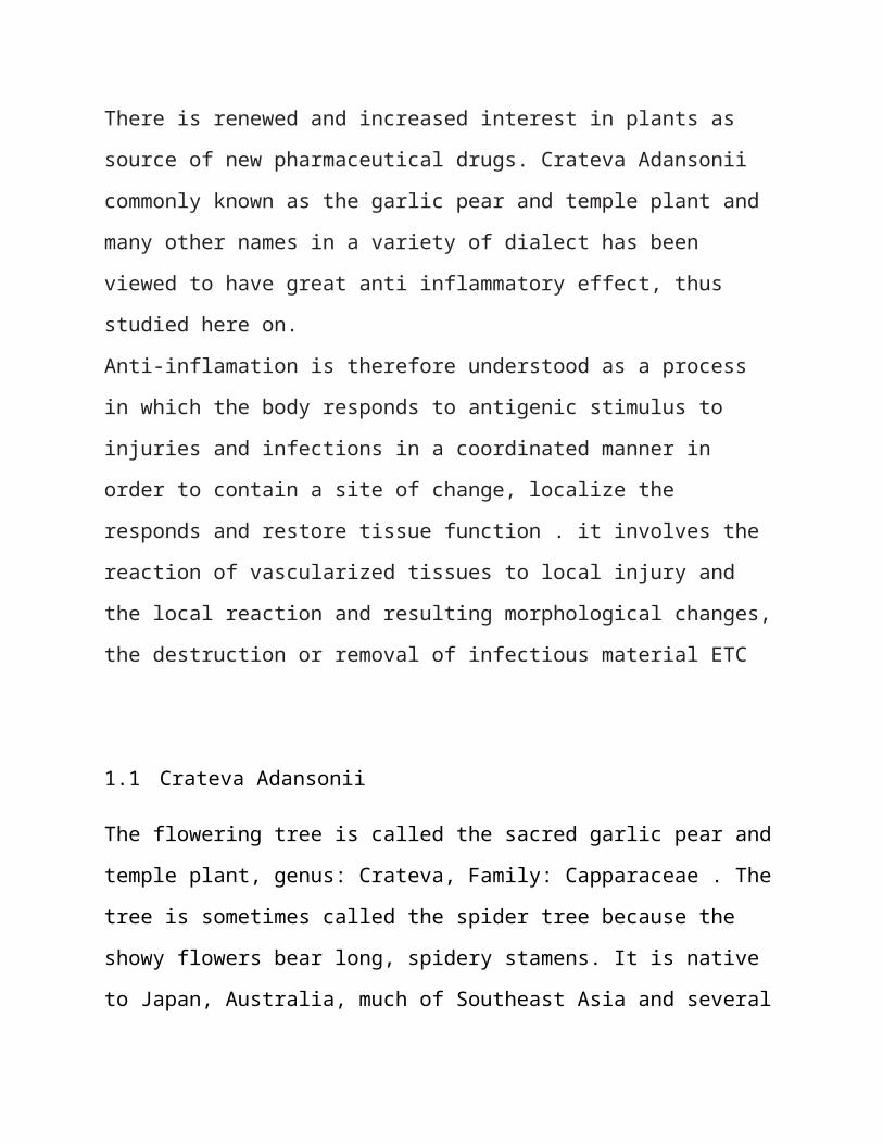
There is renewed and increased interest in plants as
source of new pharmaceutical drugs. Crateva Adansonii
commonly known as the garlic pear and temple plant and
many other names in a variety of dialect has been
viewed to have great anti inflammatory effect, thus
studied here on.
Anti-inflamation is therefore understood as a process
in which the body responds to antigenic stimulus to
injuries and infections in a coordinated manner in
order to contain a site of change, localize the
responds and restore tissue function . it involves the
reaction of vascularized tissues to local injury and
the local reaction and resulting morphological changes,
the destruction or removal of infectious material ETC
1.1 Crateva Adansonii
The flowering tree is called the sacred garlic pear and
temple plant, genus: Crateva, Family: Capparaceae . The
tree is sometimes called the spider tree because the
showy flowers bear long, spidery stamens. It is native
to Japan, Australia, much of Southeast Asia and several
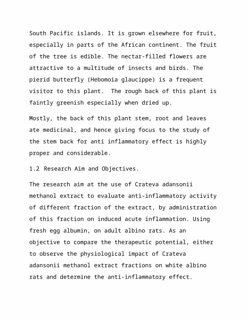
South Pacific islands. It is grown elsewhere for fruit,
especially in parts of the African continent. The fruit
of the tree is edible. The nectar-filled flowers are
attractive to a multitude of insects and birds. The
pierid butterfly (Hebomoia glaucippe) is a frequent
visitor to this plant. The rough back of this plant is
faintly greenish especially when dried up.
Mostly, the back of this plant stem, root and leaves
ate medicinal, and hence giving focus to the study of
the stem back for anti inflammatory effect is highly
proper and considerable.
1.2 Research Aim and Objectives.
The research aim at the use of Crateva adansonii
methanol extract to evaluate anti-inflammatory activity
of different fraction of the extract, by administration
of this fraction on induced acute inflammation. Using
fresh egg albumin, on adult albino rats. As an
objective to compare the therapeutic potential, either
to observe the physiological impact of Crateva
adansonii methanol extract fractions on white albino
rats and determine the anti-inflammatory effect.

CHAPTER TWO
2.1 Inflammation
Inflammation is the body's attempt at self-protection;
the aim being to remove harmful stimuli, including
damaged cells, irritants, or pathogens - and begin the
healing process.
When something harmful or irritating affects a part of
our body, there is a biological response to try to
remove it, the signs and symptoms of inflammation,
specifically acute inflammation which is a type of
inflammation, show that the body is trying to heal
itself. Inflammation does not mean infection, even when
an infection causes inflammation. Infection is caused
by a bacterium, virus or fungus, while inflammation is
the body's response to it.

INFLAMMATION AS PART OF OUR INNATE IMMUNITY.
Our innate immunity is what is naturally present in our
bodies when we are born, and not the adaptive immunity
we get after an infection or vaccination. Innate
immunity is generally non-specific, while adaptive
immunity is specific to one pathogen, for example
Whooping cough vaccine being specific to one pathogen.
2.1.1 WHAT IS ANTI-INFLAMMATION?
Anti-inflammation refers to the property of a substanceor treatment that reduces inflammation. Anti-inflammatory drugs make up about half of analgesics, remedying pain by reducing inflammation as opposed to opioids, which affect the central nervous system.
TYPES OF INFLAMMATION (acute & chronic)
Clinically, there are two main types of inflammation, active and chronic.
Active (acute) inflammation occurs on the time scale ofhours to days and is characterized by the cardinal signs of inflammation; namely, redness (rubor), swelling (tumor), heat (calor) and pain (dolor). Acute inflammation represents an initial concentrated effort to eliminate an injurious agent.

Acute inflammation is characterized histologically by the presence of neutrophils that have emigrated from blood vessels into the injured tissue. Variations in the histologic picture include the presence of eosinophil, which may be seen in parasitic infections, and basophils (termed mast cells when in tissue) seen in allergic conditions. Features of active inflammationwith components that are not usually appreciated histologically include vasodilatation, increased micro vascular permeability, and neural stimulation.
Chronic inflammation occurs on the time scale of weeks to months and is characterized by the simultaneous presence of active inflammation, tissue destruction, and attempts at repair. Clinically, the process may be characterized by the loss of proper function of the tissue, but is often an asymptomatic, subclinical response. Histologically, the process has been classically characterized by the presence of large numbers of "mononuclear cells" which is a nonspecific term referring to lymphocytes and macrophages (derived from peripheral blood monocytes) that are two cell types of entirely different function and lineage.
Histologically, chronic inflammation differs significantly from acute inflammation as the continued active attempt to eliminate a pathogen overlaps with the process of repair. Thus, the histologic picture is highly variable. The components often identified in addition to those present in active inflammation include:
Collections of lymphocytes and plasma cells, the latter of which secrete specific

immunoglobulin’s, to facilitate removal of the injurious agent
Scarring (also known as fibrosis or connective tissue replacement) involves proliferation of fibroblasts and the subsequent deposition of collagen. This process usually results when the native architecture of the tissue is unable to beregenerated or an exudate is unable to be adequately reabsorbed (a process also known as organization).
Persistent infections by a variety of agents (e.g. Mycobacterium tuberculosis) show a characteristic tissue response known as granulomatous inflammation in which collections of epithelioid macrophages serve to wall off the offending agent which is unable to be removed by typical inflammatory mechanisms
2.1.2 Steroids and non-steroidal anti-inflammatory drugs
Many steroids, to be specific glucocorticoids, reduce inflammation or swelling by binding to glucocorticoid receptors. These drugs are often referred to as corticosteroids.
Non-steroidal anti-inflammatory drugs (NSAIDs), alleviate pain by counteracting the cyclooxygenase (COX) enzyme. On its own, COX enzyme synthesizes prostaglandins, creating inflammation. In whole, the

NSAIDs prevent the prostaglandins from ever being synthesized, reducing or eliminating the pain.
Some common examples of NSAIDs are: aspirin, ibuprofen,and naproxen. The newer specific COX-inhibitors - although, it is presumed, sharing a similar mode of action - are not classified together with the traditional NSAIDs.
On the other hand, there are analgesics that are commonly associated with anti-inflammatory drugs but that have no anti-inflammatory effects. An example is paracetamol, called acetaminophen in the U.S. and sold under the brand name of Tylenol. As opposed to NSAIDs, which reduce pain and inflammation by inhibiting COX enzymes, paracetamol has recently been shown to block the reuptake of endocannabinoids, which only reduces pain, likely explaining why it has minimal effect on inflammation.
Long-term use of NSAIDs can cause gastric erosions, which can become stomach ulcers and in extreme cases can cause severe haemorrhage, resulting in death. The risk of death as a result of use of NSAIDs is 1 in 12,000 for adults aged 16–45. The risk increases almosttwentyfold for those over 75. Other dangers of NSAIDs are exacerbating asthma and causing kidney damage. Apart from aspirin, prescription and over-the-counter NSAIDs also increase the risk of myocardial infarction and stroke.
2.1.3 Immune Selective Anti-Inflammatory Derivatives (ImSAIDs)

ImSAIDs are a class of peptides being developed by IMULAN BioTherapeutics, LLC, which were discovered to have diverse biological properties, including anti-inflammatory properties. ImSAIDs work by altering the activation and migration of inflammatory cells, which are immune cells responsible for amplifying the inflammatory response. The ImSAIDs represent a new category of anti-inflammatory and are unrelated to steroid hormones or non-steroidal anti-inflammatories.
The ImSAIDs were discovered by scientists evaluating biological properties of the submandibular gland and saliva. Early work in this area demonstrated that the submandibular gland released a host of factors that regulate systemic inflammatory responses and modulate systemic immune and inflammatory reactions. It is now well accepted that the immune, nervous, and endocrine systems communicate and interact to control and modulate inflammation and tissue repair. One of the neuroendocrine pathways, when activated, results in therelease of immune-regulating peptides from the submandibular gland upon neuronal stimulation from sympathetic nerves. This pathway or communication is referred to as the cervical sympathetic trunk-submandibular gland (CST-SMG) axis, a regulatory systemthat plays a role in the systemic control of inflammation.
Early work in identifying factors that played a role inthe CST-SMG axis lead to the discovery of a seven aminoacid peptide, called the submandibular gland peptide-T.SGP-T was demonstrated to have biological activity and thermoregulatory properties related to endotoxin exposure. SGP-T, an isolate of the submandibular gland,
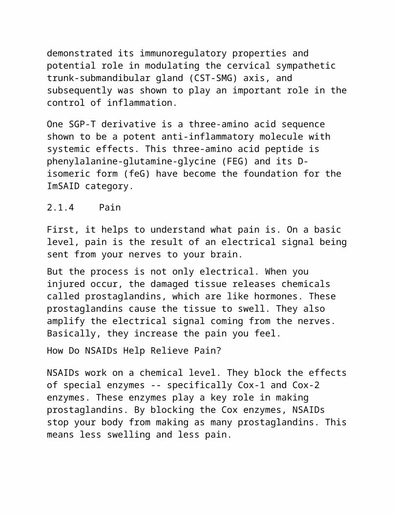
demonstrated its immunoregulatory properties and potential role in modulating the cervical sympathetic trunk-submandibular gland (CST-SMG) axis, and subsequently was shown to play an important role in thecontrol of inflammation.
One SGP-T derivative is a three-amino acid sequence shown to be a potent anti-inflammatory molecule with systemic effects. This three-amino acid peptide is phenylalanine-glutamine-glycine (FEG) and its D-isomeric form (feG) have become the foundation for the ImSAID category.
2.1.4 Pain
First, it helps to understand what pain is. On a basic level, pain is the result of an electrical signal beingsent from your nerves to your brain.But the process is not only electrical. When you injured occur, the damaged tissue releases chemicals called prostaglandins, which are like hormones. These prostaglandins cause the tissue to swell. They also amplify the electrical signal coming from the nerves. Basically, they increase the pain you feel.How Do NSAIDs Help Relieve Pain?
NSAIDs work on a chemical level. They block the effectsof special enzymes -- specifically Cox-1 and Cox-2 enzymes. These enzymes play a key role in making prostaglandins. By blocking the Cox enzymes, NSAIDs stop your body from making as many prostaglandins. Thismeans less swelling and less pain.
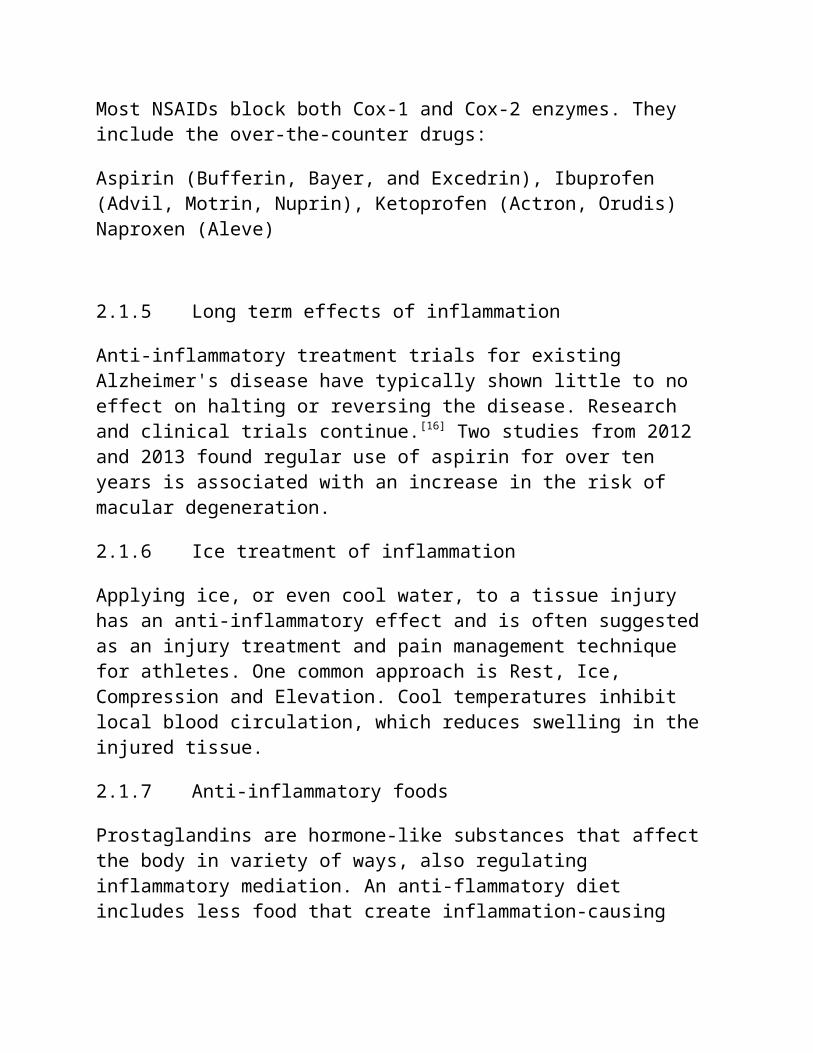
Most NSAIDs block both Cox-1 and Cox-2 enzymes. They include the over-the-counter drugs:
Aspirin (Bufferin, Bayer, and Excedrin), Ibuprofen (Advil, Motrin, Nuprin), Ketoprofen (Actron, Orudis) Naproxen (Aleve)
2.1.5 Long term effects of inflammation
Anti-inflammatory treatment trials for existing Alzheimer's disease have typically shown little to no effect on halting or reversing the disease. Research and clinical trials continue.[16] Two studies from 2012 and 2013 found regular use of aspirin for over ten years is associated with an increase in the risk of macular degeneration.
2.1.6 Ice treatment of inflammation
Applying ice, or even cool water, to a tissue injury has an anti-inflammatory effect and is often suggested as an injury treatment and pain management technique for athletes. One common approach is Rest, Ice, Compression and Elevation. Cool temperatures inhibit local blood circulation, which reduces swelling in the injured tissue.
2.1.7 Anti-inflammatory foods
Prostaglandins are hormone-like substances that affect the body in variety of ways, also regulating inflammatory mediation. An anti-flammatory diet includes less food that create inflammation-causing
prostaglandins (PGE2) in the body, and more foods that create anti-flammatory prostaglandins (PGE1 and PGE3).
Suggested diets to reduce inflammation include those rich in vegetables and low in simple carbohydrates and fats, such as saturated fats and trans fats. Anti-inflammatory foods include most colorful fruits and vegetables, oily fish (which contain higher levels of omega-3 fatty acids), nuts, seeds, and certain spices, such as ginger. Extra-virgin olive oil contains the chemical oleocanthal that acts similarly to ibuprofen. Those following an anti-inflammatory diet will avoid refined oils and sugars, and show a preference for so-called anti-inflammatory foods in their meal choices.
Omega-3 fatty acids have been shown to disrupt inflammation cell signaling pathways by binding.
2.2 Vascular event in inflammation
Acute inflammation is characterized by marked vascular
changes, including vasodilation, increased permeability
and increased blood flow, which are induced by the
actions of various inflammatory mediators. Vasodilation
occurs first at the arteriole level, progressing to the
capillary level, and brings about a net increase in the
amount of blood present, causing the redness and heat
of inflammation. Increased permeability of the vessels
results in the movement of plasma into the tissues,
with resultant stasis due to the increase in the

concentration of the cells within blood - a condition
characterized by enlarged vessels packed with cells.
Stasis allows leukocytes to migrate (move) along the
endothelium, a process critical to their recruitment
into the tissues. Normal flowing blood prevents this,
as the shearing force along the periphery of the
vessels moves cells in the blood into the middle of the
vessel.
2.2.1 Vasoconstriction
Vasoconstriction is the narrowing of the blood vessels resulting from contraction of the muscular wall of the vessels, particularly the large arteries and small arterioles. The process is the opposite of vasodilation, the widening of blood vessels. The process is particularly important in staunching hemorrhage and acute blood loss. When blood vessels constrict, the flow of blood is restricted or decreased, thus, retaining body heat or increasing vascular resistance. Cutaneously, this makes the skin turn paler because less blood reaches the surface, reducing the radiation of heat. On a larger level, vasoconstriction is one mechanism by which the body regulates and maintains mean arterial pressure.
Substances causing vasoconstriction are called vasoconstrictors, vasopressors, or simply "pressors". Generalized vasoconstriction usually results in an increase in systemic blood pressure, but it may also

occur in specific tissues causing a localized reductionin blood flow. The extent of vasoconstriction may be slight or severe depending on the substance or circumstance. Many vasoconstrictors also cause pupil dilation. Medications that cause vasoconstriction include antihistamines, decongestants and stimulants used to treat ADHD.
Mechanisms
Factors that trigger vasoconstriction can be of exogenous or endogenous origin. Ambient temperature is an example of the former. Cutaneous vasoconstriction will occur because of the body's exposure to the severecold. Examples of endogenous factors include the autonomic nervous system, circulating hormones and intrinsic mechanisms inherent to the vasculature itself(also referred to as the myogenic response).
Exogenous medications
Examples include amphetamines, antihistamines and cocaine. Many are used in medicine to treat hypotensionand as topical decongestants. Vasoconstrictors are alsoused clinically to increase blood pressure or to reducelocal blood flow. Vasoconstrictors mixed with local anesthetics are used to increase the duration of local anesthesia by constricting the blood vessels, thereby safely concentrating the anesthetic agent for an extended duration, as well as reducing hemorrhage.
2.2.2 Vasodilation

Vasodilation refers to the widening of blood vessels. It results from relaxation of smooth muscle cells within the vessel walls, particularly in the large veins, large arteries, and smaller arterioles. The process is essentially the opposite of vasoconstriction, which is the narrowing of blood vessels.
When blood vessels dilate, the flow of blood is increased due to a decrease in vascular resistance. Therefore, dilation of arterial blood vessels (mainly the arterioles) decreases blood pressure. The response may be intrinsic (due to local processes in the surrounding tissue) or extrinsic (due to hormones or the nervous system). Additionally, the response may be localized to a specific organ (depending on the metabolic needs of a particular tissue, as during strenuous exercise), or it may be systemic (seen throughout the entire systemic circulation).
Drugs that cause vasodilation are termed vasodilators
Mechanisms
Vasodilation is the result of relaxation in smooth muscle surrounding the blood vessels. This relaxation, in turn, relies on removing the stimulus for contraction, which depends on intracellular calcium ionconcentrations and, consequently, phosphorylation of the light chain of the contractile protein myosin. Thus, vasodilation mainly works either by lowering intracellular calcium concentration or the dephosphorylation of myosin. This includes stimulation
of myosin light chain phosphatase and induction of calcium symporters and antiporters that pump calcium ions out of the intracellular compartment. This is accomplished through reuptake of ions into the sarcoplasmic reticulum via exchangers and expulsion across the plasma membrane. There are three main intracellular stimuli that can result in the vasodilation of blood vessels. The specific mechanisms to accomplish these effects vary from vasodilator to vasodilator.
2.2.3 Vascular permeability
Vascular permeability, often in the form of capillary permeability or microvascular permeability, characterizes the capacity of a blood vessel wall to allow for the flow of small molecules (ions, water, nutrients) or even whole cells (lymphocytes on their way to the site of inflammation) in and out of the vessel. Blood vessel walls are lined by a single layer of endothelial cells. The gaps between endothelial cells (cell junctions) are strictly regulated dependingon the type and physiological state of the tissue.
Case of Increased Permeability:Normally, a balance exists between the amount of plasmaentering the tissues from the capillaries and the amount re-entering the circulatory system from the tissues. When the tissue is damaged as a result of infection or injury, the lining of the small blood vessels becomes leaky, thereby increasing permeability.
The balance shifts, allowing more fluid to enter the tissues.Increased Permeability PurposeWhen plasma enters damaged tissues during the inflammatory process, it carries a lot of substances with it. Clotting factors to stop the bleeding and spread of infection, antibodies to fight infection, nutrients to feed the tissue cells and proteins that attract phagocytes (cells responsible for attacking microorganisms and gobbling up the damaged cells) represent some of the substances carried by the plasma.Permeability mediators are the chemicals that cause thecapillaries to become leaky, or more permeable, when tissue is damaged. The chemicals histamine and serotonin play a significant role in increasing capillary permeability.Inflammatory Response SymptomsInflammation is accompanied by four clinical symptoms, which include redness, heat, pain and swelling. Rednessand heat are caused by increased blood flow to the damaged tissues. Pain is the result of nerve-ending stimulation by chemicals released during the inflammatory process. The accumulation of fluid in the tissue that results from increased capillary permeability produces swelling.
2.3 Cellular event
The "Cellular Event" A cell participates in many different decisions and can do so in very different

realms of information. What I call a "cellular event" is a decision involving a particular group of influences from a particular group of cells firing in aconsistent way to send a signal to a particular cell. This is a theoretical event that describes the way a cell reacts to a particular set arrangement of influences from a particular grouping of cells that areproviding input to that cell. This cellular event is a constant and theoretical because it is statistically very rare that the influences affecting a cell are everexactly the same.
The point of understanding the cellular event is to
understand how a cell can learn to change it's response
to the same influences over time. To describe this
event as a static unchanging arrangement rather than
the real changing and animated coordination of firings
is helpful to keep confusion down when trying to
understand this information.
2.3.1 Leukocyte migration as specific
hemoral/cellular immunity:
Migration of leukocytes to sites of injury or inflammation is a crucial component of both innate and adaptive immunity. Achieve this, finely co-ordinated mechanisms exist by which intravascular leukocytes are
able to penetrate the vascular wall and migrate to sites of injury or infection without causing any perceptible damage to the vessels from which they emigrate. Within this scenario, leukocyte transmigration through vessel walls (predominantly post-capillary venules) is the final stage of a stepwise cascade of leukocyte responses mediated by a series of sequential molecular interactions that initially mediate the slowing down of leukocyte rollingvelocity followed by leukocyte firm adhesion to the endothelium and eventually migration through the vesselwall. As discussed in this review, leukocyte transmigration not only acts as a means of directing the emigration of leukocytes from the vascular lumen tothe extravascular tissue but may also play a critical role in regulating the phenotype of the emigrated cellssuch that leukocyte behavior in the form of responsiveness to chemo attractants, directional migration and interactions with components of the extravascular tissue may be regulated.
The leukocytes, or white blood cells, defend the body against infecting organisms and foreign agents, both inthe tissues and in the bloodstream itself (immunity). Human blood contains about 5,000 to 10,000 leukocytes per cubic millimeter; the number increases in the presence of infection.
The migration of leukocytes from the vascular lumen tosites of infection and/or injury in the extravascular

tissue involves a series of sequential and coordinated molecular and cellular events with the resultant primary response being that of reduced leukocyte velocity within the blood stream, followed by leukocytefirm adhesion to endothelial cells lining the vessel wall and eventually migration through the vessel wall. Despite the growing knowledge of the mechanisms that mediate initial interaction of leukocytes with the endothelium, very little is known about the mechanisms that mediate and regulate leukocyte migration through the venular wall, the endothelium and its associated perivascular basement membrane.
2.3.2 Macrophages in inflammation
The macrophage is a vital type of white blood cell. Mostly acting as phagocytes gobbling invading bacteria.However, macrophages do much more than that: Not only do they act as antimicrobial warriors; they also play critical roles in immune regulation and wound-healing. They can respond to a variety of cellular signals and change their physiology in response to local cues.
The inflammatory process is usually tightly regulated, involving both signals that initiate and maintain inflammation and signals that shut the process down. Animbalance between the two signals leaves inflammation unchecked, resulting in cellular and tissue damage. Macrophages are a major component of the mononuclear phagocyte system that consists of closely related cellsof bone marrow origin, including blood monocytes, and
tissue macrophages. From the blood, monocytes migrate into various tissues and transform macrophages. In inflammation, macrophages have three major function; antigen presentation, phagocytosis, and immunomodulation through production of various cytokines and growth factors. Macrophages play a critical role in the initiation, maintenance, and resolution of inflammation. They are activated and deactivated in the inflammatory process. Activation signals include cytokines (interferon gamma, granulocyte-monocyte colony stimulating factor, and tumor necrosis factor alpha), bacterial lipopolysaccharide, extracellular matrix proteins, and other chemical mediators. Inhibition of inflammation byremoval or deactivation of mediators and inflammatory effector cells permits the host to repair damages tissues. Activated macrophages are deactivated by anti-inflammatory cytokines (interleukin 10 and transforminggrowth factor beta) and cytokine antagonists that are mainly produced by macrophages. Macrophages participatein the autoregulatory loop in the inflammatory process.Because macrophages produce a wide range of biologically active molecules participated in both beneficial and detrimental outcomes in inflammation, therapeutic interventions targeted macrophages and their products may open new avenues for controlling inflammatory diseases.

2.4 Mediators of inflammation
In general, lymphocytes are involved in the adaptive response to inflammation, and the early events of inflammation are mediated in partly by molecules produced by cells of the innate arm of the immune system. Early phase mediators are produced by mast cells and platelets. They are especially important in acute inflammation and include mainly histamine, serotonin and other vasoactive substances. Platelets may contribute to inflammatory responses resulting as aconsequence of tissue injury, through a variety of mechanisms including:
1. the release of vasoactive amines and other permeability factors,
2. the release of lysosomal enzymes,
3. the release of coagulation factors which lead to localized and generalized fibrin deposition, and
4. the formation of platelet aggregates or trombi whichresult in the blocking of vessels and capillaries. To the early phase mediators also belong chemoatractants (e.g. C5a) and cytokines such as IL-1, IL-6, and TNF-α.
Late phase mediators are responsible for the regulationof vascular events occuring later - from about 6-12 hours after initiation of inflammation. The later vascular events are mediated, at least in part, by products of arachidonic acid.
Inflammatory mediators are soluble, diffusible molecules that act locally at the site of tissue damageand infection, and at more distant sites.
Once leukocytes have arrived at a site of infection or inflammation, they release mediators which control the later accumulation and activation of other cells. However, in inflammatory reactions initiated by the immune system, the ultimate control is exerted by the antigen itself, in the same way as it controls the immune response itself. For this reason, the cellular accumulation at the site of chronic infection, or in autoimmune reactions (where the antigen cannot ultimately be eradicated), is quite different from thatat sites where the antigenic stimulus is rapidly cleared. There are four major plasma enzyme systems which have an important role in haemostasis and controlof inflammation. These are the complement system, the clotting system, the fibrinolytic (plasmin) system and the kinin system.
2.4.1 CELL-DERIVED MEDIATORS
Vasoactive Amines: Histamine and Serotonin
The two major vasoactive amines, so named because they have important actions on blood vessels, are histamine and serotonin. They are stored as preformed molecules in cells and are therefore among the first mediators tobe released during inflammation. The richest sources ofhistamine are the mast cells that are normally present
in the connective tissue adjacent to blood vessels. It is also found in blood basophils and platelets. Histamine is present in mast cell granules and is released by mast cell degranulation in response to a variety of stimuli, including (1) physical injury such as trauma, cold, or heat; (2) binding of antibodies to mast cells, which underlies allergic reactions (3) fragments of complement called anaphylatoxins (C3a and C5a); (4) histamine-releasing proteins derived from leukocytes; (5) neuropeptides (e.g., substance P); and (6) cytokines (IL-1, IL-8).
2.4.2 Histamine
Causes dilation of arterioles and increases the permeability of venules. It is considered to be the principal mediator of the immediate transient phase of increased vascular permeability, producing interendothelial gaps in venules, as we have seen. Its vasoactive effects are mediated mainly via binding to H1 receptors on microvascular endothelial cells
2.4.3 Cytokines
Are proteins produced by many cell types (principally activated lymphocytes and macrophages, but also endothelial, epithelial, and connective tissue cells) that modulate the functions of other cell types? Long known to be involved in cellular immune responses, these products have additional effects that play important roles in both acute and chronic inflammation.

Their general properties and functions are discussed in. Here we review the pro-perties of cytokines that are involved in acute inflammation
2.4.4 Serotonin (5-hydroxytryptamine)
Is a preformed vasoactive mediator with actions similarto those of histamine. It is present in platelets and certain neuroendocrine cells, e.g. in the gastrointestinal tract, and in mast cells in rodents but not humans. Release of serotonin (and histamine) from platelets is stimulated when platelets aggregate after contact with collagen, thrombin, adenosine diphosphate, and antigenantibody complexes. Thus, the platelet release reaction, which is a key component of coagulation, also results in increased vascular permeability. This is one of several links between clotting and inflammation.
2.4.5 Platelet-Activating Factor (PAF)
PAF is another phospholipid-derived mediator. Its name comes from its discovery as a factor that causes platelet aggregation, but it is now known to have multiple inflammatory effects. A variety of cell types,including platelets themselves, basophils, mast cells, neutrophils, macrophages, and endothelial cells, can elaborate PAF, in both secreted and cell-bound forms. In addition to platelet aggregation, PAF causes vasoconstriction and bronchoconstriction, and at extremely low concentrations it induces vasodilation

and increased venular permeability with a potency 100 to 10,000 times greater than that of histamine. PAF also causes increased leukocyte adhesion to endothelium(by enhancing integrin-mediated leukocyte binding), chemotaxis, degranulation, and the oxidative burst. Thus, PAF can elicit most of the vascular and cellular reactions of inflammation. PAF also boosts the synthesis of other mediators, particularly eicosanoids,by leukocytes and other cells. A role for PAF in vivo is supported by the ability of synthetic PAF receptor antagonists to inhibit inflammation in some experimental models.
2.4.6 Arachidonic Acid (AA): Metabolites: Prostaglandins, Leukotrienes, and Lipoxins
When cells are activated by diverse stimuli, such as microbial products and various mediators of inflammation, membrane AA is rapidly converted by the actions of enzymes to produce prostaglandins and leukotrienes. These biologically active lipid mediatorsserve as intracellular or extracellular signals to affect a variety of biologic processes, including inflammation and hemostasis.
• Prostaglandins (PGs) are produced by mast cells, macrophages, endothelial cells, and many other cell types, and are involved in the vascular and systemicreactions of inflammation. They are produced by the actions of two cyclooxygenases, the constitutively expressed COX-1 and the inducible enzyme COX-2.

Prostaglandins are divided into series based on structural features as coded by a letter (PGD, PGE, PGF, PGG, and PGH) and a subscript numeral (e.g., 1,2), which indicates the number of double bonds in the compound. The most important ones in inflammation are PGE2, PGD2, PGF2α, PGI2 (prostacyclin), and TxA2 (thromboxane), each of which is derived by the action of a specific enzyme on an intermediate in the pathway. Some of these enzymes have restricted tissue distribution.
• The lipoxygenase enzymes are responsible for the production of leukotrienes, which are secreted mainly by leukocytes, are chemoattractants for leukocytes, and also have vascular effects. There are three different lipoxygenases, 5-lipoxygenase being the predominant one in neutrophils. This enzyme converts AA to 5-hydroxyeicosatetraenoic acid, which is chemotactic for neutrophils, and is the precursor of the leukotrienes. LTB4 is a potent chemotactic agent and activator of neutrophils, causing aggregation and adhesion of the cells to venular endothelium, generation of ROS, and release of lysosomal enzymes.
• Lipoxins are also generated from AA by the lipoxygenase pathway, but unlike prostaglandins and leukotrienes, the lipoxins are inhibitors of inflammation. They are also unusual in that two cellpopulations are required for the transcellular

biosynthesis of these mediators. Leukocytes, particularly neutrophils, produce intermediates in lipoxin synthesis, and these are converted to lipoxins by platelets interacting with the leukocytes.
2.4.7 Free radicals as mediators of inflammation in atherosclerosis
Free radicals are chemical species that possess an unpaired electron and are often formed as intermediatesin chemical reactions. The presence of the unpaired electron makes these molecules unstable and reactive. Oxygen free radicals are reactive oxygen species (ROS) formed from the incomplete reduction of oxygen and exert a range of important effects in biological cells and tissues. Oxygen radicals and other ROS are producedby normal cellular metabolism and have critical roles in the processes of cellular signaling and injury. The four-electron reduction of molecular oxygen to water, catalyzed by the mitochondrial electron transport chain, accounts for95%of oxygen consumption in tissues.The remaining 5% proceeds via univalent reduction of oxygen with the production of superoxide anions (’02), hydrogen peroxide (H2O2), and hydroxyl radicals (’OH). These reactive products have been documented to cause cell injury. Therefore, cells have evolved several systems that function to avoid or correct damage causedby these oxygen radicals.
2.4.8 Nitric Oxide (NO)

NO was discovered as a factor released from endothelialcells that caused vasodilation and was therefore calledendothelium-derived relaxing factor. NO is a soluble gas that is produced not only by endothelial cells but also by macrophages and some neurons in the brain. It acts in a paracrine manner on target cells through induction of cyclic guanosine monophosphate, which, in turn, initiates a series of intracellular events leading to a response, such as the relaxation of vascular smooth muscle cells. Because the in vivo half-life of NO is only seconds, the gas acts only on cells in close proximity to where it is produced.
NO is synthesized from l-arginine by the enzyme nitric oxide synthase (NOS). There are three different types of NOS: endothelial (eNOS), neuronal (nNOS), and inducible (iNOS) (Fig. 2-12). eNOS and nNOS are constitutively expressed at low levels and can be activated rapidly by an increase in cytoplasmic Ca2+. iNOS, in contrast, is induced when macrophages and other cells are activated by cytokines (e.g., TNF, IFN-γ) or microbial products
2.4.9 Reactive Oxygen Species
Oxygen-derived free radicals may be released extracellularly from leukocytes after exposure to microbes, chemokines, and immune complexes, or following a phagocytic challenge.[55] Their production is dependent, as we have seen, on the activation of theNADPH oxidase system. Superoxide anion ( ), hydrogen

peroxide (H2O2), and hydroxyl radical (•OH) are the major species produced within cells, and O can combine with NO to form reactive nitrogen species. Extracellular release of low levels of these potent mediators can increase the expression of chemokines (e.g., IL-8), cytokines, and endothelial leukocyte adhesion molecules, amplifying the inflammatory response. As mentioned earlier, the physiologic function of these ROS in leukocytes is to destroy phagocytosed microbes, but release of these potent mediators can be damaging to the host They are implicated in the following responses in inflammation:
• Endothelial cell damage, with resultant increased vascular permeability. Adherent neutrophils, when activated, not only produce their own toxic speciesbut also stimulate production of ROS in the endothelial cells.
• Injury to other cell types (parenchymal cells, red blood cells).
• Inactivation of antiproteases, such as α1-antitrypsin. This leads to unopposed protease activity, with increased destruction of extracellular matrix. In the lung, such inhibition of anti-proteases contributes to destruction of elastic tissues, as in emphysema.

CHAPTER THREE
3.1 Materials, equipment, apparatus
The following materials were utilized in the course of this work: retort stand, cornical flask, funnel, spatula, water bath, burette, glass wool, syringe, silica gel paper, pipette, niddles, gloves, cages, weigh balance, test tubes, filter paper, water bottle, column chromatography tube,
3.2 Chemicals/reagent and practical technique
3.2.1 Methanol

Methanol, also known as methyl alcohol, wood alcohol, wood naphtha or wood spirits, is a chemical with the formula CH3OH (often abbreviated MeOH). Methanol acquired the name "wood alcohol" because it was once produced chiefly as a byproduct of the destructive distillation of wood. Modern methanol is perhaps produced in a catalytic industrial process directly from carbon monoxide, carbon dioxide, and hydrogen.
Methanol is the simplest alcohol, and is a light, volatile, colorless, flammable liquid with a distinctive odor very similar to, but slightly sweeter than, that of ethanol (drinking alcohol). At room temperature, it is a polar liquid, and is used as an antifreeze, solvent, fuel, and as a denaturant for ethanol. It is also used for producing biodiesel via transesterification reaction.
Methanol is produced naturally in the anaerobic metabolism of many varieties of bacteria, and is ubiquitous in small amounts in the environment. As a result, there is a small fraction of methanol vapor in the atmosphere. Over the course of several days, atmospheric methanol is oxidized with the help of sunlight to carbon dioxide and water.
Methanol burns in oxygen including open air, forming carbon dioxide and water:
2 CH3OH + 3 O2 → 2 CO2 + 4 H2O

Methanol ingested in large quantities is metabolized toformic acid or formate salts, which is poisonous to thecentral nervous system, and may cause blindness, coma, and death. Because of these toxic properties, methanol is frequently used as a denaturant additive for ethanolmanufactured for industrial uses. Also, methanol is mostly utilized in extraction process mostly in phytochemistry.
3.2.2 N-Hexane
Hexane is an alkane of six carbon atoms, with the chemical formula C6H14.
The term may refer to any of the five structural isomers with that formula, or to a mixture of them. In IUPAC nomenclature, however, hexane is the un branched isomer (n-hexane); the other four structures are named as methylated derivatives of pentane and butane. IUPAC also uses the term as the root of many compounds with alinear six-carbon backbone, such as 2-methylhexane (C7H16), which is also called "isoheptane".
Hexanes are significant constituents of gasoline. They are all colorless liquids at room temperature, with boiling points between 50 and 70 °C, with gasoline-likeodor. They are widely used as cheap, relatively safe,
largely unreactive, and easily evaporated non-polar solvents.
In industry, hexanes are used in the formulation of glues for shoes, leather products, and roofing. They are also used to extract cooking oils from seeds, for cleansing and degreasing a variety of items, and in textile manufacturing.
A typical laboratory use of hexanes is to extract oil and grease contaminants from water and soil for analysis. Since hexane cannot be easily deprotonated, it is used in the laboratory for reactions that involvevery strong bases, such as the preparation of organolithiums, e.g. Butyllithiums are typically supplied as a hexane solution.
3.2.3 Ethyl acetate
In many applications (especially pharmaceutical), the use of n-hexane is being phased out due to its long term toxicity, and often replaced by n-heptane, which will not form the toxic metabolite hexane-2,5-dione.
Ethyl acetate (systematically, ethyl ethanoate, commonly abbreviated EtOAc or EA) is the organic compound with the formula CH3-COO-CH2-CH3. This colorless liquid has a characteristic sweet smell (similar to pear drops) and is used in glues, nail polish removers, decaffeinating tea and coffee, and cigarettes. Ethyl acetate is the ester of ethanol and

acetic acid; it is manufactured on a large scale for use as a solvent.
3.2.4 Dichloromethane
Dichloromethane (DCM)—or methylene chloride—is an organic compound with the formula CH2Cl2. This colorless, volatile liquid with a moderately sweet aroma is widely used as a solvent. Although it is not miscible with water, it is miscible with many organic solvents.
DCM's volatility and ability to dissolve a wide range of organic compounds makes it a useful solvent for manychemical processes. Concerns about its health effects have led to a search for alternatives in many of these applications. dichloromethane Crateva adansonii fraction as studied in this work and so far probably proposed to have relatively high anti-inflammatory activity.
It is widely used as a paint stripper and a degreaser. In the food industry, it has been used to decaffeinate coffee and tea as well as to prepare extracts of hops

and other flavorings, and used here in practical to obtain a fraction of Crateva adansonii methanol exract.
3.3 METHODOLOGY
3.3.1 Collection and preparation of plant.
The plant was collected in May 2013 from Asata street, ogui road enugu state Nigeria.
The plant was collected fresh (plant stem back) and shade dried to obtain a sufficient estimate of gram drysample which was later coarsely powdered in a grinder to appropriately about 60-mesh size and used for solvent extraction. For sample preparation , some quantity (about 2 bucket full) of dried grinded sample was extracted by dissolving in 2500ml methanol and leftfor 48 hours.
The dissolved plant material (plant stem back) was sieved after 48 hours with the use of a sieve net. Methanol Crateva adansonii extract (1000ml) was recovered from the solution and the residue left.
3.3.2 Extraction and fractionation of Crateva adansonii
After the process of dissolving plant material in methanol for a period of about two days (48hours) and sieving afterwards (extraction), the yield (TLM), the residue was suspended in water (in a water bath). In the process of evaporation, a paste was obtained and partitioned successively with N-hexane, ethyl acetate, dichloromethane, and methanol, to yield fractions of Crateva adansonii extracts respectively. This was
possible through the use a biochemical technique known as column chromatography.
3.4 Phytochemical analysis
Phytochemicals are chemical compounds that occur naturally in plants (plant chemicals). There are responsible for most properties of plant material both chemically and other wise.
Phytochemical screening of TLM
Phytochemical screening of TLM for the presence of alkaloids, anthraquinones, glycosides, coumarins, flavonoids, saponins, phlabotannins, resins and tannin was carried out.
Test for alkaloids0.4 g of TLM was stirred with 8 ml of 1% HCl and the mixture was warmed and filtered [29]. 2 ml of filtrate was treated separately with (a) with few drops of potassium mercuric iodide (Mayer’s reagent) and (b) potassium bismuth (Dragendroff’s reagent). Turbidity orprecipitation with either of these reagents was taken as evidence for existence of alkaloids.
Test for saponinsThe ability of saponins to produce emulsion with oil was used for the screening test [29]. 20 mg of TLM was boiled in 20 ml of distilled water in a water bath for five min and filtered. 10 ml of the filtrate was mixed with 5 ml of distilled water and shaken vigorously for froth formation. 3 drops of olive oil were mixed with froth, shaken vigorously and observed for emulsion development.
Test for anthraquinones
200 mg of TLM was boiled with 6 ml of 1% HCl and filtered. The filtrate was shaken with 5 ml of benzene,filtered and 2 ml of 10% ammonia solution was added to the filtrate. The mixture was shaken and the presence of a pink, violet or red colour in the ammoniacal phaseindicated the presence of free hydroxyl anthraquinones.
Cardiac glycosides determination5 ml (10 mg/ml in methanol) of TLM was mixed with 2 ml of glacial acetic acid having one drop of FeCl3 solution. To the mixture obtained 1 ml of concentrated H2SO4 was added to form a layer. The presence of brown ring of the interface indicated deoxy sugar characteristic of cardiac glycosides.
Test for coumarinsIn a small test tube, 300 mg of TLM was covered with filter paper moistened with 1 N NaOH. The test tube wasplaced for few minutes in a boiling water bath. After removing the filter paper it was examined under UV light, yellow florescence indicated the presence of coumarins.
Test for phlobatannins80 mg of TLM was boiled in 1% aqueous hydrochloric acid; the deposition of a red precipitate indicated thepresence of phlobatannins.
Test for flavonoids50 mg of TLM was suspended in 100 ml of distilled waterto get the filtrate. 5 ml of dilute ammonia solutionwas added to 10 ml of filtrate followed by few drops ofconcentrated H2SO4. Presence of flavonoids was confirmedby yellow colouration.

Test for tannins
50 mg of TLM was boiled in 20 ml of distilled water andfiltered. A few drops of 0.1% FeCl3 was added in filtrate and observed for colour change; brownish greenor a blue-black colouration was taken as evidence for the presence of tannins.
Test for resinsA quantity, 0.12g of the extract was extracted with chloroform and the extract concentrated to dryness. Theresidue was re-dissolved in 3ml acetone and 3ml concentrated HCL added, heated in a water bath for 30minutes. A pink colour that changes to magnets red indicates the presence of resins.
3.5 Column chromatography.Column chromatography is frequently used by organic
chemists to purify liquids (and solids.) An impure sample is loaded onto a column of adsorbent, such as silica gel or alumina. An organic solvent or a mixture of solvents (the eluent) flows down through the column.Components of the sample separate from each other by partitioning between the stationary packing material (silica or alumina) and the mobile eluent. Molecules with different polarity partition to different extents,and therefore move through the column at different rates. The eluent is collected in fractions. Fractions are typically analyzed by thin-layer chromatography to see if separation of the components was successful.
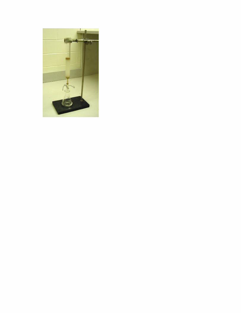
6.Using a piece of wire or stick to add a plug of cotton (glass wool) to the bottom of the column. There should be enough cotton that the sand and
silica will not fall out of the column. However, too much cotton or cotton packed too tightly will prevent the eluent from dripping at an acceptable rate. The tube is clamped to a retort stand and silica is added into the tube with the lower end tightly locked to prevent free flow if silica is dissolved while pouring into tube.
With the use of a spatula the silica was mixed withappropriate reagent E.g. dichloromethane, in a beaker before pouring and stirring to allow the silica to get into the tube, the pinch clamp is remove to allow solvent to drip into a clean flask.Taping on the side of the column with a rubber stopper or tubing to help the silica settle uniformly.
After silica has been properly packed in tube, the sample is loaded and elution with solvent/regents is done and fractions collected into separate tubes. Elution is done by placing the sample on thesilica gel packed in the tube , and elution carriedout by pouring and adding solvents, then allowed todrip.
Copyright © 2022 FDOKUMEN