Angiopoietin Like Protein 2 (ANGPTL2) Promotes Adipose Tissue Macrophage and T lymphocyte...
18
RESEARCH ARTICLE Angiopoietin Like Protein 2 (ANGPTL2) Promotes Adipose Tissue Macrophage and T lymphocyte Accumulation and Leads to Insulin Resistance Yusuke Sasaki 1,2 , Masayuki Ohta 1,2 , Dhruv Desai 1 , Jose-Luiz Figueiredo 1 , Mary C. Whelan 1 , Tomohiro Sugano 1,2 , Masaki Yamabi 1,2 , Wataru Yano 1,2 , Tyler Faits 1 , Katsumi Yabusaki 1,2 , Hengmin Zhang 1 , Andrew K. Mlynarchik 1 , Keisuke Inoue 1,2 , Ken Mizuno 1,2 , Masanori Aikawa 1 * 1 Center for Interdisciplinary Cardiovascular Sciences, Brigham and Women’s Hospital, Harvard Medical School, Boston, Massachusetts, United States of America, 2 Tokyo New Drug Research Laboratories, Kowa Company, Ltd., Tokyo, Japan * [email protected] Abstract Objectives Angiopoietin-like protein 2 (ANGPTL2), a recently identified pro-inflammatory cytokine, is mainly secreted from the adipose tissue. This study aimed to explore the role of ANGPTL2 in adipose tissue inflammation and macrophage activation in a mouse model of diabetes. Methodology/Principal Findings Adenovirus mediated lacZ (Ad-LacZ) or human ANGPTL2 (Ad-ANGPTL2) was delivered via tail vein in diabetic db/db mice. Ad-ANGPTL2 treatment for 2 weeks impaired both glu- cose tolerance and insulin sensitivity as compared to Ad-LacZ treatment. Ad-ANGPTL2 treatment significantly induced pro-inflammatory gene expression in white adipose tissue. We also isolated stromal vascular fraction from epididymal fat pad and analyzed adipose tis- sue macrophage and T lymphocyte populations by flow cytometry. Ad-ANGPTL2 treated mice had more adipose tissue macrophages (F4/80+CD11b+) and a larger M1 macrophage subpopulation (F4/80+CD11b+CD11c+). Moreover, Ad-ANGPTL2 treatment increased a CD8-positive T cell population in adipose tissue, which preceded increased macrophage accumulation. Consistent with our in vivo results, recombinant human ANGPTL2 protein treatment increased mRNA levels of pro-inflammatory gene products and production of TNF-α protein in the human macrophage-like cell line THP-1. Furthermore, Ad-ANGPTL2 treatment induced lipid accumulation and increased fatty acid synthesis, lipid metabolism related gene expression in mouse liver. PLOS ONE | DOI:10.1371/journal.pone.0131176 July 1, 2015 1 / 18 OPEN ACCESS Citation: Sasaki Y, Ohta M, Desai D, Figueiredo J-L, Whelan MC, Sugano T, et al. (2015) Angiopoietin Like Protein 2 (ANGPTL2) Promotes Adipose Tissue Macrophage and T lymphocyte Accumulation and Leads to Insulin Resistance. PLoS ONE 10(7): e0131176. doi:10.1371/journal.pone.0131176 Editor: Yoshihiro Fukumoto, Kurume University School of Medicine, JAPAN Received: February 11, 2015 Accepted: May 31, 2015 Published: July 1, 2015 Copyright: © 2015 Sasaki et al. This is an open access article distributed under the terms of the Creative Commons Attribution License, which permits unrestricted use, distribution, and reproduction in any medium, provided the original author and source are credited. Data Availability Statement: All relevant data are within the paper and its Supporting Information files. Funding: Kowa Company, Ltd., Japan. The funder provided support in the forms of salaries for authors YS, MO, TS, MY, WY, KY, KI and KM, and a research grant to MA. But the funder did not have any additional role in the study design, data collection and analysis, decision to publish, or preparation of the manuscript. The specific roles of these authors are articulated in the ‘author contributions’ section.
-
Upload
independent -
Category
Documents
-
view
7 -
download
0
Transcript of Angiopoietin Like Protein 2 (ANGPTL2) Promotes Adipose Tissue Macrophage and T lymphocyte...

RESEARCH ARTICLE
Angiopoietin Like Protein 2 (ANGPTL2)Promotes Adipose Tissue Macrophage andT lymphocyte Accumulation and Leads toInsulin ResistanceYusuke Sasaki1,2, Masayuki Ohta1,2, Dhruv Desai1, Jose-Luiz Figueiredo1, MaryC. Whelan1, Tomohiro Sugano1,2, Masaki Yamabi1,2, Wataru Yano1,2, Tyler Faits1,Katsumi Yabusaki1,2, Hengmin Zhang1, Andrew K. Mlynarchik1, Keisuke Inoue1,2,Ken Mizuno1,2, Masanori Aikawa1*
1 Center for Interdisciplinary Cardiovascular Sciences, Brigham andWomen’s Hospital, Harvard MedicalSchool, Boston, Massachusetts, United States of America, 2 Tokyo New Drug Research Laboratories, KowaCompany, Ltd., Tokyo, Japan
Abstract
Objectives
Angiopoietin-like protein 2 (ANGPTL2), a recently identified pro-inflammatory cytokine, is
mainly secreted from the adipose tissue. This study aimed to explore the role of ANGPTL2
in adipose tissue inflammation and macrophage activation in a mouse model of diabetes.
Methodology/Principal Findings
Adenovirus mediated lacZ (Ad-LacZ) or human ANGPTL2 (Ad-ANGPTL2) was delivered
via tail vein in diabetic db/db mice. Ad-ANGPTL2 treatment for 2 weeks impaired both glu-
cose tolerance and insulin sensitivity as compared to Ad-LacZ treatment. Ad-ANGPTL2
treatment significantly induced pro-inflammatory gene expression in white adipose tissue.
We also isolated stromal vascular fraction from epididymal fat pad and analyzed adipose tis-
sue macrophage and T lymphocyte populations by flow cytometry. Ad-ANGPTL2 treated
mice had more adipose tissue macrophages (F4/80+CD11b+) and a larger M1 macrophage
subpopulation (F4/80+CD11b+CD11c+). Moreover, Ad-ANGPTL2 treatment increased a
CD8-positive T cell population in adipose tissue, which preceded increased macrophage
accumulation. Consistent with our in vivo results, recombinant human ANGPTL2 protein
treatment increased mRNA levels of pro-inflammatory gene products and production of
TNF-α protein in the human macrophage-like cell line THP-1. Furthermore, Ad-ANGPTL2
treatment induced lipid accumulation and increased fatty acid synthesis, lipid metabolism
related gene expression in mouse liver.
PLOS ONE | DOI:10.1371/journal.pone.0131176 July 1, 2015 1 / 18
OPEN ACCESS
Citation: Sasaki Y, Ohta M, Desai D, Figueiredo J-L,Whelan MC, Sugano T, et al. (2015) Angiopoietin LikeProtein 2 (ANGPTL2) Promotes Adipose TissueMacrophage and T lymphocyte Accumulation andLeads to Insulin Resistance. PLoS ONE 10(7):e0131176. doi:10.1371/journal.pone.0131176
Editor: Yoshihiro Fukumoto, Kurume UniversitySchool of Medicine, JAPAN
Received: February 11, 2015
Accepted: May 31, 2015
Published: July 1, 2015
Copyright: © 2015 Sasaki et al. This is an openaccess article distributed under the terms of theCreative Commons Attribution License, which permitsunrestricted use, distribution, and reproduction in anymedium, provided the original author and source arecredited.
Data Availability Statement: All relevant data arewithin the paper and its Supporting Information files.
Funding: Kowa Company, Ltd., Japan. The funderprovided support in the forms of salaries for authorsYS, MO, TS, MY, WY, KY, KI and KM, and a researchgrant to MA. But the funder did not have anyadditional role in the study design, data collection andanalysis, decision to publish, or preparation of themanuscript. The specific roles of these authors arearticulated in the ‘author contributions’ section.

Conclusion
ANGPTL2 treatment promotes macrophage accumulation and activation. These results
suggest potential mechanisms for insulin resistance.
IntroductionObesity, a global burden, closely associates with several health problems such as type 2 diabetes,insulin resistance, atherosclerosis, dyslipidemia, cardiovascular disease, hypertension, and can-cer [1, 2]. These diseases increase mortality. Traditional views considered the adipose tissue asa mere energy storage organ. The recent knowledge, however, has recognized the adipose tissueas one of the key organs that regulate systemic metabolism [3–6]. Many macrophages accumu-late in adipose tissue in obese individuals and experimental animals. Activated macrophagesmay induce chronic low-grade inflammation in the adipose tissue, ultimately leading to sys-temic inflammation and insulin resistance [4–10].
Angiopoietin-like (ANGPTL) proteins are structurally similar to the angiopoietin familyproteins which contain both N-terminal coiled-coil domain and C-terminal fibrinogen likedomain. Unlike angiopoietins, ANGPTLs do not bind to either the angiopoietin receptor Tie 1or Tie2, indicating that ANGPTLs have different functions from those of angiopoietin proteins[11]. Some ANGPTLs may regulate metabolism. ANGPTL3 and ANGPTL4 regulate triglycer-ide metabolism by inhibiting lipoprotein lipase activity [12, 13]. ANGPTL6/Angiopoietin-related growth factor (AGF)-deficient mice develop severe obesity and insulin resistanceaccompanied by reduced energy expenditure relative to controls [14]. Likewise, ANGPTL2plays pivotal roles in various inflammatory diseases such as vascular inflammation, obesity,insulin resistance [15], cancer [16, 17] rheumatoid arthritis [18], and atherosclerosis [19, 20].Elevated serum ANGPTL2 levels positively associate with the development of type 2 diabetesin a general Japanese population [21]. Tabata et al. demonstrated that genetic deletion ofANGPTL2 in mice improved adiposity and systemic insulin resistance [15]. In contrast, Kita-zawa et al. reported that recombinant ANGPTL2 treatment improved insulin resistance in dia-betic db/db mice [22]. Thus, the function of ANGPTL2 on insulin resistance and type 2diabetes remains unclear. This study aimed to explore the role of ANGPTL2 in adipose tissueinflammation and macrophage activation in vivo.
Material and Methods
ReagentsThe following antibodies were used in the present study: FLAGM2 HRP antibody (A8692,Sigma Aldrich, St Louis, MO), Human ANGPTL2 monoclonal antibody (Clone 239829,MAB2084, R&D systems Inc., Minneapolis, MN), Beta actin antibody (Clone AC-15, NB600-501, Novus Biologicals, Littleton, CO). We used the following ELISA kits: Human ANGPTL2Assay ELISA kit (27745, Immuno-Biological Laboratories Co., Ltd, Fujioka, Japan), HumanTNF-alpha Quantikine ELISA Kit (DTA00C, R&D systems, Inc., Minneapolis, MN). Phorbol12-myristate 13-acetate (PMA, P1585-1MG) was purchased from Sigma Aldrich (St Louis, MO).
Cell culturesHuman monocytoid cell line THP-1 (TIB-202), murine macrophage like cell line RAW264.7(TIB-71), murine hapatoma cell line Hepa1c1c7 (CRL-2026), murine embryo fibroblast cellline
ANGPTL2 and Macrophage Inflammation
PLOS ONE | DOI:10.1371/journal.pone.0131176 July 1, 2015 2 / 18
Competing Interests: YS, MO, TS, MY, WY, KY, KIand KM are employees of Kowa Company, Ltd.,which funded this study. There are no patents,products in development or marketed products todeclare. This does not alter the authors' adherence toall the PLOS ONE policies on sharing data andmaterials.

3T3-L1 (CL-173), murine muscle myoblast cell line C2C12 (CRL-1722), and human hepatocel-lular carcinoma cell line HepG2 (HB-8065) were purchased from American Type Culture Col-lection (ATCC, Manassas, VA). The human umbilical vein endothelial cells (HUVECs:CC2517) were purchased from Lonza and maintained in EGM-2 bullet kit (CC-3162, Lonza,Walkersville, MD). THP-1 cells were maintained in RPMI1640 (12-702F, Lonza, Walkersville,MD) supplemented with 10% fetal bovine serum (FBS, Life Technologies, Grand Island, NY),penicillin and streptomycin (Corning, NY) at 37˚C in a humidified atmosphere of 5% CO2.THP-1 monocytes were differentiated by incubation with PMA for 24 h at 37°C. RAW264.7and Hepa1c1c7 cells were maintained in Dulbecco’s Modified Eagle’s Medium (DMEM, 12-604F, Lonza, Walkersville, MD) supplemented with10% FBS, penicillin and streptomycin at37˚C in a humidified atmosphere of 5% CO2. 3T3L1 cells were maintained with DMEM sup-plemented with10% FBS, penicillin and streptomycin at 37˚C in a humidified atmosphere of5% CO2. For adipocyte differentiation, the cells were grown to confluent for 2 days and theninduced with differentiation medium with 1μM dexamethasone (D4902, Sigma Aldrich, StLouis, MO) 0.5 mM isobutylmethylxanthine (IBMX, I-7018, Sigma Aldrich, St Louis, MO),and 10 μg/mL insulin (I-9278, Sigma Aldrich, St Louis, MO). After 2 days, the cells were main-tained in DMEM supplemented with10% FBS and 10 μg/mL insulin. Medium was changedevery 2 days. C2C12 cells were maintained with DMEM supplemented with10% FBS, penicillinand streptomycin at 37˚C in a humidified atmosphere of 5% CO2. To induce myogenic differ-entiation, when about 80% cell confluence was attained, the cells were induced with themedium containing 2% horse serum, penicillin and streptomycin. Medium was changed every2 days. HepG2 cells cells were maintained with DMEM supplemented with10% FBS, penicillinand streptomycin at 37˚C in a humidified atmosphere of 5% CO2. HUVECs were used foreach experiment before 6 passages. Human 293f (R790-07) and 293A (R705-07) cells were pur-chased from Life Technologies (Grand Island, NY) and maintained according to the manufac-turer's instructions.
Construction of adenovirus vector for expression of Angptl2 andproduction of adenovirusThe adenovirus vector that encodes FLAG-ANGPTL2 was generated by using ViraPower Ade-noviral Expression System (K4930-00, Life Technologies, Grand Island, NY).
The coding sequence (CDS) of ANGPTL2 was obtained from Genecopoeia (Cat:GC-U0900, Rockville, MD). To add FLAG tag sequences at C-terminal of ANGPTL2, the fol-lowing primer set (Forward primer: 5’GACTACAAAGACGATGACGACAAGTAACTCGAGTGCCCGCAACCCAGCTT -3 and Reverse primer: 5’CTTGTCGTCATCGTCTTTGTAGTCGTGGAAGGTGTTGGGGTTCGGTCGG-3) was used and performed FLAG tag insertionby using the site-directed mutagenesis method. FLAG-ANGPTL2 adenovirus expression clonewas prepared using pAd/CMV/ V5-DEST Gateway vector (V493-20, Invitrogen, Life Technol-ogies, Grand Island, NY). The recombinant virus (Ad-LacZ or Ad-ANTPGL2) was packagedand amplified in 293A cells according to the manufacturer's instructions. The recombinantvirus was packaged and amplified in 293A cells and purified by Fast Trap Adenovirus Purifica-tion and Concentration Kit (FTAV00003, EMDMillipore Corporation, Billerica, MA). Thevirus titer was determined by using Adeno-X Rapid Titer Kit (632250, Clontech Laboratories,Inc., Mountain View, CA).
Preparation of recombinant human ANGPTL2 proteinExpression and purification method of human ANGPTL2 protein was conducted as followedand slightly modified previous reports [22, 23]. Briefly, C-terminal FLAG ANGPTL2
ANGPTL2 and Macrophage Inflammation
PLOS ONE | DOI:10.1371/journal.pone.0131176 July 1, 2015 3 / 18

expression vector was obtained from Origene (Myc-DDK-tagged-Human angiopoietin-like 2,RC201917, Rockville, MD). C-FLAG ANGPTL2 expression vector was transfected with Free-Style max reagent (16447–100, Life Technologies, Grand Island, NY) into 293f cells. After 72hour, conditioned medium was collected and passed through a 0.2 μm pore size filter (4612,PALL Life Sciences, Ann Arbor, MI). ANTI-FLAGM2 Affinity Gel (A2220, Sigma Aldrich, StLouis, MO) was added into the filtered conditioned media. FLAGM2 Gel was washed withTris-Buffered Saline (TBS) and elute with 165 mg/mL FLAG peptide (F3290, Sigma Aldrich, StLouis, MO) or Gly-HCl (pH3.5) buffer. For Gly-HCl elution, we immediately added 10 x TBSto neutralize the solution. Protein concentration was determined by BCA protein assay kit(23225, Thermo Scientific, Rockford, MA). Protease inhibitor cocktail (P8340, Sigma Aldrich,St Louis, MO) was added throughout the procedures.
Mouse study designAll experiments were conducted using 12-wk-old male mice. We obtained 10-wk-old C57BL6Jmice and diabetic db/db mice from Jackson Laboratories (Bar Harbor, ME). The mice weremaintained on a 12-h light, 12-h dark cycle in normal cages with food and water ad libitum.Lean mice or diabetic db/db were received 5 x10^9 plaque forming units (p.f.u.) of adenovirussolution (Ad-LacZ or Ad-ANGPTL2) via tail vein at 12 week old. We monitored body weighton Day 0 (Adenovirus Injection), 3, 7, 10 and 14. Food intake and blood glucose level weremonitored once a week. Study design was described in S1 Fig. Mouse studies were conductedin accordance with federal guidelines. The Institutional Animal Care and Use Committee(Beth Israel Deaconess Medical Center, Boston, protocol #041–2012) approved all studyprotocols.
Glucose and insulin tolerance testsGlucose tolerance test was performed on Day 7 or Day14 after adenovirus injection. Mice werefasted overnight then given glucose (1 g/kg body weight) by oral administration. For insulintolerance test, mice were fasted 4 hr then given insulin solution (2 Unit/kg, Novolin, Novo Nor-disk, Plainsboro Township, NJ) via i.p. injection. The sterilized mouse tail was snipped using ascalpel and gently squeezed to obtain tail blood. Blood glucose levels were measured using gluc-ometer (One Touch Ultra, Lifescan Inc., Milpitas, CA) at indicated time points (time 0, 15, 30,60 and 120) after glucose or insulin administration.
Stromal vascular cell isolation and flow cytometry analysisIsolation of stromal vascular fraction (SVF) from mice epididymal fat pad was performed aspreviously described [24, 25]. Epididymal fat pads of db/db mice were weighed, rinsed 3 timeswith phosphate buffered saline (PBS), and then minced in the MACS buffer (PBS with bovineserum albumin, EDTA, and 0.09% Azide, 130-091-221, Miltenyi Biotec Inc., San Diego, CA).Tissue suspensions were incubated with 1 mg/mL collagenase Type II (C6885, Sigma Aldrich,St Louis, MO) and 0.2 mg/mL DNase I (DN25, Sigma Aldrich, St Louis, MO) at 37°C for30 min. with shaking (90 rpm). Cell suspension were filtered through 100 μm cell strainer(REF352360, BD Bioscience, San Jose, CA) and then centrifuged at 200 x g for 5 min to sepa-rate floating adipocytes from the SVF pellets. The SVF pellets were incubated with RBC lysisbuffer (420301, Biolegend, San Diego, CA) for 5 min before centrifuge (300 x g, 5 min) andresuspended in MACS buffer. Stromal vascular fraction cells were incubated with Fc Block(Clone2.4G2, 553142 BD Biosciences, San Jose, CA) for 20 min at 4°C. Cells were stained withthe following conjugated antibodies (30 min, 4°C in the dark): FITC anti-mouse F4/80, PE/Cy5anti-mouse CD11b, APC/Cy7 anti-mouse CD11c, PE anti-mouse CD3, FITC anti-mouse CD4,
ANGPTL2 and Macrophage Inflammation
PLOS ONE | DOI:10.1371/journal.pone.0131176 July 1, 2015 4 / 18

and APC anti-mouse CD8a (all were purchased from Biolegend, San Diego, CA). After stain-ing, cells were gently washed twice and resuspended in the fresh MACS buffer. Stained cellswere analyzed by FACS (BD FACS Aria II, BD Biosciences, San Jose, CA). Adipose tissue mac-rophages and pro-inflammatory M1 macrophages were identified as F4/80+ CD11b+ cells, andF4/80+ CD11b+ CD11c+, respectively.
Analyses of lipid profile, liver triglyceride, insulin and adipokine levelsPlasma total cholesterol and triglyceride levels were measured by Cholesterol E-test kit (Wako,Japan), and LabAssay Triglyceride test (Wako, Japan), respectively. The liver triglyceride assaywas performed as previously described [26]. 200 mg portion of the liver was extracted with10 ml of chloroform–methanol 2:1 in a Polytron (Kinematica GmbH, Luzern, Switzerland). A0.2 ml aliquot of the lower phase was taken, evaporated and dissolved in isopropanol. Livercholesterol and triglyceride levels were measured by Cholesterol E-test kit (Wako, Japan), andLabAssay Triglyceride test (Wako, Japan), respectively. Plasma adiponectin and insulin levelswere evaluated by Mouse Adiponectin/Acrp30 Quantikine ELISA Kit (MRP300, R&D systems,Minneapolis, MN), and Rat/Mouse Insulin ELISA (EZRMI-13K, EMDMillipore Corporation,Billerica, MA), respectively.
Histological analysesEpididymal fat pads were fixed with 4% paraformaldehyde and embedded in paraffin. Tissueswere sectioned serially (6 μm) and stained with hematoxylin and eosin (H&E) for general mor-phology. Liver samples were embedded in OCT compound (Sakura Finetek U.S.A., Inc, Torrance,CA). We built software tool using OpenCV (open-source image analysis library, GarageWillow,Menlopark, CA) to be able to calculate the adipocyte size accurately. Cross sectional images ofadipose tissues were processed through several image filters consisting of Canny edge filter, binar-izing filter and labeling filter to extract each adipocyte in the images. The adipocyte areas obtainedas pixel numbers were converted to physical area in um^2. For oil Red O staining, slides wererinsed with distilled water, and placed in absolute propylene glycol for 2 min. Then, slides werestained with 5 mg/mL oil red O (3125–12, EMDMillipore Corporation, Billerica, MA) solutionfor 90 min. After staining, slides were differentiated with 85% propylene glycol solution and rinsewith distilled water. Slides were stained in Gill’s hematoxylin solution. After rinse with distilledwater for several time and apply aqueous mounting solution and covered with cover glass. Stainedarea was quantified with Hue Saturation Value Analysis Program as described previously [27].
Gene expression analysisTotal RNA extracted from cells with TRIzol (Life technologies, Grand Island, NY) underwentcDNA synthesis with High-Capacity cDNA Reverse Transcription Kit (Life technologies,Grand Island, NY). Real-time quantitative PCR employed TaqMan gene expression assays(Life technologies, Grand Island, NY) and ABI 7900HT sequence detection system (Life Tech-nologies, Grand Island, NY). TaqMan probes which used in this study were summarized in theS1 Table (mouse) and S2 Table (human).
Akt phosphorylation assayAdenovirus which expressed with LacZ or ANGPTL2 was infected to differentiated 3T3L1 adi-pocytes (on Day7) differentiated C2C12 myotubes (on Day4), and HepG2 when the cellsreached confluence. For adenovirus transduction in 3T3L1 adipocytes, we used density-basedseparation followed by re-plating of enriched adipocytes in monolayer (DREAM) method
ANGPTL2 and Macrophage Inflammation
PLOS ONE | DOI:10.1371/journal.pone.0131176 July 1, 2015 5 / 18
previously reported [28]. Adenovirus particles were infected with AdenoMag (AM71000, OZbioscience, San Diego, CA) kit according to the manufacturer's instructions. The cells wereapplied serum starvation overnight and treated with medium with insulin solution (10, 100,and 1000 nM) for 15 min. The cells were then washed with ice-cold phosphate buffered saline(PBS) twice, and added lysis buffer (9803, Cell Signaling Technology, Danvers, MA) with pro-tease inhibitor and Phostop phosphatase inhibitor (Roche, Indianapolis, IN). For western blot-ting analysis, Proteins were resolved by 10% sodium dodecyl sulfate-polyacrylamide gelelectrophoresis (SDS-PAGE) and electrotransferred to nitrocellulose membranes (Life Tech-nologies, Grand Island, NY). After blocking with 2% non-fat milk, the membranes were probedwith antibodies for pSer473-PKB and total PKB (4060S and 4691S, Cell Signaling Technology,MA, USA) and the proteins were visualized by SuperSignal West Pico Chemiluminescent Sub-strate (34087, Thermo Fisher Scientific Inc., Rockford, IL). For phospho and total Akt ELISA,PathScan Phospho-Akt1 (Ser473) and Total Akt1 Sandwich ELISA Antibody Pair (#7143 and#7142, Cell Signaling Technology, Danvers, MA) according to the manufacturer's instructions.
Statistical analysisStatistical analysis was performed with GraphPad Prism 5 (GraphPad Software, La Jolla, CA).The data were analyzed using unpaired t-test or Mann-Whitney test for two group comparison.Differences involving multiple groups were first tested by one-way ANOVA, followed by post-hoc analysis with Dunnett’s test adjusting for multiple comparison when comparing multiplegroups to a control group. Differences with p< 0.05 were considered statistically significant.All data are presented as mean ± standard error of mean (SEM).
Results
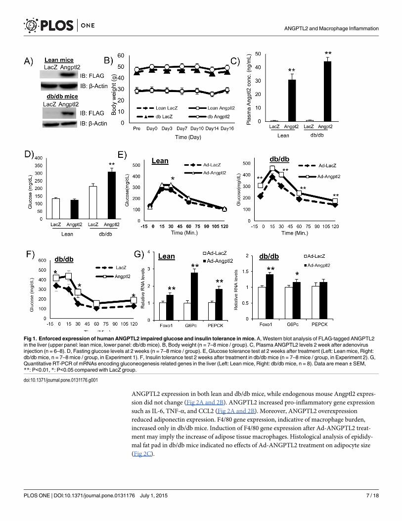
ANGPTL2 impaired glucose tolerance and insulin sensitivity in db/dbmiceWe prepared adenovirus particles expressing FLAG fusion human ANGPTL2 protein (Ad-ANGPTL2) for in vivo gain-of-function experiments. We used db/db mice, a diabetic mutantstrain, as a model of spontaneous type 2 diabetes. We injected Ad-ANGTL2 in db/db mice viatail vein. FLAG-ANGPTL2 protein expression was confirmed in the liver on Day14 (Fig 1A).Ad-ANGPTL2 treatment did not affect body weight (Fig 1B and S1B Fig). Ad-ANGPTL2 treat-ment increased circulating ANGPTL2 levels in both lean and db/db mice (Fig 1C and S1C Fig).Fasting glucose level increased in Ad-ANGPTL2 treated db/db mice on Day 14, however, therewas no difference in lean mice (Fig 1D). There was no significant difference on plasma insulin,adiponectin, total cholesterol (except for lean mice), and triglyceride levels (S1D, S1E, S1F andS1G Fig). Furthermore, enforced expression of ANGPTL2 had minimal effects on food intake(S1H Fig). We performed glucose tolerance test on Day14. Ad-ANGPTL2 treatment impairedglucose tolerance in both lean and db/db mice (S1E Fig). We also performed insulin tolerancetest using db/db mice on Day 14 (S1A Fig, experiment 2). Two week of Ad-ANGPTL2 treat-ment exacerbated insulin sensitivity (Fig 1F). After tissue harvesting, gluconeogenesis relatedgene expression was analyzed. Angptl2 overexpression enhanced Foxo1, G6Pc and Pepckexpression in both lean and obese mice (Fig 1F). These results indicate that Angptl2 overex-pression worsen both glucose tolerance and insulin sensitivity.
ANGPTL2 induced pro-inflammatory gene Expression in adipose tissueWe then assessed the effects of enforced expression of human ANGPTL2 in mouse adipose tis-sue. Real time PCR analysis showed that Ad-ANGPTL2 treatment enhanced human
ANGPTL2 and Macrophage Inflammation
PLOS ONE | DOI:10.1371/journal.pone.0131176 July 1, 2015 6 / 18
ANGPTL2 expression in both lean and db/db mice, while endogenous mouse Angptl2 expres-sion did not change (Fig 2A and 2B). ANGPTL2 increased pro-inflammatory gene expressionsuch as IL-6, TNF-α, and CCL2 (Fig 2A and 2B). Moreover, ANGPTL2 overexpressionreduced adiponectin expression. F4/80 gene expression, indicative of macrophage burden,increased only in db/db mice. Induction of F4/80 gene expression after Ad-ANGPTL2 treat-ment may imply the increase of adipose tissue macrophages. Histological analysis of epididy-mal fat pad in db/db mice indicated no effects of Ad-ANGPTL2 treatment on adipocyte size(Fig 2C).
Fig 1. Enforced expression of human ANGPTL2 impaired glucose and insulin tolerance in mice. A, Western blot analysis of FLAG-tagged ANGPTL2in the liver (upper panel: lean mice, lower panel: db/db mice). B, Body weight (n = 7–8 mice / group). C, Plasma ANGPTL2 levels 2 week after adenovirusinjection (n = 6–8). D, Fasting glucose levels at 2 weeks (n = 7–8 mice / group). E, Glucose tolerance test at 2 weeks after treatment (Left: Lean mice, Right:db/db mice, n = 7–8 mice / group, in Experiment 1). F, Insulin tolerance test 2 weeks after treatment in db/db mice (n = 7–8 mice / group, in Experiment 2). G,Quantitative RT-PCR of mRNAs encoding gluconeogenesis related genes in the liver (Left: Lean mice, Right: db/db mice, n = 8). Data are mean ± SEM,**: P<0.01, *: P<0.05 compared with LacZ group.
doi:10.1371/journal.pone.0131176.g001
ANGPTL2 and Macrophage Inflammation
PLOS ONE | DOI:10.1371/journal.pone.0131176 July 1, 2015 7 / 18

ANGPTL2 increased adipose tissue macrophages and pro-inflammatoryM1 macrophagesOur findings in Fig 2 indicate that ANGPTL2 may increase macrophage accumulation andpro-inflammatory responses in the adipose tissue. To further address this hypothesis, westained stromal vascular fractions isolated from epididymal fat pad with F4/80, CD11b andCD11c antibodies. We quantified F4/80 and CD11b double-positive macrophages and pro-inflammatory M1 macrophages (F4/80+CD11b+CD11c+) by fluorescence-activated cell sort-ing (FACS) analysis. Ad-ANGPTL2 treatment increased both adipose tissue macrophages(F4/80+CD11b+) and the pro-inflammatory M1 macrophage population (F4/80+CD11b+CD11c+) after 2-week treatment (Fig 3A, 3B and 3C), which agreed with gene expression pro-file (Fig 2A and 2B). The results indicate that ANGPTL2 may induce chronic inflammation inadipose tissue.
Fig 2. Enforced expression of human ANGPTL2 induced inflammation in adipose tissue. A, Quantitative RT-PCR of mRNAs encoding adipokines andpro-inflammatory genes in epididymal adipose tissue (lean mice, n = 8) B, Quantitative RT-PCR of mRNAs encoding adipokines and pro-inflammatory genesin epididymal adipose tissue (db/db mice, n = 5–8). C, Representative images of adipocytes after H&E staining and quantification of adipocyte size inepididymal fat (db/db mice, n = 7, Scale bar: 200 μm). Data are mean ± SEM, **: P<0.01, *: P<0.05 compared with LacZ group.
doi:10.1371/journal.pone.0131176.g002
ANGPTL2 and Macrophage Inflammation
PLOS ONE | DOI:10.1371/journal.pone.0131176 July 1, 2015 8 / 18

Increased accumulation of CD8 positive T cells by ANGPTL2 precededmacrophage accumulation in adipose tissueT lymphocytes interact with macrophages and regulate the inflammatory response. Nishimuraet al. revealed that CD8 positive T cells play a pivotal role on macrophage recruitment and adi-pose tissue inflammation [29]. To assess how ANGPTL2 participates in this process, we
Fig 3. Enforced expression of human ANGPTL2 increased adipose tissuemacrophages and promoted M1macrophage polarization in adiposetissue from db/dbmice. A, Stromal vascular fraction (SVF) was isolated from the epididymal fat pad then stained with F4/80, CD11b, and CD11c antibodiesand analyzed by FACS. B, Adipose tissue macrophage number was determined as F4/80+ and CD11b+ fraction and determined by FACS (n = 7–8). Dataare mean ± SEM, **: P<0.01, *: P<0.05 compared with LacZ group.
doi:10.1371/journal.pone.0131176.g003
ANGPTL2 and Macrophage Inflammation
PLOS ONE | DOI:10.1371/journal.pone.0131176 July 1, 2015 9 / 18
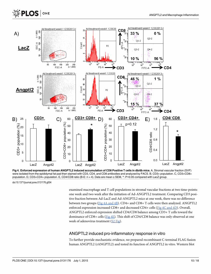
examined macrophage and T cell populations in stromal vascular fractions at two time-points:one week and two week after the initiation of Ad-ANGPTL2 treatment. Comparing CD3 posi-tive fraction between Ad-LacZ and Ad-ANGPTL2 mice at one week, there was no differencebetween two groups (Fig 4A and 4B). CD4+ and CD8+ T cells were then analyzed. ANGPTL2enforced expression increased CD8+ and decreased CD4+ cells (Fig 4C and 4D). Overall,ANGPTL2 enforced expression shifted CD4/CD8 balance among CD3+ T cells toward thedominance of CD8+ cells (Fig 4E). This shift of CD4/CD8 balance was only observed at oneweek of adenovirus treatment (S2 Fig).
ANGPTL2 induced pro-inflammatory response in vitroTo further provide mechanistic evidence, we prepared recombinant C-terminal FLAG fusionhuman ANGPTL2 (rANGPTL2) and tested its function of ANGPTL2 in vitro. Western blot
Fig 4. Enforced expression of human ANGPTL2 induced accumulation of CD8 Positive T cells in db/dbmice. A, Stromal vascular fraction (SVF)were isolated from the epididymal fat pad then stained with CD3, CD4, and CD8 antibodies and analyzed by FACS. B, CD3+ population. C, CD3+CD8+population. D, CD3+CD4+ population. E, CD4/CD8 ratio (B-E: n = 4). Data are mean ± SEM, *: P<0.05 compared with LacZ group.
doi:10.1371/journal.pone.0131176.g004
ANGPTL2 and Macrophage Inflammation
PLOS ONE | DOI:10.1371/journal.pone.0131176 July 1, 2015 10 / 18

analysis detected FLAG-rANGPTL2 protein (Fig 5A). A band shift in non-reduced conditionsuggests that rANGPTL2 formed multimers consistent with a previous report [23]. rANGPTL2increased TNF-α production in the human macrophage-like cell line THP-1 (Fig 5B) in a con-centration dependent manner. rANGPTL2 treatment increased the expression of pro-inflam-matory genes, typical of M1 macrophages, such as TNF-α, IL-1β, CCL2, and NF-κB (p105/p50) in THP-1 cells (Fig 5C). Interestingly, rANGPTL2 treatment also increased humanANGPTL2 expression, probably in an autocrine manner (Fig 5C). In the previous reports,ANGPTL2 activates migration and inflammatory response in EC [15, 19, 20]. We also observed
Fig 5. ANGPTL2 induced pro-Inflammatory responses in vitro. A, Western blotting analysis of FLAG fusion ANGPTL2 protein with anti-FLAG and anti-human Angptl2 antibody (Left: with 2-mercaptoethanol (2-ME), Right: without 2-ME). B, TNF-α concentration in rANGPTL2-treated THP-1 conditioned media(72 hr treatment, n = 3). C, Quantitative RT-PCR of mRNAs encoding Angptl2 and pro-inflammatory related genes in THP-1 cells (24 hr treatment, n = 3). D,Quantitative RT-PCR of mRNAs encoding M2 macrophage markers in THP-1 cells (24 hr treatment, n = 3). Data are mean ± SEM, **: P<0.01, *: P<0.05compared with BSA group.
doi:10.1371/journal.pone.0131176.g005
ANGPTL2 and Macrophage Inflammation
PLOS ONE | DOI:10.1371/journal.pone.0131176 July 1, 2015 11 / 18

rANGPTL2 treatment induced cell adhesion and inflammatory response in human umbilicalvein endothelial cells (HUVEC) (S3 Fig). Collectively, rANGPTL2 induced inflammatoryresponse in macrophage-like THP-1 and endothelial cells. ANGPTL2 induced pro-inflamma-tory response in THP-1 cells consistent with an increase in larger M1 macrophage subpopula-tion in adipose tissue in vivo.
ANGPTL2 increased lipid accumulation in mouse liverWhile liver weight did not differ between Ad-ANGPTL2 and Ad-LacZ, the liver of Ad-ANGPTL2 treated mice seemed more whitish than did Ad-LacZ treated mice (Fig 6A). Ad-ANGPTL2 increased hepatic triglyceride content in db/db mice as compared to Ad-LacZ (Fig6B). Oil red O staining showed that the liver of Ad-ANGPTL2 treated mice tended to havegreater stained area (p = 0.085, Fig 6C and 6D). Gene expression analysis was assessed in theliver. Human ANGPTL2 gene expression was markedly induced (Fig 6E and 6F). Ad-ANGPTL2 treatment increased the expression of genes related to fatty acid synthesis andmetabolism (PPARα, Acacb, Acly, and Fasn) in both lean and db/db mice (Fig 6E and 6F).
Fig 6. ANGPTL2 enhanced hepatic lipid accumulation in mice. A, Representative images of the liver (Left: LacZ, Right: Angptl2, db/db mice). B, Livertriglyceride levels (Left: Lean mice, Right: db/db mice, n = 7–8 animals per group). C, Representative images of oil red O staining (Left: LacZ, Right: Angptl2,db/db mice). D, Oil red O staining area (n = 7–8, db/db mice). E, Quantitative RT-PCR of mRNAs encoding genes related to fatty acid metabolism in the liverof lean mice (n = 8) F, Quantitative RT-PCR of mRNAs encoding genes related to fatty acid metabolism in the liver of db/db mice (n = 7–8). Data aremean ± SEM, **: P<0.01, *: P<0.05 compared with LacZ group.
doi:10.1371/journal.pone.0131176.g006
ANGPTL2 and Macrophage Inflammation
PLOS ONE | DOI:10.1371/journal.pone.0131176 July 1, 2015 12 / 18

Although oil red O stain results did not significantly differ, Ad-ANGPTL2 treatment increasedfatty acid synthesis, metabolism related gene expression, and lipid accumulation in the liverand may induce fatty liver in mice.
DiscussionThe present study demonstrates that adenovirus-mediated human ANGPTL2 expression inlean and diabetic db/db mice induced adipose tissue inflammation and reduced glucose toler-ance and insulin sensitivity. Enforced expression of ANGPTL2 promoted macrophage accu-mulation and pro-inflammatory M1 polarization in the adipose tissue. ANGPTL2 treatmentalso enriched CD8+ T cell population at one week of Ad-ANGPTL2 treatment prior toincreased macrophage accumulation at two weeks. rANGPTL2 induced pro-inflammatorygene expression and TNF-α protein production in the macrophage-like cell line THP-1. Thesefindings indicate that ANGPTL2 may play important roles in adipose tissue inflammation andinsulin resistance via accumulation and pro-inflammatory activation of macrophages.
A previous report by Tabata et al. demonstrated that ANGPTL2-deficient mice amelioratedadipose tissue inflammation and systemic insulin resistance [15]. The same article also demon-strated that adipose tissue-specific Angptl2 overexpression induced fat inflammation and insu-lin resistance. A more recent study, however, reported opposing effects that four week ofrecombinant ANGPTL2 treatment improved insulin resistance in db/db mice [22]. Thus, thefunctional role of ANGPTL2 on insulin resistance remains obscure. To address this issue, wecarried out adenovirus-mediated ANGPTL2 expression in lean and diabetic db/db mice, focus-ing on the function of ANGPTL2 in macrophages. Our results indicated that ANGPTL2enforced expression clearly induced adipose tissue inflammation and exacerbated insulin sensi-tivity, which is generally consistent with the results by Tabata et al [15]. A study by Kitazawaet al. and our study used db/db mice as a model of spontaneous type 2 diabetes, however, lead-ing to different outcomes. Two different conditions in two studies—ages of mice and plasmalevels of ANGPTL2—may have caused this discrepancy. The previous and our studies used 8and 12 week old of db/db mice, respectively. Severity of insulin resistance should vary depend-ing on the age of db/db mice, which may in turn affect the function of ANGPTL2 because adi-pocytes may expose more pro-inflammatory environment and produce more adipokines,leading to more insulin resistant state under more obese condition.
In our study, plasma concentrations of ANGPTL2 ranged from 15–45 ng/mL (Fig 1C andS1C Fig). But, plasma ANGPTL2 levels in the previous report exceeded 100 ng/mL after2-hour administration, which swiftly decreased afterwards [22]. Integrin α5β1 and LILRB2proteins were reported as ANGPTL2 receptors [15, 30]. Deng et al. revealed that ANGPTL2forms high-molecular-weight forms. The same study reported that ligand multimerization isrequired for activation of LILRB2 signaling and that mouse blood contain multimerizedANGPTL2 [31]. Exceeded 100 ng/mL of ANGPTL2 levels were observed in the previous reportcondition and it was over two folds as much as our condition. It may have contained moremultimerized ANGPTL2, causing the different outcomes. In addition, in our condition, plasmaANGPTL2 level was maintained at Day14 after adenovirus injection (S3 Fig), and mice wereconstantly exposed by ANGPTL2 protein since Ad-ANGPTL2 had begun hANGPTL2 proteinexpression. We speculate that this persistent stimulation of ANGPTL2 protein induced chronicinflammation and led worse insulin resistance. The functional difference between monomerand multimerized ANGPTL2 is not still fully understood and further investigations will berequired.
We assessed that circadian changes in human and mouse Angptl2 in the liver and epididy-mal white adipose tissue in both lean and db/db mice. Ad-hANGPTL2 treatment markedly
ANGPTL2 and Macrophage Inflammation
PLOS ONE | DOI:10.1371/journal.pone.0131176 July 1, 2015 13 / 18

induced expression levels of human ANGPTL2 and tended to decrease mouse Angptl2 expres-sion in both the liver and epididymal white adipose tissue (S4, S5, S6 and S7 Figs). Human andmouse Angptl2 sequences were highly conserved (over 95%) and they might exert similar effectsin the mouse bodies. Thus, elevated plasma human ANGPTL2 levels might have suppressedendogenous mouse Angptl2 expression through a negative feedback loop in the liver and epidid-ymal white adipose tissue. Additionally, we measured mRNA levels of the circadian genes Clock,Cry1 and Bmal1. As suggested by the reviewer, Angptl2 gene is known as the circadian gene [22].Kadomatsu et al. reported that human ANGPTL2 gene was significantly induced by Clock andBmal1 and markedly attenuated by Cry1 co-expression [32]. Therefore, to respond the reviewer’ssuggestion, we examined whether the enforced expression of hANGPTL2 affects these circadiangene expression. Our results suggested that increased hANGPTL2 protein levels had minimaleffects on the expression of circadian genes (S4, S5, S6 and S7 Figs). Therefore, we concluded thatincreased human ANGPTL2 levels induced adipose tissue inflammation and led to insulin resis-tance, not by affecting the circadian gene expression.
ANGPTL2 is known as a pro-inflammatory factor in keratinocytes, adipose tissue and EC[15, 19, 20]. ANGPTL2 promotes chemotaxis of monocytes and leads to macrophage accumu-lation [15]. Our data also suggest that ANGPTL2 more directly activates pro-inflammatoryresponses in macrophages (Fig 5B and 5C). In addition, ANGPTL2 expression was induced inan autocrine manner in THP-1 cells (Fig 5C). A similar phenomenon was observed in HUVEC(S8 Fig). Consistent with previous reports, macrophages and EC also produced ANGPTL2(Fig 5C and S8 Fig) [19, 20, 33]. Furthermore, we assessed the effects of enforced expression ofAd-ANGPTL2 and Ad-LacZ on Akt phosphorylation in 3T3L1 adipocytes, C2C12 murinemyoblasts, and HepG2 hepatocellular carcinoma cells, which did not effect on Akt phosphory-lation (S9 Fig). This may indicate that ANGPTL2 does not exert direct effects on the insulinsignaling pathway. Instead, ANGPTL2 may indirectly induce insulin resistance by enhancinginflammation.
Inflammation in the adipose tissue is an important factor in the development of insulinresistance. Macrophages accumulate in the white adipose tissue in obesity, leading to insulinresistance [4–10]. Recent studies have implicated that some lymphocytes are closely related tothis process of macrophage accumulation in white adipose tissue. Nishimura et al. showed thatinfiltration of CD8-positive T cells precedes and contributes to macrophage accumulation dur-ing the development of obesity [29]. When we found that Ad-ANGPTL2 treatment increasedadipose tissue macrophages and promoted pro-inflammatory M1 macrophage polarization, wespeculated that enforced expression of ANGPTL2 induces CD8-positive T cell infiltration inthe white adipose tissue before increased macrophage accumulation occurs. Ad-ANGPTL2treatment for one week substantially increased CD8-positive T cell population without af-fecting total CD3-positive T cells (Fig 4A, 4B and 4C). This CD8-positive T cell enrichmenthad disappeared at 2 weeks when macrophage accumulation and M1 polarization increased(S2 Fig and Fig 3). Further investigations are required to determine how ANGPTL2 promotesCD8-positive cell accumulation and subsequent macrophage accumulation and activation inthe adipose tissue.
We also observed that Ad-ANGPTL2 treatment induced lipid accumulation in the liver(Fig 6B), increased the expression of fatty acid synthesis and lipid metabolism related genes(Fig 6E and 6F). The function of ANGPTL2 on lipid metabolism remains unclear, but Angptl2deficient mice showed less fat accumulation in the liver compared to wild type mice [15].Our results were consistent with the results in these Angptl2 deficient mice. ANGPTL3 andANGPTL4, other ANGPTL family proteins, are known to be cleaved into the N-terminalcoiled-coil domain and the C-terminal fibrinogen like domain. The truncated N-terminalcoiled-coil domain from ANGPTL3 and ANGPTL4 can affect lipid metabolism [34, 35].
ANGPTL2 and Macrophage Inflammation
PLOS ONE | DOI:10.1371/journal.pone.0131176 July 1, 2015 14 / 18

Likewise, ANGPTL2 is also processed into the N-terminal coiled-coil domain and the C-termi-nal fibrinogen like domain [36]. Although the function of these truncated forms of ANGPTL2were not elucidated, it might have a potency to regulate lipid metabolism.
In conclusion, we propose that ANGPTL2 promotes adipose tissue inflammation as gaugedby accumulation and activation of macrophages and T lymphocytes, leading to systemic insulinresistance. Understanding the underlying mechanisms by which ANGPTL2 induces adiposetissue inflammation would provide new insight into therapeutic strategies for cardiometabolicdiseases caused by insulin resistance.
Supporting InformationS1 Fig. Ad-ANGPTL2 2 week treatment in mice. A, Scheme of experimental procedurewhich showing time line of adenovirus injection, glucose/ insulin tolerance test, and analysis inthis study. B, Body weight (n = 4 animals per group, in experiment3). C, Plasma ANGPTL2 lev-els at 1 and 2 week after adenovirus injection (Left: 1 week, Right: 2 week, n = 7–8). D, PlasmaInsulin level at 2 week after adenovirus injection (Left: Lean mice, Right: db/db mice, n = 7–8animals per group). E, Plasma adiponectin level at 2 week after adenovirus injection (Left: Leanmice, Right: db/db mice, n = 7–8 animals per group). F, Food intake (Left: Lean mice, Right:db/db mice). G, Plasma total cholesterol level (Left: Lean mice, Right: db/db mice, n = 7–8 ani-mals per group). H, Plasma triglyceride level (Left: Lean mice, Right: db/db mice, n = 7–8 ani-mals per group).Data are mean ± SEM, ��: P<0.01, �: P<0.05 compared with LacZ group.(TIFF)
S2 Fig. Ad-ANGPTL2 treatment did not effect on T Lymphocytes at 2 week. A, Stromal vas-cular fraction (SVF) were isolated from the epididymal fat pad then stained with CD3, CD4,and CD8 antibodies and analyzed by FACS. B, CD3+ population. C, CD3+CD8+ population.D, CD3+CD4+ population. E, CD4/CD8 ratio (B-E: n = 4). Data are mean ± SEM.(TIFF)
S3 Fig. Temporal plasma ANGPTL2 protein Levels in mice (Day14). A, Temporal plasmaANGPTL2 protein levels in lean mice. B, in db/db mice. C, The enlarged graph of plasmamAngptl2 protein levels in lean mice (LacZ treated). D, in db/db mice (LacZ treated). Data areexpressed as means ± S.E.M. (n = 2–3 mice for each time point).(TIFF)
S4 Fig. Temporal gene expression of human and mouse Angptl2 and circadian genes inliver of lean mice (Day14). A, Temporal human ANGPTL2 gene expression. B, mouseAngptl2. C, Clock. D, Cry1. E, Bmal1. Data are expressed as means ± S.E.M. (n = 3 mice foreach time point).(TIFF)
S5 Fig. Temporal gene expression of human and mouse Angptl2 and circadian genes inliver of db/db mice (Day14). A, Temporal human ANGPTL2 gene expression. B, mouseAngptl2. C, Clock. D, Cry1. E, Bmal1. Data are expressed as means ± S.E.M. (n = 3 mice foreach time point).(TIFF)
S6 Fig. Temporal gene expression of human and mouse Angptl2 and circadian genes in epi-didymal adipose tissue of lean mice (Day14). A, Temporal human ANGPTL2 gene expres-sion. B, mouse Angptl2. C, Clock. D, Cry1. E, Bmal1. Data are expressed as means ± S.E.M.(n = 3 mice for each time point).(TIFF)
ANGPTL2 and Macrophage Inflammation
PLOS ONE | DOI:10.1371/journal.pone.0131176 July 1, 2015 15 / 18

S7 Fig. Temporal gene expression of human and mouse Angptl2 and circadian genes in epi-didymal adipose tissue of db/db mice (Day14). A, Temporal human ANGPTL2 gene expres-sion. B, mouse Angptl2. C, Clock. D, Cry1. E, Bmal1. Data are expressed as means ± S.E.M.(n = 3 mice for each time point).(TIFF)
S8 Fig. ANGPTL2 Induced Pro-Inflammatory Response in HUVEC. Quantitative RT-PCRof mRNAs encoding Angptl2 and pro-inflammatory related genes in HUVEC (24 hr treatment,n = 3). Data are mean ± SEM,�: P<0.05 compared with BSA group.(TIFF)
S9 Fig. Ad-ANGPTL2 treatment did not effect on Akt phosphorylation in 3T3-L1 adipo-cytes, C2C12 myotubes, and HepG2 cells. A. Western blot analysis on Phospho and TotalAkt under Ad-ANGPTL2 treatment in 3T3-L1 adipocytes. MOI (multiplicity of infection):1000, Insulin stimulation, 100 nM, 15 min (n = 3, each condition). B. Phospho and Total AktELISA under Ad-ANGPTL2 treatment in C2C12 myotubes. MOI (multiplicity of infection):100 or 1000, Insulin stimulation, 100 nM, 15 min (n = 3, each condition). C. Phospho andTotal Akt ELISA under Ad-ANGPTL2 treatment in HepG2 cells. MOI (multiplicity of infec-tion): 1000, Insulin stimulation, 10, 100, and 1000 nM, 15 min (n = 3, each condition).(TIFF)
S1 Table. TaqMan probes used for real time PCR (mouse).(TIFF)
S2 Table. TaqMan probes used for real time PCR (human).(TIFF)
Author ContributionsConceived and designed the experiments: YS KI KMMA. Performed the experiments: YS MODD JLF MW TSMYWY HZ AM. Analyzed the data: YS KY TF. Contributed reagents/materi-als/analysis tools: YS KI KMMA. Wrote the paper: YS KY KMMA. Performed histologicalanalysis and staining: TF KY.
References1. Eckel RH, Grundy SM, Zimmet PZ. The metabolic syndrome. Lancet. 2005; 365:1415–1428. PMID:
15836891
2. Hotamisligil GS. Inflammation and metabolic disorders. Nature.2006; 444:860–867. PMID: 17167474
3. Rosen ED, Spiegelman BM. Adipocytes as regulators of energy balance and glucose homeostasis.Nature.2006; 444:847–853. PMID: 17167472
4. Shoelson SE, Lee J, and Goldfine AB. Inflammation and insulin resistance. J Clin Invest. 2006;116:1793–1801. PMID: 16823477
5. Ouchi N, Parker JL, Lugus JJ, Walsh K. Adipokines in inflammation and metabolic disease. Nat RevImmul 2011; 11:85–97.
6. Berg AH, Scherer PE. Adipose tissue, inflammation, and cardiovascular disease. Circ Res. 2005;96:939–949. PMID: 15890981
7. Olefsky JM, Glass CK. Macrophages, Inflammation, and Insulin Resistance. Annu Rev Physiol. 2010;72:219–46. doi: 10.1146/annurev-physiol-021909-135846 PMID: 20148674
8. Odegaard JI, Chawla A. Pleiotropic Actions of Insulin Resistance and Inflammation in MetabolicHomeostasis. Science. 2013; 339:172–177. doi: 10.1126/science.1230721 PMID: 23307735
9. Xu H, Barnes GT, Yang Q, Tan G, Yang D, Chou CJ, et al. Chronic inflammation in fat plays a crucialrole in the development of obesity-related insulin resistance. J Clin Invest. 2003; 112:1821–1830.PMID: 14679177
ANGPTL2 and Macrophage Inflammation
PLOS ONE | DOI:10.1371/journal.pone.0131176 July 1, 2015 16 / 18

10. Weisberg SP, McCann D, Desai M, RosenbaumM, Leibelet RL. Obesity is associated with macro-phage accumulation in adipose tissue. J Clin Invest. 2003; 112:1796–1808. PMID: 14679176
11. Kadomatsu T, Tabata M, Oike Y. Angiopoietin-like proteins: emerging targets for treatment of obesityand related metabolic diseases. FEBS J. 2010; 278:559–564. doi: 10.1111/j.1742-4658.2010.07979.xPMID: 21182596
12. Koishi R, Ando Y, Ono M, Shimamura M, Yasumo H, Fujiwara T, et al. Angptl3 regulates lipid metabo-lism in mice. Nat Genet. 2002; 30:151–7. PMID: 11788823
13. Yoshida K, Shimizugawa T, Ono M, Furukawa H. Angiopoietin-like protein 4 is a potent hyperlipidemia-inducing factor in mice and inhibitor of lipoprotein lipase. J Lipid Res. 2002; 43:1770–2. PMID:12401877
14. Oike Y, Akao M, Yasunaga K,Yamauchi T, Morisada T, Ito Y, et al. Angiopoietin-related growth factorantagonizes obesity and insulin resistance. Nat Med. 2005; 11:400–8. PMID: 15778720
15. Tabata M, Kadomatsu T, Fukuhara S, Miyata K, Ito Y, Endo M, et al. Angiopoietin-like protein 2 pro-motes chronic adipose tissue inflammation and obesity-related systemic insulin resistance. Cell Metab.2009; 10:178–88. doi: 10.1016/j.cmet.2009.08.003 PMID: 19723494
16. Endo M, Nakano M, Kadomatsu T, Fukuhara S, Kuroda H, Mikami S, et al. Tumor cell-derived angio-poietin-like protein ANGPTL2 is a critical driver of metastasis. Cancer Res. 2012; 72(7):1784–1794.doi: 10.1158/0008-5472.CAN-11-3878 PMID: 22345152
17. Aoi J, Endo M, Kadomatsu T, Miyata K, Nakano M, Horiguchi H, et al. Angiopoietin-like protein 2 is animportant facilitator of inflammatory carcinogenesis and metastasis. Cancer Res. 2011; 71(24):7502–7512. doi: 10.1158/0008-5472.CAN-11-1758 PMID: 22042794
18. Okada T, Tsukano H, Endo M, Tabata M, Miyata K, Kadomatsu T, et al. Synoviocyte-derived angio-poietin-like protein 2 contributes to synovial chronic inflammation in rheumatoid arthritis. Am J Pathol.2010; 176(5):2309–19. doi: 10.2353/ajpath.2010.090865 PMID: 20304962
19. Farhat N, Thorin-Trescases N, Mamarbachi M, Villeneuve L, Yu C, Martel C, et al. Angiopoietin-like 2promotes atherogenesis in mice. J Am Heart Assoc. 2013; 2(3):e000201. doi: 10.1161/JAHA.113.000201 PMID: 23666461
20. Horio E, Kadomatsu T, Miyata K, Arai Y, Hosokawa K, Doi Y, et al. Role of endothelial cell-derivedangptl2 in vascular inflammation leading to endothelial dysfunction and atherosclerosis progression.Arterioscler Thromb Vasc Biol. 2014; 34:790–800. doi: 10.1161/ATVBAHA.113.303116 PMID:24526691
21. Doi Y, Ninomiya T, Hirakawa Y, Takahashi O, Mukai N, Kitazono T, et al. Angiopoietin-like protein 2and risk of type 2 diabetes in a general Japanese population: the Hisayama study. Diabetes Care.2013; 36:98–100. doi: 10.2337/dc12-0166 PMID: 22966088
22. KitazawaM, NaganoM, Masumoto K, Shigeyoshi Y, Natsume T, Hashimoto S Angiopoietin-like 2, a cir-cadian gene, improves type 2 diabetes through potentiation of insulin sensitivity in mice adipocytes.Endocrinology. 2011; 152(7):2558–67. doi: 10.1210/en.2010-1407 PMID: 21586562
23. Kubota Y, Oike Y, Satoh S, Tabata Y, Niikura Y, Morisada T, et al. Cooperative interaction of Angiopoie-tin-like proteins 1 and 2 in zebrafish vascular development. Proc Natl Acad Sci U S A. 2005;102:13502–7. PMID: 16174743
24. Li P, Lu M, Nguyen MT, Bae EJ, Chapman J, Feng D, et. al. Functional Heterogeneity of CD11c-posi-tive Adipose Tissue Macrophages in Diet-induced Obese Mice. J. Biol. Chem. 2010; 285:15333–15345. doi: 10.1074/jbc.M110.100263 PMID: 20308074
25. Herrero L, Shapiro H, Nayer A, Lee J, Shoelson SE. Inflammation and adipose tissue macrophages inlipodystrophic mice. Proc. Natl. Acad. Sci. USA. 2010; 107:240–245. doi: 10.1073/pnas.0905310107PMID: 20007767
26. Suzuki H, Aoki T, Tamaki T, Sato F, Kitahara M, Saito Y Hypolipidemic effect of NK-104, a potent HMG-CoA reductase inhibitor, in guinea pigs. Atherosclerosis.1999; 146:259–270. PMID: 10532682
27. Yabusaki K, Faits T, McMullen E, Figueiredo JL, Aikawa M, Aikawa E. A Novel Quantitative Approachfor Eliminating Sample-To-Sample Variation Using a Hue Saturation Value Analysis Program. PlosOne 2014; 9(3): e89627. doi: 10.1371/journal.pone.0089627 PMID: 24595280
28. Kajimoto K, Takayanagi S, Sasaki S, Akita H, Harashima H. RNA interference-based silencing revealsthe regulatory role of fatty acid-binding protein 4 in the production of IL-6 and vascular endothelialgrowth factor in 3T3-L1 adipocytes. Endocrinology. 2012; 153(11):5629–36. doi: 10.1210/en.2012-1456 PMID: 23008513
29. Nishimura S, Manabe I, Nagasaki M, Eto K, Yamashita H, Ohsugi M, et al. CD8+ effector T cells contrib-ute to macrophage recruitment and adipose tissue inflammation in obesity. Nat Med. 2009; 15:914–20.doi: 10.1038/nm.1964 PMID: 19633658
ANGPTL2 and Macrophage Inflammation
PLOS ONE | DOI:10.1371/journal.pone.0131176 July 1, 2015 17 / 18

30. Zheng J, Umikawa M, Cui C, Chen X, Zhang C, Huynh H, et al. Inhibitory receptors bind ANGPTLs andsupport blood stem cells and leukaemia development. Nature. 2012; 485:656–660. doi: 10.1038/nature11095 PMID: 22660330
31. Deng M, Lu Z, Zheng J, Wan X, Chen X, Hirayasu K, et al. A motif in LILRB2 critical for Angptl2 bidingand actication. Blood. 2014; 124: 924–935. doi: 10.1182/blood-2014-01-549162 PMID: 24899623
32. Kadomatsu T, Uragami S, Akashi M, Tsuchiya Y, Nakajima H, Nakashima Y, et al. A molecular clockregulates angiopoietin-like protein 2 expression. PLoS One. 2013; 8:e57921. doi: 10.1371/journal.pone.0057921 PMID: 23469106
33. Tazume H, Miyata K, Tian Z, EndoM, Horiguchi H, Takahashi O, et al. Macrophage-Derived Angiopoie-tin-Like Protein 2 Accelerates Development of Abdominal Aortic Aneurysms. Arterioscler Thromb VascBiol. 2012; 32:1400–9. doi: 10.1161/ATVBAHA.112.247866 PMID: 22556334
34. Ono M, Shimizugawa T, Shimamura M, Yoshida K, Noji-Sakikawa C, Ando Y, et al. Protein regionimportant for regulation of lipid metabolism in angiopoietin-like 3 (ANGPTL3): ANGPTL3 is cleaved andactivated in vivo. J. Biol. Chem. 2003; 278:41804–41809. PMID: 12909640
35. Ge H, Yang G, Huang L, Motola DL, Pourbahrami T, Li C. Oligomerization and regulated proteolyticprocessing of angiopoietin-like protein 4. J. Biol. Chem. 2004; 279:2038–45 PMID: 14570927
36. Odagiri H, Kadomatsu T, Endo M, Masuda T, Morioka MS, Fukuhara M, et al. The secreted proteinANGPTL2 promotes metastasis of osteosarcoma cells through integrin α5β1, p38 MAPK, and matrixmetalloproteinases. Sci Signal. 2014; 7:ra7. doi: 10.1126/scisignal.2004612 PMID: 24448647
ANGPTL2 and Macrophage Inflammation
PLOS ONE | DOI:10.1371/journal.pone.0131176 July 1, 2015 18 / 18