Functional properties and performance of new and reprocessed coronary angioplasty balloon catheters
Upload
independentCategory
view
4download
0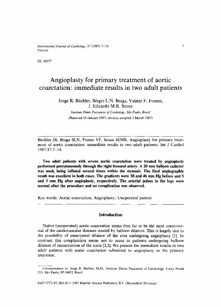
International Journal of Cardioloey 17 (1987) 7-14 Elsevier
IJC 00577
Angioplasty for primary treatment of aortic coarctation: immediate results in two adult patients
Jorge R. Biichler, SCtgio L.N. Braga, Valmir F. Fontes, J. Eduardo M.R. Sousa
Institute Dante Pazzanese of Cardiology, S5o Paula, Brazil
(Received 19 January 1987; revision accepted 3 March 1987)
Biichler JR, Braga SLN, Fontes VF, Sousa JEMR. Angioplasty for primary treat-
ment of aortic coarctation: immediate results in two adult patients. Int J Cardiol 1987;17:7-14.
Two adult patients with severe aortic coarctation were treated by angioplasty performed percutaneously through the right femoral artery. A 20 mm balloon catheter was used, being inflated several times within the stenosis. The final angiographic result was excellent in both cases. The gradients were 38 and 46 mm Hg before and 5 and 3 mm Hg after angioplasty, respectively. The arterial pulses in the legs were normal after the procedure and no complication was observed.
Key words: Aortic coarctation; Angioplasty; Unoperated patient
Introduction
Native (unoperated) aortic coarctation seems thus far to be the most controver- sial of the cardiovascular diseases treated by balloon dilation. This is largely due to
the possibility of aneurysmal dilation of the area undergoing angioplasty [l]. In contrast, this complication seems not to occur in patients undergoing balloon
dilation of recoarctation of the aorta [2,3]. We present the immediate results in two adult patients with aortic coarctation submitted to angioplasty as the primary treatment.
Correspondence ro; Jorge R. Biiichier, M.D., Institute Dante Pazzanese of Cardiology, Caixa Postal
215, SZo Paulo, SP 04012 Brazil.
0167-5273/87/$03.50 0 1987 Elsevier Science Publishers B.V. (Biomedical Division)

Fig.
in c
1. Cross-sectional echocardiography from the supra-sternal approach demonstrating the aortic ‘ase 1. A. Pre-angioplasty. The arrows show the area of coarctation. B. Post-angioplasty. The ar
show the dilated area.
arch rows

Fig.
1. A
2. Aortography performed above the coarctation area in 30 o left anterior oblique projection in Pre-angioplasty. The arrows outline the severe coarctation. B. Post-angioplasty. The arrows I
the excellent cosmetic result in the dilated area.
case show

10
TABLE 1
Pressure data.
Pre-angioplasty Ao (mm Hg) Post-angioplasty Ao (mm Hg)
Pre-coax. Post-coarc. Pre-coarc. Post-coarc.
S D S D G S D s D G
Case 1 193 115 155 109 38 120 95 115 93 5
Case 2 156 92 110 85 46 96 62 93 61 3
Ao = aorta: coax. = coarctation: D = diastolic; G = gradient; S = systolic.
Case Presentations
Case 1
A 30-year-old white woman had been known to have a heart murmur since the
age of 5 years. She was referred to us with exertional dyspnea, weakness and coldness in the legs and, more recently, headaches. The physical examination showed an arterial blood pressure of 160/100 mm Hg in the arms that could not be
measured in the legs. The femoral pulses were barely palpable and the distal leg pulses were absent. A grade 2/6 systolic murmur was audible along the left sternal border, The electrocardiogram was normal. Cross-sectional echocardiography dem- onstrated the coarctation lesion (Fig. 1A). The patient was catheterized and the hemodynamic data are presented in Table 1. The angiographic study showed a
severe aortic coarctation in the environs of the arterial ligament (Fig. 2A) and no
other heart defect.
Case 2
This 49-year-old white woman had had a transitory cerebrovascular accident 3 years previously. Since then, she had complained of exertional dyspnea, mild chest
pain, dizziness and headache. The physical examination showed an arterial blood pressure of 160/100 in the arms that could not be measured in the legs. As with the
first patient, the femoral pulses were barely palpable and the more distal pulses could not be felt. A grade 2/6 systolic murmur was heard along the left sternal border. The electrocardiogram showed a mean QRS axis of -10” together with findings suggestive of left ventricular enlargement. Cross-sectional echocardiogra-
phy confirmed the existence of aortic coarctation. She was catheterized and the pressure data are presented in Table 1. The angiographic study showed a severe aortic coarctation (Fig. 3A) and no other heart defect.
Technique
Both dilations were performed percutaneously through the right femoral artery. Through an 8F hemokit, a USC1 8F Sones catheter was introduced above the

Fig. 3. Aortography performed above the coarctation area in 15’ right anterior oblique projection in case
2. A. Pre-angioplasty. The arrows show the severe coarctation. B. Post-angioplasty. The arrows indicate the excellent cosmetic result.

Fig. 4. The arrow in Fig. A marks the waist in the balloon. This has disappeared (B) after inflatior
13
stenosis and a pull-back pressure measurement was recorded. An angiogram per-
formed above the coarctation in 15 ’ right anterior oblique projection identified the site of stenosis. A USC1 0.9 mm-220 cm straight guide wire was threaded through the catheter and placed above the stenosis. The Sones catheter was removed
carefully and a Medi-Tech 20-3/9/100 balloon catheter was guided so that it was
positioned across the coarctation. It was inflated several times for about lo-15
seconds each (Fig. 4).
The balloon catheter was removed after the procedure and the 8F hemokit was
re-introduced. An USC1 8F NIH angiographic catheter was passed above the dilated area and a pull-back pressure measurement again recorded. An angiographic
study above the dilated area was performed in 15 o right anterior oblique and 30 o
left anterior oblique projections. No complication was observed.
Results
Case 1
The pressure data are presented in Table 1. Angiographic (Fig. 2B) and echo- cardiographic (Fig. 1B) investigations demonstrated an excellent cosmetic result. The arterial blood pressure was 120/80 mm Hg in the arms and 125/100 mm Hg in
the legs. Arterial pulses in the legs were normal. The previously heard systolic murmur was absent.
Case 2
The pressure data are presented in Table 1. Angiography (Fig. 3B) and echocar- diography again showed an excellent cosmetic result. Arterial blood pressures were 110/75 mm Hg in the arms and 130/80 mm Hg in the legs. Arterial pulses in the
legs were normal. A grade l/6 systolic murmur was audible along the left sternal border. Both cases were discharged 24 hours later in excellent clinical condition.
Discussion
Studies of experimentally produced and excised coarctation lesions have demon- strated the possibility of treating the stenosis by balloon dilation by tearing the intima and media of the aorta [4-61. Studies carried out in infants and children have shown the immediate results to be satisfactory [7-111, although restenosis is
relatively common in infants [7,8]. The major concern about the procedure is the
possibility of aneurysm formation in the dilated area. Because of this possibility, a careful selection of those patients with aortic coarctation to be treated by angiop- lasty is recommended [l]. In our two cases, the immediate results were entirely
satisfactory and, technically, it seemed to be simple and safe. A previous successful angioplasty has been described in one adult patient [ll]. The Sones catheter was used because its tip is appropriate for safe passage across the tight stenoses encountered. We performed several inflations of the balloon after the “waist” had disappeared, but the ideal number of inflations has yet to be determined.

14
A close follow-up will be essential to detect as soon as possible aneurysmal formation, dissections, bacterial aortitis and restenosis should they occur. The future
of the technique will depend very much on the absence of such complications. From
our brief experience, we submit that angioplasty of native aortic coarctation in
adults seems to be a safe and simple alternative treatment to surgery. Its immediate
results are satisfactory but its efficacy has to be demonstrated over a long period of
follow-up.
Acknowledgments
We thank Lazaro V. de Miranda and ZClia Maria F.A. Rodrigues for photo-
graphical and technical assistance. We thank also Dr. Rica D.D. Btichler. Special thanks to Dr. Vera Marcia L. Gimenes by performing the cross-sectional echocar-
diography.
References
1 Lock JE, Keane JF. Fellows KE. The use of catheter intervention procedures for congenital he,art
disease. J Am Co11 Cardiol 1986;7:1420-1423.
2 Soulen RL, Kan J, Mitchell S, White RI, Jr. Balloon angioplasty of coarctation restenosis: Evaluation
by MRI (abstract). Circulation 1986;74:11-403.
3 Allen HD. Marx GR, Ovitt TW. Balloon angioplasty for coarctation: serial evaluation (abstract). J
Am Co11 Cardiol 1985;5:405.
4 Lock JE, Niemi T, Burke DA, Einsiz S, Castaneda-Zuniga WR. Transcutaneous angioplasty of
experimental aortic coarctation. Circulation 1982; 66:1280-1285.
5 Lock JE, Castaneda-Zuniga WR. Bass JL, Foker JE. Amplatz K. Anderson RW. Balloon dilatation of
excised aortic coarctation. Radiology 1982;143:689-691.
6 SOS T, Sniderman KW, Ritterk-Sos B, Strupp A, Alonso DR. Percutaneous transluminal dilatation of
thoracic aorta post mortem. Lancet 1979;2:970-971.
7 Lock JE, Bass JL. Amplats K, Fuhrman BP, Castaneda-Zuniga WR. Balloon dilatation angioplasty of
aortic coarctations in infants and children. Circulation 1983;68:109-116.
8 Finely JP, Beandien RG, Nanton MA, Roy DL. Balloon catheter dilatation of coarctation of the
aorta in young infants. Br Heart J 1983;50:411-415.
9 Sperling DR, Dorsey TJ, Rowen M, Gazzaniga AB. Percutaneous transluminal angioplasty of
congenital coarctation of the aorta. Am J Cardiol 1983;51:562-564.
10 Lababidi ZA, Daskalopoulos DA, Stoechlkett Jr. Transluminal balloon coarctation angioplasty:
experience with 27 patients. Am J Cardiol 1984;54:1288-1291.
11 Marvin WJ, Mahoney LT. Balloon angioplasty of unoperated coarctation in young children (abstract).
J Am Co11 Cardiol 1985;5:405.
Copyright © 2022 FDOKUMEN