the short term effects of low dye taping vs. calcaneal taping in ...
Upload
khangminh22Category
view
1download
0
University of Northern Iowa University of Northern Iowa
UNI ScholarWorks UNI ScholarWorks
Dissertations and Theses @ UNI Student Work
2016
The immediate effects of thearapeutic taping on musculoskeletal The immediate effects of thearapeutic taping on musculoskeletal
pain pain
Aaron Michael Krejci University of Northern Iowa
Let us know how access to this document benefits you
Copyright ©2016 Aaron Michael Krejci
Follow this and additional works at: https://scholarworks.uni.edu/etd
Part of the Therapeutics Commons
Recommended Citation Recommended Citation Krejci, Aaron Michael, "The immediate effects of thearapeutic taping on musculoskeletal pain" (2016). Dissertations and Theses @ UNI. 275. https://scholarworks.uni.edu/etd/275
This Open Access Thesis is brought to you for free and open access by the Student Work at UNI ScholarWorks. It has been accepted for inclusion in Dissertations and Theses @ UNI by an authorized administrator of UNI ScholarWorks. For more information, please contact [email protected].
THE IMMEDIATE EFFECTS OF THERAPEUTIC TAPING ON MUSCULOSKELTAL PAIN
An Abstract of a Thesis
Submitted
in Partial Fulfillment
of the Requirements for the Degree
Master of Science
Aaron Michael Krejci
University of Northern Iowa
July, 2016
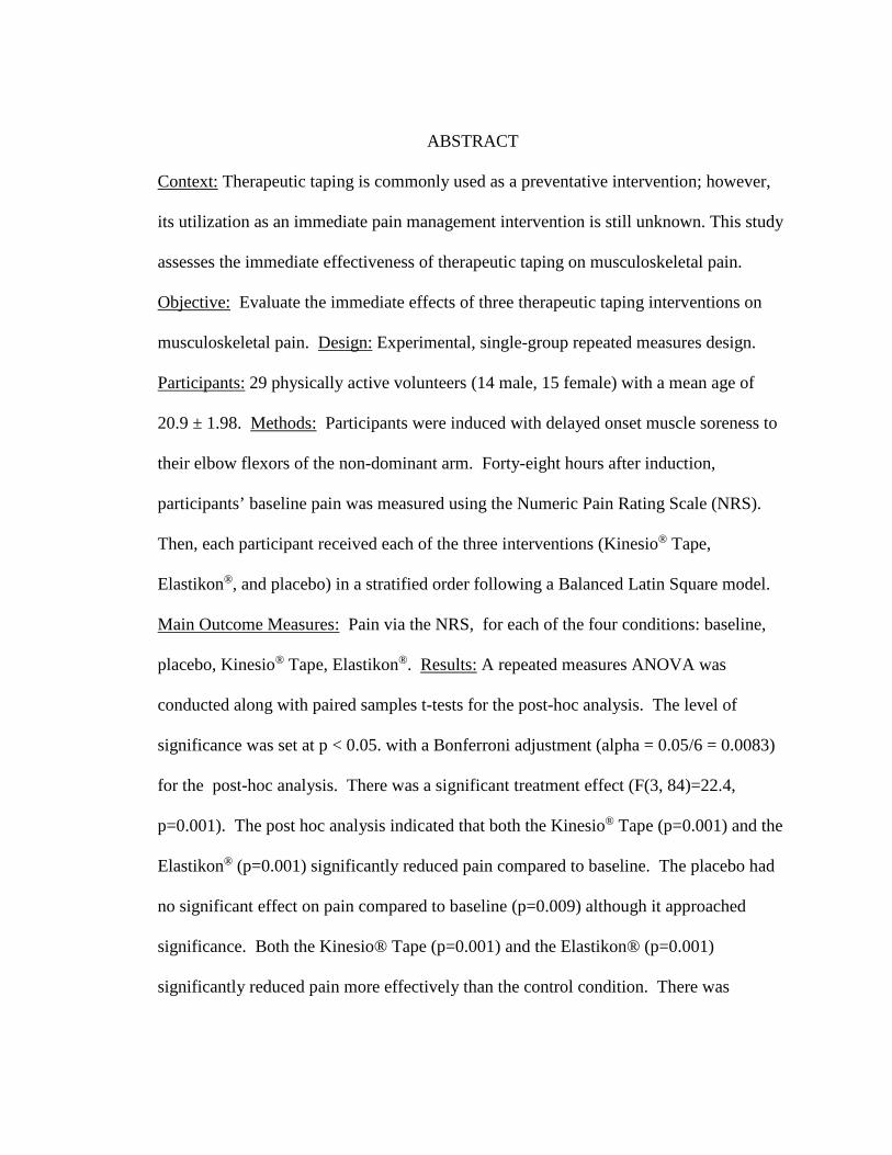
ABSTRACT
Context: Therapeutic taping is commonly used as a preventative intervention; however,
its utilization as an immediate pain management intervention is still unknown. This study
assesses the immediate effectiveness of therapeutic taping on musculoskeletal pain.
Objective: Evaluate the immediate effects of three therapeutic taping interventions on
musculoskeletal pain. Design: Experimental, single-group repeated measures design.
Participants: 29 physically active volunteers (14 male, 15 female) with a mean age of
20.9 ± 1.98. Methods: Participants were induced with delayed onset muscle soreness to
their elbow flexors of the non-dominant arm. Forty-eight hours after induction,
participants’ baseline pain was measured using the Numeric Pain Rating Scale (NRS).
Then, each participant received each of the three interventions (Kinesio® Tape,
Elastikon®, and placebo) in a stratified order following a Balanced Latin Square model.
Main Outcome Measures: Pain via the NRS, for each of the four conditions: baseline,
placebo, Kinesio® Tape, Elastikon®. Results: A repeated measures ANOVA was
conducted along with paired samples t-tests for the post-hoc analysis. The level of
significance was set at p < 0.05. with a Bonferroni adjustment (alpha = 0.05/6 = 0.0083)
for the post-hoc analysis. There was a significant treatment effect (F(3, 84)=22.4,
p=0.001). The post hoc analysis indicated that both the Kinesio® Tape (p=0.001) and the
Elastikon® (p=0.001) significantly reduced pain compared to baseline. The placebo had
no significant effect on pain compared to baseline (p=0.009) although it approached
significance. Both the Kinesio® Tape (p=0.001) and the Elastikon® (p=0.001)
significantly reduced pain more effectively than the control condition. There was

however, no difference between the Kinesio® Tape and the Elastikon® (p=0.50).
Conclusion: Based on the results, both Kinesio® Tape and Elastikon® significantly
reduced pain associated with DOMS. The placebo intervention had no significant effect
compared to the baseline, but did approach significance. Furthermore, although both
therapeutic tapes reduced pain, there was no difference between the Kinesio® Tape and
the Elastikon®. Although therapeutic taping was successful in relieving pain associated
with DOMS, the type of tape used did not matter. Therefore, clinicians can consider
using therapeutic tape to modulate pain to facilitate rehabilitation when movement is
appropriate, but limited by pain.
THE IMMEDIATE EFFECTS OF THERAPEUTIC TAPING ON MUSCULOSKELETAL PAIN
A Thesis
Submitted
in Partial Fulfillment
of the Requirements for the Degree
Master of Science
Aaron Michael Krejci
University of Northern Iowa
July, 2016

ii
APPROVAL PAGE
This Study by: Aaron Michael Krejci
Entitled: The immediate effects of therapeutic taping on musculoskeletal pain
has been approved as meeting the thesis requirement for the Degree of Master of Science in Athletic Training
___________ _____________________________________________________ Date Dr. Todd A. Evans, Chair, Thesis Committee
___________ _____________________________________________________ Date Dr. Kelli Snyder, Thesis Committee Member ___________ _____________________________________________________ Date Dr. Peter J. Neibert, Thesis Committee Member ___________ _____________________________________________________ Date Dr. Kavita Dhanwada, Dean, Graduate College

iii
DEDICATION
I would like to dedicate this thesis to my family: my parents, Cheri and Rick
Krejci; grandmas: Bonnie Sloan and Ardis Krejci; sister and brother: Lindsey and Scott
Krejci; and the rest of my aunts, uncles, and cousins. Your love, motivation, and
guidance have made my continued education possible. Thank you for the love and
support!

iv
ACKNOWLEGEMENTS
I would like to express a special thank you to Dr. Todd Evans and Dr. Kelli
Snyder who served as my committee co-chairs of this research thesis. I am truly grateful
for the time, support, guidance, and motivation you have provided me throughout the
process of this project. I would also like to extend a thank you for the help and support of
my committee and other special people; Dr. Peter Neibert, Tricia Schrage, Dr. Robin
Lund, and my co-researcher Ashley Lindahl.

v
TABLE OF CONTENTS
PAGE LIST OF TABLES ............................................................................................................ vii LIST OF FIGURES ......................................................................................................... viii INTRODUCTION ...............................................................................................................1 METHODS ..........................................................................................................................4 Research Design.............................................................................................................4 Research Participants .....................................................................................................4 Instruments .....................................................................................................................6 Procedures ......................................................................................................................7 Data Analysis ...............................................................................................................13 RESULTS ..........................................................................................................................15 DISCUSSION ....................................................................................................................17 REFERENCES ..................................................................................................................22 APPENDIX A: EXTENDED RATIONALE AND PURPOSE ........................................24 Statement of the Problem .............................................................................................25 Research Questions ......................................................................................................25 Experimental Hypotheses ............................................................................................25 Significance of the Study .............................................................................................25 Delimitations ................................................................................................................26 Limitations ...................................................................................................................27 Assumptions .................................................................................................................27
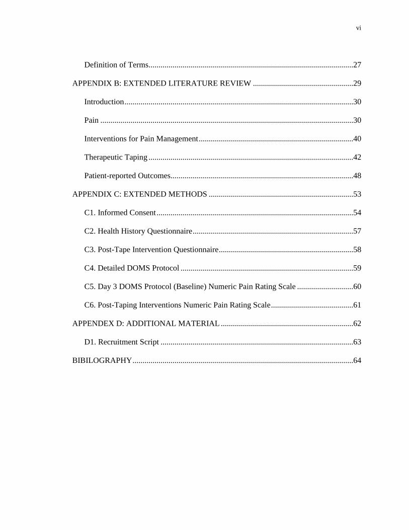
vi
Definition of Terms......................................................................................................27 APPENDIX B: EXTENDED LITERATURE REVIEW ..................................................29 Introduction ..................................................................................................................30 Pain ..............................................................................................................................30 Interventions for Pain Management .............................................................................40 Therapeutic Taping ......................................................................................................42 Patient-reported Outcomes ...........................................................................................48 APPENDIX C: EXTENDED METHODS ........................................................................53 C1. Informed Consent ..................................................................................................54 C2. Health History Questionnaire ................................................................................57 C3. Post-Tape Intervention Questionnaire ...................................................................58 C4. Detailed DOMS Protocol ......................................................................................59 C5. Day 3 DOMS Protocol (Baseline) Numeric Pain Rating Scale ............................60 C6. Post-Taping Interventions Numeric Pain Rating Scale .........................................61 APPENDEX D: ADDITIONAL MATERIAL ..................................................................62 D1. Recruitment Script ................................................................................................63 BIBILOGRAPHY ..............................................................................................................64

vii
LIST OF TABLES
TABLE PAGE
1. Participant Demographics ........................................................................................5
2. Balanced Latin Square Model ................................................................................10
3. Participant Reported Pain Severity (Descriptive Statistics)..………............…….15
4. Participant Reported Pain Relief for Each Application .........................................16

viii
LIST OF FIGURES
FIGURE PAGE
1. Participant Position with Decline Bench .................................................................8
2. Starting Position for Eccentric Exercises .................................................................9
3. Biceps Brachii Tape Application ...........................................................................11
4. Biceps Brachii Tape Application with Stockinette Sleeve ....................................12
5. Mean Pain Scores by Taping Condition ................................................................16

1
INTRODUCTION
The most common symptom for which patients seek medical attention is pain
(Coffey & Mahon, 1982). Once life-threatening conditions/illnesses are ruled out, the
primary goal for healthcare professionals is pain management. Pain can cause
dysfunction, disability, and limit activity and participation. In spite of this, one of the
main challenges is treating and measuring pain (Lara-Muñoz, Ponce de Leon, Feinstein,
Puente & Wells, 2004). Pain relief, however, can lead to the return of function, activities
of daily living and participation. Therefore, these represent meaningful patient outcomes
because they represent a sense of relief and a return to function for the patient. Outcomes
that are clinically important, or meaningful to the patient should be the future direction of
studies and research in athletic training. With clinical research that focuses on patient
outcomes, there should be a direct link between the intervention and an outcome that is
pertinent to the patient’s health (Jette, 1995). However, it is still uncertain if many of the
therapeutic interventions used by athletic trainers improve the most common reason for
patient interactions and arguably the most important outcome to measure; pain.
Many pain management interventions are already being used in the clinical setting
(Connolly, Sayers & McHugh, 2003). Examples include: over-the-counter medications
(OTC’s), electrical modalities and thermotherapies. With non-steroidal anti-
inflammatory drugs (NSAIDs), research has shown that the disruption of the body’s
natural healing process heavily outweighs the slight pain relief that may be experienced
from the medication (Gulick & Kimura, 1996). Non-pharmaceutical interventions are
often appealing to patients because they can be cheaper and less harmful to internal
2
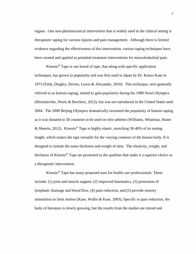
organs. One non-pharmaceutical intervention that is widely used in the clinical setting is
therapeutic taping for various injuries and pain management. Although there is limited
evidence regarding the effectiveness of this intervention, various taping techniques have
been created and applied as potential treatment interventions for musculoskeletal pain.
Kinesio® Tape is one brand of tape, that along with specific application
techniques, has grown in popularity and was first used in Japan by Dr. Kenzo Kase in
1973 (Firth, Dingley, Davies, Lewis & Alexander, 2010). This technique, now generally
referred to as kinesio-taping, started to gain popularity during the 1988 Seoul Olympics
(Mostafavifar, Wertz & Borchers, 2012), but was not introduced in the United States until
2004. The 2008 Beijing Olympics dramatically increased the popularity of kinesio taping
as it was donated to 58 countries to be used on elite athletes (Williams, Whatman, Hume
& Sheerin, 2012). Kinesio® Tape is highly elastic, stretching 30-40% of its resting
length, which makes the tape versatile for the varying contours of the human body. It is
designed to imitate the same thickness and weight of skin. The elasticity, weight, and
thickness of Kinesio® Tape are promoted as the qualities that make it a superior choice as
a therapeutic intervention.
Kinesio® Tape has many proposed uses for health care professionals. These
include: (1) joint and muscle support, (2) improved kinematics, (3) promotion of
lymphatic drainage and blood flow, (4) pain reduction, and (5) provide sensory
stimulation to limit motion (Kase, Wallis & Kase, 2003). Specific to pain reduction, the
body of literature is slowly growing, but the results from the studies are mixed and

3
inconclusive. Kinesio taping may relieve pain by stimulating sensory nerves and
cutaneous mechanoreceptors (Kachanathu, Alenazi, Seif, Hafez & Alroumim, 2014).
In a non-systematic review, Paoloni et al. (2011) suggests that kinesio taping can
relieve pain immediately in chronic low back pain patients, but the full mechanism of
action for this technique has yet to be fully understood and more quality evidence has yet
to be produced. In Montalvo, Cara and Myer’s (2014) meta-analysis, only 13 of the
eighty articles originally found qualified for inclusion due to poor levels of evidence.
Similarly, in Mostafavifar et al’s. (2012) systematic review, only six articles out of the
727 were used from the initial search. Neither the meta-analysis nor the systematic
review yielded sufficient evidence supporting the use of kinesio taping for reducing
musculoskeletal pain. However, Thelen, Dauber and Stoneman (2008) suggested that
kinesio taping can be helpful in improving pain-free active range-of-motion immediately
following application in patients with shoulder pain. By gaining pain-free motion in
one’s joint, it may seem plausible to assume that is sufficient enough for one to return to
normal functioning and/or activities of daily living. In Williams et al. (2012) meta-
analysis, they found the efficacy of kinesio taping for pain relief was questionable
because there were no clinically relevant results.
Although Kinesio® Tape and other therapeutic tapes have been implemented in
rehabilitation interventions and introduced as an intervention that reduces pain, there has
been limited research addressing its immediate impact on pain. Therefore, the purpose of
this study is to determine the effectiveness of therapeutic taping interventions on
musculoskeletal pain.

4
METHODS
Research Design
This study was experimental in nature with a single-group, repeated measures
design. The independent variables were the tape applications: Kinesio® Tape and
Elastikon® tape, and the sleeve only (placebo). The dependent variables were pain
intensity, which was quantified by the Numeric Pain Rating Scale (quantitative), and five
open-ended questions (qualitative) that were reviewed carefully by the primary
investigator.
Research Participants
Participants in this study were recruited from athletic training classes at a
university in the Midwest United States. The participants were all athletic training
majors with varying years of experience as an athletic training student. Thirty healthy
volunteers completed the first three days of the procedures. However, because one
participant was not experiencing any pain forty-eight hours after DOMS induction, the
participant was excluded from the study prior to the therapeutic taping interventions.
Therefore, twenty-nine healthy participants completed all data collection sessions (14
males, 15 females). The participants ranged in age from 19-28 years with a mean age of
20.9 ± 1.98. Additionally, means and standard deviations for the participant’s height and
weight are categorized by sex (Table 2).
5
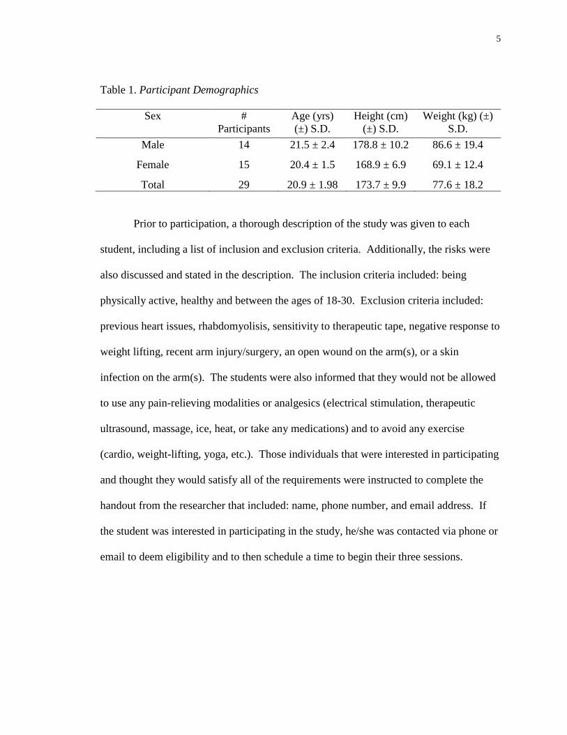
Table 1. Participant Demographics
Sex # Participants
Age (yrs) (±) S.D.
Height (cm) (±) S.D.
Weight (kg) (±) S.D.
Male 14 21.5 ± 2.4 178.8 ± 10.2 86.6 ± 19.4
Female 15 20.4 ± 1.5 168.9 ± 6.9 69.1 ± 12.4
Total 29 20.9 ± 1.98 173.7 ± 9.9 77.6 ± 18.2
Prior to participation, a thorough description of the study was given to each
student, including a list of inclusion and exclusion criteria. Additionally, the risks were
also discussed and stated in the description. The inclusion criteria included: being
physically active, healthy and between the ages of 18-30. Exclusion criteria included:
previous heart issues, rhabdomyolisis, sensitivity to therapeutic tape, negative response to
weight lifting, recent arm injury/surgery, an open wound on the arm(s), or a skin
infection on the arm(s). The students were also informed that they would not be allowed
to use any pain-relieving modalities or analgesics (electrical stimulation, therapeutic
ultrasound, massage, ice, heat, or take any medications) and to avoid any exercise
(cardio, weight-lifting, yoga, etc.). Those individuals that were interested in participating
and thought they would satisfy all of the requirements were instructed to complete the
handout from the researcher that included: name, phone number, and email address. If
the student was interested in participating in the study, he/she was contacted via phone or
email to deem eligibility and to then schedule a time to begin their three sessions.

6
Instruments
Kinesio Tex Classic Tape
One of the therapeutic tapes used in this study was the Kinesio Tex Classic Tape
(courtesy of Kinesio Taping Association International). The tape was applied to the
biceps brachii in accordance to the Clinical Therapeutic Applications of the Kinesio
Taping Method Manual for pain management (Kase et al., 2003; Kase, 2013). In order to
maintain consistency and control of this study, the tape was applied the exact same way
for each participant.
Elastikon Tape
The other therapeutic tape used in this study was Johnson & Johnson’s
ELASTIKON® Elastic Tape (control). This therapeutic tape is also highly elastic and
was also applied in accordance to the Clinical Therapeutic Applications of the Kinesio
Taping Method Manual for pain management (Kase, 2013). The same tape application
technique was used for this tape to help maintain consistency and control of this study.
Numerical Rating Scale
The participant’s subjective pain intensity was measured using the 11 point
Numerical Rating Scale (NRS) from 0-10 (0 = no pain, 10 = worst possible pain). The
participant used a line with number indicators to quantify their pain level by circling the
number which best represented their pain (Appendix C5 & C6).
Health History Form
Participants completed a health history form to identify inclusion and exclusion
criteria, including information pertaining to their physical activity level, current health

7
status, and demographics including height, weight, age, and gender. Participants who
had a current health or injury issue were asked to elaborate on the condition (Appendix
C2).
Post-Tape Intervention Questionnaire
Following each taping intervention, the participant was asked to answer the two
question, post-tape intervention questionnaire. At the conclusion of the participant’s final
session, he/she was asked to complete the three question, post-tape intervention
questionnaire. The questionnaires were used to help collect the participant’s thoughts on
the different therapeutic taping interventions (Appendix C3).
Procedures
The requirements set forth by the Institutional Review Board (IRB) at a
Midwestern Division I college were fulfilled before data collection began. The
procedures collection process occurred during two sessions for each participant over
three days. The first session began with an informed written consent form followed by
the health history questionnaire. The consent form discussed the procedures, risks and
asked for their consent to participate in the study. The health history questionnaire
included demographics, along with questions regarding the inclusion and exclusion
criteria. If any of the exclusion criterion were met following this form, the participant
was excluded from the study. Once the two forms were complete and the participant was
declared eligible, the remainder of the first session included the DOMS-inducing protocol
and pain measurements.
8
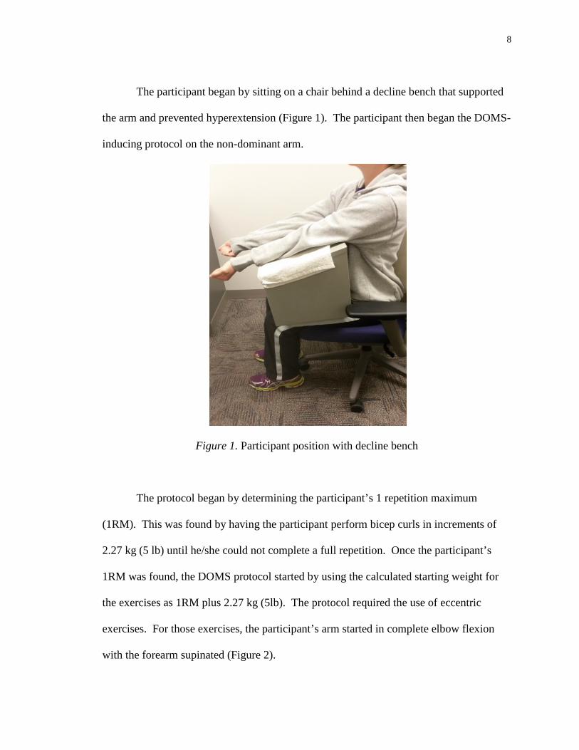
The participant began by sitting on a chair behind a decline bench that supported
the arm and prevented hyperextension (Figure 1). The participant then began the DOMS-
inducing protocol on the non-dominant arm.
Figure 1. Participant position with decline bench
The protocol began by determining the participant’s 1 repetition maximum
(1RM). This was found by having the participant perform bicep curls in increments of
2.27 kg (5 lb) until he/she could not complete a full repetition. Once the participant’s
1RM was found, the DOMS protocol started by using the calculated starting weight for
the exercises as 1RM plus 2.27 kg (5lb). The protocol required the use of eccentric
exercises. For those exercises, the participant’s arm started in complete elbow flexion
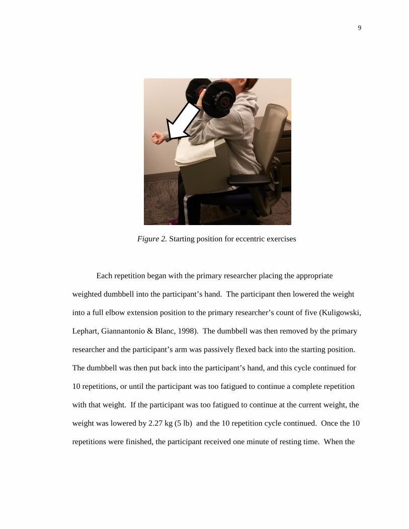
with the forearm supinated (Figure 2).
9
Figure 2. Starting position for eccentric exercises
Each repetition began with the primary researcher placing the appropriate
weighted dumbbell into the participant’s hand. The participant then lowered the weight
into a full elbow extension position to the primary researcher’s count of five (Kuligowski,
Lephart, Giannantonio & Blanc, 1998). The dumbbell was then removed by the primary
researcher and the participant’s arm was passively flexed back into the starting position.
The dumbbell was then put back into the participant’s hand, and this cycle continued for
10 repetitions, or until the participant was too fatigued to continue a complete repetition
with that weight. If the participant was too fatigued to continue at the current weight, the
weight was lowered by 2.27 kg (5 lb) and the 10 repetition cycle continued. Once the 10
repetitions were finished, the participant received one minute of resting time. When the

10
resting time was over, the exercises resumed at the last previously used weight. This
cycle of exercises continued for 5 sets of 10 repetitions.
The second session of this study took place forty-eight hours following the
DOMS protocol. The session began with the participant completing the NRS following
three bicep curls with the 2.27 kg (5lb) dumbbell. The three bicep curls were performed
doing both concentric and eccentric contractions to initiate pain in the bicep prior to
rating pain. Then, the primary researcher examined the participant’s pain rating. If the
rating was a “0”, the participant was excluded from the remainder of this study. If the
rating was a “1” or higher, the remainder of the study resumed, which consisted of the
three therapeutic taping interventions.
The participants that qualified for the remainder of the study received all of the
taping interventions, but in stratified order following the Balanced Latin Square model
(Figure 3). The order of the taping techniques were dependent upon their participant
number that was determined prior to data collection. The stratified order was used to
eliminate time as a possible factor for an increase or decrease in pain throughout the
exercises and tape application process. The order shown in Figure 3 continued
throughout all 30 participants.
Table 2. Balanced Latin Square Model
Kinesio Tape Elastikon No Tape (control) Participant 1 1 2 3 Participant 2 2 3 1 Participant 3 3 1 2 Participant 4 1 2 3

11
The Kinesio Tape (KT) was applied to the biceps brachii in accordance to the
Clinical Therapeutic Applications of the Kinesio Taping Method Manual for pain
management (Kase et al., 2003). A single strip of tape was cut into a bifurcated strip with
one end left uncut to serve as the anchor and was the length of the upper arm. The length
was measured from the distal bicep tendon (antecubital fossa) to the superior glenoid
(long head of biceps brachii origin). As shown in Figure 4, the participant is in a supine
position and the anchor piece was applied over the antecubital fossa. The two bifurcated
strips were pulled at 25% tension along each head (long and short heads) of the biceps
brachii and were attached at the origin of the muscle (supraglenoid tubercle).
Figure 3. Biceps Brachii Tape Application

12
During the tape application process, the participant had a curtain draped over the
top of the shoulder to ensure blinding from the tape (Figure 5-upper left). Additionally, a
soft stockinette sleeve was applied over the tape (Figure 5), so the participant could not
see the tape after the taping application process was complete. Then, the curtain was
removed and the participant completed the three bicep curls with the 2.27 kg (5lb)
dumbbell followed by their pain rating on the NRS. After the NRS, the participant was
asked to complete a post-tape intervention questionnaire regarding that specific
therapeutic tape application. Following the questionnaire, the participant had the curtain
draped over them again. The stockinette sleeve and tape was removed, and that
therapeutic taping intervention was complete.
Figure 4. Biceps Brachii Tape Application with Stockinette Sleeve

13
Similarly, the curtain was draped over the participant during tape application and
the stockinette sleeve was applied over the tape following application. The curtain was
then removed, the participant completed the bicep curls, the NRS, and the post-tape
intervention questionnaire. Following the questionnaire, the curtain was placed back
over the participant’s shoulder, and the stockinette sleeve and tape was removed.
The final therapeutic taping intervention required that the participant only wear
the stockinette sleeve. The participant then completed the bicep curls, the NRS, and post-
tape intervention questionnaire. The curtain was placed over the participant’s shoulder
and the sleeve was removed, marking the completion of that intervention.
Lastly, when all three therapeutic taping interventions were complete, the
participant was asked to complete three questions regarding all three of the interventions.
These questions were related to the participant’s pain and comfortability with each
intervention. At the completion of the final three questions, the primary researcher
offered the opportunity for the participant to ask any questions, or express any comments
or concerns. The final session lasted no longer than 20 minutes. The primary researcher
applied all of the therapeutic taping interventions throughout the study.
Data Analysis
The statistical analysis for this study was completed using the SPSS software with
a statistical significance of p <.05 (IBM, Armonk, NY). Descriptive reports were
calculated for the participants’ demographics. A repeated measures ANOVA was used to
determine the effect of each taping condition on DOMS induced pain. Paired samples t-
tests with Bonferroni adjustment (alpha = 0.05/6 = 0.0083) were used as post hoc

14
analysis when appropriate. The level of significance as set at p<0.05. If the participant
reported their pain as a “0” on the NRS on Day 3, they were released from the study. The
qualitative data gathered from the health history questionnaire and post-tape intervention
questionnaires were analyzed and grouped by the primary researcher.

15
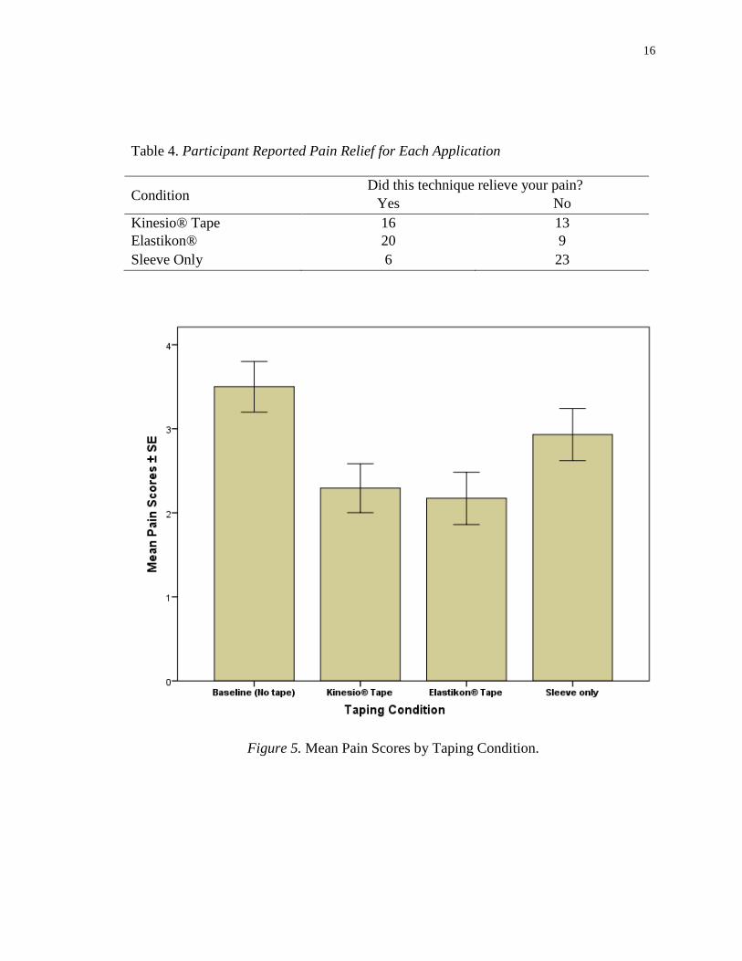
RESULTS
Mauchly’s Test of Sphericity indicated that the data was spherical (p = 0.16),
therefore, no penalty was assessed. There was a significant treatment effect (F(3,
84)=22.4, p = 0.001). Therefore, six paired samples t-tests with Bonferroni adjustment
(alpha = 0.05/6 = 0.0083) were used as post hoc analysis when appropriate. The post hoc
analysis indicated that both the Kinesio® Tape (p = 0.001) and Elastikon® (p = 0.001)
significantly reduced pain compared to baseline. The placebo (sleeve only) condition had
no significant effect on pain compared to baseline (p = 0.009); however, it did approach
significance. Furthermore, both the Kinesio® Tape (p = 0.001) and Elastikon® (p =
0.001) significantly reduced pain more effectively than the control condition. However,
there was no significant difference between the Kinesio® Tape and Elastikon® (p =
0.50). Out of the 29 participants, 55% (16/29) reported that Kinesio® Tape relieved pain,
69% (20/29) reported that Elastikon® relieved pain, and only 21% (6/29) reported that
the sleeve only relieved their pain (Table 4). The mean pain scores for each taping
condition present a significant representation of the pain reduction (Figure 5).
Table 3. Participant Reported Pain Severity (Descriptive Statistics) Condition Mean Standard Deviation N Baseline (No Tape) 3.50 1.626 29
Kinesio® Tape 2.293 1.567 29
Elastikon® 2.172 1.676 29
Sleeve Only 2.93 1.668 29
Overall 2.724 1.634 29
16
Table 4. Participant Reported Pain Relief for Each Application
Condition Did this technique relieve your pain?
Yes No Kinesio® Tape 16 13 Elastikon® 20 9 Sleeve Only 6 23
Figure 5. Mean Pain Scores by Taping Condition.

17
DISCUSSION
The purpose of this study was to determine the immediate effectiveness of
therapeutic taping on musculoskeletal pain. It was guided by the following hypotheses:
(1) therapeutic taping will decrease the self-reported pain level associated with delayed
onset muscle soreness, and (2) there would be no differences in pain relief between the
two therapeutic tapes used in this study. The results from the study confirmed that both
therapeutic tapes reduced pain, but there was no difference between the two therapeutic
tapes used. Based on the Oxford Centre for Evidence-Based Medicine Levels of
Evidence (2011), this study represents “Level 3” evidence. .
From a rehabilitation perspective, applying elastic tape can help relieve pain prior
to the specific exercises. Paoloni et al. (2011) and Zajt-Kwiatkowska, Rajkowska-Labon,
Skrobot, Bakula and Szamotulska (2007) determined that the application of therapeutic
taping cannot be used solely to replace rehabilitative exercises, but may be utilized as an
additional, short-term pain relief intervention. The key question that is unknown is why
therapeutic taping relieves pain. Numerous theories exist as to how this is possible. One
common theory is that the application of therapeutic tape might possibly alter the afferent
sensory signals, which would lead to the facilitation of pain inhibitory mechanisms,
causing a reduction in pain, which is known as the gate control theory (Osorio et al.,
2012; Paoloni et al., 2011). Also, the application of therapeutic tape may create a level of
tactile or sensory stimulation that can decrease pain. Therefore, it seems plausible to
assume that tape creates a sensory stimulation that relieves pain via the gate control
theory.
18
Recent research regarding the benefits of therapeutic tape has primarily focused
on Kinesio® Tape. Although Kinesio® Tape is widely used and highly popular, the
effectiveness of its tape and other therapeutic tapes is unknown and lacks quality research
(Mostafavifar et al., 2012). The current study determined that the application of Kinesio®
Tape and Elastikon® relieved pain and were more effective than wearing a sleeve alone.
Similarly, Osorio et al. (2012) determined that using therapeutic tape is more effective
than a control group or placebo tape when pain reduction and functional improvements
are the primary outcome. In Campolo, Babu, Dmochowska, Scariah and Varughese’s
(2013) study, they also determined that both Kinesio® Taping and McConnell Taping®
relieved pain compared to the control group, but showed no differences between the two
tapes.
The most common pain relief remedy is the use of non-steroidal anti-
inflammatory drugs (NSAIDs), which inhibit metabolic processes in the body (Cheung,
Hume & Maxwell, 2003). While therapeutic taping likely does not inhibit any metabolic
processes, it is important to understand other interventions that patients may utilize.
Other pain-relieving modalities commonly used include therapeutic ultrasound,
thermotherapy, electrical stimulation, intermittent pneumatic compression, and
hyperbaric oxygen chambers (Cheung et al., 2003). One pain-relieving intervention that
could mimic the same neurological response(s) to that of taping is, massage. Along with
the similar sensory stimulation created between tape and massage, both interventions
theoretically relieve pain by activating the gate control theory.

19
For healthcare professionals seeking immediate relief for their patients, there is a
need for more quality research focusing on short-term outcomes, rather than long-term
outcomes (24-96 hours post injury) exclusively. Although most therapeutic interventions
take time to impact patient outcomes, healthcare professionals only interact with their
patients briefly. A specialized intervention approach that focuses on providing
immediate relief from the symptoms of a condition or illness is known as palliative care
(McClay, 2010). One example is non-steroidal anti-inflammatory drugs (NSAIDs).
NSAIDs are primarily used to treat mild to moderate pain, but will not cure the illness or
resolve the cause of the pain (McClay, 2010). Therapeutic taping seems to serve the
same palliative role. When used in conjunction with rehabilitation to facilitate active
movement, recovery may be enhanced.
Pain is an important outcome for healthcare professionals and most often the first
symptom addressed. However, current treatments lack the evidence needed to justify
their use. The primary outcome measures that have been used in therapeutic taping
research include range of motion (ROM), kinematics, muscle activity, muscle strength,
proprioception, and bioelectrical activity (Montalvo et al., 2014; Williams et al., 2012).
While those outcomes can be useful for the researcher and clinician, the focus of research
needs to be on outcomes that are meaningful and beneficial to the patient. In the end,
assessing patient outcomes may lead to improved patient outcomes. However, it is
critical to have immediate treatment interventions in healthcare to satisfy patients.
The findings from this study support the use of therapeutic taping as a treatment
intervention for immediate musculoskeletal pain relief associated with delayed onset

20
muscle soreness. This study offers evidence to all healthcare professionals as it provides
evidence to support therapeutic taping for immediate musculoskeletal pain relief .
Furthermore, by applying elastic therapeutic tapes and minimizing pain, clinicians may
be able to increase patient outcomes throughout the rehabilitation process. Lastly, the
results from the present study may impact palliative care in athletic training. One of the
athletic trainer’s main goals is to treat the pathology and prevent re-injury. Therapeutic
taping through immediate pain relief may serve as a treatment intervention that increases
patient outcomes.
A limitation of this study is that it only examined the impact of tape on DOMS of
the elbow flexors. The study did not address injured participants. Future research should
examine the immediate effects of therapeutic taping on pain from musculoskeletal
injuries beyond DOMS. Although DOMS is a muscle strain and produces
musculoskeletal pain, it is important to determine if the same results surface for different
conditions. It should also be recognized that this study only addressed pain and not
function. For many patients, once pain is relieved, their focus turns to function and
participation. It is uncertain if tape can facilitate a return to function.
Future research on therapeutic taping should also focus on the long-term effects of
therapeutic taping on pain management. This study demonstrated that immediate pain
relief is possible; however, it is unknown whether therapeutic taping provides long-term
relief while it was applied (Montalvo et al., 2014). Future research should also examine
how much pain is necessary or needs to be present before a functional benefit is
observed. With pain being a subjective experience, it may be difficult to accurately

21
address that outcome. However, if possible, clinicians would then have valuable
information on the amount of pain relief needed for patients to return to their daily life.
In conclusion, this study supports the hypotheses that therapeutic taping relieves
musculoskeletal pain, and that is no differences between Kinesio® Tape and Elastikon®.
The results show that there was a significant treatment effect for therapeutic taping when
compared to the baseline and placebo. These results suggest that therapeutic taping
relieves musculoskeletal pain associated with delayed onset muscle soreness, and may
not be dependent on the type of tape utilized while serving as an efficient and cost
effective treatment option for healthcare professional to utilize with their patients.

22
REFERENCES
Campolo, M., Babu, J., Dmochowska, K., Scariah, S., & Varughese, J. (2013). A comparison of two taping techniques (Kinesio and McConnell) and their effect on anterior knee pain during functional activities. International Journal of Sports Physical Therapy, 8(2), 105-110.
Cheung, K., Hume, P., & Maxwell, L. (2003). Delayed Onset Muscle Soreness:
Treatment Strategies and Performance Factors. Sports Medicine, 33(2), 145-164. Coffey, G., & Mahon, M. (1982). Pain: Theories and a new approach to treatment. Journal of the National Medical Association, 74(2), 147-153. Connolly, D., Sayers, S., & McHugh, M. (2003). Treatment and Prevention of Delayed
Onset Muscle Soreness. Journal of Strength and Conditioning Research, 17(1), 197-208.
Firth, B., Dingley, P., Davies, E., Lewis, J., & Alexander, C. (2010). The Effect of Kinesiotape on Function, Pain, and Motoneuronal Excitability in Healthy People and People With Achilles Tendinopathy. Clin J Sport Med, 20(6), 416-421. Gulick, D. T., & Kimura, I. F. (1996). Delayed onset muscle soreness: What is it and how
do we treat it? Journal of Rehabilitation, 5, 234-243.
Jette, A. (1995). Outcomes Research: Shifting the Dominant Research Paradigm in Physical Therapy. Physical Therapy, 75(11), 965-970. Kachanathu, S., Alenazi, A., Seif, H., Hafez, A., & Alroumim, A. (2014). Comparison between Kinesio Taping and a Traditional Physical Therapy Program in Treatment of Nonspecific Low Back Pain. Journal of Physical Therapy, 26(8), 1185-1188. Kase, K. (2013, January 1). Kinesio Taping Method. Retrieved from https://www.kinesiotaping.com/about/kinesio-taping-method Kase, K., Wallis, J. & Kase, T. (2003). Clinical Therapeutic Applications of the Kinesio Taping Method. Tokyo, Japan: Ken Ikai Co Ltd. Kuligowski, L. A., Lephart, S. M., Giannantonio, F. P., & Blanc, R. O. (1998). Effect of
whirlpool therapy on signs and symptoms of delayed-onset muscle soreness. Journal of Athletic Training, 33(3), 222-228.

23
Lara-Muñoz, C., Ponce de Leon, S., Feinstein, A., Puente, A., & Wells, C. (2004). Comparison of Three Rating Scales for Measuring Subjective Phenomena in Clinical Research. I. Use of Experimentally Controlled Auditory Stimuli. Archives of Medical Research, 35, 43-48. McClay, H. (2010). Pain management in palliative care – choice of analgesia. Journal of
the Malta College of Pharmacy Practice, 16, 28-31. Montalvo, A., Cara, E., & Myer, G. (2014). Effect of Kinesiology Taping on Pain in Individuals With Musculoskeletal Injuries: Systematic Review and Meta- Analysis. The Physician and Sportsmedicine, 42(2), 48-57. Mostafavifar, M., Wertz, J., & Borchers, J. (2012). A Systematic Review of the Effectiveness of Kinesio Taping for Musculoskeletal Injury. The Physician and Sportsmedicine, 40(4), 33-40. Osorio, J., Vairo, G., Rozea, G., Bosha, P., Millard, R., Aukerman, D., & Sebastianelli,
W. (2012). The effects of two therapeutic patellofemoral taping techniques on strength, endurance, and pain responses. Physical Therapy in Sport, 1-8.
Paoloni, M., Bernetti, A., Fratocchi, G., Mangone, M., Parrinello, L., Del Pilar Cooper,
M., Sesto, L., Di Sante, L., & Santilli, V. (2011). Kinesio Taping applied to lumbar muscles influences clinical and electromyographic characteristics in chronic low back pain patients. European Journal of Physical and Rehabilitation Medicine, 47, 237-244.
Thelen, M., Dauber, J., & Stoneman, P. (2008). The Clinical Efficacy of Kinesio Tape for Shoulder Pain: A Randomized, Double-Blinded, Clinical Trial. Journal of Orthopaedic & Sports Physical Therapy, 38(7), 389-395. Williams, S., Whatman, C., Hume, P., & Sheerin, K. (2012). Kinesio Taping in Treatment and Prevention of Sports Injuries. Sports Med, 42(2), 153-164. Zajt-Kwiatkowska, J., Rajkowska-Labon, E., Skrobot, W., Bakula, S., & Szamotulska, J.
(2007). Application of Kinesio Taping® for Treatment of Sports Injuries. MedSportPress, 13(1), 130-134.

24
APPENDIX A
EXTENDED RATIONALE AND PURPOSE

25
Statement of the Problem
The purpose of this study was to determine the immediate effectiveness of
therapeutic taping on musculoskeletal pain within a healthy and active population.
Furthermore, this study will assess the differences in pain management between two
commonly used therapeutic tapes.
Research Questions
This study will attempt to answer the following question:
1. Does therapeutic taping relieve pain associated with delayed onset muscle
soreness?
2. Is there a difference between therapeutic tapes techniques for pain
management?
Experimental Hypotheses
This study will be guided by the following hypotheses:
1. It is hypothesized that therapeutic taping will decrease the self-reported
pain level in patients with delayed onset muscle soreness.
2. It is hypothesized that there will be no differences in pain relief between
the two therapeutic tapes used. However, there will be a difference
between using therapeutic taping techniques and using no tape at all.
Significance of the Study
This study will provide evidence as with the effect of therapeutic taping on pain.
This study will also be beneficial for various healthcare professionals looking to add
another treatment technique to their repertoire. Additionally, as healthcare professionals

26
one of the reasons for dysfunction and primary complaints from patients is pain. In
Williams, Whatman, Hume and Sheerin’s (2012) meta-analysis, the efficacy of kinesio
taping remained insignificant due to the ignorance of studying clinically important results
for the patient. This study has the potential to add merit to the proposed benefit of pain
relief immediately following therapeutic tape application and providing meaningful
results for the patient.
In regards to the experimental design, this study will also aid in determining any
significant or meaningful differences between two tapes commonly used in athletic
training. Moreover, this study will add to the limited, yet growing body of literature
available to healthcare professionals regarding the immediate effects of therapeutic taping
on pain management. Lastly and importantly, this study will educate patients and
consumers about the immediate effects of kinesio taping and another commonly used
elastic, adhesive therapeutic tape.
Delimitations
The following delimitations will guide this study:
1. The limited number of participants.
2. The limitation of the population, regarding age and health status limits the
findings of this study.
3. The treatments in this study will be applied only to the biceps brachii,
which makes the findings only applicable to that body part.
4. The therapeutic tapes only include Kinesio Tape and Elastikon, which
excludes other commonly used therapeutic tapes.

27
5. The therapeutic tapes will only be applied long enough to perform the
bicep curls, which limits the possibility of other proposed benefits from
the therapeutic tapes.
Limitations
The following limitations will be present during this study:
1. The participant’s response.
2. The use of healthy, non-injured participants.
Assumptions
This study will be conducted under the following assumptions:
1. Participants uphold the pain relieving limits in the consent form.
2. Participants rate their pain accurately and truthfully.
Definition of Terms
• Kinesio Taping Method: “a therapeutic taping technique that is used as an
alternative to athletic taping to support fascia, muscles, and joints; however,
unlike athletic tape, kinesio tape is theorized to decrease pain and inflammation”
(Kase, 2013; Mostafavifar, Wertz & Borchers 2012).
• Kinesio tape: “a specialized tape that is different from standard white tape in a
sense that it is elastic, can be stretched up to 140%, and its thickness mimics the
human skin” (Halseth, McChesney, DeBeliso, Vaughn & Lien, 2004).
• Elastikon® tape: a commonly used therapeutic tape that is highly elastic and
mimics the similar properties to that of kinesio tape. “Soft cotton elastic tape is
reliable compression for support of sprains, strains and muscle injuries. Also,

28
allows skin to breathe and moisture to pass through” (ELASTIKON Elastic Tape,
2015).
• Delayed Onset Muscle Soreness (DOMS): DOMS is an easy, controlled way of
introducing pain, which is usually common for those who exercise regularly and
are physically active. DOMS is the pain, soreness and stiffness felt in the muscle,
which is typically more severe from 24-72 hours following strenuous exercise
(Hazar et al., 2014).
• Pain: “an unpleasant sensory and emotional experience associated with actual or
potential tissue damage” (Moayedi & Davis, 2013).

29
APPENDIX B
EXTENDED LITERATURE REVIEW
30
Introduction
Pain is the most common reason and symptom for why individuals seek medical
attention (Coffey & Mahon, 1982). Mild to severe pain can cause dysfunction and
disability in activities of daily living. Therefore, one of the initial challenges is
measuring pain (Lara-Muñoz, Ponce de Leon, Feinstein, Puente & Wells, 2004). Multiple
valid scales exist to measure pain when the body’s tissues and/or organs are disrupted.
Musculoskeletal pain and soreness is a common experience for the active population.
Furthermore, numerous interventions have been created and applied to assist in
preventing and managing pain (Cheung, Hume & Maxwell, 2003). One progressively
popular intervention is the use of therapeutic taping. In particular, kinesio tape is being
widely used for various injuries and conditions (Kase, 2013). Many theories and
techniques have been suggested to support its proposed clinical efficacy. Therefore, the
purpose of this literature review is to elaborate on pain, measurement of pain, delayed
onset muscle soreness (DOMS), interventions for pain management, therapeutic taping,
and the importance of assessing patient-reported and patient-meaningful outcomes in
evidence-based medicine.
Pain
Pain is a subjective sensation or feeling that is one of the primary complaints seen
by clinicians in health care settings (Starkey & Sikes, 2004). For clinicians, treating pain
is typically one of the initial goals. Pain relief can be accomplished by medications,
therapeutic modalities and treatment interventions. In medical research, it is crucial to be
able to measure the effectiveness of a given treatment to relieve pain. This section of the
31
literature review will focus on pain theories, the most effective ways to measure pain, and
how healthcare professionals can impact pain in the clinical setting.
Pain plays a crucial role in the day-to-day functioning of the human body. Pain
serves as a protective mechanism and the body’s last line of self-defense (Starkey &
Sikes, 2004). Although pain is an unwanted sensation, it is vital for survival as it informs
individuals when tissues are at risk or already damaged. As defined by the International
Association for the Study of Pain, pain is “An unpleasant sensory and emotional
experience associated with actual or potential tissue damage, or described in terms of
tissue damage, or both” (International Association for The Study of Pain: Taxonomy,
2012). Many different theories have been hypothesized to describe the perception of
pain. The most influential and commonly accepted theories of pain perception include
the Specificity, Intensity, Pattern, and Gate Control Theories of Pain (Moayedi & Davis,
2013).
The Specificity Theory of Pain suggests that there is an assortment of specific
pain receptors in the tissues of the body that are transmitted to a main center in the brain
(Coffey & Mahon, 1982). Both touch and pain are encoded and transmitted along
specific pathways. Those pain or touch sensations/impulses are transmitted via A-delta
and C-fibers. A-delta fibers are large, myelinated nerves that transmit information
quickly and are often burning or stinging sensations; whereas, C-fibers are small nerves
that transmit information slowly and are often dull, aching and throbbing sensations
(Starkey & Sikes, 2004). Conclusively, the Specificity Theory of Pain states that pain is
a distinct sensation with its own pathway, autonomous of touch and any other senses.

32
The Intensity Theory of Pain, which competed with the Specificity Theory of Pain
many years ago, proposes that pain is an emotion that arises when a stimulus is stronger
than normal and there are no specific pathways for varying levels of threshold stimuli
(Moayedi & Davis, 2013). Instead of a low-threshold stimulus or a high-threshold
stimulus, it is the amount of impulses that determines the intensity of the stimulus. For
example, there can be many impulses of a subthreshold (minimal amount of pain) stimuli
and create the same amount of pain as a high intensity, suprathreshold stimuli. The
subthreshold impulses encode innocuous (unharmful) stimuli, and the suprathreshold
impulses encode noxious (painful) stimuli (Moayedi & Davis, 2013). The same
researchers came to a conclusion that there must be a summation (an overlap of electrical
activity) occurring for the subthreshold stimulus to become so painful.
The Pattern Theory of Pain became its own separate thought process by ignoring
the two previous theories and previous findings about specific nerve endings. This theory
speculates that different sensory organs respond to many stimulus intensities and have
different levels of responsivity to stimuli (Moayedi & Davis, 2013). Also, the pattern of
activity encodes the pain or touch and the location of the stimulus. To support this
theory, Lele, Sinclair and Weddell (1954) demonstrated that altering a nerve fiber would
cause action potentials to occur in any fiber. Additionally, any intense stimuli towards
the nerve fibers would cause pain. This theory is highly debated due to its lack of
explanation towards the existence of specificity (Coffey & Mahon, 1982).
The most common and practical of all theories is the Gate Control Theory of Pain,
proposed by Melzack and Wall. The two men used the evidence from the Specificity and
33
Pattern Theories of Pain and created a model to help explain the different findings and
help close the gap between the two theories. This theory proposes that there are
nociceptors (pain fibers) and touch fibers that synapse in two different cells of the spinal
cord (Moayedi & Davis, 2013). Those regions include the substantia gelatinosa (SG)
cells and the central transmission (T) cells. When the A-delta and C-fibers are
stimulated, the pain impulses are sent from the SG to the brain and will be painful as long
as the stimulus lasts. In order to relieve that pain, A-beta fibers, which are responsible
for touch and pressure, must be stimulated (Coffey & Mahon, 1982). The stimulation of
the A-beta fiber creates an inhibitory effect in the SG, where pain fibers synapse, which
“closes the gate” to pain. For example, when an injury occurs or tissue is damaged, most
people tend to rub the area because it makes it feel better. When rubbing an injured area,
it activates the Gate Control Theory of Pain by activating A-beta fibers (trying to close
the gate) to win the battle over the C-fibers (trying to open the gate). The successful
activation of appropriate fibers results in a decrease in pain from the injury (Starkey &
Sikes, 2004). Therefore, a nonpainful stimulus (rubbing) can override the painful stimuli
(injury).
The importance of these theories as it relates to this study is worthy of discussion.
The Specificity, Intensity, and Pattern Theories of Pain are least likely to impact or have
any relation to this study. The hypothesized treatment effect of pain relief from kinesio
tape and other therapeutic tapes on painful patients seems logical to be applied to the
Gate Control Theory of Pain due to the tactile stimulation. Therapeutic tape application
is similar to rubbing an injured body part as the tape is essentially applying the same
34
principles. The pulling from the adhesive tape on the skin is “closing the gate” from the
pain receptors and possibly providing pain relief. With the therapeutic tape applied, the
next important step is measuring the pain to determine if the kinesio taping is effective.
Measuring Pain
When treating and assessing pain in patients, using valid and reliable scales and
clinical measurements is critical. Whether it is in the clinical setting or in research,
choosing the appropriate scale or clinical measure for that population is an important
step. Among the most common measures of pain include the Faces Pain Scale-Revised
(FPS-R), Verbal Rating Scale (VRS), McGill Pain Questionnaire, Visual Analogue Scale
(VAS), and the Numerical Rating Scale (NRS; Ferreira-Valente, Pais-Ribeiro & Jensen,
2011). Although each measure has its own strengths and weaknesses, the validity and
reliability of each measure is supported through evidence across a variety of populations
(Ferreira-Valente et al., 2011). When choosing an appropriate measure of pain and
examining its validity, the most important part is the measure’s ability to identify change
in pain with the treatment. Of the measures previously listed, research shows that one
measure is not more clinically consistent than another measure (Ferreira-Valente et al.,
2011).
The FPS-R is a visual scale that is most commonly used with children. The scale
is made-up of six different faces resembling an increase in pain intensity (Pagé et al.,
2012). A lower score, ranging from 0-10, indicates low levels of pain and higher score
indicates higher levels of pain. The VRS is a 4-point Likert scale that is also used to
assess pain intensity and commonly used with children. The patients are asked, verbally,

35
to choose from four possible choices relating to their pain. The choice of 0 indicates “no
pain,” the 1 indicates “a little bit of pain,” the 2 indicates “a medium amount of pain,”
and the 3 indicates “a lot of pain” (Pagé et al., 2012). Unlike any of the other measures,
the McGill Pain Questionnaire is much more lengthy and takes longer to complete. This
pain measurement tool is comprised of three parts that (a) identifies where the pain is,
whether the pain is superficial, internal or both, (b) identifies the type and intensity of
pain, and (c) can also include the VAS or NRS at the end of the questionnaire (Starkey &
Sikes, 2004). This measure is primarily used in the hospital and/or clinic setting.
When sensitive and responsive measures of pain are a necessity, the VAS and
NRS may be optimal for pain measurement (Ferreira-Valente et al., 2011). The VAS is
very common in all settings and is made-up of a line that is a specific length and has
specific directions with bilateral limits that designate an absence or minimal amount of
pain on one end, and a full or maximum amount of pain on the opposite end (Lara-Muñoz
et al., 2004). The VAS is easy to administer, reliable, is consistent with and pertinent to
each patient, and can be used before and after treatments to detect any changes or the
effectiveness of a treatment.
The NRS is commonly chosen as the preferred measure of pain in many settings
and shows solid evidence of acceptability, reliability, and validity (Von Baeyer et al.,
2009). The scale can be easily administered verbally by asking the patient to indicate
their intensity of pain on a scale ranging from 0 to 10, where 0 is no pain and 10 is the
worst or most pain. If needed, this scale can also be completed by the patients
themselves. In a systematic literature review by Jensen-Hjermstad et al. (2011), it was

36
concluded that a NRS is an applicable measure of pain intensity in almost all settings.
Additionally, Bijur, Latimer and Gallagher (2003) suggest that the NRS, when
administered verbally, can replace the VAS when used for pain measurement and
detecting changes in pain from treatments. All of these measures of pain could
potentially be argued as appropriate for this study; however, the majority of the relevant
literature used the VAS and NRS (Montalvo, Cara, & Myer 2014). Before discussing
how pain is managed following measurement, it is important to understand a specific type
of pain relevant to this study.
Delayed Onset Muscle Soreness (DOMS)
Muscle soreness, stiffness and pain are common symptoms following exercise or
unfamiliar physical activity for the novice exerciser or even the elite athlete (Cheung et
al., 2003). This exercise-induced experience is known as delayed onset muscle soreness
(DOMS) and is a common model for inducing pain for research studies. DOMS is a type
I muscle strain injury and can present with tenderness throughout the muscle with
palpation and movement, decreased flexibility, and can be debilitating for the individual
towards their activities of daily living (Cheung et al., 2003; Gulick & Kimura, 1996).
These symptoms from the physical activity typically begin 12-24 hours post-exercise and
increase in intensity between 24-48 hours. The symptoms then subside within 5-7 days
(Lee, Bae, Hwang & Kim, 2015).
DOMS is typically linked to high-force or high-intensity muscle recruitment,
especially those movements involving eccentric activity. Eccentric contractions are
muscular movements where the muscle is being elongated while simultaneously

37
contracted (Gulick & Kimura, 1996). There are several theories as to why eccentric
contractions are the main cause of this pain and why DOMS occurs. The main theories
include the lactic acid theory, the muscle spasm theory, the connective tissue damage
theory, the muscle damage theory, the inflammation theory, and the enzyme efflux
theory.
The lactic acid theory assumes that lactic acid production continues following the
completion of exercising, causing a noxious stimulus and a delayed perception of pain
(Cheung et al., 2003). However, lactic acid levels typically return to pre-exercise state at
one hour following exercise. Therefore, pain that comes with fatigue in the acute stage
can be associated with the lactic acid theory, but likely not the pain that is experienced
24-48 hours after exercise (Cheung et al., 2003).
The muscle spasm theory states that following exercise, there are increased levels
of resting muscle activity, which was a sign of spasmed motor units. The localized
spasms were assumed to cause a compression of blood vessels, ischemia, and the buildup
of noxious substances (Cheung et al., 2003). The connective tissue theory considers the
connective tissue that surrounds the bundles of muscle fibers. Type I muscle fibers have
a strong, healthy structure; whereas type II muscle fibers are fast-twitch and are more
vulnerable to injuring the connective tissue due to a stretch-induced mechanism (Cheung
et al., 2003).
The muscle damage theory is likely only a partial explanation of DOMS and is
primarily associated with eccentric exercises. This theory is based on the damage to the
z-line, a contractile component, of the muscle tissue (Cheung et al., 2003). This occurs

38
during eccentric exercises because of the reduced active motor units, which then
increases the tension on each motor unit.
The inflammation theory states that there are properties of the inflammatory
response/process following eccentric activities. At the cessation of eccentric exercises,
there is breakdown of disrupted connective tissue and muscle fibers, which causes an
accumulation and attraction for different white blood cells and other tissue healing
enzymes (Cheung et al., 2003). The enzyme efflux theory states that there is an
accumulation of calcium in the muscle following exercise. The calcium accumulation
causes cellular respiration inhibition and a stall to ATP (adenosine triphosphate)
regeneration, which results in weakened z-lines and noxious nerve endings (Cheung et
al., 2003). Many researchers have come to an agreement that DOMS is likely caused by
several of these theories rather than just one alone (Gulick & Kimura, 1996). Therefore,
when aiming to prevent or treat this condition, healthcare professionals may need to
utilize and incorporate multiple techniques.
This all-to-familiar phenomenon has lead to numerous interventions for
prevention and treatment of DOMS. These interventions include warm-up and cool-
down exercises, stretching, massage, pharmacological therapy, thermotherapies,
ultrasound, electrical stimulation, compression, nutritional supplements, and hyperbaric
oxygen chambers (Bae, Lee, Kim & Kim, 2014). Of those, massage is the only treatment
intervention that has shown to be effective in minimizing the symptoms (Bae et al.,
2014). However, the research indicates that there is a lack of, or very limited amount of
evidence that investigates the immediate effects of these treatment strategies on DOMS
39
(Cheung et al., 2003). As DOMS continues to be so problematic and challenging to
solve, treatment strategies continue to evolve.
Healthcare professionals have been using tape for many years to protect joints and
ultimately reduce pain. Therefore, researchers have begun to study the effects of kinesio
taping (KT) following the induction of DOMS. The kinesio tape’s elastic properties
theoretically make it effective for pain control due its ability to increase blood and lymph
circulations (Lee et al., 2015). The current body of literature on this topic suggests that
KT helps alleviate pain by improving muscle function and strength (Hazar et al., 2014;
Lee et al., 2015). However, in these studies, the researchers examined the effects of KT
for 72 hours after DOMS induction. The reduction of pain, according to the VAS scores,
began between the 24-48 hour mark when DOMS begins to diminish in intensity (Bae et
al., 2014). There have been zero studies that have examined the immediate effects (12-24
hours after induction of DOMS) of KT on a muscle with DOMS.
While DOMS is a problematic phenomenon to solve, the main issue lies within
the debilitating pain that makes it difficult for some to accomplish their activities of daily
living, and others to optimally perform in their sport. In the clinical field, it should be the
healthcare professional’s primary goal to relieve pain in order to effectively achieve the
patient’s desired task or goal. The more efficiently pain relief can be achieved will likely
increase the likelihood of the patient’s satisfaction. Numerous interventions are being
used to assist healthcare professionals in managing pain in the clinical setting.

40
Interventions for Pain Management
Reducing and controlling pain is an essential part of treatment, and often the
primary goal. Athletic trainers and other healthcare professionals use many different
methods and techniques to do so. The most commonly utilized form of pain relief is
medicine. The medication can be administered by athletic trainers through many
different routes. The most common medications administered an/or suggested to patients
are over-the-counter medications such as, non-steroidal anti-inflammatory drugs
(NSAIDS), acetaminophen, and topical analgesics. NSAIDs are often taken orally and
are one of the most widely used analgesics in sports medicine (Hertel, 1997). These
drugs are used to relieve mild to moderate pain by disrupting the body’s natural
metabolic processes, and likely will not cure the illness or be the solution to the condition
(McClay, 2010). Therefore, NSAIDs are palliative. Another route for medication(s) is
transdermally by administering a phonophoresis or iontophoresis treatment (Starkey &
Sikes, 2004). Both treatment techniques use therapeutic ultrasound to assist in the
diffusion of the medication through a large area of the skin. Since it is known that
medications can have a direct impact on pain relief, the following treatment
methods/interventions are proposed in theory.
Many different modalities are commonly used in the health care setting to achieve
pain relief (Slocum, 2014). The most common and basic of them all are the thermal
modalities, which include cryotherapy (ice) and thermotherapy (heat). The application of
ice directly to the skin results in alterations to the cutaneous, subcutaneous,
intramuscular, and joint temperatures. By decreasing the tissue temperature, the ice

41
causes vasoconstriction, reduces the entire inflammatory response, and decreases the rate
of metabolism; therefore, reducing pain (Cheung et al., 2003). Another modality used to
relieve pain is thermotherapy, or the use of heat as a treatment intervention. A few
common types of thermotherapies include warm whirlpool, heating pad, therapeutic
ultrasound, and shortwave diathermy. While the whirlpool and heating pad only increase
the temperature of the superficial skin, both of the other modalities penetrate the tissue
deeply and produce changes through thermal and nonthermal mechanisms (Starkey &
Sikes, 2004). Furthermore, Cheung et al. (2003) suggests that the increase in temperature
exacerbates the inflammatory process by increasing blood flow to the applied area.
Various electrical current interventions are commonly used in the clinical setting
to achieve different results (Denegar & Perrin, 1992). Electrical currents can relieve pain
by accelerating the healing process for wounds and fractures, or affecting the perception
and transmission of pain (Cheung et al., 2003; Starkey & Sikes, 2004). The most
common types of electrical currents include high-volt pulsed current electrical
stimulation, Russian stimulation, interferential current, microcurrent, and transcutaneous
electrical nerve stimulation (TENS). Because of its convenient use, TENS is one of the
most common forms of electrical stimulation used in research and clinically during
rehabilitation to facilitate and strengthen muscles, and relieve pain (Gulick & Kimura,
1996; Kuru, Yaliman & Dereli, 2012).
For healthcare settings that may not have access to some of the previously listed
modalities, a common pain relief technique is therapeutic massage. Multiple theories
exist for the reasoning behind this pain relief. Massage is suggested to reduce pain by

42
increasing oxygenated blood flow, which inhibits prostaglandin production and,
therefore, limits any further damage or disruption to the inflammatory process (Cheung et
al., 2003). Another common theory is that therapeutic massage relieves pain via the Gate
Control Theory (Slocum, 2014). The massage creates a tactile stimulation on the skin
that leads to the pain reduction. Similarly, two studies determined that applying tape
directly on the skin created a sensory and cutaneous mechanoreceptor stimulation, which
resulted in a reduction in pain (Campolo, Babu, Dmochowska, Scariah & Varughese,
2013; Osorio et al., 2012). Lastly, one of the most convenient and efficient ways to
hypothetically achieve a reduction in pain is adhesive tape. There are many different
brands and types of tape, along with a variety of taping techniques being used to affect
one’s pain perception (Mostafavifar et al., 2012).
Pain is an important symptom and sensation that is seen on a daily basis by
healthcare professionals. Being able to understand the most practical and current theories
behind pain should give clinicians a solid base for treating the pain. Assessing the
patient’s pain using the most valid and reliable measures is not only important in a
clinical setting, but also when conducting research. Clinicians are continuously looking
for new and innovative techniques to reduce pain and provide the best possible care to
every patient in an efficient manner.
Therapeutic Taping
The traditional uses of therapeutic tape have been around for many years. The
primary uses for tape include first-aid assistance, prevention, and stabilization. For
EMT’s and other emergency medical personnel, tape is an effective supply for applying

43
gauze and other bandages for first aid purposes. For individuals and clinicians involved
in the sports medicine setting, therapeutic tape is widely used for its preventative
purposes. While utilizing and applying specific techniques to different body parts, taping
has been found to be an effective prophylactic intervention that provides the same
amount of stabilization that a prophylactic brace could (Olmsted, Vela, Denegar &
Hertel, 2004). Those uses are common and widely used; however, new tapes and
techniques continue to evolve.
The non-traditional or novel uses of therapeutic taping are commonly used, but
lack quality evidence to support their efficacy. Those uses include: improving joint
kinematics and biomechanics, improving proprioception, and providing pain relief. The
most common therapeutic tapes and taping techniques applied for these uses are
McConnell tape, NUCAP Medical Upper Knee Spider® (Spider® Technique), RockTape,
standard elastic tapes, and kinesio tape (Kase, Wallis & Kase, 2003; Osorio et al., 2012).
Although mentioned in the previous section of this literature review, Campolo et
al’s. (2013) and Osorio et al’s. (2012) studies were benchmark studies for the results they
produced regarding the comparisons of two different tapes. First, in Campolo et al’s.
(2013) study, they compared the effectiveness of kinesio tape and McConnell tape on
patients with anterior knee pain during functional activities. Conclusively, they
determined that both tapes and techniques were effective in relieving pain, but one was
not more effective than the other. Second, in Osorio et al’s. (2012) study, they examined
the effectiveness of McConnell tape and Spider® technique (patellofemoral taping
techniques) on strength, endurance, and pain. The results from the study also showed

44
that the taping techniques improved all of their clinical measures, but there were no
differences between the two techniques. However, the interesting part is that both of
these techniques were created to alter the position and biomechanics of the patella. The
results demonstrated that no patella alterations occurred, which led the authors to suggest
that the tape did not improve the biomechanics or kinematics of the patella, but likely
relieved pain via the gate control theory and other tactile stimulating theories. Although
the primary focus from those studies was on patellofemoral pain syndrome patients
(PFPS), the same tapes can be applied to other body parts as well. However, one such
taping technique with increasing popularity is the Kinesio Taping Method (Kase, 2013).
This method consists of the kinesio tape and a specific taping technique, and when used
together has several proposed benefits similar to the one’s previously mentioned.
The Kinesio Taping Method is a therapeutic and rehabilitative taping technique
used to treat a variety of injuries and conditions (Kase, 2013). The technique is applied
using kinesio tape, which is an elastic therapeutic tape with qualities that are intended to
imitate human skin (Firth, Dingley, Davies, Lewis & Alexander, 2010). The kinesio tape
is latex free, made of cotton, has roughly the same thickness as the epidermis, and has the
potential to be stretched up to 130-140% of its original length, giving it unique properties
and application possibilities (Martínez-Gramage, Merino-Ramirez, Amer-Cuenca &
Lisón, 2014). Created in Japan in the 1970s by Dr. Kenzo Kase, the kinesio tape is
specifically applied to a body part based upon the goals of the treatment and can be
applied in hundreds of different ways (Williams et al., 2012). Although this method has
been around for more than 35 years, its popularity grew due to widespread use on

45
national television. During the 2008 Olympic Games in Beijing, kinesio tape was
donated to over 50 countries for use on their elite athletes (Williams et al., 2012). Since
then, it has become a popular treatment intervention for athletic trainers and other
healthcare professionals due to the several proposed benefits.
The Kinesio Taping Method has been designed for numerous uses, which include
(a) aiding the body’s healing process, (b) providing muscle & joint support and stability
without restricting range of motion, (c) facilitating lymphatic drainage, (d) re-educating
the neuromuscular system, (e) enhancing performance, (f) preventing further injury or re-
injury, and (g) reducing pain (Kase, 2013). Each proposed use is supported theoretically,
but these theories or hypotheses have not been demonstrated or supported with evidence
(Martínez-Gramage et al., 2014). To fully understand why kinesio taping is becoming so
popular, it is important to understand the suggested theories.
One of the predominant hypotheses related to kinesio taping is that the kinesio
tape microscopically lifts the skin. The lifting affect creates convolutions in the skin,
which creates more interstitial space and leads to a decrease in inflammation and increase
in lymphatic drainage from the area (Ujino, Eberman, Kahanov, Renner & Demchak,
2013). Muscle and joint support and stability, re-education of the neuromuscular system,
and enhancement of performance can be explained through two theories. First, Dr. Kase
states that when applied from origin to insertion, kinesio tape facilitates muscle activity.
Conversely, application of kinesio tape from insertion to origin inhibits muscle activity.
If the previous theory were to be true, it is imperative that the clinician is properly
educated on the Kinesio Taping Method. Second, a positional stimulus is gained through

46
the skin and aligning the underlying tissues. Also, by correctly aligning the fascia, a
pain-free range-of-motion is allowed, which can improve the kinematics of the affected
joint (Ujino et al., 2013).
Preventing further injury or re-injury could theoretically be linked to improved
proprioception (Halseth et al., 2004). One such mechanism to improve proprioception
has been proposed by Murray (2000), which states this is accomplished through an
increase in cutaneous mechanoreceptors stimulation. Many hypotheses exist regarding
the pain relief from kinesio taping. First, the lifting of the skin theory could be argued to
decrease pain due to the decrease in inflammation, which is a painful process (Ujino et
al., 2013). Second, when the kinesio tape loses form it stimulates the cutaneous
mechanoreceptors that may possibly inhibit noxious impulses in the spinal cord and
relieve pain via an ascending pathway (Montalvo et al., 2014; Lim & Tay, 2015). Lastly,
the kinesio taping technique alleviates irritation and pressure of the neurosensory
receptors that can produce painful sensations (Mostafavifar et al., 2012). The proposed
theories and hypotheses are important in understanding how kinesio taping may affect
patients. However, producing results through research is what clinicians should be
basing their clinical decisions on.
The Kinesio Taping Method is a rather novel treatment technique; therefore, the
amount of quality literature is scarce. Although the body of literature continues to grow,
the results contributing to the efficacy of kinesio taping remain inconclusive (Montalvo et
al., 2014). In Williams et al’s. (2012) meta-analysis, few results were concluded from the
10 articles that met the inclusion criteria. First, there was a minimal amount of quality
47
evidence to suggest the use of kinesio taping over other elastic taping techniques in the
management and/or prevention of injuries. Second, kinesio taping may be beneficial in
improving strength and range of motion when compared with other tapes. Overall, the
authors determined that higher quality research is needed so that clinicians can be
confident in using kinesio tape on their patients. Similarly, in Mostafavifar et al’s. (2012)
systematic review, insufficient evidence was found to support the use of kinesio taping
after a musculoskeletal injury.
Although multiple meta-analyses and systematic reviews concluded that the
efficacy of kinesio taping is inconclusive, it is worthy to review the few pieces of
literature that produced significant findings. In a study that examined stretching with
kinesio tape applied, the authors found that kinesio taping can increase shoulder range of
motion (Ujino et al., 2013). Another study also focused on the shoulder, but studied
shoulder pain and found that kinesio taping may be relevant and beneficial in improving
pain-free range of motion immediately following kinesio tape application (Thelen,
Dauber & Stoneman, 2008). Furthermore, Paoloni et al. (2011) found that kinesio taping
relieved pain and normalized lumbar muscle function when the kinesio tape was applied
to chronic low back pain patients. The last study worthy of discussing also discovered
significant findings. In patients with shoulder impingement syndrome, kinesio taping
was more effective than a variety of therapeutic modalities after the first two weeks, and
may also be a possible treatment option for shoulder impingement syndrome when an
immediate effect is necessary (Kaya, Zinnuroglu & Tugcu, 2011).

48
The Kinesio Taping Method and other therapeutic taping techniques are rather
simple treatment techniques that have the potential to provide merit for its use on patients
with musculoskeletal injuries or other conditions. The abundancy of theories for the
numerous amount of proposed uses for these techniques are continually being studied.
The current body of literature is evaluating a variety of dependent variables ranging from
strength output to proprioception changes and pain relief. With the purpose of keeping
patients satisfied and providing optimal patient care, future research needs to focus on
improving the patient’s function and meaningful patient outcomes, such as pain.
Patient-reported Outcomes
As evidence-based medicine and research continues to evolve, a rather novel
approach should be taken. Many studies reviewed in this body of literature focus solely
on numbers and values that have no meaning or value to the patient (i.e. range of motion,
proprioception, strength output). The focus of clinical research needs to be based upon
outcomes that are meaningful to the patient. Patient-centered, or patient-reported
outcomes are essentially the primary way to determine if patients have been made better
by certain treatments (Wright, 1999).
Outcomes research refers to all types of clinical research that examines health
outcomes. More specifically, the types of research that consider the impact of the
treatment on the patient (Rogers, 2005). Furthermore, patient-centered outcomes
research is the production of evidence that examines the positives and negatives of
treatment methods to prevent, diagnose, and treat specific conditions to improve the
quality of care given by the clinician (Jampel, 2013). The outcomes obtained by the

49
patients themselves, referred to as patient-reported outcomes (PROs), are seemingly more
important than any other outcome reported by another individual. This growing
awareness of patient-reported outcomes is crucial for optimal functioning of all
healthcare systems (Deshpande, Rajan, Sudeepthi & Nazir, 2011).
A patient-reported outcome is any type of report or survey regarding the status of
a patient’s health, and comes directly from the patient without alteration or interpretation
of their response by the clinician (Deshpande et al., 2011). It has been supported by
evidence that when providing attention to the patient’s feedback on health care
interventions and outcomes, that treatment and overall outcomes can be improved. In all
health care settings, treating symptoms is often the main objective. However, when
treating a patient’s symptoms, one important aspect is often overlooked or completely
forgotten and that is Health Related Quality of Life (HRQOL; Deshpande et al., 2011).
Per the World Health Organization (WHO) classification, one’s HRQOL is a measure of
disability. The term disability presents itself as a rather broad term and varies from
patient to patient. When treating a patient with the goal of improving their disability as
the outcome, a few key measures have been addressed. Those measures include quality
of life, patient satisfaction, and functional status (Rogers, 2005). Pertaining to the
physically active population, treating a patient experiencing pain should focus on
improving functionality, returning to activities of daily living, and any other meaningful
patient outcomes.
Meaningful patient outcomes are goals or aspired results from a treatment
intervention that has meaning and value to the patient such as pain relief, returning to

50
activities of daily living, improving function, and improving disability, for example.
Furthermore, because patients are often concerned about how their injury or condition
affects their lifestyle, being able to properly function is crucial (McLeod et al., 2008). In
the physically active population, functional outcomes are tasks such as balancing,
jumping, running, throwing, etc. (Williams, Miller III, Sebastianelli & Vairo, 2013).
Functionality is a vital outcome as it can lead to other important measures in patient care.
Improving one’s function and/or achieving their aspired outcomes, can lead to
patient satisfaction and happiness (Wright, 1999). Satisfaction can be described as the
relationship between experience and expectations. Additionally, satisfaction is an
important measure in the quality of care because it shows the clinician’s success based
upon the patient’s values and expectations (Donabedian, 1988). Patient satisfaction is an
important outcome because satisfied patients are likely to have a better outlook on the
treatment. Also, patient satisfaction can be correlated with the outcome and/or process of
care (Wright, 1999). Although theoretically measuring patient satisfaction sounds
beneficial, there are problems that arise. First, not all patients may understand exactly
what satisfaction means. Second, many other factors besides outcomes of treatment
could affect the patient’s satisfaction. Lastly, not all patients may seek treatment to be
satisfied. Rather, patients seek treatment and medical attention to relieve pain and
improve health or function (Coffey & Mahon, 1982; Wright, 1999). Therefore, it should
be understood that patient satisfaction is an important measure in the process of care, but
not the primary measure in the outcome of care. When looking at all of these possible
measures, assessing clinical outcomes is of most importance.
51
To properly understand the effectiveness of treatment interventions on health
care, assessing clinical outcomes is necessary. Clinical outcomes assessment is the study
and examination of health status changes for a patient or many patients at the end of
health care (Evans & Lam, 2011). The clinical outcomes assessment is made up of
PROs. The significance of using PROs is that many different outcomes can be measured.
Outcomes such as physical function, satisfaction with care, HRQOL, clinical trial
outcomes, and many more can be measured by allowing the patient to report to the
clinician (Deshpande et al., 2011). Most importantly, by using PROs it gives a sense of
empowerment to the patient, and is helpful in interpreting clinical outcomes and decision
making about treatment interventions. When integrating PROs into clinical practice, it is
crucial that the correct measure is selected based upon the injury and population (Evans
& Lam, 2011).
When searching for the appropriate PRO measure to use, lack of availability is not
an issue. Currently, there are more than 50 outcome measurements for the foot and
ankle, and more than 40 measurements for the shoulder (Evans & Lam, 2011). Also,
there are many measurements related to overall health, specific diseases, and
individualized measures, for example. Aside from using PROs, there are disablement
theories and paradigms that can be directly applied to measuring outcomes. Disablement
is a series of related events that result in an injury or specific condition that leads to
disability and/or decreased function (Denegar, Vela & Evans, 2008). The disablement
models use a specific framework for various reasons. Denegar et al. (2008) suggests that
the paradigms help understand one’s disability, they create concepts that can be defined
52
and measured, and can clarify the relationships throughout the model. Clinical outcomes
research and patient-centered outcomes research should be the new foundation in making
decisions regarding patient care. The use of clinical outcomes assessments will help
improve patient care by producing evidence to clinicians regarding the effectiveness of
the most current and common treatment interventions (McLeod et al., 2008).
Conclusively, it is apparent that evidence supports the use incorporating the
patient into treatment decision and clinical outcomes. In clinical research, the use of
patient-reported outcomes has many benefits for everyone involved in the healthcare
setting. Understanding the outcomes that are meaningful to the patient, along with
realizing their needs from the treatment (eg, improving function) is beneficial for the
entire treatment process. The use of clinical outcome assessments following health care
is how clinicians should base their decision-making towards medical treatments.
In summary, there is value to be gained in examining how a specific treatment
technique can impact one’s pain perception. The use of therapeutic taping proposed
many uses to improve the overall health of patients with musculoskeletal injuries or other
conditions. Whether pain is relieved or satisfaction is gained, the primary outcome when
treating patients should focus on improving function and/or returning to activities of daily
living. The research pertaining to therapeutic taping is sporadic and inconclusive
amongst all outcome measures. Therefore, the purpose of this study is to determine the
immediate effectiveness of therapeutic taping on musculoskeletal pain associated with
delayed onset muscle soreness.

53
APPENDIX C
EXTENDED METHODS
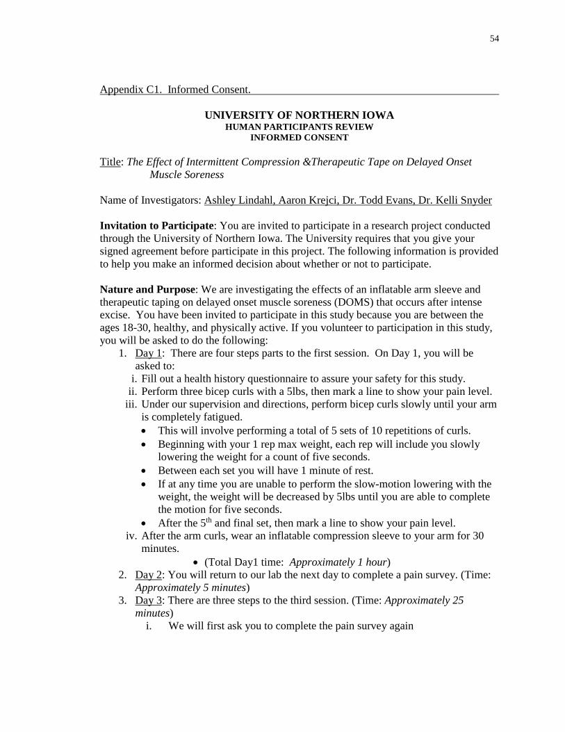
54
Appendix C1. Informed Consent.
UNIVERSITY OF NORTHERN IOWA HUMAN PARTICIPANTS REVIEW
INFORMED CONSENT Title: The Effect of Intermittent Compression &Therapeutic Tape on Delayed Onset
Muscle Soreness Name of Investigators: Ashley Lindahl, Aaron Krejci, Dr. Todd Evans, Dr. Kelli Snyder Invitation to Participate: You are invited to participate in a research project conducted through the University of Northern Iowa. The University requires that you give your signed agreement before participate in this project. The following information is provided to help you make an informed decision about whether or not to participate. Nature and Purpose: We are investigating the effects of an inflatable arm sleeve and therapeutic taping on delayed onset muscle soreness (DOMS) that occurs after intense excise. You have been invited to participate in this study because you are between the ages 18-30, healthy, and physically active. If you volunteer to participation in this study, you will be asked to do the following:
1. Day 1: There are four steps parts to the first session. On Day 1, you will be asked to:
i. Fill out a health history questionnaire to assure your safety for this study. ii. Perform three bicep curls with a 5lbs, then mark a line to show your pain level.
iii. Under our supervision and directions, perform bicep curls slowly until your arm is completely fatigued. • This will involve performing a total of 5 sets of 10 repetitions of curls. • Beginning with your 1 rep max weight, each rep will include you slowly
lowering the weight for a count of five seconds. • Between each set you will have 1 minute of rest. • If at any time you are unable to perform the slow-motion lowering with the
weight, the weight will be decreased by 5lbs until you are able to complete the motion for five seconds.
• After the 5th and final set, then mark a line to show your pain level. iv. After the arm curls, wear an inflatable compression sleeve to your arm for 30
minutes. • (Total Day1 time: Approximately 1 hour)
2. Day 2: You will return to our lab the next day to complete a pain survey. (Time: Approximately 5 minutes)
3. Day 3: There are three steps to the third session. (Time: Approximately 25 minutes)
i. We will first ask you to complete the pain survey again

55
ii. If your arm is still sore, we will ask you to perform 3 sets of arm curls, for 3 repetitions in each set, with little or no weight, while wearing one of three taping conditions. The three tape conditions include two different tape applications and one condition with no tape. The tape applications will include strips of tape applied over your biceps region. You be asked to rate you pain after each of the sets of arm curls.
iii. Finally, we will ask a few questions regarding your opinion of the tape and compression sleeve.
Important requirements for you to consider: • Participation in our study will make your bicep muscle sore; probably for 3 – 5
days. • We anticipate that your participation will take approximately 1.5 hours over the
three sessions. • You might not be able to participate if you have (or had): heart issues,
rhabdomyolysis, sensitivity to therapeutic tape, negative response to weight lifting, recent arm injury/surgery, open wound on your arm(s), or a skin infection on your arm. We will perform a history screening before you begin to determine your eligibility.
• We are also asking that you 1. Do not exercise between the Day 1 and Day 3 session. 2. Do not use any other pain relieving techniques such as:
Pain relieving medications such as ibuprofen or aspirin Applying hot or cold packs to the affected area for the duration of
this study • We may withdraw you from this research if your eligibility status changes during
the study (e.g. Illness, begin additional weight lifting, take pain medication, etc.) Discomfort and Risks: • You will experience mild to moderate pain/soreness from the bicep curl protocol.
This biceps pain may be uncomfortable and may be similar to discomfort you may feel after beginning a new physical activity/exercise. This pain is often described as achy, tender, or annoying.
• There are treatments used in this study, which utilize therapeutic tape. If you are sensitive to tape or other adhesives on your skin, you might develop redness on your skin after the treatment.
• The compression treatment and tape treatments should not be uncomfortable or painful AND you can discontinue your participation at any time.
• If your health status requires further medical consultation, the researcher is obligated to refer you to the appropriate physician. If you do become sore, the researcher and university are not obligated to provide you with any other treatment. Any costs for injuries or other medical attention are solely your responsibility.

56
Benefits and Compensation: Although your participation may be of no direct benefit, you will be entered in a drawing to win 1 of 4 $20 pre-paid VISA cards (there will be 20 participants). At the end of the study, all of the participants’ identifying codes will placed in a hat. The last participant of the study will draw four codes out of the hat. The primary investigator will then match the codes with the participant’s information and notify them of their winnings by email or phone. If you do not complete the entire session, you will still be eligible for the drawing. Confidentiality: Information obtained which could identify you will be kept confidential. The summarized findings with no identifying information may be published in an academic journal or presented at a scholarly conference. Right to Refuse or Withdraw: Your participation is completely voluntary. You are free to withdraw from participation at any time or to choose not to participate at all, and by doing so, you will not be penalized or lose benefits to which you are otherwise entitled. Questions: If you have any questions or concerns about you rights as a research participant related to this study or the study itself, now or in the future, please contact Ashley Lindahl (319-354-0941), Aaron Krejci [(507) 440-6958, [email protected]] or Todd Evans [(319)273-6152 [email protected]]. You can also contact the office of the IRB Administrator, University of Northern Iowa, at 319-273-6148, for answers to questions about rights of research participants and the participant review process. Agreement: Include the following statement:
I am fully aware of the nature and extent of my participation in this project as stated above and the possible risks arising from it. I hereby agree to participate in this project. I acknowledge that I have received a copy of this consent statement. I am 18 years of age or older.
_________________________________ ____________________ (Signature of participant) (Date) _________________________________ (Printed name of participant) _________________________________ ____________________ (Signature of investigator) (Date) _________________________________ ____________________ (Signature of instructor/advisor) (Date)

57
Appendix C2. Health History Questionnaire.
Health History Questionnaire. Participant Number: __________
Health History Form PLEASE DO NOT PUT YOUR NAME ON THIS PAPER
Ht. feet inches Wt. pounds Age: Gender: M F
1. Does the statement below best describe your physical activity level? Yes No I engage in moderate- intensity aerobic physical activity for a minimum of 30 minutes a day, 5 days a week or a vigorous intensity aerobic activity for a minimum of 20 minutes a day, 3 days a week.
2. Are you currently participating in a weight training program? Yes No 3. Do you incorporate bicep curls in your workout? Yes No 4. Are you sensitive or allergic to therapeutic tape of other types of adhesives? For
example: do you get a rash or itchy shin from tape or ban aids? . Yes No
5. Have you ever had severe adverse effect when weight lifting? (More severe than soreness) Yes No
6. Have you ever been diagnosed with Malignancy, rhabdomyolysis, infection of the skin or joint, or a cardiac disease? Yes No
7. Have you had an injury or surgery to your upper extremity in the past 6 months? (i.e. shoulder, elbow, arm, wrist, hand) Yes No
8. Do you currently have any other injury or condition that limits your activity level? Yes No
9. Do you currently have pain in your arms? Yes No If you answered “YES”, to any questions, or you are unsure about any of your answers, you will be asked for more detail to help us determine if is safe for you to participate in our study.
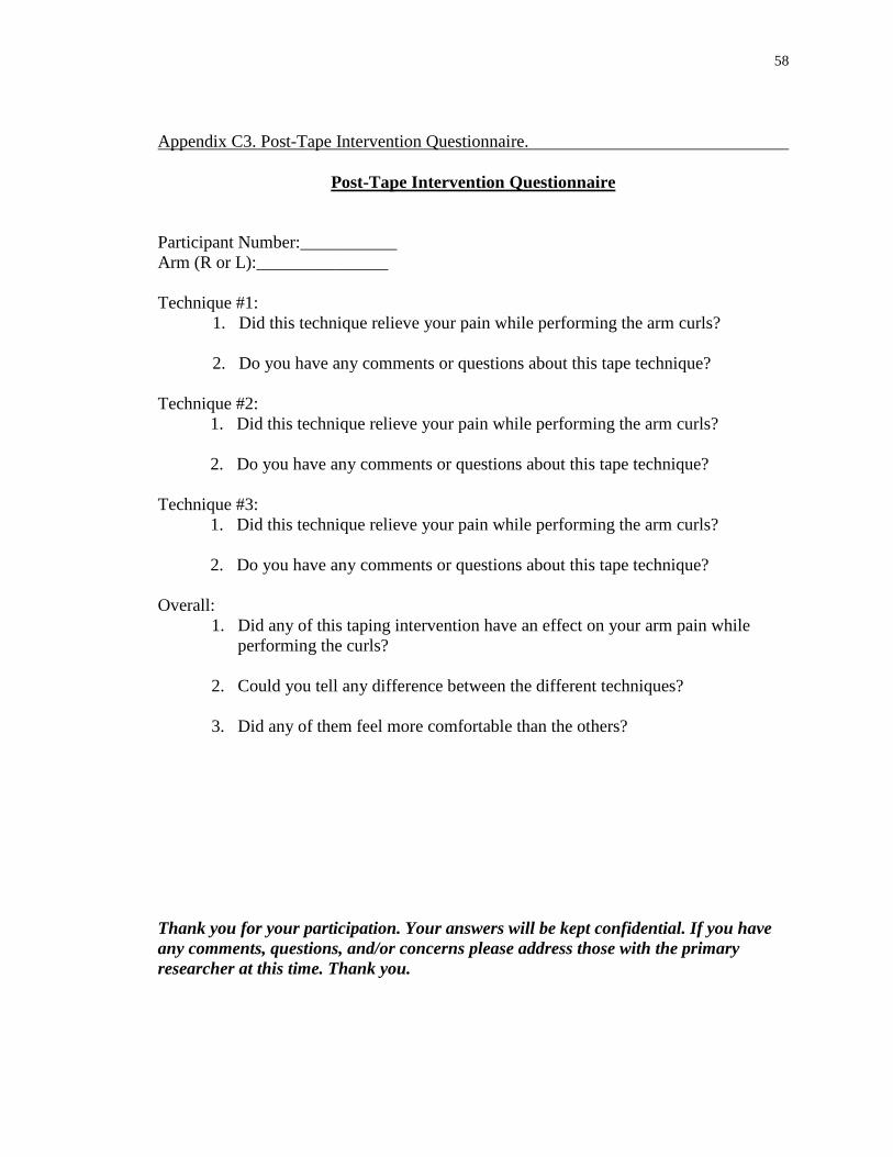
58
Appendix C3. Post-Tape Intervention Questionnaire.
Post-Tape Intervention Questionnaire
Participant Number:___________ Arm (R or L):_______________
Technique #1:
1. Did this technique relieve your pain while performing the arm curls? 2. Do you have any comments or questions about this tape technique?
Technique #2:
1. Did this technique relieve your pain while performing the arm curls? 2. Do you have any comments or questions about this tape technique?
Technique #3:
1. Did this technique relieve your pain while performing the arm curls? 2. Do you have any comments or questions about this tape technique?
Overall:
1. Did any of this taping intervention have an effect on your arm pain while performing the curls?
2. Could you tell any difference between the different techniques? 3. Did any of them feel more comfortable than the others?
Thank you for your participation. Your answers will be kept confidential. If you have any comments, questions, and/or concerns please address those with the primary researcher at this time. Thank you.
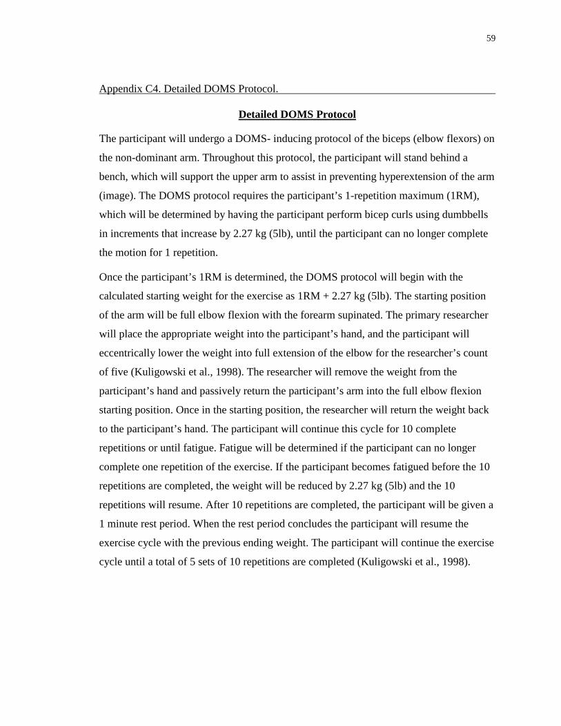
59
Appendix C4. Detailed DOMS Protocol.
Detailed DOMS Protocol
The participant will undergo a DOMS- inducing protocol of the biceps (elbow flexors) on
the non-dominant arm. Throughout this protocol, the participant will stand behind a
bench, which will support the upper arm to assist in preventing hyperextension of the arm
(image). The DOMS protocol requires the participant’s 1-repetition maximum (1RM),
which will be determined by having the participant perform bicep curls using dumbbells
in increments that increase by 2.27 kg (5lb), until the participant can no longer complete
the motion for 1 repetition.
Once the participant’s 1RM is determined, the DOMS protocol will begin with the
calculated starting weight for the exercise as 1RM + 2.27 kg (5lb). The starting position
of the arm will be full elbow flexion with the forearm supinated. The primary researcher
will place the appropriate weight into the participant’s hand, and the participant will
eccentrically lower the weight into full extension of the elbow for the researcher’s count
of five (Kuligowski et al., 1998). The researcher will remove the weight from the
participant’s hand and passively return the participant’s arm into the full elbow flexion
starting position. Once in the starting position, the researcher will return the weight back
to the participant’s hand. The participant will continue this cycle for 10 complete
repetitions or until fatigue. Fatigue will be determined if the participant can no longer
complete one repetition of the exercise. If the participant becomes fatigued before the 10
repetitions are completed, the weight will be reduced by 2.27 kg (5lb) and the 10
repetitions will resume. After 10 repetitions are completed, the participant will be given a
1 minute rest period. When the rest period concludes the participant will resume the
exercise cycle with the previous ending weight. The participant will continue the exercise
cycle until a total of 5 sets of 10 repetitions are completed (Kuligowski et al., 1998).

60
Appendix C5. Day 3 DOMS Protocol (Baseline) Numeric Pain Rating Scale
Numeric Pain Rating Scale Participant Number:______________ Arm (R or L):_______________
Please circle or “X” the number on the scale that represents the intensity of the pain you experience at this time.
NO PAIN WORST PAIN POSSIBLE

61
Appendix C6. Post-Taping Interventions Numeric Pain Rating Scale
Subject Number:______________ Ankle (R or L):_______________
Please circle or “X” the number on the scale that represents the intensity of the pain you experience at this time. Time 1
NO PAIN WORST PAIN POSSIBLE Time 2
NO PAIN WORST PAIN POSSIBLE Time 3
NO PAIN WORST PAIN POSSIBLE

62
APPENDIX D
ADDITIONAL MATERIAL

63
Appendix D1. Recruitment Script.
Classroom Recruiting Script (*Instructors will not be present) Hello Everyone, For those that don’t know me, my name is ____. I’m an athletic training master’s student here at UNI and I am here to invite you to participate in my research study. I am studying the effects of different modalities on pain; specifically, the effects of a new type of compression sleeve and different types of taping techniques. You might have seen these being used already in the athletic training room. If you participate in my study it will involve 3 sessions with me in the athletic training research lab.
1. Day 1: On the first day, probably a Sunday, I will ask you to: a. Complete a series of arm curls to the point of nearly exhausting your
biceps. The purpose of these curls is to induce delayed onset muscle soreness; you’ve probably heard it called DOMS. This is what you feel a few days after you begin working out and you are very sore for the next several days. So if you participate, I will be asking you to give yourself DOMS to your biceps.
b. Complete a pain scale several times c. Wear the inflatable arm sleeve for 30 minutes before you leave the
lab. 2. On Day 2, I will ask you to return to the lab to repeat the pain scale. 3. On Day 3, I will ask you to complete the pain scale again. If your arm is still
sore, I will then apply three different taping techniques to your arm, ask you to perform 3 curls with 5 lbs while wearing the tape, then rate your pain for each technique.
4. Your total approximate time commitment over the three session is 1.5 hours. Please note, If you agree to participate:
• You will be asked not to participate in any exercise including weight lifting and cardio activity during the duration of this study, approximately 3 days.
• You will also be asked not to use any other pain relieving techniques. This could include taking pain relieving medications such as ibuprofen or aspirin as well as applying hot or cold packs to the affected area for the duration of this study.
If you are interested in participating, please write your name, number and email address on the paper I distributed and I will contact you to set up the first session. Thank you!
64
BIBLIOGRAPHY
Bae, S., Lee, Y., Kim, G., & Kim, K. (2014). The Effects of Kinesio-taping Applied to Delayed Onset Muscle Soreness on Changes in Pain. International Journal of Bio-Science and Bio-Technology, 6(3), 133-142. Bijur, P., Latimer, C., & Gallagher, J. (2003). Validation of a Verbally Administered Numerical Rating Scale of Acute Pain for Use in the Emergency Department. Academy of Emergency Medicine, 10(4), 390-392. Campolo, M., Babu, J., Dmochowska, K., Scariah, S., & Varughese, J. (2013). A
comparison of two taping techniques (Kinesio and McConnell) and their effect on anterior knee pain during functional activities. International Journal of Sports Physical Therapy, 8(2), 105-110.
Cheung, K., Hume, P., & Maxwell, L. (2003). Delayed Onset Muscle Soreness:
Treatment Strategies and Performance Factors. Sports Medicine, 33(2), 145-164. Coffey, G., & Mahon, M. (1982). Pain: Theories and a new approach to treatment. Journal of the National Medical Association, 74(2), 147-153. Denegar, C., & Perrin, D. (1992). Effect of Transcutaneous Electrical Nerve Stimulation,
Cold, and a Combination Treatment on Pain, Decreased Range of Motion, and Strength Loss Associated with Delayed Onset Muscle Soreness. Journal of Athletic Training, 27(3), 200-206.
Denegar, C., Vela, L. & Evans, T. (2008). Evidence-Based Sports Medicine: Outcomes Instruments for Active Populations. Clinics in Sports Medicine, 1-8. Deshpande, P., Rajan, S., Lakshmi Sudeepthi, B. & Nazir, A. (2011). Patient-reported outcomes: A new era in clinical research. Perspectives in Clinical Research, 2(4), 137-144. Donabedian, A. (1988). The Quality of Care. Journal of the American Medical Association, 260(12), 1743-1748. ELASTIKON Elastic Tape. (2015). Retrieved from http://www.jnjsportsmed.com/page- product-elastikon.html. Evans, T. & Lam, K. (2011). Clinical Outcomes Assessment in Sport Rehabilitation. Journal of Sport Rehabilitation, 20, 8-16.
65
Ferreira-Valente, M., Pais-Ribeiro, J., & Jensen, M. (2011). Validity of four pain intensity rating scales. International Association for the Study of Pain, 152, 2399- 2404. Firth, B., Dingley, P., Davies, E., Lewis, J., & Alexander, C. (2010). The Effect of Kinesiotape on Function, Pain, and Motoneuronal Excitability in Healthy People and People With Achilles Tendinopathy. Clin J Sport Med, 20(6), 416-421. Gulick, D. T., & Kimura, I. F. (1996). Delayed onset muscle soreness: What is it and how
do we treat it? Journal of Rehabilitation, 5, 234-243.
Halseth, T., McChesney, J., DeBeliso, M., Vaughn, R., & Lien, J. (2004). The Effects of Kinesio Taping on Proprioception At The Ankle. Journal of Sports Science and Medicine, 3, 1-7. Hazar, Z., Citaker, S., Yilmaz Demirtas, C., Celik Bukan, N., Kafa, N., & Celik, B. (2014). Effects of kinesiology taping on delayed onset muscle soreness: a randomized controlled pilot study. Journal of Exercise Therapy and Rehabilitation, 1(2), 49-54. Hertel, J. (1997). The Role of Nonsteroidal Anti-Inflammatory Drugs in the Treatment of Acute Soft Tissue Injuries. Journal of Athletic Training, 32(4), 350-358. International Association for The Study of Pain: Taxonomy. (2012, May 22). Retrieved
from http://iasp-pain.org/Content/NavigationMenu/GeneralResou rceLinks/PainDefinitions/default.htm
Jampel, H. (2013). Patient-Centered Outcomes Research: Evolution, Definition, and Implications. American Academy of Opthalmology, 655-656. Jensen Hjermstad, M., Fayers, P., Haugen, D., Caraceni, A., Hanks, G., Loge, J., . . . Kaasa, S. (2011). Studies Comparing Numerical Rating Scales, Verbal Rating Scales, and Visual Analogue Scales for Assessment of Pain Intensity in Adults: A Systematic Literature Review. Journal of Pain and Symptom Management, 41(6), 1073-1093. Kase, K. (2013, January 1). Kinesio Taping Method. Retrieved from https://www.kinesiotaping.com/about/kinesio-taping-method. Kase, K., Wallis, J. & Kase, T. (2003). Clinical Therapeutic Applications of the Kinesio Taping Method. Tokyo, Japan: Ken Ikai Co Ltd.

66
Kaya, E., Zinnuroglu, M. & Tugcu, I. (2011). Kinesio taping compared to physical therapy modalities for the treatment of shoulder impingement syndrome. Clinical Rheumatology 30, 201-207. Kuligowski, L. A., Lephart, S. M., Giannantonio, F. P., & Blanc, R. O. (1998). Effect of
whirlpool therapy on signs and symptoms of delayed-onset muscle soreness. Journal of Athletic Training, 33(3), 222-228.
Kuru, T., Yaliman, A., & Dereli, E. (2012). Comparison of efficiency of Kinesio taping and electrical stimulation in patients with patellofemoral pain syndrome. Acta Orthop Traumatol Turc, 46(5), 385-392.
Lara-Muñoz, C., Ponce de Leon, S., Feinstein, A., Puente, A., & Wells, C. (2004). Comparison of Three Rating Scales for Measuring Subjective Phenomena in Clinical Research. I. Use of Experimentally Controlled Auditory Stimuli. Archives of Medical Research, 35, 43-48. Lee, Y., Bae, S., Hwang, J., & Kim, K. (2015). The effects of kinesio taping on architecture, strength and pain of muscles in delayed onset muscle soreness of biceps brachii. Journal of Physical Therapy Science, 27, 457-459. Lele, P., Sinclair, D., & Weddell, G. (1954). The Reaction Time To Touch. Journal of Physiology, 123, 187-203. Lim, E., & Tay, M. (2015). Kinesio taping in musculoskeletal pain and disability that
lasts for more than 4 weeks: is it time to peel off the tape and throw it out with the sweat? A systematic review with meta-analysis focused on pain and also methods of tape application. British Journal of Sports Medicine, 0, 1-10.
Martinez-Gramage, J., Merino-Ramirez, M., Amer-Cuenca, J. & Lison, J. (2014). Effect of Kinesio Taping on gastrocnemius activity in ankle range of movement during gait in healthy adults: A randomized controlled trial. Physical Therapy in Sport, 1-6. McClay, H. (2010). Pain management in palliative care – choice of analgesia. Journal of
the Malta College of Pharmacy Practice, 16, 28-31. McLeod, T., Snyder, A., Parsons, J., Bay, C., Michener, L. & Sauers, E. (2008). Using Disablement Models and Clinical Outcomes Assessment to Enable Evidence- Based Athletic Training Practice, Part II: Clinical Outcomes Assessment. Journal of Athletic Training, 43(4), 437-445. Moayedi, M., Davis, K. (2013). Theories of pain: from specificity to gate control. Journal of Neurophysiology, 109, 5-12.

67
Montalvo, A., Cara, E., & Myer, G. (2014). Effect of Kinesiology Taping on Pain in Individuals With Musculoskeletal Injuries: Systematic Review and Meta- Analysis. The Physician and Sportsmedicine, 42(2), 48-57. Mostafavifar, M., Wertz, J., & Borchers, J. (2012). A Systematic Review of the Effectiveness of Kinesio Taping for Musculoskeletal Injury. The Physician and Sportsmedicine, 40(4), 33-40. Murray, H. (2000). Effects of Kinesio Taping on Muscle Strength after ACL-Repair. Journal of Orthopedic and Sports Physical Therapy, 30(1), 1-2. Olmsted, L., Vela, L., Denegar, C., & Hertel, J. (2004). Prophylactic Ankle Taping and
Bracing: A Numbers-Needed-to-Treat and Cost-Benefit Analysis. Journal of Athletic Training, 39(1), 95-100.
Osorio, J., Vairo, G., Rozea, G., Bosha, P., Millard, R., Aukerman, D., & Sebastianelli,
W. (2012). The effects of two therapeutic patellofemoral taping techniques on strength, endurance, and pain responses. Physical Therapy in Sport, 1-8.
Paoloni, M., Bernetti, A., Fratocchi, G., Mangone, M., Parrinello, L., Del Pilar Cooper,
M., Sesto, L., Di Sante, L., & Santilli, V. (2011). Kinesio Taping applied to lumbar muscles influences clinical and electromyographic characteristics in chronic low back pain patients. European Journal of Physical and Rehabilitation Medicine, 47, 237-244.
Pagé, M., Katz, J., Stinson, J., Isaac, L., Martin-Pichora, A., & Campbell, F. (2012). Validation of the Numerical Rating Scale for Pain Intensity and Unpleasantness in Pediatric Acute Postoperative Pain: Sensitivity to Change Over Time. The Journal of Pain, 13(4), 359-369. Rogers, S. (2005). Outcomes Research in Surgery: A Call to Arms. Journal of Surgical Research, 128, 218-220. Slocum, D. (2014). The Effects of Kinesio Taping for Pain Management. Undergraduate Research Journal for the Human Sciences, 13. Starkey, C., & Sikes, R. (2004). The Physiology and Psychology of Pain. In Therapeutic Modalities (3rd ed., pp. 36-67). Philadelphia, PA: F.A. Davis Company. Thelen, M., Dauber, J., & Stoneman, P. (2008). The Clinical Efficacy of Kinesio Tape for Shoulder Pain: A Randomized, Double-Blinded, Clinical Trial. Journal of Orthopaedic & Sports Physical Therapy, 38(7), 389-395.
68
Ujino, A., Eberman, L., Kahanov, L., Renner, C., & Demchak, T. (2013). The Effects of Kinesio Tape and Stretching on Shoulder ROM. International Journal of Athletic Therapy & Training, 18(2), 24-28. Von Baeyer, C., Spagrud, L., McCormick, J., Choo, E., Neville, K., & Connelly, M. (2009). Three new datasets supporting use of the Numerical Rating Scale (NRS- 11) for children's self-reports of pain intensity. International Association for the Study of Pain, 143, 223-227. Williams, E., Miller III, S., Sebastianelli, W. & Vairo, G. (2013). Comparative Immediate Functional Outcomes Among Cryotherapeutic Interventions at the Ankle. International Journal of Sports Physical Therapy, 8(6), 828-837. Williams, S., Whatman, C., Hume, P., & Sheerin, K. (2012). Kinesio Taping in Treatment and Prevention of Sports Injuries. Sports Med, 42(2), 153-164. Wright, J. (1999). Outcomes Research: What to Measure. World Journal of Surgery 23, 1224-1226.
Copyright © 2022 FDOKUMEN