the short term effects of low dye taping vs. calcaneal taping in ...
85
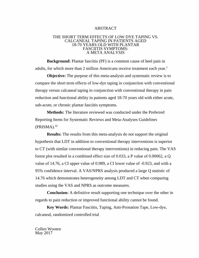
ABSTRACT THE SHORT TERM EFFECTS OF LOW DYE TAPING VS. CALCANEAL TAPING IN PATIENTS AGED 18-70 YEARS OLD WITH PLANTAR FASCIITIS SYMPTOMS: A META ANALYSIS Background: Plantar fasciitis (PF) is a common cause of heel pain in adults, for which more than 2 million Americans receive treatment each year. 1 Objective: The purpose of this meta-analysis and systematic review is to compare the short term effects of low-dye taping in conjunction with conventional therapy versus calcaneal taping in conjunction with conventional therapy in pain reduction and functional ability in patients aged 18-70 years old with either acute, sub-acute, or chronic plantar fasciitis symptoms. Methods: The literature reviewed was conducted under the Preferred Reporting Items for Systematic Reviews and Meta-Analyses Guidelines (PRISMA). 32 Results: The results from this meta-analysis do not support the original hypothesis that LDT in addition to conventional therapy interventions is superior to CT (with similar conventional therapy interventions) in reducing pain. The VAS forest plot resulted in a combined effect size of 0.033, a P value of 0.00062, a Q value of 14.76, a CI upper value of 0.989, a CI lower value of -0.923, and with a 95% confidence interval. A VAS/NPRS analysis produced a large Q statistic of 14.76 which demonstrates heterogeneity among LDT and CT when comparing studies using the VAS and NPRS as outcome measures. Conclusion: A definitive result supporting one technique over the other in regards to pain reduction or improved functional ability cannot be found. Key Words: Plantar Fasciitis, Taping, Anti-Pronation Tape, Low-dye, calcaneal, randomized controlled trial Collen Wooten May 2017
-
Upload
khangminh22 -
Category
Documents
-
view
3 -
download
0
Transcript of the short term effects of low dye taping vs. calcaneal taping in ...
ABSTRACT
THE SHORT TERM EFFECTS OF LOW DYE TAPING VS. CALCANEAL TAPING IN PATIENTS AGED
18-70 YEARS OLD WITH PLANTAR FASCIITIS SYMPTOMS:
A META ANALYSIS
Background: Plantar fasciitis (PF) is a common cause of heel pain in
adults, for which more than 2 million Americans receive treatment each year.1
Objective: The purpose of this meta-analysis and systematic review is to
compare the short term effects of low-dye taping in conjunction with conventional
therapy versus calcaneal taping in conjunction with conventional therapy in pain
reduction and functional ability in patients aged 18-70 years old with either acute,
sub-acute, or chronic plantar fasciitis symptoms.
Methods: The literature reviewed was conducted under the Preferred
Reporting Items for Systematic Reviews and Meta-Analyses Guidelines
(PRISMA).32
Results: The results from this meta-analysis do not support the original
hypothesis that LDT in addition to conventional therapy interventions is superior
to CT (with similar conventional therapy interventions) in reducing pain. The VAS
forest plot resulted in a combined effect size of 0.033, a P value of 0.00062, a Q
value of 14.76, a CI upper value of 0.989, a CI lower value of -0.923, and with a
95% confidence interval. A VAS/NPRS analysis produced a large Q statistic of
14.76 which demonstrates heterogeneity among LDT and CT when comparing
studies using the VAS and NPRS as outcome measures.
Conclusion: A definitive result supporting one technique over the other in
regards to pain reduction or improved functional ability cannot be found.
Key Words: Plantar Fasciitis, Taping, Anti-Pronation Tape, Low-dye,
calcaneal, randomized controlled trial
Collen Wooten May 2017


THE SHORT TERM EFFECTS OF LOW DYE TAPING VS.
CALCANEAL TAPING IN PATIENTS AGED
18-70 YEARS OLD WITH PLANTAR
FASCIITIS SYMPTOMS:
A META ANALYSIS
by
Collen Wooten
A project
submitted in partial
fulfillment of the requirements for the degree of
Doctor of Physical Therapy
in the Department of Physical Therapy
College of Health and Human Services
California State University, Fresno
May 2017

APPROVED
For the Department of Physical Therapy:
We, the undersigned, certify that the project of the following student meets the required standards of scholarship, format, and style of the university and the student's graduate degree program for the awarding of the doctoral degree. Collen Wooten
Project Author
Deborah Walker (Chair) Physical Therapy
Jenna Sawdon-Bea Physical Therapy
For the University Graduate Committee:
Dean, Division of Graduate Studies

AUTHORIZATION FOR REPRODUCTION
OF DOCTORAL PROJECT
X I grant permission for the reproduction of this project in part or in
its entirety without further authorization from me, on the
condition that the person or agency requesting reproduction
absorbs the cost and provides proper acknowledgment of
authorship.
Permission to reproduce this project in part or in its entirety must
be obtained from me.
Signature of project author:

ACKNOWLEDGMENTS
I would like to thank my chair, Dr. Walker, my committee member, Dr.
Jenna, and my advisor, Dr. Singh for guiding me throughout the EBP process and
supporting me. Your patience and time spent guiding me is appreciated more than
you can ever imagine. A special thanks to Fresno State’s Physical Therapy
Department and faculty for helping me develop the necessary skills needed to
become a successful physical therapist. And a very special thanks to my family
and friends, specifically my mom, dad, and brother, for helping me become the
person I am today and supporting me throughout my life path. I love you guys SO
much.

TABLE OF CONTENTS
Page
LIST OF TABLES ................................................................................................. vii
LIST OF FIGURES ............................................................................................... viii
BACKGROUND ...................................................................................................... 1
Prevalence, Risk Factors, and Clinical Presentation of Plantar Fasciitis .......... 1
Anatomy ............................................................................................................ 2
Mechanism of Injury ......................................................................................... 3
Interventions ...................................................................................................... 4
Review of Current Taping Literature ................................................................ 7
Outcome Measures .......................................................................................... 14
Importance, Hypothesis, and Purpose of this Meta-Analysis and Systematic Review ............................................................................... 14
METHODS ............................................................................................................. 16
Literature Search Strategy ............................................................................... 16
Inclusion Criteria ............................................................................................. 16
Exclusion Criteria ............................................................................................ 17
Study Selection ................................................................................................ 17
Study Quality .................................................................................................. 17
Data Collection ................................................................................................ 18
Analysis of Data .............................................................................................. 18
RESULTS ............................................................................................................... 20
Meta-Analysis and Systematic Review Study Selection ................................ 20
Included Study Characteristics of Meta-Analysis ........................................... 20
Data Analysis of Individual Studies Used in Meta-Analysis ......................... 24
Included Study Characteristics of Systematic Review ................................... 25

Page
vi vi
DISCUSSION ......................................................................................................... 29
Meta-Analysis Review .................................................................................... 29
Limitations of Meta-Analysis Studies Causing Heterogeneity ....................... 31
Analysis of Systematic Review ....................................................................... 35
Limitations of Literature ................................................................................. 37
Clinical Implications of Meta-Analysis Combined with Systematic Review ................................................................................................. 38
Risk of Bias in This Meta-Analysis and Systematic Review ......................... 40
Future Studies .................................................................................................. 41
Conclusion ....................................................................................................... 42
REFERENCES ....................................................................................................... 43
TABLES ................................................................................................................. 49
FIGURES ............................................................................................................... 54
APPENDICES ........................................................................................................ 59
APPENDIX A: GAIT CYCLE ............................................................................... 60
APPENDIX B: VAS, NPRS OUTCOME MEASURES ....................................... 62
APPENDIX C: PATIENT SPECIFIC FUNCTIONAL SCALE ........................... 64
APPENDIX D: WINDLASS MECHANISM ........................................................ 66
APPENDIX E: MARTIN ET AL. CLINICAL PRACTICE GUIDELINES SCORING ................................................................................................... 68
APPENDIX F: STAGES OF INJURY .................................................................. 70
APPENDIX G: MODIFIED FORMS OF TAPING .............................................. 72
APPENDIX H: EVIDENCE HEIRARCHY TRIANGLE ..................................... 74

LIST OF TABLES
Page
Table 1. List of Conventional Therapy Interventions ............................................ 50
Table 2. Studies Included in Meta-Analysis .......................................................... 51
Table 3. PEDro Scores of Articles Used in Meta-Analysis ................................... 52
Table 4. Studies Used in Systematic Review ......................................................... 53

LIST OF FIGURES
Page
Figure 1. Low-Dye Taping Technique ................................................................... 55
Figure 2. Calcaneal Taping Technique ................................................................... 56
Figure 3. Consort Diagram ..................................................................................... 57
Figure 4. VAS Forest Plot ...................................................................................... 58

BACKGROUND
Prevalence, Risk Factors, and Clinical Presentation of Plantar Fasciitis
Plantar fasciitis (PF) is a common cause of heel pain in adults. It is
estimated that more than 2 million Americans receive treatment each year for
plantar fasciitis.1 Plantar Fasciitis has been reported to affect a wide sample of
patients, including both the non-athletic and athletic population.2 In the non-
athletic population, it is most frequently seen in weight bearing occupations, with
70% of cases involving unilateral symptoms. In the athletic population, 10% of all
running athletes involved in basketball, tennis, football, long distance running, and
dancing have noted high prevalence of PF.1-3 Plantar Fasciitis can be equally
present in both male and female patients within a wide age range.
Men and women with poor gait mechanics that include excessive pronation
with resultant loss of plantar fascia extensibility are common risk factors for PF
symptoms. Individuals with these poor gait mechanics, especially during loading
and pre-swing phases, can affect both the athletic and sedentary populations.4-7
Other risk factors include limited ankle dorsiflexion range of motion, high body
mass index in non-athletic individuals, consistent running, and work-related
weight-bearing activities particularly under conditions with poor shock absorption
such as worn shoe wear and unforgiving surfaces.2 Plantar fasciitis symptoms,
such as pain location, can vary in individuals.
Typically, plantar fasciitis symptoms are characterized by pain and palpable
tenderness in the area of the medial tubercle of the calcaneus (medial heel), pain
that is increased when taking the first few steps in the morning, and pain that is
worse when weightbearing.8-10 Medial heel pain is considered a common clinical

2 2
finding as well. In order to better understand PF and its associated pathologies
however, knowledge of the anatomy of plantar fascia is crucial.
Anatomy
The plantar fascia, or plantar aponeurosis, is the investing fascia of the sole
of the foot and forms a strong mechanical linkage between the calcaneus and the
toes. Synonymous with the deep fascia, the plantar aponeurosis arises
predominantly from the medial process of the calcaneal tuberosity and attaches
distally, through several slips, to the plantar aspect of the forefoot as well as the
medial and lateral intermuscular septa.11 Anatomically the fascia can be divided
into 3 components or bands: the lateral, medial, and central bands.12,13
When present, the lateral band arises from the lateral margin of the medial
tubercle and provides a partial origin for the abductor digiti minimi muscle. At the
level of the cuboid, the lateral band bifurcates into medial and lateral crura. The
stronger lateral crux inserts into the base of the fifth metatarsal to form the plantar
ligament of the sole. The medial crux, in contrast, courses distally and receives a
contribution of the central band of the plantar fascia before coursing deep and
inserting deep into the plantar plate of either the third, fourth, or fifth
metatarsophalangeal joint. The central aponeurotic band is often cited as the major
component of the plantar fascia both structurally and functionally. The apex
originates from the plantar aspect of the medial process of the calcaneal tuberosity
where it serves as a partial origin for flexor digitorum brevis as it conforms to the
convex plantar surface of the calcaneus.11
Overall, the role of the plantar fascia is twofold, to provide support of the
longitudinal arch while also serving as a dynamic shock absorber for the foot and
entire leg.14 Due to its vital function (supporting body weight and providing shock
3 3
absorption) and anatomical location, plantar fasciitis tissue can be exposed to
various mechanisms of injury (see Appendix A).
Mechanism of Injury
Typically plantar heel pain comprises of inflamed fasciitis as well as tissue
surrounding this fasciitis structure, hence termed plantar fasciitis. Plantar fasciitis
is due to collagen disarray in the absence of inflammatory cells. This pathology
can resemble that of tendinosis and the condition can also be referred to as plantar
fasciosis or fasciopathy.15 The 2 most common underlying causes of plantar heel
pain are mechanical and degenerative in nature and are believed to result from
either trauma or overuse and can result in acute, sub-acute, and chronic conditions.
A common mechanically-related injury includes a predisposing factor such
as excessive pronation. This contributes to the biomechanics of an adducted talus
and everted calcaneus and can result in increased tension in the structures on the
plantar surface of the foot, causing arch collapse and creating excessive tensile
strain within the plantar fascia which can produce microscopic tears causing
acute/sub-acute inflammation.14 Another form of injury can occur from prolonged
degeneration.
Degeneration caused by repetitive stress to the origin of this fascia on the
calcaneus may cause tissue breakdown and calcium deposits, forming spurs
known as calcaneal spurs. This is indicative of chronic PF symptoms.16 Calcaneal
spurs can be an irritating component of plantar fascia, present as heel pain, and are
often developed through similar mechanisms. Due to this, both heel pain and PF
pathologies are treated similar and are often times considered together.16 Heel pain
can be caused by mechanical causes as well. Various interventions can be used in
order to manage these mechanisms of injury (see Appendices B-D).

4 4
Interventions
The commonly prescribed treatment options for PF are conservative and
surgical interventions. 1,5,17,21 Some conservative interventions used to treat PF
typically include ultrasound, stretching, and taping and are more conventional type
interventions. Other conservative interventions include manual therapy, foot
orthoses, night splints, etc. (see Table 1 for full list).17 This meta-analysis and
systematic review will focus on conventional therapy interventions: ultrasound,
stretching, and taping.
Ultrasound therapy is a common choice of treatment for reducing pain and
inflammation in plantar fasciitis.18 Therapeutic ultrasound is a method of applying
deep heat to connective tissue.19 This treatment strategy relieves plantar heel pain
by both thermal and mechanical effect.20,21 Therapeutic ultrasound (US), described
as a high-frequency mechanical wave, transmits energy through vibration, and is
extensively used in clinics.7 Ultrasonic generators are able to deliver energy in 2
modalities: continuous or pulsed. In the continuous form, the wave power
(measured as w/cm²) remains steady, and its expected effects involve the
production of deep heat, increased local blood flow, and pain relief. If used in high
powers (1.3 to 3.0 w/cm²), it also acts on fibrosis termination.7 Monophasic pulsed
current can be used to promote fascia, wound, and pressure ulcer healing
processes. Delivering of electrical current using electrodes has been shown to
induce cellular actions and histological responses (such as collagen and
deoxyribonucleic acid synthesis). Polarity from pulsed ultrasonic current can cause
a negatively charged cathode to attract the positively charged fibroblast cells to
promote and accelerate proliferation phases of plantar fascia.51
Ultrasound is used commonly in PT treatment settings.7 However, the
evidence for the use of US for PF is controversial. Shanks et al. in 2010 conducted

5 5
a literature review of the effectiveness of ultrasound in lower extremity
musculoskeletal conditions and deemed only 1 study out of 10 as high quality
evidence (16+) and found no statistical significance between sham ultrasound and
ultrasound treatments for both pain relief according to VAS and a pain
questionnaire and functional outcome according to objective assessment of
function.22 Additionally, research done by Martin et al. in 2014, used to revise the
clinical practice guidelines, provides weak evidence for the recommendation that
US alone cannot be recommended for individuals with PF.17
Another intervention used to treat PF is stretching, a technique that has
been shown to be useful in individuals presenting with PF symptoms.7,23,25
Stretching techniques have also been used to alleviate symptoms associated with
plantar heel pain as well. Stretching of the Achilles tendon and plantar fascia,
performed 3 to 5 times daily, has been shown to be effective in decreasing the pain
at the plantar fascia.5 Stretching of both the plantar fascia and Achilles tendon is
also considered a conventional type of therapy in patients with PF symptoms.
Sweeting et al. in 2011 conducted a systematic review of 6 studies concerning
stretching techniques in patients with PF.23 They reported that most participants
improved over the course of the studies in both visual analog scale (VAS) scores
and functional scores according to patient specific functional scale (PSFS), foot
function index (FFI), and the foot health status questionnaire (FHSQ) but when
stretching was compared to alternative or control interventions, the changes only
reached statistical significance in one study that used a combination of calf muscle
stretches and plantar fascia stretches in their stretching program. However, these
authors revealed there is some evidence that plantar fascia stretching may be more
effective than Achilles tendon stretching alone in the short-term. Martin et al. also
reported that stretching of the plantar fascia and/or calf musculature received an A

6 6
score (indicating strong evidence) and can be used to provide short term relief for
patients experiencing heel pain or PF.17 Even though stretching is shown to
alleviate symptoms, it does not address the underlying pathology of poor foot
biomechanics and therefore may only provide temporary relief.5 An intervention
that can provide relief of pain symptoms while also providing stability for those
with poor foot biomechanics is taping (see Appendices E-G).
Martin et al. gave taping an A score indicating strong evidence that
supports the use of taping for patients with PF symptoms.17 There are various
types of taping techniques available to treat plantar fasciitis. Two techniques are
low dye taping24 and calcaneal taping.24,25 These taping strategies can be valuable
since tape typically remains in place for 24 hours a day for 7 to 10 days.21 These
taping techniques are thought to improve the biomechanics of the faulty foot and
can correct either rearfoot, midfoot or forefoot region alignment.21
Low-dye taping in particular involves an adhesive taping material meant to
combat excessive pronation.26 A detailed picture of this process with steps can be
seen in Figure 1. This taping strategy includes placing an anchor strip with 1 to 1.5
inch tape first around the metatarsal heads followed by taping another anchor strip
covering the medial and lateral borders of the foot. Then, another strip of taping
begins at the fifth metatarsal head followed by the taping of the lateral border
upwards towards the heel, coming around the calcaneus and across to finish where
the taping originally began. After this, these steps are completed 3-4 more times
starting and finishing at the 1st metatarsal head. A circling of the arch (3 times)
followed by covering the original anchor strips with 2 more anchor strips is then
applied.27 This ensures supportive taping of the rearfoot and midfoot thus
preventing excessive pes planus and pronation. Another form of taping is
calcaneal taping.
7 7
Calcaneal taping involves taping of the posterior heel. Calcaneal taping is
done using similar tape which is cut into 4 parts. A detailed picture of this process
with steps can be seen in Figure 2. The first strip is applied distally to the lateral
malleolus, pulls the calcaneus medially, and attaches to the medial aspect of the
foot (distal to the medial malleolus). A second and third strip is then applied
following the same pattern with overlap of approximately one third of the tape
width moving in the distal direction. A fourth strip circles around the back of the
heel starting distally to the lateral malleolus, wrapping around the posterior aspect
of the calcaneus, and finally anchoring distal to the medial malleolus.14 Literature
concerning conventional therapy such as ultrasound, stretching, low-dye taping,
and calcaneal taping was gathered and assessed for this meta-analysis and
systematic review.
Review of Current Taping Literature
Taping and Pain Relief
The low-dye taping technique and calcaneal taping technique have been
shown to be effective treatment options for pain relief.5 Vicenzino et al.
investigated the effect of 2 taping techniques applied vertically to support
navicular height. Low-dye taping and LDT with calcaneal slings were compared.
Low-dye tape with calcaneal slings was found to be more effective immediately
after application and after exercising according to patient VAS reports.27
Aishwarya et al. conducted a study that compared the effects of CT versus a
modified LDT technique (less arch coverage, tape oriented in an X pattern on the
surface of the underfoot) on the calcaneal angle in subjects with PF symptoms.44
These authors found a pre-post comparison that showed reduced calcaneal
eversion angle and reduced pain intensity in both treatment groups. They deemed

8 8
this reduction in calcaneal eversion and pain insignificant. However, a between-
group analysis showed a significant reduction of calcaneal eversion angle in the
CT group of 3.4⁰ with a 95% CI range of -4.52⁰ to 2.4⁰ than the modified LDT
group. The reduction in pain intensity in the CT group was 20.7 points with a 95%
CI range of -30.61 to -10.72 and considered greater than the modified LDT group.
They concluded that CT provided better relief of pain with improved
biomechanical correction in the short term when compared to a modified LDT
technique.
Park et al. compared the effects of LDT on pain and stability. The subjects
were divided into 2 groups: a LDT group and a conservative treatment group. The
low-dye taping group received LDT and TENS and the conservative group
received TENS alone.27 They found a within-group comparison of the VAS
demonstrated that the LDT group and conservative group values significantly
decreased after taping, but the LDT group value decreased more significantly than
the conservative treatment group value and shown through a post-test between-
group comparison (p<0.05). The authors conducted a within-group comparison of
the transfer area of the center of gravity value and found that this significantly
increased after the taping intervention, but increased more significantly in the LDT
group compared to the CT group as shown by another post-test between-group
comparison (p<0.05).
Ranjan et al. conducted a study that compared LDT (however they did not
specify a control group) and found a decrease in VAS and an increase in FFI
scores among subjects. The average VAS score pre-treatment was 65.41 and
following treatment, 28.83. The average FHSQ score pre-treatment was 51.45 and
following treatment, 71.54 (demonstrating an increase in functional ability). The

9 9
authors concluded LDT was effective in both reducing pain and increasing
function in patients with PF symptoms.
Ernst et al. examined a modified LDT technique (same modifications as
Aishwarya et al.) supporting the longitudinal arch to relieve pain.5 Nineteen of 20
subjects reported a decrease in pain post-taping. However there is differing
evidence regarding pain reduction for taping as well.
In 2010, Van Der Water et al. conducted a systematic review of controlled
trials to determine the efficacy of taping in patients with PF symptoms and found
limited evidence that taping (both LDT and CT techniques) can reduce pain in the
short term in patients with plantar fasciitis but the effects on disability were
inconclusive.29 Studies assessing arch height, foot pressure, and pronation before
and after taping techniques also exist.
Taping and Arch Height, Foot Pressure, Ankle Dorsiflexion, and Pronation
Holmes et al. conducted an experiment to determine the effects of LDT in
regards to subtalar neutral joint (STJN) position.26 They found that a modified
LDT procedure could be an effective tool for the placement of the subtalar joint
into the neutral position. Low-dye taping may also be effective for maintaining the
STJN position through light exercise up to 10 minutes. However, the study did not
investigate whether or not the tape will continue to maintain the neutral position of
the subtalar joint through more vigorous or longer periods of exercise.
Vincenzino et al. conducted a study concerning the initial effects of LDT on
the medial and longitudinal arch during walking and running.42 They found that
compared with the no tape control condition, LDT produced a significant mean

10 10
increase in the medial longitudinal arch height index during standing, walking, and
jogging (p<0.05).
Vincenzino et al. also conducted a study concerning plantar foot pressures
following an augmented LDT (tape covering the first metatarsal head, around the
posterior heel, and finished on the fifth metatarsal head followed by transverse
strips anchored on the medial side covering the plantar surface) LDT
predominantly increased plantar pressures in the lateral midfoot during walking
and jogging.43 Additionally, LDT reduced mean maximum pressure at the medial
forefoot and at the medial rearfoot during walking.
Kang et al. conducted a study to investigate the effects of walking with CT
in subjects with limited ankle dorsiflexion passive range of motion (DF PROM).51
Fifteen ankles in this study presented with limited DF PROM and were examined.
After CT was applied, the subjects ambulated on a walkway for 10 minutes. A
goniometer was used to measure the ankle DF PROM while the knee was in an
extended position before and after walking with CT. The difference in ankle DF
PROM between before and after walking with CT was analyzed using a paired t-
test. The authors found that the ankle DF PROM was significantly increased after
walking with CT. The authors concluded that walking with CT is effective for
increasing the ankle DF PROM in individuals with limited ankle DF PROM.
Kang et al. conducted another study to determine the effects of walking
with CT on ankle dorsiflexion (DF) and heel-off time during the stance phase of
gait and ankle DF passive range of motion (PROM).52 Ten subjects participated in
this study and were analyzed. Sixteen ankles with limited ankle DF PROM were
tested. The subject’s ankle DF PROM was measured using a goniometer and the
ankle DF before heel-off and time to heel-off in the stance phase of gait was also
measured using a 3D motion analysis system before and after walking with CT.

11 11
Data was analyzed using a paired t-test. The authors found that ankle DF before
heel-off (p = 0.001), time to heel-off during the stance phase of gait (p = 0.005),
and ankle DF PROM (p < 0.001) was significantly increased post-CT compared
with pre-CT. They concluded that ambulating with CT is an effective self-exercise
for improving ankle kinematics during gait and increasing ankle DF PROM in
individuals with limited ankle DF PROM.
Yoon et al. also conducted a study to investigate the changes in passive
ankle dorsiflexion range of motion (ROM), maximum plantar force, force-time,
and time to heel off during walking between pre and post application of modified
CT (calcaneal taping strategy meant to mobilize with movement).53 Eighteen feet
with limited ankle dorsiflexion in 13 people were examined. Subjects were first
asked to walk before and immediately after applying the modified CT followed by
walking for 5 minutes with the modified CT. A floor mat pressure measurement
system (HR-mat) was used to measure maximum plantar force, force-time, and
time to heel off. Passive ankle dorsiflexion ROM was measured using a standard
goniometer. They found that passive ankle dorsiflexion ROM and time to heel off
were significantly increased after 5-minute walking with the modified CT
compared with walking before and immediately after applying the tape.
Significantly increased plantar force and force-time on the hindfoot and
significantly decreased force-time on the forefoot during walking after 5 minutes
was observed in the modified CT group. They found no significant difference
between before and immediately after applying the tape was observed in any
variable. The authors concluded that walking an additional 5 minutes with
modified CT increased passive ankle dorsiflexion ROM and time to heel off and
improved dynamic plantar loading during walking.

12 12
Weon et al. conducted a study to determine the effects of CT on peak
plantar pressure of the rearfoot and forefoot while ambulating.54 Fifteen subjects
with plantar heel pain participated in this study. A Pedoscan was used for the
recording of plantar pressure data during ambulation. The participants walked
along a 12 meter walkway before and after application of CT. The plantar pressure
gait was measured 3 times under barefoot and CT conditions randomly at a normal
walking speed. The peak plantar pressure data was calculated for the medial and
lateral areas of the rearfoot and the forefoot. A paired t-test was used to determine
significant differences in peak plantar pressures of the rearfoot and the forefoot
before and after the application of CT. The authors found that CT resulted in
statistically significant decreases in peak plantar pressure of the rearfoot (medial
side: p=0.03; lateral side: p=0.01). However, there was no significant change in
peak plantar pressure of the forefoot (medial side: p=0.45; lateral side: p=0.40).
The authors concluded that CT is recommended to reduce plantar pressure of the
rearfoot in weight-bearing activities in subjects with plantar heel pain.
Sanzo et al. conducted an experiment on vertical foot pressure in patients
with PF symptoms and found that LDT significantly decreased the pressure
transmitted through the foot during contact phases of gait.35 They did not find a
significant effect on the path of the center of pressure (COP). However, they did
find that the COP line was moved laterally thus decreasing medial plantar surface
pressure. They concluded that LDT may assist with the healing process to prevent
chronicity and degeneration due to excessive vertical foot pressure during contact
phases of gait and may be a possible consideration for treatment in the acute stages
of PF by unloading the foot as well.
Harradine et al. conducted a study concerning LDT and rearfoot
motion/position before and after exercise and found that taping immediately

13 13
reduced resting calcaneal stance position significantly (P<0.05) but this control
was lost after exercise.37 They concluded that LDT made no significant difference
in total pronation or maximum pronation velocity during walking.
O’Sullivan et al. conducted a study concerning LDT on rearfoot motion and
plantar pressure during the stance phase of gait and found LDT is associated with
reduced peak plantar pressure in the midfoot and forefoot that indicate reduced
pronation with LDT.38 LDT appears to reduce both pronation and supination in the
rearfoot, rather than simply reducing pronation, when assessed using 3D motion
analysis. The authors concluded that there is a potential that LDT can reduce
pronation by restricting rearfoot motion rather than pronation specifically.
However, the degree of change observed with LDT was very small, and further
research is needed to clarify the clinical significance of these initial findings.
Radford et al. conducted a systematic review of the effect of LDT on
kinematic, kinetic, and electromyographic variables and found LDT provides a
small change in navicular height post application, although it is unclear whether
this change is clinically important.39 There was high heterogeneity between some
trials examining other variables, indicating that more research is needed to confirm
the results of previous trials.
Russo et al. conducted a study concerning the effect of LDT on peak
plantar pressures of normal feet during gait and found overall, LDT significantly
reduced the peak plantar pressures of normal feet during gait.40 Of particular
interest was that a significant reduction in mean peak plantar pressure was
observed in the medial midfoot (1.4 N/cm2) and an increase occurred in the lateral
midfoot (2.6 N/cm2).
Keenan and Tanner examined the effects of high-dye and low-dye taping of
the rear foot.6 Eighteen subjects were tested under 3 conditions: barefoot, low-dye

14 14
taping, and high-dye taping (taping of low-dye style with additional taping that
crosses the talocrural joint). Two-dimensional motion of the rearfoot was assessed
for each condition. The results indicated that inversion was increased with both
high-dye and low-dye taping as compared with no taping at all. However, only
high-dye taping, significantly reduced the eversion of the rearfoot and was
considered more effective for control of pronation. Various outcome measures can
be used to gauge the effects of taping on patients with PF symptoms.
Outcome Measures
Typical and clinically accepted outcome measurements gauging progress of
individuals with plantar fasciitis symptoms include the visual analog scale (VAS),
foot function index (FFI), plantar fasciitis disability scale (PFDS), foot and ankle
outcome score (FAOS), foot health status questionnaire (FHSQ), patient specific
functional scale (PSFS), numerical pain rating scale (NPRS), foot pain and
disability scale (FPDS), McGill pain questionnaire, and the American orthopedic
foot and ankle score (AOFAS). Of these outcome measures, the VAS and NPRS
have been shown to have a high correlation between each other.31 The VAS is of
particular interest in this meta-analysis due to its reliability and clinical use in
measuring pain. The VAS includes a subjective report from a patient that ranges
from 0 (no pain) to 10 (worst pain) or 0-100, depending on the VAS template.
Importance, Hypothesis, and Purpose of this Meta-Analysis and Systematic Review
Taping as an intervention, or as part of an intervention, for the treatment of
plantar fasciitis has been used for at least 70 years.5 During this literature review,
only one systematic review by Podolsky et al. was found assessing the efficacy of
low-dye and calcaneal taping treatment strategies.57 There is limited evidence and

15 15
studies done comparing the efficacy of low-dye taping and calcaneal taping in
treatment of plantar fasciitis.18 Therefore, it was considered relevant to review the
literature concerning this topic.5 Studies have shown low-dye taping techniques to
improve PF symptoms in regards to pain reduction. 5,18,27,29 It is hypothesized that
low-dye taping combined with conventional therapy (ultrasound and stretching)
will be more effective in reducing pain than calcaneal taping combined with
conventional therapy (ultrasound and stretching) in patients with PF symptoms. A
meta-analysis comparing VAS and NPRS scores was conducted in order to assess
both taping techniques and their effects on reported pain in patients with PF
symptoms. Due to this meta-analysis only comparing pain as an outcome measure
among subjects, a systematic review was also considered appropriate to further
investigate LDT and CT effects on function. The systematic review conducted
included articles concerning patients with PF symptoms, taping interventions, and
functional outcome measures such as the FFI, PSFS, FHSQ, and FPDS in order to
assess the functional effects of both taping techniques. This ensures a comparison
of articles that include a subjective pain assessment (VAS, NPRS) and a functional
questionnaire assessment (see Appendix H).
The purpose of this meta-analysis and systematic review is to compare the
short term effects of low-dye taping in conjunction with conventional therapy
versus calcaneal taping in conjunction with conventional therapy in pain reduction
and functional ability in patients aged 18-70 years old with either acute, sub-acute,
or chronic plantar fasciitis symptoms.

METHODS
Literature Search Strategy
The literature reviewed was conducted under the Preferred Reporting Items
for Systematic Reviews and Meta-Analyses Guidelines (PRISMA).32 The
databases searched consisted of: CINAHL plus with full text (EBSCO), Proquest
Physical Education Index, Pubmed, Academic Search Complete (EBSCO),
Cochrane Central Register of Controlled Trials, and Cochrane Database of
Systematic Reviews. All databases were accessed through Henry Madden Library
under the physical therapy subject heading. Search terms included: “plantar
fasciitis AND randomized control trial, randomized controlled trial, randomized
clinical trial, systematic review, antipronation, taping, and tape.” All articles
screened were published from 2005-2016, written in the English language,
concerning the human species, and peer reviewed. Inclusion and exclusion criteria
were established for this meta-analysis and systematic review as well.
Inclusion Criteria
Articles screened for this meta-analysis underwent an inclusion/exclusion
criteria based on relevancy. The inclusion criteria consisted of studies needing to
contain the VAS or NPRS as an outcome measure, involving patients between the
ages of 18-70 years old with either acute (4 weeks or less), sub-acute (within 4 to
16 weeks), or chronic (16 weeks or more) plantar fasciitis symptoms, studies
involving male and female individuals, and study designs with appropriate
scientific evidence. Systematic reviews, randomized clinical trials, cohort studies,
and case control studies were considered studies with appropriate scientific
evidence.
17 17
Exclusion Criteria
Studies were excluded if they were deemed outside of the inclusion criteria.
Additionally, studies including subjects having a clinical disorder where
therapeutic ultrasound is contraindicated, spasticity throughout the lower
extremity, previous surgery or treatment for plantar fasciitis, history of ankle or
foot fractures, congenital deformities of the foot or ankle, impaired circulation to
the lower extremities, referred pain due to sciatica and/or other neurological
disorders, history of any skin condition where myofascial release is
contraindicated, subjects with arthritis, subjects with traumatic heel pain
(including use of pain or anti-inflammatory medication), those with corticosteroids
injections preceding 3 months, and subjects with known tape allergies were
excluded from this meta-analysis.
Study Selection
Studies were gathered and assessed using a standardized method by a single
reviewer. Studies that did not meet the inclusion criteria were excluded. Those
studies that were appropriate after inclusion and exclusion criteria screening were
then assessed for this meta-analysis. An in-depth outline of this process, including
a list of the inclusion and exclusion criteria, can be seen in Figure 3. Individual
study quality was assessed using the PEDro scale.
Study Quality
Studies used for this meta-analysis were assessed via the 11-point PEDro
scale in order to gauge quality.33 A higher score indicates a higher validity and a
lower score indicates a risk of study bias. Individual points are given to studies
based on whether or not certain validity criterion are met. The first criterion of the
PEDro scale is not used for the total score of the study assessed, making the total

18 18
point value 10 points. Studies included in this meta-analysis ranged from 1 to 4 on
the PEDro scale and can be seen in Table 2. Remaining studies were then entered
into a MetaAnalyst© software.
Data Collection
Selected studies were then entered and interpreted by MetaAnalyst©
software program in order to produce statistical results. Treatment statistics
including outcome measure effect sizes, statistical homogeneity, statistical
heterogeneity, and a forest plot was produced by this software. Data from the
results sections of each study were extracted and used for the statistical portion.
Individual control and experimental group results were extracted from studies and
combined during this meta-analysis based on similar sample sizes and outcome
measures used in the studies. Combining of data was necessary due to limited
amounts of studies involving the same parameters (similar sample size, outcome
measures used, duration of study, and conventional therapy parameters).
Extracted data from the studies used in this meta-analysis included means,
standard deviations, and sample sizes and are outlined in a forest plot. This forest
plot can be seen in Figure 4. An in-depth discussion of results can be found in the
section titled Results below. Only post-test data was used during the statistical
analysis portion of this meta-analysis.
Analysis of Data
Lower extremity pain was determined by the VAS and NPRS outcome
measure. For the purpose of the meta-analysis, potential homogeneity and effect
size was assessed using a Q value based on Cohen’s standards. These standards
indicate a Q value of >.8 as being a large effect, a value between .3-.8 as a
moderate effect, and a value of <.3 as a small effect. A p-value of <.05 is

19 19
considered to be statistically significant with a 95% confidence interval. Forest
plots were used to assess the grand effect size between studies. A random effect
size model was considered appropriate due to the small amount of studies included
in this meta-analysis.

RESULTS
Meta-Analysis and Systematic Review Study Selection
A total of 687 potential articles were obtained and screened from databases
consisting of CINAHL plus with full text (EBSCO), Proquest Physical Education
Index, Pubmed, Academic Search Complete (EBSCO), Cochrane Central Register
of Controlled Trials, and Cochrane Database of Systematic Reviews. Further
screening of these articles was done in order to eliminate duplicates, leaving 586
articles. These remaining articles were then assessed based on appropriateness of
their abstracts and whether or not they were written in the English language. Those
not written in the English language or showing no relevancy in abstracts were
discarded from the study, leaving 28 articles. The remaining 28 articles were
screened based on free full text availability, peer reviewed, and non-experimental
study criteria. All 28 articles were deemed appropriate after this screening. These
articles were then eliminated or kept based on experiment design, those that did
not have an experimental design were eliminated. After this elimination, 9
remaining articles were used for qualitative synthesis. Of these 9 articles, only 4
used similar outcome measures (VAS, NPRS and FFI) and presented with similar
treatment time parameters (1 week), this deemed them appropriate to compare and
use. For a detailed flow diagram outlining this process, see Figure 3. For a list of
characteristics of studies extracted from databases, hand searches, reference
list/citations see Figure 3. Included study characteristics were also assessed during
this meta-analysis and systematic review.
Included Study Characteristics of Meta-Analysis
A characteristic list of all 4 studies used for this meta-analysis can be seen
in Table 2. The 4 studies included in this meta-analysis were randomized

21 21
controlled trials and were within the publication dates of 2010-2016. PEDro scores
of these articles ranged from 1-4 out of a 10 point scale. A detailed description of
individual PEDro scores can be seen in Table 3. Sankhe et al., Vishal et al.,
Dhillon et al., and Agrawal et al. were the authors of the remaining 4 studies used
for this meta-analysis. All 4 studies had similar age ranges and length of treatment
times (7 days). Dhillon et al. and Agrawal et al. used a similar subject amount (30
subjects) with the exception of Vishal et al. who conducted their study using 60
subjects and Sankhe et al. who used 52 subjects.
Sankhe et al. compared low-dye taping to calcaneal taping using 2 groups
(Group A and Group B).18 Group A received US on a continuous mode, with an
intensity of 1 W/cm2 and a frequency of 1 MHz for 5 minutes in a prone position
for 7 sittings, 1 sitting per day. This was followed by CT. Group B received US
with the same parameters as Group A followed by LDT. Both groups received
these 2 interventions daily for 7 days but did not receive stretching interventions.
This study gave moderate description of their methods but did not include pictures
of the 2 taping techniques and did not give reproducible specifics regarding both
taping techniques. Sankhe et al. used both the VAS and FFI as outcome measures
to gauge patient pain and functional levels before and after 7 daily treatment
sessions. The authors revealed that during the comparison of group A and group B
there was significant reduction in pain in group B (p< 0.001). However, the
authors failed to report their FFI results and used a 0-100 VAS scale that was
scaled down to a 0-10 scale in order to input the statistics for this meta-analysis.
The authors still concluded that LDT was significantly more effective than CT in
reducing pain and increasing foot function in patients with plantar fasciitis without
quality functional results.
22 22
Vishal et al. also compared low-dye taping to calcaneal taping using 2
groups.21 Group A received US at a continuous mode with an intensity of 1W/cm2
and a frequency of 1 MHz for 5 minutes in prone lying for 7 sittings with 1 sitting
per day. Stretching was also given in the form of passive stretching of the ankle
flexors and plantar fascia performed in supine lying with the soleus muscle
stretched during knee flexion and a gastrocnemius stretch with the knee extended.
Overpressure was placed upon the bottom of the foot while the ankle was in
dorsiflexion, a passive stretch was then applied to the big toe flexors to incorporate
stretch to the plantar fascia. These stretches were given for 3 repetitions each and
held for a count of 30 seconds per stretch in addition to CT with reproducible
detail and pictures. Group B received the same US and stretching interventions in
addition to LDT with reproducible detail and pictures. Vishal et al. also used both
the VAS and FFI as outcome measures to gauge patient pain and functional levels
before and after 7 daily treatment sessions. They found that there was significant
change in pain relief as per VAS score (p<0.001) and improvement in functional
ability as per FFI (p<0.0001). They concluded that both groups revealed a
decrease in pain and an increase in function but that group A demonstrated better
results than group B.
Dhillon et al. conducted a study comparing low-dye taping versus
myofascial release in patients with PF symptoms.55 They placed subjects into 3
groups, Group A, B, and C. All groups received US (US with an output of 1w/cm2,
pulsed mode 1:4 ratio, for 5 minutes with frequency of 1 MHz) and stretching
(patient was asked to complete active calf stretch in standing by leaning against
the wall, holding each stretch for 1 minute, and repeating 5 times each session).
Group A received just US and stretching. Group B received the same US and
stretching interventions combined with a myofascial technique to the underside of

23 23
the foot. Group C received the same US and stretching interventions combined
with low-dye taping given in reproducible detail (similar to Vishal et al.).
Interventions were given daily for 7 days. Data was collected on the 1st and 7th day
using the NPRS as a pain outcome measure. The authors concluded that all 3
groups showed significant reduction of pain. However, a between groups analysis
was done that demonstrated group B as showing more significant reduction of
pain.
Agrawal et al. compared calcaneal taping to sham taping using 2 groups.56
Group A received US set at 3 Mhz with 1.0 w/cm2 and continuous for 7 minutes
with stretching (a passive stretch applied to the big toe flexors in order to stretch
the plantar fascia for 3 repetitions. Each stretch was held for a count of 30
seconds) and CT was given described in reproducible detail with a picture.
However, a modified calcaneal taping technique was used (tape passes medially
and superiorly to the tendon of the tibialis anterior to avoid problems while
ambulating first strip is placed obliquely around the back of the heel while the
calcaneus is externally rotated and extended around the lower leg, then a second
strip of tape is used to reinforce the first). The patient is asked to keep the tape in
place for 24 hours. Group B received the same US and stretching parameters as
group A in addition to sham taping. The VAS and patient specific functional scale
(PSFS) were outcome measures used in this study to gauge patient pain and
functionality. A significant difference was found post treatment among the groups
for VAS (p=0.0265). A highly significant difference was found post treatment
among the groups for PSFS (p= 0.0062). There were significant differences found
between pre and post VAS and PSFS scores in both the experimental and control
group. The authors concluded that a modified CT was shown to be a more

24 24
effective tool for the relief of plantar heel pain than sham taping when combined
with conventional physical therapy.
These 4 studies used slightly differing US parameters (with the exception
of Vishal et al. and Sankhe et al.) and also had differing stretching techniques
used, with the exception of Sankhe et al. (which did not include a stretching
intervention). This, amongst other components, adds to the potential risk of bias
amongst the 4 studies used for this meta-analysis and is discussed further in the
Discussion section. Data analysis of all 4 studies was assessed during this meta-
analysis.
Data Analysis of Individual Studies Used in Meta-Analysis
The effect sizes were assessed using a Q-value, with a Q value of more than
0.8 as having a large effect, between 0.3-0.8 as having a moderate effect, and less
than 0.3 as having a small effect. Potential homogeneity was assessed by using a P
value, with a P value of more than 0.05 considered homogenous and a P value of
less than .05 considered heterogeneous. A random effect size model was
considered appropriate due to the differing I2 values amongst the articles. This
random effects model compared individual article group means and correlating
standard deviations for this meta-analysis. In this meta-analysis, individual study
statistics were also assessed.
The VAS forest plot resulted in a combined effect size of 0.033, a P value
of 0.00062, a Q value of 14.76, a CI upper value of 0.989, a CI lower value of -
0.923, and with a 95% confidence interval. It should be noted that a meta-analysis
Q value lower than the degrees of freedom and a P value more than .05, labels a
group of studies as homogeneous. When comparing the VAS statistics Q value for
this meta-analysis, the Q value (14.76) is significantly higher than the degrees of

25 25
freedom (2) and the P value (0.00062) is significantly lower than .05 which also
shows that the VAS results are heterogeneous, similar to the forest plot results.
This makes the results for the VAS forest plot heterogeneous with a moderate
effect size. A total of 4 studies were used to produce these results to make 3
comparisons, or plots, in the VAS forest plot results. A detailed forest plot can be
seen in Figure 4. For this systematic review, studies were screened via the same
parameters as the articles used for the meta-analysis. However, studies were
included if they contained any type of functional outcome measure correlated with
either LDT or CT and any duration of treatment. A total of 5 studies were deemed
appropriate for this systematic review and are assessed below.
Included Study Characteristics of Systematic Review
The purpose of this systematic review is to compare the short term effects
of low-dye taping in conjunction with conventional therapy versus calcaneal
taping in conjunction with conventional therapy on functional ability in patients
aged 18-70 years old with either acute, sub-acute, or chronic plantar fasciitis
symptoms. A characteristic list of all 5 studies used for this systematic review can
be seen in Table 4. The studies included were 5 randomized controlled trials and
were within the publication dates of 2006-2016. These studies reviewed failed to
meet the criteria for this meta-analysis for a variety of reasons including differing
time parameters, differing outcome measures used, and differing interventions
compared to taping. Studies reviewed are meant to add to the understanding of
both LDT and CT and their effects on function in patients with acute, sub-acute,
and chronic PF symptoms.
Abd El Salam et al. conducted a RCT aimed to compare 2 methods of arch
support, LDT and medial arch support (MAS) in patients with PF symptoms.45

26 26
Thirty patients, 23 men and 7 women, with unilateral plantar fasciitis were
randomly assigned to either the LDT or MAS group. Both groups were assessed
before and after experiment conduction for foot function and pain. Both groups
received 9 sessions over 3 weeks consisting of ultrasound and calf muscles
stretching in addition to either LDT or MAS. They were instructed to maintain
supportive intervention (LDT or MAS) throughout this period as well. Pre-post
comparison showed reduced pain and improved function in both groups. Between-
groups analysis showed non-significant difference in pre-VAS and pre-FPDS.
Post-VAS and post-FPDS showed significant improvement in patients in the MAS
group. Results indicate that MAS is more efficacious for short-term management
of pain and disability in patients with plantar fasciitis than LDT.
Goyal et al. conducted a RCT meant to compare LDT and iontophoresis.46
The purpose of the study was to observe the effect of a combination of LDT and
iontophoresis or LDT alone in the treatment of PF symptoms. A total of 30
patients (16 males; 14 females) were selected as subjects and were further divided
into 2 groups. Each group comprised of 15 subjects (8 males; 7 females). The
results of the study show an improvement in the mean values of both VAS and FFI
scores after treatment in both groups. A statistical significance was found between
LDT combined with iontophoresis than LDT alone. It was concluded that the
patients with PF that were treated with a combination therapy (LDT and
iontophoresis) experienced significantly more recovery from pain and an increase
in functional ability.
Hyland et al. conducted a RCT comparing CT, sham taping, and plantar
fascia stretching.47 A total of 41 subjects were randomly assigned into 4 groups,
(1) stretching of the plantar fascia, (2) CT, (3) control (no treatment), and (4) sham
taping. A visual analog scale (VAS) for pain and a patient-specific functional scale

27 27
(PSFS) for functional activities were measured pretreatment and after 1 week of
treatment (posttreatment). A significant difference was found post-treatment
among the groups for the VAS (P<.001). Specifically, significant differences were
found between stretching and CT (mean ± SD, 4.6 ± 0.7 versus 2.7 ± 1.8; P =
.006), stretching and control (mean ± SD, 4.6 ± 0.7 versus 6.2 ± 1.0; P = .026), CT
and control (mean ± SD, 2.7 ± 1.8 versus 6.2 ± 1.0; P<.001), and CT and sham
taping (mean ± SD, 2.7 ± 1.8 versus 6.0 ± 0.9; P<.001). However, no significant
difference among groups was found for posttreatment PSFS (P = .078). Calcaneal
taping was shown to be a more effective tool for the relief of plantar heel pain than
stretching, sham taping, or no treatment.
Patil et al. conducted a RCT comparing LDT and myofascial release in
patients with PF symptoms.48 A total of 30 patients suffering from chronic PF
were randomized and divided into 2 groups. 15 patients underwent a myofascial
release technique and 15 patients underwent a LDT administered for 1 week (7
treatment sessions total). Outcome measures used included the VAS and FFI and
were collected at baseline and 1 week after initiation of therapy. The data was
analyzed using statistical testing which was performed using a SPSS 17 software
package. Results revealed significant improvement for all outcome measures in
each group (p<0.05). Additionally significantly greater improvements were
detected in favor of the myofascial release group (p < 0.05). They concluded
findings that indicate a potential benefit after giving a myofascial release
technique in patients with chronic planter fasciitis over LDT.
Radford et al. conducted a RCT to determine the effectiveness of LDT on
pain and function.49 Ninety-two participants with plantar heel pain (mean age 50 ±
14 years; mean body mass index 30 ± 6; and a median self-reported duration of
symptoms 10 months, range of 2 to 240 months) were recruited from the general

28 28
public. Participants were randomly allocated to (i) low-Dye taping and sham
ultrasound or (ii) sham ultrasound alone. The duration of follow-up for each
participant was 1 week. No participants were lost to follow-up. Outcome measures
included 'first-step' pain (measured on a 100 mm Visual Analogue Scale) and the
Foot Health Status Questionnaire domains of foot pain, foot function and general
foot health. Participants treated with LDT reported a small improvement in 'first-
step' pain after 1 week of treatment compared to those who did not receive taping.
The estimate of effect on 'first-step' pain favored the LDT (ANCOVA adjusted
mean difference -12.3 mm; 95% CI -22.4 to -2.2; P = 0.017). There were no other
statistically significant difference between groups. Thirteen participants in the
taping group experienced an adverse event however most were mild to moderate
and short-lived. When used for the short-term treatment of plantar heel pain, LDT
provides a small improvement in 'first-step' pain compared with a sham
intervention after a 1-week period.

DISCUSSION
Meta-Analysis Review
Four studies were deemed appropriate for this meta-analysis. All studies
were assessed with the same internal and exclusion criteria. Of these 4 studies, all
were deemed randomized controlled trials by the authors. The purpose of this
meta-analysis was to determine the effectiveness of low-dye taping versus
calcaneal taping on pain reduction in patients with plantar fasciitis symptoms. This
meta-analysis compares both low-dye taping and calcaneal taping interventions
and can add to the existing body of literature in order to determine which of these
taping methods is more beneficial or requires further available literature.
It should be noted that a traditional meta-analysis typically uses studies that
include both interventions of interest in order to produce results and compare
interventions directly. However, this meta-analysis was unique in that 2 studies,
Dhillon et al. and Agrawal et al., did not compare either taping techniques directly
(within the same study) yet had intervention combinations that met the inclusion
criteria.55,56 In order to add to this meta-analysis comparison, it was necessary to
combine LDT and conventional therapy intervention statistics with CT and
conventional therapy intervention statistics from 2 separate studies which
generated 3 comparisons and a subsequent forest plot. These generations produced
results that are not significant however.
The results from this meta-analysis do not support the original hypothesis
that LDT in addition to conventional therapy interventions is superior to CT (with
similar conventional therapy interventions) in reducing pain. The VAS forest plot
resulted in a combined effect size of 0.033, a P value of 0.00062, a Q value of
14.76, a CI upper value of 0.989, a CI lower value of -0.923, and with a 95%

30 30
confidence interval. A VAS/NPRS analysis produced a large Q statistic of 14.76
which demonstrates heterogeneity among LDT and CT when comparing studies
using the VAS and NPRS as outcome measures. Two studies (Dhillon et
al./Agrawal et al. and Sankhe et al.) favored LDT over CT and one study (Vishal
et al.) favored CT over LDT when comparing VAS and NPRS results.18,21,55,56
Therefore, it cannot definitively be said that one taping technique is superior to the
other in reducing pain through VAS and NPRS in patients with PF symptoms.
In a traditional meta-analysis, effect sizes are typically assessed after
several studies have been compared in order to more definitively review
interventions being assessed. For this meta-analysis, only 4 studies comprising 3
comparisons were produced. Review of these comparisons demonstrated a
moderate effect size between the 2 taping interventions. This effect size indicates a
moderate difference between taping techniques but again, this should be
interpreted with caution due to the limited amount of studies assessed.
After review of the results of this meta-analysis, there cannot be definitive
support of the original hypothesis of LDT as being more effective than CT in
reducing pain in patients with PF symptoms. Results of this meta-analysis
demonstrate heterogeneity, meaning these results should be interpreted with
caution and implications of this analysis will be discussed to help interpret the
results produced. Some of the limitations causing heterogeneity across these
studies include lowered PEDro scores, differing US intervention parameters,
differing stretching intervention parameters, differing baseline patient
characteristics, and lack of detail in taping techniques.
31 31
Limitations of Meta-Analysis Studies Causing Heterogeneity
A significant component causing heterogeneity in this meta-analysis
involves the poor methodological quality of all 4 studies used. PEDro scores of the
4 studies used for this meta-analysis ranged from 1-4 out of a 10 point scale and
can be considered to have high to moderate risk of bias. Of significant concern
includes all 4 studies lacking similarities in baseline subject characteristics,
lacking blinding of patients, lacking blinding of treating therapists, and lacking
concealed allocation thus presenting an increased chance for risk of bias due to
differing subject presentations, patient performance/perception bias, and treating
therapist care. Those studies with the lowest PEDro score included Dhillon et al.
and Sankhe et al.18,55 Specifically, Dhillon et al. report vague findings and make it
difficult to assess true patient baseline characteristics and interpretation of
statistics.55 Sankhe et al. demonstrate similarity in vague reporting as Dhillon et al.
and also fail to report functional outcomes, even though a functional outcome
measure was intended to have been utilized.18,55 Additionally, writing of both
studies present with grammatical errors that warrant concern in regards to
authenticity of being peer reviewed. In addition to poor methodological quality,
differing US parameters were also present among studies.
Vishal et al. and Sankhe et al. had identical US parameters used but
Agrawal et al. and Dhillon et al. had differing parameters.18,21,55,56 These
differences among parameters contribute to the heterogeneity between studies.
Vishal et al. and Sankhe et al. outlined US parameters with a frequency of 1
MHz.18,21 A lower frequency US, such as a 1 MHz setting, is typically intended to
induce healing effects on deeper tissue.22 Plantar fascia is considered superficial
tissue and therefore may not experience the intended effects of a lower frequency
US setting. Agrawal et al. and Dhillon et al. utilized a high frequency setting of 3

32 32
MHz which is more appropriate for inducing healing effects on superficial tissue
such as plantar fascia.55,56 However as stated earlier, Shanks et al. concluded that
there is no current high-quality evidence available that supports therapeutic US in
the treatment of lower limb musculoskeletal conditions.22 Additionally, Martin et
al. in 2014 revised the clinical practice guidelines concerning plantar fasciitis and
deemed evidence supporting US and its effects as weak.17 Stretching parameters
varied among the articles used in this meta-analysis as well.
All 4 studies outlined differing stretching techniques (with the exception of
Sankhe et al. which did not include a stretching intervention) thus furthering the
heterogeneity in this meta-analysis.18 Vishal et al. conducted stretching in subjects
that involved passive stretching of the ankle flexors and plantar fascia, Agrawal et
al. conducted plantar fascia specific stretching where a passive stretch was applied
to the toe flexors, and Dhillon et al. conducted stretching interventions that
involved subjects completing active calf stretching in standing by leaning against a
wall.21,55,56
Stretching of the plantar fascia and plantar flexors have been shown to have
positive effects in patients with plantar fasciitis symptoms.5,11,15 However,
Sweeting et al. in 2011 conducted a systematic review of 6 studies concerning
stretching techniques in patients with PF and concluded there were too few studies
to assess whether stretching is effective compared to control or other interventions,
for either pain or function. However, they did find some evidence that plantar
fascia stretching may be more effective than Achilles tendon stretching alone in
the short-term in regards to pain reduction.23
Martin et al. also reported that stretching of the plantar fascia and/or calf
musculature received an A score (indicating strong evidence) and can be used to
provide short term relief for patients experiencing heel pain or PF as well.17 Those

33 33
subjects receiving just plantar fascia stretching versus plantar fascia stretching
combined with plantar flexor stretching could have reported an increase in pain
reduction regardless of whether they underwent LDT or CT. Baseline
characteristics of the studies used in this meta-analysis varied.
Baseline characteristics of subjects in all 4 studies varied from acute, sub-
acute, and chronic conditions. These factors can further add to the cause for
heterogeneity. Patients with acute PF symptoms can respond differently than
patients with chronic PF symptoms. Vishal et al. and Sankhe et al. reported
patients who presented with symptoms more than 4 weeks, both sub-acute and
acute conditions of PF, while Agrawal et al. included patients with symptoms
more than 1 week but less than 6 weeks, which included acute and sub-acute
symptoms.18,21,56 Dhillon et al. included subjects with symptoms more than 6
weeks, ranging from sub-acute to chronic presentations of PF symptoms.55 These
differing timelines of symptoms among patients are a cause of heterogeneity,
considering acute and chronic presentations of PF symptoms can respond
differently to treatment even though both taping techniques were done under short
term (1 week) conditions. As mentioned earlier, taping can be considered a short
term intervention and may have a different effect on patients with an acute
presentation of PF versus chronic PF. Patients with acute PF symptoms could
present with more immediate pain relief than patients with chronic conditions
because acute symptoms may be more inflammation based than the chronic
presentation in which inflammation has been replaced by a change in the pliability
of soft tissue, ie. scar tissue. In the case of acute PF symptoms, studies suggest that
taping can provide immediate biomechanical changes in pronation and ankle
position that can place less damaging forces and plantar pressure on already
inflamed tissue.47,48,49 Biomechanical alignment changes in patients with acute PF

34 34
symptoms can lead to a biased improvement in patient reported pain. Conversely,
those with chronic PF symptoms may have tissue scarring, calcaneal spurs, and
tissue restrictions resulting in a limit in response to pain relief following taping.
Furthermore, calcaneal spurs can also impinge on tissue after newly aligned ankle
and foot positioning thus causing continued or increased patient reported pain.
Authors of all 4 studies also included a wide age range of subjects.
Agrawal et al. reported patients with ages from 18 to 35, Vishal et al.
reported ages 18 to 52, Dhillon et al. reported ages 18-40, and Sankhe et al.
reported ages from 18 to 65.18,21,55,56 Authors of the studies did not stratify subjects
by BMI, activity level, or high arch versus low arch as well. For example, an 18
year old athletic subject may have varying responses compared to a 65 year old
sedentary subject. Taping of a sedentary person with an increased BMI may not
have as much biomechanical change (decreased plantar pressure difference)
compared to the same taping of a person who is active and within a normal BMI.
Additionally, authors did not mention arch height in subjects prior to
interventions. Both LDT and CT are considered biomechanical adjustments and a
person who already has an increased arch may not report decreased pain from
either taping technique. Studies presented with limited reproducible taping detail
which is important for interventions such as taping.
Taping detail ensures that taping techniques are done correctly and can be
compared effectively. Vishal et al. and Dhillon et al. included detailed taping
strategies with accompanying pictures.21,55 Agrawal et.al. also outlined
reproducible taping detail through steps of taping but did not show a picture of
their final taping.56 Conversely, Sankhe et al. provided moderate detail to their
taping technique but did not include a picture of the finished taping.18 Agrawal et
al. utilized a modified CT technique that included tape passing medially and
35 35
superiorly to the tendon of the tibialis anterior with the first strip being placed
obliquely around the back of the heel while the calcaneus is externally.56 A second
strip of tape was used to reinforce the first. This taping method varies from the CT
mentioned in other studies assessed. Agrawal et al. demonstrated a CT technique
that could cause decreased biomechanical adjustment due to less calcaneal anti-
pronation support compared to the more prevalent method of CT seen in other
studies.56 This vague reporting of taping by Sankhe et al. and modified CT method
conducted by Agrawal et al. could further increase the reason for heterogeneity in
this meta-analysis.18,56
Analysis of Systematic Review
The purpose of this systematic review was to compare the short term effects
of low-dye taping and calcaneal taping on functional ability in patients aged 18-70
years old with either acute, sub-acute, or chronic plantar fasciitis symptoms. This
review was conducted in order to add to the understanding of both LDT and CT
and their effects on function. The studies included were 5 randomized controlled
trials and were within the publication dates of 2006-2016. Again, these studies
reviewed failed to meet the criteria for this meta-analysis for a variety of reasons
including differing time parameters, differing outcome measures used, and
differing interventions compared to taping.
All 5 studies assessed in this systematic review demonstrated a decrease in
pain through VAS scores of both LDT study groups and CT study groups. Of
these, 4 studies conducted by Abd El Salam et al., Goyal et al., Patil et al., and
Radford et al. compared LDT with conventional therapy interventions which
included US, Achilles tendon stretching, foot intrinsic muscle strengthening, and
iontophoresis (with the exception of Radford who paired LDT with sham US as

36 36
interventions).45,46,48,49 And the remaining study, Hyland et al. compared CT
independently.47 However, this analysis of the systematic review will focus on the
functional outcome measure results found among these studies.
Of the 5 studies reviewed, 3 studies reported an increase in functional
ability scores after LDT via functional outcome measures (the MFPDS, FFI, and
FHSQ) that have been validated and cited as reliable throughout literature.45,46,48
These 3 studies reported subject populations that ranged from 24-70 subjects. This
amount range of subjects can be considered clinically relevant and can add to the
current understanding of available research concerning LDT and function. There
was one study that demonstrated LDT as having no significant effect on functional
outcomes (Radford et al.).47 This study compared LDT and sham US versus sham
US alone found no significant difference between groups using the FHSQ.47 One
study concerning CT was assessed during this systematic review (Hyland et al.).49
This final study compared CT versus stretching of the plantar fascia, sham
taping, and a no treatment control group in patients with PF symptoms.49 Authors
found no significant difference between groups using the PSFS. After review of
the literature, a total of 3 studies were found (2 of which were included in this
meta-analysis) concerning CT and its effects on function. Vishal et al. and
Agrawal et al. conducted studies that were reviewed in this meta-analysis but also
reported functional outcomes via the FFI and PSFS.21,56 Vishal et al. reported a
significant improvement in PSFS scores in both LDT and CT groups.21 Agrawal et
al. reported a significant improvement in FFI scores in subjects receiving CT.56
Therefore, CT also presents with limited evidence regarding its effect on
improving functional ability in patients with PF symptoms. There is a significant
gap in the literature concerning CT that warrants review however. This lack of
literature concerning CT could be due to Middleton et al. describing a LDT
37 37
technique in 1992 that has been well cited and prevalent in the literature since,
thus standardizing the LDT technique.16 A standardization of CT does not seem to
exist and therefore may not be utilized as much as the LDT method.
The results of this systematic review yield limited evidence that both LDT
and CT can improve functional ability in patients with PF symptoms. It is difficult
to compare both techniques directly due to the lack of literature and available
literature quality concerning these 2 techniques. Therefore a definitive result
supporting one technique as being more functionally beneficial over the other
cannot be found. This lack of literature warrants further review.
Limitations of Literature
Podolsky et al. conducted a systematic review of taping in 2015 and
analyzed 5 randomized control trials, one cross-over study and 2 single group
repeated measures studies.57 They concluded taping in the short term is beneficial
during the treatment of plantar fasciitis. They also found that best evidence exists
for low dye taping and calcaneal taping. They recommended more research being
needed to investigate the long-term effect and effectiveness of specific taping
techniques. However, gaps in the literature concerning low-dye taping compared
to calcaneal taping exist. Currently, it seems only 2 studies, Sankhe et al. and
Vishal et al., compare both low dye and calcaneal taping directly.18,21 In addition
to this, taping as an independent intervention was uncommon in majority of
articles screened for this meta-analysis. Majority of studies screened included
other interventions coupled with taping techniques. These additional interventions
included stretching, ultrasound, foot intrinsic strengthening, modifying foot wear,
orthopedic inserts, and iontophoresis medications. Of these interventions,
stretching coupled with ultrasound were most prevalent. Another component to a

38 38
quality meta-analysis involves the comparison of articles that compare low-dye
taping versus calcaneal taping with similar intervention parameters. For example,
a study comparing low-dye taping and stretching versus calcaneal taping and
stretching would benefit by including similar stretching interventions (since
plantar fascia stretching can produce differing results than plantar flexor
stretching). This component adds to the existing limitations due to the possibility
of different stretching techniques confounding results. Reporting of reproducible
interventions varies among the literature as well.
Interventions described in some of the articles assessed were not specific
enough to reproduce. For taping, reproducible detail is important in order to
accurately assess articles when comparing intervention effectiveness. As discussed
previously there is a standardized technique for LDT that has been alluded to in
the literature however CT does not have the same standardization. Three of the 4
articles gathered for this meta-analysis used reproducible and detailed taping
techniques with the exception of Sankhe et al. (which was assumed to have used
the same LDT and CT technique as Vishal et al.).18,21 Literature gaps such as these
should be addressed in order to raise our understanding of the types of taping
techniques and improve clinical implications. The clinical implications that can be
gathered from this meta-analysis are still useful and can be used for currently
treating therapists however.
Clinical Implications of Meta-Analysis Combined with Systematic Review
Results of this meta-analysis do not support the original hypothesis of LDT
as being superior to CT in reducing pain for patients with plantar fasciitis
symptoms. Again, these results should be interpreted with caution due to the
heterogeneity found in this meta-analysis. However, LDT (in addition to

39 39
conventional therapy) may demonstrate a positive effect in VAS and NPRS as
seen in the literature.3,16,45,46,47 Several studies support that LDT can provide relief
of pain according to VAS scoring.5,27,29 A moderate amount of studies exist that
support LDT (in addition to conventional therapy) as improving functional ability
as well.6,7,45,46,48 For example, Landorf et al. and Patil et al. demonstrated a
positive effect in both VAS and functional scores when LDT was compared
against sham taping.18,34 Clinicians can use a LDT technique to improve short term
subjective pain perception and functional ability in patients with PF symptoms.
Especially individuals who present with plantar fasciitis due to a high BMI, having
limited dorsiflexion range of motion, or being a consistent runner. In this situation
the application of LDT can support the medial longitudinal arch thereby
decreasing the ground reaction forces on plantar fascia tissue.
A review of the literature conducted by Martin et al. show that medial arch
inserts can demonstrate pain reduction in patients with PF symptoms as well,
however, it can be argued that taping can be a quick and relatively inexpensive
first attempt at biomechanical adjustments of the foot.14 Low-dye taping can
further inform the therapist and patient suffering from PF symptoms whether or
not the patient could benefit (presenting with reduced pain and improved function)
from biomechanical adjustments that more rigid taping and arch support provide.
However, it should be noted that majority of studies available concerning LDT
involve LDT in combination with other interventions and therefore makes it
difficult to compare the effects of LDT as an independent intervention. Calcaneal
taping should be considered too.
The lack of literature concerning CT further weakens the evidence of this
technique in reducing pain and improving function in patients with PF symptoms.
However, a few studies have demonstrated calcaneal taping as improving VAS

40 40
scores and functional ability in patients with PF symptoms.45,47 Due to this benefit,
this technique should not be excluded from potential treatment plans. Calcaneal
taping could be an appropriate alternative technique utilized in patients that report
no difference in pain level or functional ability after a LDT method is applied.
Low-dye tape is considered a short term treatment with a short term effect
and could be used in patients with forms of acute, sub-acute, and chronic PF
symptoms. Low-dye taping can be an appropriate intervention to decrease current
patient reported pain levels during treatment for example. Although research does
not support that LDT can have lasting effects after removal of tape, the short term
effects on pain relief can benefit patients during physical therapy treatments in
order to proceed with other planned interventions. Studies demonstrate LDT as an
adjunct to therapy.5,18,21,27,29 Low-dye taping is an appropriate adjunct tool that a
physical therapist can utilize in combination with other conventional therapy
techniques including foot intrinsic strengthening, plantar flexor stretching, plantar
fascia stretching, and other modalities in order to better treat patients with PF
symptoms. Literature concerning both plantar fascia and plantar flexor stretching
has been proven to reduce pain in patients with PF symptoms and can therefore be
used in combination with LDT.11 However, modalities such as ultrasound have
weak supporting evidence concerning pain reduction in patients with PF
symptoms and should be considered as an alternative method of treatment for
plantar fasciitis. Risk of bias within this meta-analysis exists and is discussed
below.
Risk of Bias in This Meta-Analysis and Systematic Review
Risk of bias in this meta-analysis include selective reporting in studies and
combining data amongst studies. Studies used for this meta-analysis had
41 41
individual interventions selected and combined in order to produce a forest plot
and present the results in an organized fashion. For example, intervention statistics
were retrieved from Agrawal et al. and Dhillon et al. and combined in order to
generate 3 comparisons for this meta-analysis.55,56 Combining data of studies that
have similar parameters with differing authors reporting results can increase the
risk of bias. However, all 4 studies used for this meta-analysis presented results in
a table that included experimental and control pre/post means and standard
deviations helping to decrease the chance for bias. Combining studies with
differing methodological quality (especially differing PEDro scores) can further
increase the risk of bias and must be acknowledged. It should also be noted that
authors can be responsible for withholding or misinterpreting results in order to
support a hypothesis as well. Future studies can benefit from this information and
add to the current literature concerning LDT and CT in patients with PF
symptoms.
Future Studies
Future studies should include a more similar baseline of patient
characteristics. Differing acuity of PF symptoms could be a starting point and help
to limit heterogeneous findings. Furthermore, more studies could be designed with
higher quality and higher PEDro scores. For example, blinding of treating
therapists and patients could be conducted to further decrease risk of bias within
studies and produce more credible results. An increase of studies with randomized
controlled trials and systematic review designs could add to the current literature
and help diminish existing gaps as well. Less confounding interventions like
ultrasound could be omitted in future research. Ultrasound can be a component
that adds to heterogeneity found in other meta-analyses and systematic reviews.

42 42
Suggestions like these would contribute to improved homogeneous pools of data
possibly providing a more accurate analysis between taping techniques.
Conclusion
The results from this meta-analysis do not support the original hypothesis
that LDT in addition to conventional therapy is superior to CT (with similar
conventional therapy) in reducing pain. A systematic review comparing LDT to
CT for improvement of functional abilities in patients with PF symptoms was also
conducted. The results of this systematic review yield limited evidence that both
LDT and CT can improve functional ability in patients with PF symptoms.
Therefore a definitive result supporting one technique over the other in regards to
pain reduction or improved functional ability cannot be found.
However, several studies have been found demonstrating LDT (in addition
to conventional therapy) as reducing pain in patients with PF symptoms.5,27,29
Additionally, a moderate amount of studies have been found that demonstrates
LDT (in addition to conventional therapy) improving functional abilities in
patients.45,46,48 Therefore, one technique is not more efficacious than the other for
pain reduction or functional ability. However, low-dye taping can continue to be
used in clinical settings when treating patients with PF symptoms.

REFERENCES

REFERENCES
1. Pfeffer G, Bacchetti P, Deland J, et al. Comparison of custom and
prefabricated orthoses in the initial treatment of proximal plantar fasciitis.
Foot Ankle Int. 1999;20.
2. Schepsis et al. Plantar Fasciitis: Etiology Treatment, Surgical Results, and
Review of the Literature. Clin Orthop Relat Res. 1991;266:185-196.
3. Goyal et al. Treatment of Plantar Fasciitis by Taping vs. Iontophoresis: A
Randomized Clinical Trial. JESP. 2013;9(1):36-39.
4. Crosby W, Humble RN. Rehabilitation of plantar fasciitis. Clin Podatr Med
Surg. 2001;18(2):225-231.
5. Hyland MR, Webber-Gaffney A, Cohen L, Lichtman PT. Randomized
controlled trial of calcaneal taping, sham taping, and plantar fascia
stretching for the short-term management of plantar heel pain. J Orthop
Sports Phys Ther. 2006;36(6):364-371.
6. Barrett Sea. Plantar Fasciitis and Other Causes of Heel Pain. Am Fam
Physician. 1999;59(8):2200-2206.
7. Zanon Rea. Continuous Ultrasound for Chronic Plantar Fasciitis Treatment.
Acta Orthop Bras. 2006;14(3).
8. Thomas JL. The Diagnosis and Treatment of Heel Pain. J Foot Ankle Surg.
2001;40:329-340.
9. Crawford et al. How effective is therapeutic ultrasound in the treatment of
heel pain? Ann Rheum Dis. 1996;56:265-267.
10. Singh D ea. Fortnightly Review: Plantar Fasciitis. BMJ. 1997;315:172-175.
11. Mitchell L. Deep Fascia of the Foot: Anatomical and Clinical
Considerations. J Am Podiatr Med Assoc. 1991;81:373-378.
12. Newal et al. Conservative Treatment of Plantar Fascia Strain. Phys
Sportsmed. 1977;5:68-73.
13. Herick MR et al. The Plantar Aponeurosis. Foot Ankle Int. 1996;17:646-
649.

45 45
14. Aishwarya et al. Immediate Effect of Calcaneal Taping Versus Windlass
Taping on Calcaneal Angle in Subjects with Plantar Fasciitis. IJTA.
2016;33:28-32.
15. Bruckner et al. Clin J Sport Med. 6 ed. Sydney1996.
16. Middleton Jea. Plantar Fasciitis - Heel Pain in Athletes. J Athl Train.
1992;27(1):70-75.
17. Martin RL, Davenport TE, Reischl SF, et al. Heel pain-plantar fasciitis:
revision 2014. J Orthop Sports Phys Ther. 2014;44(11):A1-33.
18. Sankhe A, Shukila G, rathod v, Alagesan J, Manickavasagam I. The effect
of Calcaneal taping versus Low dye taping in treating patients with plantar
fasciitis - Randomized Clinical Trial. IJTRR. 2016;5(3):41.
19. Draper et al. Rate of Temperature Decay in Human Muscle Following 3
MHz Ultrasound: The Stretching Window Revealed. J Athl Train.
1995;30(4):304-307.
20. Draper et al. Rate of Temperature Increase in Human Muscle During 1
MHz and 3 MHz Continuous Ultrasound. J Orthop Sports Phys Ther.
1995;22(4):142-151.
21. Vishal ea. Effectiveness of plantar fasciitis taping and calcaneal taping in
plantar heel pain - A randomized clinical trial. Indian J Physiother Occup
Ther. 2010;Vol. 4, No. 3:86-90.
22. Shanks P, Curran M, Fletcher P, Thompson R. The effectiveness of
therapeutic ultrasound for musculoskeletal conditions of the lower limb: A
literature review. J Foot Ankle Res. 2010;20(4):133-139.
23. Sweeting D, Parish B, Hooper L, Chester R. The effectiveness of manual
stretching in the treatment of plantar heel pain: a systematic review. J Foot
Ankle Res. 2011;4(1):19.
24. Saxelby J, Betts RP, Bygrave CJ. ‘Low-Dye’ taping on the foot in the
management of plantar-fasciitis. J Foot Ankle Res. 1997;7(4):205-209.
25. Holmes et al. Effect of a Modified, Low-Dye Medial Longitudinal Arch
Taping Procedure on the Subtalar Joint Neutral Position Before and After
Ligth Exercise. J Orthop Sports Phys Ther. 2002;32(5):194-201.

46 46
26. Vicenzino Bea. Plantar Foot Pressures After the Augmented Low Dye
Taping Technique. J Athl Train. 2007.
27. Park C, Lee S, Lim D-y, Yi C-W, Kim JH, Jeon C. Effects of the
application of Low-Dye taping on the pain and stability of patients with
plantar fasciitis. J Phys Ther Sci. 2015;27(8):2491-2493.
28. Martin JE, Hosch JC, Goforth WP, Murff RT, Lynch DM, Odom RD.
Mechanical treatment of plantar fasciitis: a prospective study. J Am Podiatr
Med Assoc. 2001;91.
29. Van der Water Aea. Efficacy of Taping for the Treatment of Plantar
Fasciosis J Am Podiatr Med Assoc. 2010;100(1):41-51.
30. Khan et al. Effectiveness of Plantar Fasciitis Taping and Calcaneal Taping
in Plantar Heel Pain. JRCRS. 2015.
31. Jensen MP, Turner JA, Romano JM, Fisher LD. Comparative reliability and
validity of chronic pain intensity measures. PAIN. 1999;83(2):157-162.
32. Liberati A. The PRISMA Statement for Reporting Systematic Reviews and
Meta-Analysis of Studies That Evaluate Health Care Interventions:
Explanation and Elaboration. Ann Intern Med. 2009;151(4):W65-W94.
33. Maher CG, Sherrington C, Herbert RD, Moseley AM, Elkins M. Reliability
of the PEDro Scale for Rating Quality of Randomized Controlled Trials.
Phys Ther. 2003;83(8):713-721.
34. Patil S, Gaigole R. Effectiveness of myofascial release technique and taping
technique on pain and disability in patients with chronic plantar fasciitis:
Randomized Clinical trial. IJTRR. 2016;5(1):61.
35. Franettovich M, Chapman A, Blanch P, Vicenzino B. A Physiological and
Psychological Basis for Anti-Pronation Taping from a Critical Review of
the Literature. Am J Sports Med. 2008;38(8):617-631.
36. Sanzo et al. The Effects of Low Dye Taping on Vertical Foot Pressure in
Subjects with Plantar Fasciitis. IJTRR. 2015.
37. Harradine P, Herrington L, Wright R. The effect of Low Dye taping upon
rearfoot motion and position before and after exercise. J Foot Ankle Res.
2001;11(2):57-60.
47 47
38. O'Sullivan K, Kennedy N, O'Neill E, Ni Mhainin U. The effect of low-dye
taping on rearfoot motion and plantar pressure during the stance phase of
gait. BMC Musculoskelet Disor. 2008;9:111-111.
39. Radford JA, Burns J, Buchbinder R, Landorf KB, Cook C. The effect of
low-Dye taping on kinematic, kinetic, and electromyographic variables: a
systematic review. J Orthop Sports Phys Ther. 2006;36(4):232-241.
40. Russo SJ, Chipchase LS. The effect of low-Dye taping on peak plantar
pressures of normal feet during gait. Aust J Physiother. 2001;47(4):239-
244.
41. Stuber K, Kristmanson K. Conservative therapy for plantar fasciitis: a
narrative review of randomized controlled trials. JCCA. 2006;50(2):118-
133.
42. Vicenzino B, Franettovich M, McPoil T, Russell T, Skardoon G. Initial
effects of anti-pronation tape on the medial longitudinal arch during
walking and running. Br J Sports Med. 2005;39(12):939-943; discussion
943.
43. Vicenzino Bea. Plantar Foot Pressures After the Augmented Low Dye
Taping Technique. J Athl Train. 2007.
44. Aishwarya N, Sai KV. Immediate Effect of Calcaneal Taping Versus
Windlass Taping on Calcaneal Angle in Subjects with Plantar Fasciitis.
IJTA. 2016.
45. Abd El Salam MS, Abd Elhafz YN. Low-dye taping versus medial arch
support in managing pain and pain-related disability in patients with plantar
fasciitis. Foot Ankle Spec. 2011;4(2):86-91.
46. Goyal M, Kumar A, Mahajan N, Moitra M. Treatment of plantar fasciitis
by taping vs. iontophoresis: A randomized clinical trial. JESP.
2013;9(1):34.
47. Hyland MR, Webber-Gaffney A, Cohen L, Lichtman PT. Randomized
controlled trial of calcaneal taping, sham taping, and plantar fascia
stretching for the short-term management of plantar heel pain. J Orthop
Sports Phys Ther. 2006;36(6):364-371.
48. Patil S, Gaigole R. Effectiveness of myofascial release technique and taping
technique on pain and disability in patients with chronic plantar fasciitis:
Randomized Clinical trial. IJTRR. 2016;5(1):61.

48 48
49. Radford JA, Landorf KB, Buchbinder R, Cook C. Effectiveness of low-Dye
taping for the short-term treatment of plantar heel pain: a randomised trial.
BMC Musculoskelet Disord. 2006;7:64.
50. Ranjan R, Kumar R, Sahu RL, Agarwal A, Singh A. Role of Low Dye
Taping for Short Term Treatment of Plantar Fasciitis. JEMDS.
2016;5(16):753-754.
51. Kang M-H, Kim J-W, Kim M-H, Park T-J, Park J-H, Oh J-S. Influence of
Walking with Talus Taping on the Ankle Dorsiflexion Passive Range of
Motion. J Phys Ther Sci. 2013;25(8):1011-1013.
52. Kang M-h, Kim J-w, Choung S-d, Park K-n, Kwon O-y, Oh J-s. Immediate
effect of walking with talus-stabilizing taping on ankle kinematics in
subjects with limited ankle dorsiflexion. Int J Sports Phys Ther.
2014;15(3):156-161.
53. Yoon J-y, Hwang Y-i, An D-h, Oh J-s. Changes in Kinetic, Kinematic, and
Temporal Parameters of Walking in People With Limited Ankle
Dorsiflexion: Pre-Post Application of Modified Mobilization With
Movement Using Talus Glide Taping. JMPT. 2014;37(5):320-325.
54. Weon J-H, Kim G-S, Jung D-Y. Effect of Calcaneal Taping on Peak
Plantar Pressure of Forefoot and Rearfoot during Gait. J Korean Phys Ther.
2015;27(6):434-438.
55. Dhillon ea. Effect of Myofascial Release vs. Low-Dye Taping on pain in
Patients with Plantar Fasciitis. IJSR. 2013.
56. Agrawal Sea. Effectiveness of Mulligan's Taping for the Short Term
Management of Plantar Heel Pain - Randomized Control Trial. IJBAR.
2015;6(07):531-536.
57. Podolsky R, Kalichman L. Taping for plantar fasciitis. J Back
Musculoskelet Rehabil. 2015;28(1):1-6.

TABLES

50 50
Table 1. List of Conventional Therapy Interventions
Manual therapy
Therapeutic exercises
Foot orthoses
Night splints
Neuromuscular re-education
Dry needling
Other physical agents (electrotherapy, low-level laser therapy, and phonopohoresis
Modifying shoe-wear
Weight loss education

51 51
Table 2. Studies Included in Meta-Analysis
Article # of
subjects
Age range of
subjects
interventions Outcome
measures
Length
of
study
Type of
study
Agrawal et
al. 2015
(symptoms
more than
1 week,
less than 6
weeks)
30 18-35 Ultrasound +
Plantar Fascia
Stretching +
Sham Taping vs.
Calcaneal
Taping +
Ultrasound +
Plantar Fascia
Stretching
VAS,
PSFS
7 days RCT
Vishal et
al. 2010
(symptoms
more than
1 month)
60 38.4 ± 14
(mean age:
38.5 ± 15.35
for calcaneal
taping, 38.3 ±
14.1 low dye
taping
Low-Dye
Taping +
Ultrasound +
stretching vs.
Calcaneal
Taping +
Ultrasound +
Plantar Fascia
Stretching
VAS, FFI 7 days RCT
Dhillon et
al. 2013
(symptoms
more than
6 weeks)
30 18-40 Ultrasound + calf
stretching (group
A) vs.
Ultrasound + calf
stretching +
myofascial
release (group B)
vs. Low-Dye
Taping +
Ultrasound +
Stretching
(group C)
Numerical
Pain
Rating
Scale
(NPRS),
similar to
VAS
10 days
(data
taken
on 7th
day)
RCT
Sankhe et
al. 2016
(pain for
more than
4 weeks)
52 18-65 Calcaneal
Taping +
Ultrasound vs.
Low-Dye
Taping +
Ultrasound
VAS, FFI 7 days RCT

52 52
Table 3. PEDro Scores of Articles Used in Meta-Analysis PEDro Criteria Dhillon
et al.
2013
Agrawal et
al.
2015
Sankhe et
al.
2016
Vishal
et al.
2010
Eligibility criteria were specified ✓ ✓ ✓ ✓
Subjects were randomly allocated to groups ✓ ✓
Allocation was concealed ✓
Groups were similar at baseline ✓
There was blinding of all subjects
There was blinding of all therapists ✓
There was blinding of assessors measuring
outcomes
✓
Measures of at least one key outcome were
obtained from more than 85% of the subjects
initially allocated to groups
All subjects received the treatment or control
as allocated
✓ ✓
The results of between group comparisons
are reported for at least one outcome
✓ ✓ ✓ ✓
The study provides both key measures and
measures of variability
Total 1 4 3 4

53 53
Table 4. Studies Used in Systematic Review
Study Study
Design
Population Size
(N) and
Characteristics
Interventions Outcome
Measures
Treatment
Parameters/
Study Length
Abd El
Salam et al.
2011
RCT
N= 30
Age Range= 40-60
years
Duration of
Symptoms= 4
weeks or more
LDT + US vs.
Medial Arch
Support + US
VAS,
MFPDS
3 sessions
weekly, x3
weeks
Goyal et al.
2013
RCT N= 30
Age Range= 24-58
years
Duration of
symptoms=
Unknown
LDT +
Iontophoresis
vs.
Iontophoresis
VAS, FFI Daily sessions,
x7 days
Hyland et al.
2010
RCT N= 41
Age Range= 18-65
years
Duration of
Symptoms=
Unknown
Stretching of
PF vs. CT vs.
Control (no
treatment) vs.
Sham Taping
VAS,
PSFS
Daily sessions,
x7 days
Patil et al.
2016
RCT N= 30
Age Range= 30-70
years
Duration of
Symptoms=
Unknown
LDT + US +
Foot Intrinsic
Muscle
Strengthening +
AT stretching
vs. Myofascial
Release + US +
Foot Intrinsic
Muscle
Strengthening +
TA stretching
VAS, FFI Daily sessions,
x7 days
Radford et
al. 2006
RCT N= 92
Age Range= 18
years or older
Duration of
Symptoms= 2-240
Months
LDT + Sham
US vs. Sham
US
VAS,
FHSQ
Daily sessions,
x7 days

FIGURES

55 55
Figure 1. Low-Dye Taping Technique

56 56
Figure 2. Calcaneal Taping Technique

57 57
Figure 3. Consort Diagram

58 58
(Effect Size: 0.033, P Value: 0.00062, Q Value: 14.76, Heterogeneous, Moderate
Effect, Upper CI: 0.989, Lower CI: -0.923, 95% CI)
Figure 4. VAS Forest Plot

APPENDICES

APPENDIX A: GAIT CYCLE

6
1

APPENDIX B: VAS, NPRS OUTCOME MEASURES

6
3

64
APPENDIX C: PATIENT SPECIFIC FUNCTIONAL SCALE

65 65

66
APPENDIX D: WINDLASS MECHANISM

67 67

68
APPENDIX E: MARTIN ET AL. CLINICAL PRACTICE GUIDELINES SCORING

69 69

70
APPENDIX F: STAGES OF INJURY

71 71

72
APPENDIX G: MODIFIED FORMS OF TAPING

73 73

74
APPENDIX H: EVIDENCE HEIRARCHY TRIANGLE

75 75