Prediction of immediate and future rewards differentially recruits cortico-basal ganglia loops
7
ARTICLES In daily life, people make decisions based on the prediction of rewards at different time scales; for example, one might do daily exercise to achieve a future fitness goal, or resist the temptation of sweets to avoid future weight gain. Damage to the prefrontal cortex often impairs daily decision making, which requires assessment of future outcomes 1,2 . Lesions in the core of the nucleus accumbens in rats result in a ten- dency to choose small immediate rewards over larger future rewards 3 . Low activity of the central serotonergic system is associated with impulsive behavior in humans 4 , and animals with lesions in the ascending serotonergic pathway tend to choose small immediate rewards over larger future rewards 5,6 . A possible mechanism underly- ing these observations is that different sub-loops of the topographi- cally organized cortico-basal ganglia network are specialized for reward prediction at different time scales and that they are differen- tially activated by the ascending serotonergic system 7 . To test whether there are distinct neural pathways for reward prediction at different time scales, we developed a ‘Markov decision task’ in which an action affects not only the immediate reward but also future states and rewards. Using fMRI, we analyzed brain activity in human subjects as they performed this task. Recent functional brain imaging studies have shown the involvement of specific brain areas, such as the orbitofrontal cortex (OFC) and the ventral striatum, in prediction and perception of rewards 8–11 . In these previous studies, however, rewards were given either independent of the subject’s actions or as a function of the current action. Our Markov decision task probes decision mak- ing in a dynamic context, with small losses followed by a large positive reward. The results of the block-design analysis suggest differential involvement of brain areas in decision making by prediction of rewards at different time scales. By analyzing subjects’ performance data according to a theoretical model of reinforcement learning, we found a gradient of activation within the insula and the striatum for prediction of rewards at different time scales. RESULTS Behavioral results In the Markov decision task, a visual signal (one of three shapes) was presented at the start of each trial to indicate one of three states, and the subject selected one of two actions: pressing the right or left but- ton with the right hand (Fig. 1a; see Methods for details). For each state, the subject’s action choice affected not only the immediate reward, but also the state subsequently presented (Fig. 1b,c). The rule of state transition was fixed during the entire experiment (Fig. 1), but the rules of reward delivery changed according to the task condition. In the SHORT condition, action a 1 gives a small pos- itive reward (+r 1 = 20 yen average; see Methods) and action a 2 gives a small loss (–r 1 ) in all three states (Fig. 1b). The optimal behavior for maximizing total reward in the SHORT condition is to collect small positive rewards by taking action a 1 at each state. In the LONG condition, action a 2 at state s 3 gives a big bonus (+r 2 = 100 yen aver- age; see Methods), and action a 1 at state s 1 results in a big loss (–r 2 ; Fig. 1c). The optimal behavior is to receive small losses at state s 1 and s 2 to obtain a large positive reward at state s 3 by taking action a 2 at each state; this is opposite to the optimal behavior in the SHORT condition. Whereas the optimal strategy in the SHORT condition results in small, immediate rewards at each step, the optimal strat- egy in the LONG condition results in small immediate losses but a net positive reward by the end of one cycle. Thus, for successful action in the LONG condition, subjects must consider both the 1 Department of Bioinformatics and Genomics, Nara Institute of Science and Technology, 8916-5 Takayama, Ikoma, Nara 630-0101, Japan. 2 Department of Computational Neurobiology, ATR Computational Neuroscience Laboratories, 2-2-2 Hikaridai, Keihanna Science City, Kyoto 619-0288, Japan. 3 CREST, Japan Science and Technology Agency, 2-2-2 Hikaridai, Keihanna Science City, Kyoto 619-0288, Japan. 4 Department of Psychiatry and Neurosciences, Hiroshima University, 1-2-3 Kasumi, Minamiku, Hiroshima 734-8551, Japan. Correspondence should be addressed to K.D. ([email protected]). Published online 4 July 2004; doi:10.1038/nn1279 Prediction of immediate and future rewards differentially recruits cortico-basal ganglia loops Saori C Tanaka 1–3 , Kenji Doya 1–3 , Go Okada 3,4 , Kazutaka Ueda 3,4 ,Yasumasa Okamoto 3,4 & Shigeto Yamawaki 3,4 Evaluation of both immediate and future outcomes of one’s actions is a critical requirement for intelligent behavior. Using functional magnetic resonance imaging (fMRI), we investigated brain mechanisms for reward prediction at different time scales in a Markov decision task. When human subjects learned actions on the basis of immediate rewards, significant activity was seen in the lateral orbitofrontal cortex and the striatum. When subjects learned to act in order to obtain large future rewards while incurring small immediate losses, the dorsolateral prefrontal cortex, inferior parietal cortex, dorsal raphe nucleus and cerebellum were also activated. Computational model–based regression analysis using the predicted future rewards and prediction errors estimated from subjects’ performance data revealed graded maps of time scale within the insula and the striatum: ventroanterior regions were involved in predicting immediate rewards and dorsoposterior regions were involved in predicting future rewards. These results suggest differential involvement of the cortico-basal ganglia loops in reward prediction at different time scales. NATURE NEUROSCIENCE VOLUME 7 | NUMBER 8 | AUGUST 2004 887 © 2004 Nature Publishing Group http://www.nature.com/natureneuroscience
-
Upload
independent -
Category
Documents
-
view
0 -
download
0
Transcript of Prediction of immediate and future rewards differentially recruits cortico-basal ganglia loops

A R T I C L E S
In daily life, people make decisions based on the prediction of rewardsat different time scales; for example, one might do daily exercise toachieve a future fitness goal, or resist the temptation of sweets to avoidfuture weight gain. Damage to the prefrontal cortex often impairs dailydecision making, which requires assessment of future outcomes1,2.Lesions in the core of the nucleus accumbens in rats result in a ten-dency to choose small immediate rewards over larger future rewards3.Low activity of the central serotonergic system is associated withimpulsive behavior in humans4, and animals with lesions in theascending serotonergic pathway tend to choose small immediaterewards over larger future rewards5,6. A possible mechanism underly-ing these observations is that different sub-loops of the topographi-cally organized cortico-basal ganglia network are specialized forreward prediction at different time scales and that they are differen-tially activated by the ascending serotonergic system7. To test whetherthere are distinct neural pathways for reward prediction at differenttime scales, we developed a ‘Markov decision task’ in which an actionaffects not only the immediate reward but also future states andrewards. Using fMRI, we analyzed brain activity in human subjects asthey performed this task. Recent functional brain imaging studies haveshown the involvement of specific brain areas, such as theorbitofrontal cortex (OFC) and the ventral striatum, in prediction andperception of rewards8–11. In these previous studies, however, rewardswere given either independent of the subject’s actions or as a functionof the current action. Our Markov decision task probes decision mak-ing in a dynamic context, with small losses followed by a large positivereward. The results of the block-design analysis suggest differentialinvolvement of brain areas in decision making by prediction ofrewards at different time scales. By analyzing subjects’ performance
data according to a theoretical model of reinforcement learning, wefound a gradient of activation within the insula and the striatum forprediction of rewards at different time scales.
RESULTSBehavioral resultsIn the Markov decision task, a visual signal (one of three shapes) waspresented at the start of each trial to indicate one of three states, andthe subject selected one of two actions: pressing the right or left but-ton with the right hand (Fig. 1a; see Methods for details). For eachstate, the subject’s action choice affected not only the immediatereward, but also the state subsequently presented (Fig. 1b,c).
The rule of state transition was fixed during the entire experiment(Fig. 1), but the rules of reward delivery changed according to thetask condition. In the SHORT condition, action a1 gives a small pos-itive reward (+r1 = 20 yen average; see Methods) and action a2 givesa small loss (–r1) in all three states (Fig. 1b). The optimal behaviorfor maximizing total reward in the SHORT condition is to collectsmall positive rewards by taking action a1 at each state. In the LONGcondition, action a2 at state s3 gives a big bonus (+r2 = 100 yen aver-age; see Methods), and action a1 at state s1 results in a big loss (–r2;Fig. 1c). The optimal behavior is to receive small losses at state s1and s2 to obtain a large positive reward at state s3 by taking action a2at each state; this is opposite to the optimal behavior in the SHORTcondition. Whereas the optimal strategy in the SHORT conditionresults in small, immediate rewards at each step, the optimal strat-egy in the LONG condition results in small immediate losses but anet positive reward by the end of one cycle. Thus, for successfulaction in the LONG condition, subjects must consider both the
1Department of Bioinformatics and Genomics, Nara Institute of Science and Technology, 8916-5 Takayama, Ikoma, Nara 630-0101, Japan. 2Department ofComputational Neurobiology, ATR Computational Neuroscience Laboratories, 2-2-2 Hikaridai, Keihanna Science City, Kyoto 619-0288, Japan. 3CREST, JapanScience and Technology Agency, 2-2-2 Hikaridai, Keihanna Science City, Kyoto 619-0288, Japan. 4Department of Psychiatry and Neurosciences, HiroshimaUniversity, 1-2-3 Kasumi, Minamiku, Hiroshima 734-8551, Japan. Correspondence should be addressed to K.D. ([email protected]).
Published online 4 July 2004; doi:10.1038/nn1279
Prediction of immediate and future rewardsdifferentially recruits cortico-basal ganglia loopsSaori C Tanaka1–3, Kenji Doya1–3, Go Okada3,4, Kazutaka Ueda3,4, Yasumasa Okamoto3,4 & Shigeto Yamawaki3,4
Evaluation of both immediate and future outcomes of one’s actions is a critical requirement for intelligent behavior. Usingfunctional magnetic resonance imaging (fMRI), we investigated brain mechanisms for reward prediction at different time scalesin a Markov decision task. When human subjects learned actions on the basis of immediate rewards, significant activity was seenin the lateral orbitofrontal cortex and the striatum. When subjects learned to act in order to obtain large future rewards whileincurring small immediate losses, the dorsolateral prefrontal cortex, inferior parietal cortex, dorsal raphe nucleus and cerebellumwere also activated. Computational model–based regression analysis using the predicted future rewards and prediction errorsestimated from subjects’ performance data revealed graded maps of time scale within the insula and the striatum: ventroanteriorregions were involved in predicting immediate rewards and dorsoposterior regions were involved in predicting future rewards.These results suggest differential involvement of the cortico-basal ganglia loops in reward prediction at different time scales.
NATURE NEUROSCIENCE VOLUME 7 | NUMBER 8 | AUGUST 2004 887
©20
04 N
atur
e P
ublis
hing
Gro
up
http
://w
ww
.nat
ure.
com
/nat
uren
euro
scie
nce
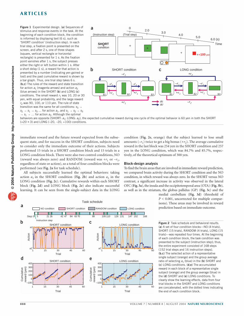
condition (Fig. 2e, orange) that the subject learned to lose smallamounts (–r1) twice to get a big bonus (+r2). The average cumulativereward in the last block was 254 yen in the SHORT condition and 257yen in the LONG condition, which was 84.7% and 85.7%, respec-tively, of the theoretical optimum of 300 yen.
Block-design analysisTo find the brain areas that are involved in immediate reward prediction,we compared brain activity during the SHORT condition and the NOcondition, in which reward was always zero. In the SHORT versus NOcontrast, a significant increase in activity was observed in the lateralOFC (Fig. 3a), the insula and the occipitotemporal area (OTA) (Fig. 3b),as well as in the striatum, the globus pallidus (GP) (Fig. 3c) and the
medial cerebellum (Fig. 3d) (threshold ofP < 0.001, uncorrected for multiple compar-isons). These areas may be involved in rewardprediction based on immediate outcome.
Figure 2 Task schedule and behavioral results.(a) A set of four condition blocks—NO (4 trials),SHORT (15 trials), RANDOM (4 trials), LONG (15trials)—was repeated four times. At the beginningof each condition block, the task condition waspresented to the subject (instruction step); thus,the entire experiment consisted of 168 steps(152 trial steps and 16 instruction steps).(b,c) The selected action of a representativesingle subject (orange) and the group averageratio of selecting a1 (blue) in the (b) SHORT and(c) LONG conditions. (d,e) The accumulatedreward in each block of a representative singlesubject (orange) and the group average (blue) inthe (d) SHORT and (e) LONG conditions. Toclearly show the learning effects, data from fourtrial blocks in the SHORT and LONG conditionsare concatenated, with the dotted lines indicatingthe end of each condition block.
immediate reward and the future reward expected from the subse-quent state, and for success in the SHORT condition, subjects needto consider only the immediate outcome of their actions. Subjectsperformed 15 trials in a SHORT condition block and 15 trials in aLONG condition block. There were also two control conditions, NO(reward was always zero) and RANDOM (reward was +r1 or –r1,regardless of state or action), so a total of four condition blocks wereperformed (see Fig. 2a for task schedule).
All subjects successfully learned the optimal behaviors: takingaction a1 in the SHORT condition (Fig. 2b) and action a2 in theLONG condition (Fig. 2c). Cumulative rewards within each SHORTblock (Fig. 2d) and LONG block (Fig. 2e) also indicate successfullearning. It can be seen from the single-subject data in the LONG
Figure 1 Experimental design. (a) Sequences ofstimulus and response events in the task. At thebeginning of each condition block, the conditionis informed by displaying text (6 s), such as‘SHORT condition’ (instruction step). In eachtrial step, a fixation point is presented on thescreen, and after 2 s, one of three shapes(square, vertical rectangle or horizontalrectangle) is presented for 1 s. As the fixationpoint vanishes after 1 s, the subject presseseither the right or left button within 1 s. After a short delay (1 s), a reward for that action ispresented by a number (indicating yen gained orlost) and the past cumulative reward is shown bya bar graph. Thus, one trial step takes 6 s. (b,c) The rules of the reward and state transitionfor action a1 (magenta arrows) and action a2(blue arrows) in the SHORT (b) and LONG (c)conditions. The small reward r1 was 10, 20 or 30yen, with equal probability, and the large rewardr2 was 90, 100, or 110 yen. The rule of statetransition was the same for all conditions: s3 →s2 → s1 → s3 … for action a1, and s1 → s2 → s3→ s1 → … for action a2. Although the optimalbehaviors are opposite (SHORT: a1; LONG: a2), the expected cumulative reward during one cycle of the optimal behavior is 60 yen in both the SHORT (+20 × 3) and LONG (–20, –20, +100) conditions.
A R T I C L E S
888 VOLUME 7 | NUMBER 8 | AUGUST 2004 NATURE NEUROSCIENCE
a
b
+100 yen
2.03.0
4.05.0
6.0 (s)
Time
0
a2a1
–r1
–r1–r1
–r2
+r2
–r1
+r1 +r1+r1+r1
+r1
–r1
s1 s2 s3 s1 s2 s3
SHORT condition LONG conditionc
SHORTcondition
(Instruction step) (Trial step)
1 15 30 45 60
–100
0
100
200
300
400
Trial
1 15 30 45 60
–100
0
100
200
300
400
Trial
SHORT condition LONG condition
1 15 30 45 60a2
a1
a2
a1SHORT condition
Trial
Act
ion
Cum
ulat
ive
rew
ard
(yen
)
Cum
ulat
ive
rew
ard
(yen
)
1 15 30 45 60
LONG condition
Trial
Act
ion
NO condition SHORT condition LONG conditionRANDOM condition
0 42 84 126 168 5 21 26 47 63 70 89 105 110 131 147 152
Task schedule
Instruction step
Step:
a
b c
ed
©20
04 N
atur
e P
ublis
hing
Gro
up
http
://w
ww
.nat
ure.
com
/nat
uren
euro
scie
nce
analysis. We took the theoretical framework of temporal difference(TD) learning12, which has been successfully used for explainingreward-predictive activations of the midbrain dopaminergic systemas well as those of the cortex and the striatum8,11,13–16. In TD learningtheory, the predicted amount of future reward starting from a states(t) is formulated as the ‘value function’
V(t) = E[r(t + 1) + γ r(t + 2) + γ2r(t + 3) + …]. (1)
Any deviation from the prediction is given by the TD error
δ(t) = r(t) + γ V(t) – V(t – 1), (2)
which is a crucial learning signal for reward prediction and actionselection. The ‘discount factor’ γ (0 ≤ γ < 1) controls the time scaleof prediction: when γ = 0, only the immediate reward r(t + 1) is con-sidered, but as γ approaches 1, rewards in the further future aretaken into account.
We estimated the time courses of reward prediction V(t) and pre-diction error δ(t) from each subject’s performance data and usedthem as the explanatory variables in multiple regression analysis withfMRI data (see Methods). In our Markov decision task, the mini-mum value of γ needed to find the optimal action in the LONG con-dition is 0.36, and any small value of γ is sufficient in the SHORTcondition. From the results of our block-design analysis, we assumedthat different networks involving the cortex and basal ganglia arespecialized for reward prediction at different time scales and thatthey work in parallel, depending on the requirement of the task.Thus, we varied the discount factor γ as 0, 0.3, 0.6, 0.8, 0.9 and 0.99:small γ for immediate reward prediction and large γ for long futurereward prediction. An example of these time courses is shown inSupplementary Figure 1 online.
We observed a significant correlation with reward prediction V(t) inthe medial prefrontal cortex (mPFC; including the anterior cingulatecortex (ACC) and the medial OFC) (Fig. 6a) and bilateral insula (Fig.
6b), left hippocampus and left temporal pole(P < 0.001, uncorrected; see SupplementaryTable 2 online). Figure 6 shows the correlated
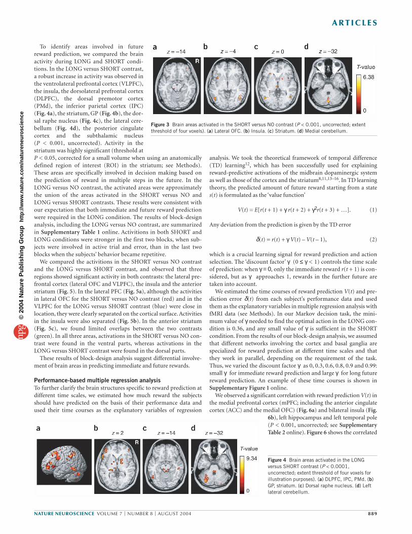
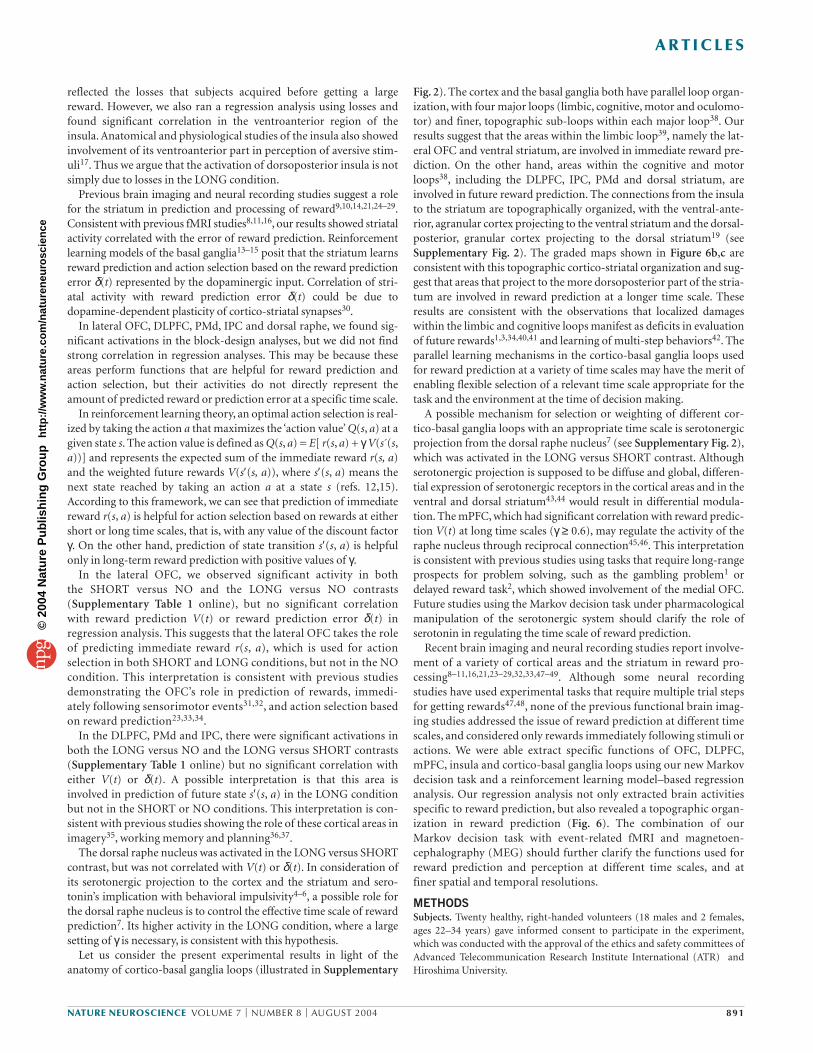
To identify areas involved in futurereward prediction, we compared the brainactivity during LONG and SHORT condi-tions. In the LONG versus SHORT contrast,a robust increase in activity was observed inthe ventrolateral prefrontal cortex (VLPFC),the insula, the dorsolateral prefrontal cortex(DLPFC), the dorsal premotor cortex(PMd), the inferior parietal cortex (IPC)(Fig. 4a), the striatum, GP (Fig. 4b), the dor-sal raphe nucleus (Fig. 4c), the lateral cere-bellum (Fig. 4d), the posterior cingulatecortex and the subthalamic nucleus (P < 0.001, uncorrected). Activity in thestriatum was highly significant (threshold atP < 0.05, corrected for a small volume when using an anatomicallydefined region of interest (ROI) in the striatum; see Methods).These areas are specifically involved in decision making based onthe prediction of reward in multiple steps in the future. In theLONG versus NO contrast, the activated areas were approximatelythe union of the areas activated in the SHORT versus NO andLONG versus SHORT contrasts. These results were consistent withour expectation that both immediate and future reward predictionwere required in the LONG condition. The results of block-designanalysis, including the LONG versus NO contrast, are summarizedin Supplementary Table 1 online. Activitions in both SHORT andLONG conditions were stronger in the first two blocks, when sub-jects were involved in active trial and error, than in the last twoblocks when the subjects’ behavior became repetitive.
We compared the activitions in the SHORT versus NO contrastand the LONG versus SHORT contrast, and observed that threeregions showed significant activity in both contrasts: the lateral pre-frontal cortex (lateral OFC and VLPFC), the insula and the anteriorstriatum (Fig. 5). In the lateral PFC (Fig. 5a), although the activitiesin lateral OFC for the SHORT versus NO contrast (red) and in theVLPFC for the LONG versus SHORT contrast (blue) were close inlocation, they were clearly separated on the cortical surface. Activitiesin the insula were also separated (Fig. 5b). In the anterior striatum(Fig. 5c), we found limited overlaps between the two contrasts(green). In all three areas, activations in the SHORT versus NO con-trast were found in the ventral parts, whereas activations in theLONG versus SHORT contrast were found in the dorsal parts.
These results of block-design analysis suggest differential involve-ment of brain areas in predicting immediate and future rewards.
Performance-based multiple regression analysisTo further clarify the brain structures specific to reward prediction atdifferent time scales, we estimated how much reward the subjectsshould have predicted on the basis of their performance data andused their time courses as the explanatory variables of regression
Figure 4 Brain areas activated in the LONGversus SHORT contrast (P < 0.0001,uncorrected; extent threshold of four voxels forillustration purposes). (a) DLPFC, IPC, PMd. (b)GP, striatum. (c) Dorsal raphe nucleus. (d) Leftlateral cerebellum.
A R T I C L E S
NATURE NEUROSCIENCE VOLUME 7 | NUMBER 8 | AUGUST 2004 889
Figure 3 Brain areas activated in the SHORT versus NO contrast (P < 0.001, uncorrected; extentthreshold of four voxels). (a) Lateral OFC. (b) Insula. (c) Striatum. (d) Medial cerebellum.
©20
04 N
atur
e P
ublis
hing
Gro
up
http
://w
ww
.nat
ure.
com
/nat
uren
euro
scie
nce

DISCUSSIONThe results of the block-design and performance-based regressionanalyses suggest differential involvement of brain areas in actionlearning by prediction of rewards at different time scales. Both block-design and performance-based regression analyses showed activity inthe insula and the anterior striatum. Activations of the ventral regionin the SHORT versus NO contrast and the dorsal region in the LONGversus SHORT contrast in each area (Fig. 5) are consistent with theventroanterior-dorsoposterior maps of the discount factor γ found inperformance-based regression analysis (Fig. 6).
The insula takes a pivotal position in reward processing by receiv-ing primary taste and visceral sensory input17 and sending output tothe OFC18 and the striatum19. Previous studies showed that the insulais activated with anticipation of primary reward10 and that insularlesion causes deficits in incentive learning for primary reward20. Our
results confirm the role of the insula in pre-diction of non-primary, monetary reward21,and further suggest heterogeneous organiza-tion within the insula. Previous imagingstudies also showed involvement of theinsula, especially the ventroanterior region,in processing aversive outcomes22,23. Thus apossible interpretation of the activation ofthe insula in the LONG condition is that it
voxels within these areas using a gradient ofcolors for different γvalues (red for γ= 0, bluefor γ = 0.99). Activity in the mPFC, temporalpole and hippocampus correlated withreward prediction with a longer time scale (γ≥ 0.6). Furthermore, in the insula, we found agraded map of activity for reward predictionat different time scales (Fig. 6b). Whereas activity in the ventroanteriorregion correlated with reward prediction at a shorter time scale, activ-ity in the dorsoposterior region correlated with reward prediction at alonger time scale.
We also found, in the basal ganglia, significant correlation withreward prediction error δ(t) using a wide range of time scales (Fig. 6c; P < 0.001, uncorrected; see Supplementary Table 3 onlineand Methods). Again, we found a graded map, which had a shorttime scale in the ventroanterior part and a long time scale in thedorsoposterior part. The coincidence of the ventroanterior-dorso-posterior maps and the ventroanterior-dorsoposterior shifts inactivities (Fig. 6b,c) indicate that, while the ventroanterior regionswith smaller γ were predominantly active in the SHORT condition,the dorsoposterior regions with larger γ became more active in theLONG condition.
Figure 6 Voxels with a significant correlation(height threshold P < 0.001, uncorrected; extentthreshold of four voxels) with reward predictionV(t) and prediction error δ(t) are shown indifferent colors for different settings of thediscount factor γ. Voxels correlated with two or more regressors are shown by a mosaic ofcolors. (a, b) Significant correlation with reward prediction V(t). (a) mPFC. (b) Insula. (c) Significant correlation with reward predictionerror δ(t) restricted to ROI in the striatum (sliceat white line in horizontal slice at z = 2 mm).Note the ventroanterior-to-dorsoposteriorgradient with the increase in γ both in the insulaand the striatum. Red and blue lines correspondto the z-coordinate levels of activation peaks inthe insula and striatum shown in Figure 5b,c (redfor the SHORT versus NO and blue for the LONGversus SHORT contrasts).
Figure 5 Comparison of brain areas activated inthe SHORT versus NO contrast (red) and theLONG versus SHORT contrast (blue). (a–c) Thesefigures show activation maps focused on (a) thelateral OFC (red (x, y, z) = (38, 46, –14); blue (46,47, 3)) (b) the insula (red (–36, 13, –4); blue(–30, 18, 1)), and (c) the striatum (red (18, 10,0); blue (18, 12, 3)) where we observedsignificant activation in both contrasts. The areaswhere activity overlapped area are shown in green.
A R T I C L E S
890 VOLUME 7 | NUMBER 8 | AUGUST 2004 NATURE NEUROSCIENCE
©20
04 N
atur
e P
ublis
hing
Gro
up
http
://w
ww
.nat
ure.
com
/nat
uren
euro
scie
nce
A R T I C L E S
reflected the losses that subjects acquired before getting a largereward. However, we also ran a regression analysis using losses andfound significant correlation in the ventroanterior region of theinsula. Anatomical and physiological studies of the insula also showedinvolvement of its ventroanterior part in perception of aversive stim-uli17. Thus we argue that the activation of dorsoposterior insula is notsimply due to losses in the LONG condition.
Previous brain imaging and neural recording studies suggest a rolefor the striatum in prediction and processing of reward9,10,14,21,24–29.Consistent with previous fMRI studies8,11,16, our results showed striatalactivity correlated with the error of reward prediction. Reinforcementlearning models of the basal ganglia13–15 posit that the striatum learnsreward prediction and action selection based on the reward predictionerror δ(t) represented by the dopaminergic input. Correlation of stri-atal activity with reward prediction error δ(t) could be due todopamine-dependent plasticity of cortico-striatal synapses30.
In lateral OFC, DLPFC, PMd, IPC and dorsal raphe, we found sig-nificant activations in the block-design analyses, but we did not findstrong correlation in regression analyses. This may be because theseareas perform functions that are helpful for reward prediction andaction selection, but their activities do not directly represent theamount of predicted reward or prediction error at a specific time scale.
In reinforcement learning theory, an optimal action selection is real-ized by taking the action a that maximizes the ‘action value’ Q(s, a) at agiven state s. The action value is defined as Q(s, a) = E[ r(s, a) + γV(s´(s,a))] and represents the expected sum of the immediate reward r(s, a)and the weighted future rewards V(s′(s, a)), where s′(s, a) means thenext state reached by taking an action a at a state s (refs. 12,15).According to this framework, we can see that prediction of immediatereward r(s, a) is helpful for action selection based on rewards at eithershort or long time scales, that is, with any value of the discount factorγ. On the other hand, prediction of state transition s′(s, a) is helpfulonly in long-term reward prediction with positive values of γ.
In the lateral OFC, we observed significant activity in both the SHORT versus NO and the LONG versus NO contrasts(Supplementary Table 1 online), but no significant correlationwith reward prediction V(t) or reward prediction error δ(t) inregression analysis. This suggests that the lateral OFC takes the roleof predicting immediate reward r(s, a), which is used for actionselection in both SHORT and LONG conditions, but not in the NOcondition. This interpretation is consistent with previous studiesdemonstrating the OFC’s role in prediction of rewards, immedi-ately following sensorimotor events31,32, and action selection basedon reward prediction23,33,34.
In the DLPFC, PMd and IPC, there were significant activations inboth the LONG versus NO and the LONG versus SHORT contrasts(Supplementary Table 1 online) but no significant correlation witheither V(t) or δ(t). A possible interpretation is that this area isinvolved in prediction of future state s′(s, a) in the LONG conditionbut not in the SHORT or NO conditions. This interpretation is con-sistent with previous studies showing the role of these cortical areas inimagery35, working memory and planning36,37.
The dorsal raphe nucleus was activated in the LONG versus SHORTcontrast, but was not correlated with V(t) or δ(t). In consideration ofits serotonergic projection to the cortex and the striatum and sero-tonin’s implication with behavioral impulsivity4–6, a possible role forthe dorsal raphe nucleus is to control the effective time scale of rewardprediction7. Its higher activity in the LONG condition, where a largesetting of γ is necessary, is consistent with this hypothesis.
Let us consider the present experimental results in light of theanatomy of cortico-basal ganglia loops (illustrated in Supplementary
Fig. 2). The cortex and the basal ganglia both have parallel loop organ-ization, with four major loops (limbic, cognitive, motor and oculomo-tor) and finer, topographic sub-loops within each major loop38. Ourresults suggest that the areas within the limbic loop39, namely the lat-eral OFC and ventral striatum, are involved in immediate reward pre-diction. On the other hand, areas within the cognitive and motorloops38, including the DLPFC, IPC, PMd and dorsal striatum, areinvolved in future reward prediction. The connections from the insulato the striatum are topographically organized, with the ventral-ante-rior, agranular cortex projecting to the ventral striatum and the dorsal-posterior, granular cortex projecting to the dorsal striatum19 (seeSupplementary Fig. 2). The graded maps shown in Figure 6b,c areconsistent with this topographic cortico-striatal organization and sug-gest that areas that project to the more dorsoposterior part of the stria-tum are involved in reward prediction at a longer time scale. Theseresults are consistent with the observations that localized damageswithin the limbic and cognitive loops manifest as deficits in evaluationof future rewards1,3,34,40,41 and learning of multi-step behaviors42. Theparallel learning mechanisms in the cortico-basal ganglia loops usedfor reward prediction at a variety of time scales may have the merit ofenabling flexible selection of a relevant time scale appropriate for thetask and the environment at the time of decision making.
A possible mechanism for selection or weighting of different cor-tico-basal ganglia loops with an appropriate time scale is serotonergicprojection from the dorsal raphe nucleus7 (see Supplementary Fig. 2),which was activated in the LONG versus SHORT contrast. Althoughserotonergic projection is supposed to be diffuse and global, differen-tial expression of serotonergic receptors in the cortical areas and in theventral and dorsal striatum43,44 would result in differential modula-tion. The mPFC, which had significant correlation with reward predic-tion V(t) at long time scales (γ ≥ 0.6), may regulate the activity of theraphe nucleus through reciprocal connection45,46. This interpretationis consistent with previous studies using tasks that require long-rangeprospects for problem solving, such as the gambling problem1 ordelayed reward task2, which showed involvement of the medial OFC.Future studies using the Markov decision task under pharmacologicalmanipulation of the serotonergic system should clarify the role ofserotonin in regulating the time scale of reward prediction.
Recent brain imaging and neural recording studies report involve-ment of a variety of cortical areas and the striatum in reward pro-cessing8–11,16,21,23–29,32,33,47–49. Although some neural recordingstudies have used experimental tasks that require multiple trial stepsfor getting rewards47,48, none of the previous functional brain imag-ing studies addressed the issue of reward prediction at different timescales, and considered only rewards immediately following stimuli oractions. We were able extract specific functions of OFC, DLPFC,mPFC, insula and cortico-basal ganglia loops using our new Markovdecision task and a reinforcement learning model–based regressionanalysis. Our regression analysis not only extracted brain activitiesspecific to reward prediction, but also revealed a topographic organ-ization in reward prediction (Fig. 6). The combination of ourMarkov decision task with event-related fMRI and magnetoen-cephalography (MEG) should further clarify the functions used forreward prediction and perception at different time scales, and atfiner spatial and temporal resolutions.
METHODSSubjects. Twenty healthy, right-handed volunteers (18 males and 2 females,ages 22–34 years) gave informed consent to participate in the experiment,which was conducted with the approval of the ethics and safety committees ofAdvanced Telecommunication Research Institute International (ATR) andHiroshima University.
NATURE NEUROSCIENCE VOLUME 7 | NUMBER 8 | AUGUST 2004 891
©20
04 N
atur
e P
ublis
hing
Gro
up
http
://w
ww
.nat
ure.
com
/nat
uren
euro
scie
nce
A R T I C L E S
Behavioral task. In the Markov decision task (Fig. 1), one of three states wasvisually presented to the subject using three different shapes, and the subjectselected one of two actions by pressing one of two buttons using their righthand (Fig. 1a). The rule of state transition was the same for all conditions: s3
→ s2 → s1 → s3 … for action a1, and s1 → s2 → s3 → s1 → … for action a2. Therules for reward, however, changed in each condition. In the SHORT condition(Fig. 1b), action a1 results in a small positive reward (+r1 = 10, 20 or 30 yen,with equal probabilities), whereas action a2 results in a small loss (–r1) at anyof the three states. Thus, the optimal behavior is to collect small positiverewards at each state by performing action a1. In the LONG condition (Fig. 1c), however, the reward setting is such that action a2 gives a large positivereward (+r2 = 90, 100 or 110 yen) at state s3, and action a1 gives a large loss(–r2) at state s1. Thus, the optimal behavior is to receive small losses at states s1
and s2 to obtain a large positive reward at state s3 by taking action a2 at eachstate. There were two control conditions: the NO condition, where the rewardwas always zero, and the RANDOM condition, where the reward was positive(+r1) or negative (–r1) with equal probability, regardless of state or action.
Subjects completed 4 trials in a NO condition block, 15 trials in a SHORTcondition block, 4 trials in a RANDOM condition block and 15 trials in aLONG condition block. A set of four condition blocks (NO, SHORT, RAN-DOM, LONG) was repeated four times (Fig. 2a). Subjects were informed ofthe current condition at the beginning of each condition block by text on thescreen (first slide in Fig. 1a); thus, the entire experiment consisted of 168 steps(152 trial steps and 16 instruction steps), taking about 17 min. The mappingsof the three states to the three figures, and the two buttons to the two actions,were randomly set at the beginning of each experiment, so that subjects wererequired to learn the amount of reward associated with each figure-buttonpair in both SHORT and LONG conditions. Furthermore, in the LONG condi-tion, subjects had to learn the subsequent figure for each figure-action pairand take into account the amount of reward expected from the subsequent fig-ure in selecting a button.
fMRI imaging. A 1.5-tesla scanner (Shimadzu-Marconi, Magnex Eclipse) wasused to acquire both structural T1-weighted images (repetition time,TR = 12 ms, TE = 4.5 ms, flip angle = 20°, matrix = 256 × 256, FoV = 256 mm,thickness = 1 mm, slice gaP = 0 mm) and T2*-weighted echo planar images(TR = 6 s, TE = 55 ms, flip angle = 90°, 50 transverse slices, matrix = 64 × 64,FoV = 192 mm, thickness = 3 mm, slice gap = 0 mm) showing blood oxygenlevel–dependent (BOLD) contrasts.
Because the aim of the present study was to identify brain activity underlyingreward prediction over multiple trial steps, we acquired functional images every6 s (TR = 6 s), in synchrony with single trials. Although shorter TRs and event-related designs are often used in experiments that aim to distinguish brainresponses to events within a trial9,11,21,26, analysis of those finer events in timewere not the focus of the current study. With this longer TR, the BOLD signal ina single scan contained a mixture of responses for a reward-predictive stimulusand reward feedback. However, because of the progress of learning and the sto-chastic nature of the amount of reward, the time courses of reward predictionV(t) and prediction error δ(t) over the 168 trial steps were markedly different.Thus, we could separate activity corresponding to reward prediction from thatcorresponding to outcomes by using both reward prediction V(t) and rewardoutcome r(t) in multiple regression analysis, as described below.
Data analysis. The data were pre-processed and analyzed with SPM99(www.fil.ion.ucl.ac.uk/spm/spm99.html). The first two volumes of imageswere discarded to avoid T1 equilibrium effects. The images were realigned tothe first image as a reference, spatially normalized with respect to the MontrealNeurological Institute EPI template, and spatially smoothed with a Gaussiankernel (8 mm, full-width at half-maximum).
We conducted two types of analysis. One was block-design analysis usingfour boxcar regressors covering the whole experiment, convolved with ahemodynamic response function as the reference waveform for each condition(NO, SHORT, RANDOM, LONG). We did not find substantial differencesbetween SHORT versus NO and SHORT versus RANDOM contrasts, orbetween LONG versus NO and LONG versus RANDOM contrasts. Thereforewe report here only the results with the NO condition as the control condition.The other method was multivariate regression analysis using explanatory vari-
ables, representing the time course of the reward prediction V(t) or rewardprediction error δ(t) at six different timescales γ, estimated from subjects’ per-formance data (described below).
In both analyses, images of parameter estimates for the contrast of interestwere created for each subject. These were then entered into a second-levelgroup analysis using a one-sample t test at a threshold of P < 0.001, uncor-rected for multiple comparisons (random effects analysis) and extent thresh-old of four voxels. Small-volume correction (SVC) was done at a threshold ofP < 0.05 using an ROI within the striatum (including the caudate and puta-men), which was defined anatomically based on a normalized T1 image.
Procedures of performance-based regression analysis. The time courses ofreward prediction V(t) and reward prediction error δ(t) were estimated fromeach subject’s performance data—state s(t), action a(t) and reward r(t)—asdescribed below.
Reward prediction. To estimate how much of a forthcoming reward a subjectwould have expected at each step during the Markov decision task, we took thedefinition of the value function (equation 1) and reformulated it based on therecursive structure of the task. Namely, if the subject starts from a state s(t) andcomes back to the same state after k steps, the expected cumulative reward V(t)should satisfy the consistency condition V(t) = r(t + 1) + γ r(t + 2) + … + γ k–1r(t + k) + γ kV(t).
Thus, for each time t of the data file, we calculated the weighted sum of therewards acquired until the subject returned to the same state and estimated thevalue function for that episode as
(1)
The estimate of the value function V(t) at time t was given by the average of allprevious episodes from the same state as at time t
(2)
where {t1, …, tL} are the indices of time visiting the same state as s(t), that is,s(t1) = … = s(tL) = s(t).
Reward prediction error. The TD error (equation 2) was calculated from thedifference between the actual reward r(t) and the temporal difference of theestimated value function V(t).
We separately calculated the time courses of V(t) and δ(t) during SHORTand LONG conditions; we concatenated data of four blocks in the SHORTcondition, and calculated V(t) and δ(t) as described above. We used the sameprocess for the LONG condition data. During the NO and RANDOM condi-tions, the values of V(t) and δ(t) were fixed at zero. Finally, we reconstructedthe data corresponding to the real time course of the experiment. Examples ofthe time course of these variables are shown in Supplementary Figure 1online. We used either V(t) or δ(t) as the explanatory variable in a regressionanalysis by SPM. To remove any effects of factors other than reward predic-tion, we concurrently used other variables in the regression, namely the fourbox-car functions representing each condition (NO, SHORT, RANDOM,LONG). Because the immediate reward prediction V(t) with γ = 0 can coin-cide with reward outcome r(t) if learning is perfect, we included the rewardoutcome r(t) in regression analyses with V(t). Thus, the significant correla-tion with V(t) (Fig. 6a,b) should represent a predictive component ratherthan a reward outcome.
The amplitude of explanatory variables δ(t) with all γwere large in early trialsand decreased as subjects learned the task (Supplementary Fig. 1 online). Thisdecreasing trend causes a risk that areas that are activated early in trials, such asthose responsible for general attentiveness or novelty, have correlations withδ(t). Because our aim in regression analysis was to clarify the brain structuresinvolved in reward prediction at specific time scales, we removed the areas thathad similar correlation to δ(t) at all settings of γ from considerations in Figure 6and Supplementary Table 3 online. To compare the results of regression analysis
( ) ( )∑=
=L
lltV
LtV
1
1 ∧
( ) ( ) ( ) ( )[1
21 1
k
k ktrtrtrtV
γ−
++++++=−Λ [∧ γ
γ
892 VOLUME 7 | NUMBER 8 | AUGUST 2004 NATURE NEUROSCIENCE
©20
04 N
atur
e P
ublis
hing
Gro
up
http
://w
ww
.nat
ure.
com
/nat
uren
euro
scie
nce
A R T I C L E S
with six different values of γ, we used display software that can overlay multipleactivation maps in different colors on a single brain structure image. When avoxel is significantly activated in multiple values of γ, it is shown by a mosaic ofmultiple colors, with apparent subdivision of the voxel (Fig. 6).
Note: Supplementary information is available on the Nature Neuroscience website.
ACKNOWLEDGMENTSWe thank K. Samejima, N. Schweighofer, M. Haruno, H. Imamizu, S. Higuchi,T. Yoshioka, T. Chaminade and M. Kawato for helpful discussions and technicaladvice. This research was funded by ‘Creating the Brain,’ Core Research forEvolutional Science and Technology (CREST), Japan Science and Technology Agency.
COMPETING INTERESTS STATEMENTThe authors declare that they have no competing financial interests.
Received 5 March; accepted 2 June 2004Published online at http://www.nature.com/natureneuroscience/
1. Bechara, A., Damasio, H. & Damasio, A.R. Emotion, decision making and theorbitofrontal cortex. Cereb. Cortex 10, 295–307 (2000).
2. Mobini, S. et al. Effects of lesions of the orbitofrontal cortex on sensitivity to delayedand probabilistic reinforcement. Psychopharmacology (Berl.) 160, 290–298(2002).
3. Cardinal, R.N., Pennicott, D.R., Sugathapala, C.L., Robbins, T.W. & Everitt, B.J.Impulsive choice induced in rats by lesions of the nucleus accumbens core. Science292, 2499–2501 (2001).
4. Rogers, R.D. et al. Dissociable deficits in the decision-making cognition of chronicamphetamine abusers, opiate abusers, patients with focal damage to prefrontal cor-tex, and tryptophan-depleted normal volunteers: evidence for monoaminergic mech-anisms. Neuropsychopharmacology 20, 322–339 (1999).
5. Evenden, J.L. & Ryan, C.N. The pharmacology of impulsive behaviour in rats: theeffects of drugs on response choice with varying delays of reinforcement.Psychopharmacology (Berl.) 128, 161–170 (1996).
6. Mobini, S., Chiang, T.J., Ho, M.Y., Bradshaw, C.M. & Szabadi, E. Effects of central5-hydroxytryptamine depletion on sensitivity to delayed and probabilistic reinforce-ment. Psychopharmacology (Berl.) 152, 390–397 (2000).
7. Doya, K. Metalearning and neuromodulation. Neural Net. 15, 495–506 (2002).8. Berns, G.S., McClure, S.M., Pagnoni, G. & Montague, P.R. Predictability modulates
human brain response to reward. J. Neurosci. 21, 2793–2798 (2001).9. Breiter, H.C., Aharon, I., Kahneman, D., Dale, A. & Shizgal, P. Functional imaging of
neural responses to expectancy and experience of monetary gains and losses.Neuron 30, 619–639 (2001).
10. O’Doherty, J.P., Deichmann, R., Critchley, H.D. & Dolan, R.J. Neural responses dur-ing anticipation of a primary taste reward. Neuron 33, 815–826 (2002).
11. O’Doherty, J.P., Dayan, P., Friston, K., Critchley, H. & Dolan, R.J. Temporal differ-ence models and reward-related learning in the human brain. Neuron 38, 329–337(2003).
12. Sutton, R.S. & Barto, A.G. Reinforcement Learning (MIT Press, Cambridge,Massachusetts, 1998).
13. Houk, J.C., Adams, J.L. & Barto, A.G. in Models of Information Processing in theBasal Ganglia (eds. Houk, J.C., Davis, J.L. & Beiser, D.G.) 249–270 (MIT Press,Cambridge, Massachusetts, 1995).
14. Schultz, W., Dayan, P. & Montague, P.R. A neural substrate of prediction and reward.Science 275, 1593–1599 (1997).
15. Doya, K. Complementary roles of basal ganglia and cerebellum in learning and motorcontrol. Curr. Opin. Neurobiol. 10, 732–739 (2000).
16. McClure, S.M., Berns, G.S. & Montague, P.R. Temporal prediction errors in a passivelearning task activate human striatum. Neuron 38, 339–346 (2003).
17. Mesulam, M.M. & Mufson, E.J. Insula of the old world monkey. III: Efferent corticaloutput and comments on function. J. Comp. Neurol. 212, 38–52 (1982).
18. Cavada, C., Company, T., Tejedor, J., Cruz-Rizzolo, R.J. & Reinoso-Suarez, F. Theanatomical connections of the macaque monkey orbitofrontal cortex. Cereb. Cortex10, 220–242 (2000).
19. Chikama, M., McFarland, N.R., Amaral, D.G. & Haber, S.N. Insular cortical projec-tions to functional regions of the striatum correlate with cortical cytoarchitectonicorganization in the primate. J. Neurosci. 17, 9686–9705 (1997).
20. Balleine, B.W. & Dickinson, A. The effect of lesions of the insular cortex on instru-mental conditioning: evidence for a role in incentive memory. J. Neurosci. 20,8954–8964 (2000).
21. Knutson, B., Fong, G.W., Bennett, S.M., Adams, C.M. & Hommer, D. A region ofmesial prefrontal cortex tracks monetarily rewarding outcomes: characterization withrapid event-related fMRI. Neuroimage 18, 263–272 (2003).
22. Ullsperger, M. & von Cramon, D.Y. Error monitoring using external feedback: specificroles of the habenular complex, the reward system, and the cingulate motor arearevealed by functional magnetic resonance imaging. J. Neurosci. 23, 4308–4314(2003).
23. O’Doherty, J., Critchley, H., Deichmann, R. & Dolan, R.J. Dissociating valence ofoutcome from behavioral control in human orbital and ventral prefrontal cortices. J. Neurosci. 23, 7931–7939 (2003).
24. Koepp, M.J. et al. Evidence for striatal dopamine release during a video game.Nature 393, 266–268 (1998).
25. Elliott, R., Friston, K.J. & Dolan, R.J. Dissociable neural responses in human rewardsystems. J. Neurosci. 20, 6159–6165 (2000).
26. Knutson, B., Adams, C.M., Fong, G.W. & Hommer, D. Anticipation of increasingmonetary reward selectively recruits nucleus accumbens. J. Neurosci. 21, RC159(2001).
27. Pagnoni, G., Zink, C.F., Montague, P.R. & Berns, G.S. Activity in human ventralstriatum locked to errors of reward prediction. Nat. Neurosci. 5, 97–98 (2002).
28. Elliott, R., Newman, J.L., Longe, O.A. & Deakin, J.F. Differential response patternsin the striatum and orbitofrontal cortex to financial reward in humans: a parametricfunctional magnetic resonance imaging study. J. Neurosci. 23, 303–307 (2003).
29. Haruno, M. et al. A neural correlate of reward-based behavioral learning in caudatenucleus: a functional magnetic resonance imaging study of a stochastic decisiontask. J. Neurosci. 24, 1660–1665 (2004).
30. Reynolds, J.N. & Wickens, J.R. Dopamine-dependent plasticity of corticostriatalsynapses. Neural Net. 15, 507–521 (2002).
31. Tremblay, L. & Schultz, W. Reward-related neuronal activity during go-nogo task per-formance in primate orbitofrontal cortex. J. Neurophysiol. 83, 1864–1876 (2000).
32. Critchley, H.D., Mathias, C.J. & Dolan, R.J. Neural activity in the human brain relat-ing to uncertainty and arousal during anticipation. Neuron 29, 537–545 (2001).
33. Rogers, R.D. et al. Choosing between small, likely rewards and large, unlikelyrewards activates inferior and orbital prefrontal cortex. J. Neurosci. 19, 9029–9038(1999).
34. Rolls, E.T. The orbitofrontal cortex and reward. Cereb. Cortex 10, 284–294 (2000).35. Hanakawa, T. et al. The role of rostral Brodmann area 6 in mental-operation tasks:
an integrative neuroimaging approach. Cereb. Cortex 12, 1157–1170 (2002).36. Owen, A.M., Doyon, J., Petrides, M. & Evans, A.C. Planning and spatial working
memory: a positron emission tomography study in humans. Eur. J. Neurosci. 8,353–364 (1996).
37. Baker, S.C. et al. Neural systems engaged by planning: a PET study of the Tower ofLondon task. Neuropsychologia 34, 515–526 (1996).
38. Middleton, F.A. & Strick, P.L. Basal ganglia and cerebellar loops: motor and cogni-tive circuits. Brain Res. Brain Res. Rev. 31, 236–250 (2000).
39. Haber, S.N., Kunishio, K., Mizobuchi, M. & Lynd-Balta, E. The orbital and medialprefrontal circuit through the primate basal ganglia. J. Neurosci. 15, 4851–4867(1995).
40. Eagle, D.M., Humby, T., Dunnett, S.B. & Robbins, T.W. Effects of regional striatallesions on motor, motivational, and executive aspects of progressive-ratio perform-ance in rats. Behav. Neurosci. 113, 718–731 (1999).
41. Pears, A., Parkinson, J.A., Hopewell, L., Everitt, B.J. & Roberts, A.C. Lesions of theorbitofrontal but not medial prefrontal cortex disrupt conditioned reinforcement inprimates. J. Neurosci. 23, 11189–11201 (2003).
42. Hikosaka, O. et al. Parallel neural networks for learning sequential procedures.Trends Neurosci. 22, 464–471 (1999).
43. Mijnster, M.J. et al. Regional and cellular distribution of serotonin 5-hydroxytrypta-mine2a receptor mRNA in the nucleus accumbens, olfactory tubercle, and caudateputamen of the rat. J. Comp. Neurol. 389, 1–11 (1997).
44. Compan, V., Segu, L., Buhot, M.C. & Daszuta, A. Selective increases in serotonin 5-HT1B/1D and 5-HT2A/2C binding sites in adult rat basal ganglia following lesions ofserotonergic neurons. Brain Res. 793, 103–111 (1998).
45. Celada, P., Puig, M.V., Casanovas, J.M., Guillazo, G. & Artigas, F. Control of dorsalraphe serotonergic neurons by the medial prefrontal cortex: involvement of sero-tonin-1A, GABA(A), and glutamate receptors. J. Neurosci. 21, 9917–9929 (2001).
46. Martin-Ruiz, R. et al. Control of serotonergic function in medial prefrontal cortex byserotonin-2A receptors through a glutamate-dependent mechanism. J. Neurosci.21, 9856–9866 (2001).
47. Hikosaka, K. & Watanabe, M. Delay activity of orbital and lateral prefrontal neuronsof the monkey varying with different rewards. Cereb. Cortex 10, 263–271 (2000).
48. Shidara, M. & Richmond, B.J. Anterior cingulate: single neuronal signals related todegree of reward expectancy. Science 296, 1709–1711 (2002).
49. Matsumoto, K., Suzuki, W. & Tanaka, K. Neuronal correlates of goal-based motorselection in the prefrontal cortex. Science 301, 229–232 (2003).
NATURE NEUROSCIENCE VOLUME 7 | NUMBER 8 | AUGUST 2004 893
©20
04 N
atur
e P
ublis
hing
Gro
up
http
://w
ww
.nat
ure.
com
/nat
uren
euro
scie
nce