Androgen-Induced Rhox Homeobox Genes Modulate the Expression of AR-Regulated Genes
16
Androgen-Induced Rhox Homeobox Genes Modulate the Expression of AR-Regulated Genes Zhiying Hu, Dineshkumar Dandekar, Peter J. O’Shaughnessy, Karel De Gendt, Guido Verhoeven, and Miles F. Wilkinson Department of Reproductive Medicine (M.F.W.), The University of California, San Diego, California 92093; Department of Biochemistry & Molecular Biology (Z.H., D.D., M.F.W.), University of Texas M.D. Anderson Cancer Center, Houston, Texas 77030; The Institute of Comparative Medicine (P.J.O.), University of Glasgow Veterinary School, Glasgow G61 1QH, United Kingdom; and Laboratory for Experimental Medicine and Endocrinology (K.D.G., G.V.), Catholic University of Leuven, B-3000 Leuven, Belgium Rhox5, the founding member of the reproductive homeobox on the X chromosome (Rhox) gene cluster, encodes a homeodomain-containing transcription factor that is selectively expressed in Sertoli cells, where it promotes the survival of male germ cells. To identify Rhox5-regulated genes, we generated 15P-1 Sertoli cell clones expressing physiological levels of Rhox5 from a stably transfected expression vector. Microarray analysis identified many genes altered in expression in response to Rhox5, including those encoding proteins controlling cell cycle regulation, apoptosis, metabolism, and cell-cell interactions. Fifteen of these Rhox5-regulated genes were chosen for further analysis. Analysis of Rhox5-null male mice indicated that at least nine of these are Rhox5- regulated in the testes in vivo. Many of them have distinct postnatal expression patterns and are regulated by Rhox5 at different postnatal time points. Most of them are expressed in Sertoli cells, indicating that they are candidates to be directly regulated by Rhox5. Transfection analysis with expression vectors encoding different mouse and human Rhox family members revealed that the regulatory response of a subset of these Rhox5-regulated genes is both conserved and redundant. Given that Rhox5 depends on androgen receptor (AR) for expression in Sertoli cells, we examined whether some Rhox5-regulated genes are also regulated by AR. We provide several lines of evidence that this is the case, leading us to propose that RHOX5 serves as a key intermediate transcription factor that directs some of the actions of AR in the testes. (Molecular Endocrinology 24: 60 –75, 2010) H omeobox genes encode transcription factors harbor- ing a 60-amino acid DNA-binding motif called a ho- meodomain. These transcription factors govern key em- bryonic developmental processes, including body-axis formation, organogenesis, and limb development (1). Whereas the roles of homeobox genes in embryonic de- velopment have been studied intensely for over two de- cades, their functions in controlling postembryonic devel- opmental events have only begun to be examined. Homeobox genes are good candidates to control postem- bryonic events, because many homeobox genes are ex- pressed during postnatal development and in adult tis- sues. Indeed, mouse knockout studies have shown that the HoxA9, Hoxc13, and Pdx1 homeobox genes are cru- cial for hematopoiesis, hair growth, and gut homeostasis, respectively (2– 4). Another postembryonic process likely to be controlled by homeobox genes is spermatogenesis. This idea is sup- ported by the fact that over 50 of the approximately 200 known mouse homeobox genes are expressed in the testis and other male reproductive organs (5–12). Although this suggests a role for homeobox transcription factors in male reproduction, the functions of most homeobox tran- scription factors in the postnatal and adult male repro- ductive tract are not yet clear, because efforts to elucidate their roles by use of knockout mice have been clouded by ISSN Print 0888-8809 ISSN Online 1944-9917 Printed in U.S.A. Copyright © 2010 by The Endocrine Society doi: 10.1210/me.2009-0303 Received July 31, 2009. Accepted September 18, 2009. First Published Online November 9, 2009 Abbreviations: AR, Androgen receptor; ARE, androgen-response element; qPCR, quanti- tative PCR; SAR, secondary androgen-response. ORIGINAL RESEARCH 60 mend.endojournals.org Mol Endocrinol, January 2010, 24(1):60 –75
-
Upload
independent -
Category
Documents
-
view
1 -
download
0
Transcript of Androgen-Induced Rhox Homeobox Genes Modulate the Expression of AR-Regulated Genes

Androgen-Induced Rhox Homeobox Genes Modulatethe Expression of AR-Regulated Genes
Zhiying Hu, Dineshkumar Dandekar, Peter J. O’Shaughnessy, Karel De Gendt,Guido Verhoeven, and Miles F. Wilkinson
Department of Reproductive Medicine (M.F.W.), The University of California, San Diego, California92093; Department of Biochemistry & Molecular Biology (Z.H., D.D., M.F.W.), University of Texas M.D.Anderson Cancer Center, Houston, Texas 77030; The Institute of Comparative Medicine (P.J.O.), University ofGlasgow Veterinary School, Glasgow G61 1QH, United Kingdom; and Laboratory for Experimental Medicineand Endocrinology (K.D.G., G.V.), Catholic University of Leuven, B-3000 Leuven, Belgium
Rhox5, the founding member of the reproductive homeobox on the X chromosome (Rhox) genecluster, encodes a homeodomain-containing transcription factor that is selectively expressed inSertoli cells, where it promotes the survival of male germ cells. To identify Rhox5-regulated genes,we generated 15P-1 Sertoli cell clones expressing physiological levels of Rhox5 from a stablytransfected expression vector. Microarray analysis identified many genes altered in expression inresponse to Rhox5, including those encoding proteins controlling cell cycle regulation, apoptosis,metabolism, and cell-cell interactions. Fifteen of these Rhox5-regulated genes were chosen forfurther analysis. Analysis of Rhox5-null male mice indicated that at least nine of these are Rhox5-regulated in the testes in vivo. Many of them have distinct postnatal expression patterns and areregulated by Rhox5 at different postnatal time points. Most of them are expressed in Sertoli cells,indicating that they are candidates to be directly regulated by Rhox5. Transfection analysis withexpression vectors encoding different mouse and human Rhox family members revealed that theregulatory response of a subset of these Rhox5-regulated genes is both conserved and redundant.Given that Rhox5 depends on androgen receptor (AR) for expression in Sertoli cells, we examinedwhether some Rhox5-regulated genes are also regulated by AR. We provide several lines of evidencethat this is the case, leading us to propose that RHOX5 serves as a key intermediate transcription factorthat directs some of the actions of AR in the testes. (Molecular Endocrinology 24: 60–75, 2010)
Homeobox genes encode transcription factors harbor-ing a 60-amino acid DNA-binding motif called a ho-
meodomain. These transcription factors govern key em-bryonic developmental processes, including body-axisformation, organogenesis, and limb development (1).Whereas the roles of homeobox genes in embryonic de-velopment have been studied intensely for over two de-cades, their functions in controlling postembryonic devel-opmental events have only begun to be examined.Homeobox genes are good candidates to control postem-bryonic events, because many homeobox genes are ex-pressed during postnatal development and in adult tis-sues. Indeed, mouse knockout studies have shown that
the HoxA9, Hoxc13, and Pdx1 homeobox genes are cru-cial for hematopoiesis, hair growth, and gut homeostasis,respectively (2–4).
Another postembryonic process likely to be controlledby homeobox genes is spermatogenesis. This idea is sup-ported by the fact that over 50 of the approximately 200known mouse homeobox genes are expressed in the testisand other male reproductive organs (5–12). Although thissuggests a role for homeobox transcription factors inmale reproduction, the functions of most homeobox tran-scription factors in the postnatal and adult male repro-ductive tract are not yet clear, because efforts to elucidatetheir roles by use of knockout mice have been clouded by
ISSN Print 0888-8809 ISSN Online 1944-9917Printed in U.S.A.Copyright © 2010 by The Endocrine Societydoi: 10.1210/me.2009-0303 Received July 31, 2009. Accepted September 18, 2009.First Published Online November 9, 2009
Abbreviations: AR, Androgen receptor; ARE, androgen-response element; qPCR, quanti-tative PCR; SAR, secondary androgen-response.
O R I G I N A L R E S E A R C H
60 mend.endojournals.org Mol Endocrinol, January 2010, 24(1):60–75
Miles Wilkinson

putative functional redundancies and embryonic lethality(6). These efforts have identified some homeobox genesthat control the formation of the male reproductive tractduring embryogenesis, but the identity of homeoboxgenes that have roles in the male reproductive tract afterbirth have remained largely obscure (5, 12–14).
To our knowledge, the only mammalian homeoboxgene demonstrated to have a role in spermatogenesis isRhox5 (previously known as Pem), the founding memberof the reproductive homoeobox genes on the X chromo-some (Rhox) gene family (7). All the members of the Rhoxgene family form a tandem array on the X chromosome inmammals. The mouse Rhox gene cluster contains 33 mem-bers, and thus is the largest homeobox gene cluster known inany species (7–11, 15). Virtually all of the Rhox genes areselectively expressed in reproductive tissues and the pla-centa, suggesting that they encode transcription factors de-voted to regulating gametogenesis and embryogenesis (7).
Rhox5 was first identified in a search for genes differ-entially expressed between two related T-lymphoma cellclones that exhibited marked differences in developmen-tal status and tumorigenicity (16). Rhox5 was initially ofinterest because it was found to be selectively expressed inT-cell lymphomas, not normal T cells, and also in a widevariety of other tumor cells of diverse developmental andtissue origins (17). Later investigations revealed thatRhox5 encodes a tumor antigen (18), interacts with pro-tein involved in tumorigenesis (19, 20), and its expressionis induced by the proto-oncogene Ras (21, 22). Althoughthese studies have suggested the possibility that Rhox5has a role in tumorigenesis, its precise role in malignancyhas not yet been determined. Other studies have focusedon Rhox5’s normal expression pattern and biologicalroles. Rhox5 is normally expressed in the early mouseembryo in a developmentally regulated manner (23), andis later expressed in placenta (24, 25) and in both maleand female germ cells in developing fetal gonads (26, 27).In neonatal and adult mice and rats, Rhox5 expression isprimarily confined to somatic cells in the testis, epididy-mis, and ovary (28, 29). In the testis, its expression isrestricted to Sertoli cells, where it is expressed in a stage-specific manner during the seminiferous epithelial cycleby an androgen- and androgen receptor (AR)-dependentmechanism (30, 31).
The expression of Rhox5 in somatic cells in the malereproductive tract is crucial for normal spermatogenesisand sperm maturation. Targeted mutation of Rhox5 inmale mice leads to subfertility, marked by increased germ-cell apoptosis, reduced sperm number, and reduced spermmotility (7). Rhox5-null mice exhibit an increased fre-quency of apoptotic germ cells in seminiferous epithelialcycle stages that normally contain dying germ cells (stages
I–IV and stage XII) and they also have apoptotic germcells in stages that normally lack dying germ cells (stagesV–XI). Rhox5 is unlikely to act directly on germ cells tocontrol germ cell survival and motility, because it is notdetectably expressed in germ cells, but instead is ex-pressed in Sertoli cells, the nurse cells in intimate contactwith germ cells (29, 30). This suggests a model in whichRhox5 functions in spermatogenesis by virtue of its abil-ity to regulate genes encoding Sertoli cell secreted and cellsurface proteins that, in turn, control key events in germcells, including their differentiation and survivial (32–34).
To begin to address the molecular mechanisms bywhich Rhox5 helps drive spermatogenesis, we used tran-scriptional profiling to identify genes regulated by physi-ological levels of Rhox5 in 15P-1 cells, a well-establishedSertoli cell line (35–37). This genome-wide analysis led tothe discovery of many Rhox5-regulated genes, a largeproportion of which encode cell surface proteins. Manyof these were expressed in Sertoli cells, which was ofinterest because such genes could mediate Rhox5-inducedsignals that impinge on germ cells. Also identified wereRhox5-regulated genes encoding other kinds of proteins,including secreted factors, transcription factors, and met-abolic enzymes. We focused on a subset of these genes andfound that most are also Rhox5-regulated in the testes invivo. We determined the testicular cell types and develop-mental stages in which the regulation of each gene occurs.We also identified other Rhox family members that reg-ulate their expression, and provided evidence that theRhox-dependent regulatory circuitry controlling thesegenes is conserved. Finally, we addressed whether any ofthe Rhox5 gene targets are secondary androgen-response(SAR) genes. These are long-sought-after genes that arenot directly regulated by androgen and AR, but rather arecontrolled by androgen-dependent regulators, such asandrogen-induced transcription factors (38, 39). BecauseRhox5 is an AR- and androgen-inducible gene in Sertolicells, it is a good candidate to regulate SAR genes. Indeed,we obtained many lines of evidence that several Rhox5-regulated genes are SAR genes. We also showed that otherandrogen-inducible Rhox genes (besides Rhox5) are capa-ble of regulating these candidate SAR genes. The discoveryof genes coregulated by AR and RHOX transcription fac-tors has the potential to begin illuminating the molecularmechanisms by which androgens drive spermatogenesis.
Results
Establishment of Sertoli cell clones expressingphysiological levels of Rhox5
To identify Rhox5 target genes by a gain-of-functionapproach, we screened Sertoli cell lines for lines that ex-
Mol Endocrinol, January 2010, 24(1):60–75 mend.endojournals.org 61

press low endogenous Rhox5 mRNA levels. Our ratio-nale was that a low Rhox5-expressing line would providean optimal baseline for identifying genes regulated byphysiological levels of Rhox5 expressed from a Rhox5-expression vector. We predicted that most Sertoli celllines would express low levels of Rhox5 because we pre-viously found that primary mouse Sertoli cells dramati-cally down-regulate Rhox5 mRNA expression after only1 d in culture (30). Indeed, we found that all three Sertolicell lines that we tested—15P-1, MSC-1, and Tm4—expressed very low levels of Rhox5 mRNA, as judged byquantitative (q) PCR analysis (supplemental Fig. S1,which is published as supplemental data on The Endo-crine Society’s Journals Online web site at http://mend.endojournals.org). The two cell lines with the lowestexpression, 15P-1 and Tm4, expressed approximately300-fold less Rhox5 mRNA than adult testes. This is aconservative estimate of the difference given thatRhox5 mRNA is only expressed in Sertoli cells in theadult testis (26, 29). Indeed, RHOX5 protein was un-detectable in 15P-1 cells, as judged by Western blotanalysis (supplemental Fig. S2). We chose 15P-1 overTm4 for our gain-of-function study because we foundthat 15P-1 cells could be transfected more efficientlythan Tm4 cells (using Lipofectamin; data not shown).We also chose the 15P-1 cell line because it exhibitsmany normal Sertoli cell characteristics, including theexpression of genes normally expressed by Sertoli cells(e.g. WT1 and Steel) and the ability to support thetransit of male germ cells through meiosis (35, 40, 41).
To express Rhox5 in 15P-1 cells, a murine Rhox5full-length cDNA was cloned into an expression vectorunder the control of the CMV immediate early pro-moter. This construct was then stably transfected into15P-1 Sertoli cells and several independent G418-resis-tant subclones were obtained. Northern blot analysisdemonstrated that many of the cell clones expressedRhox5 mRNA at levels comparable to that in postnataland adult testes (data not shown). To confirm thatRhox5 was expressed, we used Western blot analysis.This showed that the stably transfected cell clones alsoexpressed RHOX5 protein (Fig. 1A). To obtain a quan-titative assessment of Rhox5 mRNA levels, we usedqPCR analysis (Fig. 1B). This demonstrated that clones6 and 24 expressed the highest level of Rhox5 mRNAand thus these two clones were chosen as the Rhox5-positive clones for microarray analysis. Clones 14 and16, which had the lowest level of Rhox5 mRNA (com-parable with the level in parental 15P-1 cells; data notshown), were chosen as the Rhox5-negative clones formicroarray analysis.
Identification of genes regulated by Rhox5 inSertoli cells
Total cellular RNA was isolated from clones 6, 24, 14,and 16 and analyzed by microarray analysis. The digitallycaptured data was analyzed by use of GeneSpring soft-ware to determine the genes that were differentially ex-pressed between the four samples in all pairwise combi-nations. This analysis revealed that 182 genes were eitherup-regulated or down-regulated by 2-fold or more in theRhox5-positive cell clones compared with the Rhox5-negative cell clones (supplemental Tables 1 and 2). TheRhox5-regulated genes that we identified by this ap-proach encode proteins encompassing a broad range offunctions. We used OntoExpress software to classifythem and put them into categories based on gene ontologyclassification (42). Supplemental Fig. 3 provides a graph-ical representation of the molecular function analysis, bi-ological process analysis, and cellular component analysisfor the proteins encoded by all 182 genes. SupplementalFigs. 4 and 5 provide the same type of information foronly the Rhox5 up-regulated and down-regulated genes,respectively. Compared with the whole mouse genome,
A
RHOX5
!!Actin
3 4 17 19 5 24 6 SL12.4
Stably transfected 15P-1clone
B
6 24 14 16 9 10 12 11 15 Testis0
20
40
60
80
100
120
140
160
180
Stably transfected 15P-1 cell cloneRh
ox5
mRN
A le
vel
FIG. 1. Generation of 15P-1 Sertoli cell clones expressingphysiological levels of Rhox5. A, Western blot analysis of RHOX5protein expression in 15P-1 stable clones stably transfected with aRHOX5-expression vector. SL12.4 cells served as positive control forRHOX5 expression. After probing with RHOX5 antiserum, the blot wasstripped and reprobed with a !-actin antiserum as an internal controlfor loading. B, qPCR analysis of total cellular RNA from the 15P-1Sertoli cell clones. Rhox5 mRNA expression levels in the 15P-1 cellclones relative to parental 15P-1 cells (which was arbitrarily given avalue of 1), all normalized against ribosomal protein L19 transcriptlevels. Adult testis served as a positive control. Values denote the meanfold change ! SEM.
62 Hu et al. Rhox5-Regulated Genes Mol Endocrinol, January 2010, 24(1):60–75

the Rhox5-regulated genes were not statistically overrep-resented in any one category, but instead were involved ina wide array of molecular and cellular functions.
In vitro regulation and cellular site of expressionin vivo
To verify the regulation observed by microarray anal-ysis, we performed qPCR analysis on 15 of the 182 genes(Table 1). Nine of them encode membrane-bound pro-teins, which we chose because such proteins could medi-ate Rhox5-dependent signaling between Sertoli cells andgerm cells. The other six genes were selected for variousreasons. Carboxypeptidase X1 (Cpxm1) and ganglioside-induced differentiation-associated protein 1 (Gdap1)were chosen because they are dramatically regulated byRhox5, according to our microarray analysis (Table 1).BTB and CNC Homology 2 (Bach2) and kruppel-likefactor 9 (Klf9) encode transcription factors with well-established functions; the latter is known to have a role infemale reproduction and neuronal development (43, 44).Last, isocitrate dehydrogenase 3 (Idh3a) and 2,3-bisphos-phoglycerate mutase (Bpgm) were selected because theyencode proteins directly involved in metabolism. This wasof interest because we have found that Rhox5 regulatesmany other genes involved in metabolism (determined byuse of an independent approach to the one we took here;MacLean, J., Z. Hu, and M. F. Wilkinson, unpublishedobservations).
To verify the regulation of these 15 genes, qPCR wasperformed on total cellular RNA from the four original15P-1 clones used for microarray hybridization (6, 24,14, and 16), and other 15P-1 stable clones expressing
different levels of Rhox5 mRNA (clones 1, 4, 11, 19, and26; data not shown). This analysis demonstrated that all15 of the transcripts that we tested were regulated inprecisely the same manner as indicated by microarrayanalysis (Table 1). Furthermore, the magnitude of theregulation for most of the transcripts was similar whenmeasured by either qPCR analysis or microarray analysis.
To assess which of these 15 genes are candidates to bedirect targets of Rhox5, we assessed whether they areexpressed in Sertoli cells, the site of Rhox5 expression(29). To do this, we generated an enriched Sertoli cellfraction from adult testes by use of a modified protocolinvolving gravity sedimentation and enzymatic treat-ments (see Materials and Methods). This procedure alsogenerates purified interstitial (mainly Leydig) cells, whichallowed us to determine whether any of these Rhox5-regulated genes are also expressed in this somatic cellsubset. Hypotonic shock was used to lyse most of thecontaminating germ cells in both Sertoli- and interstitial-enriched fractions (45). By comparing gene expression inSertoli and interstitial cell fractions prepared with or with-out hypotonic shock, we were able to assess the contributionof germ cells. Although it is not possible to generate entirelypure populations of these cell types, our protocol yieldedfractions that were substantially enriched, based on theirrelative expression of Sertoli (Gata1 and Rhox5) (29–31,46) and Leydig cell markers (LH receptor [Lhr]) (47). Toassess the effectiveness of hypotonic shock in removing germcells, we assessed the levels of two germ cell-specific genes:testicular orphan receptor-2 (Tr2) and Deleted in Azoosper-mia-like (Dazl) (48, 49).
TABLE 1. Rhox5-regulated genes
Genesymbol Gene name Array qPCR
Molecularfunction
Cellularcomponent
Tmem47 Transmembrane protein 47 253 519 Signal transduction MembraneCd24a CD24a antigen 16.6 15.6 Apoptosis MembraneGdap1 Ganglioside-induced differentiation associated
protein 110.5 4.0 Signal transduction Cytoplasm
Tfrc Transferrin receptor 8.5 2.8 Transport MembraneBpgm 2,3-bisphosphoglycerate mutase 3.0 2.7 Metabolism UnknownIdh3a Isocitrate dehydrogenase 3 2.6 5.0 Metabolism MitochondrionFzd2 Frizzled homolog 2 "2.1 "2.7 Signal transduction MembraneTmem176b Transmembrane protein 176B "2.1 "1.6 Signal transduction MembraneTmem176a Transmembrane protein 176A "2.2 "2.2 Unknown MembraneBach2 BTB and CNC homology 2 "2.6 "6.6 Transcription factor NucleusPltp Phospholipid transfer protein "3.2 "5.0 Transport MembraneKlf9 Kruppel-like factor 9 "3.2 "2.3 Signal transduction NucleusIfnar2 Interferon (" and !) receptor 2 "3.5 "3.2 Signal transduction MembraneUnc5c Unc5 homolog C "3.5 "9.5 Signal transduction MembraneCpxm1 Carboxypeptidase #1 "8.1 "10.3 Transport Extracellular
The $Array$ column indicates the average change in mRNA level in the Rhox5-positive expressing 15P-1 Sertoli cell clones (nos. 6 and 24) relative tothe Rhox5-negative cell clones (nos. 14 and 16), as judged by microarray analysis. The $qPCR$ column lists the average mRNA levels of the samegenes, as judged by use of qPCR analysis, normalized against the level of L19 mRNA (see Fig. 1B). Positive and negative values denote geneexpression that was up-regulated and down-regulated, respectively, in response to Rhox5. The genes were classified into $molecular function$ and$cellular component$ by use of OntoExpress software.
Mol Endocrinol, January 2010, 24(1):60–75 mend.endojournals.org 63

Table 2 shows the expression of the 15 Rhox5-regu-lated genes in the different cell subsets, as assessed byqPCR analysis. Five of the genes were predominantly ex-pressed in Sertoli cells (group 1), based on two criteria.First, they were more highly expressed in the Sertoli cellfraction that underwent hypotonic shock and than theone that did not (under the headings Sertoli cells andSertoli no-shock, respectively in Table 2). Second, theywere more highly expressed in the purified Sertoli cellfraction than in total testes (the latter of which was givena value of 1). Of note, however, we found that the mag-nitude of the increased expression in the purified Sertolicell fraction (relative to total testes) varied considerablybetween genes. This may, in part, be the result of some ofthe genes being expressed in other cell types in the testes.Another possible contributing factor is that our proce-dure may not equally purify Sertoli cells in all stages of theseminiferous epithelial cycle. This would lead to differentlevels of enrichment, depending on the pattern of expres-sion of a given gene during the seminiferous epithelialcycle. For example, the positive control, Gata1, has beenshown to be expressed exclusively by Sertoli cells, butbecause it exhibits stage-specific expression (50), thiscould explain why we found its expression was increasedby less in the purified Sertoli cell fraction (%4-fold) thanone of the Rhox5-regulated genes, interferon receptor-2(Ifnar2), which exhibited approximately 8-fold increasedexpression. As another example, the Rhox5-regulatedgenes, Tfc, which encodes the transferrin receptor, was pre-viously shown to be specifically expressed only by Sertolicells in the testes (51), but we found it was only expressed atapproximately 2-fold higher levels in the purified Sertoli cellfraction than in total testes. We suggest that this may beexplained by its stage-specific expression (52). We did findthat Tfc expression was relatively Sertoli cell-specific, as TfcmRNA was at approximately 10-fold higher levels in thepurified Sertoli cell fraction than in the purified Leydig cellfraction.
We divided the other 9 Rhox5-regulated genes intothree categories: 1) those expressed in both the Sertoli-and interstitial-cell fractions (group 2), 2) those expressedpredominantly in the interstitial-cell fraction (group 3),and 3) those predominantly expressed in testicular celltypes other than Sertoli cells or interstitial cells (group 4).The group 2 genes are candidates for directly regulationby Rhox5, whereas those in groups 3 and 4 probably areindirectly regulated through paracrine signaling from Ser-toli cells. We note, however, that Bpgm, which is in group4 because it was less expressed in the Sertoli and intersti-tial fractions than in total testes, exhibited significantlyincreased expression in purified Sertoli cells than in non-shocked Sertoli cells, which suggests that it is expressed in
at least a subset of Sertoli cells. Together, these resultsprovide evidence that approximately 60% of the Rhox5-regulated genes that we selected for in-depth analysis areexpressed in Sertoli cells in the adult testes in vivo.
In vivo regulationAlthough the 15P-1 cell line has many Sertoli cell char-
acteristics (35, 37), it is an immortalized cell line adaptedto tissue culture, and thus it does not completely mimicnormal Sertoli cells. Hence, it was crucial to determinewhether the genes regulated by Rhox5 in 15P-1 cells werealso regulated by Rhox5 in vivo. We took three approachesto address this question. First, we compared their expressionin adult testes from Rhox5-null and control mice. Second,we compared their expression in purified somatic cell frac-tions from Rhox5-null and control adult testes. Third, wecompared their postnatal pattern of expression in Rhox5-null and control mice. The first approach revealed that 5 ofthe 15 genes were significantly regulated by Rhox5 in theadult testis: cluster-w4 antigen (Cd24a), transmembraneprotein (Tmem) 176a, Tmem176b, Klf9, and Cpxm1 (Fig.2). All five of these genes were up-regulated in the Rhox5-null adult testes, indicating that they are negatively regulatedby Rhox5 in vivo. Consistent with this finding, four of thesegenes were repressed by Rhox5 in 15P-1 cells (Table 1). Thelone exception, Cd24a, was strongly up-regulated by Rhox5in 15P-1 cells, whereas it was modestly down-regulated byRhox5 in the adult testes. Three of the genes—Tmem176a,Tmem176b, and Klf9—were down-regulated by similarmagnitudes in 15P-1 cells and the adult testis.
Because molecular analysis of the whole testes will oftennot reveal genes regulated by Rhox5 in only a specific subsetof testicular cells, we also examined Rhox5-mediated regu-lation in adult testicular cell subsets. Because we found thatmost of the Rhox5-regulated genes are expressed in the en-
Rhox5Bac
h2Bpgm
Cd24a
Cpxm1
Fzd2
Idh3aIfn
ar2Gdap
1Klf9 Pltp Tfrc
Tmem17
6a
Tmem17
6b
Tmem47
Unc5c
0
1
2
3
4
5 Rhox5-WTRhox5-KO
*
**
*
***
mRN
A l e
vel
0.00
2
FIG. 2. Genes regulated by Rhox5 in the adult testis. qPCR analysis oftotal cellular RNA from adult Rhox5-null (KO) and wild-type (WT)littermate testes. mRNA levels were quantified as described in Fig. 1B.Statistical analysis was performed by use the two-tailed Student’s ttest: *, P & 0.05; **, P & 0.01.
64 Hu et al. Rhox5-Regulated Genes Mol Endocrinol, January 2010, 24(1):60–75

riched Sertoli cell and interstitial cell fractions, we examinedthese two cell subsets. Figure 3 shows the ratio of gene ex-pression in these two fractions prepared from Rhox5-nulland control mice (note that the ratio is the average of twoindependent experiments and is presented on a log scale).The data indicate that several genes are regulated by Rhox5in the Sertoli cell fraction, the interstitial cell fraction, orboth. Of the five genes up- or down-regulated by 2-fold ormore in Sertoli cells (indicated with an a in Fig. 3, upperpanel), four were regulated in the same manner in responseto Rhox5 in 15P-1 cells (Table 1). The lone exception, Friz-zled homolog 2 (Fzd2), was up-regulated by Rhox5 inthe Sertoli cell fraction in vivo and modestly down-regulated by Rhox5 in 15P-1 cells. However, Fzd2 ex-pression was down-regulated by Rhox5 in the intersti-tial fraction, indicating that it is regulated in the samemanner by Rhox5 in interstitial cells in vivo as it is in15P-1 cells. Three of the other four genes regulated by2-fold or more in the interstitial cell fraction (indicated withan a in Fig. 3, lower panel) were also regulated in the samemanner in vitro (Table 1). Together, these data indicatedthat four additional genes (beyond those identified in totaladult testes) are Rhox5 regulated in vivo: Fzd2, Gdap1, Pltp,and Tmem47 (also known as Tm4sf10). All four of thesegenes are expressed in purified Sertoli cells (Table 2) andregulated by Rhox5 in Sertoli cells (Fig. 3), indicating thatthey are all candidates to be direct targets of RHOX5.
As a final approach to assess the regulation of these 15genes by Rhox5 in vivo, we examined their expression pat-tern during the first wave of spermatogenesis in Rhox5-nulland control mice. This approach provided an independentassessment of Rhox5-mediated regulation and it had thepotential to pinpoint the stage(s) of spermatogenesis atwhich Rhox5 acts on its target genes. This analysis demon-strated that 5 of the 15 genes were significantly regulated byRhox5 at one or more time points during the first wave ofspermatogenesis (Fig. 4 and supplemental Fig. S6). Althoughsome of these genes were regulated by Rhox5 only tran-siently during the first wave of spermatogenesis (i.e. at veryspecific postnatal time points), the regulation was reproduc-ible. Furthermore, all five of these genes exhibited the sameregulation in vitro. Thus, three of the genes (Fzd2, Unc5c,and Tmem176a) were down-regulated by Rhox5 both invivo and in 15P-1 cells, whereas the other two genes (Cd24aand Gdap1) were up-regulated by Rhox5 both in vivo and in15P-1 cells (Fig. 4 and Table 1). Three of the genes (Fzd2,Gdap1, and Tmem176a) were also regulated in adult testesin the same manner as they were in postnatal testes (Figs. 2and 3). Four of the genes were expressed and/or regulated inpurified Sertoli cells (Table 2 and Fig. 3), indicating that theyare candidates to be direct targets of Rhox5.
Together, the data derived from these three differentapproaches indicated that 10 of the 15 genes regulatedby Rhox5 in the 15P-1 Sertoli cell line are also regu-
TABLE 2. Cell types expressing Rhox5-regulated genes
Gene Sertoli cells Interstitial cells Sertoli no-shock Interstitial no-shock Primary sourceGroup 1
Gdap1 2.2 1.0 1.2 1.6 SertoliTfrc 1.9 0.2 0.7 0.5 SertoliTmem176b 2.7 0.4 0.2 0.3 SertoliTmem176a 2.8 0.8 0.3 0.3 SertoliIfnar2 8.3 0.9 0.4 1.4 Sertoli
Group 2Tmem47 8.9 2.2 2.7 0.1 Sertoli ' interstitialFzd2 8.3 6.3 1.7 0.5 Sertoli ' interstitialPltp 5.5 4.3 0.9 1.6 Sertoli ' interstitialUnc5c 1.9 2.0 1.1 1.4 Sertoli ' interstitial
Group 3Cd24a 0.7 7.7 0.7 1.3 Interstitial
Group 4Bpgm 0.4 0.7 0.1 0.4 Non-Sertoli/interstitialIdh3a 0.9 0.1 1.1 1.1 Non-Sertoli/interstitialBach2 0.5 0.4 0.8 1.1 Non-Sertoli/interstitialKlf9 0.8 0.7 1.1 0.9 Non-Sertoli/interstitialCpxm1 0.2 0.04 0.4 0.9 Non-Sertoli/interstitial
MarkersGata1 4.2 0.1 3.8 0.1 SertoliRhox5 5.3 0.9 1.5 0.2 SertoliLhr 0.6 19.9 0.2 1.0 InterstitialTr2 0.7 0.2 1.0 1.9 GermDazl 0.5 0.1 2.2 1.0 Germ
qPCR analysis of total cellular RNA from purified Sertoli and interstitial cells obtained from adult testes. The $no shock$ columns refer to thesemipurified Sertoli and interstitial cell fractions that had not undergone hypotonic shock to remove most germ cells. The numbers are mRNA level(normalized against L19 mRNA; see Fig. 1B) in the cell fractions relative to total testes, the latter of which was arbitrarily given a value of $1.$
Mol Endocrinol, January 2010, 24(1):60–75 mend.endojournals.org 65

lated by Rhox5 in the testis in vivo. Seven of these 10genes are expressed in the enriched Sertoli cell fractionand thus may be directly regulated by Rhox5 (Table 2).
Rhox5 targets Ar-regulated genesThe Rhox5 gene depends on the nuclear hormone re-
ceptor AR and androgen for expression in Sertoli cells (seethe introductory section and Discussion). This led us tohypothesize that a subset of Rhox5-regulated genes arealso regulated by AR and androgen. In other words, wehypothesized that a subset of Rhox5-regulated genes areSAR genes that are androgen-regulated through the ac-tion of Rhox5. As a first test of this hypothesis, we deter-mined whether any of the 15 Rhox5-regulated genes de-scribed above are regulated by AR in vivo. To do this, weexamined their expression in Sertoli cell AR-knockout(Scarko) mice, which have Ar mutated conditionally inSertoli cells (53), and testicular feminized (Tfm) mice,
which have a naturally occurring mu-tation in the Ar gene that inactivatesAR function (54). qPCR analysis ofadult testes from these mutant mice re-vealed that 11 of the 15 Rhox5-regu-lated genes had significantly altered ex-pression compared with control testes(Fig. 5). Eight of these genes were reg-ulated in the same manner by Ar andRhox5 (either positively or negativelyin response to both), indicating thatthese are candidate SAR genes. Six ofthem had significantly altered expres-sion in both Scarko and Tfm mice, pro-viding strong evidence that these genesare bona fide AR-regulated genes. Theother two genes, Fzd2 and Pltp, wereonly regulated in Scarko mice, not Tfmmice. Although many possible expla-nations for this exist, a likely one is thatbecause Tfm mice are cryptorchid (55),the abnormally high temperature of thetestes in these mice prevents normalAR-mediated regulation of these twogenes.
The signature feature of SAR genesis that they do not directly respond toAR (39, 56). To determine whether theeight candidate SAR genes have the po-tential to respond to AR, we examinedtheir 5(- and 3(-flanking regions forandrogen-response elements (AREs) byuse of the Gemomatix program. Fourof these genes had no consensus AREs,whereas the other four had either one
or two consensus AREs (supplemental Table 3). To em-pirically determine whether the eight candidate SARgenes do not directly respond to AR, we examinedwhether their expression was altered in response to ARand androgen under conditions when Rhox5 is not in-duced. This was accomplished by use of 15P-1 cells,which do not exhibit increased Rhox5 mRNA levels inresponse to the testosterone analog R1881 and transfec-tion with an AR expression plasmid (supplemental Fig.S6). We found that AR and R1881 did not significantlyaffect the expression of any of the eight candidate SARgenes in the 15P-1 cell line (supplemental Fig. S7). Incontrast, AR and R1881 did increase the expression of theAR-regulated gene Fabp (data not shown).
As another test of AR/androgen responsiveness, wegenerated a luciferase reporter construct containing 3 kbof 5(-flanking sequence from the candidate SAR gene
Bach2
BpgmCd24
a
Cpxm1
Fzd2
Idh3aIfn
ar2Gdap
1Klf9 Pltp Tfr
Tmem17
6a
Tmem17
6b
Tmem47
0.01
0.1
1
10
100 Interstitial
aa
a
a aUp-regulatedDown-regulated
Rhox
5 W
T/KO
Rat
io
Bach2
BpgmCd24
a
Cpxm1
Fzd2Idh3a
Ifnar2
Gdap1
Klf9 Pltp Tfr
Tmem17
6a
Tmem17
6b
Tmem47
Rhox50.01
0.1
1
10
100 Sertoli
aa
a
a
a
a
Up-regulatedDown-regulated
Rho
x5 W
T/K
O R
atio
FIG. 3. Cell subset expression pattern of Rhox5-regulated genes. qPCR analysis of totalcellular RNA from purified Sertoli and interstitial cells (prepared by use of hypotonic shock toremove germ cells) from adult Rhox5-null (KO) and wild-type (WT) littermate mice testes. Thevalue shown for each gene is the average WT/KO mRNA ratio from two independent cellpreparations, quantified as described in Fig. 1B. a, mRNA ratios of either #2 or $0.5(i.e. )2-fold change).
66 Hu et al. Rhox5-Regulated Genes Mol Endocrinol, January 2010, 24(1):60–75

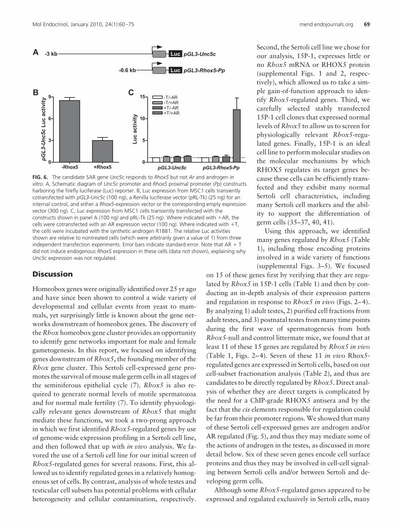
Unc5c (Fig. 6A). We chose Unc5c because itharbors no consensus AREs in this region (Ta-ble 3 and data not shown). When cotrans-fected into the MSC1 Sertoli cell line with anAR expression plasmid and incubated withR1881, this Unc5c promoter construct did notexhibit altered luciferase expression (Fig. 6B).In contrast, the positive control, a luciferasereporter construct containing the Rhox5 prox-imal promoter (Pp) (Fig. 6A), expressedgreatly elevated levels of luciferase in responseto AR and R1881 (Fig. 6B). Note that,whereas the Rhox5 Pp reporter was greatlyincreased in expression in response to AR andR1881, the endogenous Rhox5 gene was notsignificantly elevated in expression (as judgedby qPCR analysis; data not shown), probablybecause it is in a repressed state because ofDNA methylation (17, 57). The ability ofUnc5c to respond to AR only under conditionsin which Rhox5 expression is also induced(Figs. 5 and 6), coupled with the ability ofUnc5c to be regulated by Rhox5 both in vitroand in vivo (Table 1 and Fig. 4, respectively),provides strong evidence that Unc5c is regu-lated by AR through the action of Rhox5.
Identification of common targets ofAR-regulated Rhox genes
In the previous section, we provided evidencethat we had identified SAR genes that are regu-lated by AR through the action of the AR-induc-ible gene Rhox5. As another means to addresswhether we identified bona fide SAR genes, wedetermined whether they are regulated by otherAR-inducible Rhox genes. We previously dem-onstrated that Rhox2, Rhox3, Rhox10, andRhox11 are all strongly up-regulated in expres-sion in response to AR and androgen in Sertolicells (7). Thus, we examined whether any ofthese other AR-inducible Rhox genes were capa-ble of regulating the putative SAR genes we hadidentified. The putative SAR genes that we se-lected for this analysis were those that fulfilledthree criteria: 1) regulated by Rhox5 in vivo, asjudged by our three-prong analysis (Figs. 2–4);2) regulated by AR in vivo (Fig. 5); and 3) signif-icantly expressed in the purified Sertoli cell frac-tion (Table 2, category 1 or 2). The six genes thatfulfilled these criteria were Gdap1, Tmem176a,Tmem176b, Fzd2, Pltp, and Unc5c. To deter-mine whether these six genes are regulated by
0
11
22
33
44
55 Fzd2
0
1000
2000
3000
4000
5000
6000 Rhox5-wtRhox5-koRhox5
5.0 7.5 10.0 12.5 15.0 17.5 20.0 22.5 25.0 27.5 30.00
5
10
15
20 Unc5c
posnatal time
0
2
4
6 Tmem176a
0
1
2
3
4 CD24a
0
6
12
18
24 Gdap1
mRN
A le
vel
FIG. 4. Developmental expression pattern of Rhox5-regulated genes. qPCR analysis oftestes RNA from Rhox5-null (KO) and wild-type (WT) littermate mice of the indicatedpostnatal ages (six mice per time point). All values were quantified as described in Fig.1B. Postnatal time points at which Rhox5 regulates the indicated genes are denoted byarrows (upward and downward arrows indicate genes that are up-regulated and down-regulated, respectively). A value of 1 was arbitrarily given to the lowest expression levelamong the time points examined for a given gene. Error bars indicate standard error.
Mol Endocrinol, January 2010, 24(1):60–75 mend.endojournals.org 67

AR-regulated Rhox genes, we transiently transfected ex-pression vectors encoding RHOX2, RHOX3, RHOX5,RHOX10, and RHOX11 into 15P-1 cells (we previouslyshowed that all these RHOX proteins are all expressed atsimilar levels from these expression vectors) (32). qPCRanalysis revealed that each of the six SAR genes was signif-icantly regulated in response to at least two of the AR-in-ducible Rhox genes (Fig. 7A). For example, Unc5c was neg-atively regulated by Rhox3, Rhox5, and Rhox10.Tmem176a and Tmem176b were also regulated by Rhox3and Rhox5, but instead of being regulated by Rhox10, thesetwo genes responded to Rhox11. Pltp had the same patternof expression as Tmem176a and Tmem176b, except thatRhox2 also negatively regulated its expression. Fzd2 had an
unusual pattern of expression in that itwas negatively regulated by Rhox2,Rhox3, Rhox5, and Rhox11, but waspositively regulated by Rhox10. To as-sess specificity, we also evaluated the ef-fect of Rhox8, which is not AR regu-lated, either as assessed in a Sertoli cellline (7) or in postnatal and adult testes invivo (MacLean, J., K. de Gendt, G. Verho-even, and M. F. Wilkinson, unpublishedobservations). We found that forcedexpression of Rhox8 did not signifi-cantly affect the expression of any ofthe SAR genes except for Gdap1.Rhox8 positively regulated Gdap1, asdid Rhox5 and Rhox10. It is noteworthythat we found that transiently trans-fected Rhox5 only modestly regulatedthe SAR genes (Fig. 7A), which con-trasted with its more dramatic effects onsome of the SAR genes in cells stablytransfected with Rhox5 (Table 1; i.e.Gdap1, Pltp, and Unc5c). Regardless ofthis quantitative difference, Rhox5 hadthe same qualitative effect on the six SARgenes in transiently and stably trans-fected cells (i.e. Rhox5 negatively regu-lated all the SAR genes except for Gdap1under both conditions).
To determine whether the regulationof the putative SAR genes is a conservedresponse, we examined the effect offorced expression of the human RHOXgenes RHOXF1 and RHOXF2. Theseare the only human RHOX genes thathave been characterized; both are se-lectively expressed in human testes(58, 59). RHOXF1 is an androgen-induced gene, whereas RHOXF2 has
not been characterized in this regard (58). We found thattransient transfection of expression vectors encodingeither RHOXF1 or RHOXF2 triggered the down-regula-tion of Unc5c and Pltp (Fig. 7B). RHOXF2, but notRHOXF1, up-regulated Gdap1 expression, whereasRHOXF1, but not RHOXF2, modestly decreased the ex-pression of both Tmem176 and Tmem176b. The only pu-tative SAR gene that did not significantly respond toeither RHOXF1 or RHOXF2 was Fzd2. We concludethat human RHOXF1 and RHOXF2 share with andro-gen-regulated mouse Rhox family members the abilityto regulate most of the SAR genes, suggesting that thisis a conserved response.
A
B
Rhox5Bac
h2Bpgm
Cd24a
Cpxm1
Fzd2
Idh3aIfn
ar2Gdap
1Klf9 Pltp Tfrc
Tmem17
6a
Tmem17
6b
Tm4sf10
Unc5c
0
1
2
3
4 ControlScarko
**
*
*
**
*
****
*
*
*
**
**
mRN
A le
vel
Rhox5Bac
h2Bpgm
Cd24a
Cpxm1
Fzd2
Idh3aIfn
ar2Gdap
1Klf9 Pltp Tfrc
Tmem17
6a
Tmem17
6b
Tm4sf10
Unc5c
0
1
2
3
4
ControlTfm
11
19
27
35
** ** **
***
*
**
mRN
A le
vel
0.01
0.008
FIG. 5. Identification of Ar- and Rhox5-regulated genes. A, qPCR analysis of testes RNA fromadult Tfm and littermate control mice. B, qPCR analysis of testes RNA from adult Scarko andlittermate control mice. All values were quantified as described in Fig. 1B. Control mRNAlevels were arbitrarily given a value of 1. Statistical analysis was performed by use the two-tailed Student’s t test: *, P & 0.05; **, P & 0.01; ***, P & 0.001.
68 Hu et al. Rhox5-Regulated Genes Mol Endocrinol, January 2010, 24(1):60–75
Miles Wilkinson
Discussion
Homeobox genes were originally identified over 25 yr agoand have since been shown to control a wide variety ofdevelopmental and cellular events from yeast to mam-mals, yet surprisingly little is known about the gene net-works downstream of homeobox genes. The discovery ofthe Rhox homeobox gene cluster provides an opportunityto identify gene networks important for male and femalegametogenesis. In this report, we focused on identifyinggenes downstream of Rhox5, the founding member of theRhox gene cluster. This Sertoli cell-expressed gene pro-motes the survival of mouse male germ cells in all stages ofthe seminiferous epithelial cycle (7). Rhox5 is also re-quired to generate normal levels of motile spermatozoaand for normal male fertility (7). To identify physiologi-cally relevant genes downstream of Rhox5 that mightmediate these functions, we took a two-prong approachin which we first identified Rhox5-regulated genes by useof genome-wide expression profiling in a Sertoli cell line,and then followed that up with in vivo analysis. We fa-vored the use of a Sertoli cell line for our initial screen ofRhox5-regulated genes for several reasons. First, this al-lowed us to identify regulated genes in a relatively homog-enous set of cells. By contrast, analysis of whole testes andtesticular cell subsets has potential problems with cellularheterogeneity and cellular contamination, respectively.
Second, the Sertoli cell line we chose forour analysis, 15P-1, expresses little orno Rhox5 mRNA or RHOX5 protein(supplemental Figs. 1 and 2, respec-tively), which allowed us to take a sim-ple gain-of-function approach to iden-tify Rhox5-regulated genes. Third, wecarefully selected stably transfected15P-1 cell clones that expressed normallevels of Rhox5 to allow us to screen forphysiologically relevant Rhox5-regu-lated genes. Finally, 15P-1 is an idealcell line to perform molecular studies onthe molecular mechanisms by whichRHOX5 regulates its target genes be-cause these cells can be efficiently trans-fected and they exhibit many normalSertoli cell characteristics, includingmany Sertoli cell markers and the abil-ity to support the differentiation ofgerm cells (35–37, 40, 41).
Using this approach, we identifiedmany genes regulated by Rhox5 (Table1), including those encoding proteinsinvolved in a wide variety of functions(supplemental Figs. 3–5). We focused
on 15 of these genes first by verifying that they are regu-lated by Rhox5 in 15P-1 cells (Table 1) and then by con-ducting an in-depth analysis of their expression patternand regulation in response to Rhox5 in vivo (Figs. 2–4).By analyzing 1) adult testes, 2) purified cell fractions fromadult testes, and 3) postnatal testes from many time pointsduring the first wave of spermatogenesis from bothRhox5-null and control littermate mice, we found that atleast 11 of these 15 genes are regulated by Rhox5 in vivo(Table 1, Figs. 2–4). Seven of these 11 in vivo Rhox5-regulated genes are expressed in Sertoli cells, based on ourcell-subset fractionation analysis (Table 2), and thus arecandidates to be directly regulated by Rhox5. Direct anal-ysis of whether they are direct targets is complicated bythe need for a ChIP-grade RHOX5 antisera and by thefact that the cis elements responsible for regulation couldbe far from their promoter regions. We showed that manyof these Sertoli cell-expressed genes are androgen and/orAR regulated (Fig. 5), and thus they may mediate some ofthe actions of androgen in the testes, as discussed in moredetail below. Six of these seven genes encode cell surfaceproteins and thus they may be involved in cell-cell signal-ing between Sertoli cells and/or between Sertoli and de-veloping germ cells.
Although some Rhox5-regulated genes appeared to beexpressed and regulated exclusively in Sertoli cells, many
-3 kb pGL3-Unc5c
pGL3-Unc5c pGL3-Rhox5-Pp0
5
10
15-T/+AR+T/-AR
-T/-AR
+T/+AR
Luc
activ
ity
A
pGL3-Rhox5-Pp
B
Luc
-0.6 kb Luc
-Rhox5 +Rhox50
3
6
9
pGL3
-Unc
5c L
uc a
ctiv
ity
C
FIG. 6. The candidate SAR gene Unc5c responds to Rhox5 but not Ar and androgen invitro. A, Schematic diagram of Unc5c promoter and Rhox5 proximal promoter (Pp) constructsharboring the firefly luciferase (Luc) reporter. B, Luc expression from MSC1 cells transientlycotransfected with pGL3-Unc5c (100 ng), a Renilla luciferase vector (pRL-Tk) (25 ng) for aninternal control, and either a Rhox5-expression vector or the corresponding empty expressionvector (300 ng). C, Luc expression from MSC1 cells transiently transfected with theconstructs shown in panel A (100 ng) and pRL-Tk (25 ng). Where indicated with 'AR, thecells were cotransfected with an AR expression vector (100 ng). Where indicated with 'T,the cells were incubated with the synthetic androgen R1881. The relative Luc activitiesshown are relative to nontreated cells (which were arbitrarily given a value of 1) from threeindependent transfection experiments. Error bars indicate standard error. Note that AR ' Tdid not induce endogenous Rhox5 expression in these cells (data not shown), explaining whyUnc5c expression was not regulated.
Mol Endocrinol, January 2010, 24(1):60–75 mend.endojournals.org 69

Rhox5-regulated genes were expressed and/orregulated in other testicular cell types. For ex-ample, we found that a number of the Rhox5-regulated genes were expressed in both theSertoli and interstitial cell fractions (Table 2,category 2); some of these were regulated byRhox5 in the interstitial cell fraction (Fig. 3).We also identified a gene exclusively expressedin the interstitial cell fraction—Cd24a (Table2, category 3)—that exhibited modestly al-tered expression in Rhox5-null mice (Figs. 3and 4), suggesting that Cd24a is regulated byRhox5 in a paracrine manner. Other Rhox5-regulated genes were expressed at very lowlevels in both the Sertoli and interstitial cellfractions (Table 2, category 4) and thus thesegenes may be regulated by Rhox5 in other tes-ticular cell types, such as germ cells or myoidcells. The overall picture that emerges fromthis analysis of 15 Rhox5-regulated genes isthat, even though RHOX5 protein is restrictedto Sertoli cells (30, 31), its web of regulationextends to various cell types in the testes.
Because Rhox5 is an androgen- and AR-induced homeobox gene (29–31), it is a can-didate to encode a transcription factor thatmediates some of the actions of androgen inthe testes. This predicts that a subset ofRHOX5 targets are SAR genes; i.e. genes in-directly regulated by AR through the action ofintermediate factors (60). Although severalSAR genes have been defined in the prostate(39, 61), to our knowledge none have beendefined in the testes. It is crucial to define SARgenes in the testis, as such genes are likely to bemediators of androgen action in this male re-productive organ. Progress on this front hasbeen stymied because very few androgen-reg-ulated transcription factors have been identi-fied in the Sertoli cell, the only cell type defin-itively known to mediate androgen action inthe testes (6). To our knowledge, the only an-drogen-induced transcription factor genes thathave been defined in Sertoli cells are Rhox5and c-myc. Rhox5 is androgen- and AR-induced in both cultured Sertoli cells and Sertolicells in vivo (29–31, 46, 53, 62–64), whereasc-myc is androgen-inducible in cultured Sertolicells (65), but it appears not to be AR-regu-lated in vivo (53, 55). In this paper, we provideseveral lines of evidence that we have identi-fied eight SAR genes that are regulated by AR
Unc5c
Tmem17
6a
Tmem17
6bGdap
1Fzd
2pltp
0
1
2
3
4 Rhox8
*
mRN
A le
vel
0.0
0.2
0.4
0.6
0.8
1.0
1.2
1.4Rhox11
* **
*
mRN
A le
vel
0
1
2
3Rhox10
*
*
mRN
A le
vel
0.00
0.25
0.50
0.75
1.00Rhox3
*
* *
** *
mRN
A le
vel
0.0
0.2
0.4
0.6
0.8
1.0
1.2Rhox2
*
*
mR
NA le
vel
0.0
0.2
0.4
0.6
0.8
1.0
1.2
1.4Rhox5
** *
*
* *
mRN
A le
vel
0.0
0.2
0.4
0.6
0.8
1.0
1.2RHOXF1
*
**
*
mRN
A le
vel
Unc5c
Tmem17
6a
Tmem17
6bGdap
1Fzd
2Pltp
0.0
0.5
1.0
1.5
2.0
2.5 RHOXF2
*
*
*mRN
A le
vel
A B
FIG. 7. Regulation of candidate SAR genes by Ar-inducible Rhox genes. A, qPCRanalysis of total cellular RNA from 15P-1 Sertoli cells transiently transfected withexpression vectors encoding the indicated mouse Rhox genes or the empty expressionvector (800 ng). A value of 1 was arbitrarily given to the expression of the indicatedgenes in cells transfected with the empty expression vector. All values were quantified asdescribed in Fig. 1B. B, Same as panel A, except the cells were transiently transfectedwith expression vectors encoding the human RHOXF1 and RHOXF2 proteins. Statisticalanalysis was performed by using the two-tailed student’s t-test (*, P & 0.05).
70 Hu et al. Rhox5-Regulated Genes Mol Endocrinol, January 2010, 24(1):60–75

through the action of Rhox5 in the testes. First, all eight ofthese genes are regulated by Rhox5 in 15P-1 Sertoli cells(Table 1), and all but one of them (Ifnar2) are regulatedby Rhox5 in the testes in vivo (Figs. 2–4). Second, ouranalysis of Ar-deficient mice indicated that all eight ofthese genes are AR-regulated in adult testes in vivo (Fig.5). As confirmation of this, we found that five of thesegenes (Bpgm, Cpxm1, Ifnar2, Tmem176a, andTmem176b) were independently defined as AR-regulatedby a study that used microarray analysis to identify genesdifferentially expressed in postnatal Tfm Ar-deficient andcontrol mice testes (55). Third, our analysis of the puta-tive regulatory regions in the candidate SAR genesshowed that four of them had no consensus AREs and theother four had few consensus AREs (Table 3). Because, bydefinition, SAR genes are regulated indirectly by AR, thispaucity of AREs is consistent with these genes being bonafide SAR genes. We should note, however, that ARE con-sensus sites are often not functional, and functional AREsoften differ considerably from the consensus sequence(66). Fourth, none of the eight genes were regulated byAR and androgen under conditions in which Rhox5 wasnot induced (supplemental Fig. 7). Fifth, a reporter con-struct harboring the promoter region of one of the candi-date SAR genes (Unc5c) was responsive to Rhox5, butwas not responsive to AR and androgen under conditionsin which Rhox5 was not induced (Fig. 6). Sixth, severalandrogen/AR-inducible mouse Rhox genes besidesRhox5 regulated many of the candidate SAR genes (Fig.7A). Finally, RHOXF1, an androgen-inducible humanRHOX gene (58), was also capable of regulating someof the candidate SAR genes (Fig. 7B), providing evi-dence that this is a conserved response. HumanRHOXF2 also regulated many of these genes (Fig. 7B),but whether it is an androgen-inducible gene has notyet been determined.
Consistent with the often noted connection betweenthe testis and the brain (67), we found that a remarkablylarge proportion of the candidate SAR genes encode pro-teins with known functions in the nervous system. Unc5cencodes a transmembrane receptor of the immunoglobu-lin superfamily that binds to netrin-1, a protein importantfor many developmental events in the brain, includinggoverning axon migration by triggering chemorepulsion(68). UN5C also has a death domain, which engenders itwith proapoptotic activity that may also be important forits roles in brain development (69). We recently showedthat UNC5C functions similarly in the testes by promot-ing the death of male germ cells (32). This germ celldeath-promoting property, coupled with the fact thatUnc5c gene expression is negatively regulated byRhox5, makes Unc5c a good candidate to have a role in
Rhox5’s ability to promote the survival of germ cells(7). Another candidate SAR gene, Gdap1, is up-regu-lated during neuron differentiation (70) and, when mu-tated in humans, is responsible for many cases of Char-cot-Marie-Tooth disease, the most common inheritedform of peripheral neuropathy. GDAP1 is a member ofthe glutathione-S-transferase family, and thus, its neu-rological protective role may derive from its antioxi-dant properties. Tmem176b (also known as Clast1, Lr8,and TORID) encodes a ubiquitously expressed four-transmembrane cell surface protein that is crucial for nor-mal brain development and function. Targeted deletion ofTmem176b in mice causes defects in the cerebellum andsevere ataxia (71). It has not been reported whetherTmem176b-mutant mice have defects in male fertility,but our finding that both Ar and Rhox5 regulate its ex-pression suggests it may have some role in androgen-me-diated events in the testes. A related four-transmembraneprotein-encoding gene, Tmem176a (also known asGS188 and HCA112), is another putative SAR gene. Atpresent, there is no information on either the biochemicalor biological function of Tmem176a. Another candidateSAR gene, Pltp, encodes a protein that acts both insideand outside cells to mediate the transfer of lipids. Al-though PLTP is fairly widely expressed and thereforeprobably acts in many cell types, it appears to be partic-ularly important for brain function, in part, by virtue ofits being secreted by neurons and glial cells. PLTP is likelyto have roles in neurodegenerative and inflammatorybrain diseases, including Alzheimer’s disease, becausePLTP levels are altered in many of these disease states.PLTP also alters the phosphorylation state of TAU, aneuron-specific protein that undergoes shifts in phos-phorylation status in many neurological diseases (72).
The remaining three candidate SAR genes, Fzd2, Ifnr2,and Bpgm, are likely to have roles in many cell types.Fzd2, encodes one of the 10 known frizzled receptors inmice; these are cell surface receptors that bind to theWNT family of secreted glycoproteins. On binding ofWNT proteins, frizzled receptors elicit signal transduc-tion events crucial for conserved developmental steps inmany organisms. Because frizzled receptors tend to actredundantly, it has been difficult to assign precise func-tions to them, including FZD2 (73). Ifnar2 encodes thecell surface receptor for antiviral cytokines called inter-ferons. On interferon binding, IFNAR2 activates theSTAT signaling pathway, which in turn elicits an antiviralresponse, including rapid mRNA decay and translationalrepression (74). Bpgm encodes a multifunctional enzymebest known for its role in controlling the metabolism of2,3-diphosphoglycerate in erythrocytes (75). It will be
Mol Endocrinol, January 2010, 24(1):60–75 mend.endojournals.org 71

interesting to know what specific roles BPGM has inthe testes.
In addition to the candidate SAR genes describedabove, we identified four other genes regulated by Rhox5in the testes in vivo: Cd24a, Klf9, Cpxm1, and Tmem47(Figs. 2–4). Two of these, Cd24a and Klf9, have knownroles in the nervous system. Cd24a encodes aglycosylphosphatidylinositol-anchored glycoprotein thatwas originally identified based on its expression in im-mune cells, but it was subsequently shown to also beexpressed and have a role in neurons. In the adult mousecentral nervous system, CD24 expression is restricted toimmature proliferating neurons in regions of the brainundergoing neurogenesis. Cd24a-knockout mice exhibitincreased numbers of proliferating neurons in thesezones, indicating that CD-24 serves to dampen the gen-eration of adult neurons (76). Kfl9 (also known as Bteb1)encodes a zinc-finger transcription factor that whenknocked out in mice leads to cerebellar Purkinje cell de-fects and behavioral defects consistent with its havingroles in the cerebellum, amygdala, and hippocampus (77).Whereas exactly how KLF9 functions in the brain is notknown, in vitro experiments have suggested it has a rolein neurite extension and branching in vitro (44). Tmem47encodes a four-transmembrane protein that is a memberof a different subfamily than Tmem176a and Tmem176b,the other four-transmembrane protein genes regulated byRhox5. The functional role of TMEM47 in mammals isnot known, but its ortholog in Caenorhabditis elegans,VAB-9, is crucial for the formation of adherens junctionsin epithelial cells (78). Thus, it is tempting to speculatethat TMEM47 might function in the formation and/ormaintenance of the testes-specific adherens junctions thatform between Sertoli cells and germ cells: the ectoplasmicspecialization.
Future studies will be required to precisely define thegene networks downstream of AR and the AR-regulatedRhox genes and to identify their roles in spermatogenesis.One essential question will be to delineate which func-tions of AR require the action of androgen-regulatedRhox genes. Studies in AR-deficient mice harboring eithera nonfunctional Ar allele in Sertoli cells or a hypomorphicAr allele in all cells have revealed that AR has at least threefunctions: 1) it promotes the progression of meiotic sper-matocytes, 2) it promotes the maturation and/or survivalof round spermatids, allowing the generation of elongat-ing spermatids, and 3) it promotes the final maturationand release of elongated spermatids into the lumen of theseminiferous tubules (62, 64, 79, 80). Thus, androgen-inducible Rhox genes may be involved in any or all ofthese events. Another question for the future will be todetermine which Rhox5-regulated genes in Sertoli cells
are direct targets of RHOX5. This will be a challenge toanswer, because a RHOX5-binding consensus sequencehas yet to be defined and RHOX5 may act on enhancersfar upstream or downstream of its target genes. Finally, itwill be intriguing to understand how Rhox5 regulatesgenes in cell types in addition to the Sertoli cell, the onlycell type that significantly expresses Rhox5 (26, 29).Given that a large proportion of Rhox5-regulated genesencode cell surface and secreted proteins (supplementalFig. S3), an obvious mechanism by which it could act is toinfluence cell-cell interactions and signaling events occur-ring between Sertoli cells and other cell types in the testis.By defining and characterizing Rhox-regulated genes invitro and in vivo, we believe that the work presented inthis paper provides a foundation to address these manyfuture issues.
Materials and Methods
Cell culture and transfectionThe 15P-1 and MSC1 cell lines were maintained in DMEM
supplemented with 10% fetal bovine serum and 50 mg/ml ofboth penicillin and streptomycin. The cells were grown at 37 Cin 5% CO2 atmosphere and split when approximately 80%confluent.
To generate stable Rhox5 expression plasmid-transfectedclones, 15P-1 cells cultured to approximately 60% confluencein 100-mm plates were transfected with 2 %g of Rhox5 expres-sion plasmid by use of lipofectamine (Invitrogen, Carlsbad, CA)and clones resistant to antibiotic (700 %g/ml G418) wereselected.
The concentration of plasmids used for transfection was in-dependently determined by use of both a fluorometer and ana-lytical gel electrophoresis. For transient transfection analysis,15P-1 cells were plated in 60-mm dishes the day before trans-fection at a density of approximately 2 # 106 cells per dishes in4 ml of growing medium. On the day of transfection, 800 ng ofplasmid DNA was diluted with 150 %l of serum-free mediumand mixed with 150 %l of serum-free media containing 3 %l ofLipofectamine (Invitrogen, Carlsbad, CA) and incubated atroom temperature for 30 min; then, the DNA-lipid complex wasadded to the wells and incubated at 37 C in a CO2 incubator for5 h. Cells were harvested 36 h post transfection for RNA anal-ysis. MSC1 cells were transfected by use of Fugene 6 (RocheDiagnostics, GmbH, Mannheim, Germany) according to themanufacturer’s instructions. Cells were plated on 12-well cul-ture dishes and then cotransfected with 100 ng of the Rhox5/Unc5c-containing plasmids and 100 ng of either the AR expres-sion plasmid or empty vector. After a 6-h incubation in DMEMsupplemented with 10% charcoal-stripped bovine serum (Hy-Clone Laboratories, Inc., Logan, UT), R1881 was added at finalconcentration of 10 nM. Cells were harvested 36 h post trans-fection for luciferase assay by use of the Dual LuciferaseAssay System (Promega, Madison, WI), according to themanufacturer’s instructions. The relative luciferase activitywas calculated by normalizing against the cotransfected internalcontrol pRL-null. The results shown are the mean ! SEM of threeindependent transfection experiments.
72 Hu et al. Rhox5-Regulated Genes Mol Endocrinol, January 2010, 24(1):60–75

PlasmidsThe Unc5c-P (G734), Rhox5-Pp (Pem250), and Rhox ex-
pression plasmids were generated previously (32, 46). The hu-man AR pcDNA 3.1 plasmid (G541) was kindly provided byZhengxin Wang (The University of Texas M.D. Anderson Can-cer Center, Houston, TX).
RNA and protein analysisTotal cellular RNA was prepared from cell lines and tissues
by guanidinium isothiocyanate lysis and centrifugation over a5.7 M CsCl cushion as described previously (81). The Rhox5cDNA probe used for Northern blot analysis was prepared asdescribed previously (24). Real-time RT-PCR analysis was per-formed as described previously (7) with primers as described inTable 1. Microarray analysis with 15P-1 stable cell clones wasperformed at the University of Iowa DNA core facility on totalcellular RNA from clones 6, 24, 14, and 16, using an Affymetrixmouse genome GeneChip 430 2.0 array. This gene chip inde-pendently samples approximately 45,101 probe sets, which cor-responds to approximately 39,000 genes.
For Western blot analysis, 20 %g of total cell lysates wereelectrophoresed in a 10% SDS-polyacrylamide, transferred toHybond ECL nitrocellulose (Amersham, Piscataway, NJ), andprobed with antibodies against RHOX5 or !-actin (SigmaChemical Co., St. Louis, MO). The membranes were given three10-min washes with 0.1% Tween 20 in PBS at room tempera-ture and then incubated for 45 min at room temperature withthe secondary antibody (ECL kit antirabbit or antimouse fromAmersham) at a dilution of 1:5000. The filter was developedby using the ECL-Plus reagent according to the manufactur-er’s protocol (Amersham).
Testis cell fractionationSertoli and interstitial cells were purified from testes as pre-
viously described (7, 82). In brief, testes were decapsulated andthe seminiferous tubules were allowed to settle in PBS, followedby incubation in collagenase (C2674; Sigma). After anotherround of settling, the supernatant, which was enriched for in-terstitial cells, was subjected to hypotonic shock (by incubationin 1:7 diluted PBS for 3 min) to remove germ cells, and thesurviving cells were pelleted and the RNA extracted. The pelletobtained after collagenase treatment, which was enriched forSertoli cells, was incubated in a solution containing a mixture ofenzymes [0.1% collagenase, 0.2% hyaluronidase (H6254;Sigma), 0.04% DNase I (D5025; Sigma), and 0.03% trypsininhibitor (T6522; Sigma) in 1# PBS, pH 7.4] at 30 C for 40 min.The cells were pelleted and subjected to the same hypotonicshock as the interstitial cells, followed by centrifugation andRNA extraction of the pellet.
Acknowledgments
We thank Miriam Buttigieg for mouse tissue collection, and Drs.James MacLean and Wai-kin Chan for helpful advice through-out the course of this project.
Address all correspondence and requests for reprints to: MilesF. Wilkinson, Department of Reproductive Medicine, University ofCalifornia, San Diego, 9500 Gilman Drive, MC 0864, La Jolla,California 92093-0864. E-mail: [email protected].
This work was supported by NIH Grants HD-45595.Present address for D.D.: Fox Chase Cancer Center, Phila-
delphia, Pennsylvania 19111.Disclosure Summary: The authors have nothing to disclose.
References
1. Ingham PW, Martinez Arias A 1992 Boundaries and fields in earlyembryos. Cell 68:221–235
2. Chiba S 1998 Homeobox genes in normal hematopoiesis and leu-kemogenesis. Int J Hematol 68:343–353
3. Awgulewitsch A 2003 Hox in hair growth and development. Natur-wissenschaften 90:193–211
4. Lee CS, Kaestner KH 2004 Clinical endocrinology and metabolism.Development of gut endocrine cells. Best Pract Res Clin EndocrinolMetab 18:453–462
5. Wilhelm D, Palmer S, Koopman P 2007 Sex determination andgonadal development in mammals. Physiol Rev 87:1–28
6. Maclean 2nd JA, Wilkinson MF 2005 Gene regulation in spermat-ogenesis. Curr Top Dev Biol 71:131–197
7. MacLean JA, Chen MA, Wayne CM, Bruce SR, Rao MK, MeistrichML, MacLeod CL, Wilkinson MF 2005 Rhox: a new homeoboxgene cluster. Cell 120:369–382
8. Jackson M, Watt AJ, Gautier P, Gilchrist D, Driehaus J, GrahamGJ, Keebler J, Prugnolle F, Awadalla P, Forrester LM 2006 A mu-rine specific expansion of the Rhox cluster involved in embryonicstem cell biology is under natural selection. BMC Genomics 7:212
9. MacLean 2nd JA, Lorenzetti D, Hu Z, Salerno WJ, Miller J, WilkinsonMF 2006 Rhox homeobox gene cluster: recent duplication of threefamily members. Genesis 44:122–129
10. Morris L, Gordon J, Blackburn CC 2006 Identification of a tandemduplicated array in the Rhox alpha locus on mouse chromosome X.Mamm Genome 17:178–187
11. Wang X, Zhang J 2006 Remarkable expansions of an X-linkedreproductive homeobox gene cluster in rodent evolution. Genomics88:34–43
12. Rao M, Wilkinson MF 2002 Homeobox genes and the male repro-ductive system. In: Robaire B, Hinton BT, eds. The epididymis:From molecules to clinical practice. New York: Kluwer Academic/Plenum Publishers
13. Lindsey S, Wilkinson MF 1996 Homeobox genes and male repro-ductive development. J Assist Reprod Genet 13:182–192
14. MacLaughlin DT, Donahoe PK 2004 Sex determination and differ-entiation. N Engl J Med 350:367–378
15. Geyer CB, Eddy EM 2008 Identification and characterization ofRhox13, a novel X-linked mouse homeobox gene. Gene 423:194–200
16. MacLeod CL, Fong AM, Seal BS, Walls L, Wilkinson MF 1990Isolation of novel complementary DNA clones from T lymphomacells: one encodes a putative multiple membrane-spanning protein.Cell Growth Differ 1:271–279
17. Sasaki AW, Doskow J, MacLeod CL, Rogers MB, Gudas LJ,Wilkinson MF 1991 The oncofetal gene Pem encodes a homeodo-main and is regulated in primordial and pre-muscle stem cells. MechDev 34:155–164
18. Ono T, Sato S, Kimura N, Tanaka M, Shibuya A, Old LJ,Nakayama E 2000 Serological analysis of BALB/C methylchol-anthrene sarcoma Meth A by SEREX: identification of a cancer/testis antigen. Int J Cancer 88:845– 851
19. Agarwal SK, Lee Burns A, Sukhodolets KE, Kennedy PA, Obungu VH,Hickman AB, Mullendore ME, Whitten I, Skarulis MC, Simonds WF,Mateo C, Crabtree JS, Scacheri PC, Ji Y, Novotny EA, Garrett-Beal L,Ward JM, Libutti SK, Richard Alexander H, Cerrato A, Parisi MJ,Santa Anna-A S, Oliver B, Chandrasekharappa SC, Collins FS, SpiegelAM, Marx SJ 2004 Molecular pathology of the MEN1 gene. Ann NYAcad Sci 1014:189–198
Mol Endocrinol, January 2010, 24(1):60–75 mend.endojournals.org 73

20. Guo F, Huang X, Li S, Sun L, Li Y, Li H, Zhou Y, Chu Y, Zhou T2007 Identification of prosaposin as a novel interaction partner forRhox5. J Genet Genomics 34:392–399
21. Rao MK, Maiti S, Ananthaswamy HN, Wilkinson MF 2002 Ahighly active homeobox gene promoter regulated by Ets and Sp1family members in normal granulosa cells and diverse tumor celltypes. J Biol Chem 277:26036–26045
22. MacLean 2nd JA, Rao MK, Doyle KM, Richards JS, Wilkinson MF2005 Regulation of the Rhox5 homeobox gene in primary granu-losa cells: preovulatory expression and dependence on SP1/SP3 andGABP. Biol Reprod 73:1126–1134
23. Hamatani T, Carter MG, Sharov AA, Ko MS 2004 Dynamics ofglobal gene expression changes during mouse preimplantation de-velopment. Dev Cell 6:117–131
24. Wilkinson MF, Kleeman J, Richards J, MacLeod CL 1990 A noveloncofetal gene is expressed in a stage-specific manner in murineembryonic development. Dev Biol 141:451–455
25. Lin TP, Labosky PA, Grabel LB, Kozak CA, Pitman JL, Kleeman J,MacLeod CL 1994 The Pem homeobox gene is X-linked and ex-clusively expressed in extraembryonic tissues during early murinedevelopment. Dev Biol 166:170–179
26. Pitman JL, Lin TP, Kleeman JE, Erickson GF, MacLeod CL 1998Normal reproductive and macrophage function in Pem homeoboxgene-deficient mice. Dev Biol 202:196–214
27. Daggag H, Svingen T, Western PS, van den Bergen JA, McClive PJ,Harley VR, Koopman P, Sinclair AH 2008 The rhox homeoboxgene family shows sexually dimorphic and dynamic expression dur-ing mouse embryonic gonad development. Biol Reprod 79:468–474
28. Maiti S, Doskow J, Sutton K, Nhim RP, Lawlor DA, Levan K,Lindsey JS, Wilkinson MF 1996 The Pem homeobox gene: rapidevolution of the homeodomain, X chromosomal localization, andexpression in reproductive tissue. Genomics 34:304–316
29. Lindsey JS, Wilkinson MF 1996 Pem: a testosterone- and LH-regu-lated homeobox gene expressed in mouse Sertoli cells and epididymis.Dev Biol 179:471–484
30. Sutton KA, Maiti S, Tribley WA, Lindsey JS, Meistrich ML, BucanaCD, Sanborn BM, Joseph DR, Griswold MD, Cornwall GA,Wilkinson MF 1998 Androgen regulation of the Pem homeodo-main gene in mice and rat Sertoli and epididymal cells. J Androl19:21–30
31. Rao MK, Wayne CM, Meistrich ML, Wilkinson MF 2003 Pemhomeobox gene promoter sequences that direct transcription in aSertoli cell-specific, stage-specific, and androgen-dependent man-ner in the testis in vivo. Mol Endocrinol 17:223–233
32. Hu Z, Shanker S, MacLean 2nd JA, Ackerman SL, Wilkinson MF2008 The RHOX5 homeodomain protein mediates transcriptionalrepression of the netrin-1 receptor gene Unc5c. J Biol Chem 283:3866–3876
33. Hu Z, MacLean JA, Bhardwaj A, Wilkinson MF 2007 Regulationand function of the Rhox5 homeobox gene. Ann NY Acad Sci1120:72–83
34. Shanker S, Hu Z, Wilkinson MF 2008 Epigenetic regulation anddownstream targets of the Rhox5 homeobox gene. Int J Androl31:462–470
35. Rassoulzadegan M, Paquis-Flucklinger V, Bertino B, Sage J, JasinM, Miyagawa K, van Heyningen V, Besmer P, Cuzin F 1993 Trans-meiotic differentiation of male germ cells in culture. Cell 75:997–1006
36. Grandjean V, Sage J, Ranc F, Cuzin F, Rassoulzadegan M 1997Stage-specific signals in germ line differentiation: control of Sertolicell phagocytic activity by spermatogenic cells. Dev Biol 184:165–174
37. Jabado N, Canonne-Hergaux F, Gruenheid S, Picard V, Gros P2002 Iron transporter Nramp2/DMT-1 is associated with the mem-brane of phagosomes in macrophages and Sertoli cells. Blood 100:2617–2622
38. Payne AH 1990 Hormonal regulation of cytochrome P450 en-
zymes, cholesterol side-chain cleavage and 17 alpha-hydroxylase/C17–20 lyase in Leydig cells. Biol Reprod 42:399–404
39. Verhoeven G, Swinnen JV 1999 Indirect mechanisms and cascadesof androgen action. Mol Cell Endocrinol 151:205–212
40. Del Rio-Tsonis K, Covarrubias L, Kent J, Hastie ND, Tsonis PA1996 Regulation of the Wilms’ tumor gene during spermatogenesis.Dev Dyn 207:372–381
41. Rossi P, Albanesi C, Grimaldi P, Geremia R 1991 Expression of themRNA for the ligand of c-kit in mouse Sertoli cells. Biochem Bio-phys Res Commun 176:910–914
42. Rogers MF, Ben-Hur A 2009 The use of gene ontology evidencecodes in preventing classifier assessment bias. Bioinformatics (Oxf)25:1173–1177
43. Velarde MC, Geng Y, Eason RR, Simmen FA, Simmen RC 2005Null mutation of Kruppel-like factor9/basic transcription elementbinding protein-1 alters peri-implantation uterine development inmice. Biol Reprod 73:472–481
44. Bonett RM, Hu F, Bagamasbad P, Denver RJ 2009 Stressor andglucocorticoid-dependent induction of the immediate early genekruppel-like factor 9: implications for neural development and plas-ticity. Endocrinology 150:1757–1765
45. Wagle JR, Heindel JJ, Steinberger A, Sanborn BM 1986 Effect ofhypotonic treatment on Sertoli cell purity and function in culture. InVitro Cell Dev Biol 22:325–331
46. Bhardwaj A, Rao MK, Kaur R, Buttigieg MR, Wilkinson MF 2008GATA factors and androgen receptor collaborate to transcription-ally activate the Rhox5 homeobox gene in Sertoli cells. Mol cell Biol28:2138–2153
47. Kuopio T, Pelliniemi LJ, Huhtaniemi I 1989 Rapid Leydig cellproliferation and luteinizing hormone receptor replenishment in theneonatal rat testis after a single injection of human chorionic go-nadotropin. Biol Reprod 40:135–143
48. Zhang H, Denhard LA, Zhou H, Liu LH, Lan ZJ 20080610009K11Rik, a testis-specific and germ cell nuclear receptor-interacting protein. Biochem Biophys Res Commun 366:898–904
49. Lifschitz-Mercer B, Elliott DJ, Issakov J, Leider-Trejo L, SchreiberL, Misonzhnik F, Eisenthal A, Maymon BB 2002 Localization of aspecific germ cell marker, DAZL1, in testicular germ cell neopla-sias. Virchows Arch 440:387–391
50. Yomogida K, Ohtani H, Harigae H, Ito E, Nishimune Y, Engel JD,Yamamoto M 1994 Developmental stage- and spermatogenic cycle-specific expression of transcription factor GATA-1 in mouse Sertolicells. Development (Camb) 120:1759–1766
51. Roberts KP, Griswold MD 1990 Characterization of rat transferrinreceptor cDNA: the regulation of transferrin receptor mRNA intestes and in Sertoli cells in culture. Mol Endocrinol 4:531–542
52. Linder CC, Heckert LL, Roberts KP, Kim KH, Griswold MD 1991Expression of receptors during the cycle of the seminiferous epithe-lium. Ann NY Acad Sci 637:313–321
53. Denolet E, De Gendt K, Allemeersch J, Engelen K, Marchal K, VanHummelen P, Tan KA, Sharpe RM, Saunders PT, Swinnen JV,Verhoeven G 2006 The effect of a Sertoli cell-selective knockout ofthe androgen receptor on testicular gene expression in prepubertalmice. Mol Endocrinol 20:321–334
54. He WW, Kumar MV, Tindall DJ 1991 A frame-shift mutation inthe androgen receptor gene causes complete androgen insensitivityin the testicular-feminized mouse. Nucleic Acids Res 19:2373–2378
55. O’Shaughnessy PJ, Abel M, Charlton HM, Hu B, Johnston H,Baker PJ 2007 Altered expression of genes involved in regulation ofvitamin A metabolism, solute transportation, and cytoskeletal func-tion in the androgen-insensitive tfm mouse testis. Endocrinology148:2914–2924
56. Heemers HV, Verhoeven G, Swinnen JV 2006 Androgen activationof the sterol regulatory element-binding protein pathway: currentinsights. Mol Endocrinol 20:2265–2277
57. Oda M, Yamagiwa A, Yamamoto S, Nakayama T, Tsumura A,Sasaki H, Nakao K, Li E, Okano M 2006 DNA methylation regu-
74 Hu et al. Rhox5-Regulated Genes Mol Endocrinol, January 2010, 24(1):60–75
lates long-range gene silencing of an X-linked homeobox gene clus-ter in a lineage-specific manner. Genes Dev 20:3382–3394
58. Geserick C, Weiss B, Schleuning WD, Haendler B 2002 OTEX, anandrogen-regulated human member of the paired-like class of ho-meobox genes. Biochem J 366:367–375
59. Wayne CM, MacLean JA, Cornwall G, Wilkinson MF 2002 Twonovel human X-linked homeobox genes, hPEPP1 and hPEPP2, se-lectively expressed in the testis. Gene 301:1–11
60. Dean DM, Sanders MM 1996 Ten years after: reclassification ofsteroid-responsive genes. Mol Endocrinol 10:1489–1495
61. Collett GP, Betts AM, Johnson MI, Pulimood AB, Cook S, NealDE, Robson CN 2000 Peroxisome proliferator-activated receptoralpha is an androgen-responsive gene in human prostate and ishighly expressed in prostatic adenocarcinoma. Clin Cancer Res6:3241–3248
62. De Gendt K, Swinnen JV, Saunders PT, Schoonjans L, DewerchinM, Devos A, Tan K, Atanassova N, Claessens F, Lecureuil C, HeynsW, Carmeliet P, Guillou F, Sharpe RM, Verhoeven G 2004 A Sertolicell-selective knockout of the androgen receptor causes spermato-genic arrest in meiosis. Proc Natl Acad Sci USA 101:1327–1332
63. Sneddon SF, Walther N, Saunders PT 2005 Expression of androgenand estrogen receptors in Sertoli cells: studies using the mouse SK11cell line. Endocrinology 146:5304–5312
64. Tan KA, De Gendt K, Atanassova N, Walker M, Sharpe RM,Saunders PT, Denolet E, Verhoeven G 2005 The role of androgens inSertoli cell proliferation and functional maturation: studies in micewith total or Sertoli cell-selective ablation of the androgen receptor.Endocrinology 146:2674–2683
65. Lim K, Yoo JH, Kim KY, Kweon GR, Kwak ST, Hwang BD 1994Testosterone regulation of proto-oncogene c-myc expression in pri-mary Sertoli cell cultures from prepubertal rats. J Androl 15:543–550
66. Wang Q, Li W, Liu XS, Carroll JS, Janne OA, Keeton EK,Chinnaiyan AM, Pienta KJ, Brown M 2007 A hierarchical network oftranscription factors governs androgen receptor-dependent prostatecancer growth. Mol Cell 27:380–392
67. Guo J, Zhu P, Wu C, Yu L, Zhao S, Gu X 2003 In silico analysisindicates a similar gene expression pattern between human brainand testis. Cytogenet Genome Res 103:58–62
68. Round J, Stein E 2007 Netrin signaling leading to directed growthcone steering. Curr Opin Neurobiol 17:15–21
69. Mehlen P, Llambi F 2005 Role of netrin-1 and netrin-1 dependencereceptors in colorectal cancers. Br J Cancer 93:1–6
70. Liu H, Nakagawa T, Kanematsu T, Uchida T, Tsuji S 1999 Isola-tion of 10 differentially expressed cDNAs in differentiated Neuro2a
cells induced through controlled expression of the GD3 synthasegene. J Neurochem 72:1781–1790
71. Maeda Y, Fujimura L, O-Wang J, Hatano M, Sakamoto A, ArimaM, Ebara M, Ino H, Yamashita T, Saisho H, Tokuhisa T 2006 Roleof Clast1 in development of cerebellar granule cells. Brain Res1104:18–26
72. Dong W, Albers JJ, Vuletic S 2009 Phospholipid transfer proteinreduces phosphorylation of tau in human neuronal cells. J NeurosciRes 87:3176–3185
73. Fischer T, Guimera J, Wurst W, Prakash N 2007 Distinct but re-dundant expression of the Frizzled Wnt receptor genes at signalingcenters of the developing mouse brain. Neuroscience 147:693–711
74. Domanski P, Fish E, Nadeau OW, Witte M, Platanias LC, Yan H,Krolewski J, Pitha P, Colamonici OR 1997 A region of the betasubunit of the interferon alpha receptor different from box 1 inter-acts with Jak1 and is sufficient to activate the Jak-Stat pathway andinduce an antiviral state. J Biol Chem 272:26388–26393
75. Joulin V, Peduzzi J, Romeo PH, Rosa R, Valentin C, Dubart A,Lapeyre B, Blouquit Y, Garel MC, Goossens M, Rosa J, Cohen-Solal M 1986 Molecular cloning and sequencing of the humanerythrocyte 2,3-bisphosphoglycerate mutase cDNA: revised aminoacid sequence. EMBO J 5:2275–2283
76. Belvindrah R, Rougon G, Chazal G 2002 Increased neurogenesis inadult mCD24-deficient mice. J Neurosci 22:3594–3607
77. Morita M, Kobayashi A, Yamashita T, Shimanuki T, Nakajima O,Takahashi S, Ikegami S, Inokuchi K, Yamashita K, Yamamoto M,Fujii-Kuriyama Y 2003 Functional analysis of basic transcriptionelement binding protein by gene targeting technology. Mol Cell Biol23:2489–2500
78. Simske JS, Koppen M, Sims P, Hodgkin J, Yonkof A, Hardin J 2003The cell junction protein VAB-9 regulates adhesion and epidermalmorphology in C. elegans. Nat Cell Biol 5:619–625
79. Chang C, Chen YT, Yeh SD, Xu Q, Wang RS, Guillou F, Lardy H,Yeh S 2004 Infertility with defective spermatogenesis and hypotes-tosteronemia in male mice lacking the androgen receptor in Sertolicells. Proc Natl Acad Sci USA 101:6876–6881
80. Abel MH, Baker PJ, Charlton HM, Monteiro A, Verhoeven G, DeGendt K, Guillou F, O’Shaughnessy PJ 2008 Spermatogenesis andSertoli cell activity in mice lacking Sertoli cell receptors for follicle-stimulating hormone and androgen. Endocrinology 149:3279–3285
81. Wilkinson M 2000 Purification of RNA. New York: Oxford Uni-versity Press
82. Anway MD, Folmer J, Wright WW, Zirkin BR 2003 Isolation ofSertoli cells from adult rat testes: an approach to ex vivo studies ofSertoli cell function. Biol Reprod 68:996–1002
Mol Endocrinol, January 2010, 24(1):60–75 mend.endojournals.org 75