Barhl1, a gene belonging to a new subfamily of mammalian homeobox genes, is expressed in migrating...
10
© 2000 Oxford University Press Human Molecular Genetics, 2000, Vol. 9, No. 9 1443–1452 Barhl1, a gene belonging to a new subfamily of mammalian homeobox genes, is expressed in migrating neurons of the CNS Alessandro Bulfone 1,+ , Emilio Menguzzato 1,+,§ , Vania Broccoli 1 , Anna Marchitiello 1 , Claudio Gattuso 1 , Margherita Mariani 2 , Gian Giacomo Consalez 2 , Salvador Martinez 4 , Andrea Ballabio 1,3 and Sandro Banfi 1,¶ 1 Telethon Institute of Genetics and Medicine (TIGEM), 2 Department of Biological and Technological Research (DIBIT), 3 Universita’ Vita-Salute San Raffaele, San Raffaele Biomedical Science Park, Via Olgettina 58, 20132 Milan, Italy and 4 Department of Morphological Sciences, Faculty of Medicine, University of Murcia, Murcia, Spain Received 7 February 2000; Revised and Accepted 27 March 2000 The BarH1 and BarH2 (Bar) Drosophila genes are homeobox-containing genes, which are required for the fate determination of external sensory organs in the fly. By means of a bioinformatic approach, we have identified in mouse and human two homeobox genes highly related to the Bar Drosophila genes, Barhl1 and Barhl2. While Barhl1 represents a novel gene, Barhl2 turned out to correspond to the mBH1 cDNA recently described in rat. We isolated and sequenced the full-length mouse Barhl1 and mapped both the human BARHL1 and BARHL2 genes to chro- mosomes 9q34 and 1p22, respectively. Detailed anal- ysis of the murine Barhl1 expression pattern by in situ hybridization revealed that this transcript is exclusively expressed in restricted domains of the developing CNS, which suggests that this gene, similar to its Drosophila counterparts BarH1 and BarH2, may play a crucial role in cell fate determin- ation of neural structures. In particular, Barhl1 showed specific domains of expression in the dien- cephalon and in the rhombencephalon where it was found to be expressed in migrating cells giving rise to the cerebellar external granular layer and to specific populations of dorsal sensory interneurons of the spinal cord. Thus, Barhl1 function may be required for the generation of these specific subtypes of neuronal progenitors. Furthermore, the mapping assignment and the expression pattern make BARHL1 an attractive positional candidate gene for a form of Joubert syndrome, a rare develop- mental anomaly of the cerebellum in humans. INTRODUCTION The development of the mammalian neural system is a very complex process that requires an ordered series of steps begin- ning with an initial growth phase of dividing precursor cells and the subsequent process of differentiation of post-mitotic cells. The molecular mechanisms controlling these events still remain largely elusive and involve many genes, most of which have not yet been identified and characterized. One way to gain insight into the genetic program of neural development in mammals is represented by the study of model organisms. In fact, many of the pathways involved in neurogenesis have been found to share similarities during evolution and numerous genes have been found to be conserved and to play similar roles even in distantly related species. In particular, the fruitfly Drosophila melanogaster is one of the most valuable model organisms to study development (1). Many genes playing a role in neurogenesis in the fly have been characterized at both the genetic and molecular levels (2,3). This has led to the understanding of some of the basic pathways underlying neural development in Drosophila, particularly of the sensory organs. The proneural genes such as the four members of the Achaete– scute complex, daughterless and atonal, act as positive regula- tors for sensory organ formation, as opposed to other genes like hairy, Enhancer of split and extramacrochetae, which act as negative regulators (for review see refs 4,5). A number of upstream regulators of proneural genes have also been identi- fied and several of them are transcription factors which are required for the initial activation of proneural genes in sensory organs (6–9). Among these are the BarH1 and BarH2 genes. They are required for the fate determination of external sensory organs and for normal eye development in the fly (10–12). Recently, they have also been found to play a crucial role in regional pre-patterning of specific structures of the Drosophila nervous system, such as the notum. The BarH1 and BarH2 genes exert this function through an upstream regulation of the proneural achaete–scute genes (13). We recently started a project aimed at the identification and characterization of novel human cDNAs similar to Drosophila mutant genes. By screening the Expressed Sequence Tag data- base (dbEST) using the BLAST software, we identified >200 such human cDNAs and we termed them Drosophila-related expressed sequences (DRES) (14,15). One of the DRES genes + These authors contributed equally to this work § Present address: Telethon Institute of Gene Therapy (TIGET), San Raffaele Biomedical Science Park, Milan, Italy ¶ To whom correspondence should be addressed. Tel: +39 02 215601; Fax: +39 02 21560220; Email: [email protected]
-
Upload
independent -
Category
Documents
-
view
1 -
download
0
Transcript of Barhl1, a gene belonging to a new subfamily of mammalian homeobox genes, is expressed in migrating...
© 2000 Oxford University Press Human Molecular Genetics, 2000, Vol. 9, No. 9 1443–1452
Barhl1, a gene belonging to a new subfamily ofmammalian homeobox genes, is expressed inmigrating neurons of the CNSAlessandro Bulfone1,+, Emilio Menguzzato1,+,§, Vania Broccoli1, Anna Marchitiello1,Claudio Gattuso1, Margherita Mariani2, Gian Giacomo Consalez2, Salvador Martinez4,Andrea Ballabio1,3 and Sandro Banfi1,¶
1Telethon Institute of Genetics and Medicine (TIGEM), 2Department of Biological and Technological Research(DIBIT), 3Universita’ Vita-Salute San Raffaele, San Raffaele Biomedical Science Park, Via Olgettina 58, 20132 Milan,Italy and 4Department of Morphological Sciences, Faculty of Medicine, University of Murcia, Murcia, Spain
Received 7 February 2000; Revised and Accepted 27 March 2000
The BarH1 and BarH2 (Bar) Drosophila genes arehomeobox-containing genes, which are required forthe fate determination of external sensory organs inthe fly. By means of a bioinformatic approach, wehave identified in mouse and human two homeoboxgenes highly related to the Bar Drosophila genes,Barhl1 and Barhl2. While Barhl1 represents a novelgene, Barhl2 turned out to correspond to the mBH1cDNA recently described in rat. We isolated andsequenced the full-length mouse Barhl1 and mappedboth the human BARHL1 and BARHL2 genes to chro-mosomes 9q34 and 1p22, respectively. Detailed anal-ysis of the murine Barhl1 expression pattern by insitu hybridization revealed that this transcript isexclusively expressed in restricted domains of thedeveloping CNS, which suggests that this gene,similar to its Drosophila counterparts BarH1 andBarH2, may play a crucial role in cell fate determin-ation of neural structures. In particular, Barhl1showed specific domains of expression in the dien-cephalon and in the rhombencephalon where it wasfound to be expressed in migrating cells giving riseto the cerebellar external granular layer and tospecific populations of dorsal sensory interneuronsof the spinal cord. Thus, Barhl1 function may berequired for the generation of these specificsubtypes of neuronal progenitors. Furthermore, themapping assignment and the expression patternmake BARHL1 an attractive positional candidategene for a form of Joubert syndrome, a rare develop-mental anomaly of the cerebellum in humans.
INTRODUCTION
The development of the mammalian neural system is a verycomplex process that requires an ordered series of steps begin-
ning with an initial growth phase of dividing precursor cellsand the subsequent process of differentiation of post-mitoticcells. The molecular mechanisms controlling these events stillremain largely elusive and involve many genes, most of whichhave not yet been identified and characterized. One way togain insight into the genetic program of neural development inmammals is represented by the study of model organisms. Infact, many of the pathways involved in neurogenesis have beenfound to share similarities during evolution and numerousgenes have been found to be conserved and to play similarroles even in distantly related species. In particular, the fruitflyDrosophila melanogaster is one of the most valuable modelorganisms to study development (1). Many genes playing arole in neurogenesis in the fly have been characterized at boththe genetic and molecular levels (2,3). This has led to theunderstanding of some of the basic pathways underlying neuraldevelopment in Drosophila, particularly of the sensory organs.The proneural genes such as the four members of the Achaete–scute complex, daughterless and atonal, act as positive regula-tors for sensory organ formation, as opposed to other geneslike hairy, Enhancer of split and extramacrochetae, which actas negative regulators (for review see refs 4,5). A number ofupstream regulators of proneural genes have also been identi-fied and several of them are transcription factors which arerequired for the initial activation of proneural genes in sensoryorgans (6–9). Among these are the BarH1 and BarH2 genes.They are required for the fate determination of external sensoryorgans and for normal eye development in the fly (10–12).Recently, they have also been found to play a crucial role inregional pre-patterning of specific structures of the Drosophilanervous system, such as the notum. The BarH1 and BarH2genes exert this function through an upstream regulation of theproneural achaete–scute genes (13).
We recently started a project aimed at the identification andcharacterization of novel human cDNAs similar to Drosophilamutant genes. By screening the Expressed Sequence Tag data-base (dbEST) using the BLAST software, we identified >200such human cDNAs and we termed them Drosophila-relatedexpressed sequences (DRES) (14,15). One of the DRES genes
+These authors contributed equally to this work§Present address: Telethon Institute of Gene Therapy (TIGET), San Raffaele Biomedical Science Park, Milan, Italy¶To whom correspondence should be addressed. Tel: +39 02 215601; Fax: +39 02 21560220; Email: [email protected]
1444 Human Molecular Genetics, 2000, Vol. 9, No. 9
identified in the course of our dbEST screening, DRES115,was found to bear significant homology to the homeodomainof the Drosophila genes BarH1 and BarH2. Here, we report adetailed characterization of the Dres115 (Barhl1) geneincluding cDNA identification, sequence and mapping assign-ment in both human and mouse. In addition, we have isolatedand mapped the human homolog of mBH1 (Barhl2), a genehighly related to Barhl1 which has been described previouslyin rat (16). A comprehensive study of Barhl1 expression byRNA in situ hybridization revealed that this gene is expressedin discrete domains of the CNS, particularly in migratingprogenitor cells, giving rise to the cerebellar external granularlayer and to specific populations of dorsal sensory interneuronsof the spinal cord.
RESULTS
cDNA identification and sequence analysis
By analyzing the DRES Search Engine (17), a collection ofTBLASTN searches obtained using all Drosophila proteinsequences as queries, we identified a mouse EST (GenBankaccession no. W36375), significantly similar to the homeo-domain of the Drosophila proteins BARH1 and BARH2(GenBank accession nos B39369 and S27806, respectively).The EST W36375 was derived from the IMAGE mouseembryo cDNA clone 335997 and we named this cloneDRES115. Due to its sequence similarity, the DRES115 cDNAwas renamed BarH (Drosophila)-like 1 (Barhl1). Using theentire cDNA insert as a probe, we screened two differentmouse cDNA libraries: an embryonic day 11.5 (E11.5) embryoand an embryonic carcinoma cDNA library. These screeningsled to the identification of five cDNA clones, among whichclone ME9 was found to span the entire Barhl1 transcript(1593 bp). Sequence analysis of Barhl1 revealed a single openreading frame of 984 bp (GenBank accession no. AJ237590).The putative initiation codon was identified at position 18within a nucleotide sequence meeting Kozak’s criteria (18).The open reading frame ends with a TGA stop codon atnucleotide 999. The Barhl1 gene is therefore predicted toencode a protein product of 327 amino acids.
To identify the human BARHL1 gene, we screened a humanlambda genomic library and identified one positive clonewhich, by sequence analysis, was found to contain the last twoexons of the human gene. By joining together these two exons,we obtained a partial BARHL1 coding sequence of 518 bpcontaining the homeobox and the stop codon (GenBank acces-sion no. AJ237816). The degree of sequence identity betweenthe partial human BARHL1 coding sequence and the corre-sponding murine sequence was 94% at the nucleotide level and99% at the amino acid level (with only one amino acid changein the two proteins; Fig. 1A).
Sequence comparison analysis revealed that the Barhl1 geneis a new member of the bar subgroup of homeobox genes, char-acterized by the presence of a tyrosine residue at position 49(within the third helix) of the homeodomain rather than the morecommon phenylalanine. This class includes a limited number ofproteins, namely the Drosophila BARH1 and BARH2, theCaenorhabditis elegans CEH-30, the hydra CNOX3, themammalian BARX1 and BARX2, and the rat mBH1 proteins(Fig. 1B). The BARHL1 predicted protein shows the highest
level of sequence similarity to the rat mBH1 protein and, asexpected, to the Drosophila proteins BARH1 and BARH2. Inparticular, BARHL1 was found to be closely related to themBH1 protein, recently described by Saito et al. (16), as the twoproteins show 71% similarity and 67% identity. The stretch ofhighest sequence conservation between the two proteins wasfound in a region of 107 amino acids encompassing the homeo-domain: only seven amino acid changes were detected in thisportion. The degree of similarity is much lower in the N- and theC-termini (Fig. 1A); in the N-terminus, high similarity betweenthe two proteins is found in two regions that have been desig-nated by Saito et al. (16) as ‘FIL’ domains (FIL1 and FIL2domains; Fig. 1A) because their consensus sequence iscomposed of phenylalanine, isoleucine and leucine. The degreeof sequence similarity between the murine BARHL1 and the ratmBH1 in the N- and C-termini (very low compared with thealmost complete identity found in the human and murine Barhl1genes in the C-terminal region of the proteins), as well as theirdifferent patterns of expression (see below), indicated that theyare not orthologous genes but rather paralogous genes belongingto a new subclass of mammalian homeobox genes. Weconfirmed this hypothesis by PCR analysis on human andmurine genomic DNA (Materials and Methods), by isolating afragment of 208 bp corresponding to the human and murinehomologs of the rat mBH1 gene, hereafter termed Barhl2(HGNC-approved gene symbol). The human BARHL2 fragment(GenBank accession no. AJ251753) shared 92 and 74% nucleo-tide identity with the rat Barhl2 and the human BARHL1 cDNAsequences, respectively.
The similarity of BARHL1 to the Drosophila BARH1 andBARH2, on the other hand, is mostly restricted to a region of80 amino acids (Fig. 1A) including the homeodomain, and 20amino acids immediately downstream (81% identity withBARH1 and 85% identity with BARH2). Also, the nematodeprotein CEH-30 shows a striking homology to BARHL1 in thesame portion (70% identity). The degree of similarity betweenthe murine BARHL1 and the murine BARX1 and BARX2proteins is less significant (63 and 65% identity, respectively)and is restricted to the homeodomain. This indicates that theBARX proteins, previously reported to be very closely relatedto the Bar Drosophila genes, belong to a different subfamily ofhomeodomain-containing proteins which is also suggested bythe different expression pattern (19,20).
Genomic mapping of Barhl1 and Barhl2
To establish the mapping assignment of the human BARHL1gene, we performed a PCR analysis on the GeneBridge4 radia-tion hybrid panel (21) using a sequence tagged site generatedin the genomic region downstream of the third coding exon ofthis gene. As a result, BARHL1 was assigned to chromosome9q34, between markers WI-9685 and WI-6494. Interestingly,Saar et al. (22) recently reported on the genetic mapping of alocus for Joubert syndrome (a rare developmental disordercharacterized by aplasia/hypoplasia of the cerebellar vermis) to9q34, between the markers D9S114 and D9S1826. The latterregion overlaps with the BARHL1 radiation hybrid (RH)mapping interval suggesting that BARHL1 is a positionalcandidate gene for this form of Joubert syndrome.
We also decided to establish the localization of Barhl1 in themouse genome. By haplotype and linkage analysis of the BSS

Human Molecular Genetics, 2000, Vol. 9, No. 9 1445
backcross DNA panel generated and maintained at The JacksonLaboratory (Bar Harbor, ME), we mapped the Barhl1 mousegene to murine chromosome 2 (Fig. 2). Mapping of Barhl1 wasmade possible by the existence of an EcoRI polymorphismbetween the parental DNAs (C57BL/6JEi and SPRET/Ei). Suchpolymorphism allowed us to distinguish BS heterozygotes fromSS homozygotes in the N2 progeny of the cross. Barhl1 mapsbetween D2Ertd217e (q = 1.09; LOD = 25.3) distally and
D2Ucl22 (q = 9.78; LOD = 14.9) proximally and cosegregateswith Notch1. This region of murine chromosome 2 has beenfound to be syntenic to human chromosome 9q34 because thehuman homolog of Notch1 has been mapped to 9q34.3.
We also decided to establish the mapping assignment ofBARHL2, the human homolog of the rat mBH1 gene. For thispurpose, we designed oligonucleotide primers on the BARHL2fragment and performed a PCR analysis on the GeneBridge4
Figure 1. Sequence analysis of the Barhl1 cDNA. (A) Sequence alignment of the murine BARHL1 predicted protein (mBARHL1) with the human partialBARHL1 protein (hBARHL1), the rat BARHL2 (rBARHL2) and the Drosophila BARH1 and BARH2 proteins (dBARH1 and dBARH2, respectively). Aminoacids that are identical in two or more proteins are shaded. The homeodomain is underlined and the two ‘FIL’ domains are boxed. The asterisk indicates the onlyamino acid difference between the murine and the partial human BARHL1 proteins. (B) Amino acid sequence alignment of the BARHL1 murine homeodomainand other related homeodomains, namely the rat BARHL2, the Drosophila BARH1 and BARH2, the C.elegans CEH-30, the murine BARX1 and BARX2(mBARX1 and mBARX2, respectively), and the hydra CNOX3. Residues identical to the BARHL1 protein are indicated by dots. All these homeodomains arecharacterized by the presence of a tyrosine residue at position 49 (see asterisk) instead of the more common phenylalanine.

1446 Human Molecular Genetics, 2000, Vol. 9, No. 9
RH panel. This analysis revealed that the BARHL2 gene mapsto chromosome 1p22, between the markers AFMA205XD5 andWI-7719.
Expression of the murine Barhl1 gene during embryonicdevelopment
To determine the temporal and spatial expression of Barhl1during mouse development, we used in situ hybridization tohistological sections and whole-mount preparations at severaldevelopmental stages. Barhl1 expression was highly dynamicover the course of development but exclusively restricted to
the CNS. The earliest embryonic day examined was E7.5 andthe first expression was detected at 9.5 days’ gestation in thecaudal part of the diencephalic anlage. At later stages otherdomains of expression appeared in the mesencephalon,rhombencephalon and spinal cord. To interpret Barhl1 expres-sion, we used the terminology of the prosomeric model whichsuggests that the forebrain (prosencephalon) can be dividedinto two preneuromeric domains: the diencephalon and thesecondary prosencephalon. These two structures can beregionally subdivided into three diencephalic (p1–p3) andthree secondary prosencephalic (p4–p6) molecular compart-ments called prosomeres (23,24). In its alar components, thediencephalon consists of the pretectum (p1), dorsal thalamusand epithalamus (p2), and ventral thalamus (p3) (Fig. 3O). Thesecondary prosencephalon includes the telencephalic and opticvesicles, the preoptic area and the hypothalamus (Fig. 3O).
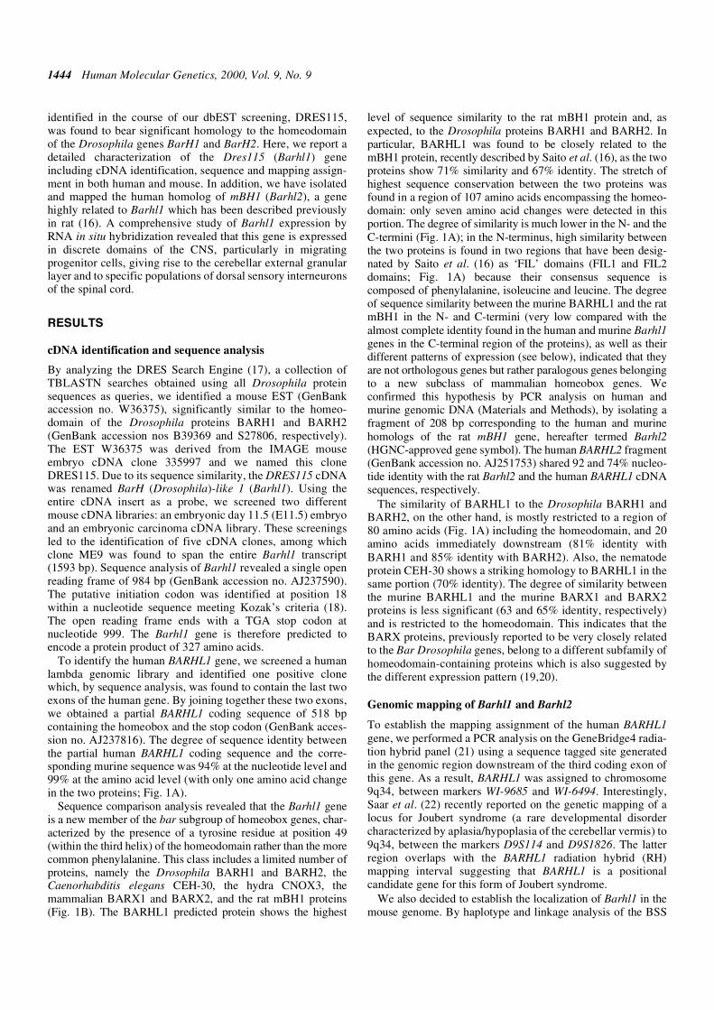
In the forebrain, Barhl1 is expressed exclusively in arestricted domain of the caudal diencephalon at early neuraltube stage (E9.5), and more specifically in the ventral area ofp1 and p2 (Fig. 3A). By E10.5, Barhl1 expression is detectedalong the neural tube in two separate rostral and caudaldomains (Figs 3B, and 4A, D and G). The rostral diencephalicdomain extends: (i) anteriorly, into the basal plate ofprosomeres 3 and 4 (p3, p4), anlage of the mammillary area;(ii) dorsally, along the p2–p3 boundary corresponding to thezona limitans (ZL), which will separate the dorsal from theventral thalamus; and (iii) posteriorly to the pretectum (p1)(Fig. 3B, E, F, G and M). The mammillary area corresponds tothe only region where the mouse Barhl1 gene is expressed inthe ventral midline (p4 in Fig. 3B and E). In the pretectum, twodistinct domains of expression can be recognized, one in thebasal plate and the other in the most alar and caudal region(Fig. 3B and M). This latter expression, corresponding to thecommissural area, will extend basally by E11.5 (Fig. 3C, K, Nand O). At E10.5, the caudal domain extends from themidbrain/isthmic boundary, through the rhombencephalon(cerebellar plates and rhombic lips) to the entire extent of thespinal cord. In these structures, this gene is expressed as alongitudinal stripe along the most dorsal area of the alar plate,but not in the roof plate (Figs 3B and H, and 4A, D and G).
By E11.5 the expression pattern of Barhl1 changes dynami-cally: a positive band appears in the mesencephalic basal plate(Fig. 3C, L, N and O), and in the spinal cord the expressionexpands laterally, following the ventral migrating cells fromthe dorsal alar plate to the boundary between the basal and alarplate (Fig. 4E and H). At E12.5, the expression shows anothersignificant change of pattern: in the diencephalon, expressionbecomes mainly restricted to the mammillary region (basal p4)(Fig. 3D, I and J), whereas mesencephalic expression is nowlocalized in the rostral alar plate in the presumptive area of thesuperior colliculus (Figs 3D and 4C). In the caudal rhomben-cephalon, a large lateral band of expression is observed andprobably corresponds to the cells migrating ventrally, whichwill form the pontine and inferior olivary nuclear complex(25,26) (Figs 3D and 4C). At this stage, most of the expressionin the spinal cord is confined to the mantle layer in a lateralband between the basal and alar plate (Figs 3D, and 4F and I)and this pattern will not change during later stages of develop-ment (data not shown).
The sites of expression at late embryonic stages (E16.5),besides the spinal cord, are the mantle layer of the inferior
Figure 2. Mapping of Barhl1 in the mouse genome. (A) Haplotype and linkageanalysis of Barhl1 and flanking loci on mouse chromosome 2 through the anal-ysis of the BSS backcross (Jackson Laboratory). The distinction between BSheterozygotes and SS homozygotes in the N2 progeny of the cross is made pos-sible by the analysis of an EcoRI polymorphism (Materials and Methods).Empty squares indicate the Mus spretus allele; solid squares indicate theC57BL/6J allele; shaded squares signify that the genotype was not determined.Numbers to the right, between rows, indicate recombination fractions ± SE,and LOD scores. Columns show different haplotypes observed on chromo-some 2. Numbers below columns define the number of individuals sharingeach haplotype. (B) Position of Barhl1 on chromosome 2 with respect tonearby markers independently mapped by others on the BSS backcross. Num-bers on the left represent approximate genetic distances (cM) from the mostcentromeric chromosome 2 marker in this cross. The chromosomal location ofa syntenic human gene (NOTCH1) and the two markers defining the BARHL1RH mapping interval (see text) is also indicated. The relative order of thehuman loci was determined by analysis of the Genetic Location Database(LDB; http://cedar.genetics.soton.ac.uk/public_html/ldb.html ).
Human Molecular Genetics, 2000, Vol. 9, No. 9 1447
colliculus anlage, with a clear caudo–rostral gradient, thepontine nucleus and the cerebellar external granular layer(Fig. 5A and B). Another site of expression, only evident at
this stage of development, is the sensory epithelium of theinner ear, where Barhl1 expression is possibly restricted to thesensory hair cells (Fig. 5C).
Figure 3. Expression of the murine Barhl1 gene during embryonic development. The Barhl1 transcript was detected by in situ hybridization in whole-mountpreparations (A–D and M) and in tissue sections (E–L): Barhl1 expression is visualized at E9.5 (A), E10.5 (B, E, F, G, H and M), E11.5 (C, K, L and N) and E12.5(D, I and J). At E9.5 Barhl1 expression is detected in the caudal part of the diencephalic anlage corresponding to the ventral area of prosomeres 1 and 2 (p1, p2)(A). At E10.5 Barhl1 is expressed in two separate domains (B). The rostral diencephalic domain extends from the basal plate of prosomeres 3 and 4 (p3, p4) ante-riorly, anlage of the mammillary area, to the pretectum (p1) (B, E, F and G) posteriorly. The mammillary area corresponds to the only region where the mouseBarhl1 gene is expressed in the ventral midline [p4 in (B) and (E)]. The basal expression in P2 and P3 extends dorsally into the ZL between the anlage of the ventraland dorsal thalamus [best seen at E11.5 in (N) and (O)]. In the pretectum it is expressed in two distinct bands: one in the basal plate and the other in the most alarand caudal region (B and M); the arrows in (B) and (C) show the boundary between mesencephalon and diencephalon. In the caudal domain, extending from themidbrain/isthmic boundary to the entire extent of the spinal cord, Barhl1 is expressed as a longitudinal stripe along the most dorsal area of the alar plate, but notin the roof plate (B and H). At E11.5 in the commissural area of the pretectum Barhl1 expression extends basally and a positive band appears in the mesencephalicbasal plate (C, K, L, N and O) corresponding to the ventral periaqueductal grey [see arrow in (L)]. The signal immediately posterior to the optic vesicle, detectedat E10.5 (B) and E11.5 (C and N) seems to correspond to the primordium of the extra-ocular muscles. At E12.5 the rostral diencephalic expression becomesrestricted to the mammillary region (basal p4) and prerubral tegmentum, encompassing retromammillar (basal p3), posterior tubercle (basal p2) and subpretectal(basal p1) tegmental zones (D, I and J). In the mesencephalon Barhl1 expression is localized in the rostral alar plate in the presumptive area of the superior collic-ulus (D). At this stage in the caudal rhombencephalon a large stream of positive cells probably migrating ventrally is evident [see arrow in (D)]. While in the spinalcord expression is definitively confined to the mantle layer of a lateral band between the basal and alar plates. AP, alar plate; BP, basal plate; Cb, cerebellar plate;cm, posterior commissure; D, diencephalon; ET, eminentia thalami; FP, floor plate; Hy, hypothalamus; IC, inferior colliculus; LT, lamina terminalis; M, mesen-cephalon; MM, mammillary region; Mb, mandibulary component of the first branchial arch; Mx, maxillary component of the first branchial arch; ov, optic vesicle;PT, pretectum; Rh, rhombencephalon; rl, rhombic lips; RP, roof plate; SC, superior colliculus; SpC, spinal cord; T, telencephalon; V, fifth cranial nerve ganglia;ZL, zona limitans.

1448 Human Molecular Genetics, 2000, Vol. 9, No. 9
The expression pattern of Barhl1 was also examined in post-natal brain. Expression was observed in brain regions thatderived from sites of earlier expression. In particular, Barhl1 isexpressed at postnatal days 4 and 7 (P4 and P7) in the super-ficial layers of the superior colliculus, in the entire inferiorcolliculus, in the external cerebellar granular layer, in thepontine nucleus and in a restricted hypothalamic area (Fig. 5Dand E, and data not shown).
DISCUSSION
In this paper, we describe the isolation and characterization ofa novel mammalian gene, Barhl1, which represents a newmember of the Bar subgroup of homeobox genes, character-ized by the presence of a tyrosine residue at position 49 of thehomeodomain instead of the more common phenylalanine(Fig. 1B). In particular, Barhl1 is highly related in terms of
Figure 4. Expression of Barhl1 in migrating neurons, detected by in situ hybridization in whole-mount embryos at E10.5 (A, D and G), E11.5 (B, E and H) andE12.5 (C, F and I). The expression is visualized in a view from posterior for the rhombencephalon (A–C), and in transverse (D–F) and lateral views (G–I) for thespinal cord. At these stages, Barhl1 is expressed in a domain extending from the midbrain/isthmic boundary, through the rhombencephalon, to the entire extent ofthe spinal cord. In these structures, this gene is expressed at E10.5 as a longitudinal stripe in the mantle zone along the most dorsal area of the alar plate (A, Dand G). From E11.5 the expression domain in the caudal rhombencephalon and of spinal cord expands laterally following the ventral migrating cells from thedorsal alar plate [see arrows in (B), (E) and (H)]. By E12.5, in the spinal cord, Barhl1 is a expressed almost exclusively in two columns localized in the mantle zonebetween the alar and the basal plates (F and I). At this stage Barhl1 is intensively expressed in the entire superior colliculi of the mesencephalon (C). AP, alar plate;BP, basal plate; Cb, cerebellar plate; FP, floor plate; IC, inferior colliculi; M, mesencephalon; rl, rhombic lip; SC, superior colliculi.

Human Molecular Genetics, 2000, Vol. 9, No. 9 1449
sequence similarity to another homeobox-containing gene,mBH1, recently described in rat by Saito et al. (16). Thepredicted proteins encoded by the mouse Barhl1 and the ratmBH1 cDNAs show very significant sequence similarityparticularly in a central portion of 107 amino acids spanningthe homeodomain, while the N- and C-termini show muchlower homology (Fig. 1A). To rule out the possibility that themBH1 gene could be the rat homolog of Barhl1, we isolatedtwo partial cDNA fragments belonging to the murine andhuman counterparts of mBH1 and verified that they aredifferent from Barhl1. Also, the expression patterns of mBH1(Barhl2) and Barhl1 are different, since mBH1 has beenreported to be expressed in retina and in the dorsal thalamus(16), sites where Barhl1 was never detected in our experi-ments. Therefore, Barhl1 and mBH1 (now renamed Barhl2,HGNC-approved symbol) represent two members of a newsubfamily of Bar homeobox genes. Barhl1 and Barhl2 are themammalian genes showing the highest sequence similarity tothe two Bar Drosophila genes BarH1 and BarH2. In fact, themurine proteins BARX1 and BARX2, which are also charac-terized by the presence of a Bar-like homeodomain and havepreviously been reported to be closely related to the BarDrosophila proteins (19,20), show a much lower sequence
similarity (restricted to the homeodomain) to the BARH1 andBARH2 proteins (Fig. 1B). These data indicate that the Barhlgenes represent the mammalian transcripts most closely relatedto the Bar Drosophila genes.
Barhl1 expression and neural development
Barhl1 is exclusively expressed in restricted domains of thedeveloping CNS. The regionalization of gene expression in theembryonic CNS is believed to provide positional informationthat regulates the generation of defined populations of neuronsand appropriate guidance of their axons. On the basis of theanalysis of its expression pattern, we argue that Barhl1 can beinvolved in such processes at different levels of the neural axis.
Barhl1 is first detected at E9.5 in the basal and caudal regionsof the prosencephalon (p1 and p2) that correspond to the anlageof the diencephalon, a structure that will differentiate into thepretectum and the thalamus. From E10.5, expression is detectedin the basal zone of the secondary prosencephalon and dien-cephalon. In the pretectum, Barhl1 is expressed in the mostventral part of the basal zone (basal p1) and, separately, in thealar domain (alar p1). In the latter region, three other homeoboxgenes are expressed: Pax6, Lim1 and Gsh1 (27). Unlike Pax6,which begins expression at an earlier stage, Barhl1, Lim1 and
Figure 5. Late embryonic (E16.5) (A–C) and postnatal (P4) (D and E) expression of Barhl1. At E16.5 mesencephalic expression is prevalent in the caudal colliculusanlage with a distinct caudo–rostral gradient, while in the rhombencephalon it is expressed in the EGL of the cerebellar anlage (A) and in the pontine nucleus [(B) andarrow in (C)]. Another site of expression is the sensory epithelium of the inner ear (C). At P4 Barhl1 is selectively expressed in the superior and inferior colliculi of themesencephalon (D), in the most external part of the EGL (D and E), in the pontine nucleus (D) as well as in mammillary nuclei of the hypothalamus (D).
1450 Human Molecular Genetics, 2000, Vol. 9, No. 9
Gsh1 expression in p1 initiate at E10.5. It is therefore reasonableto hypothesize that these genes are involved in establishing theidentity of p1 cells as different from mesencephalic cells, andthat the timing of Lim1, Gsh1 and Barhl1 expression suggeststhat these genes may be downstream of Pax6.
At E11.5, Barhl1 expression extends into the basal plate ofthe caudal hypothalamus, stopping abruptly at the bordersbetween the mammillary and infundibulary region, the limitbetween the prosomeres 4 and 5 (Fig. 3). More caudally, basalexpression in P2 and P3 extends dorsally into the ZL betweenthe ventral and dorsal thalamus. Expression in the hypothal-amus also persists postnatally: Barh1l is clearly detected at P4in the anterior and lateral mammillary nuclei and, at lowerlevels, in the retromammillary domains (Fig. 5). The extensiveexpression in the above mentioned embryonic structures, suchas the thalamus and the mammillary region, suggests thatBarhl1 may have a role in the development of these centralprocessing stations. In adult brain, the thalamus connectsrostral brain centers with the brainstem and the spinal cord,while the mammillary region is selectively involved in someaspects of learning and memory.
In the mesencephalon, Barhl1 starts to be expressed in thebasal plate in a region corresponding to the periaqueductalgrey at E11.5. By E12.5, strong expression is detected in themantle of the tectum. At this stage, Barhl1 is first expressed inthe entire superior colliculi, whereas by E16.5, its expressionin the tectum is confined to the caudal colliculus anlage, with aclear caudo–rostral gradient. The inferior colliculi, function-ally related to the auditory system, and the superior colliculi,layered structures which are part of the visual system, aregenerated in the alar plate region of the mesencephalonthrough a complex neuroblast migration process.
Barhl1 is a novel marker of cerebellar migrating neurons
The post-mitotic neurons generated in the alar rhombencephalonand, in particular, in the region of incomplete closure of thedorsal neural tube named rhombic lip, migrate to various desti-nations, giving rise to cerebellar granule cells and the precere-bellar nuclei. Here, we show that Barhl1 is strongly expressed inthe rhombic lip as well as in the structures deriving from it. Thecerebellar granule cell precursors originate from the rostral lip,while the caudal lip will give rise to the ventrally migratingneurons generating the precerebellar nuclei of the medulla andof the pons. Within this migrating stream from the lower lip,Barhl1 is expressed only in neurons migrating to the pontinenuclei and not in the neurons migrating to the medulla. Interest-ingly, the formation of these nuclei is selectively disrupted inseveral knock-out mice, such as the mouse mutants for Pax6(26), Math1 (28), Netrin1 (25,29) and its receptor Deleted incolorectal cancer (Dcc) (30). Cells at the dorsal margin of therostral rhombic lip give rise to granule neuron progenitors(31,32). These progenitors, specified to become granule cellprogenitors, express the transcription factor genes Zic1, Zic2(33,34), Zipro1 (35) and the basic HLH gene Math1 (36,37).Recently, it has been shown that Math1 function is required forthe generation of granule cell progenitors (38). Nevertheless, themolecular steps involved in the specification of granule cell fateremain largely unknown.
Recent studies in the development of the cerebellar primor-dium suggest the involvement of patterning signals, such as
FGF and Wnt proteins, that control regional identity at theboundary between the mesencephalon and metencephalon(mes/met boundary) (39), and inductive factors, such as theBMP proteins, which have an important role in the specifica-tion of the granule cell precursors (40). The first specifiedgranule neuron progenitors appear in the rhombic lip laterallyto the roof plate, a source of several BMPs, at around E10 andthey seem to express both the Math1 (36,37,41) and Barhl1genes. The ability of several BMPs (BMP6 and 7, and GDF7)to induce the expression of Math1 and other dorsal markers(Zic1, Zic2 and Wnt3a) has been proved recently (40),suggesting that the induction of the granule precursors maydepend on BMP signaling with a mechanism similar to theinduction of dorsal interneurons of the spinal cord (42). Oncethe specification of the granule progenitors has occurred, theputative coexpression of the Math1 and Barhl1 genes is main-tained. The co-expression is also maintained in the subsequentevents characterized by migration to the external granular layer(EGL), expansion of the progenitor population and final differ-entiation and migration into the internal granular layer (IGL)(36,37; data not shown). Therefore, it is reasonable to hypoth-esize that the generation of the EGL may depend on a BMP-triggered transduction pathway involving not only Math1 (38)but also Barhl1 which has a similar spatial and temporalpattern of expression.
Barhl1 expression and development of dorsal sensoryinterneurons of the spinal cord
The spinal cord is composed of different cell types which aregenerated and occupy different positions along the dorso–ventral axis of the neural tube (43). The motor neurons aregenerated laterally to the floor plate, which represents theventral midline of the neural tube. Dorsally, after the migrationof the neural crest cells and the closure of the neural tube, theroof plate is formed along the dorsal midline and cells lateral toit differentiate into different types of dorsal sensory inter-neurons. The specification of ventral and dorsal cell types, aswell as of individual neuronal subtypes, seems to be regulatedby inductive signaling factors, provided by the notochord andfloor plate ventrally, and by the epidermal ectoderm and roofplate dorsally. Recent evidence provides strong support for arole of the BMP subclass of TGFβ-related proteins in thecontrol of the differentiation of dorsal neurons (42,44). Inparticular, it has been demonstrated that GDF7, a BMP familymember expressed selectively by roof plate cells, is able toinduce the differentiation of dorsal commissural interneurons(42). These neurons are generated close to the roof plate, andcan be divided in D1A and D1B neurons (45). The Math1 geneis specifically expressed by the progenitor cells adjacent to theroof plate which generate both subclasses of the D1 neurons(36,37,41,42,44). At E10.5, the Barhl1 and Math1 domains ofexpression seem to overlap entirely in this region. However, atE12.5, Barhl1 expression becomes restricted to a more ventraldomain corresponding to the region where the D1B subclass ofneurons migrate (45), while Math1 expression is confined tothe D1A neurons which, for the most part, remain close to theroof plate (42). Therefore, both D1A and D1B neurons seem tooriginate from Math1+ and Barhl1+ progenitors, but theysubsequently reside in different sites of the dorsal spinal cordwith D1A neurons maintaining the Math1 expression and D1B

Human Molecular Genetics, 2000, Vol. 9, No. 9 1451
neurons maintaining the expression of Barhl1. It is worthnoting that in GDF7 mutants, only the late Math1+ cells, corre-sponding to the D1A neurons, are lost, whereas the D1B differ-entiate normally (42). Thus, there might be different TGFβ-related proteins from the roof plate regulating the formationand identity of the specific subtypes of dorsal interneurons.
The expression pattern of Barhl1 in the developing CNSstrongly suggests that this gene, similar to its Drosophila coun-terparts BarH1 and BarH2, may play a crucial role in cell fatedetermination of neural structures and may therefore beinvolved in the pathogenesis of CNS developmental anoma-lies. Interestingly, the human BARHL1 gene maps to chromo-some 9q34, in a region overlapping the critical interval for oneform of Joubert syndrome described recently by Saar et al.(22). Joubert syndrome, is a rare syndrome with autosomalrecessive inheritance characterized by aplasia/hypoplasia ofthe cerebellar vermis with abnormal eye movements, ataxiaand mental retardation. Both the mapping assignment and itsremarkable expression in the primordia of the cerebellum andthe brainstem, render BARHL1 an attractive candidate gene forJoubert syndrome. Mutation analysis in patients as well as thegeneration of a Barhl1 knockout mouse will be necessary todetermine whether the Barhl1 gene plays a causative role inJoubert syndrome or in other developmental defects of themammalian nervous system.
MATERIALS AND METHODS
cDNA identification
To identify the murine Barhl1 full-length transcript, twomurine cDNA libraries were used: an E11 embryo (ClontechML3003a) and an embryonic carcinoma (P19 cell line; Strata-gene 937317) cDNA library. For the isolation of the BARHL1gene, we used a fibroblast genomic library (Stratagene946204). Plating, hybridization and washing conditions wereas described previously (46).
To isolate the human and murine BARHL2 genes, we carriedout PCR analysis on human genomic DNA using the oligo-nucleotide primers TO-9557 and TO-9558 designed on thesequence of the rat mBH1 cDNA (16). Oligonucleotidesequences were as follows: TO-9557, 5'-GTGGAAGCGGCA-GACTGCAG-3' and TO-9558, 5'-CAGGCCGTGGATGAG-CACGC-3'. PCR conditions were 35 cycles of 94°C for 45 s,50°C for 45 s and 72°C for 45 s.
cDNA sequence analysis
cDNA sequence analysis, and nucleotide and protein databasesearches were performed as described previously (14). Data onsimilarity/identity were obtained using the Bestfit program ofthe GCG software package, version 8.1. The multiple align-ment analyses were generated using the PileUp program of theWisconsin GCG software package, version 8.1.
Genomic mapping in human and mouse
For the RH mapping assignment of the human BARHL1 andBARHL2, we used a previously described procedure (14). PCRanalysis on the GeneBridge4 radiation hybrid panel (21) wasperformed using the following two primers designed in the
BARHL1 genomic region, 2 kb downstream of the beginning ofthe third exon: TO-5571, 5'-TTCTCAACTTGCCCCAGTCC-3'and TO-5572, 5'-CCTCATAAACGCCGACTCAAC-3'. For themapping of the BARHL2 gene, we used oligonucleotide primersTO-9840, 5'-CCGAGGCAGGGAACTACTCGG-3' and TO-9841, 5'-CCGCTGCAGCTGGGGATGG-3'. PCR conditionswere 35 cycles of 94°C for 45 s, 52°C for 45 s and 72°C for 45 sfor both BARHL1 and BARHL2.
Genetic mapping was achieved using a (C57BL/6j×SPRET/Ei)F1×SPRET/Ei (BSS) backcross generated and distributed bythe Jackson Laboratory (47). An EcoRI RFLP was identified byhybridization of C57BL/6j and SPRET/Ei parental DNAs (cutwith each of the following six restriction enzymes: EcoRI,EcoRV, KpnI, MspI, TaqI and XbaI) with a Barhl1 cDNA probecorresponding to the insert of the IMAGE clone 335997. Thepolymorphism consisted of a single SPRET/Ei-specific EcoRIband, and of two C57BL/6JEi-specific EcoRI fragments. FourSouthern panels containing EcoRI-cut parental DNAs and N2progeny (n = 94) DNAs were hybridized with a Barhl1 cDNAprobe. The analysis of the strain distribution pattern observed inthe N2 progeny (n = 92) of the BSS backcross (47) was analyzedwith the Map Manager 2.6 program (48) and permitted the local-ization of Barhl1 to chromosome 2.
Expression studies
Mouse embryo tissue sections were prepared and RNA in situhybridization experiments were performed as described (49).The IMAGE EST clone 335997 containing part of the Barhl1transcript was linearized with appropriate restriction enzymesto transcribe either sense or antisense 35S-labeled riboprobes.Slides were exposed for 10 days. Micrographs are doubleexposures: red color represents the in situ hybridization signal,and the blue shows the nuclei stained with Hoechst 33258 dye.
ACKNOWLEDGEMENTS
We thank E. Roncoroni and the TIGEM sequencing staff fortechnical support, and M. Smith for preparation of the manu-script. This work was supported by the Italian Telethon Found-ation, by the Merck Genome Research Institute (grant no. 37),and by the Armenise Harvard Foundation. S.M. is supportedby grants from the EC (BIO4-CT98-0303, DGICYT PM98-0056) and from Fundacion la Caixa 97/101.
REFERENCES
1. Rubin, G.M. (1988) Drosophila melanogaster as an experimental organ-ism. Science, 240, 1453–1459.
2. The FlyBase Consortium (1994) FlyBase—the Drosophila database.Nucleic Acids Res., 22, 3456–3458.
3. Bellen, H.J. and Smith, R.F. (1995) FlyBase: a virtual Drosophila cornu-copia. Trends Genet., 11, 456–457.
4. Jan, Y.N. and Jan, L.Y. (1994) Genetic control of cell fate specification inDrosophila peripheral nervous system. Annu. Rev. Genet., 28, 373–393.
5. Campuzano, S. and Modolell, J. (1992) Patterning of the Drosophila nerv-ous system: the achaete–scute gene complex. Trends Genet., 8, 202–208.
6. Gomez Skarmeta, J.L., del Corral, R.D., de la Calle Mustienes, E., FerreMarco, D. and Modolell, J. (1996) Araucan and caupolican, two membersof the novel iroquois complex, encode homeoproteins that control prone-ural and vein-forming genes. Cell, 85, 95–105.
7. Leyns, L., Gomez Skarmeta, J.L. and Dambly Chaudiere, C. (1996)iroquois: a prepattern gene that controls the formation of bristles on thethorax of Drosophila. Mech. Dev., 59, 63–72.

1452 Human Molecular Genetics, 2000, Vol. 9, No. 9
8. Kehl, B.T., Cho, K.O. and Choi, K.W. (1998) mirror, a Drosophila home-obox gene in the Iroquois complex, is required for sensory organ and alulaformation. Development, 125, 1217–1227.
9. Ramain, P., Heitzler, P., Haenlin, M. and Simpson, P. (1993) pannier, anegative regulator of achaete and scute in Drosophila, encodes a zincfinger protein with homology to the vertebrate transcription factor GATA-1. Development, 119, 1277–1291.
10. Higashijima, S., Michiue, T., Emori, Y. and Saigo, K. (1992) Subtypedetermination of Drosophila embryonic external sensory organs by redun-dant homeo box genes BarH1 and BarH2. Genes Dev., 6, 1005–1018.
11. Kojima, T., Ishimaru, S., Higashijima, S., Takayama, E., Akimaru, H.,Sone, M., Emori, Y. and Saigo, K. (1991) Identification of a different-typehomeobox gene, BarH1, possibly causing Bar (B) and Om (1D) mutationsin Drosophila. Proc. Natl Acad. Sci. USA, 88, 4343–4347.
12. Higashijima, S., Kojima, T., Michiue, T., Ishimaru, S., Emori, Y. andSaigo, K. (1992) Dual Bar homeo box genes of Drosophila required intwo photoreceptor cells, R1 and R6, and primary pigment cells for normaleye development. Genes Dev., 6, 50–60.
13. Sato, M., Kojima, T., Michiue, T. and Saigo, K. (1999) Bar homeoboxgenes are latitudinal prepattern genes in the developing Drosophila notumwhose expression is regulated by the concerted functions of decapenta-plegic and wingless. Development, 126, 1457–1466.
14. Banfi, S., Borsani, G., Rossi, E., Bernard, L., Guffanti, A., Rubboli, F.,Marchitiello, A., Giglio, S., Coluccia, E., Zollo, M., Zuffardi, O. and Bal-labio, A. (1996) Identification and mapping of human cDNAs homolo-gous to Drosophila mutant genes through EST database searching. NatureGenet., 13, 167–174.
15. Banfi, S., Borsani, G., Bulfone, A. and Ballabio, A. (1997) Drosophila-related expressed sequences. Hum. Mol. Genet., 6, 1745–1753.
16. Saito, T., Sawamoto, K., Okano, H., Anderson, D.J. and Mikoshiba, K.(1998) Mammalian BarH homologue is a potential regulator of neuralbHLH genes. Dev. Biol., 199, 216–225.
17. Guffanti, A., Banfi, S., Simon, G., Ballabio, A. and Borsani, G. (1997)DRES search engine: of flies, men and ESTs. Trends Genet., 13, 79–80.
18. Kozak, M. (1984) Compilation and analysis of sequences upstream fromthe translational start site in eukaryotic mRNAs. Nucleic Acids Res., 12,857–873.
19. Tissier Seta, J.P., Mucchielli, M.L., Mark, M., Mattei, M.G., Goridis, C.and Brunet, J.F. (1995) Barx1, a new mouse homeodomain transcriptionfactor expressed in cranio-facial ectomesenchyme and the stomach. Mech.Dev., 51, 3–15.
20. Jones, F.S., Kioussi, C., Copertino, D.W., Kallunki, P., Holst, B.D. andEdelman, G.M. (1997) Barx2, a new homeobox gene of the Bar class, isexpressed in neural and craniofacial structures during development. Proc.Natl Acad. Sci. USA, 94, 2632–2637.
21. Gyapay, G., Schmitt, K., Fizames, C., Jones, H., Vega-Czary, N., Spillett,D., Muselet, D., Prud’Homme, J.-F., Dib, C., Auffray, C. et al. (1996) Aradiation hybrid map of the human genome. Hum. Mol. Genet., 5, 339–346.
22. Saar, K., Al-Gazali, L., Sztriha, L., Rueschendorf, F., Nur, E.K.M., Reis,A. and Bayoumi, R. (1999) Homozygosity mapping in families withJoubert syndrome identifies a locus on chromosome 9q34.3 and evidencefor genetic heterogeneity. Am. J. Hum. Genet., 65, 1666–1671.
23. Bulfone, A., Puelles, L., Porteus, M.H., Frohman, M.A., Martin, G. andRubenstein, J.L.R. (1993) Spatially restricted expression of Dlx-1, Dlx-2(Tes-1), Gbx-2 and Wnt-3 in the embryonic day 12.5 mouse forebraindefines potential transverse and longitudinal segmental boundaries. J.Neurosci., 13, 3155–3172.
24. Rubenstein, J.L. and Puelles, L. (1994) Homeobox gene expression duringdevelopment of the vertebrate brain. Curr. Top. Dev. Biol., 29, 1–63.
25. Yee, K.T., Simon, H.H., Tessier-Lavigne, M. and O’Leary, D.M. (1999)Extension of long leading processes and neuronal migration in the mamma-lian brain directed by the chemoattractant netrin-1. Neuron, 24, 607–622.
26. Engelkamp, D., Rashbass, P., Seawright, A. and van Heyningen, V.(1999) Role of Pax6 in development of the cerebellar system. Develop-ment, 126, 3585–3596.
27. Mastick, G.S., Davis, N.M., Andrew, G.L. and Easter Jr, S.S. (1997) Pax 6functions in boundary formation and axon guidance in the embryonicmouse forebrain. Development, 124, 1985–1997.
28. Ben-Arie, N., Hassan, B.A., Bermingham, N.A., Malicki, D.M.,Armstrong, D., Matzuk, M., Bellen, H.J. and Zoghbi, H.Y. (2000) Func-tional conservation of atonal and math1 in the CNS and PNS. Develop-ment, 127, 1039–1048.
29. Serafini, T., Colamarino, S.A., Leonardo, E.D., Wang, H., Beddington,R., Skarnes, W.C. and Tessier-Lavigne, M. (1996) Netrin-1 is required forcommissural axon guidance in the developing vertebrate nervous system.Cell, 87, 1001–1014.
30. Fazeli, A., Dickinson, S.L., Hermiston, M.L., Tighe, R.V., Steen, R.G.,Small, C.G., Stoeckli, E.T., Keino-Masu, K., Masu, M., Rayburn, H. et al.(1997) Phenotype of mice lacking functional Deleted in colorectal cancer(Dcc) gene. Nature, 386, 796–804.
31. Hatten, M.E. and Heintz, N. (1995) Mechanisms of neural patterning andspecification in the developing cerebellum. Annu. Rev. Neurosci., 18,385–408.
32. Alder, J., Cho, N.K. and Hatten, M.E. (1996) Embryonic precursor cellsfrom the rhombic lip are specified to a cerebellar granule neuron identity.Neuron, 17, 389–399.
33. Aruga, J., Yokota, N., Hashimoto, M., Furuichi, T., Fukuda, M. andMikoshiba, K. (1994) A novel zinc finger protein, zic, is involved in neu-rogenesis, especially in the cell lineage of cerebellar granule cells. J. Neu-rochem., 63, 1880–1890.
34. Nagai, T., Aruga, J., Takada, S., Gunther, T., Sporle, R., Schughart, K. andMikoshiba, K. (1997) The expression of the mouse Zic1, Zic2, and Zic3gene suggests an essential role for Zic genes in body pattern formation.Dev. Biol., 182, 299–313.
35. Yang, X.W., Zhong, R. and Heintz, N. (1996) Granule cell specification inthe developing mouse brain as defined by expression of the zinc fingertranscription factor RU49. Development, 122, 555–566.
36. Ben Arie, N., McCall, A.E., Berkman, S., Eichele, G., Bellen, H.J. andZoghbi, H.Y. (1996) Evolutionary conservation of sequence and expres-sion of the bHLH protein Atonal suggests a conserved role in neurogene-sis. Hum. Mol. Genet., 5, 1207–1216.
37. Akazawa, C., Ishibashi, M., Shimizu, C., Nakanishi, S. and Kageyama, R.(1995) A mammalian helix–loop–helix factor structurally related to theproduct of Drosophila proneural gene atonal is a positive transcriptionalregulator expressed in the developing nervous system. J. Biol. Chem., 270,8730–8738.
38. Ben-Arie, N., Bellen, H.J., Armstrong, D.L., McCall, A.E., Gordadze,P.R., Guo, Q., Matzuk, M.M. and Zoghbi, H.Y. (1997) Math1 is essentialfor genesis of cerebellar granule neurons. Nature, 390, 169–172.
39. Joyner, A.L. (1996) Engrailed, Wnt and Pax genes regulate midbrain–hindbrain development. Trends Genet., 12, 15–20.
40. Alder, J., Lee, K.J., Jessell, T.M. and Hatten, M.E. (1999) Generation ofcerebellar granule neurons in vivo by transplantation of BMP-treatedneural progenitor cells. Nature Neurosci., 2, 535–540.
41. Helms, A.W. and Johnson, J.E. (1998) Progenitors of dorsal commissuralinterneurons are defined by MATH1 expression. Development, 125, 919–928.
42. Lee, K.J., Mendelsohn, M. and Jessell, T.M. (1998) Neuronal patterningby BMPs: a requirement for GDF7 in the generation of a discrete class ofcommissural interneurons in the mouse spinal cord. Genes Dev., 12,3394–3407.
43. Tanabe, Y. and Jessell, T.M. (1996) Diversity and pattern in the develop-ing spinal cord [published erratum appears in Science (1997) 276, 21].Science, 274, 1115–1123.
44. Liem Jr, K.F., Tremml, G. and Jessell, T.M. (1997) A role for the roofplate and its resident TGFbeta-related proteins in neuronal patterning inthe dorsal spinal cord. Cell, 91, 127–138.
45. Lee, K.J. and Jessell, T.M. (1999) The specification of dorsal cell fates inthe vertebrate central nervous system. Annu. Rev. Neurosci., 22, 261–294.
46. Franco, B., Guioli, S., Pragliola, A., Incerti, B., Bardoni, B., Tonlorenzi,R., Carrozzo, R., Maestrini, E., Pieretti, M., Taillon-Miller, P. et al. (1991)A gene deleted in Kallmann’s syndrome shares homology with neural celladhesion and axonal path-finding molecules. Nature, 353, 529–536.
47. Rowe, L.B., Nadeau, J.H., Turner, R., Frankel, W.N., Letts, V.A., Eppig,J.T., Ko, M.S.H., Thurston, S.J. and Birkenmeier, E.H. (1994) Maps fromtwo interspecific backcross DNA panels available as a community geneticmapping resource. Mamm. Genome, 5, 253–274.
48. Manly, K.F. and Elliott, R.W. (1991) RI manager, a microcomputerprogram for the analysis of data from recombinant inbred strains. Mamm.Genome, 1, 123–126.
49. Rugarli, E.I., Lutz, B., Kuratani, S.C., Wawersik, S., Borsani, G.,Ballabio, A. and Eichele, G. (1993) Expression pattern of the Kallmannsyndrome gene in the olfactory system suggests a role in neuronal target-ing. Nature Genet., 4, 19–25.