Small-scale biogeographical patterns in some groundwater Crustacea, the syncarid, Parabathynellidae
Upload
independentCategory
view
1download
0
Zoological Journal of the Linnean Society (1995), 113: 351–459. With 73 figures
Taxonomic revision of the subfamilyToradjiinae (Crustacea: Oniscidea:Scleropactidae)
F. FERRARA, C. MELI and S. TAITI
Centro di Studio per la Faunistica ed Ecologia Tropicali del Consiglio Nazionale delle Ricerche,Via Romana 17, 50125 Firenze, Italy
Received June 1994, accepted for publication December 1994
In the present revision four genera and 32 species are recognized. One genus, Paratoradjia, and17 species, namely Adinda malaccensis, A. sumatrana, A. palniensis, A. carli, A. nilgiriensis, A. lobata, A.triangulifera, A. riedeli, A. lamellata, Protoradjia paeninsulae, P. insularis, P. montana, P. pilosa, Paratoradjiaberoni, P. vietnamensis, P. sulcata and Toradjia hirsuta, are described as new. The neotype of Toradjiacephalica and lectotypes of Adinda weberi, Protoradjia jacobsoni, Toradjia gorgona and T. celebensis areestablished. Adinda calegarii is considered to be a junior synonym of Adinda weberi, and A. conglobator,a species inquirenda. Toradjia dollfusi is transferred to the genus Adinda, and T. indosinensis to Paratoradjia.The characters of the group and its taxonomic position within the Oniscidea are discussed: it istransferred from the Eubelidae to the Scleropactidae, of which it constitutes a separate subfamily,the Toradjiinae. A map and comments on the distribution of genera and species are included.
ADDITIONAL KEY WORDS:—Isopoda – taxonomy – new genus – new species – distribution –Oriental Region.
CONTENTS
Introduction . . . . . . . . . . . . . . . . . . . 352History . . . . . . . . . . . . . . . . . . . . 352Material . . . . . . . . . . . . . . . . . . . . 353Abbreviations . . . . . . . . . . . . . . . . . . . 354Morphological characters . . . . . . . . . . . . . . . . 354Definition and systematic position . . . . . . . . . . . . . . 358Evolution . . . . . . . . . . . . . . . . . . . . 360Biogeography . . . . . . . . . . . . . . . . . . . 361Taxonomy . . . . . . . . . . . . . . . . . . . . 362
Genus Adinda Budde-Lund, 1904 . . . . . . . . . . . . . . 362Weberi group . . . . . . . . . . . . . . . . . . 364Adinda weberi (Dollfus, 1898) . . . . . . . . . . . . . . . 364Adinda malaccensis sp. nov. . . . . . . . . . . . . . . 368Adinda sumatrana sp. nov. . . . . . . . . . . . . . . 371Adinda conglobator (Budde-Lund, 1902) . . . . . . . . . . . . . 373Stebbingi group . . . . . . . . . . . . . . . . . . 375Adinda stebbingi (Collinge, 1914) . . . . . . . . . . . . . . 375Adinda travancorensis (Stebbing, 1911) . . . . . . . . . . . . . 377Adinda gigas (Collinge, 1915) . . . . . . . . . . . . . . . 381Adinda palniensis sp. nov. . . . . . . . . . . . . . . 381Adinda carli sp. nov. . . . . . . . . . . . . . . . . 385Adinda nilgiriensis sp. nov. . . . . . . . . . . . . . . 388Adinda lobata sp. nov. . . . . . . . . . . . . . . . 392
3510024–4082/95/040351+109 $08.00/0 © 1995 The Linnean Society of London
352 F. FERRARA ET AL.
Adinda triangulifera sp. nov. . . . . . . . . . . . . . . 396Adinda scabra (Collinge, 1916) . . . . . . . . . . . . . . . 398Adinda pulchra (Collinge, 1916). . . . . . . . . . . . . . . 402Riedeli group . . . . . . . . . . . . . . . . . . 404Adinda riedeli sp. nov. . . . . . . . . . . . . . . . 404Adinda platyperaeon (Schultz, 1982) . . . . . . . . . . . . . . 409Adinda lamellata sp. nov. . . . . . . . . . . . . . . 413Dollfusi group . . . . . . . . . . . . . . . . . . 416Adinda dollfusi (Richardson Searle, 1922) comb. nov. . . . . . . . . . 416Genus Protoradjia Arcangeli, 1955 . . . . . . . . . . . . . . 419Protoradjia jacobsoni Arcangeli, 1955 . . . . . . . . . . . . . 419Protoradjia paeninsulae sp. nov. . . . . . . . . . . . . . 422Protoradjia insularis sp. nov. . . . . . . . . . . . . . 426Protoradjia montana sp. nov. . . . . . . . . . . . . . 429Protoradjia pilosa sp. nov. . . . . . . . . . . . . . . 430Genus Paratoradjia nov. . . . . . . . . . . . . . . . 433Paratoradjia beroni sp. nov. . . . . . . . . . . . . . . 434Paratoradjia indosinensis (Arcangeli, 1948) comb. nov. . . . . . . . . . 436Paratoradjia vietnamensis sp. nov. . . . . . . . . . . . . 439Paratoradjia sulcata sp. nov. . . . . . . . . . . . . . . 443Genus Toradjia Dollfus, 1898 . . . . . . . . . . . . . . . 443Toradjia gorgona Dollfus, 1898 . . . . . . . . . . . . . . . 445Toradjia celebensis Dollfus, 1898 . . . . . . . . . . . . . . 449Toradjia cephalica Dollfus, 1898 . . . . . . . . . . . . . . 449Toradjia hirsuta sp. nov. . . . . . . . . . . . . . . . 454
Key to genera and species . . . . . . . . . . . . . . . . 455Acknowledgements . . . . . . . . . . . . . . . . . . 458References . . . . . . . . . . . . . . . . . . . . 459
INTRODUCTION
Taiti, Ferrara & Schmalfuss (1991) revised the family Eubelidae giving a newdefinition and listing all the genera belonging to that family. They also statedthat three genera from the Oriental region (Toradjia Dollfus, 1898, AdindaBudde-Lund, 1904, of which Paraperiscyphis Stebbing, 1911 is a junior synonym,and Protoradjia Arcangeli, 1955), previously included in the Eubelidae, belonginstead to the Scleropactidae.
In this contribution we discuss this statement in detail and give a revisionof these three genera together with the description of a related new genus.This study represents the first comprehensive review of this enigmatic and, upto now, poorly known group, and once more points out how scanty is theknowledge of the Oniscidea in general and from tropical areas in particular.
HISTORY
The first record of this group was made by Dollfus (1898), who describedthree species of the new genus Toradjia (T. celebensis, T. gorgona and T. cephalica)and one of the genus Periscyphus (a mis-spelling of Periscyphis) (P. weberi). Budde-Lund (1902) described a new species of Toradjia from Malaysia (T. conglobator)and transferred to this genus also Periscyphis weberi. Two years later, Budde-Lund (1904: 37) erected the new genus Adinda for P. weberi without giving anydefinition or discussion. Stebbing (1911) established the genus Paraperiscyphis fora species from south-western India (P. travancorensis), stating that it was probablethat Periscyphis weberi would also have to be ascribed to this genus, obviouslyignoring the existence of Adinda. Collinge (1914) described a new species ofParaperiscyphis (P. stebbingi) from southern India and a year later (Collinge, 1915)
353REVISION OF TORADJIINAE
yet another, again from southern India, which he included in the genusPeriscyphis (P. gigas), without any justification for this ascription. Collinge (1916)described two more new species of Paraperiscyphis from Sri Lanka (P. pulcher andP. scabrus ) and corrected the mistake made the year before by transferringPeriscyphis gigas to the genus Paraperiscyphis. Later, Richardson Searle (1922)described a new species of Toradjia from Java (T. dollfusi), and Omer-Cooper(1926), in his revision of the genus Periscyphis, first recognized ParaperiscphisStebbing, 1911 as a junior synonym of Adinda Budde-Lund, 1904. Arcangeli(1927), who apparently was not aware of Omer-Cooper’s paper, described anew species of Paraperiscyphis from Sumatra (P. calegarii) and later Jackson (1936)recorded this species in Malaysia and, tentatively, Adinda scabra in northernBorneo.
Arcangeli (1948, 1952) is the first author to discuss this group, establishingthe new subfamily Toradjiinae (family Eubelidae) for the genera Toradjia andAdinda, mainly characterized by the presence of Trachelipus-type pleopodal lungsin pleopods 1 and 2. Arcangeli (1955) defined a new genus, Protoradjia, for thenew species P. jacobsoni, and better characterized this group highlighting, besidesthe respiratory structures of the pleopods, the exoantennate ability to roll upinto a ‘semicomplete’ ball and the peculiar structure of telson and uropods,similar to that of the ‘Circoniscinae’, at that time also included in the Eubelidae(now instead in the Scleropactidae).
According to Arcangeli (1955) the Toradjiinae included the following species:
(1) Toradjia celebensis Dollfus, 1898 from Sulawesi;(2) T. gorgona Dollfus, 1898 from an unknown locality in ‘East Dutch India’
and Java;(3) T. cephalica Dollfus, 1898 from Java;(4) T. dollfusi Richardson Searle, 1922 from Java;(5) Adinda weberi (Dollfus, 1898) from Sumatra;(6) A. conglobator (Budde-Lund, 1902) from Malaysia;(7) A. travancorensis (Stebbing, 1911) from southern India;(8) A. stebbingi (Collinge, 1914) from southern India;(9) A. gigas (Collinge, 1915) from southern India;
(10) A. pulchra (Collinge, 1916) from Sri Lanka;(11) A. scabra (Collinge, 1916) from Sri Lanka;(12) A. calegarii (Arcangeli, 1927) from Sumatra;(13) Protoradjia jacobsoni Arcangeli, 1955 from Sumatra.
Two more species must be added to this list, i.e. Toradjia indosinensis Arcangeli,1948 from Vietnam (obviously forgotten by Arcangeli) and Paraperiscyphisplatyperaeon from Borneo, described by Schultz (1982).
MATERIAL
Specimens of the species described in this paper were collected in Malaysiaduring field trips organized by the Centro di Studio per la Faunistica edEcologia Tropicali, Florence (these have been deposited in the Museo di StoriaNaturale, Sezione di Zoologia ‘La Specola’, Florence), and in southern India,Sri Lanka, Vietnam, Indonesia, Brunei, Philippines and New Guinea bycolleagues from various institutions.
354 F. FERRARA ET AL.
The type specimens of Adinda weberi, Paraperiscyphis platyperaeon, Toradjia dollfusi,Protoradjia jacobsoni, T. gorgona and T. celebensis were re-examined. However, wecould not re-examine those described by Stebbing (1911) (A. travancorensis) andCollinge (1914, 1915, 1916) (A. stebbingi, A. gigas, A. pulchra and A. scabra) whichwere originally deposited in the collections of the Zoological Survey of India,Calcutta. Since our requests for the loan of material were not granted, we donot know whether the specimens are still present in the collections of thatinstitution or have been lost. The type specimens of Toradjia indosinensis Arcangeli,1948 and T. cephalica Dollfus, 1898, which should be in the Museo Regionaledi Scienze Naturali, Turin, and the Zoologisch Museum, Amsterdam, respectively,are no longer present in the collections of those museums and must beconsidered as lost (E. Gavetti and D. Platvoet, personal communication).
Identifications are based on morphological characters. Normally, species arevery well characterized by the male sexual dimorphisms, which in several casesare truly spectacular.
Abbreviations
BM � Natural History Museum, LondonHNHM � Hungarian Natural History Museum, BudapestMHNG � Museum d’Histoire Naturelle, GenevaMNHN � Museum National d’Histoire Naturelle, ParisMZUF �Museo di Storia Naturale, Sezione di Zoologia ‘La Specola’
dell’Universita, FlorenceNHMB � Naturhistorisches Museum, Basel
NNHMS � National Natural History Museum, SofiaNNM � Nationaal Natuurhistorisch Museum, LeidenSMF � Senckenberg-Museum, Frankfurt
SMNS � Staatliches Museum fur Naturkunde, StuttgartUSNM � National Museum of Natural History, Smithsonian Institution,
Washington, D.C.ZIUL � Zoological Institute, University of LundZMA � Zoologisch Museum, AmsterdamZMC � Zoologisk Museum, CopenhagenZMH � Zoologisches Museum, Hamburg.
MORPHOLOGICAL CHARACTERS
In the taxa considered here, the length of the body ranges from 3 mm inToradjiahirsuta to 27 mm in Adinda platyperaeon.
Usually the dorsal surface bears small scale-spines, which are triangular orpetaliform, but sometimes piliform. The noduli laterales (one per side) are smalland inserted on the posterior margin of the pereonites. In most species thedorsal surface is smooth, in some it is rough or distinctly granulated.
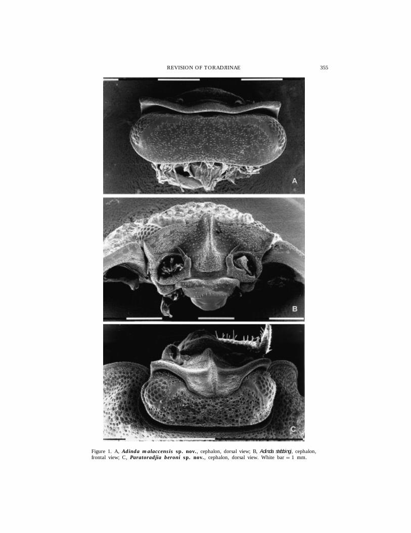
The structure of the cephalon shows great differences within the group,according to the degree of specialization for rolling up and for antennalprotection. In the less specialized forms (Fig. 1A) it is antero-posteriorlycompressed, the profrons is flattened with no special structures to hold theantennae when the animal rolls up into a ball, and the upper margin of the
355REVISION OF TORADJIINAE
Figure 1. A, Adinda malaccensis sp. nov., cephalon, dorsal view; B, Adinda stebbingi, cephalon,frontal view; C, Paratoradjia beroni sp. nov., cephalon, dorsal view. White bar � 1 mm.
356 F. FERRARA ET AL.
frontal shield protrudes above the vertex from which it is separated by a deepgroove. This groove has transverse stripes on both anterior and posterior wallsand often a row of lamellar scales on its posterior margin; it is continuous inAdinda and Protoradjia, but interrupted in the middle in Paratoradjia and Toradjia.In more specialized forms the profrons shows a median longitudinal carina andtwo paramedian depressions which hold the antennae (as in species of Adindaof the stebbingi, riedeli and dollfusi groups) (Fig. 1B). The frontal margin may bebent over the vertex and fused with it in the middle as in Paratoradjia (Fig.1C) or even interrupted as in Toradjia (see Fig. 65A–E); in the latter genus twodeep paramedian antennal grooves run across the whole vertex. Eyes aregenerally large and the number of ommatidia varies (11–28) according to thesize of the species.
Pereonite 1 does not bear any particular structure connected to the rollingup ability in Adinda, with the exception of A. dollfusi where a rounded lobe ispresent at the sides of the posterior margin. In the other genera a schisma ispresent: in Protoradjia this cuts the posterior corner and is very shallow, withthe outer lobe distinctly larger and protruding backwards in relation to theinner one; in Paratoradjia and Toradjia it is far from the posterior corner andseparates two lobes normally protruding backwards to the same degree. Inspecies of Toradjia, pereonite 1 has two variably-developed transverse grooveson the anterior margin, which are continuous with those present on thecephalon and hold the distal segment of the peduncle and flagellum of theantenna.
In the forms possessing a schisma, pereonite 2 (and sometimes 3), bears atransverse ridge or a rounded lobe on the ventral surface, close to the anteriormargin of the epimeron; only in Toradjia celebensis is a ventral tooth present onpereonite 2. Pleonites 3–5, with rectangular epimera, continue the body outline.The telson is semicircular, except in Adinda triangulifera and Paratoradjia beroni,where it is triangular. In all the species it is shorter than the posterior marginof the uropodal protopod and endopod.
The antennule consists of three articles; the distal one is the longest, isconical and has one or more superimposed rows of aesthetascs on the medialmargin.
In the forms less specialized for rolling up, the antennae are long and frail(e.g. Adinda platyperaeon), while in the most specialized forms they are short andstout (Toradjia spp.). The antennal flagellum consists of two articles, the secondone with some aesthetascs, and it ends with a long apical organ (half the totallength of the flagellum in Toradjia and Paratoradjia).
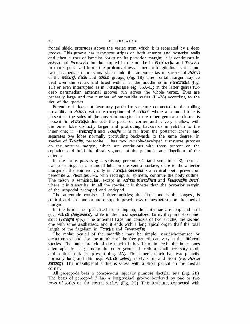
The molar penicil of the mandible may be simple, semidichotomized ordichotomized and also the number of the free penicils can vary in the differentspecies. The outer branch of the maxillule has 10 main teeth, the inner onesoften apically cleft; among the outer group of teeth a small accessory toothand a thin stalk are present (Fig. 2A). The inner branch has two penicils,normally long and thin (e.g. Adinda weberi), rarely short and stout (e.g. Adindastebbingi). The maxillipedal endite is setose with a short penicil on the medialcorner.
All pereopods bear a conspicuous, apically plumose dactylar seta (Fig. 2B).The basis of pereopod 7 has a longitudinal groove bordered by one or tworows of scales on the rostral surface (Fig. 2C). This structure, connected with
357REVISION OF TORADJIINAE
Figure 2. A, Adinda malaccensis sp. nov., particular of the outer branch of maxillule withstalk and accessory tooth; B, Adinda scabra, dactylar seta; C, Adinda malaccensis sp. nov., {,pereopod 7; D, Adinda malaccensis sp. nov., water conducting system on ventral epimeronof pereonite 7. White bar � 0.1 mm (A, B); 1 mm (C, D).

358 F. FERRARA ET AL.
the ventral grooves of the water conducting system (Fig. 2D), is similar to thatpresent in Ligia and other Oniscidea and shows that also in this group thesystem is open. This is very unusual among Crinocheta, in which only theScyphacoidea (sensu Schmalfuss, 1989) possess an open water conducting system,while all the others having a closed one (Hoese, 1981, 1982).
The uropodal protopod is flattened with conspicuous exopod and endopod,both medially inserted, that fill the gap between telson and protopod, completingthe body outline.
The rolling up ability is exoantennate in all the species, i.e. antennae reston the dorsum and remain outside the ball. As in other ‘rollers’ (forinstance Armadillidiidae, Armadillidae and Eubelidae) there are great differencesconcerning the specialization of the rolling up ability: there are forms withincomplete ability, in which the animal simply bends in half like a pocketknife(e.g. Adinda platyperaeon), and others with a very convex body, in which the ballbecomes completely spherical (e.g. Toradjia spp.).
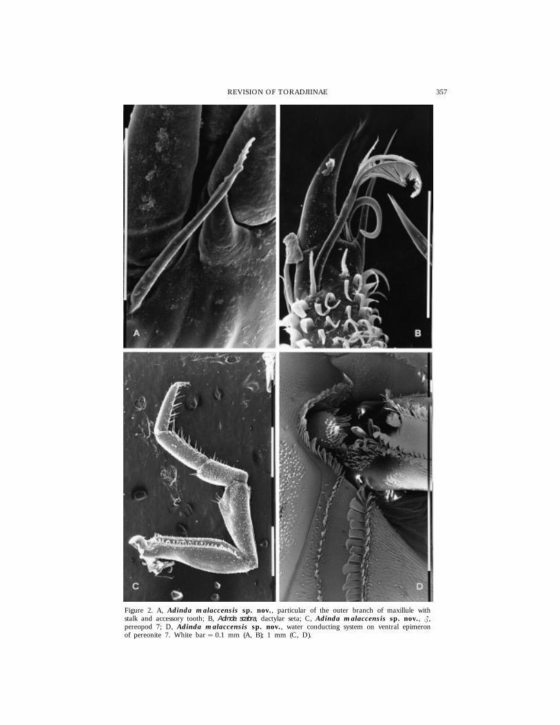
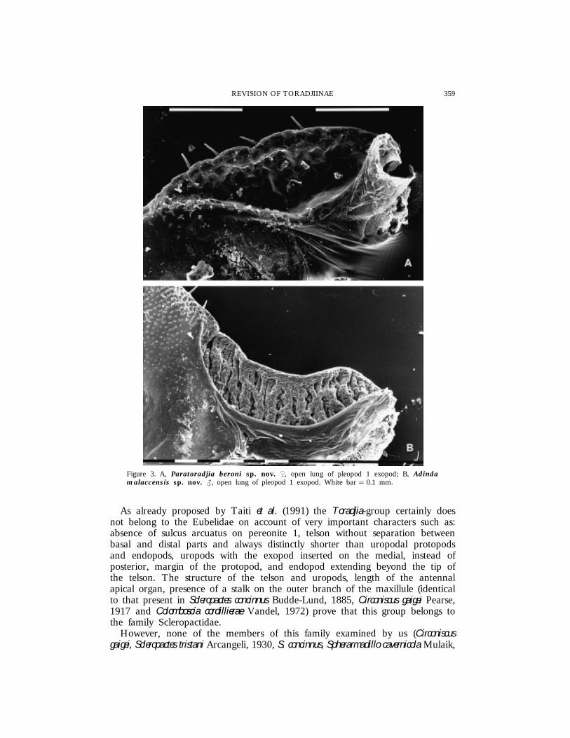
Only pleopods 1 and 2 exopod possess, in a dorsal position, open lungs (i.e.a respiratory area without spiracles), with two different degrees of complexitywithin the group. In the more simple type (Fig. 3A) the respiratory surface isonly waved and completely exposed. This type, similar to that in species ofthe European genus Oniscus and the tropical genus Nagurus, is present in speciesof the genera Paratoradjia and Toradjia, as well as in Adinda dollfusi. In the morecomplex type (Fig. 3B) the respiratory surface is composed of irregularanastomosed folds; moreover, this surface is partly covered by the dorsal wallof the exopod. Compared to the simpler type the respiratory surface is greatlyincreased and the partial invagination of the respiratory folds leads to greaterprotection from desiccation. This type, similar to that described in the Palaearcticgenus Trachelipus, is present in most species of Adinda and in Protoradjia.
DEFINITION AND SYSTEMATIC POSITION
All the species considered certainly constitute a homogeneous group definedby: (1) exoantennate rolling up ability (which is more or less developed); (2) acephalon that, though with remarkable modifications (carina, grooves to holdthe antennae) within the group due to the adaptations connected with antennalprotection, shows a common basic pattern: a frontal shield with upper marginprotruding above the vertex and separated from it by a deep groove withtransverse stripes on its walls; (3) a convex telson, normally semicircular,distinctly shorter than the tips of pleonite 5 and uropodal protopod; (4) antennalflagellum with two articles and a long apical organ; (5) maxillule inner branchwith two penicils, outer one with 4 + 6 main teeth and a long stalk; (6)maxillipedal endite setose and bearing a penicil on the medial corner; (7)pereopods with long, conspicuous dactylar seta, setose in the distal part; basisof pereopod 7 rostrally with a longitudinal groove; (8) open lungs more or lessdeveloped on pleopods 1 and 2 exopod; (9) uropodal protopod flattened withboth exopod and endopod medially inserted, distinctly surpassing posteriormargin of telson and filling the space between protopod and telson; and (10)an open water conducting system. No character is exclusive to this group butthe combination of all the characters easily distinguishes and well characterizesit.
359REVISION OF TORADJIINAE
Figure 3. A, Paratoradjia beroni sp. nov. |, open lung of pleopod 1 exopod; B, Adindamalaccensis sp. nov. {, open lung of pleopod 1 exopod. White bar � 0.1 mm.
As already proposed by Taiti et al. (1991) the Toradjia-group certainly doesnot belong to the Eubelidae on account of very important characters such as:absence of sulcus arcuatus on pereonite 1, telson without separation betweenbasal and distal parts and always distinctly shorter than uropodal protopodsand endopods, uropods with the exopod inserted on the medial, instead ofposterior, margin of the protopod, and endopod extending beyond the tip ofthe telson. The structure of the telson and uropods, length of the antennalapical organ, presence of a stalk on the outer branch of the maxillule (identicalto that present in Scleropactes concinnus Budde-Lund, 1885, Circoniscus gaigei Pearse,1917 and Colomboscia cordillierae Vandel, 1972) prove that this group belongs tothe family Scleropactidae.
However, none of the members of this family examined by us (Circoniscusgaigei, Scleropactes tristani Arcangeli, 1930, S. concinnus, Spherarmadillo cavernicola Mulaik,
360 F. FERRARA ET AL.
1960, Chileoniscus marmoratus Taiti, Ferrara & Schmalfuss, 1986, Neosanfilippia zoiaiManicastri, 1991, Colomboscia bituberculata Taiti, Allspach & Ferrara, in press,and C. cordillierae) possesses a groove on the basis of pereopod 7 and thus anopen water conducting system as found in the Toradjia-group, nor do othergenera certainly related to the Scleropactidae, such as Hekelus Barnard, 1932and Exzaes Barnard, 1932, both from Africa. We consider this characterimportant enough to define a separate subfamily within the Scleropactidae, theToradjiinae, the members of which also show a homogeneous distribution.
Arcangeli (1948, 1955) first established this subfamily within the Eubelidae,pointing out the similarity with the other subfamily Cyrconiscinae (nowScleropactidae). Curiously, though, none of the defining characters of theToradjiinae listed by that author, i.e. ‘semicomplete’ exoantennate ability toroll up, shape of the telson, inner ramus of the maxillule with two penicils,pleopod 1 and 2 exopods with Trachelipus-type lungs, is exclusive to the group,since they are present together also in the typical Scleropactidae (for instancein Scleropactes concinnus).
EVOLUTION
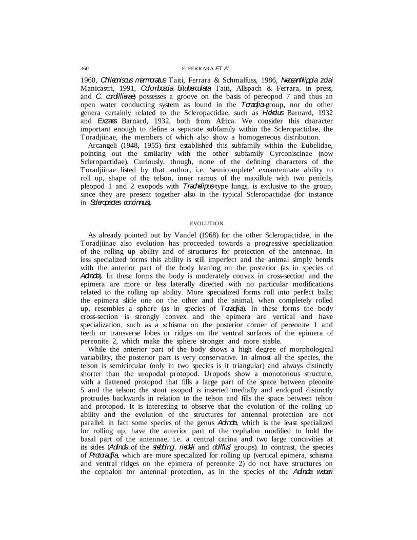
As already pointed out by Vandel (1968) for the other Scleropactidae, in theToradjiinae also evolution has proceeded towards a progressive specializationof the rolling up ability and of structures for protection of the antennae. Inless specialized forms this ability is still imperfect and the animal simply bendswith the anterior part of the body leaning on the posterior (as in species ofAdinda). In these forms the body is moderately convex in cross-section and theepimera are more or less laterally directed with no particular modificationsrelated to the rolling up ability. More specialized forms roll into perfect balls;the epimera slide one on the other and the animal, when completely rolledup, resembles a sphere (as in species of Toradjia). In these forms the bodycross-section is strongly convex and the epimera are vertical and havespecialization, such as a schisma on the posterior corner of pereonite 1 andteeth or transverse lobes or ridges on the ventral surfaces of the epimera ofpereonite 2, which make the sphere stronger and more stable.
While the anterior part of the body shows a high degree of morphologicalvariability, the posterior part is very conservative. In almost all the species, thetelson is semicircular (only in two species is it triangular) and always distinctlyshorter than the uropodal protopod. Uropods show a monotonous structure,with a flattened protopod that fills a large part of the space between pleonite5 and the telson; the stout exopod is inserted medially and endopod distinctlyprotrudes backwards in relation to the telson and fills the space between telsonand protopod. It is interesting to observe that the evolution of the rolling upability and the evolution of the structures for antennal protection are notparallel: in fact some species of the genus Adinda, which is the least specializedfor rolling up, have the anterior part of the cephalon modified to hold thebasal part of the antennae, i.e. a central carina and two large concavities atits sides (Adinda of the stebbingi, riedeli and dollfusi groups). In contrast, the speciesof Protoradjia, which are more specialized for rolling up (vertical epimera, schismaand ventral ridges on the epimera of pereonite 2) do not have structures onthe cephalon for antennal protection, as in the species of the Adinda weberi

361REVISION OF TORADJIINAE
group. While species of Adinda show such plasticity for antennal protection, themodifications in the species of the other genera are very stable.
BIOGEOGRAPHY
The opinion of Vandel (1968), that the Scleropactidae are endemic to theNeotropical Region, with a distribution between the Tropic of Cancer and theTropic of Capricorn, was commonly accepted until a few years ago. TheFrench author pointed out, however, the exception of the genus SphaerobathytropaVerhoeff, 1908 with two species, S. antarctica Vandel, 1963 from Patagonia andS. ribauti Verhoeff, 1908 from the Pyrenees (according to Schmalfuss (1980),this genus most probably belongs to the Armadillidae). Yet a form ofScleropactidae (Kefalloniscus hauseri Schmalfuss, 1986) has been described fromCephalonia in Greece and there are two genera from South Africa, Hekelusand Exzaes, which probably belong to that family (Ferrara & Schmalfuss, 1976;Schmalfuss, 1986). Furthermore, the Toradjia-group (Taiti et al., 1991) has beententatively related to the Scleropactidae, suggesting a much wider distributionof this family. Our conclusions fully confirm this suggestion: the Toradjiinae infact occur in a large part of the Oriental Region and in the Austro-malayanSubregion (Fig. 4). They would appear to be absent throughout much of India,the Indochinese peninsula, Philippines, etc., but this is certainly due to the lackof investigations in those regions.
While the species of Protoradjia are concentrated in northern Sumatra, in thesouthern part of West Malaysia and Singapore, and those of Toradjia are limited
Figure 4. Distribution of the subfamily Toradjiinae: 1, Adinda weberi; 2, A. malaccensis; 3, A. sumatrana;4, A. conglobator; 5, A. stebbingi; 6, A. travancorensis; 7, A. gigas; 8, A. palniensis; 9, A. carli; 10, A.nilgiriensis; 11, A. lobata; 12, A. triangulifera; 13, A. scabra; 14, A. pulchra; 15, A. riedeli; 16, A.platyperaeon; 17, A. lamellata; 18, A. dollfusi; 19, Protoradjia jacobsoni; 20, P. paeninsulae; 21, P. insularis;22, P. montana; 23, P. pilosa; 24, Paratoradjia beroni; 25, P. indosinensis; 26, P. vietnamensis; 27, P.sulcata; 28, Toradjia gorgona; 29, T. celebensis; 30, T. cephalica; 31, T. hirsuta.

362 F. FERRARA ET AL.
to Java, Sarawak and Sulawesi, the species of Adinda and Paratoradjia are morewidely distributed. Species of Adinda populate a vast area between southernIndia and the Philippines and those of Paratoradjia are distributed, apparentlyin a discontinuous manner, in a still larger area from Sri Lanka to NewIreland.
Even if very little is known about the ecology of the single species, theToradjiinae seem to populate exclusively montane and lowland tropical rainforests.
TAXONOMY
GenericlevelIn all the species examined many characters, such as the telson and uropods,
antenna, mandible, maxilliped, dactylar seta, water conducting system andpleopodal lungs, are very similar. Yet the species can be grouped according tomorphological specializations such as the ability to roll up into a ball andstructures for antennal protection. Considering the large number of speciesexamined and the stability of these specializations, they can be considered tobe good characters with which genera can be defined. Four genera arerecognized: Adinda, the richest in number of species (18) with no distinctstructures connected with the ability to roll up into a ball; Protoradjia (5 species)characterized by a shallow schisma at the posterior corner of the pereonite 1;Paratoradjia (4 species) with frontal shield bent over vertex in the middle andpartially fused with it; and Toradjia (4 species), with deep grooves for antennalprotection on the cephalon and pereonite 1.
SpecificlevelSpecies are well characterized by the male sexual modifications that can
involve all pereopods, pleopods and, in some species of Adinda, even sternitesof pereonites 2–5. In some cases the shape of the cephalon and telson andthe colour pattern are distinct enough to allow safe specific identifications,whereas dorsal granulation must be considered carefully since in some species(e.g. Adinda palniensis) juveniles are strongly granulated while adults are almostsmooth.
Genus Adinda Budde-Lund, 1904
Type species: Mesarmadillo [Budde-Lund’s mistake for Periscyphus] weberi Dollfus,1898 by original designation and monotypy.
Synonym: Paraperiscyphis Stebbing, 1911. Type species: Paraperiscyphis travancorensisStebbing, 1911 by original designation and monotypy.
DiagnosisAnimals with incomplete exoantennate ability to roll up into a ball. Cephalon:
profrons flattened or with a longitudinal carina in the middle, more or lessprotruding; large frontal lateral lobes, often distinctly protruding above vertexand directed forwards; frontal lamina separated from vertex by a deep groove.

363REVISION OF TORADJIINAE
Pereonite 1 with posterior margin usually concave, sometimes straight or evendeeply incised at base of epimera; posterior corner without schisma; no lobesor teeth on ventral surface. Epimera of pereonite 2 ventrally with no transverseridge. Telson normally semicircular, rarely triangular, always shorter thanuropodal protopods. Antennule of three articles. Antenna with flagellum of twoarticles and long apical organ. Mandible with molar penicil simple (i.e. consistingof a single plumose seta) or semidichotomized (i.e. consisting of some plumosesetae arising from a common stem). Outer branch of maxillule with 4 + 6teeth, an accessory small tooth and a slender stalk with small nodes in thedistal part; inner branch with two penicils. Maxilliped with setose endite bearinga penicil on medial corner. Pereopods with long dactylar seta, bifid and setoseat apex. Basis of pereopod 7 with a longitudinal deep groove bordered withscales on rostral surface. Pleopodal exopods 1–2 with open lungs similar tothose in Trachelipus. Uropods with protopod flattened, posteriorly broadly roundedor subtruncated; exopod inserted in a notch of the medial margin of protopod;endopod distinctly surpassing posterior margin of telson.
RemarksBudde-Lund (1904: 37), in his key to the Spherilloninae, established the
genus Adinda for the species Mesarmadillo weberi Dollfus, 1898, a combinationnever published by the French author; obviously Budde-Lund meant Periscyphusweberi Dollfus, 1898. According to Omer-Cooper (1926: 351), ParaperiscyphisStebbing, 1911 is a junior synonym of Adinda.
The species present some differences in the structure of the cephalon andbuccal pieces. In A. weberi the frontal shield is almost flattened, with no distinctcarina in the middle, and with small lateral lobes not protruding forwards,while in A. platyperaeon a sharp medial carina is present and the lateral lobesare broad and protrude forwards. These differences must be considered aswithin genus variability since, between these two extreme conditions, intermediatestates are also present. As regards the buccal pieces, the differences concernmandibles and maxillules. In some species (e.g. A. riedeli), the molar penicil ofthe mandible is clearly simple, while in others (e.g. A. weberi) it issemidichotomized. The maxillule also varies: the teeth of the outer branch maybe entire (A. stebbingi) or some apically cleft (A. weberi); the two penicils of theinner branch may be long and thin (A. weberi) or short and stout (A. stebbingi).In the systematics of the Oniscidea the structure of buccal pieces is consideredto be of high taxonomic value in some groups (e.g. Philosciidae) but not inothers (e.g. Armadillidae). For the genus Adinda, we cannot exclude that thesedifferences may be important enough to characterize distinct taxa (such assubgenera). However, considering the uniformity of all the other characters, thestill fragmentary knowledge of this group (certainly several other species will bediscovered), that some species have not been properly investigated (A. conglobator,A. travancorensis and A. gigas) and that also in species of Toradjia, certainly ahomogenous group, there are some differences in the buccal appendages (seebelow), we prefer for the time being to delineate only species-groups. Althoughthey have no taxonomic status, they serve as a useful means of grouping speciesthat have combinations of characters in common. Four groups are recognized:the weberi group, the stebbingi group, the riedeli group and the dollfusi group.Their characters are summarized in Table 1.
364 F. FERRARA ET AL.
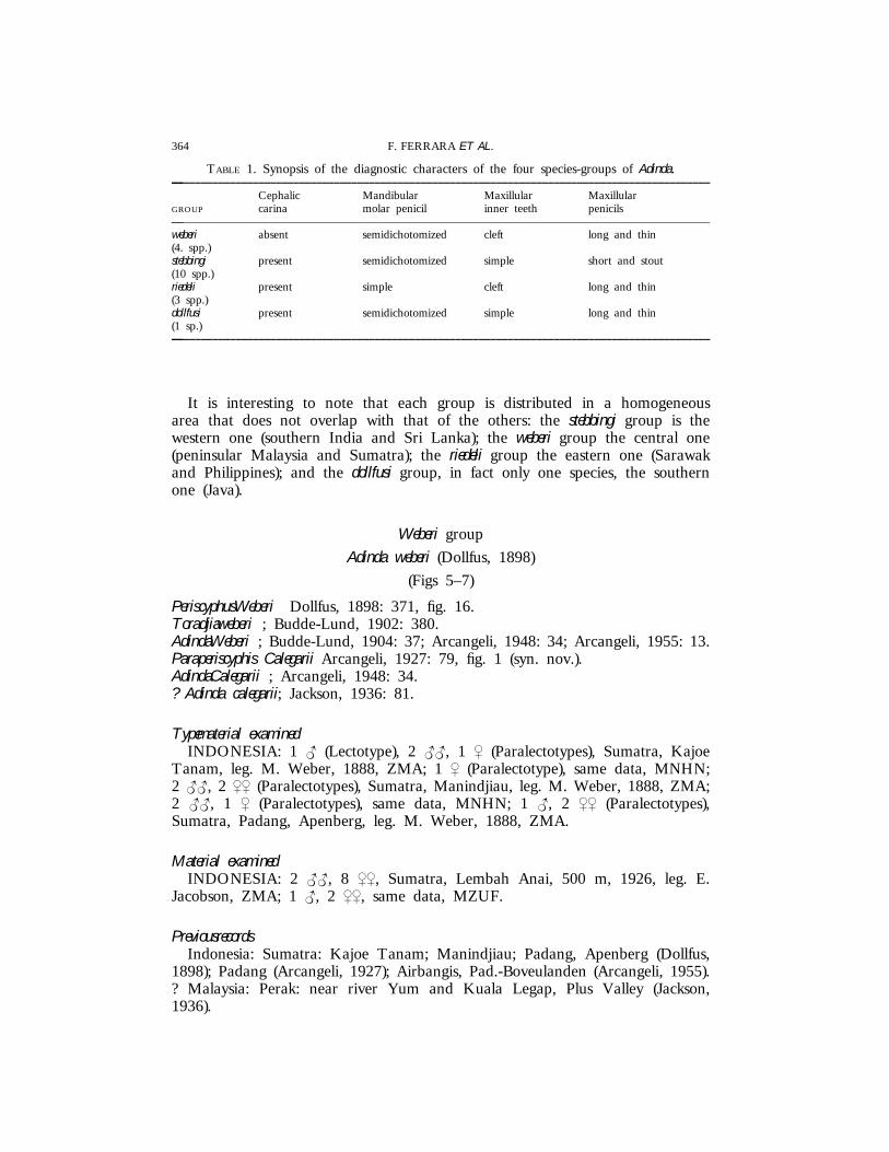
TABLE 1. Synopsis of the diagnostic characters of the four species-groups of Adinda.—–––––––––––––––––––––––––––––––––––––––––––––––––––––––––––––––––––––––––––––––––––––––––––––
Cephalic Mandibular Maxillular MaxillularGROUP carina molar penicil inner teeth penicils—–––––––––––––––––––––––––––––––––––––––––––––––––––––––––––––––––––––––––––––––––––––––––––––weberi absent semidichotomized cleft long and thin(4. spp.)stebbingi present semidichotomized simple short and stout(10 spp.)riedeli present simple cleft long and thin(3 spp.)dollfusi present semidichotomized simple long and thin(1 sp.)—–––––––––––––––––––––––––––––––––––––––––––––––––––––––––––––––––––––––––––––––––––––––––––––
It is interesting to note that each group is distributed in a homogeneousarea that does not overlap with that of the others: the stebbingi group is thewestern one (southern India and Sri Lanka); the weberi group the central one(peninsular Malaysia and Sumatra); the riedeli group the eastern one (Sarawakand Philippines); and the dollfusi group, in fact only one species, the southernone (Java).
Weberi group
Adinda weberi (Dollfus, 1898)
(Figs 5–7)
PeriscyphusWeberi Dollfus, 1898: 371, fig. 16.Toradjiaweberi ; Budde-Lund, 1902: 380.AdindaWeberi ; Budde-Lund, 1904: 37; Arcangeli, 1948: 34; Arcangeli, 1955: 13.Paraperiscyphis Calegarii Arcangeli, 1927: 79, fig. 1 (syn. nov.).AdindaCalegarii ; Arcangeli, 1948: 34.? Adinda calegarii; Jackson, 1936: 81.
Typematerial examinedINDONESIA: 1 { (Lectotype), 2 {{, 1 | (Paralectotypes), Sumatra, Kajoe
Tanam, leg. M. Weber, 1888, ZMA; 1 | (Paralectotype), same data, MNHN;2 {{, 2 || (Paralectotypes), Sumatra, Manindjiau, leg. M. Weber, 1888, ZMA;2 {{, 1 | (Paralectotypes), same data, MNHN; 1 {, 2 || (Paralectotypes),Sumatra, Padang, Apenberg, leg. M. Weber, 1888, ZMA.
Material examinedINDONESIA: 2 {{, 8 ||, Sumatra, Lembah Anai, 500 m, 1926, leg. E.
Jacobson, ZMA; 1 {, 2 ||, same data, MZUF.
PreviousrecordsIndonesia: Sumatra: Kajoe Tanam; Manindjiau; Padang, Apenberg (Dollfus,
1898); Padang (Arcangeli, 1927); Airbangis, Pad.-Boveulanden (Arcangeli, 1955).? Malaysia: Perak: near river Yum and Kuala Legap, Plus Valley (Jackson,1936).
365REVISION OF TORADJIINAE
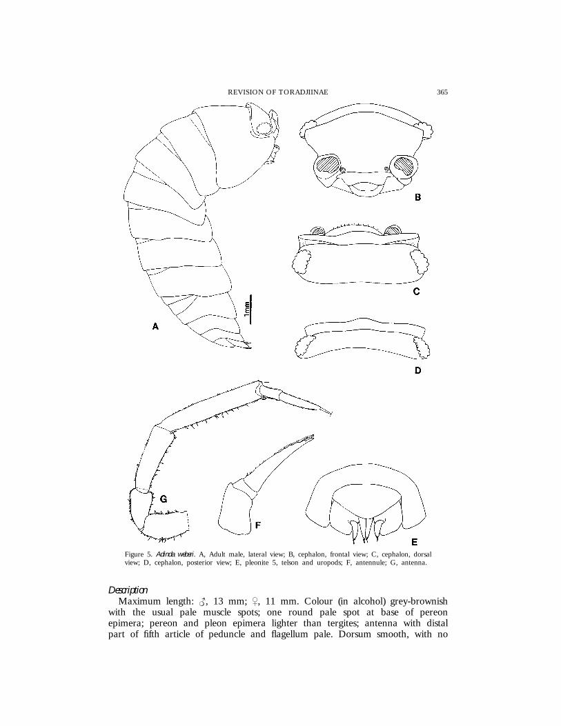
Figure 5. Adinda weberi. A, Adult male, lateral view; B, cephalon, frontal view; C, cephalon, dorsalview; D, cephalon, posterior view; E, pleonite 5, telson and uropods; F, antennule; G, antenna.
DescriptionMaximum length: {, 13 mm; |, 11 mm. Colour (in alcohol) grey-brownish
with the usual pale muscle spots; one round pale spot at base of pereonepimera; pereon and pleon epimera lighter than tergites; antenna with distalpart of fifth article of peduncle and flagellum pale. Dorsum smooth, with no
366 F. FERRARA ET AL.
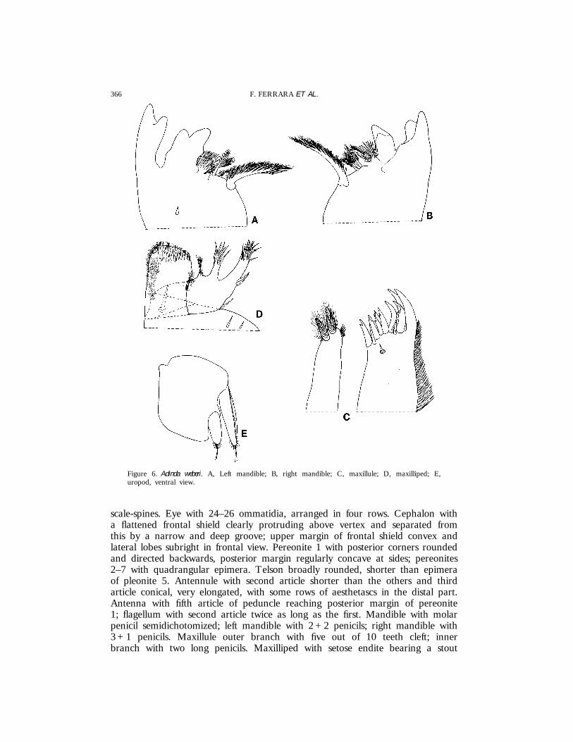
Figure 6. Adinda weberi. A, Left mandible; B, right mandible; C, maxillule; D, maxilliped; E,uropod, ventral view.
scale-spines. Eye with 24–26 ommatidia, arranged in four rows. Cephalon witha flattened frontal shield clearly protruding above vertex and separated fromthis by a narrow and deep groove; upper margin of frontal shield convex andlateral lobes subright in frontal view. Pereonite 1 with posterior corners roundedand directed backwards, posterior margin regularly concave at sides; pereonites2–7 with quadrangular epimera. Telson broadly rounded, shorter than epimeraof pleonite 5. Antennule with second article shorter than the others and thirdarticle conical, very elongated, with some rows of aesthetascs in the distal part.Antenna with fifth article of peduncle reaching posterior margin of pereonite1; flagellum with second article twice as long as the first. Mandible with molarpenicil semidichotomized; left mandible with 2 + 2 penicils; right mandible with3 + 1 penicils. Maxillule outer branch with five out of 10 teeth cleft; innerbranch with two long penicils. Maxilliped with setose endite bearing a stout
367REVISION OF TORADJIINAE
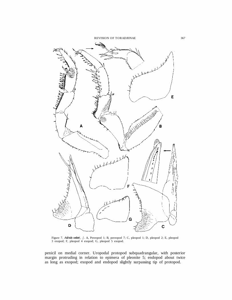
Figure 7. Adinda weberi, {. A, Pereopod 1; B, pereopod 7; C, pleopod 1; D, pleopod 2; E, pleopod3 exopod; F, pleopod 4 exopod; G, pleopod 5 exopod.
penicil on medial corner. Uropodal protopod subquadrangular, with posteriormargin protruding in relation to epimera of pleonite 5; endopod about twiceas long as exopod; exopod and endopod slightly surpassing tip of protopod.
368 F. FERRARA ET AL.
Male. Pereopods 1–3 carpus and, to a lesser extent, merus with lines of longtrifid spines; sternal margin of carpus of pereopods 1–3 and merus of pereopods1–4 with a brush of small scales. Pereopod 7 ischium with concave sternalmargin, a setose area at the base and one on the distal part of the rostralsurface; merus with a setose, clearly protruding lobe. Pleopod 1 exopod witha long triangular posterior point, outer margin with several spines; endopodstraight, with a hyaline lobe at apex. Pleopod 2 endopod longer than exopod.Pleopods 3–5 exopod as in Fig. 7E–G.
DistributionIndonesia (Sumatra) and Malaysia (?).
RemarksThis species is characterized by the cephalon with the frontal shield flattened
and protruding over the vertex, the male pereopod 7 ischium with concavesternal margin and merus with a setose basal lobe, the male pleopod 1 exopodwith a long triangular posterior point.
According to its original description (Arcangeli, 1927), Paraperiscyphis calegariipresents no differences from A. weberi, and the locality where the two specieswere collected is the same (Sumatra, Padang). In our opinion P. calegarii mustbe considered as a junior synonym of A. weberi.
Jackson’s record (1936) from Malaysia is doubtful and re-examination of thismaterial is necessary. Most probably these specimens belong to one of theother species recorded from Malaysia (see below).
Adinda malaccensis sp. nov.
(Figs 1A, 2A, C, D, 3B, 8, 9)
Material examinedMALAYSIA: 1 { (Holotype), 12 {{, 11 ||, 3 juvs (Paratypes), Pahang,
Genting Highlands, 1725 m, edge of forest, leg. S. Taiti & L. Bartolozzi,18.xi.1987, MZUF; 1 {, 1 | (Paratypes), same data, SMNS; 2 || (Paratypes),same locality, 1600 m, edge of forest, leg. S. Taiti & L. Bartolozzi, 20.xi.1987,MZUF; 1 {, 2 || (Paratypes), Pahang, along the road to Genting Highlands,1320 m, leg. S. Taiti, 7.xi.1985, MZUF; 4 {{, 3 || (Paratypes), same locality,1650 m, leg. S. Taiti & L. Bartolozzi, 13.xi.1987, MZUF; 3 ||, 1 juv.(Paratypes), Pahang, Genting Tea Estate, about 40 km NE of Kuala Lumpur,610 m, leg. S. Taiti, 2.xi.1985, MZUF; 1 { (Paratype), same locality, leg. S.Taiti & L. Bartolozzi, 16.xi.1987, MZUF; 2 || (Paratypes), Pahang, BukitRengit, near Lanchang, 120 m, lowland forest, near stream, leg. S. Taiti & L.Bartolozzi, 30.xi.1987, MZUF; 5 {{, 2 ||, 11 juvs (Paratypes), same locality,leg. S. Taiti, 15.xi.1987, MZUF; 1 juv. (Paratype), Selangor, Ulu Gombak, 24km NE of Kuala Lumpur, 230 m, lowland forest, leg. S. Taiti, 20.x.1985,MZUF; 1 juv. (Paratype), same locality, leg. S. Taiti, 22.x.1985, MZUF.
DescriptionMaximum length: {, 19 mm; |, 24 mm. Colour (in alcohol) grey-brownish
with the usual pale muscle spots; one pale spot at the base of pereon and
369REVISION OF TORADJIINAE
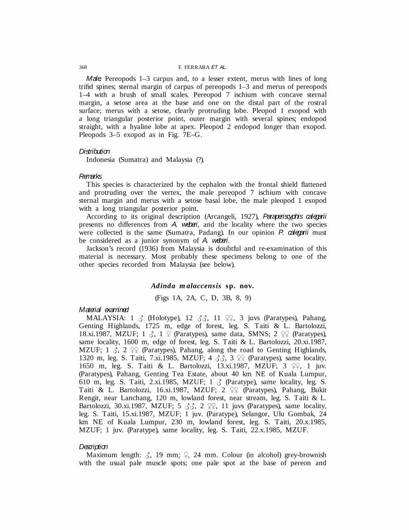
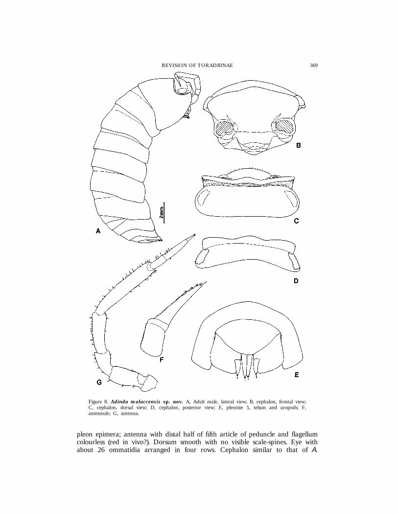
Figure 8. Adinda malaccensis sp. nov. A, Adult male, lateral view; B, cephalon, frontal view;C, cephalon, dorsal view; D, cephalon, posterior view; E, pleonite 5, telson and uropods; F,antennule; G, antenna.
pleon epimera; antenna with distal half of fifth article of peduncle and flagellumcolourless (red in vivo?). Dorsum smooth with no visible scale-spines. Eye withabout 26 ommatidia arranged in four rows. Cephalon similar to that of A.

370 F. FERRARA ET AL.
Figure 9. Adinda malaccensis sp. nov., {. A, Pereopod 1; B, pereopod 7; C, pleopod 1; D,pleopod 2; E, pleopod 3 exopod; F, pleopod 4 exopod; G, pleopod 5 exopod.
weberi, but with frontal shield having upper margin slightly sinuous and laterallobes more rounded. Pereon, pleon, telson, antennule, antenna, buccal piecesand uropod as in A. weberi.
Male. Pereopods 1–4 with lines of trifid spines on carpus and, more sparsely,
371REVISION OF TORADJIINAE
on merus; a brush of small scales on sternal margin of carpus and merus.Pereopod 7 ischium narrower and longer than in A. weberi, with straight sternalmargin; merus with basal lobe little protruding and bearing long setae. Pleopod1 exopod with a short triangular posterior point with some spines; straightendopod with no particular modifications at apex. Pleopods 2–5 similar tothose of A. weberi.
EtymologyThe name refers to the Malay peninsula (Malacca) where these specimens
were collected.
DistributionMalaysia.
RemarksAdinda malaccensis is morphologically very similar to A. weberi, from which it
is distinguished by the larger maximum dimensions, the frontal shield with theupper margin sinuous instead of regularly convex, the male pereopod 7 whichhas a narrower ischium with straight, instead of concave, sternal margin, meruswith the basal lobe less protruding, and the male pleopod 1 exopod withshorter posterior point.
Adinda sumatrana sp. nov.
(Figs 10–12)
Material examinedINDONESIA: 1 { (Holotype), 1 | (Paratype), Sumatra, west coast,
Tandjunggadang, 1000 m, leg. E. Jacobson, 1926, ZMA; 1 | (Paratype), samelocality, leg. E. Jacobson, 1925, MZUF; 2 ||, 1 juv. (Paratypes), Sumatra,Bukittingi, Gunung Merapi, 2000–2200 m, leg. A. Riedel, 18.x.1990, SMNS;1 { (Paratype), same data, MZUF; 1 {, 1 |, 1 juv. (Paratypes), Sumatra,Bukittingi, Gunung Singgalang, leg. A. Riedel, 16.x.1990, SMNS; 1 | (Paratype),same locality, 1800 m, leg. E. Jacobson, 1925, ZMA.
DescriptionMaximum length: {, 19 mm; |, 22 mm. Colour (in alcohol), as in A. weberi.
Dorsum with some small triangular scale-spines. Cephalon with frontal shieldsimilar to A. malaccensis. Pereonites 1–3 with posterior corners subacute. Pleon,telson, antennule, antenna and buccal pieces as in A. weberi. Uropodal protopodnarrower than in the previous two species.
Male. Pereopods 1–4 carpus and, to a lesser extent, merus with lines of longtrifid spines and a brush of small scales on sternal margin. Pereopods 3–4ischium with some conspicuous tubercles on sternal margin. Pereopod 7 ischiumwith sternal margin sinuous in the distal part; merus with basal lobe similarto that of A. malaccensis, but with shorter setae. Pleopod 1 exopod with a stout
372 F. FERRARA ET AL.
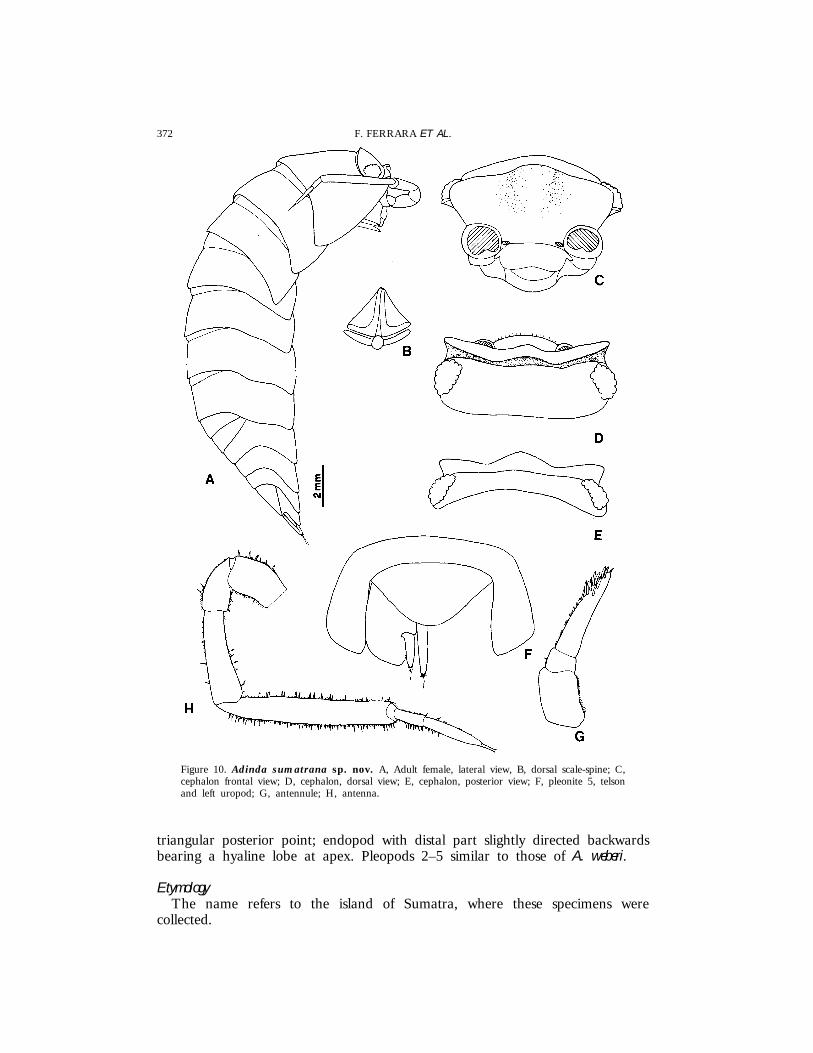
Figure 10. Adinda sumatrana sp. nov. A, Adult female, lateral view, B, dorsal scale-spine; C,cephalon frontal view; D, cephalon, dorsal view; E, cephalon, posterior view; F, pleonite 5, telsonand left uropod; G, antennule; H, antenna.
triangular posterior point; endopod with distal part slightly directed backwardsbearing a hyaline lobe at apex. Pleopods 2–5 similar to those of A. weberi.
EtymologyThe name refers to the island of Sumatra, where these specimens were
collected.
373REVISION OF TORADJIINAE
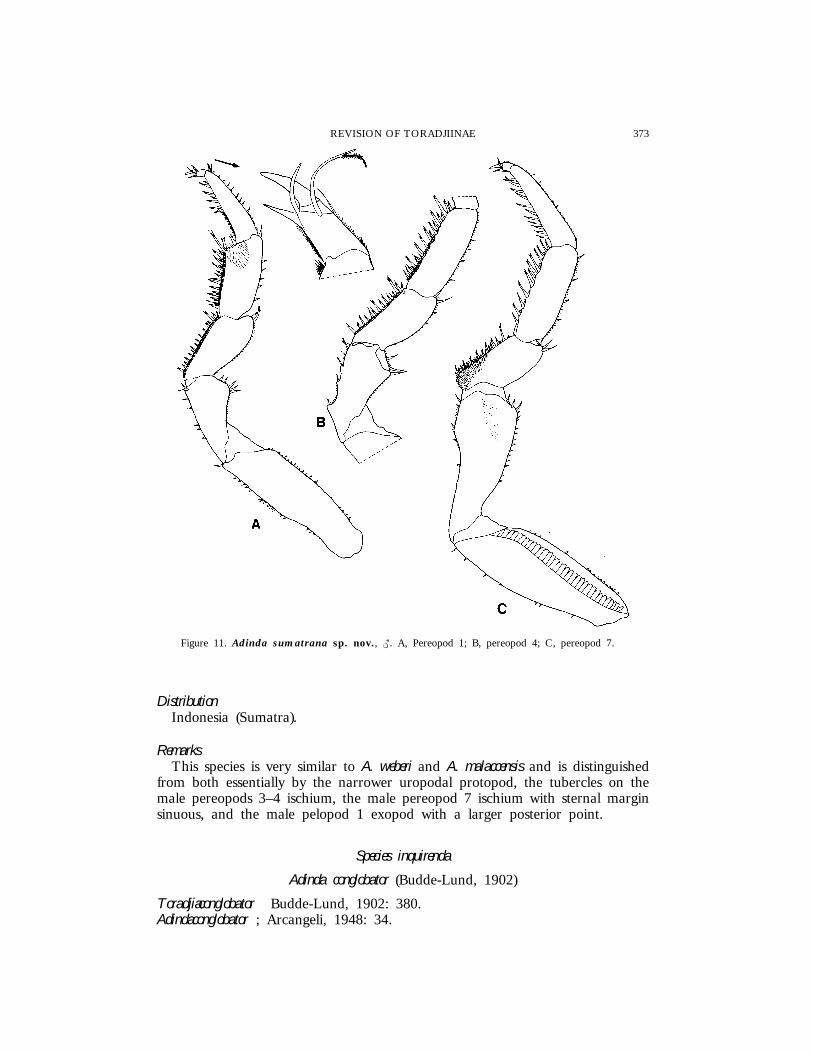
Figure 11. Adinda sumatrana sp. nov., {. A, Pereopod 1; B, pereopod 4; C, pereopod 7.
DistributionIndonesia (Sumatra).
RemarksThis species is very similar to A. weberi and A. malaccensis and is distinguished
from both essentially by the narrower uropodal protopod, the tubercles on themale pereopods 3–4 ischium, the male pereopod 7 ischium with sternal marginsinuous, and the male pelopod 1 exopod with a larger posterior point.
Species inquirenda
Adinda conglobator (Budde-Lund, 1902)
Toradjiaconglobator Budde-Lund, 1902: 380.Adindaconglobator ; Arcangeli, 1948: 34.

374 F. FERRARA ET AL.
Figure 12. Adinda sumatrana sp. nov., {. A, Pleopod 1; B, pleopod 2; C, pleopod 3 exopod;D, pleopod 4 exopod; E, pleopod 5 exopod.
PreviousrecordsMalaysia: Ajenz (?Aring), Kelantan (Budde-Lund, 1902).
DistributionMalaysia.
RemarksBudde-Lund (1902) did not properly describe or illustrate this species. He
only stated that it was ‘‘most nearly allied’’ to A. weberi from which it differedin having ‘‘shorter antennae, with the flagellum short, white, and basal jointvery short’’. Without a re-examination of the type material, which appears notto be deposited in any of the institutions contacted by us, nothing can be saidabout this species, except that it certainly belongs to Adinda.

375REVISION OF TORADJIINAE
Stebbingi group
Adinda stebbingi (Collinge, 1914)
(Figs 1B, 13–16)
Paraperiscyphisstebbingi Collinge, 1914: 207, pl. XXIV figs 1–10; Collinge, 1916:115.AdindaStebbingi ; Arcangeli, 1948: 34.
Material examinedINDIA: 11 {{, 14 ||, 9 juvs, Anamalai Hills, Valparai, under rotting logs,
leg. J. Carl, 4.iii.1927, MHNG; 2 {{, 1 |, same data, MZUF; 1 {, 1 |,same data, SMNS.
PreviousrecordsIndia: Anamalai Hills, Madras Pres. (Collinge, 1914); Kavalai, Cochin State
(Collinge, 1916).
DescriptionMaximum length: {, 23 mm; |, 20 mm. Colour (in alcohol) pale brown,
probably due to the long conservation (dark brown according to Collinge,1914). Dorsum strongly granulated with larger granules on tergites and onposterior margin of pereonites; several petaliform scale-spines surrounded bysmall scales. Eye with 28 ommatidia disposed in four rows. Cephalon with adistinct carina on profrons; frontal shield protruding above vertex and separatedfrom this by a deep groove bordered by a row of scales; frontal shield withmedial lobe triangularly raised; lateral lobes directed forwards, more protrudingthan carina. Pereonite 1 with posterior margin clearly concave at the base ofepimera; posterior corner acute. Pereonites 2–4 with epimera directed backwardsand with posterior corners progressively less acute. Pereonites 5–7 with posteriormargin slightly sinuous at sides. Telson wider than long, with broadly roundedapex. Antennule with second article slightly shorter than first; third articleconical with several rows of aesthetascs on medial margin. Antenna with fiftharticle of peduncle reaching the posterior margin of pereonite 2; second articleof flagellum about 1.5 times as long as first. Mandibles with molar penicilsemidichotomized; left mandible with 6 + 2 penicils; right mandible with 4 + 1penicils. Maxillule outer branch with all teeth apically entire; inner branch withtwo stout subequal penicils. Uropodal protopod with distal margin rounded;endopod and exopod slightly protruding in relation to the protopod tip.
Male. Sternites of pereonites 2–5 with two medial points protruding downwards.Pereopods 1–4 carpus and merus with lines of long trifid spines and a largearea of short setae on the rostral surface. Pereopods 2–4 merus with sternalmargin clearly sinuous, and ischium with some large tubercles disposed in twolines on sternal margin. Pereopods 5–7 ischium, merus and carpus with linesof long spines on sternal margin. Pleopod 1 exopod with a long triangularposterior point bearing some spines on outer and medial margin; endopod withno particular modifications. Pleopod 2 endopod slightly longer than exopod.Pleopods 3–5 exopod as in Fig. 16D–F.
376 F. FERRARA ET AL.
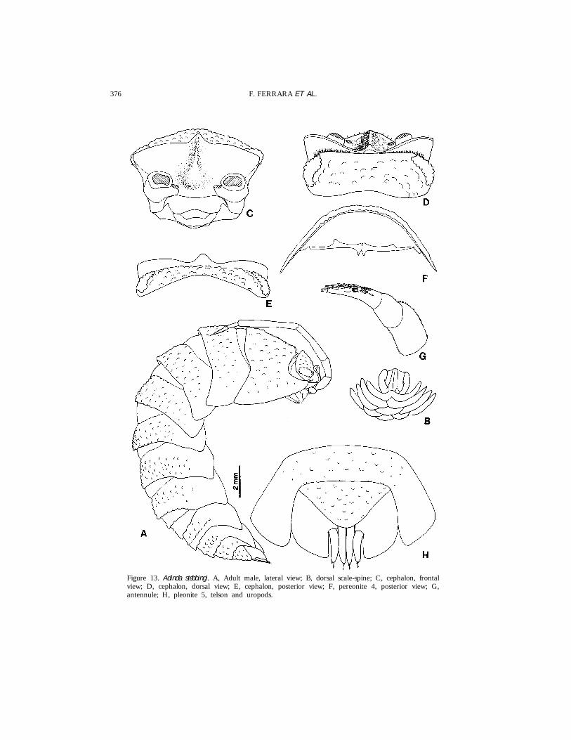
Figure 13. Adinda stebbingi. A, Adult male, lateral view; B, dorsal scale-spine; C, cephalon, frontalview; D, cephalon, dorsal view; E, cephalon, posterior view; F, pereonite 4, posterior view; G,antennule; H, pleonite 5, telson and uropods.
377REVISION OF TORADJIINAE
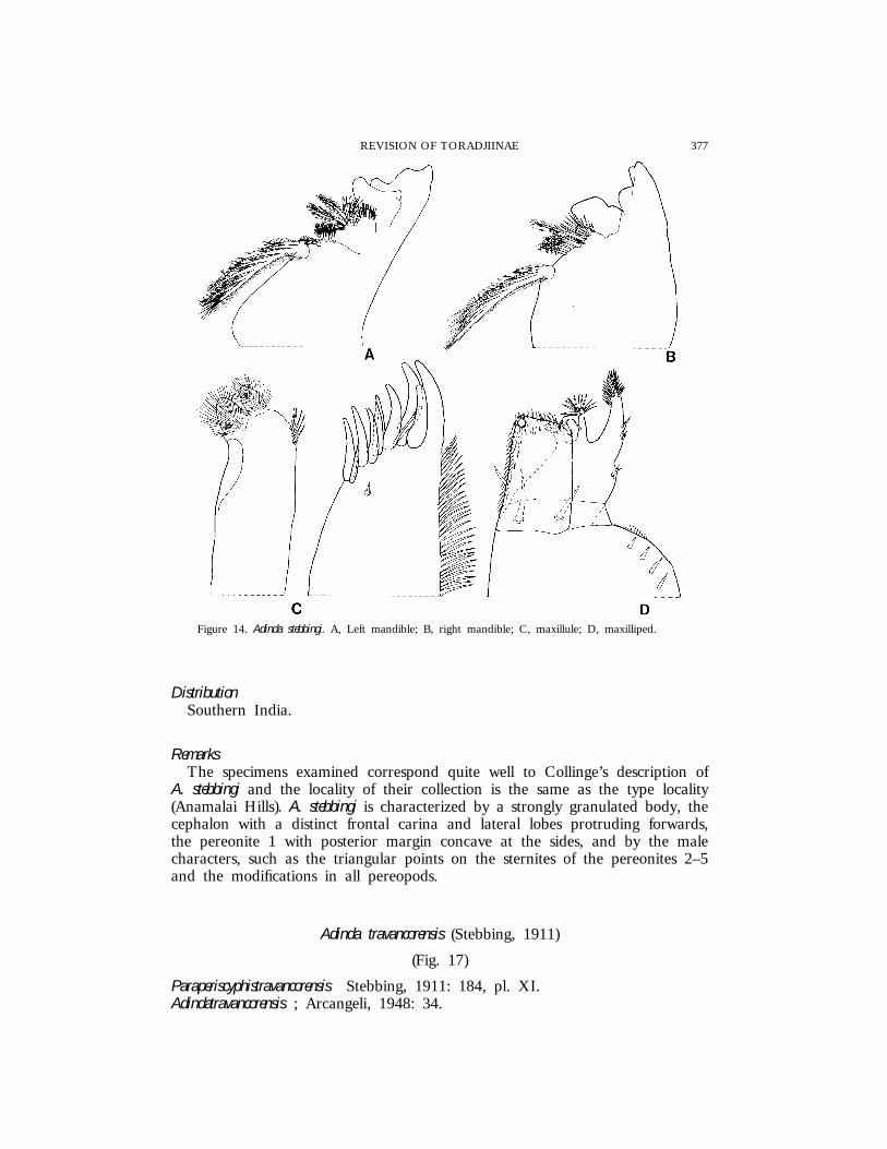
Figure 14. Adinda stebbingi. A, Left mandible; B, right mandible; C, maxillule; D, maxilliped.
DistributionSouthern India.
RemarksThe specimens examined correspond quite well to Collinge’s description of
A. stebbingi and the locality of their collection is the same as the type locality(Anamalai Hills). A. stebbingi is characterized by a strongly granulated body, thecephalon with a distinct frontal carina and lateral lobes protruding forwards,the pereonite 1 with posterior margin concave at the sides, and by the malecharacters, such as the triangular points on the sternites of the pereonites 2–5and the modifications in all pereopods.
Adinda travancorensis (Stebbing, 1911)
(Fig. 17)
Paraperiscyphistravancorensis Stebbing, 1911: 184, pl. XI.Adindatravancorensis ; Arcangeli, 1948: 34.

378 F. FERRARA ET AL.
Figure 15. Adinda stebbingi, {. A, Pereopod 1; B, pereopod 2; C, pereopod 3; D, pereopod 4; E,pereopod 5; F, pereopod 6.
379REVISION OF TORADJIINAE
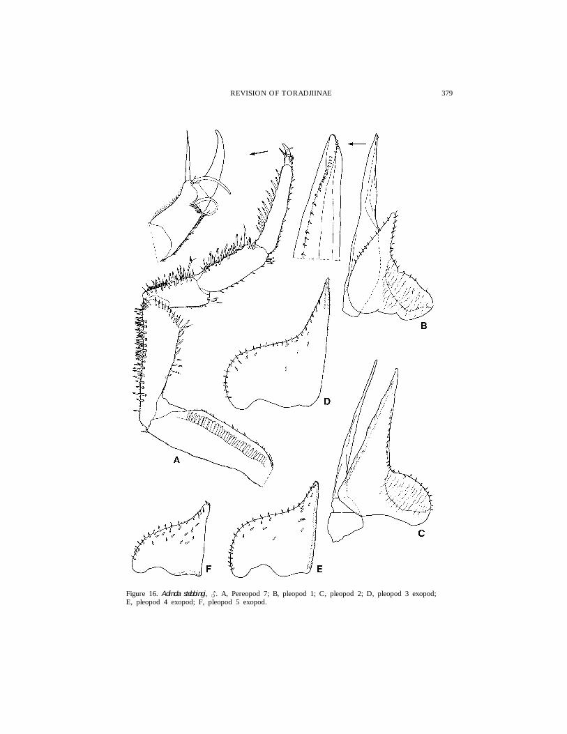
Figure 16. Adinda stebbingi, {. A, Pereopod 7; B, pleopod 1; C, pleopod 2; D, pleopod 3 exopod;E, pleopod 4 exopod; F, pleopod 5 exopod.
380 F. FERRARA ET AL.
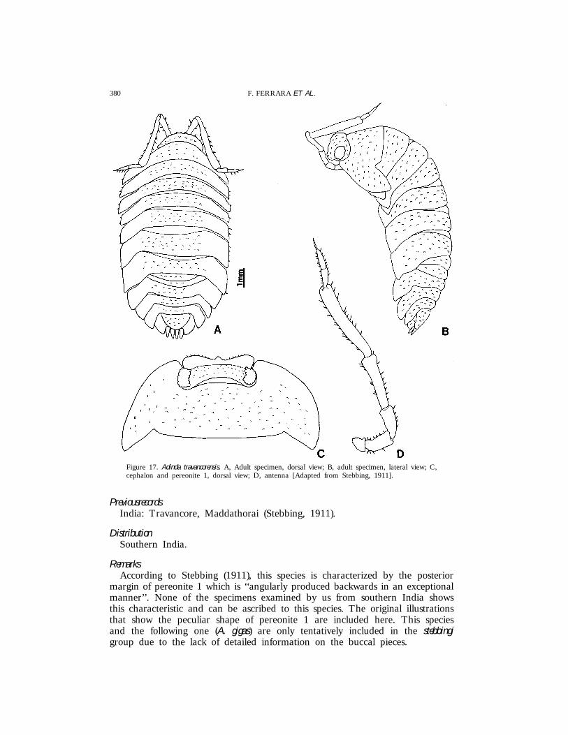
Figure 17. Adinda travancorensis. A, Adult specimen, dorsal view; B, adult specimen, lateral view; C,cephalon and pereonite 1, dorsal view; D, antenna [Adapted from Stebbing, 1911].
PreviousrecordsIndia: Travancore, Maddathorai (Stebbing, 1911).
DistributionSouthern India.
RemarksAccording to Stebbing (1911), this species is characterized by the posterior
margin of pereonite 1 which is ‘‘angularly produced backwards in an exceptionalmanner’’. None of the specimens examined by us from southern India showsthis characteristic and can be ascribed to this species. The original illustrationsthat show the peculiar shape of pereonite 1 are included here. This speciesand the following one (A. gigas) are only tentatively included in the stebbingigroup due to the lack of detailed information on the buccal pieces.
381REVISION OF TORADJIINAE
Adinda gigas (Collinge, 1915)
Periscyphisgigas Collinge, 1915: 148, pl. IX figs 1–10.Paraperiscyphisgigas ; Collinge, 1916: 116.Adindagigas ; Arcangeli, 1948: 34.
PreviousrecordsIndia: Ponmudi, Travancore [about 35 km NE of Trivandrum, Kerala State]
(Collinge, 1915).
RemarksThe original description and illustrations of A. gigas are not useful to fully
characterize this species. Nevertheless, none of the specimens examined by usfrom southern India shows the peculiar colour pattern described by Collinge,1915 (‘‘horny-brown with the lateral plates of the 1st, 5th and 6th mesosomaticand the 3rd and 4th metasomatic segments yellow’’) and can be ascribed tothis species.
Adinda palniensis sp. nov.
(Figs 18–20)
Material examinedINDIA: 1 { (Holotype), 2 {{, 3 ||, 6 juvs (Paratypes), Upper Palni Hills,
Vandaravu, Sholas, about 2300 m, leg. J. Carl, 7.iv.1927, MHNG; 5 ||, 1juv. (Paratypes), Upper Palni Hills, Kukkal-Shola, about 1900 m, under rottinglogs, leg. J. Carl, 1.iv.1927, MHNG; 1 { (Paratype), same data, MZUF; 1 {,7 || (Paratypes), same data, MHNG; 3 {{, 5 ||, 3 juvs (Paratypes), samelocality and collector, 3.iv.1927, MHNG; 2 {{, 3 || (Paratypes), Upper PalniHills, Bombay-Shola, near Kodaikanal, 2200 m, under logs and stones, leg. J.Carl, 21.iii.1927, MHNG; 1 {, 1 | (Paratypes), same data, MZUF; 1 {
(Paratype), same data, SMNS; 1 |, 2 juvs (Paratypes), Lower Palni Hills, Sholanear Shembaganur (Tiger-Shola?), about 1600 m, leg. J. Carl, 17.iv.1927,MHNG; 1 {, 1 |, 1 juv. (Paratypes), Upper Palni Hills, Mariyan-Shola, 2400m, forest, leg. J. Carl, 11–14.iv.1927, MHNG; 1 {, 5 juvs (Paratypes),Travancore, Vattavadai Valley (between Palni and Anamalai Hills), primaryforest, 1800–1850 m, leg. J. Carl, 10.iv.1927, MHNG.
DescriptionMaximum length: {, 17 mm; |, 19 mm. Colour (in alcohol) brownish with
pale muscle spots and a round pale spot at the base of pereonal epimera.Dorsum almost smooth in adults, distinctly granulated in young specimens, withseveral small petaliform scale-spines. Eye with about 25 ommatidia. Cephalonwith a very narrow frontal carina protruding forwards; lateral lobes at rightangle, directed forwards, clearly separated from vertex by a deep groove.Pereonite 1 with posterior margin slightly sinuous, posterior corners acute withrounded apex. Telson broadly rounded, about twice as long as wide. Antennulesimilar to that of A. stebbingi but with articles proportionally shorter. Antennawith fifth article reaching the posterior margin of pereonite 1 and with firstarticle of flagellum about half the length of the second. Buccal pieces as in A.
382 F. FERRARA ET AL.
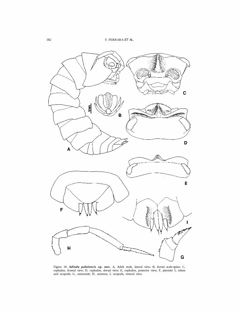
Figure 18. Adinda palniensis sp. nov. A, Adult male, lateral view; B, dorsal scale-spine; C,cephalon, frontal view; D, cephalon, dorsal view; E, cephalon, posterior view; F, pleonite 5, telsonand uropods; G, antennule; H, antenna; I, uropods, ventral view.
383REVISION OF TORADJIINAE
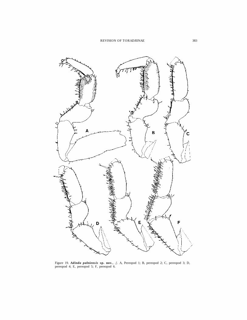
Figure 19. Adinda palniensis sp. nov., {. A, Pereopod 1; B, pereopod 2; C, pereopod 3; D,pereopod 4; E, pereopod 5; F, pereopod 6.
384 F. FERRARA ET AL.
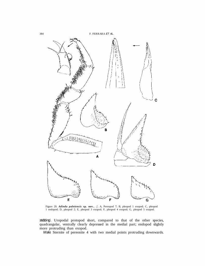
Figure 20. Adinda palniensis sp. nov., {. A, Pereopod 7; B, pleopod 1 exopod; C, pleopod1 endopod; D, pleopod 2; E, pleopod 3 exopod; F, pleopod 4 exopod; G, pleopod 5 exopod.
stebbingi. Uropodal protopod short, compared to that of the other species,quadrangular, ventrally clearly depressed in the medial part; endopod slightlymore protruding than exopod.
Male. Sternite of pereonite 4 with two medial points protruding downwards.

385REVISION OF TORADJIINAE
Pereopods 1–2 carpus with a setose area on the rostral surface close to thesternal margin. Pereopods 1–4 carpus and distal part of merus with a brushof short scales on sternal margin. Pereopods 2–4 ischium with distinct tubercleson sternal margin. Pereopods 5–7 carpus and merus with lines of long spineson sternal margin. Pereopod 7 ischium with lines of spines on the sternalmargin of the distal part and with a triangular lobe at the base, directedproximally. Pleopods 1–5 similar to those of A. stebbingi.
EtymologyThe name of the new species refers to the site of collection of the specimens:
the Palni Hills.
DistributionSouthern India.
RemarksAdinda palniensis is characterized by the dorsal surface of the body with no
granulations, the frontal carina distinctly protruding forwards, short andquadrangular uropodal protopods, the presence of two medial points on thesternite of the male pereonite 4, and the modifications of the male pereopods.
In the almost smooth dorsum it corresponds to A. gigas from which it isreadily distinguished by the colour pattern. It is also similar to A. stebbingi butis distinct from that species essentially by the absence of dorsal granulations,telson relatively larger, uropods larger and shorter, and the different malecharacters, mainly the presence of medial points only on the sternite of thepereonite 4, the absence of distinct modifications on pereopods 1–4, and thepresence of a spur-like lobe directed proximally at the base of the pereopod 7ischium.
Adinda carli sp. nov.
(Figs 21, 22)
Material examinedINDIA: 1 { (Holotype), 1 {, 5 ||, 3 juvs (Paratypes), Lower Palni Hills,
Tandikudi, Cardamon Estate, about 1500 m, under logs, leg. J. Carl, 23.iv.1927,MHNG; 1 {, 1 | (Paratypes), same data, MZUF; 1 { (Paratype), same data,SMNS; 1 {, 2 ||, 2 juvs (Paratypes), Lower Palni Hills, Maryland, Shola,1600 m, leg. J. Carl, 20.iv.1927, MHNG.
DescriptionMaximum length: {, 14 mm; |, 17 mm. Colour (in alcohol) brownish with
several pale spots. Dorsum strongly granulated with more developed granulesin the central part and on the posterior margin of tergites. Eye with 22–23ommatidia. Cephalon with frontal shield bearing a narrow central carina, lessprotruding forwards than lateral lobes; frontal shield separated from vertex bya deep groove bordered by same scales; a supra-antennal line interrupted inthe middle is present. Pereonite 1 with posterior margin slightly sinuous atbase of epimera; posterior corner acute. Telson about twice as wide as long

386 F. FERRARA ET AL.
Figure 21. Adinda carli sp. nov. A, Adult male, lateral view; B, dorsal scale-spine; C, cephalon,frontal view; D, cephalon, dorsal view; E, cephalon, posterior view; F, pleonite 5, telson anduropods; G, antennule; H, antenna.
with apex broadly rounded. Antennule similar to previous species. Antennawith fifth article of peduncle reaching half of pereonite 2; second article offlagellum about twice as long as first. Buccal pieces as in A. stebbingi. Uropodalprotopod subquadrangular, slightly longer than wide, ventrally depressed in themedial part; exopod less and endopod more protruding than protopod apex.
Male. Sternites of pereonites 4 and, to a lesser extent, 5 with two medialpoints protruding downwards. Pereopod 1 carpus with a setose area close to
387REVISION OF TORADJIINAE
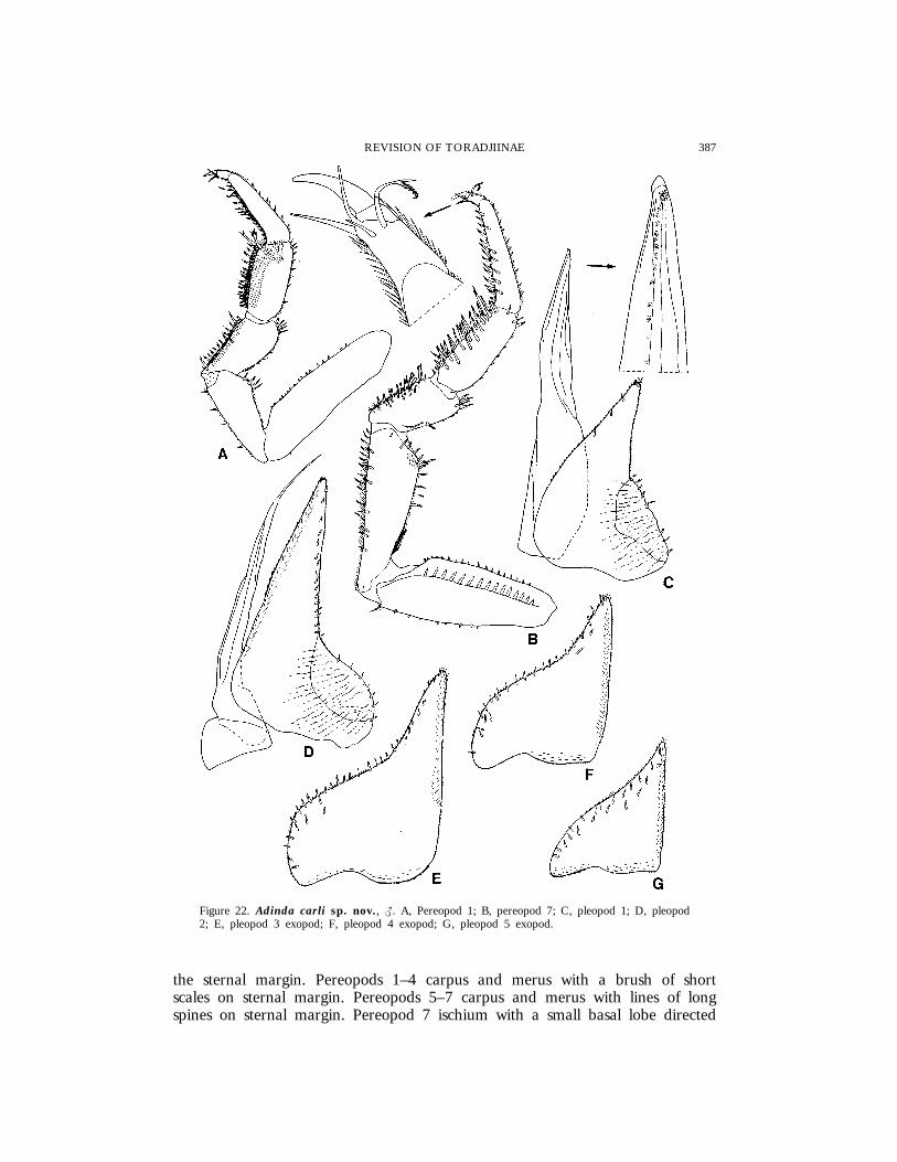
Figure 22. Adinda carli sp. nov., {. A, Pereopod 1; B, pereopod 7; C, pleopod 1; D, pleopod2; E, pleopod 3 exopod; F, pleopod 4 exopod; G, pleopod 5 exopod.
the sternal margin. Pereopods 1–4 carpus and merus with a brush of shortscales on sternal margin. Pereopods 5–7 carpus and merus with lines of longspines on sternal margin. Pereopod 7 ischium with a small basal lobe directed

388 F. FERRARA ET AL.
proximally; merus with a rounded setose lobe on rostral surface. Pleopods 1–5similar to those of A. palniensis.
EtymologyThe new species is named after Prof. J. Carl, Geneva, who collected these
specimens.
DistributionSouthern India.
RemarksThis species is very similar to A. stebbingi and A. palniensis. It is distinguished
from the former by the different shape of the uropod, modifications on theischium and merus of the male anterior pereopods, and the presence of asmall lobe at the base of the ischium and rounded lobe on the merus of themale pereopod 7. It is distinguished from the latter by the presence of dorsalgranulations, longer uropodal protopod, absence of tubercles on the ischium ofthe male pereopods 2–4, and presence of a rounded lobe on the merus of themale pereopod 7.
Adinda nilgiriensis sp. nov.
(Figs 23–25)
Material examinedINDIA: 1 { (Holotype), 6 {{, 8 ||, 7 juvs (Paratypes), Nilgiri Hills,
Avalanche, about 2000 m, forest, under relatively dry logs, leg. J. Carl,18.i.1927, MHNG; 1 {, 1 | (Paratypes), same data, MZUF; 1 { (Paratype),same data, SMNS; 5 {{, 4 || (Paratypes), Nilgiri Hills, jungle below Coonoor,1600 m, under dead leaves and stones on deep soil, leg. J. Carl, 24.xii.1926,MHNG; 1 { (Paratype), same data, MZUF; 1 juv. (Paratype), Nilgiri Hills,Coonoor, Lady Cumings-Seat, about 1700 m, under very damp dead leaves,leg. J. Carl, 29.xii.1926, MHNG; 1 { (Paratype), Nilgiri Hills, Dodabetta,reserved forest, 2400 m, Ootacamund Region, leg. J. Carl, 11.i.1927, MHNG;2 {{, 2 ||, 1 juv. (Paratypes), Nilgiri Hills, Old Street of Nilgiri, 1–4 mibelow Coonoor, 1600 m, under dead leaves and ground, leg. J. Carl, 4.i.1927,MHNG; 1 | (Paratype), Nilgiri Hills, S of Coonoor, forest, 1600 m, underdead leaves, leg. J. Carl, 21–24.xii.1936, MHNG; 1 | (Paratype), Nilgiri Hills,Coonoor, forest, under leaves, 1800 m, leg. J. Carl, 17.xii.1926, MHNG.
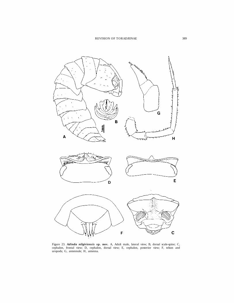
DescriptionMaximum length: {, 18 mm; |, 19 mm. Colour (in alcohol) brownish with
several small pale spots; antennae with distal part of the fifth article of peduncleand first article of flagellum pale. Dorsum slightly granulated, with several smallpetaliform scale-spines. Eye with about 21 ommatidia. Cephalon with frontalshield bearing a narrow carina protruding forwards and upwards; lateral lobestriangular, directed forwards, separated from vertex by a deep groove with arow of scales; a supra-antennal line interrupted in the middle is present.Pereonite 1 with posterior margin slightly sinuous at base of epimera, posterior
389REVISION OF TORADJIINAE
Figure 23. Adinda nilgiriensis sp. nov. A, Adult male, lateral view; B, dorsal scale-spine; C,cephalon, frontal view; D, cephalon, dorsal view; E, cephalon, posterior view; F, telson anduropods; G, antennule; H, antenna.
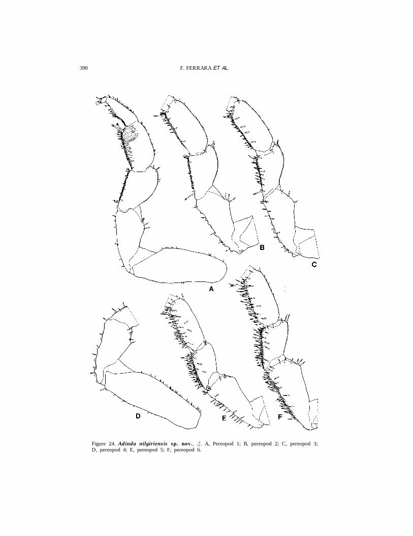
390 F. FERRARA ET AL.
Figure 24. Adinda nilgiriensis sp. nov., {. A, Pereopod 1; B, pereopod 2; C, pereopod 3;D, pereopod 4; E, pereopod 5; F, pereopod 6.

391REVISION OF TORADJIINAE
Figure 25. Adinda nilgiriensis sp. nov., {. A, Pereopod 7; B, pleopod 1; C, pleopod 2; D,pleopod 3 exopod; E, pleopod 4 exopod; F, pleopod 5 exopod.
392 F. FERRARA ET AL.
corner acute with rounded apex. Telson twice as wide as long, broadly rounded.Antennule as in the previous species. Antenna with fifth article of pedunclereaching the posterior margin of pereonite 1; second article of flagellum abouttwice as long as first. Buccal pieces as in A. stebbingi. Uropodal protopod wideand ventrally depressed in the medial part; exopod slightly less and endopodmore protruding in relation to protopod apex.
Male. Sternites of pereonites 2–4 with two small points and pereonite 5 withtwo well-developed ones, directed downwards. Pereopods very similar to thoseof A. palniensis, except pereopod 7 which presents ischium with basal lobeperpendicular to sternal margin and merus with a protruding rounded lobe ontergal margin. Pleopods 1–5 as in A. palniensis.
EtymologyThe name refers to the Nilgiri Hills, where the specimens were collected.
DistributionSouthern India.
RemarksThis species, because of the presence of a spur-like lobe on the proximal
part of the ischium of the male pereopod 7, is similar to A. palniensis and A.carli. It is distinguished from the former in having a slight dorsal granulation,frontal carina less developed, sternal spines on the male pereonites 2–5 insteadof only on pereonite 4, absence of a setose area on the male pereopod 2carpus, and the different orientation of the basal lobe of the pereopod 7ischium. It is distinguished from the latter species by the posterior corner ofpereopod 1 protruding further backwards, shorter uropodal protopod, presenceof tubercles on the male pereopods 2–4 ischium and the larger and differentlyoriented basal lobe of the male pereopod 7 ischium.
Adinda lobata sp. nov.
(Figs 26–28)
Material examinedINDIA: 1 { (Holotype), 1 | (Paratype), Anamalai Hills, Valparai, Naduar
Estate, about 1200 m, leg. J. Carl, iii.1927, MHNG.
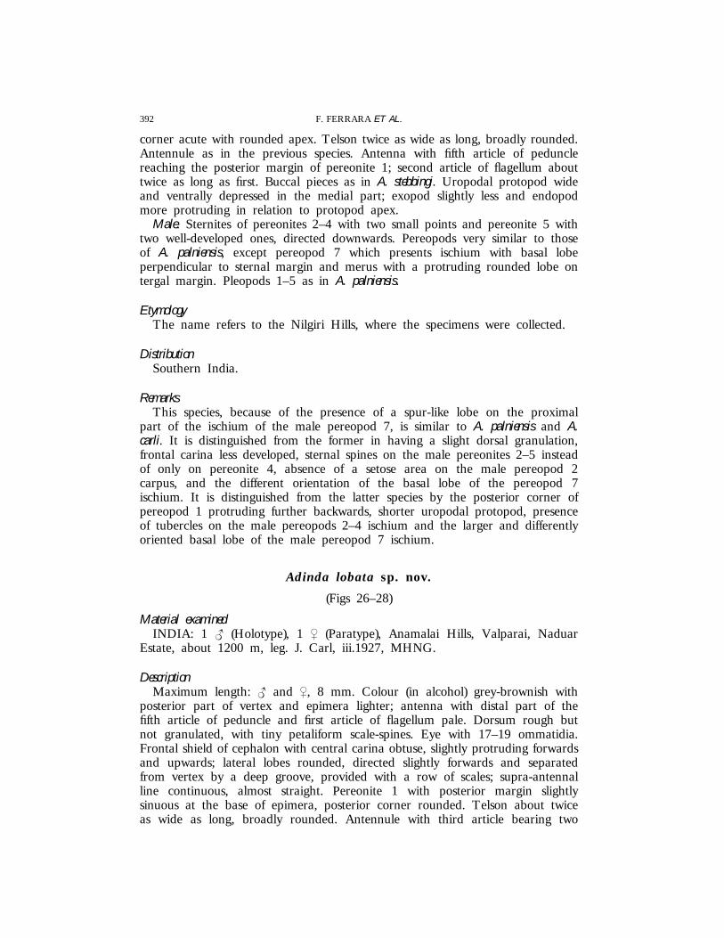
DescriptionMaximum length: { and |, 8 mm. Colour (in alcohol) grey-brownish with
posterior part of vertex and epimera lighter; antenna with distal part of thefifth article of peduncle and first article of flagellum pale. Dorsum rough butnot granulated, with tiny petaliform scale-spines. Eye with 17–19 ommatidia.Frontal shield of cephalon with central carina obtuse, slightly protruding forwardsand upwards; lateral lobes rounded, directed slightly forwards and separatedfrom vertex by a deep groove, provided with a row of scales; supra-antennalline continuous, almost straight. Pereonite 1 with posterior margin slightlysinuous at the base of epimera, posterior corner rounded. Telson about twiceas wide as long, broadly rounded. Antennule with third article bearing two
393REVISION OF TORADJIINAE
Figure 26. Adinda lobata sp. nov. A, Adult female, lateral view; B, dorsal scale-spine; C,cephalon, frontal view; D, cephalon, dorsal view; E, cephalon, posterior view; F, pleonite 5, telsonand uropods; G, antennule; H, antenna.
rows of aesthetascs on medial margin and two aesthetascs at apex. Antennawith fifth article of peduncle reaching posterior margin of pereonite 1; secondarticle of flagellum about 1.5 times as long as first. Buccal pieces as in A.stebbingi, but the penicils of mandibles are 2 + 1 in the right one and 2 + 2 inthe left one. Uropodal protopod with the distal part narrower than in all theprevious species; stout exopod and endopod, distinctly protruding in relation toprotopod tips.
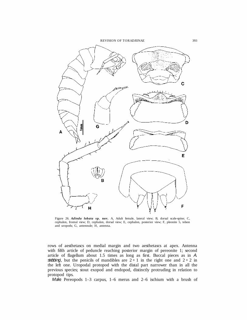
Male. Pereopods 1–3 carpus, 1–6 merus and 2–6 ischium with a brush of
394 F. FERRARA ET AL.
Figure 27. Adinda lobata sp. nov., {. A, Pereopod 1; B, pereopod 2; C, pereopod 6; D,pereopod 7.
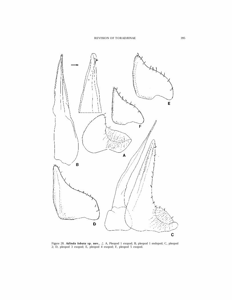
395REVISION OF TORADJIINAE
Figure 28. Adinda lobata sp. nov., {. A, Pleopod 1 exopod; B, pleopod 1 endopod; C, pleopod2; D, pleopod 3 exopod; E, pleopod 4 exopod; F, pleopod 5 exopod.
396 F. FERRARA ET AL.
small scales on sternal margin. Pereopod 7 ischium distally enlarged to form arounded lobe, with 2 strong spines, sternal margin straight; merus slightlyswollen on sternal margin. Pleopod 1 exopod wider than long, with short androunded posterior point; endopod straight with rounded apex. Pleopod 2 andpleopods 3–5 exopod as in Fig. 28C–F.
EtymologyThe name refers to the male pereopod 7 ischium which is distally enlarged
in the form of a round lobe.
DistributionSouthern India.
RemarksAdinda lobata is readily distinguished from the other species by the uropodal
protopods distally narrower and exopods stouter, the male pereopod 7 ischiumclearly enlarged, and the male pleopod 1 exopod with a short rounded posteriorpoint.
Adinda triangulifera sp. nov.
(Fig 29)
Material examinedINDIA: 1 | (Holotype), 2 || (Paratypes), Anamalai Hills, Attakatti, Shola,
Ibex-Hill, 1200 m, leg. J. Carl, 26.ii.1927, MHNG; 1 | (Paratype), same data,MZUF.
DescriptionMaximum length: 18 mm. Colour (in alcohol) yellow-brownish; articles 1–3
and distal part of fifth article of antennal peduncle pale. Dorsum stronglygranulated and covered by several small petaliform scale-spines. Eye with about20 ommatidia. Cephalon with frontal shield bearing an acute central carinaprotruding forwards and upwards; lateral lobes triangular, directed forwards,separated from vertex by a deep groove bordered with scales; supra-antennalline interrupted in the middle. Pereonite 1 with posterior margin slightly sinuous,posterior corner with rounded apex. Telson about twice as wide as long,triangular, with slightly concave sides and rounded apex. Antennule with thirdarticle provided with four rows of aesthetascs on medial margin and twoaesthetascs at apex. Antenna with fifth article of peduncle reaching posteriormargin of pereonite 1; second article of flagellum twice as long as the first.Buccal pieces as in A. stebbingi, except mandibles which have 3+1 penicils onthe right and 3 + 2 on the left. Uropodal protopod subquadrangular; exopodand endopod at the same level, not protruding in relation to the posteriormargin of protopod.

397REVISION OF TORADJIINAE
Figure 29. Adinda triangulifera sp. nov. A, Adult female, lateral view; B, dorsal scale-spine;C, cephalon, frontal view; D, cephalon, dorsal view; E, cephalon, posterior view; F, pleon andtelson; G, pleonite 5, telson and uropods.
EtymologyThe name refers to the triangular shape of the telson.
DistributionSouthern India.
398 F. FERRARA ET AL.
RemarksEven if no male specimens have been examined, this species deserves to be
described due to its peculiar telson shape (triangular with concave sides) whichimmediately distinguishes it from all the other species in the group. In themarked dorsal granulations, A. triangulifera resembles A. stebbingi, A. travancorensis,A. carli and A. scabra; however, it is readily distinguished form those species,besides its telson shape, also by the frontal carina more acute and protruding,and the uropodal protopod subquadrangular.
Adinda scabra (Collinge 1916)
(Figs 2B, 30–32)
Paraperiscyphisscabrus Collinge, 1916: 117, pl. IX figs 6–10.Adindascabrus ; Arcangeli, 1948: 34.necAdinda scabrus ; Jackson, 1936: 84.
Material examinedSRI LANKA: 2 ||, Kandy, Udawattakele Sanctuary, 2100 ft, D. R. Davis
& W. H. Rowe, 10–23.i.1970, USNM; 1 {, 1 |, Kandy, leg. C. Besuchet &I. Lobl, 14.ii.1970, MHNG; 1 {, same locality and collectors, 22.i.1970,MHNG; 1 {, 2 ||, same locality, forest, under stones, leg. O. Low-Beer,21.ii.1912, SMF; 1 {, Haputale, leg. C. Besuchet & I. Lobl, 23.vii.1970,MHNG; 1 {, Central Prov., Horton Plains, 11 mi SSE of Nuwara-Eliya, 6700ft, indigenous forest slope, leg. Lund University Ceylon Expedition, 19–20.iii.1962, ZIUL; 1 |, 1 juv., same data, ZIUL; 1 |, same locality, leg. C.Besuchet & I. Lobl, 15.ii.1970, MHNG; 1 {, Sabaragamuwa Prov., Deerwood,Kuruwita, 6 mi NNW of Ratnapura, leg. Lund University Ceylon Expedition,18–21.ii.1962, ZIUL; 1 |, 1 juv., Kuruwita, leg. P. Beron, 1.xii.1984, NNHMS;1 |, Hatton, leg. C. Besuchet & I. Lobl, 9.xi.1970, MHNG; 1 {, WesternProv., Yongammulla, 3 mi E Yakkala, 18 mi NE of Colombo, leg. LundUniversity Ceylon Expedition, 19.i.1962, ZIUL; 3 {{, 1 |, same data, MZUF;1 {, 2 ||, 5 juvs, same locality, leg. Lund University Ceylon Expedition, 24.i–6.iii.1962, ZIUL; 1 |, 2 juvs, Peradeniya, leg. C. Besuchet & I. Lobl, 19.i.1970,MHNG.
PreviousrecordsSri Lanka: Peradeniya (Collinge, 1916).
DescriptionMaximum length: {, 12 mm; |, 18 mm. Colour (in alcohol) grey-brownish
with yellowish spots disposed as follows: pereonites with two paramedian spotsclose to the anterior margin and a median one, an oblique one at the baseof epimera; pleonites 3–5 with one spot at the base of epimera; anterior andposterior corners of pereonites and pleonites pale, telson with two paramedianspots; antenna with distal part of articles 3–5 of peduncle pale. Dorsumdistinctly granulated, each granulation bearing a petaliform scale-spine. Eyewith about 23 ommatidia. Cephalon with frontal shield provided with an obtusemedian carina slightly protruding forwards and upwards, and separated from
399REVISION OF TORADJIINAE
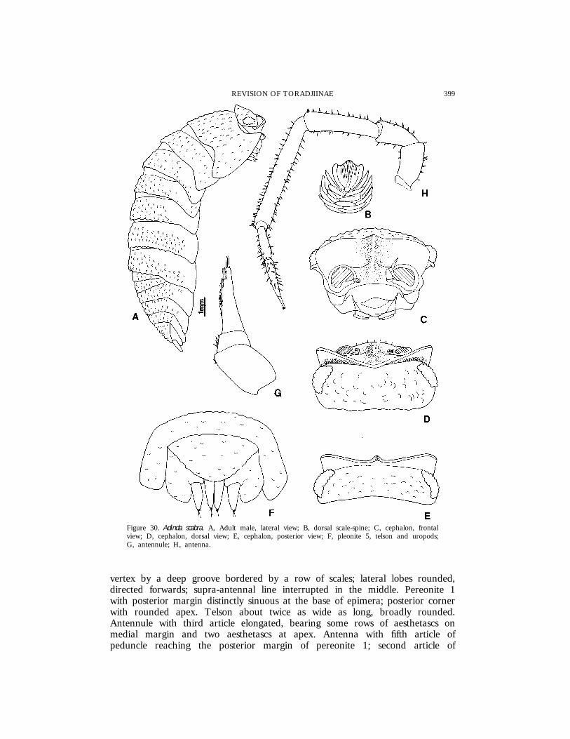
Figure 30. Adinda scabra. A, Adult male, lateral view; B, dorsal scale-spine; C, cephalon, frontalview; D, cephalon, dorsal view; E, cephalon, posterior view; F, pleonite 5, telson and uropods;G, antennule; H, antenna.
vertex by a deep groove bordered by a row of scales; lateral lobes rounded,directed forwards; supra-antennal line interrupted in the middle. Pereonite 1with posterior margin distinctly sinuous at the base of epimera; posterior cornerwith rounded apex. Telson about twice as wide as long, broadly rounded.Antennule with third article elongated, bearing some rows of aesthetascs onmedial margin and two aesthetascs at apex. Antenna with fifth article ofpeduncle reaching the posterior margin of pereonite 1; second article of
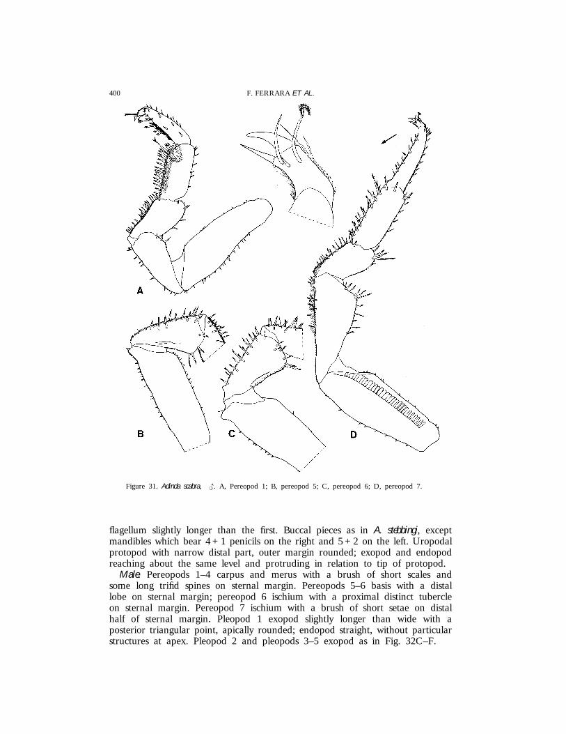
400 F. FERRARA ET AL.
Figure 31. Adinda scabra, {. A, Pereopod 1; B, pereopod 5; C, pereopod 6; D, pereopod 7.
flagellum slightly longer than the first. Buccal pieces as in A. stebbingi, exceptmandibles which bear 4 + 1 penicils on the right and 5 + 2 on the left. Uropodalprotopod with narrow distal part, outer margin rounded; exopod and endopodreaching about the same level and protruding in relation to tip of protopod.
Male. Pereopods 1–4 carpus and merus with a brush of short scales andsome long trifid spines on sternal margin. Pereopods 5–6 basis with a distallobe on sternal margin; pereopod 6 ischium with a proximal distinct tubercleon sternal margin. Pereopod 7 ischium with a brush of short setae on distalhalf of sternal margin. Pleopod 1 exopod slightly longer than wide with aposterior triangular point, apically rounded; endopod straight, without particularstructures at apex. Pleopod 2 and pleopods 3–5 exopod as in Fig. 32C–F.

401REVISION OF TORADJIINAE
Figure 32. Adinda scabra, {. A, Pleopod 1 exopod; B, pleopod 1 endopod; C, pleopod 2; D,pleopod 3 exopod; E, pleopod 4 exopod; F, pleopod 5 exopod.
DistributionSri Lanka.
RemarksThis species is essentially characterized by the dorsal surface distinctly
granulated, uropodal protopod with a narrow distal part, presence of a distalprocess on the male pereopods 5 and 6 basis and a tubercle on pereopod 6merus, presence of a brush of setae in the distal half of the sternal margin ofthe male pereopod 7 ischium.
Jackson (1936) tentatively ascribed some specimens from Borneo (Sabah, from
402 F. FERRARA ET AL.
Kiau to Tenompak) to A. scabra, but this record certainly refers to anotherspecies.
Adinda pulchra (Collinge, 1916)
(Figs 33, 34)
Paraperiscyphispulcher Collinge, 1916: 116, pl. IX figs 1–5.Adindapulcher ; Arcangeli, 1948; 34.
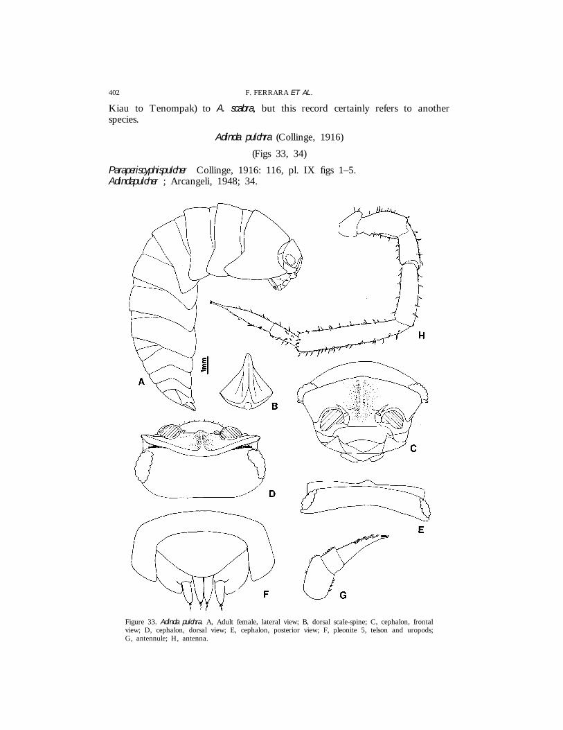
Figure 33. Adinda pulchra. A, Adult female, lateral view; B, dorsal scale-spine; C, cephalon, frontalview; D, cephalon, dorsal view; E, cephalon, posterior view; F, pleonite 5, telson and uropods;G, antennule; H, antenna.

403REVISION OF TORADJIINAE
Figure 34. Adinda pulchra, {. A, Pereopod 1; B, pereopod 7; C, pleopod 1 exopod; D, pleopod 1endopod; E, pleopod 2; F, pleopod 3 exopod; G, pleopod 4 exopod; H, pleopod 5 exopod.
404 F. FERRARA ET AL.
Material examinedSRI LANKA: 1 |, Kuruwita, leg. P. Beron, 1.xii.1984, NNHMS; 2 juvs,
Central Prov., Ramboda, 7 mi NW of Nuwara-Eliya, leg. Lund UniversityCeylon Expedition, 4.iii.1962, ZIUL; 1 {, same data, MZUF; 1 {, 1 juv.,Kegalla, leg. C. Besuchet & I. Lobl, 14.i.1970, MHNG.
PreviousrecordsSri Lanka: Peradeniya (Collinge, 1916).
DescriptionMaximum length: {, 12 mm; |, 17 mm. Colour (in alcohol) brownish with
several yellowish spots; antenna as in A. scabra. Dorsum rough not granulated,with several small triangular scale-spines. Eye with about 22 ommatidia.Cephalon with frontal shield bent over vertex in the median part, bearing amedial carina slightly protruding forwards; lateral lobes rounded and slightlydirected forwards, separated from vertex by a deep groove provided with arow of scales; supra-antennal line visible only at sides. Pereonite 1 with posteriormargin distinctly sinuous at the base of epimera; posterior corner acute withrounded apex. Telson broadly rounded, about twice as wide as long. Antennulewith third article provided with four rows of aesthetascs on medial margin andtwo aesthetascs at apex. Antenna with fifth article of peduncle reaching posteriormargin of pereonite 1; second article of flagellum about 1.5 times as long asfirst. Buccal pieces as in A. stebbingi, except mandibles with 4 + 1 penicils onthe right and 3 or 4 + 2 on the left. Uropodal protopod with rounded posteriormargin; stout exopod and endopod, distinctly protruding in relation to protopodtip.
Male. Pereopods 1–3 carpus, merus, ischium and pereopods 4–6 ischium witha brush of small scales on sternal margin. Pereopod 7 ischium with sternalmargin almost straight, bearing a brush of long setae in the distal half, meruswith a brush of setae on sternal margin. Pleopod 1 exopod slightly wider thanlong, posterior point rounded; endopod with no particular modifications. Pleopod2 and pleopods 3–5 exopod as in Fig. 34E–H.
DistributionSri Lanka.
RemarksAdinda pulchra is very similar to A. scabra from which it is distinguished by
the absence of dorsal granulations, frontal carina less protruding forwards, andabsence of distinct sexually dimorphic modifications on the male pereopods 5–6.
Riedeli group
Adinda riedeli sp. nov.
(Figs 35–38)
Material examinedMALAYSIA: 1 { (Holotype), 2 || (Paratypes), Sarawak, Belaga District,
Long Linau, leg. A. Riedel, 17–21.iii.1990, SMNS; 1 |, 3 juvs (Paratypes),
405REVISION OF TORADJIINAE
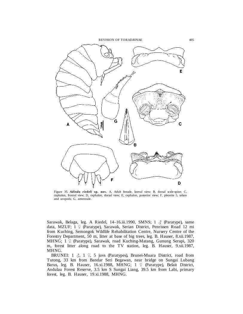
Figure 35. Adinda riedeli sp. nov. A, Adult female, lateral view; B, dorsal scale-spine; C,cephalon, frontal view; D, cephalon, dorsal view; E, cephalon, posterior view; F, pleonite 5, telsonand uropods; G, antennule.
Sarawak, Belaga, leg. A Riedel, 14–16.iii.1990, SMNS; 1 { (Paratype), samedata, MZUF; 1 | (Paratype), Sarawak, Serian District, Penrissen Road 12 mifrom Kuching, Semongok Wildlife Rehabilitation Centre, Nursery Centre of theForestry Department, 50 m, litter at base of big trees, leg. B. Hauser, 8.xii.1987,MHNG; 1 | (Paratype), Sarawak, road Kuching-Matang, Gunung Serapi, 320m, forest litter along road to the TV station, leg. B. Hauser, 9.xii.1987,MHNG.
BRUNEI: 1 {, 1 |, 5 juvs (Paratypes), Brunei-Muara District, road fromTutong, 33 km from Bandar Seri Begawan, near bridge on Sungai LubangBarus, leg. B. Hauser, 16.xi.1988, MHNG; 1 | (Paratype), Belait District,Andulau Forest Reserve, 3.5 km S Sungai Liang, 39.5 km from Labi, primaryforest, leg. B. Hauser, 19.xi.1988, MHNG.

406 F. FERRARA ET AL.
Figure 36. Adinda riedeli sp. nov. A, Right mandible; B, left mandible; C, maxillule; D,maxilliped.
DescriptionMaximum length: {, 9 mm; |, 11 mm. Very convex body with almost
vertical epimera. Colour (in alcohol) grey-brownish with some irregular yellowishspots; antenna with distal part of the fifth article of peduncle and flagellumpale. Dorsum rough and pruinose due to the presence of many long triangularscale-spines. Eye with about 25 ommatidia. Cephalon with frontal shieldprotruding over vertex, median carina distinctly protruding with a depressionin the upper part; lateral lobes triangular, directed forwards; frontal shieldseparated from vertex by a deep groove, wider at sides. Pereonite 1 withposterior margin slightly sinuous at the base of epimera; posterior corner

407REVISION OF TORADJIINAE
Figure 37. Adinda riedeli sp. nov., {. A, Antenna; B, pereopod 1; C, pereopod 7.
rounded. Telson with posterior margin subtruncated, twice as wide as long.Antennule with third article bearing five rows of aesthetascs on medial marginand two aesthetascs at apex. Stout antenna reaching posterior margin ofpereonite 2; second article of flagellum about twice as long as first. Mandibleswith molar penicil simple, 1 + 1 penicils on the right and 1 + 2 on the left.Maxillule outer branch with five out of 10 teeth cleft; inner branch withtwo long penicils. Maxilliped endite setose with penicil. Uropodal protopodsubquadrangular; exopod and endopod equally protruding in relation to protopodtip.
Male. Anterior pereopods with no particular modifications, only pereopods 1–4carpus with small scales on sternal margin. Pereopod 7 ischium with straightsternal margin and a relief covered with setae in the distal part of the rostralsurface; merus with a distinct conical lobe covered with short setae. Pleopod 1exopod longer than wide, with a long acute posterior point; endopod stout, withtuft of setae at apex. Pleopod 2 and pleopods 3–5 exopod as in Fig. 38B–E.
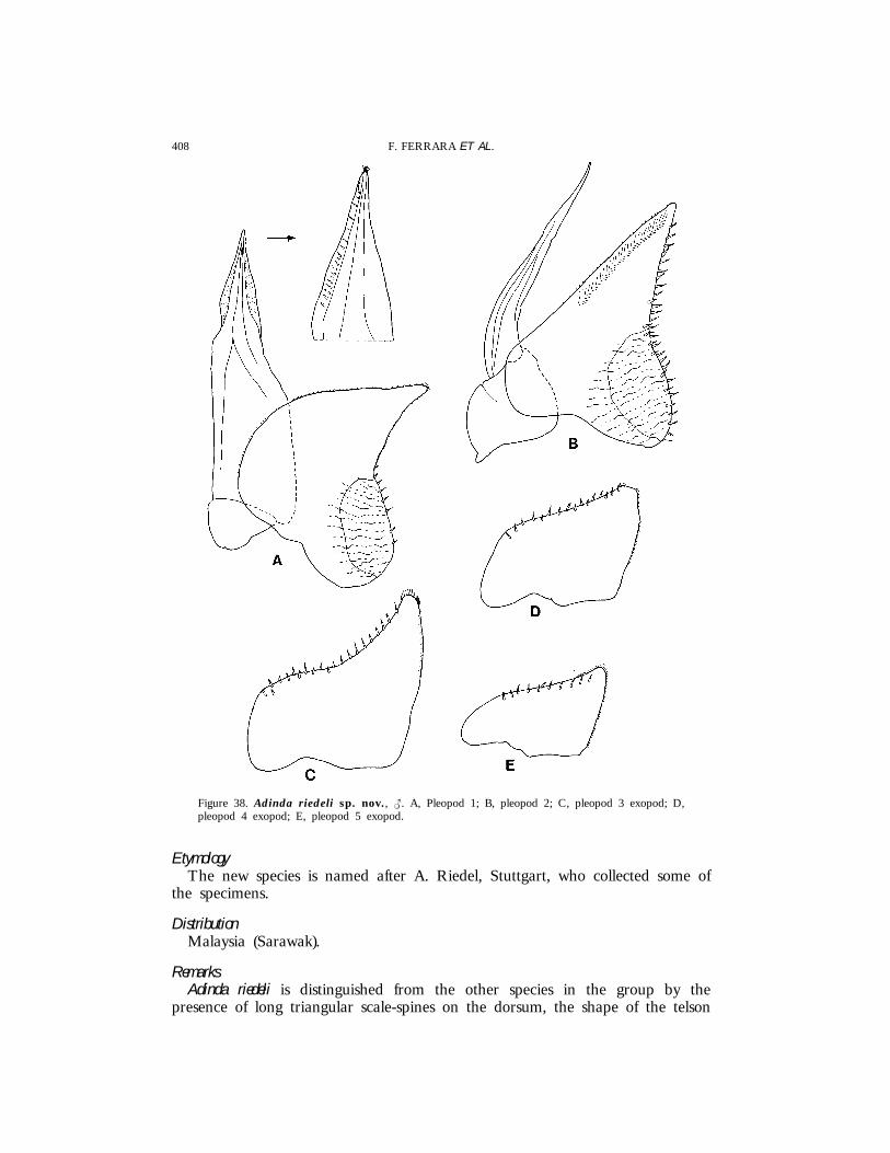
408 F. FERRARA ET AL.
Figure 38. Adinda riedeli sp. nov., {. A, Pleopod 1; B, pleopod 2; C, pleopod 3 exopod; D,pleopod 4 exopod; E, pleopod 5 exopod.
EtymologyThe new species is named after A. Riedel, Stuttgart, who collected some of
the specimens.
DistributionMalaysia (Sarawak).
RemarksAdinda riedeli is distinguished from the other species in the group by the
presence of long triangular scale-spines on the dorsum, the shape of the telson
409REVISION OF TORADJIINAE
and uropods, and by the presence of a distinct conical lobe on the malepereopod 7 ischium.
Adinda platyperaeon (Schultz, 1982)
(Figs 39–42)
Paraperiscyphisplatyperaeon Schultz, 1982: 104, figs 3–5.
Typematerial examinedMALAYSIA: 1 { (Holotype), Sarawak, Gunung Mulu National Park, Gua
Air Jernih (Clearwater Cave), BM 1981 : 332 : 1; 1 | (Allotype), same data, BM1981 : 333 : 1; 3 {{, 1 | (Paratypes), same data, BM 1981 : 334 : 15.
Material examinedMALAYSIA: 1 {, 2 ||, Sarawak, Gunung Mulu National Park, Gua Ajaib
(Wonder Cave), BM 1981 : 335 : 3; 3 {{, 1 |, Sarawak, Gunung Mulu NationalPark, Lubang Angin, Lubang Hijau, Lubang Ular (Cave of the Winds, GreenCave, Snake Cave), BM 1981 : 336 : 7; 2 {{, Sarawak, Gunung Mulu NationalPark, Windsor Cave, Clearwater River Cave, BM 1981:337:2 (specimensidentified by Schultz, 1982).
PreviousrecordsMalaysia: Sarawak, Gunung Mulu National Park: Gua Air Jernih, Lubang
Angin, Lubang Hijau, Lubang Ular, Windsor Cave, Clearwater River Cave,Gua Ajaib (Schultz, 1982).
DescriptionMaximum length: {, 21.5 mm; |, 27 mm. Body enlarged and flattened,
convex only in the median part, epimera directed obliquely outwards. Antennaeand pereopods frail and long. Colour brown-yellowish, antenna with distal partof the fifth article of peduncle and flagellum pale. Dorsum smooth with smallsetae. Eye with about 12 ommatidia, about 20 in the specimens from GuaAjaib. Cephalon with frontal shield bearing a median carina distinctly protrudingforwards and upwards; lateral lobes triangular, directed forwards and separatedfrom vertex by a deep groove. Pereonite 1 with posterior margin straight andposterior corner at right angle. Telson about 1.5 times as wide as long, broadlyrounded. Antennule with third article bearing five superimposed rows ofaesthetascs and two aesthetascs at apex. Antenna reaching the posterior marginof pereonite 5; fifth article of peduncle about twice as long as flagellum;flagellar articles subequal. Buccal pieces as in A. riedeli. Uropodal protopod withouter margin rounded; exopod and endopod slightly protruding in relation toprotopod tip.
Male. Pereopods 1–4 carpus and merus with lines of long trifid spines anda brush of short scales on sternal margin. Pereopod 7 ischium with sternalmargin concave and provided with a distal setose lobe on rostral surface; meruswith a brush of setae on basal part of sternal margin and a small setose lobeon rostral surface. Pleopod 1 exopod with a long triangular posterior point
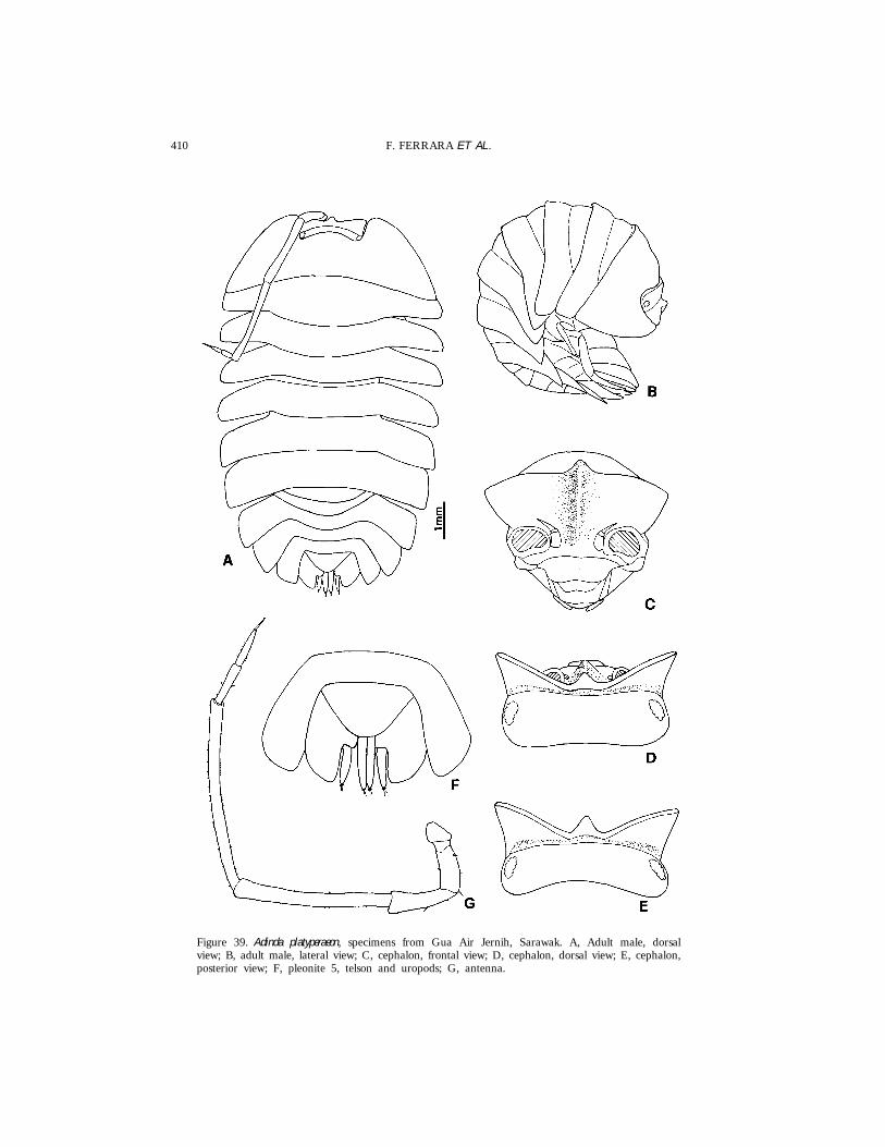
410 F. FERRARA ET AL.
Figure 39. Adinda platyperaeon, specimens from Gua Air Jernih, Sarawak. A, Adult male, dorsalview; B, adult male, lateral view; C, cephalon, frontal view; D, cephalon, dorsal view; E, cephalon,posterior view; F, pleonite 5, telson and uropods; G, antenna.
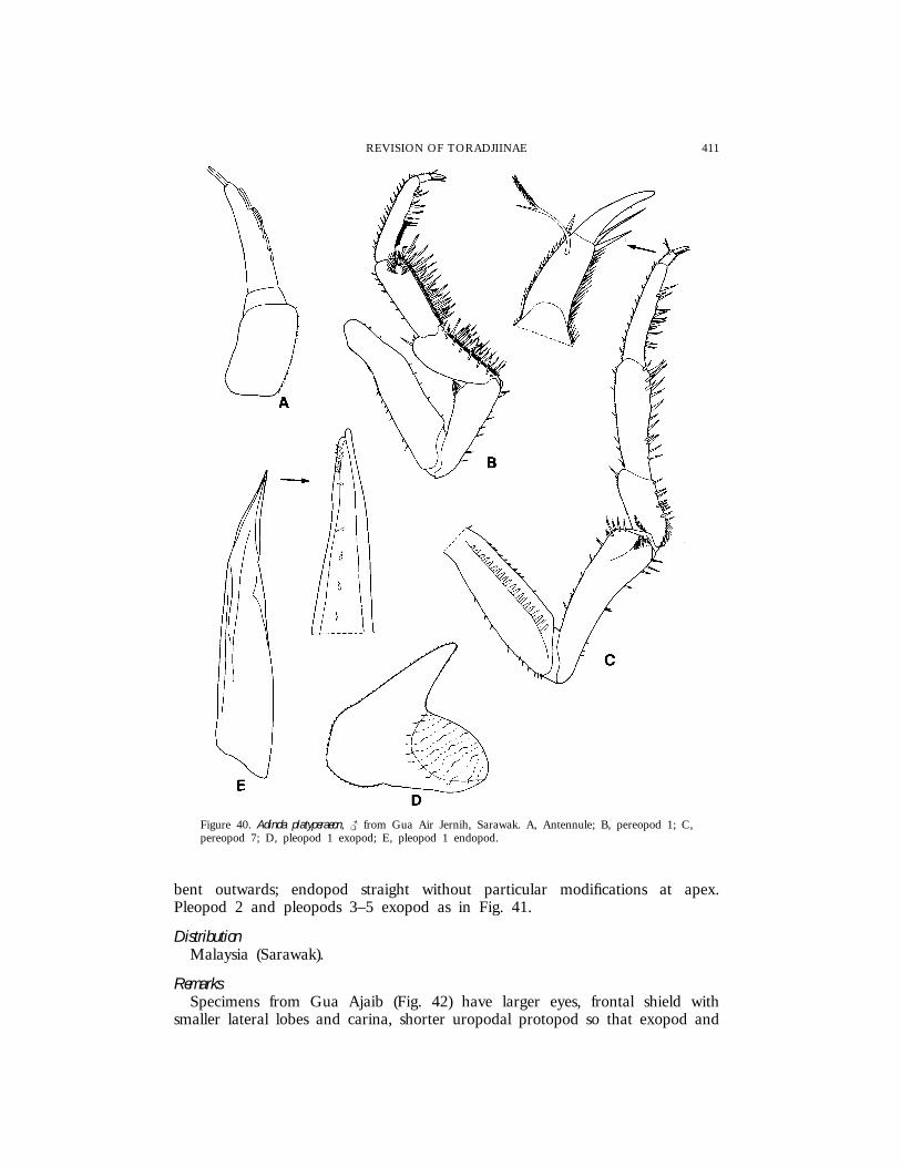
411REVISION OF TORADJIINAE
Figure 40. Adinda platyperaeon, { from Gua Air Jernih, Sarawak. A, Antennule; B, pereopod 1; C,pereopod 7; D, pleopod 1 exopod; E, pleopod 1 endopod.
bent outwards; endopod straight without particular modifications at apex.Pleopod 2 and pleopods 3–5 exopod as in Fig. 41.
DistributionMalaysia (Sarawak).
RemarksSpecimens from Gua Ajaib (Fig. 42) have larger eyes, frontal shield with
smaller lateral lobes and carina, shorter uropodal protopod so that exopod and
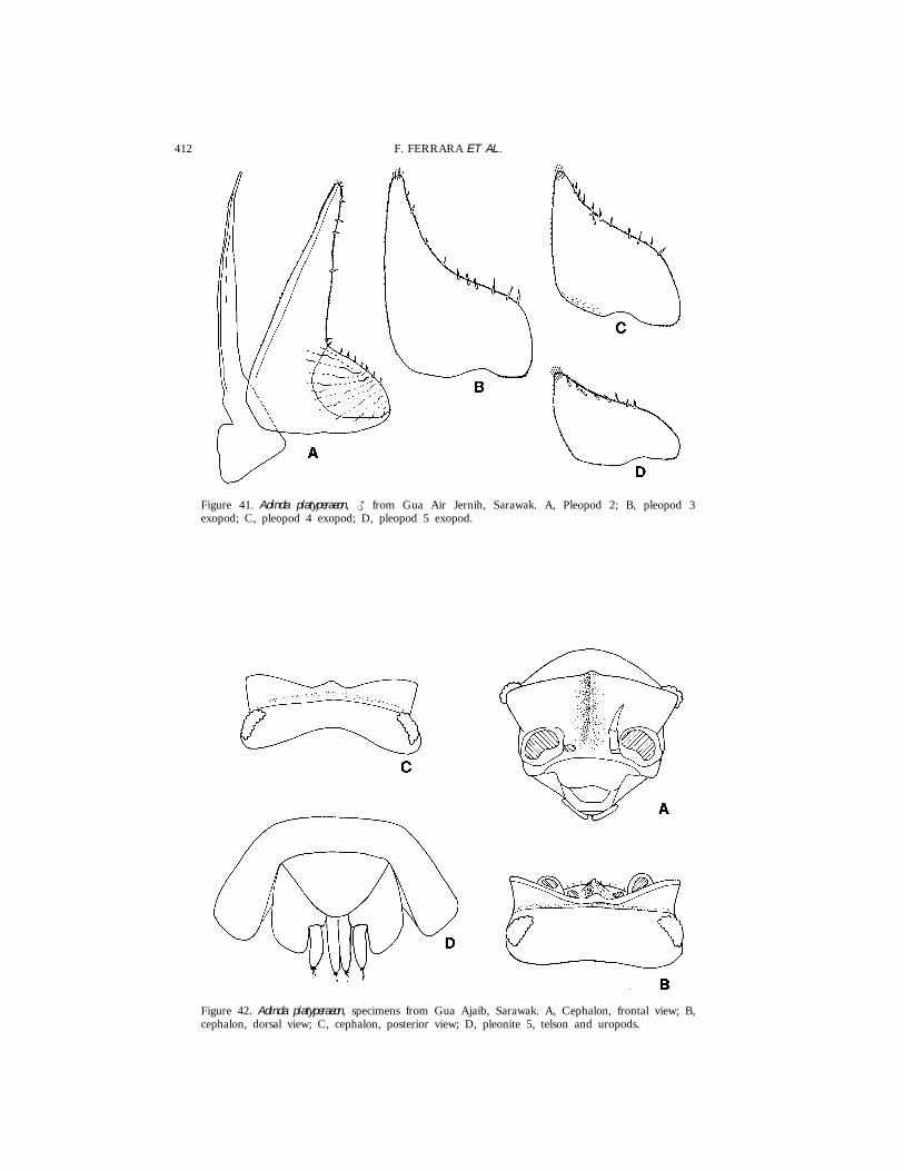
412 F. FERRARA ET AL.
Figure 41. Adinda platyperaeon, { from Gua Air Jernih, Sarawak. A, Pleopod 2; B, pleopod 3exopod; C, pleopod 4 exopod; D, pleopod 5 exopod.
Figure 42. Adinda platyperaeon, specimens from Gua Ajaib, Sarawak. A, Cephalon, frontal view; B,cephalon, dorsal view; C, cephalon, posterior view; D, pleonite 5, telson and uropods.
413REVISION OF TORADJIINAE
endopod distinctly protrude backwards. For all the other characters, includingthe male modifications, these specimens correspond to the types. Though itcannot be excluded that they belong to a distinct taxon, we agree with Schultz(1982) that these differences are too small to justify specific separation.
Adinda platyperaeon is readily distinguished from the other species in the groupby the enlarged and flat body, the elongated antennae and pereopods, and thestructure of the male pereopod 7.
Adinda lamellata sp. nov.
(Figs 43, 44)
Material examinedBRUNEI: 1 { (Holotype), 1 {, 1 { juv., 4 ||, 35 juvs (Paratypes), Brunei-
Muara District, Bandar Seri Begawan, leg. B. Hauser, 15.xi.1988, MHNG; 1{, 1 | (Paratypes), same data, MZUF; 2 {{, 2 {{ juvs, 2 || juvs (Paratypes),Brunei-Muara District, road from Tutong, 33 km from Bander Seri Begawan,near bridge on Sungai Lubang Barus, leg. C. Lienhard, 16.xi.1988, MHNG; 1{, 3 || juvs (Paratypes), Brunei-Maura District, road from Tutong, 33 kmfrom Bandar Seri Begawan, near bridge on Sungai Lubang Barus, leg. B.Hauser, 16.xi.1988, MHNG; 1 {, 2 juvs (Paratypes), Brunei-Muara District,Berakas Forest Reserve, N Bandar Seri Begawan, 19.5 km from Muara, tropicalheath forest, leg. C. Lienhard, 20.xi.1988, MHNG.
MALAYSIA: 1 { juv., Sarawak, Bau, environs Gua Puang near KampongPelaman Sekiang, escarpment of Gunung Jambusan, 10–30 m, leg. C. Lienhard,4.xii.1987, MHNG.
PHILIPPINES: 1 |, 2 juvs, Palawan, S. Lucia, Puerto Princesa, leg. G.Osella, 5.ii.1981, MZUF; 1 |, Palawan, Mantalingajan, Tagerubung, 1150 m,leg. Noona Dan Expedition, 19.ix.1961, ZMC; 1 |, Balabac, Dalawan Bay, inMalaise-traps, leg. Noona Dan Expedition, 6.x.1961, ZMC.
DescriptionMaximum length: {, 9 mm; |, 11 mm. Colour (in alcohol) yellowish with
many small brown spots; antenna with articles 1–3 of peduncle pale. Dorsumslightly rough with many small triangular scale-spines. Eye with about 24ommatidia. Cephalon with shield bearing an obtuse median carina protrudingupwards but protruding much less forwards in relation to the lateral lobes;frontal shield separated from vertex by a very narrow groove, more apparentat the sides; supra-antennal line interrupted in the middle. Pereonite 1 withposterior margin distinctly sinuous at the base of epimera; posterior cornerrounded. Pereonite 2 with a transverse depression on anterior margin ofepimera. Telson semicircular, twice as wide as long. Antennule with third articleapically pointed, bearing five superimposed rows of aesthetascs. Short antennareaching rear margin of pereonite 2; fifth article of peduncle about 1.5 timeslonger than flagellum; second flagellar article about twice as long as first. Buccalpieces as in A. riedeli. Uropod with quadrangular protopod; exopod and endopodprotruding in relation to protopod tip.
Male. Pereopods 1–4 carpus and merus with lines of long trifid spines anda brush of short scales. Pereopod 7 ischium with sternal margin distinctly
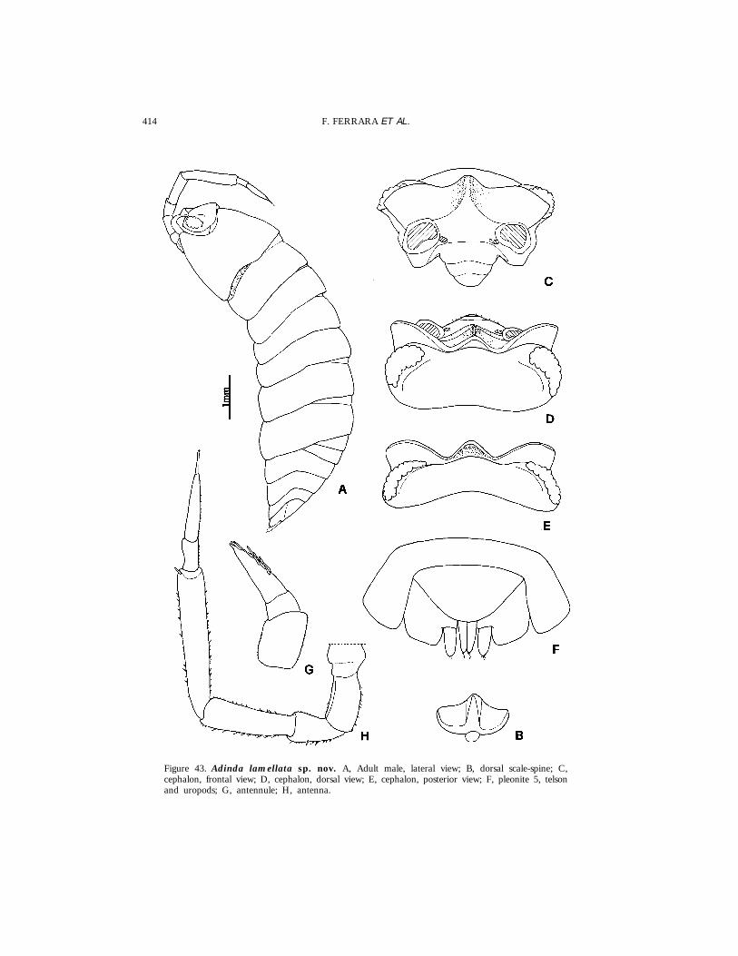
414 F. FERRARA ET AL.
Figure 43. Adinda lamellata sp. nov. A, Adult male, lateral view; B, dorsal scale-spine; C,cephalon, frontal view; D, cephalon, dorsal view; E, cephalon, posterior view; F, pleonite 5, telsonand uropods; G, antennule; H, antenna.
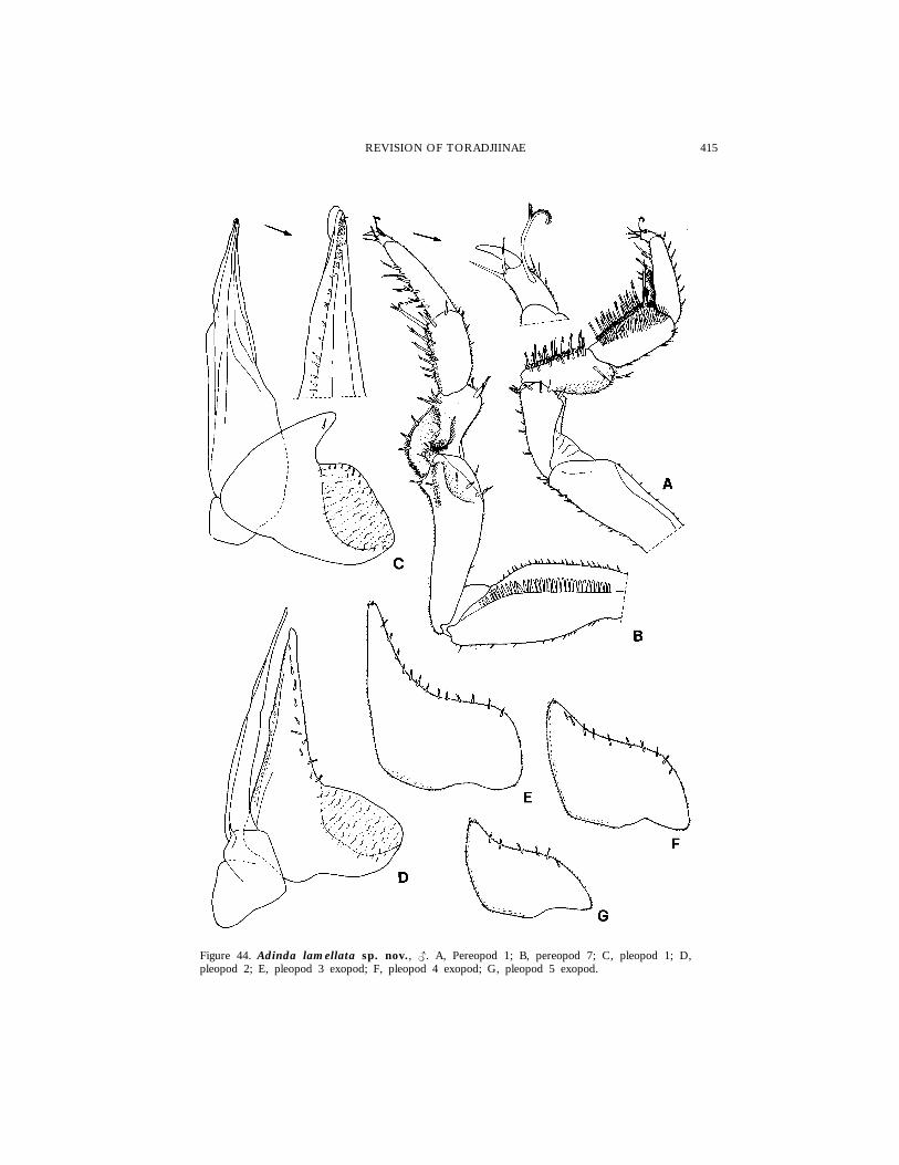
415REVISION OF TORADJIINAE
Figure 44. Adinda lamellata sp. nov., {. A, Pereopod 1; B, pereopod 7; C, pleopod 1; D,pleopod 2; E, pleopod 3 exopod; F, pleopod 4 exopod; G, pleopod 5 exopod.

416 F. FERRARA ET AL.
concave, distally with a small triangular protrusion, rostral surface distally witha setose process; merus with a large lamellar semicircular protrusion on sternalmargin, rostral surface with a large setose lobe. Pleopod 1 exopod withtriangular posterior point, bent outwards; endopod with a small hyaline lobeat apex. Pleopod 2 and pleopods 3–5 exopod as in Fig. 44D–G.
EtymologyThe name refers to the large lamellar protrusion on the sternal margin of
the male pereopod 7 merus.
DistributionBrunei, Malaysia (Sarawak) and Philippines.
RemarksThe specimens from Malaysia (Sarawak) and the Philippines correspond quite
well in general morphology to the type series from Brunei and they are ascribedto this species. However, since no adult males are present in the material fromthese two countries, we prefer not to designate those specimens as paratypes.
Adinda lamellata is easily distinguished from all the other species in the genusby the structure of the male pereopod 7 and pleopod 1.
Dollfusi group
Adinda dollfusi (Richardson Searle, 1922) comb. nov.
(Figs 45, 46)
Toradjia dollfusi Richardson Searle, 1922 : 3, pl. I fig. 2.
Typematerial examinedINDONESIA: 1 { (‘Type’ � Holotype), Java, Nongkodjadjar, 1200 m, USNM;
1 { (‘Cotype’ � Paratype), same data, NNM.
PreviousrecordsIndonesia: Java, Nongkodjadjar (Richardson Searle, 1922).
DescriptionMaximum length: 9 mm. Body enlarged, moderately convex. Colour (according
to Richardson Searle, 1922) brown with yellow spots. Dorsum smooth, pruinosedue to the presence of numerous small scale-spines. Eye with about 20ommatidia. Cephalon with frontal shield bearing an obtuse carina protrudingupwards; lateral lobes right-angled, directed forwards; frontal shield separatedfrom vertex by a narrow and shallow groove, provided with a row of shortscales. Pereonite 1 with epimera directed outwards; posterior margin straightwith a rounded lobe at sides which slides under the anterior margin of pereonite2. Telson semicircular, slightly wider than long. Antennule with third articlelong and conical, provided with aesthetascs, Antennae absent in the specimensexamined. Mandibles with molar penicils semidichotomized. Maxillule outerbranch with all teeth simple; inner branch with two long penicils. Maxillipedwith a setose endite and a penicil on medial corner. Pleopods 1–2 exopod

417REVISION OF TORADJIINAE
Figure 45. Adinda dollfusi. A, Holotype, dorsal view; B, cephalon, frontal view; C, cephalon, dorsalview; D, cephalon, posterior view; E, pereonite 1, right side, dorsal view; F, pleonite 5, telsonand uropods.
provided with open lungs with a large respiratory surface similar to those inOniscus. Uropodal protopod subquadrangular; exopod and endopod not protrudingin relation to the posterior margin of protopod.
Male. Pereopod 7 ischium with sternal margin almost straight with a distal

418 F. FERRARA ET AL.
Figure 46. Adinda dollfusi, {. A, Pereopod 7, caudal surface; B, pereopod 7, rostral surface; C,pleopod 1; D, pleopod 2; E, pleopod 3 exopod; F, pleopod 4 exopod; G, pleopod 5 exopod.
setose rounded process. Pleopod 1 exopod with a short rounded posterior point;endopod with no particular modifications. Pleopod 2 exopod with very longtriangular posterior point. Pleopods 3–5 exopod as in Fig. 46E–G.
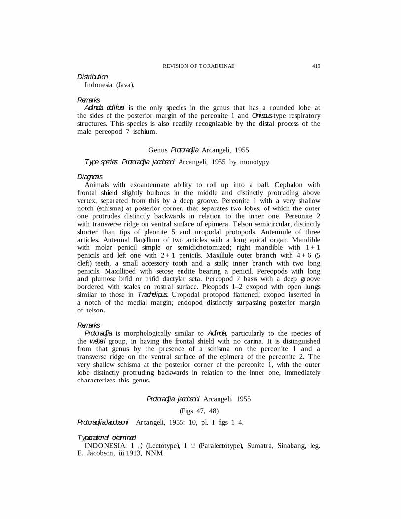
419REVISION OF TORADJIINAE
DistributionIndonesia (Java).
RemarksAdinda dollfusi is the only species in the genus that has a rounded lobe at
the sides of the posterior margin of the pereonite 1 and Oniscus-type respiratorystructures. This species is also readily recognizable by the distal process of themale pereopod 7 ischium.
Genus Protoradjia Arcangeli, 1955
Type species: Protoradjia jacobsoni Arcangeli, 1955 by monotypy.
DiagnosisAnimals with exoantennate ability to roll up into a ball. Cephalon with
frontal shield slightly bulbous in the middle and distinctly protruding abovevertex, separated from this by a deep groove. Pereonite 1 with a very shallownotch (schisma) at posterior corner, that separates two lobes, of which the outerone protrudes distinctly backwards in relation to the inner one. Pereonite 2with transverse ridge on ventral surface of epimera. Telson semicircular, distinctlyshorter than tips of pleonite 5 and uropodal protopods. Antennule of threearticles. Antennal flagellum of two articles with a long apical organ. Mandiblewith molar penicil simple or semidichotomized; right mandible with 1 + 1penicils and left one with 2 + 1 penicils. Maxillule outer branch with 4 + 6 (5cleft) teeth, a small accessory tooth and a stalk; inner branch with two longpenicils. Maxilliped with setose endite bearing a penicil. Pereopods with longand plumose bifid or trifid dactylar seta. Pereopod 7 basis with a deep groovebordered with scales on rostral surface. Pleopods 1–2 exopod with open lungssimilar to those in Trachelipus. Uropodal protopod flattened; exopod inserted ina notch of the medial margin; endopod distinctly surpassing posterior marginof telson.
RemarksProtoradjia is morphologically similar to Adinda, particularly to the species of
the weberi group, in having the frontal shield with no carina. It is distinguishedfrom that genus by the presence of a schisma on the pereonite 1 and atransverse ridge on the ventral surface of the epimera of the pereonite 2. Thevery shallow schisma at the posterior corner of the pereonite 1, with the outerlobe distinctly protruding backwards in relation to the inner one, immediatelycharacterizes this genus.
Protoradjia jacobsoni Arcangeli, 1955
(Figs 47, 48)
ProtoradjiaJacobsoni Arcangeli, 1955: 10, pl. I figs 1–4.
Typematerial examinedINDONESIA: 1 { (Lectotype), 1 | (Paralectotype), Sumatra, Sinabang, leg.
E. Jacobson, iii.1913, NNM.
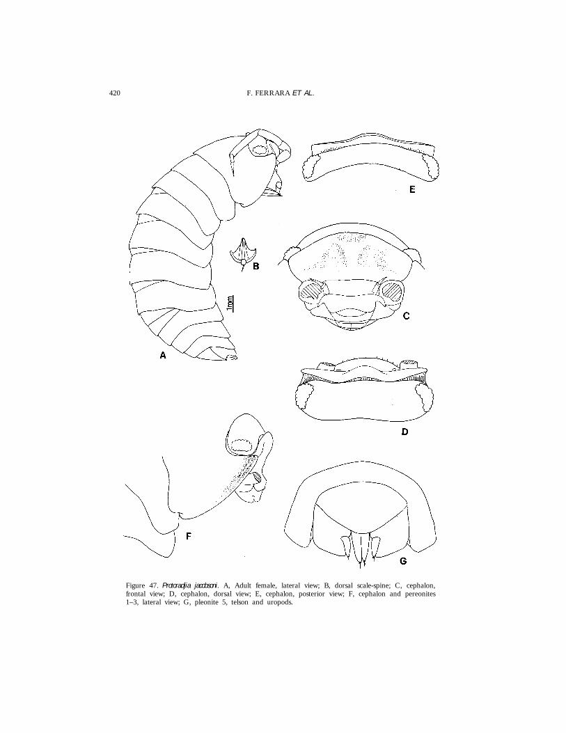
420 F. FERRARA ET AL.
Figure 47. Protoradjia jacobsoni. A, Adult female, lateral view; B, dorsal scale-spine; C, cephalon,frontal view; D, cephalon, dorsal view; E, cephalon, posterior view; F, cephalon and pereonites1–3, lateral view; G, pleonite 5, telson and uropods.
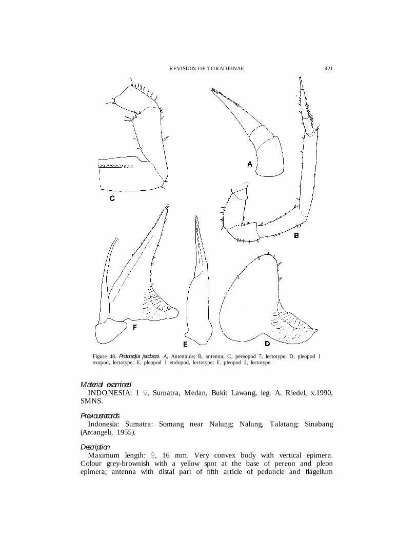
421REVISION OF TORADJIINAE
Figure 48. Protoradjia jacobsoni. A, Antennule; B, antenna; C, pereopod 7, lectotype; D, pleopod 1exopod, lectotype; E, pleopod 1 endopod, lectotype; F, pleopod 2, lectotype.
Material examinedINDONESIA: 1 |, Sumatra, Medan, Bukit Lawang, leg. A. Riedel, x.1990,
SMNS.
PreviousrecordsIndonesia: Sumatra: Somang near Nalung; Nalung, Talatang; Sinabang
(Arcangeli, 1955).
DescriptionMaximum length: |, 16 mm. Very convex body with vertical epimera.
Colour grey-brownish with a yellow spot at the base of pereon and pleonepimera; antenna with distal part of fifth article of peduncle and flagellum

422 F. FERRARA ET AL.
pale. Dorsum smooth with small triangular scale-spines. Eye with about 25ommatidia. Cephalon with frontal shield bulbous in the median part, slightlybent over vertex, from which it is separated by a deep groove bordered byscales; supra-antennal line straight. Pereonite 1 dorsally with a thin groove inthe anterior part, close and parallel to the lateral margin; posterior marginslightly sinuous; outer lobe of schisma protruding in relation to inner one.Pereonites 2–7 with rectangular epimera. Pereonite 2 ventrally with a thintransverse ridge. Telson semicircular, about twice as wide as long. Antennulewith third article long, conical, with five rows of superimposed aesthetascs andtwo subapical aesthetascs. Antenna reaching posterior margin of pereonite 2;second article of flagellum about twice as long as first. Mandibles with molarpenicil semidichotomized. Uropod with protopod subquadrangular; endopodslightly protruding in relation to exopod and protopod tip.
Male. Pereopod 7 ischium with sternal margin slightly concave. Pleopod 1exopod with large rounded posterior lobe; endopod with no particularmodifications. Pleopod 2 as in Fig. 48F.
DistributionIndonesia (Sumatra).
RemarksProtoradjia jacobsoni, type species of the genus, is characterized by the presence
of a thin but distinct groove near the lateral margin of the pereonite 1 andthe male pleopod 1 exopod with a large rounded posterior lobe.
Protoradjia paeninsulae sp. nov.
(Figs 49–51)
Material examinedMALAYSIA: 1 { (Holotype), 1 {, 2 ||, 4 juvs (Paratypes), Pahang, Cameron
Highlands, Gunung Jasar, forest, 1500–1600 m, leg. S. Taiti & L. Bartolozzi,29.xi.1987, MZUF; 1 |, 2 juvs (Paratypes), Pahang, Cameron Highlands, leg.A. Riedel, 1–8.iv.1990, SMNS; 3 ||, 1 juv. (Paratypes), Pahang, CameronHighlands, Gunung Beremban, edge of forest, 1500 m, leg. S. Taiti & L.Bartolozzi, 28.xi.1987, MZUF; 1 | (Paratype), same data, SMNS; 1 |, 1 juv.(Paratypes), same locality, 1700 m, 28.xi.1987, MZUF; 3 {{, 2 ||, 1 juv.(Paratypes), Pahang, Genting Highlands, forest, 1500 m, leg. S. Taiti & L.Bartolozzi, 17.ix.1987, MZUF; 6 {{, 5 ||, 17 juvs (Paratypes), Pahang, FraserHill, edge of forest, along the path to waterfall, leg. S. Taiti, 8.xi.1985, MZUF.
DescriptionMaximum length: {, 17 mm; |, 13 mm. Colour (in alcohol) grey-brown,
with a median yellow spot on pereonites 1–7, two paramedian ones onpereonites 1–6 and pleonites, and one per side at the base of pereon andpleon epimera. Dorsum slightly rough with many small petaliform scale-spines.Eye with about 22 ommatidia. Cephalon with frontal shield bulbous in themiddle, protruding above vertex, from which it is separated by a narrowgroove; supra-antennal line not visible. Pereonite 1 with posterior margin almost
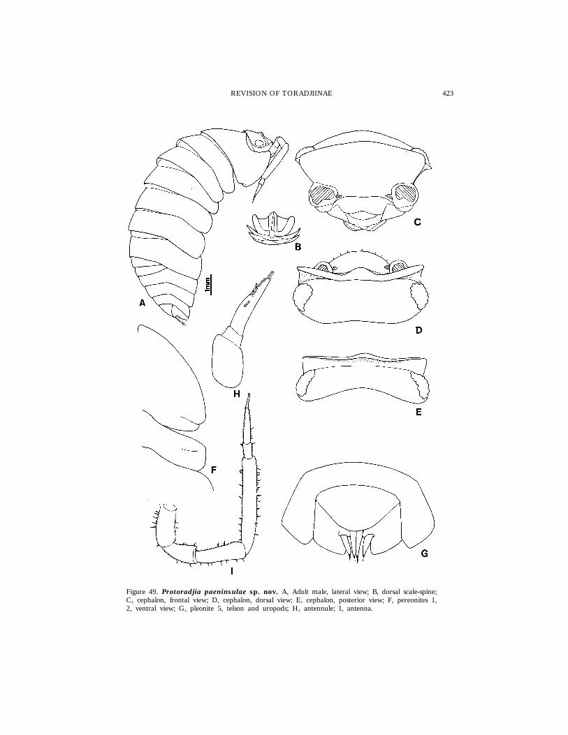
423REVISION OF TORADJIINAE
Figure 49. Protoradjia paeninsulae sp. nov. A, Adult male, lateral view; B, dorsal scale-spine;C, cephalon, frontal view; D, cephalon, dorsal view; E, cephalon, posterior view; F, pereonites 1,2, ventral view; G, pleonite 5, telson and uropods; H, antennule; I, antenna.
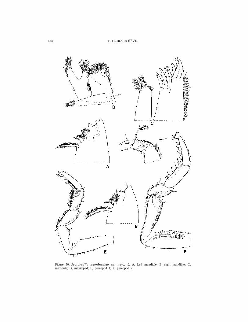
424 F. FERRARA ET AL.
Figure 50. Protoradjia paeninsulae sp. nov., {. A, Left mandible; B, right mandible; C,maxillule; D, maxilliped; E, pereopod 1; F, pereopod 7.
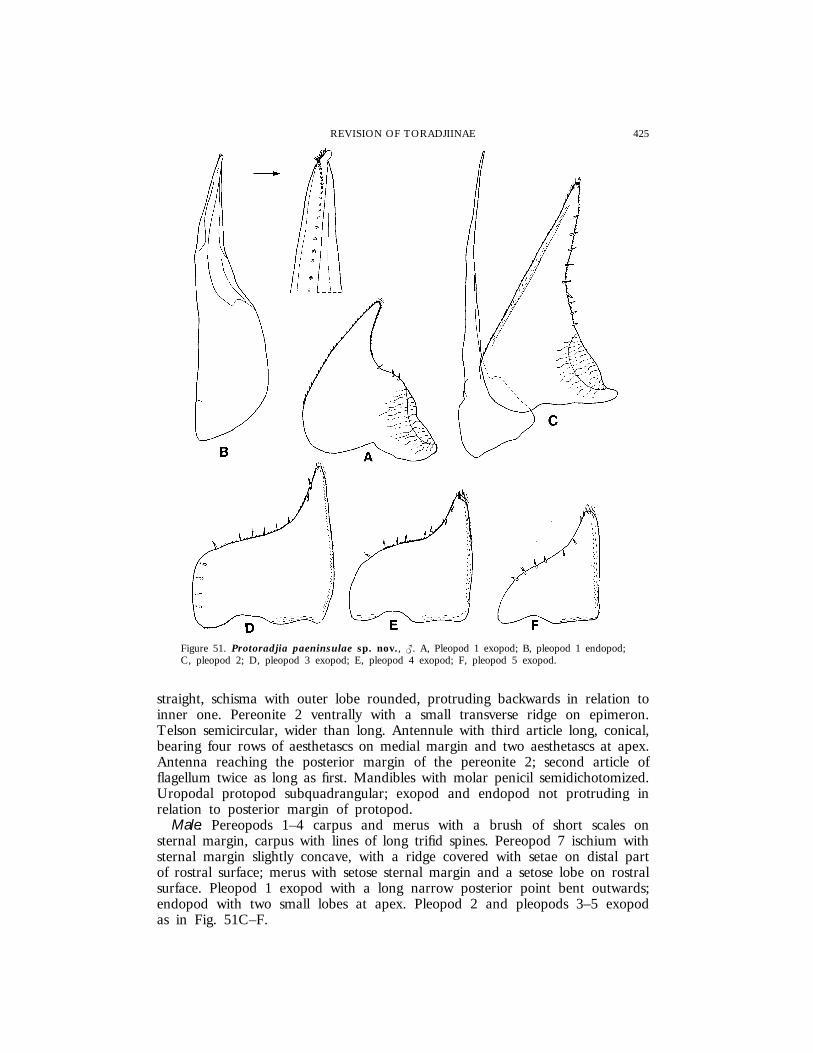
425REVISION OF TORADJIINAE
Figure 51. Protoradjia paeninsulae sp. nov., {. A, Pleopod 1 exopod; B, pleopod 1 endopod;C, pleopod 2; D, pleopod 3 exopod; E, pleopod 4 exopod; F, pleopod 5 exopod.
straight, schisma with outer lobe rounded, protruding backwards in relation toinner one. Pereonite 2 ventrally with a small transverse ridge on epimeron.Telson semicircular, wider than long. Antennule with third article long, conical,bearing four rows of aesthetascs on medial margin and two aesthetascs at apex.Antenna reaching the posterior margin of the pereonite 2; second article offlagellum twice as long as first. Mandibles with molar penicil semidichotomized.Uropodal protopod subquadrangular; exopod and endopod not protruding inrelation to posterior margin of protopod.
Male. Pereopods 1–4 carpus and merus with a brush of short scales onsternal margin, carpus with lines of long trifid spines. Pereopod 7 ischium withsternal margin slightly concave, with a ridge covered with setae on distal partof rostral surface; merus with setose sternal margin and a setose lobe on rostralsurface. Pleopod 1 exopod with a long narrow posterior point bent outwards;endopod with two small lobes at apex. Pleopod 2 and pleopods 3–5 exopodas in Fig. 51C–F.

426 F. FERRARA ET AL.
EtymologyThe name refers to the Malay peninsula where the specimens were collected.
DistributionMalaysia.
RemarksThis species is distinguished from P. jacobsoni by the absence of the groove
near the lateral margin of the pereonite 1, the presence of lobes on thepereopod 7 ischium and merus, and the male pleopod 1 exopod with a longnarrow posterior point.
Protoradjia insularis sp. nov.(Figs 52, 53)
Material examinedINDONESIA: 1 { (Holotype), 2 juvs (Paratypes), Sumatra, Doerian (� Durian),
Pulau Durian, Riau Archipelago, leg. K. W. Dammerman, xi.1923, NNM; 1| (Paratype), same data, MZUF.
SINGAPORE: 2 ||, 1 juv. (Paratypes), Island Country Club, between LowerPeirce Reservoir and Windsor Park Estate, remains of primary forest, leg. B.Hauser, 12.xi.1988, MHNG.
DescriptionMaximum length: { and |, 8 mm. Colour (in alcohol) brown-yellowish with
antennae, cephalon and epimera pale. Dorsum smooth with tiny scale-spines.Eye with about 24 ommatidia. Cephalon with frontal shield slightly bulbous inthe middle, bent over vertex, from which it is separated by a groove visibleonly at the sides; lateral lobes rounded, not protruding forwards. Pereonite 1with posterior margin straight; outer lobe of schisma broadly rounded. Pereonites2 ventrally with a transverse ridge on epimera. Pereonites 2–4 with epimeratriangular, apically rounded. Telson semicircular, about twice as wide as long.Antennule with third article long, conical, bearing some rows of aesthetascs onmedial margin. Antenna with second flagellar article about twice as long asfirst. Mandible with molar penicil semidichotomized. Uropod as in previousspecies.
Male. Pereopods 1–4 merus and carpus with a brush of short scales; carpuswith lines of long trifid spines. Pereopod 7 ischium with sternal margin concave,with an area of long setae in the basal and distal part; merus with sternalmargin covered with long setae in the basal part. Pleopod 1 exopod withposterior point triangular; endopod straight with a small rounded lobe at apex.Pleopod 2 and pleopods 3–5 exopod as in Fig. 53E–H.
EtymologyThe name refers to the Riau and Singapore islands, where these specimens
were collected.
DistributionIndonesia (Sumatra) and Singapore.
RemarksThis species is distinguished from P. jacobsoni by the frontal shield clearly
bent over the vertex, the absence of the groove on the lateral margin of the

427REVISION OF TORADJIINAE
Figure 52. Protoradjia insularis sp. nov. A, Adult male, lateral view; B, cephalon, frontalview; C, cephalon, dorsal view; D, cephalon, posterior view; E, pleonite 5, telson and uropods;F, antennule; G, antenna.
pereonite 1, the pereonites 2–4 with triangular instead of quadrangular epimera,the presence of setose areas on the male pereopod 7 ischium and merus, andthe male pleopod 1 exopod with the posterior point triangular instead ofrounded. It is distinguished from P. paeninsulae by the frontal shield bent overthe vertex, the absence of lobes and ridge on the male pereopod 7 ischiumand merus, the male pleopod 1 exopod with a posterior point much largerand straight instead of bent outwards.

428 F. FERRARA ET AL.
Figure 53. Protoradjia insularis sp. nov., {. A, Pereopod 1; B, pereopod 7; C, pleopod 1exopod; D, pleopod 1 endopod; E, pleopod 2; F, pleopod 3 exopod; G, pleopod 4 exopod; H,pleopod 5 exopod.
429REVISION OF TORADJIINAE
Protoradjia montana sp. nov.
(Figs 54, 55)
Material examinedINDONESIA: 1 { (Holotype), 1 {, 4 ||, 4 juvs (Paratypes), North Sumatra,
Kotacane, Gunung Sinabung, 2000 m, leg. A. Riedel, 7–8.x.1990, SMNS; 1{, 1 | (Paratypes), same data, MZUF.
DescriptionMaximum length: {, 8 mm; |, 7 mm. Colour (in alcohol) brownish, lighter
in females, with pale spots disposed as follows: a median one on posteriormargin of cephalon, pereonites and pleonites 1–4; two paramedian ones andtwo at the base of epimera of pereonites; two on telson. Dorsum smooth withmany small petaliform scale-spines. Eye with about 20 ommatidia. Cephalonwith frontal shield bulbous in the middle, separated from vertex by a narrowgroove; lateral lobes rounded, not protruding forwards. Pereonite 1 with posteriormargin slightly sinuous at sides; outer lobe of schisma rounded. Pereonite 2with epimera rectangular with rounded corners, ventrally with a thin ridge.Telson semicircular. Antennule with third article long, conical, with four rowsof aesthetascs on medial margin. Antenna reaching about middle of pereonite2; second flagellar article more than twice as long as first. Mandible with molarpenicil simple. Uropodal protopod subtriangular with rounded outer margin;exopod and endopod protruding backwards in relation to posterior margin ofprotopod.
Male. Pereopods 1–3 carpus and merus with a brush of short scales andsome long trifid spines on sternal margin. Pereopod 7 ischium with sternalmargin slightly concave, covered with setae; merus with sternal margin convexand an area of long setae on rostral surface. Pleopod 1 exopod with posteriorpoint short, triangular, broadly rounded apically; endopod with no particularmodifications. Pleopod 2 and pleopods 3–5 exopod as in Fig. 55D–G.
EtymologyThe name refers to the site of collection of the specimens, Mt Sinabung, at
a height of about 2000 m.
DistributionIndonesia (Sumatra).
RemarksThis species is distinguished from P. jacobsoni by the absence of a groove
near the lateral margin of the pereonite 1, the male pereopod 7 ischium andmerus with setose areas, and male pleopod 1 exopod with a shorter posteriorpoint. It is distinguished from P. paeninsulae by the absence of lobes on themale pereopod 7 ischium and merus, and male pleopod 1 exopod with posteriorpoint wider. P. montana is morphologically close to P. insularis from which it isrecognisable by the frontal shield of cephalon not bent over the vertex, widermale pereopod 7 merus with a larger setose area, and the male pleopod 1exopod with posterior point shorter and rounded apically.
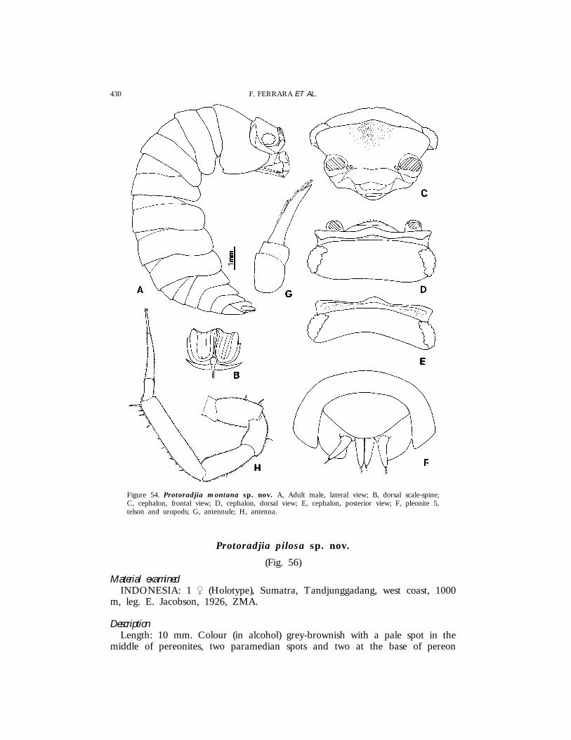
430 F. FERRARA ET AL.
Figure 54. Protoradjia montana sp. nov. A, Adult male, lateral view; B, dorsal scale-spine;C, cephalon, frontal view; D, cephalon, dorsal view; E, cephalon, posterior view; F, pleonite 5,telson and uropods; G, antennule; H, antenna.
Protoradjia pilosa sp. nov.
(Fig. 56)
Material examinedINDONESIA: 1 | (Holotype), Sumatra, Tandjunggadang, west coast, 1000
m, leg. E. Jacobson, 1926, ZMA.
DescriptionLength: 10 mm. Colour (in alcohol) grey-brownish with a pale spot in the
middle of pereonites, two paramedian spots and two at the base of pereon
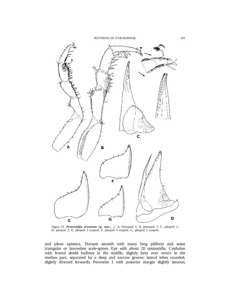
431REVISION OF TORADJIINAE
Figure 55. Protoradjia montana sp. nov., {. A, Pereopod 1; B, pereopod 7; C, pleopod 1;D, pleopod 2; E, pleopod 3 exopod; F, pleopod 4 exopod; G, pleopod 5 exopod.
and pleon epimera. Dorsum smooth with many long piliform and sometriangular or lanceolate scale-spines. Eye with about 20 ommatidia. Cephalonwith frontal shield bulbous in the middle, slightly bent over vertex in themedian part, separated by a deep and narrow groove; lateral lobes rounded,slightly directed forwards. Pereonite 1 with posterior margin slightly sinuous;

432 F. FERRARA ET AL.
Figure 56. Protoradjia pilosa sp. nov. A, Holotype, lateral view; B-D, dorsal scale-spines; E,cephalon, frontal view; F, cephalon, dorsal view; G, cephalon, posterior view; H, pleonite 5, telsonand uropods; I, antennule.
outer lobe of schisma broadly rounded. Pereonite 2 ventrally with a thin butdistinct transverse ridge on epimeron. Telson twice as wide as long, verybroadly rounded. Antennule with third article long, conical, bearing four rowsof aesthetascs on medial margin. Antenna reaching the posterior margin ofpereonite 2; second flagellar article about twice as long as first. Mandibles with

433REVISION OF TORADJIINAE
molar penicil semidichotomized. Uropod with subquadrangular protopod; exopodand endopod slightly protruding in relation to protopod tip.
EtymologyThe name refers to the presence of long piliform scale-spines on the dorsum.
DistributionIndonesia (Sumatra).
RemarksEven if this species is known only from one female specimen, it is worth a
description because it is readily distinguished from all the other species in thegenus by the presence of piliform scale-spines on the dorsal body surface.
Genus Paratoradjia nov.
Typespecies: Paratoradjia beroni sp. nov.
DiagnosisAnimals with exoantennate ability to roll up. Cephalon with frontal shield
distinctly protruding above vertex at the sides, separated from this by a deepgroove bordered with scales, and bent over vertex and partially fused with itin the median part; a median carina along the whole frontal shield is present.Pereonite 1 with a notch (schisma) on posterior margin, far from the posteriorcorner; inner and outer lobes of schisma rounded, equally protruding backwards.Pereonite 2 with a transverse ridge on ventral surface of epimera. Telsonsemicircular or triangular, shorter than tips of pleonite 5 and uropodal protopod.Antennule of three articles. Antennal flagellum of two articles with long apicalorgan. Pereopods with long dactylar seta, distally plumose. Pereopod 7 basiswith groove bordered by a row of scales. Mandibles with molar penicil simple.Maxillule outer branch with 4 + 6 teeth, a small accessory tooth and a stalk;inner branch with two long penicils. Maxilliped with setose endite and penicil.Pleopods 1–2 exopod with open lung similar to that in Oniscus. Uropods withflattened protopod distinctly surpassing apex of telson; exopod inserted in anotch of the medial margin of protopod.
RemarksThe four species described here, even if clearly differentiated by the male
modifications, constitute a very homogeneous group characterized mainly bythe cephalic structure. Moreover, Paratoradjia is easily distinguished from Adindaby the presence of a schisma on the pereonite 1 and a ventral lobe on thepereonite 2, and from Protoradjia by the deeper schisma, far from the posteriorcorner. The new genus is morphologically similar to Toradjia, from which it isdistinguished essentially by the different structure of the cephalon: in Paratoradjiathe frontal shield is bent over the vertex and partially fused with it in themiddle, the frontal margin is continuous and, when the animal rolls up, theantennae simply rest on the vertex; in Toradjia the frontal shield is interruptedin the middle by two deep antennal grooves which continue on the vertex.
434 F. FERRARA ET AL.
The peculiar cephalic structure immediately distinguishes Paratoradjia from allthe other genera in the subfamily.
Paratoradjia beroni sp. nov.
(Figs 1C, 3A, 57–59)
Material examinedSRI LANKA: 1 { (Holotype), 3 || (Paratypes), Kuruwita, leg. P. Beron,
1.xii.1984, NNHMS; 1 | (Paratype), same data, MZUF; 1 | (Paratype),Sabaragamuwa Prov., Deerwood, Kuruwita, 6 mi NNW of Ratnapura, underlogs, leg. Lund University Ceylon Expedition, 18–21.ii.1962, ZIUL; 1 {
(Paratype), same data, ZIUL; 1 { (Paratype), same data, MZUF; 1 | (Paratype),same locality, little gorge, leg. Lund University Ceylon Expedition, 18.ii.1962,ZIUL; 2 ||, 1 juv. (Paratypes), Southern Prov., Haycock Mountain, 21 miNNE of Galle, 300–450 m, under stones, leg. Lund University CeylonExpedition, 29.i.1962, ZIUL.
DescriptionMaximum length: {, 7 mm; |, 6 mm. Colour (in alcohol) grey-brownish;
antennae pale. Dorsum with distinct prominent granulations; dorsal surface withnumerous circular fossettae and small petaliform scale-spines. Eye with about19 ommatidia. Cephalon with frontal shield bent over vertex in the middle,separated from this by a ridge; lateral lobes obliquely directed forwards,separated from vertex by a groove bordered with scales; a protruding carinaon profrons, which reaches posterior margin of frontal shield, separates twolongitudinal concavities against which the peduncles of antennae lean when theanimal rolls up; profile of carina sinuous; supra-antennal line interrupted in themiddle. Pereonite 1 with posterior margin almost straight. Telson triangularwith slightly concave sides and rounded apex. Antennule with third article stoutand short, bearing a tuft of aesthetascs subapically and two aesthetascs apically.Short antenna almost reaching posterior margin of pereonite 1; second flagellararticle about twice as long as first, with long apical organ. Right mandiblewith 1 + 1 penicils; left mandible with 1 + 2 penicils. Maxillule outer branchwith some cleft teeth. Uropodal protopod with outer margin regularly rounded;exopod and endopod slightly protruding in relation to protopod tip.
Male. Pereopod 7 ischium with sternal margin straight and setose in thebasal part, a small lobe covered with short setae distally on rostral surface;merus with a distinct setose triangular lobe on rostral surface. Pleopod 1 exopodcordiform with some spines on outer margin; endopod with enlarged apex.Pleopod 2 and pleopods 3–5 exopod as in Fig. 59D–G.
EtymologyThe new species is named after Dr P. Beron, Sofia, who collected some of
the specimens.
DistributionSri Lanka.
RemarksParatoradjia beroni is characterized by the distinct dorsal granulation, the telson
with triangular apex, the modifications of the male pereopod 7 and the shapeof the male pleopod 1.

435REVISION OF TORADJIINAE
Figure 57. Paratoradjia beroni sp. nov. A, Adult female, lateral view; B, dorsal scale-spine;C, cephalon, frontal view; D, cephalon, dorsal view; E, cephalon, posterior view; F, cephalon,lateral view; G, pereonites 1, 2, dorsal view; H, pereonites 1, 2, ventral view; I, pleonite 5, telsonand uropods.

436 F. FERRARA ET AL.
Figure 58. Paratoradjia beroni sp. nov. A, Antennule; B, antenna; C, left mandible; D, rightmandible; E, maxillule; F, maxilliped.
Paratoradjia indosinensis (Arcangeli, 1948) comb. nov.
(Fig. 60)
Toradjiaindosinensis Arcangeli, 1948: 30, pls I–IV figs 1–14.
PreviousrecordsVietnam: Nha-Trang; Da-Lat; Banov (Arcangeli, 1948).
DistributionVietnam.
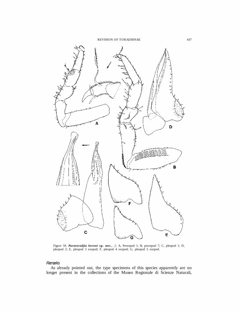
437REVISION OF TORADJIINAE
Figure 59. Paratoradjia beroni sp. nov., {: A, Pereopod 1; B, pereopod 7; C, pleopod 1; D,pleopod 2; E, pleopod 3 exopod; F, pleopod 4 exopod; G, pleopod 5 exopod.
RemarksAs already pointed out, the type specimens of this species apparently are no
longer present in the collections of the Museo Regionale di Scienze Naturali,

438 F. FERRARA ET AL.
Figure 60. Paratoradjia indosinensis. A, Cephalon, dorsal view; B, pereonite 1, right side, dorsal view;C, pleonite 5, telson and uropods; D, antennal flagellum; E, left uropod; F, pereopod 7, {; G,pleopod 1 exopod, {; H, pleopod 2 exopod, { [Adapted from Arcangeli, 1948].
Turin. However, the description and illustrations provided by Arcangeli (1948)show that Toradjia indosinensis possesses all the characters of Paratoradjia, particularlythe structure of the cephalon and of the pereonite 1, and thus it must betransferred to this genus. P. indosinesis has the largest body size in the genus

439REVISION OF TORADJIINAE
(up to 12 mm long) and is characterized by the slightly tuberculated dorsum,the male modifications of pereopod 7 with the ischium bearing a longitudinallamellar process on the sternal margin, and the shape of the male pleopod 1exopod with outer margin deeply incised and a short posterior point apicallyrounded. The main characters of P. indosinensis, redrawn from Arcangeli’soriginal illustrations, are shown in Fig. 60.
Paratoradjia vietnamensis sp. nov.
(Figs 61–63)
Material examinedVIETNAM: 1 { (Holotype), Cuc Phuong Prov., Ninh-Binh, extracted from
soil of Asplenium nidusavis, leg. Topal, 6.v.1966, HNHM.
DescriptionLength: 7 mm. Colour (in alcohol) brown-reddish; antennae lighter. Dorsum
with distinct granulations on anterior half with many small scale-spines. Eyewith 16 ommatidia. Cephalon similar to that of P. beroni, from which it isdistinguished by the profile of carina not sinuous in the central part, and thefrontal shield which in the median part is bent over the vertex but is notdistinctly separated from it. Pereonite 1 with epimeron slightly bent outwards,posterior margin almost straight. Telson semicircular with two dorsal paramedianlongitudinal reliefs. Antenna reaching pereonite 2; flagellum with second articleabout twice as long as first. Buccal pieces not observed due to their poorpreservation. Uropodal protopod subquadrangular, with rounded posteriormargin; exopod and endopod protruding in relation to posterior margin ofprotopod. Pereopods 1–5 merus with lines of small triangular spines on sternalmargin. Pereopod 6 ischium strongly enlarged in the distal part, sternal marginconcave covered with long setae in the distal half, tergal margin with a distinctprocess bearing a strong spine apically; merus with a subrectangular processand two stout spines on tergal margin. Pereopod 7 ischium with sternal marginalmost straight and a distal depression with small triangular spines on rostralsurface; merus with a rounded setose lobe on rostral surface. Pleopod 1 exopodwith a short triangular posterior point apically rounded; endopod with noparticular modifications. Pleopod 2 and pleopods 3–5 exopod as in Fig. 63C–F.
EtymologyThe name refers to Vietnam where the specimen was collected.
DistributionVietnam.
RemarksThis species is readily distinguished from all the others in the genus by the
distinct granulations on the anterior part of the body, the two paramedianlongitudinal reliefs on the telson, and the peculiar features of the male pereopod6.
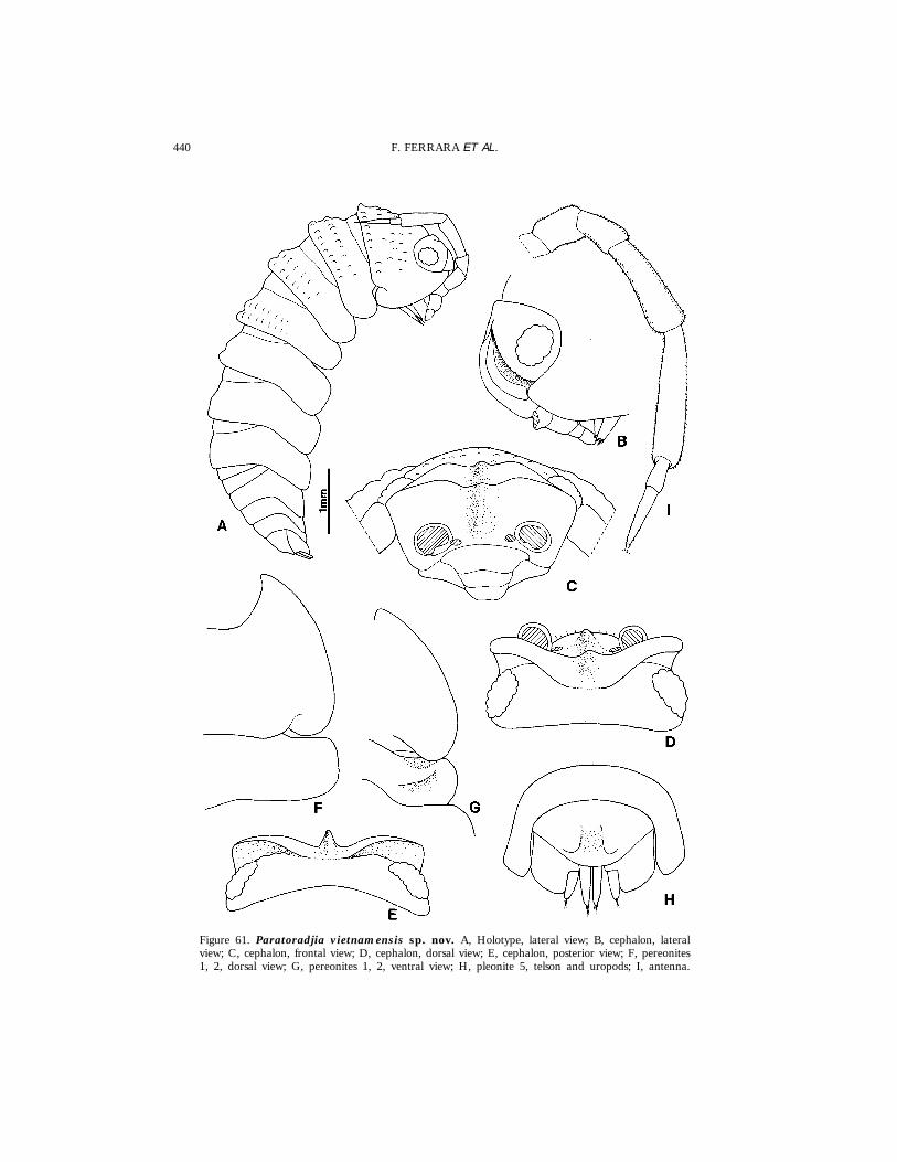
440 F. FERRARA ET AL.
Figure 61. Paratoradjia vietnamensis sp. nov. A, Holotype, lateral view; B, cephalon, lateralview; C, cephalon, frontal view; D, cephalon, dorsal view; E, cephalon, posterior view; F, pereonites1, 2, dorsal view; G, pereonites 1, 2, ventral view; H, pleonite 5, telson and uropods; I, antenna.
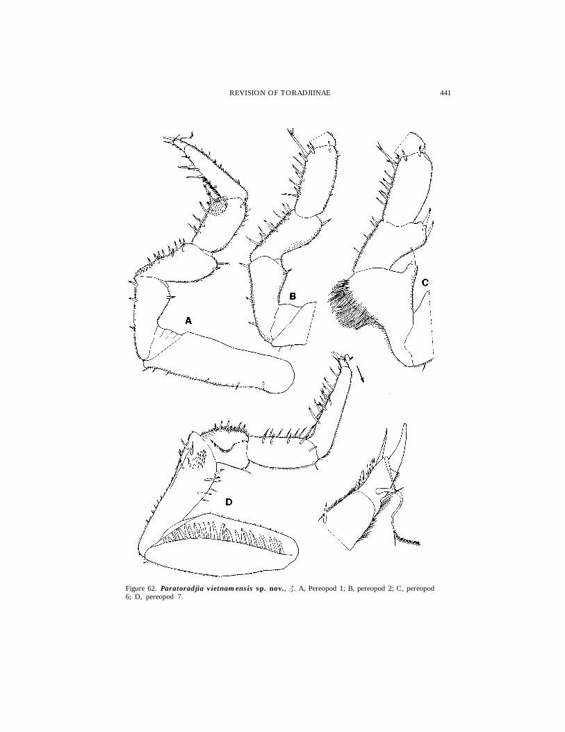
441REVISION OF TORADJIINAE
Figure 62. Paratoradjia vietnamensis sp. nov., {. A, Pereopod 1; B, pereopod 2; C, pereopod6; D, pereopod 7.

442 F. FERRARA ET AL.
Figure 63. Paratoradjia vietnamensis sp. nov., {. A, Pleopod 1 exopod; b, pleopod 1endopod; C, pleopod 2; D, pleopod 3 exopod; E, pleopod 4 exopod; F, pleopod 5 exopod.

443REVISION OF TORADJIINAE
Paratoradjia sulcata sp. nov.
(Fig. 64)
Material examinedPAPUA NEW GUINEA: 1 | (Holotype), Bismarck Islands, New Ireland,
Lelet Plateau, soil, leg. R. Enery, viii.1979, MHNG.
DescriptionLength: 7 mm. Colour (in alcohol) light brown. Dorsum smooth, pruinose
due to the presence of small scale-spines. Eye with about 20 ommatidia.Cephalon with frontal shield bent over vertex in the median part, separatedfrom this by a distinct ridge; lateral lobes obliquely directed forwards, separatedfrom vertex by a groove bordered with short scales; median carina obtuse,slightly protruding. Pereonite 1 with a narrow depression parallel to the lateralmargin; posterior margin straight. Telson semicircular, about twice as wide aslong. Antennule as in P. beroni. Antenna reaching posterior margin of pereonite1; second flagellar article twice as long as first. Buccal pieces, dactylar seta ofthe pereopods and respiratory structures as in P. beroni. Uropodal protopodwith posterior margin oblique; exopod and endopod protruding in relation toprotopod tip.
EtymologyThe name refers to the depression parallel to the lateral margin of the
pereonite 1.
DistributionNew Guinea.
RemarksEven if only a female specimen has been examined, this species is worthy
of description because it is clearly distinguishable from all the other speciesand considerably enlarges the distributional range of the genus. The new speciesis easily distinguished by the smooth dorsum, less developed frontal carina andmainly by the presence of a distinct depression close the lateral margin of thepereonite 1.
Genus Toradjia Dollfus, 1898
Type species: Toradjia celebensis Dollfus, 1898, by original designation.
DiagnosisAnimals with exoantennate and complete ability to roll up into a ball.
Cephalon with frontal shield protruding above vertex at the sides, separatedfrom this by a deep groove; frontal shield interrupted in the middle by twodeep antennal grooves, separated by a narrow carina; these grooves, parallelin the frontal region, continue obliquely on the vertex and transversely on theanterior part of pereonite 1; when the animals roll up, the antenna fits intothese grooves. Pereonite 1, in addition to the above-mentioned grooves, dorsallywith a transverse ridge (not visible in T. hirsuta); a schisma on posterior margin
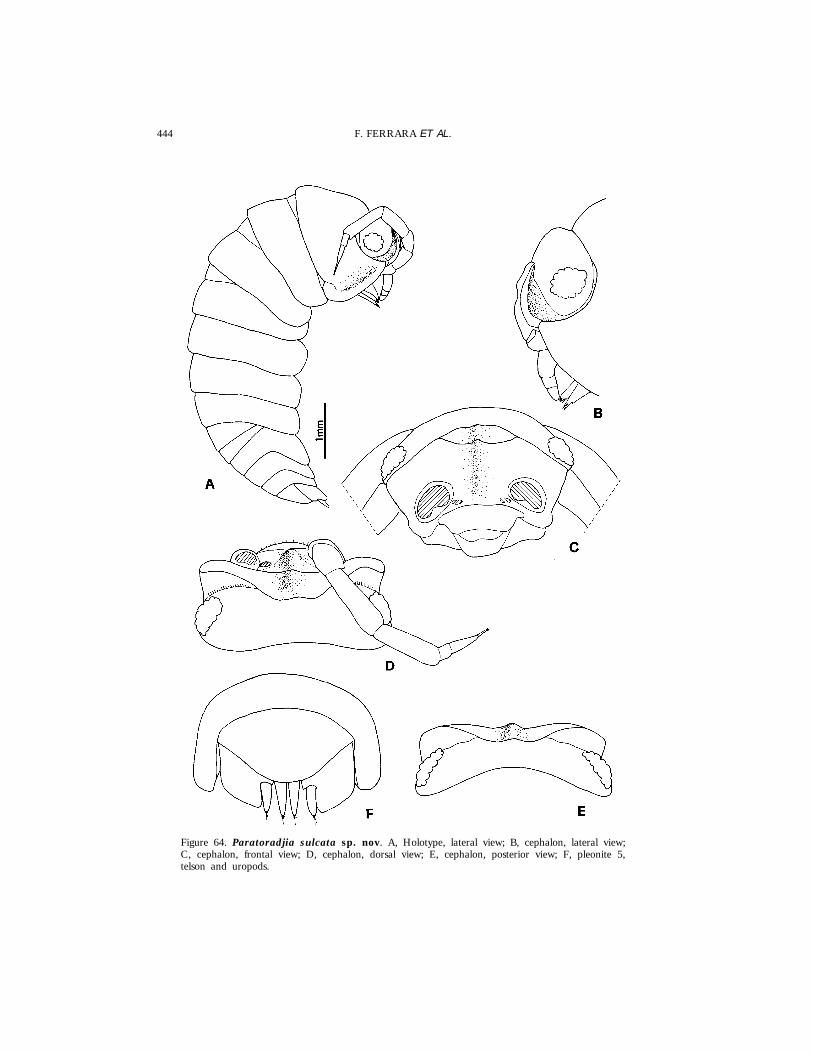
444 F. FERRARA ET AL.
Figure 64. Paratoradjia sulcata sp. nov. A, Holotype, lateral view; B, cephalon, lateral view;C, cephalon, frontal view; D, cephalon, dorsal view; E, cephalon, posterior view; F, pleonite 5,telson and uropods.

445REVISION OF TORADJIINAE
similar to that present in Paratoradjia, i.e. far from the posterior corner. Pereonite2 ventrally with triangular tooth or a transverse lobe more or less developed.Telson semicircular. Antennule of three articles. Antenna with flagellum of twoarticles and long apical organ. Mandibles with molar penicil simple ordichotomized. Maxillule outer branch with 4 + 6 teeth, a small additional toothand a stalk; inner branch with two long penicils. Maxilliped with setose enditeand a penicil on medial corner. Pereopods with long dactylar seta, bifid andsetose in the distal part. Pereopod 7 basis with a groove bordered with scales.Pleopods 1 and 2 exopod with open lungs as in Paratoradjia (Oniscus-like).Uropodal protopod with posterior margin rounded, exopod inserted mediallyin a notch of the medial margin of protopod.
RemarksToradjia is characterized by the presence of antennal grooves on the frontal
region, vertex and pereonite 1. Within the Toradjiinae, this genus is the mostspecialized for rolling up and for antennal protection.
Toradjia gorgona Dollfus, 1898
(Figs 65–67)
ToradjiaGorgona Dollfus, 1898: 366, fig. 11a–c, tab. XIV fig. 11; Arcangeli,1955: 13.
Typematerial examinedINDONESIA: 1 { (Lectotype), uncertain locality, MNHN; 1 | (Paralectotype),
uncertain locality, leg. M. Weber, 1889, ZMA.
Material examinedINDONESIA: 1 |, Java, ‘Tjibodas’ (� Cibodas), leg. Kraepelin (specimen
identified by Budde-Lund), ZMH; 1 |, Java, Cibodas, Lithocarpus-Castanopsisforest above Botanical Garden, tourist path to the waterfall, 1300–1380 m, leg.C. Lienhard, 26.xi.1987, MHNG.
PreviousrecordsIndonesia: locality ? (Dollfus, 1898); Java (Arcangeli, 1955).
DescriptionMaximum length: |, 7 mm. Very convex body with vertical epimera. Colour
grey-brownish. Dorsum of the anterior part of the body rough with largegranules. Eye with about 11 ommatidia. Cephalon with thick lateral lobesslightly protruding above vertex and separated from it by a deep groove;profrons with a narrow lamelliform carina that continues on vertex. Pereonite1 with epimeron slightly directed outwards and posterior margin straight; innerand outer lobes of schisma rounded, equally protruding backwards. Pereonite2 ventrally with a transverse rounded lobe on epimeron. Pereonites 3 and 4ventrally with a transverse ridge on epimeron. Mandibles with molar penicilsimple; right mandible with 1 + 1 penicils; left mandible with 1 + 2 penicils.
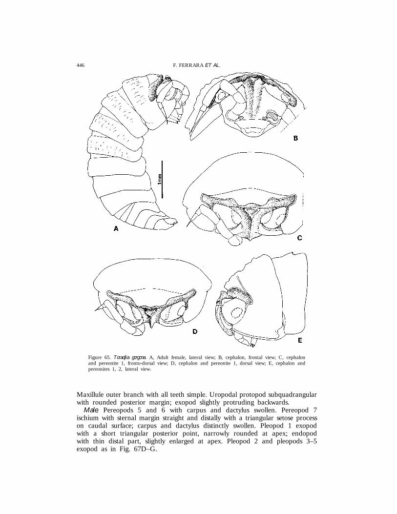
446 F. FERRARA ET AL.
Figure 65. Toradjia gorgona. A, Adult female, lateral view; B, cephalon, frontal view; C, cephalonand pereonite 1, fronto-dorsal view; D, cephalon and pereonite 1, dorsal view; E, cephalon andpereonites 1, 2, lateral view.
Maxillule outer branch with all teeth simple. Uropodal protopod subquadrangularwith rounded posterior margin; exopod slightly protruding backwards.
Male. Pereopods 5 and 6 with carpus and dactylus swollen. Pereopod 7ischium with sternal margin straight and distally with a triangular setose processon caudal surface; carpus and dactylus distinctly swollen. Pleopod 1 exopodwith a short triangular posterior point, narrowly rounded at apex; endopodwith thin distal part, slightly enlarged at apex. Pleopod 2 and pleopods 3–5exopod as in Fig. 67D–G.

447REVISION OF TORADJIINAE
Figure 66. Toradjia gorgona. A, Pereonites 1–3, ventral view; B, pleonite 5, telson and uropods; C,right mandible; D, left mandible; E, maxillule; F, maxilliped.
DistributionIndonesia (Java).
RemarksDollfus described this species on the basis of two specimens from an unknown
locality without indicating the sex or designating the holotype. The malespecimen deposited in MNHN is designated here as lectotype of T. gorgona.Arcangeli (1955: 13) stated that he examined material of this species from Java,with no information about the number, sex or collection sites of the specimens,
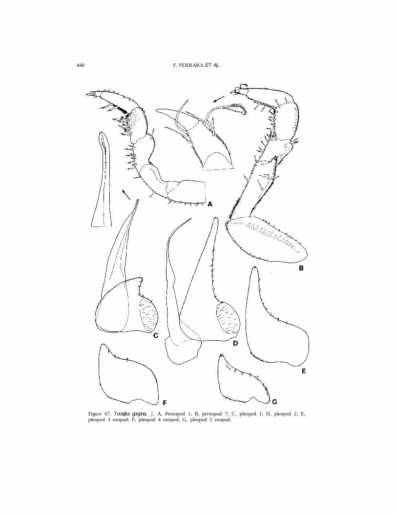
448 F. FERRARA ET AL.
Figure 67. Toradjia gorgona, {. A, Pereopod 1; B, pereopod 7; C, pleopod 1; D, pleopod 2; E,pleopod 3 exopod; F, pleopod 4 exopod; G, pleopod 5 exopod.
449REVISION OF TORADJIINAE
and that probably also the specimens identified by Dollfus were collected onthat island. Our records confirm this assumption.
Toradjia celebensis Dollfus, 1898
(Fig. 68)
Toradjiacelebensis Dollfus, 1898: 365, fig. 10a–c, pl. XIV fig. 10.
Typematerial examinedINDONESIA: 1 | (Lectotype), Celebes (�Sulawesi), Soputan, 1200 m, leg.
F. and P. Sarasin, MNHN; 1 | (Paralectotype), same data, humus, NHMB.
PreviousrecordsIndonesia: Sulawesi, Soputan (Dollfus, 1898).
DescriptionMaximum length: 5 mm. Very convex body with vertical epimera. Colour
(according to Dollfus, 1898) grey with small pale stripes; telson and uropodsred. Dorsum smooth except pereonite 1 which has some granulations. Eye withabout 20 ommatidia. Cephalon and pereonite 1 similar to those of T. gorgona,except the lateral margin of pereonite 1 that is distinctly directed outwards.Pereonite 2 ventrally with an oblique triangular tooth on epimeron. Telsonsemicircular, about twice as wide as long. Buccal pieces as in T. gorgona, exceptmolar penicil of mandible that is dichotomized. Uropodal protopod with roundedposterior margin; exopod and endopod equally protruding backwards in relationto protopod tip.
RemarksToradjia celebensis is easily distinguished from all the other species by the
lateral margin of the pereonite 1 directed outwards and the presence of aventral tooth on the pereonite 2. The specimen deposited in the MNHN isdesignated here as lectotype of this species.
Toradjia cephalica Dollfus, 1898
(Figs 69–71)
Toradjia cephalica Dollfus, 1898: 367, fig. 12a, pl. XIV fig. 12.
Material examinedINDONESIA: 1 { (Neotype), 1 {, 1 juv., Java, Cibodas, Lithocarpus-Castanopsis
forest above botanical Garden, along a gorge, 1350–1480 m, leg. B. Hauser,26.xi.1987, MHNG; 1 {, 1 |, same data, MZUF.

450 F. FERRARA ET AL.
Figure 68. Toradjia celebensis. A, Cephalon and pereonite 1, dorsal view; B, cephalon and pereonite1, frontal view; C, cephalon and pereonites 1, 2, lateral view; D, pereonites 1–3, ventral view; E,telson and uropods; F, right mandible; G, left mandible.
PreviousrecordsIndonesia: Java, Tjibodas (�Cibodas) (Dollfus, 1898).
DescriptionMaximum length: {, 5.2 mm. Very convex body with vertical epimera.
Grey-brownish, some irregular pale spots on pereonites 1–3; telson and uropods
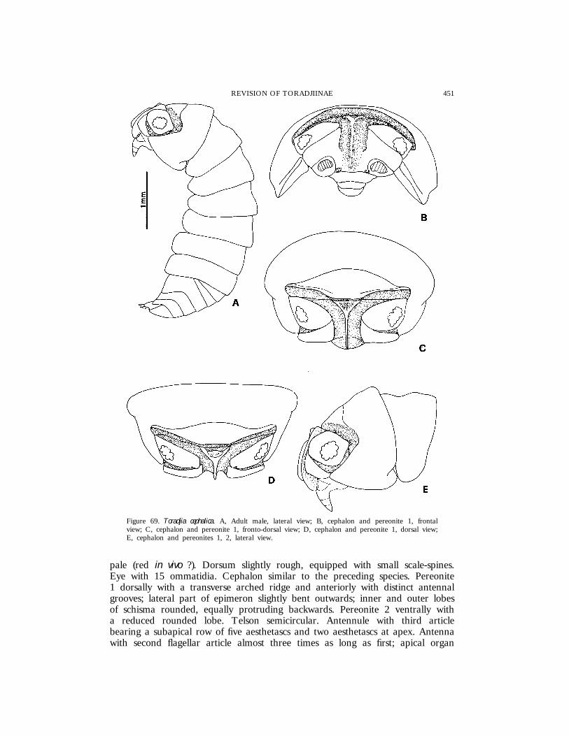
451REVISION OF TORADJIINAE
Figure 69. Toradjia cephalica. A, Adult male, lateral view; B, cephalon and pereonite 1, frontalview; C, cephalon and pereonite 1, fronto-dorsal view; D, cephalon and pereonite 1, dorsal view;E, cephalon and pereonites 1, 2, lateral view.
pale (red in vivo ?). Dorsum slightly rough, equipped with small scale-spines.Eye with 15 ommatidia. Cephalon similar to the preceding species. Pereonite1 dorsally with a transverse arched ridge and anteriorly with distinct antennalgrooves; lateral part of epimeron slightly bent outwards; inner and outer lobesof schisma rounded, equally protruding backwards. Pereonite 2 ventrally witha reduced rounded lobe. Telson semicircular. Antennule with third articlebearing a subapical row of five aesthetascs and two aesthetascs at apex. Antennawith second flagellar article almost three times as long as first; apical organ

452 F. FERRARA ET AL.
Figure 70. Toradjia cephalica, {. A, Pereonites 1–3, ventral view; B, pleonite 5, telson and uropods;C, antennule; D, antenna; E, pereopod 1; F, pereopod 7.
shorter than second article of flagellum. Buccal pieces as in T. gorgona. Uropodalprotopod quadrangular; endopod distinctly surpassing exopod and protopod tip.
Male. Pereopod 7 ischium with sternal margin straight and distally with atriangular setose process on caudal surface. Pleopod 1 exopod with very short

453REVISION OF TORADJIINAE
Figure 71. Toradjia cephalica, {. A, Pleopod 1 exopod; B, pleopod 1 endopod; C, pleopod 2; D,pleopod 3 exopod; E, pleopod 4 exopod; F, pleopod 5 exopod.
rounded posterior point; endopod with straight narrow distal part. Pleopod 2and pleopods 3–5 exopod as in Fig. 71C–F.
DistributionIndonesia (Java).
RemarksThe specimens examined correspond well to Dollfus’ description of T. cephalica
except for the pereonite 2 which has a distinct, even if reduced, ventral lobeon the epimeron. According to Dollfus the pereonite 2 ‘‘paraissant depourvude duplicature coxale’’ but the French author probably overlooked this character.Moreover also the collection locality (Cibodas) of the material examined is thesame as that of the single type specimen of T. cephalica, which, as stated in

454 F. FERRARA ET AL.
the Introduction, is no longer present in the collections of ZMA and must beconsidered lost. All the species in Toradjia are morphologically very similar toeach other and the description of T. cephalica does not permit a safe recognitionof the species. Thus in order to ensure taxonomic stability and material forfuture comparisons, we designate one male specimen deposited in the MHNGas the neotype of Toradjia cephalica, since all the conditions for designation of aneotype specified in Art. 75 of the International Code of Zoological Nomenclature(International Commission on Zoological Nomenclature, 1985) are satisfied.
This species is morphologically very similar to T. gorgona, from which itdiffers in the absence of dorsal granules, the posterior margin of uropodalprotopod subtruncated instead of rounded, and the male pleopod 1 exopodwith shorter posterior point. Moreover the position of the triangular process onthe male pereopod 7 ischium is different in the two species: in T. gorgona it isclose to the tergal margin while in T. cephalica it is close to the sternal one.
Toradjia hirsuta sp. nov.
(Figs 72, 73)
Material examinedMALAYSIA: 1 | (Holotype), Sarawak, Serian District, Penrissen Road, 12
mi from Kuching, Semongok Wildlife Rehabilitation Centre, Nursery Centre ofthe Forestry Department, 20–50 m, leg. B. Hauser, 8.xii.1987, MHNG.
DescriptionLength: 3 mm. Colour grey-brownish. Dorsum with long piliform scale-spines.
Eye with 12 ommatidia. Cephalon similar to that of the other species in thegenus. Pereonite 1 with no transverse ridge and distinct antennal grooves onanterior part, only a wide depression parallel to the anterior margin is present;posterior margin straight; lateral margin slightly directed outwards; inner lobeof schisma rounded, protruding backwards in relation to outer one. Pereonite2 ventrally with a rounded lobe on epimeron. Telson semicircular. Antennawith second article of flagellum twice as long as first; apical organ as long assecond flagellar article. Buccal pieces as in Toradjia gorgona except outer lobe ofmaxillule which has five out of 10 teeth apically cleft. Uropodal protopod withposterior margin rounded; endopod and exopod slightly surpassing protopodtip.
EtymologyThe name refers to the characteristic piliform scale-spines on the dorsum.
RemarksEven if only a female specimen has been studied, it deserves to be described
because it represents a distinct and well characterized species. T. hirsuta differsfrom all the other species by the presence of piliform scale-spines, absence ofa distinct antennal groove on the pereonite 1, reduction of the outer lobe ofthe schisma, and outer lobe of the maxillule with some apically cleft teeth.
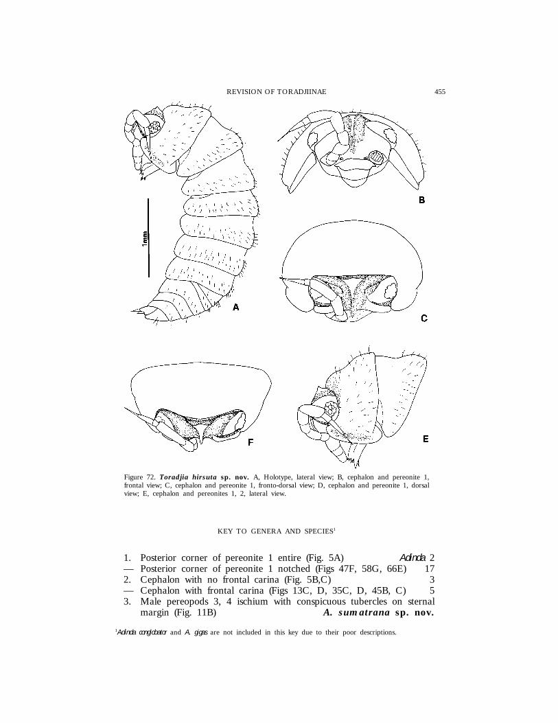
455REVISION OF TORADJIINAE
Figure 72. Toradjia hirsuta sp. nov. A, Holotype, lateral view; B, cephalon and pereonite 1,frontal view; C, cephalon and pereonite 1, fronto-dorsal view; D, cephalon and pereonite 1, dorsalview; E, cephalon and pereonites 1, 2, lateral view.
KEY TO GENERA AND SPECIES1
1. Posterior corner of pereonite 1 entire (Fig. 5A) Adinda 2— Posterior corner of pereonite 1 notched (Figs 47F, 58G, 66E) 172. Cephalon with no frontal carina (Fig. 5B,C) 3— Cephalon with frontal carina (Figs 13C, D, 35C, D, 45B, C) 53. Male pereopods 3, 4 ischium with conspicuous tubercles on sternal
margin (Fig. 11B) A. sumatrana sp. nov.
1Adinda conglobator and A. gigas are not included in this key due to their poor descriptions.
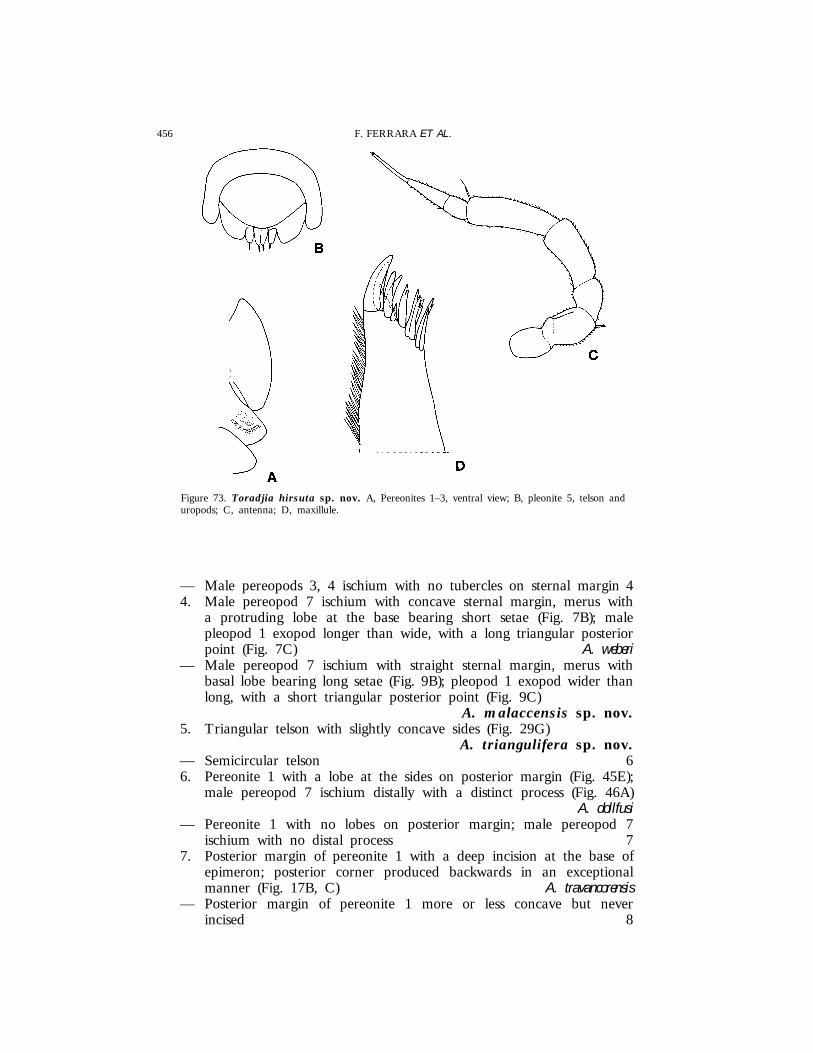
456 F. FERRARA ET AL.
Figure 73. Toradjia hirsuta sp. nov. A, Pereonites 1–3, ventral view; B, pleonite 5, telson anduropods; C, antenna; D, maxillule.
— Male pereopods 3, 4 ischium with no tubercles on sternal margin 44. Male pereopod 7 ischium with concave sternal margin, merus with
a protruding lobe at the base bearing short setae (Fig. 7B); malepleopod 1 exopod longer than wide, with a long triangular posteriorpoint (Fig. 7C) A. weberi
— Male pereopod 7 ischium with straight sternal margin, merus withbasal lobe bearing long setae (Fig. 9B); pleopod 1 exopod wider thanlong, with a short triangular posterior point (Fig. 9C)
A. malaccensis sp. nov.5. Triangular telson with slightly concave sides (Fig. 29G)
A. triangulifera sp. nov.— Semicircular telson 66. Pereonite 1 with a lobe at the sides on posterior margin (Fig. 45E);
male pereopod 7 ischium distally with a distinct process (Fig. 46A)A. dollfusi
— Pereonite 1 with no lobes on posterior margin; male pereopod 7ischium with no distal process 7
7. Posterior margin of pereonite 1 with a deep incision at the base ofepimeron; posterior corner produced backwards in an exceptionalmanner (Fig. 17B, C) A. travancorensis
— Posterior margin of pereonite 1 more or less concave but neverincised 8
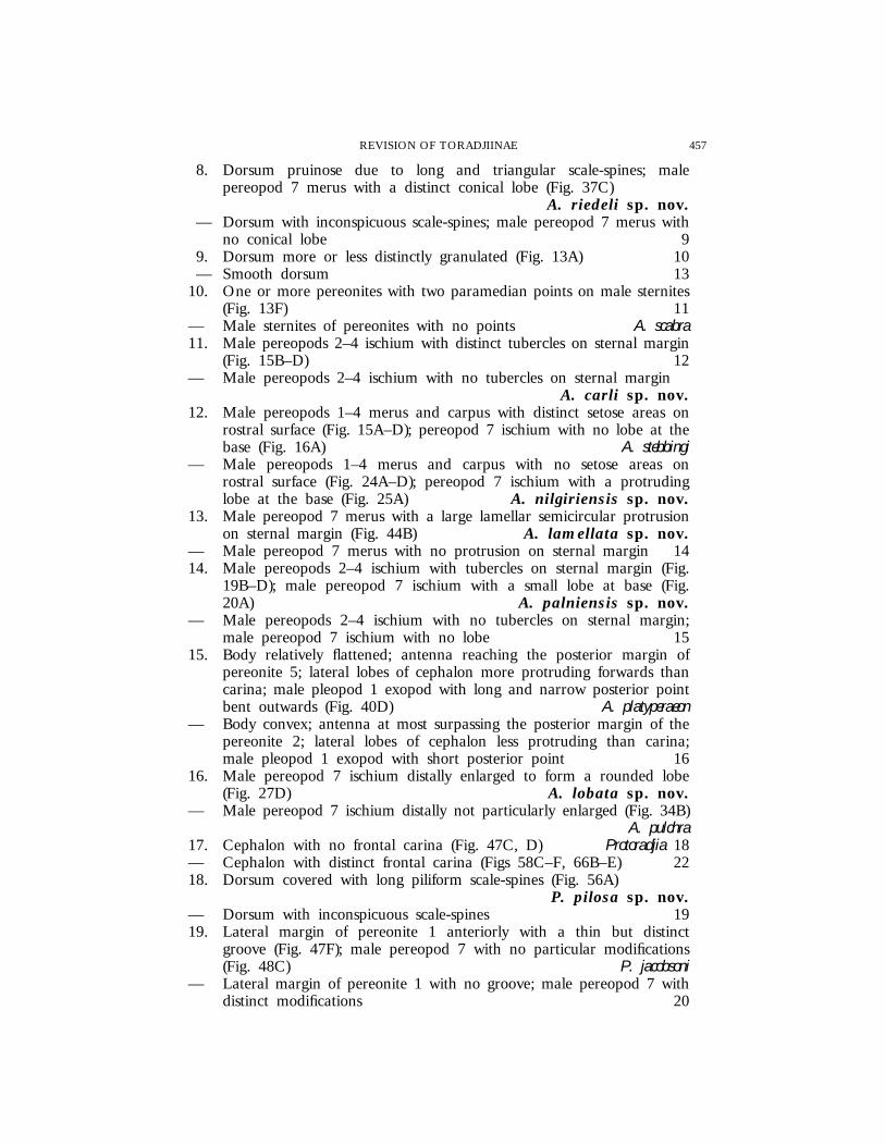
457REVISION OF TORADJIINAE
8. Dorsum pruinose due to long and triangular scale-spines; malepereopod 7 merus with a distinct conical lobe (Fig. 37C)
A. riedeli sp. nov.— Dorsum with inconspicuous scale-spines; male pereopod 7 merus with
no conical lobe 99. Dorsum more or less distinctly granulated (Fig. 13A) 10— Smooth dorsum 13
10. One or more pereonites with two paramedian points on male sternites(Fig. 13F) 11
— Male sternites of pereonites with no points A. scabra11. Male pereopods 2–4 ischium with distinct tubercles on sternal margin
(Fig. 15B–D) 12— Male pereopods 2–4 ischium with no tubercles on sternal margin
A. carli sp. nov.12. Male pereopods 1–4 merus and carpus with distinct setose areas on
rostral surface (Fig. 15A–D); pereopod 7 ischium with no lobe at thebase (Fig. 16A) A. stebbingi
— Male pereopods 1–4 merus and carpus with no setose areas onrostral surface (Fig. 24A–D); pereopod 7 ischium with a protrudinglobe at the base (Fig. 25A) A. nilgiriensis sp. nov.
13. Male pereopod 7 merus with a large lamellar semicircular protrusionon sternal margin (Fig. 44B) A. lamellata sp. nov.
— Male pereopod 7 merus with no protrusion on sternal margin 1414. Male pereopods 2–4 ischium with tubercles on sternal margin (Fig.
19B–D); male pereopod 7 ischium with a small lobe at base (Fig.20A) A. palniensis sp. nov.
— Male pereopods 2–4 ischium with no tubercles on sternal margin;male pereopod 7 ischium with no lobe 15
15. Body relatively flattened; antenna reaching the posterior margin ofpereonite 5; lateral lobes of cephalon more protruding forwards thancarina; male pleopod 1 exopod with long and narrow posterior pointbent outwards (Fig. 40D) A. platyperaeon
— Body convex; antenna at most surpassing the posterior margin of thepereonite 2; lateral lobes of cephalon less protruding than carina;male pleopod 1 exopod with short posterior point 16
16. Male pereopod 7 ischium distally enlarged to form a rounded lobe(Fig. 27D) A. lobata sp. nov.
— Male pereopod 7 ischium distally not particularly enlarged (Fig. 34B)A. pulchra
17. Cephalon with no frontal carina (Fig. 47C, D) Protoradjia 18— Cephalon with distinct frontal carina (Figs 58C–F, 66B–E) 2218. Dorsum covered with long piliform scale-spines (Fig. 56A)
P. pilosa sp. nov.— Dorsum with inconspicuous scale-spines 1919. Lateral margin of pereonite 1 anteriorly with a thin but distinct
groove (Fig. 47F); male pereopod 7 with no particular modifications(Fig. 48C) P. jacobsoni
— Lateral margin of pereonite 1 with no groove; male pereopod 7 withdistinct modifications 20

458 F. FERRARA ET AL.
20. Male pereopod 7 ischium and merus with distinct lobes on rostralsurface (Fig. 50F) P. paeninsulae sp. nov.
— Male pereopod 7 ischium and merus with no lobes on rostral surface21
21. Frontal shield bent over vertex in the median part (Fig. 52C); pleopod1 exopod with triangular posterior point, apically acute (Fig. 53C)
P. insularis sp. nov.— Frontal shield not bent over vertex (Fig. 54D); pleopod 1 exopod
with posterior point apically rounded (Fig. 55C)P. montana sp. nov.
22. Frontal shield bent over vertex and fused with it in the median part,with two paramedian concavities (Fig. 58D, E)
Paratoradjia gen. nov. 23— Frontal shield interrupted in the middle by deep antennal grooves
which continue on the vertex and, more or less developed, on theanterior part of pereonite 1 (Fig. 66A–E) Toradjia 26
23. Smooth dorsum; pereonite 1 with narrow depression parallel to thelateral margin (Fig. 65A) P. sulcata sp. nov.
— More or less granulated dorsum; pereonite without lateral depression24
24. Triangular telson with concave sides (Fig. 58I) P. beroni sp. nov.— Semicircular telson 2525. Telson with two paramedian longitudinal reliefs (Fig. 62H); male
pereopod 6 ischium strongly modified (Fig. 63C); male pereopod 7ischium with no process on sternal margin
P. vietnamensis sp. nov.— Telson with no paramedian reliefs (Fig. 61C); male pereopod 6
ischium without modifications; male pereopod 7 ischium with adistinct longitudinal lamellar process on sternal margin (Fig. 61F)
P. indosinensis comb. nov.6. Dorsum covered with long piliform scale-spines (Fig. 73A)
T. hirsuta sp. nov.— Dorsum without piliform scale-spines 2727. Epimera of pereonite 2 ventrally with tooth-like process (Fig. 69D)
T. celebensis— Epimera of pereonite 2 ventrally with transverse lobes (Figs 67A,
71A) 2828. Anterior part of body dorsally with large granules; male pleopod 1
exopod with distinct triangular posterior point (Fig. 68C) T. gorgona— Dorsum with no granules; male pleopod 1 exopod with short rounded
posterior point (Fig. 72A) T. cephalica
ACKNOWLEDGEMENTS
The authors wish to thank the following persons and institutions for the loanof material: Ms J. Ellis (BM), Dr L. Forro (HNHM), Dr D. Defaye and DrD. Guinot (MNHN), Mrs C. Stocker (NHMB), Dr P. Beron (NNHMS), Prof.L. B. Holthuis and Dr C. H. M. Fransen (NNM), Dr M. Turkay and Mr A.Allspach (SMF), Dr H. Schmalfuss (SMNS), Dr B. Kensley (USNM), Prof. P.

459REVISION OF TORADJIINAE
Brinck (ZIUL), Mr D. Platvoet (ZMA), Dr J. Just (ZMC), and Prof. G.Hartmann (ZMH). Dr B. Hauser, Geneva (MHNG), is particularly acknowledgedalso for the facilities provided during two short visits to his institution by oneof the authors (F. Ferrara). Many thanks are due to Prof. H. S. Yong (KualaLumpur) for his kind help and hospitality during S. Taiti’s visits to Malaysia(1985, 1987).
REFERENCES
Arcangeli A. 1927. Paraperiscyphis Calegarii Arc. nuova specie di Isopodo terrestre di Sumatra. Bollettino delLaboratorio di Zoologia Generale e Agraria della R. Scuola Superiore di Agricoltura di Portici 20: 79–82.
Arcangeli A. 1948. Una specie nuova del genere Toradjia Dollf. (Crostacei Isopodi terrestri). Bollettino delMuseo Zoologico dell’Universita di Torino 1 (1943–48): 29–36, pls I–IV.
Arcangeli A. 1952. Le caratteristiche della famiglia Eubelidae. Crostacei Isopodi terrestri. Sue sottofamigliee suoi generi. Bollettino dell’Istituto e Museo Zoologico dell’Universita di Torino 3 (1951–52): 61–80.
Arcangeli A. 1955. Contributo alle conoscenze sugli Eubelidi di Asia (Crostacei Isopodi terrestri). Bollettinodell’Istituto e Museo Zoologico dell’Universita di Torino 4 (1953–54): 9–17, pl. I.
Budde-Lund G. 1902. A list of Terrestrial Isopods. In: Lanchester WF, ed. On the Crustacea collectedduring the ‘‘Skeat Expedition’’ to the Malay Peninsula. Proceedings of the Zoological Society of London: 379–381.
Budde-Lund G. 1904. A revision of ‘‘Crustacea Isopoda terrestria’’ with additions and illustrations. 2 Spherilloninae. 3Armadillo. Kjo�benhavn: H. Hagerup, pp. 33–144, pls VI–X.
Collinge WE. 1914. On some new terrestrial Isopods from the Andaman Islands and southern India.Records of the Indian Museum 10: 207–210, pls XXIV–XXV.
Collinge WE. 1915. Contributions to a knowledge of the Terrestrial Isopoda of India, Pt. I. Records of theIndian Museum 11: 143–151, pls IV–XII.
Collinge WE. 1916. Contributions to a knowledge of the Terrestrial Isopoda of India, Pt. II. Records of theIndian Museum 12: 115–128, pls IX–XIX.
Dollfus A. 1898. Isopodes terrestres des Indes Neerlandaises recueillis par M. le Professeur Max Weber etpar M. M. le Docteurs Fritz et Paul Sarasin (de Bale). In: Weber W, ed. Zoologische Ergebnisse einer Reisein Niederlandisch Ost-Indien, Leiden 4: 357–382, pls XIII–XV.
Ferrara F, Schmalfuss H. 1976. Terrestrial Isopods from West Africa. Part 1: Family ‘‘Eubelidae’’Budde-Lund, 1899. Monitore Zoologico Italiano (N.S.) Supplemento 7: 1–114.
Hoese B. 1981. Morphologie und Funktion des Wasserleitungssystems der terrestrischen Isopoden (Crustacea,Isopoda, Oniscoidea). Zoomorphology 98: 135–167.
Hoese B. 1982. Der Ligia-Typ des Wasserleitungssystems bei terrestrischen Isopoden und seine Entwicklungin der Familie Ligiidae (Crustacea, Isopoda, Oniscoidea). Zoologische Jahrbucher. Abteilung fur Anatomie undOntogenie der Tiere 108: 225–261.
International Commission on Zoological Nomenclature. 1985. International Code of Zoological Nomenclature.Third Edition adopted by the XX General Assembly of the International Union of Biological Sciences. (Ed. InternationalTrust for Zoological Nomenclature.)
Jackson HG. 1936. Terrestrial Isopods from Malaysia. Bulletin of the Raffles Museum, Singapore, Straits Settlements12: 77–87.
Omer-Cooper J. 1926. A revision of the genus Periscyphis Gerst. (Isopoda Terrestria). Proceedings of theZoological Society of London 24: 349–400.
Richardson Searle H. 1922. Terrestrial Isopoda collected in Java by Dr. Edward Jacobson with descriptionsof five new species. Proceedings of the United States National Museum 60: 1–7, pls 1–2.
Schmalfuss H. 1980. Die ersten Landasseln aus Dominikanischem Bernstein mit einer systematisch-phylogenetischen Revision der Familie Sphaeroniscidae (Stuttgarter Bernsteinsammlung: Crustacea, Isopoda,Oniscoidea). Stuttgarter Beitrage zur Naturkunde. Serie B (Geologie und Palaontologie) 61: 1–12.
Schmalfuss H. 1986. Die Landisopoden (Oniscidea) Griechenlands. 8. Beitrag: Gattung Kefalloniscus nov.gen. (Scleropactidae). Revue Suisse de Zoologie 93: 279–289.
Schmalfuss H. 1989. Phylogenetics in Oniscidea. Monitore Zoologico Italiano (N.S.) Monografia 4: 3–27.Schultz GA. 1982. Terrestrial isopod crustaceans (Oniscoidea) from Mulu Caves, Sarawak, Borneo. Journal
of Natural History 16: 101–117.Stebbing TRR. 1911. Indian Isopods. Records of the Indian Museum 6: 179–191.Taiti S, Ferrara F, Schmalfuss H. 1991. Evolution and biogeography of the family Eubelidae (Crustacea,
Oniscidea). In: Juchault P, Mocquard JP, eds. The Biology of terrestrial Isopods. III. Proceedings of the ThirdInternational Symposium on the Biology of Terrestrial Isopods, Poitiers (France), July 10–12, 1990. Poitiers: Universitede Poitiers, 23–30.
Vandel A. 1968. I. Isopodes terrestres. In: Mission zoologique belge aux ıles Galapagos et en Ecuador (N. et J.Leleup, 1964–1965). Resultats scientifiques. Premiere partie. Bruxelles: L’Imprimerie des Sciences 1: 37–168.
Copyright © 2022 FDOKUMEN