Life cycle of the temporary fish parasite, gnathia africana (crustacea: isopoda: gnathiidae)
8
135 Life cycle of the temporary fish parasite, Gnathia africana (Crustacea: Isopoda: Gnathiidae) Nico J. Smit 1,3 , Linda Basson 2 and Jo G. Van As 2 1 School of Life Sciences, Kingston University, Penrhyn Road, Kingston upon Thames, Surrey, KT1 2EE, UK; 2 Department of Zoology and Entomology, University of the Free State, P.O. Box 339, Bloemfontein, 9300, South Africa; 3 Present address: Department of Zoology, University of Cape Town, Private Bag, Rondebosch, 7701, South Africa Key words: Isopoda, Gnathiidae, Gnathia africana, life cycle, fish parasite Abstract. Laboratory work was conducted to elucidate the life cycle of the South African gnathiid isopod, Gnathia africana Barnard, 1914. The natural fish hosts of this temporary parasite, the super klipfish Clinus superciliosus (Linnaeus, 1758), were exposed to gnathiid larvae in the laboratory. It was found that G. africana has three larval stages, consisting of three unfed (zuphea) and three fed (praniza) stages. First-, second- and third-stage zuphea larvae took an average of 2 h 18 min, 2 h 43 min and 10 h 8 min respectively to complete their feeding and the first- and second-stage praniza moulted at 8 and 10 days respectively into the next zuphea stage. Three to six days after its last blood meal, the sex of the third and final praniza stage could be determined by the presence of either a testis or two ovaries in the dorsal pereon. Male larvae moulted into adult males between 8 and 10 days post feeding. Female larvae moulted at approximately 17 days into adult females. Fertilisation of the eggs by the male took place within 24 hours of completion of the female moult. The development of the embryos and subsequent release of the young larvae between 15 and 23 days post fertilisation completed the cycle. This entire cycle took approximately 62 days in water temperatures of 20–25°C. The life cycle and ecology of members of the family Gnathiidae have intrigued scientists for more than two centuries. Even now the information available on these aspects of gnathiid biology is scanty. To the authors’ knowledge the life cycle of only the following six of the more than 170 described gnathiid species has been researched in any detail: Gnathia maxillaris (Montagu, 1804) by Smith (1904) and Mouchet (1928); G. piscivora Paperna et Por, 1977 by Paperna and Por (1977); Paragnathia formica (Hesse, 1864) by Monod (1926), Mouchet (1928), Stoll (1962, 1963), Amanieu (1963) and Upton (1987a, b); Caecognathia calva (Vanhöffen, 1914) by Wägele (1987, 1988); C. abys- sorum (Sars, 1872) by Klitgaard (1991, 1997); and Elaphognathia cornigera (Nunomura, 1992) by Tanaka and Aoki (1998, 1999, 2000). Gnathia africana Barnard, 1914 was the first gnathiid species to be described from southern Africa (Barnard 1914a, b). This species is very common along the South African west and south coasts where the parasitic larvae feed on the blood and lymph fluid of residential intertidal fishes (Smit and Davies 1999, Smit et al. 1999). The resting larvae and adult stages are found in a variety of sponges, tunicates and tubes of serpulid worms (Barnard 1914b, Smit et al. 1999). The abundance of these gnathiids in the intertidal zone and the availability of their fish hosts make them perfect research specimens, especially for life-cycle experi- ments. It has recently been shown by Davies and Smit (2001) that G. africana acts as a vector for the blood parasite Haemogregarina bigemina Laveran et Mesnil, 1901. This fact underlines the importance of further research into the life cycle and history of gnathiids in general. This paper reports on the life cycle of G. africana constructed from field and laboratory work. MATERIALS AND METHODS Material used in the laboratory experiments during the present study was collected during October 1998 at Jeffreys Bay. Fish were collected and examined for parasites as described by Smit and Davies (1999), Smit et al. (1999) and Davies and Smit (2001). Gnathiids were also collected from sponges (see Smit et al. 1999). Live final-stage larvae were kept in 50-ml plastic screw-top containers of seawater and transported back to the Department of Zoology and Entomology, University of the Free State, where a marine aquarium was set up to maintain fish for use as hosts in feeding experiments with parasitic larval stages of Gnathia africana. The fish host used for these experiments was the super klipfish Clinus superciliosus (Linnaeus, 1758). This widespread intertidal fish species was found to be a common intertidal host for G. africana larvae (Smit and Davies 1999). Initially, artificial sponges, as used by Wägele (1988) were built to serve as a resting place for adults and resting larvae, but it was found that the animals survived perfectly well in the 50-ml screw-top containers of seawater alone. It was, however, important to exchange the water regularly (once every third day) and to clean each gnathiid with a small brush in order to remove debris from the body, especially from the pleopods and dorsal pereon. All animals were kept in seawater FOLIA PARASITOLOGICA 50: 135–142, 2003 Address for correspondence: N.J. Smit, Department of Zoology, University of Cape Town, Private Bag, Rondebosch, 7701, South Africa. Phone: ++27 21 650 3610; Fax: ++ 27 21 650 3301; E-mail: [email protected]
-
Upload
independent -
Category
Documents
-
view
0 -
download
0
Transcript of Life cycle of the temporary fish parasite, gnathia africana (crustacea: isopoda: gnathiidae)
135
Life cycle of the temporary fish parasite, Gnathia africana(Crustacea: Isopoda: Gnathiidae)
Nico J. Smit1,3, Linda Basson2 and Jo G. Van As2
1School of Life Sciences, Kingston University, Penrhyn Road, Kingston upon Thames, Surrey, KT1 2EE, UK;2Department of Zoology and Entomology, University of the Free State, P.O. Box 339, Bloemfontein, 9300, South Africa;3Present address: Department of Zoology, University of Cape Town, Private Bag, Rondebosch, 7701, South Africa
Key words: Isopoda, Gnathiidae, Gnathia africana, life cycle, fish parasite
Abstract. Laboratory work was conducted to elucidate the life cycle of the South African gnathiid isopod, Gnathia africanaBarnard, 1914. The natural fish hosts of this temporary parasite, the super klipfish Clinus superciliosus (Linnaeus, 1758), wereexposed to gnathiid larvae in the laboratory. It was found that G. africana has three larval stages, consisting of three unfed(zuphea) and three fed (praniza) stages. First-, second- and third-stage zuphea larvae took an average of 2 h 18 min, 2 h 43 minand 10 h 8 min respectively to complete their feeding and the first- and second-stage praniza moulted at 8 and 10 daysrespectively into the next zuphea stage. Three to six days after its last blood meal, the sex of the third and final praniza stagecould be determined by the presence of either a testis or two ovaries in the dorsal pereon. Male larvae moulted into adult malesbetween 8 and 10 days post feeding. Female larvae moulted at approximately 17 days into adult females. Fertilisation of the eggsby the male took place within 24 hours of completion of the female moult. The development of the embryos and subsequentrelease of the young larvae between 15 and 23 days post fertilisation completed the cycle. This entire cycle took approximately62 days in water temperatures of 20–25°C.
The life cycle and ecology of members of the familyGnathiidae have intrigued scientists for more than twocenturies. Even now the information available on theseaspects of gnathiid biology is scanty. To the authors’knowledge the life cycle of only the following six of themore than 170 described gnathiid species has beenresearched in any detail: Gnathia maxillaris (Montagu,1804) by Smith (1904) and Mouchet (1928); G.piscivora Paperna et Por, 1977 by Paperna and Por(1977); Paragnathia formica (Hesse, 1864) by Monod(1926), Mouchet (1928), Stoll (1962, 1963), Amanieu(1963) and Upton (1987a, b); Caecognathia calva(Vanhöffen, 1914) by Wägele (1987, 1988); C. abys-sorum (Sars, 1872) by Klitgaard (1991, 1997); andElaphognathia cornigera (Nunomura, 1992) by Tanakaand Aoki (1998, 1999, 2000).
Gnathia africana Barnard, 1914 was the firstgnathiid species to be described from southern Africa(Barnard 1914a, b). This species is very common alongthe South African west and south coasts where theparasitic larvae feed on the blood and lymph fluid ofresidential intertidal fishes (Smit and Davies 1999, Smitet al. 1999). The resting larvae and adult stages arefound in a variety of sponges, tunicates and tubes ofserpulid worms (Barnard 1914b, Smit et al. 1999). Theabundance of these gnathiids in the intertidal zone andthe availability of their fish hosts make them perfectresearch specimens, especially for life-cycle experi-ments. It has recently been shown by Davies and Smit(2001) that G. africana acts as a vector for the blood
parasite Haemogregarina bigemina Laveran et Mesnil,1901. This fact underlines the importance of furtherresearch into the life cycle and history of gnathiids ingeneral. This paper reports on the life cycle of G.africana constructed from field and laboratory work.
MATERIALS AND METHODS
Material used in the laboratory experiments during thepresent study was collected during October 1998 at JeffreysBay. Fish were collected and examined for parasites asdescribed by Smit and Davies (1999), Smit et al. (1999) andDavies and Smit (2001). Gnathiids were also collected fromsponges (see Smit et al. 1999). Live final-stage larvae werekept in 50-ml plastic screw-top containers of seawater andtransported back to the Department of Zoology andEntomology, University of the Free State, where a marineaquarium was set up to maintain fish for use as hosts infeeding experiments with parasitic larval stages of Gnathiaafricana. The fish host used for these experiments was thesuper klipfish Clinus superciliosus (Linnaeus, 1758). Thiswidespread intertidal fish species was found to be a commonintertidal host for G. africana larvae (Smit and Davies 1999).Initially, artificial sponges, as used by Wägele (1988) werebuilt to serve as a resting place for adults and resting larvae,but it was found that the animals survived perfectly well in the50-ml screw-top containers of seawater alone. It was,however, important to exchange the water regularly (onceevery third day) and to clean each gnathiid with a small brushin order to remove debris from the body, especially from thepleopods and dorsal pereon. All animals were kept in seawater
FOLIA PARASITOLOGICA 50: 135–142, 2003
Address for correspondence: N.J. Smit, Department of Zoology, University of Cape Town, Private Bag, Rondebosch, 7701, South Africa.Phone: ++27 21 650 3610; Fax: ++ 27 21 650 3301; E-mail: [email protected]
136
at temperatures between 20 and 25ºC. Gnathiids wereexamined daily under a dissection microscope to monitor theircondition.
During feeding experiments a single host fish was placed ina 25-l aerated aquarium. A single zuphea larva from thelaboratory-reared culture was placed with the fish by means ofa plastic pipette. The feeding period of 20 zuphea (unfed)larvae of each stage (Z1–Z3) was measured from the time ofattachment on the host to detachment from the host to thenearest five minutes. The positions of attachment of each larvawere also recorded. Twenty larvae of each stage weremeasured to the nearest 0.1 mm in order to determine theincrease in length between different stages. All measurementsare presented as range (mean ± standard deviation). The timeof digestion of 20 praniza (fed) larvae of each stage (P1–P2)was measured from the day of detachment to the day on whichthey moulted into the next stage.
RESULTS
A total of 33 final-stage larvae were collected fromtheir fish hosts and 14 from sponges at Jeffreys Bay. Ofthe 33 larvae collected from fish, 16 moulted into malesand 17 into females. Of these females, 11 survived thewhole term of the experimental period (from detach-ment from fish host to release of stage 1 zuphea larvae).All 14 final-stage larvae collected from spongesmoulted into males.
To obtain the feeding and moulting data for 60larvae, 73 larvae were used. The reason for this was thatthe fish killed 13 of the larvae by removing them fromtheir bodies (in most cases from the caudal fin) withtheir teeth. Of these larvae, the fish ate six, the otherseven were spat out.
Zuphea 1 (Z1). The larvae (zuphea 1) released byfemales started to feed immediately on the fish hosts inthe laboratory. The cohorts of Z1 larvae from different-sized females were almost uniform in size (1.0–1.1mm).During feeding experiments, Z1 larvae attached to thehost fish for between 1 hour 15 minutes and 4 hours 10minutes (mean 2 h 18 min). On comparing the length ofthe feeding period of Z1 larvae attached to differentareas on the fish host, a correlation was evident betweenthe attachment site on the host and the length of thefeeding period. Larvae attached to the body of the hostwere able to finish their feeding much faster than thoseattached to the fins. Upon completion of feeding thelarvae left the fish host as praniza 1 larvae.
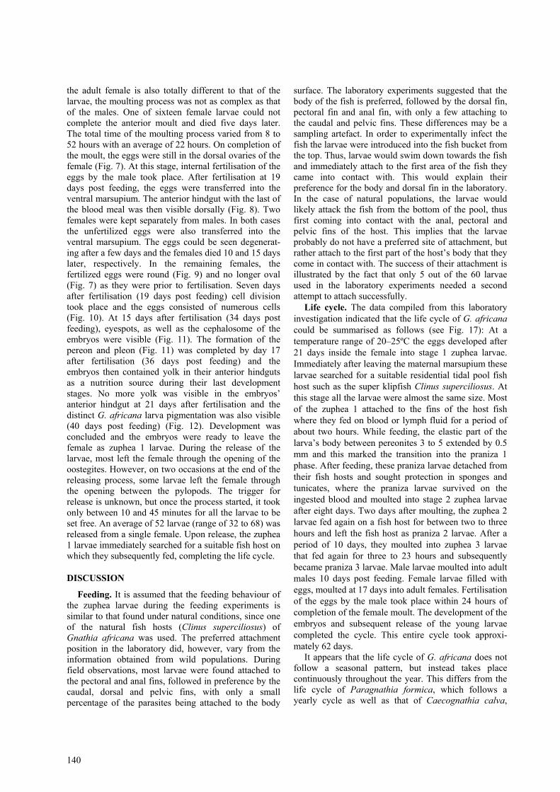
Praniza 1 (P1). The detached P1 larvae ranged inlength from 1.4 to 1.6 mm (mean 1.5 ± 0.06 mm). Thismeans that the first feeding period led to an averageincrease of 0.5 mm in length. The P1 larvae started tomoult into zuphea 2 larvae at seven to nine days(average 8 days) post feeding. The moulting processwas a characteristic isopod moult, consisting of aposterior moult followed by an anterior moult (Fig. 1).The moulting process took between 40 and 90 minutesto complete. The newly moulted larvae were able to use
their limbs after 15 minutes and could swim 30 minutesafter completion of the moult.
Zuphea 2 (Z2). On completion of the moult, theremainder of the blood/lymph meal was still visible inthe anterior hindgut of the Z2 larvae. The length of theZ2 larvae was 1.5–1.8 mm (mean 1.7 ± 0.09 mm), thusresulting in an increase of 0.2 mm in length. The Z2larvae were again ready to feed two to three days post-moulting. At that stage none of the previous meal wasvisible in the anterior hindgut. The feeding period of theZ2 larvae ranged from 1 hour 15 minutes to 4 hours(mean 2 h 43 min), not much longer than that of the Z1larvae. As in the case of the Z1 larvae, Z2 larvae thatattached to the body of the host fish were able tocomplete their feeding more rapidly than those attachedto the fins of the host. The results of the second feedingphase were praniza 2 larvae.
Praniza 2 (P2). The P2 larvae were 2.2–2.6 mm long(mean 2.4 ± 0.1 mm). The average increase in length of0.7 mm produced by the second feeding period wasalmost one and a half times more than that of the firstfeeding stage. The size range of the larvae also in-creased. The P2 larvae spent between 7 and 16 days(mean 10 ± 2.5 days) in a resting and digesting phasebefore they moulted into the next larval stage. Moultingagain followed the same pattern as for the P1 larvae,taking 40–100 min to complete. The resulting larvae(zuphea 3) were the final feeding stages.
Zuphea 3 (Z3). The Z3 larvae ranged in size from2.3 to 2.8 mm (mean 2.6 ± 0.16 mm). As in the case ofthe Z2 larvae, the Z3 larvae gained an average of 0.2mm in length through moulting. Five days aftermoulting, the last of the blood meal was digested andthe larvae were ready to feed again. The feeding periodof the Z3 larvae (mean 10 h 8 min) was, on average,almost five times as long as those of the Z1 and Z2larvae. In some cases a feeding period of more than 20hours was recorded. The feeding process resulted in anaverage increase of 1.2 mm in length. The now P3larvae varied in length from 3.1 to 4.5 mm (mean 3.8 ±0.35 mm). The P3 larvae (Fig. 2) were the final larvalstages and could be separated into male and femalelarvae.
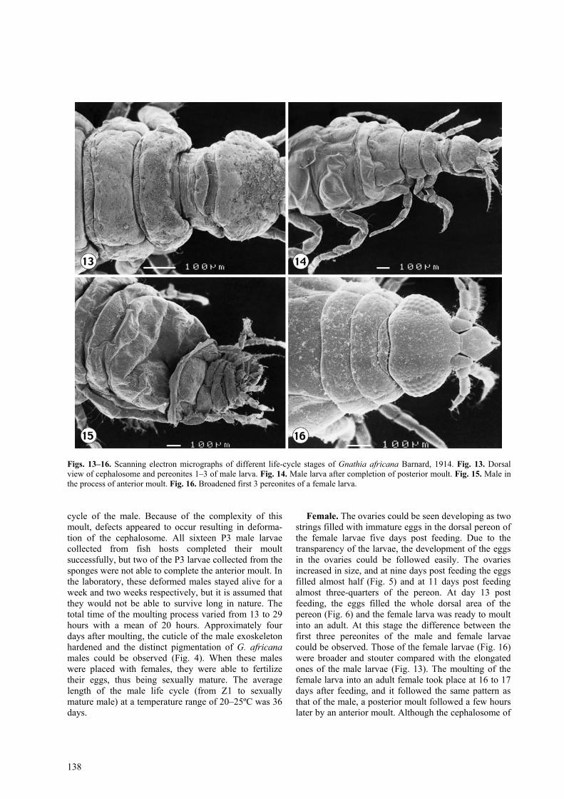
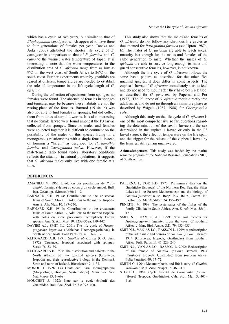
Male. In the P3 larvae that were going to moult intomales, the testes could be seen situated in the dorsalpereon between three to five days after feeding (Fig. 3).The male larvae of G. africana were also characterisedby the elongation of the first pereonite forming a dilatedregion behind the cephalosome (Fig. 13) as described byMonod (1926) for Paragnathia formica. After 8 to 10days, the male larva underwent the final moult into anadult male. In a single case the testis was observed 10days post feeding in the male larva and the moultingonly commenced after 19 days. The moulting process ofG. africana P3 larvae also consisted of a posterior moultfollowed by an anterior moult, as described for other

Smit et al.: Life cycle of Gnathia africana
137
Figs. 1–12. Light micrographs of the different developmental stages of Gnathia africana Barnard, 1914. Fig. 1. Praniza 1 duringthe anterior moult. Fig. 2. Dorsal view of a fully fed praniza 3. Fig. 3. Dorsal view of a male praniza 3 with testis (T). Fig. 4.Adult male with characteristic pigmentation. Fig. 5. Female larva with eggs filling half of dorsal pereon. Fig. 6. Female larvawith eggs filling complete dorsal pereon. Fig. 7. Dorsal view of recently moulted adult female. Fig. 8. Anterior hindgut (AH) offemale with remaining blood meal dorsally visible. Fig. 9. Round fertilized eggs in the ventral marsupium of an adult female.Fig. 10. Eggs in marsupium undergoing cell division. Fig. 11. Eyes, cephalosome, pereonites and pleonites of embryos visible.Fig. 12. First-stage larva ready to leave marsupium. Scale bars: Figs. 1, 11, 12 = 100 µm; Figs. 2–10 = 1 mm.
gnathiid species. The posterior moult took about one totwo hours to complete. During this process formation ofeach adult pleonite could be seen. Upon completion ofthe posterior moult, the new adult’s pereopods were notused for at least eight hours. At that stage, the adultposterior end could easily be distinguished from the
larval anterior end (Fig. 14). About 14 hours after theposterior moult, the anterior moult commenced (Fig.15). This was a much more complex moult and took upto four hours to complete. The newly formed cephalo-some is soft and without the distinct G. africana pig-mentation. This is a very vulnerable stage in the life
138
Figs. 13–16. Scanning electron micrographs of different life-cycle stages of Gnathia africana Barnard, 1914. Fig. 13. Dorsalview of cephalosome and pereonites 1–3 of male larva. Fig. 14. Male larva after completion of posterior moult. Fig. 15. Male inthe process of anterior moult. Fig. 16. Broadened first 3 pereonites of a female larva.
cycle of the male. Because of the complexity of thismoult, defects appeared to occur resulting in deforma-tion of the cephalosome. All sixteen P3 male larvaecollected from fish hosts completed their moultsuccessfully, but two of the P3 larvae collected from thesponges were not able to complete the anterior moult. Inthe laboratory, these deformed males stayed alive for aweek and two weeks respectively, but it is assumed thatthey would not be able to survive long in nature. Thetotal time of the moulting process varied from 13 to 29hours with a mean of 20 hours. Approximately fourdays after moulting, the cuticle of the male exoskeletonhardened and the distinct pigmentation of G. africanamales could be observed (Fig. 4). When these maleswere placed with females, they were able to fertilizetheir eggs, thus being sexually mature. The averagelength of the male life cycle (from Z1 to sexuallymature male) at a temperature range of 20–25ºC was 36days.
Female. The ovaries could be seen developing as twostrings filled with immature eggs in the dorsal pereon ofthe female larvae five days post feeding. Due to thetransparency of the larvae, the development of the eggsin the ovaries could be followed easily. The ovariesincreased in size, and at nine days post feeding the eggsfilled almost half (Fig. 5) and at 11 days post feedingalmost three-quarters of the pereon. At day 13 postfeeding, the eggs filled the whole dorsal area of thepereon (Fig. 6) and the female larva was ready to moultinto an adult. At this stage the difference between thefirst three pereonites of the male and female larvaecould be observed. Those of the female larvae (Fig. 16)were broader and stouter compared with the elongatedones of the male larvae (Fig. 13). The moulting of thefemale larva into an adult female took place at 16 to 17days after feeding, and it followed the same pattern asthat of the male, a posterior moult followed a few hourslater by an anterior moult. Although the cephalosome of
Smit et al.: Life cycle of Gnathia africana
139
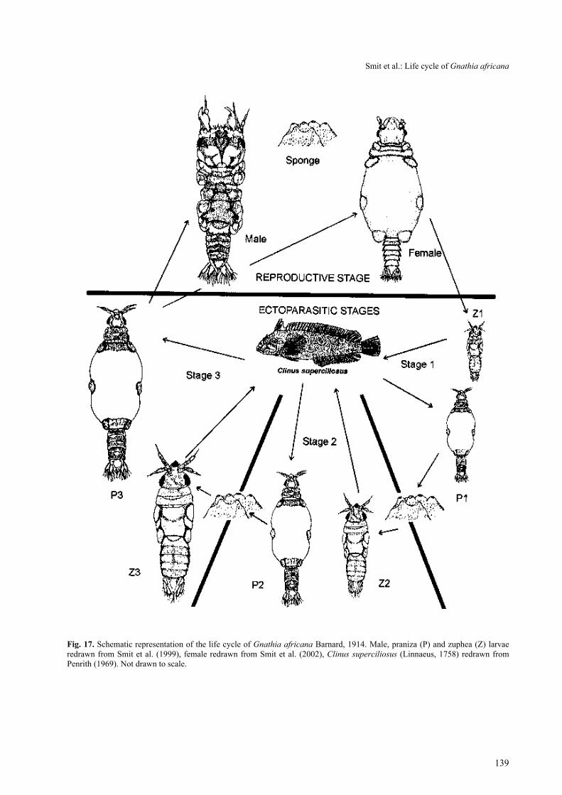
Fig. 17. Schematic representation of the life cycle of Gnathia africana Barnard, 1914. Male, praniza (P) and zuphea (Z) larvaeredrawn from Smit et al. (1999), female redrawn from Smit et al. (2002), Clinus superciliosus (Linnaeus, 1758) redrawn fromPenrith (1969). Not drawn to scale.
140
the adult female is also totally different to that of thelarvae, the moulting process was not as complex as thatof the males. One of sixteen female larvae could notcomplete the anterior moult and died five days later.The total time of the moulting process varied from 8 to52 hours with an average of 22 hours. On completion ofthe moult, the eggs were still in the dorsal ovaries of thefemale (Fig. 7). At this stage, internal fertilisation of theeggs by the male took place. After fertilisation at 19days post feeding, the eggs were transferred into theventral marsupium. The anterior hindgut with the last ofthe blood meal was then visible dorsally (Fig. 8). Twofemales were kept separately from males. In both casesthe unfertilized eggs were also transferred into theventral marsupium. The eggs could be seen degenerat-ing after a few days and the females died 10 and 15 dayslater, respectively. In the remaining females, thefertilized eggs were round (Fig. 9) and no longer oval(Fig. 7) as they were prior to fertilisation. Seven daysafter fertilisation (19 days post feeding) cell divisiontook place and the eggs consisted of numerous cells(Fig. 10). At 15 days after fertilisation (34 days postfeeding), eyespots, as well as the cephalosome of theembryos were visible (Fig. 11). The formation of thepereon and pleon (Fig. 11) was completed by day 17after fertilisation (36 days post feeding) and theembryos then contained yolk in their anterior hindgutsas a nutrition source during their last developmentstages. No more yolk was visible in the embryos’anterior hindgut at 21 days after fertilisation and thedistinct G. africana larva pigmentation was also visible(40 days post feeding) (Fig. 12). Development wasconcluded and the embryos were ready to leave thefemale as zuphea 1 larvae. During the release of thelarvae, most left the female through the opening of theoostegites. However, on two occasions at the end of thereleasing process, some larvae left the female throughthe opening between the pylopods. The trigger forrelease is unknown, but once the process started, it tookonly between 10 and 45 minutes for all the larvae to beset free. An average of 52 larvae (range of 32 to 68) wasreleased from a single female. Upon release, the zuphea1 larvae immediately searched for a suitable fish host onwhich they subsequently fed, completing the life cycle.
DISCUSSION
Feeding. It is assumed that the feeding behaviour ofthe zuphea larvae during the feeding experiments issimilar to that found under natural conditions, since oneof the natural fish hosts (Clinus superciliosus) ofGnathia africana was used. The preferred attachmentposition in the laboratory did, however, vary from theinformation obtained from wild populations. Duringfield observations, most larvae were found attached tothe pectoral and anal fins, followed in preference by thecaudal, dorsal and pelvic fins, with only a smallpercentage of the parasites being attached to the body
surface. The laboratory experiments suggested that thebody of the fish is preferred, followed by the dorsal fin,pectoral fin and anal fin, with only a few attaching tothe caudal and pelvic fins. These differences may be asampling artefact. In order to experimentally infect thefish the larvae were introduced into the fish bucket fromthe top. Thus, larvae would swim down towards the fishand immediately attach to the first area of the fish theycame into contact with. This would explain theirpreference for the body and dorsal fin in the laboratory.In the case of natural populations, the larvae wouldlikely attack the fish from the bottom of the pool, thusfirst coming into contact with the anal, pectoral andpelvic fins of the host. This implies that the larvaeprobably do not have a preferred site of attachment, butrather attach to the first part of the host’s body that theycome in contact with. The success of their attachment isillustrated by the fact that only 5 out of the 60 larvaeused in the laboratory experiments needed a secondattempt to attach successfully.
Life cycle. The data compiled from this laboratoryinvestigation indicated that the life cycle of G. africanacould be summarised as follows (see Fig. 17): At atemperature range of 20–25ºC the eggs developed after21 days inside the female into stage 1 zuphea larvae.Immediately after leaving the maternal marsupium theselarvae searched for a suitable residential tidal pool fishhost such as the super klipfish Clinus superciliosus. Atthis stage all the larvae were almost the same size. Mostof the zuphea 1 attached to the fins of the host fishwhere they fed on blood or lymph fluid for a period ofabout two hours. While feeding, the elastic part of thelarva’s body between pereonites 3 to 5 extended by 0.5mm and this marked the transition into the praniza 1phase. After feeding, these praniza larvae detached fromtheir fish hosts and sought protection in sponges andtunicates, where the praniza larvae survived on theingested blood and moulted into stage 2 zuphea larvaeafter eight days. Two days after moulting, the zuphea 2larvae fed again on a fish host for between two to threehours and left the fish host as praniza 2 larvae. After aperiod of 10 days, they moulted into zuphea 3 larvaethat fed again for three to 23 hours and subsequentlybecame praniza 3 larvae. Male larvae moulted into adultmales 10 days post feeding. Female larvae filled witheggs, moulted at 17 days into adult females. Fertilisationof the eggs by the male took place within 24 hours ofcompletion of the female moult. The development of theembryos and subsequent release of the young larvaecompleted the cycle. This entire cycle took approxi-mately 62 days.
It appears that the life cycle of G. africana does notfollow a seasonal pattern, but instead takes placecontinuously throughout the year. This differs from thelife cycle of Paragnathia formica, which follows ayearly cycle as well as that of Caecognathia calva,
Smit et al.: Life cycle of Gnathia africana
141
which has a cycle of two years, but similar to that ofElaphognathia cornigera, which appeared to have threeto four generations of females per year. Tanaka andAoki (2000) attributed the shorter life cycle of E.cornigera in comparison to that of P. formica and C.calva to the warmer water temperature of Japan. It isinteresting to note that the water temperatures in thedistribution area of G. africana range from as low as9ºC on the west coast of South Africa to 26ºC on thesouth coast. Further experiments whereby gnathiids arereared at different temperatures are needed to establishthe role of temperature in the life-cycle length of G.africana.
During the collection of specimens from sponges, nofemales were found. The absence of females in spongesand tunicates may be because these habitats are not theresting-place of the females. Barnard (1914a, b) wasalso not able to find females in sponges, but did collectthem from tubes of serpulid worms. It is also interestingthat no female larvae were found amongst the P3 larvaecollected from sponges. Since no males and femaleswere collected together it is difficult to comment on thepossibility of the males of this species living in amonogamous relationships with a single female insteadof forming a “harem” as described for Paragnathiaformica and Caecognathia calva. However, if themale/female ratio found under laboratory conditionsreflects the situation in natural populations, it suggeststhat G. africana males only live with one female at atime.
This study also shows that the males and females ofG. africana do not follow asynchronous life cycles asdocumented for Paragnathia formica (see Upton 1987a,b). The males of G. africana are able to reach sexualmaturity fast enough for the males and females of thesame generation to mate. Whether the males of G.africana are able to survive long enough to mate andguard consecutive females, however, is not known.
Although the life cycle of G. africana follows thesame basic pattern as described for the other fivegnathiid species, it does differ in some aspects. Thezuphea 1 larvae of G. africana immediately start to feedand do not need to moult after they have been released,as described for G. piscivora by Paperna and Por(1977). The P3 larvae of G. africana moult directly intoadult males and do not go through an immature phase asdescribed by Wägele (1987, 1988) for Caecognathiacalva.
Although this study on the life cycle of G. africana isone of the most comprehensive so far, questions regard-ing the determination of the sex in larvae (is the sexdetermined in the zuphea 1 larvae or only in the P3larval stage?), the effect of temperature on the life span,and the trigger for the release of the zuphea 1 larvae bythe females, still remain unanswered.
Acknowledgement. This study was funded by the marineresource program of the National Research Foundation (NRF)of South Africa.
REFERENCES
AMANIEU M. 1963: Evolution des populations de Para-gnathia formica (Hesse) au cours d’un cycle annuel. Bull.Inst. Océanogr. (Monaco) 60: 1–12.
BARNARD K.H. 1914a: Contributions to the crustaceanfauna of South Africa. 1. Additions to the marine Isopoda.Ann. S. Afr. Mus. 10: 197–230.
BARNARD K.H. 1914b: Contributions to the crustaceanfauna of South Africa. 3. Additions to the marine Isopoda,with notes on some previously incompletely knownspecies. Ann. S. Afr. Mus. 10: 325a–358a, 359–442.
DAVIES A.J., SMIT N.J. 2001: The life cycle of Haemo-gregarina bigemina (Adeleina: Haemogregarinidae) inSouth African hosts. Folia Parasitol. 48: 169–177.
KLITGAARD A.B. 1991: Gnathia abyssorum (G.O. Sars,1872) (Crustacea, Isopoda) associated with sponges.Sarsia 76: 33–39.
KLITGAARD A.B. 1997: The distribution and habitats in theNorth Atlantic of two gnathiid species (Crustacea,Isopoda) and their reproductive biology in the DenmarkStrait and north of Iceland. Bioscience 47: 1–32.
MONOD T. 1926: Les Gnathiidae. Essai monographique(Morphologie, Biologie, Systématique). Mem. Soc. Sci.Nat. Maroc 13: 1–668.
MOUCHET S. 1928: Note sur le cycle évolutif desGnathiidae. Bull. Soc. Zool. Fr. 53: 392–400.
PAPERNA I., POR F.D. 1977: Preliminary data on theGnathiidae (Isopoda) of the Northern Red Sea, the BitterLakes and the Eastern Mediterranean and the biology ofGnathia piscivora n. sp. Rapp. P.-v. Réun. Comm. Int.Explor. Sci. Mer Méditerr. 24: 195–197.
PENRITH M. 1969: The systematics of the fishes of thefamily Clinidae in South Africa. Ann. S. Afr. Mus. 55: 1–121.
SMIT N.J., DAVIES A.J. 1999: New host records forHaemogregarina bigemina from the coast of southernAfrica. J. Mar. Biol. Assoc. U.K. 79: 933–935.
SMIT N.J., VAN AS J.G., BASSON L. 1999: A redescriptionof the adult male and praniza of Gnathia africana Barnard,1914 (Crustacea, Isopoda, Gnathiidae) from southernAfrica. Folia Parasitol. 46: 229–240.
SMIT N.J., VAN AS J.G., BASSON L. 2002: Redescriptionof the female of Gnathia africana Barnard, 1914(Crustacea: Isopoda: Gnathiidae) from southern Africa.Folia Parasitol. 49: 67–72.
SMITH G. 1904: Metamorphosis and life-history of Gnathiamaxillaris. Mitt. Zool. Neapel 16: 469–474.
STOLL C. 1962: Cycle évolutif de Paragnathia formica(Hesse) (Isopoda: Gnathiidae). Cah. Biol. Mar. 3: 401–416.
142
STOLL C. 1963: La mue en deux temps chez Paragnathiaformica (Hesse) (Isopoda, Gnathiidae). Zool. Exp. Gen.104: 1–23.
TANAKA K., AOKI M. 1998: Crustacean infauna of thedemosponge Halichondria okadai (Kadota) with referenceto the life cycle of Gnathia sp. (Isopoda: Gnathiidea). In:Y. Watanabe and N. Fusetani (Eds.), Sponge Science:Multidisciplinary Perspectives. Springer-Verlag, Tokyo,pp. 259–267.
TANAKA K., AOKI M. 1999: Spatial distribution patterns ofthe sponge dwelling gnathiid isopod Elaphognathiacornigera (Nunomura) on an intertidal rocky shore of theIzu Peninsula, southern Japan. Crustacean Res. 28: 160–167.
TANAKA K., AOKI M. 2000: Seasonal traits of reproductionin gnathiid isopod Elaphognathia cornigera (Nunomura,1992). Zool. Sci. 17: 467–475.
UPTON N.P.D. 1987a: Asynchronous male and female lifecycles in the sexually dimorphic, harem-forming isopodParagnathia formica (Crustacea: Isopoda). J. Zool. Lond.212: 677–690.
UPTON N.P.D. 1987b: Gregarious larval settlement within arestricted intertidal zone and sex differences in subsequentmortality in the polygynous saltmarsh isopod Paragnathiaformica (Crustacea: Isopoda). J. Mar. Biol. Assoc. U.K.67: 663–678.
WÄGELE J.W. 1987: Description of the postembryonal stagesof the Antarctic fish parasite Gnathia calva Vanhöffen(Crustacea: Isopoda) and synonymy with HeterognathiaAmar & Roman. Polar Biol. 7: 77–92.
WÄGELE J.W. 1988: Aspects of the life cycle of the Antarc-tic fish parasite Gnathia calva Vanhöffen (Crustacea: Iso-poda). Polar Biol. 8: 287–291.
Received 1 May 2002 Accepted 17 January 2003