Analgesic effects of intramuscular administration of meloxicam in Hispaniolan parrots ( Amazona...
6
AJVR, Vol 70, No. 12, December 2009 1471 A dequate pain control is an essential part of thera- peutic intervention in acute and chronic disease for all animals, including birds. In humans, pain control is part of a multimodal program for accel- erated postsurgical recovery. 1 Consideration of pain management options is also important to the prac- tice of veterinary medicine and is mandated by the Institute of Laboratory Animal Resources for pain management in all vertebrate species. 2 Nonsteroidal anti-inflammatory drugs are characterized by their anti-inflammatory effects on peripheral tissues and also have centrally acting antinociceptive effects. 3,4 Analgesic effects of intramuscular administration of meloxicam in Hispaniolan parrots ( Amazona ventralis) with experimentally induced arthritis Gretchen A. Cole, DVM; Joanne Paul-Murphy, DVM; Lisa Krugner-Higby, DVM, PhD; Julia M. Klauer, BS; Scott E. Medlin, DVM; Nicholas S. Keuler, MS; Kurt K. Sladky, MS, DVM Objective—To evaluate the analgesic efficacy of meloxicam in parrots with experimentally induced arthritis, with extent of weight bearing and rotational perch walking used as out- come measures. Animals—15 adult Hispaniolan parrots (Amazona ventralis). Procedures—Arthritis was experimentally induced via intra-articular injection of micro- crystalline sodium urate suspension (MSU) into 1 intertarsal joint. Parrots were treated in a crossover design. Five treatments were compared as follows: meloxicam (4 dos- ages) at 0.05, 0.1, 0.5, and 1.0 mg/kg (IM, q 12 h, 3 times) and 0.03 mL of saline (0.9% NaCl) solution (IM, q 12 h, 3 times). The first treatment was given 6 hours following MSU administration. Lameness was assessed by use of a biomechanical perch to re- cord weight-bearing load and a rotational perch to determine dexterity. Feces were col- lected to assay for occult blood. Results—Parrots treated with meloxicam at 1.0 mg/kg had significantly better return to normal (baseline) weight bearing on the arthritic pelvic limb, compared with control parrots or parrots treated with meloxicam at 0.05, 0.1, and 0.5 mg/kg. All fecal samples collected from parrots following induction of arthritis and treatment with meloxicam had negative results for occult blood. Conclusions and Clinical Relevance—Meloxicam administered at 1.0 mg/kg, IM, every 12 hours effectively relieved arthritic pain in parrots. (Am J Vet Res 2009;70:1471–1476) Nonsteroidal anti-inflammatory drugs block the binding of arachidonic acid to COXs, preventing the conversion of thromboxane A 2 to thromboxane B 2 and the production of prostaglandins, which are potent mediators of inflammation. Cyclooxygenases are expressed in the CNS of vertebrates, and their relative expression varies depending on the species. 5 A broad tissue distribution of COX is found in chick- ens. 6 Prostaglandins and COXs participate in avian nociception and act peripherally and centrally simi- lar to prostaglandins and COXs of mammals. 7,8 Galliformes and Anseriformes have been the pri- mary avian orders used to evaluate pharmacokinetics and physiologic and analgesic effects of COX-1 and COX-2 inhibitors. 7–13 In a study 14 that evaluated foot withdrawal latency to a thermal stimulus in chickens, naproxen administration attenuated inflammation and hyperalgesia. In another study, 11 chickens with natu- rally occurring chronic lameness improved their walk- ing speed and ability after carprofen administration and self-selected greater amounts of carprofen-laced feed, compared with control chickens. 11 Ducks have Received November 24, 2008. Accepted February 12, 2009. From the Department of Surgical Sciences, School of Veterinary Medi- cine (Cole, Paul-Murphy, Krugner-Higby, Klauer, Medlin, Sladky), and the Department of Statistics, College of Letters and Science (Keuler), University of Wisconsin, Madison, WI 53706. Dr. Paul- Murphy’s present address is Department of Medicine and Epidemi- ology, School of Veterinary Medicine, University of California-Da- vis, Davis, CA 95616. Presented in abstract form at the American Association of Zoo Veteri- narians Conference, Los Angeles, October 2008. Supported by the Morris Animal Foundation (grant No. D08ZO- 086). The authors thank Brian Higby for engineering the rotating perch ap- paratus and Claudia Hirsch for assistance with animal care. Address correspondence to Dr. Paul-Murphy ([email protected]). ABBREVIATIONS COX Cyclooxygenase MSU Microcrystalline sodium urate suspension
Transcript of Analgesic effects of intramuscular administration of meloxicam in Hispaniolan parrots ( Amazona...
AJVR, Vol 70, No. 12, December 2009 1471
Adequate pain control is an essential part of thera-peutic intervention in acute and chronic disease
for all animals, including birds. In humans, pain control is part of a multimodal program for accel-erated postsurgical recovery.1 Consideration of pain management options is also important to the prac-tice of veterinary medicine and is mandated by the Institute of Laboratory Animal Resources for pain management in all vertebrate species.2 Nonsteroidal anti-inflammatory drugs are characterized by their anti-inflammatory effects on peripheral tissues and also have centrally acting antinociceptive effects.3,4
Analgesic effects of intramuscular administration of meloxicam in Hispaniolan parrots (Amazona ventralis) with experimentally induced arthritis
Gretchen A. Cole, DVM; Joanne Paul-Murphy, DVM; Lisa Krugner-Higby, DVM, PhD; Julia M. Klauer, BS; Scott E. Medlin, DVM; Nicholas S. Keuler, MS; Kurt K. Sladky, MS, DVM
Objective—To evaluate the analgesic efficacy of meloxicam in parrots with experimentally induced arthritis, with extent of weight bearing and rotational perch walking used as out-come measures.Animals—15 adult Hispaniolan parrots (Amazona ventralis).Procedures—Arthritis was experimentally induced via intra-articular injection of micro-crystalline sodium urate suspension (MSU) into 1 intertarsal joint. Parrots were treated in a crossover design. Five treatments were compared as follows: meloxicam (4 dos-ages) at 0.05, 0.1, 0.5, and 1.0 mg/kg (IM, q 12 h, 3 times) and 0.03 mL of saline (0.9% NaCl) solution (IM, q 12 h, 3 times). The first treatment was given 6 hours following MSU administration. Lameness was assessed by use of a biomechanical perch to re-cord weight-bearing load and a rotational perch to determine dexterity. Feces were col-lected to assay for occult blood.Results—Parrots treated with meloxicam at 1.0 mg/kg had significantly better return to normal (baseline) weight bearing on the arthritic pelvic limb, compared with control parrots or parrots treated with meloxicam at 0.05, 0.1, and 0.5 mg/kg. All fecal samples collected from parrots following induction of arthritis and treatment with meloxicam had negative results for occult blood.Conclusions and Clinical Relevance—Meloxicam administered at 1.0 mg/kg, IM, every 12 hours effectively relieved arthritic pain in parrots. (Am J Vet Res 2009;70:1471–1476)
Nonsteroidal anti-inflammatory drugs block the binding of arachidonic acid to COXs, preventing the conversion of thromboxane A
2 to thromboxane
B2 and the production of prostaglandins, which are
potent mediators of inflammation. Cyclooxygenases are expressed in the CNS of vertebrates, and their relative expression varies depending on the species.5 A broad tissue distribution of COX is found in chick-ens.6 Prostaglandins and COXs participate in avian nociception and act peripherally and centrally simi-lar to prostaglandins and COXs of mammals.7,8
Galliformes and Anseriformes have been the pri-mary avian orders used to evaluate pharmacokinetics and physiologic and analgesic effects of COX-1 and COX-2 inhibitors.7–13 In a study14 that evaluated foot withdrawal latency to a thermal stimulus in chickens, naproxen administration attenuated inflammation and hyperalgesia. In another study,11 chickens with natu-rally occurring chronic lameness improved their walk-ing speed and ability after carprofen administration and self-selected greater amounts of carprofen-laced feed, compared with control chickens.11 Ducks have
Received November 24, 2008.Accepted February 12, 2009.From the Department of Surgical Sciences, School of Veterinary Medi-
cine (Cole, Paul-Murphy, Krugner-Higby, Klauer, Medlin, Sladky), and the Department of Statistics, College of Letters and Science (Keuler), University of Wisconsin, Madison, WI 53706. Dr. Paul-Murphy’s present address is Department of Medicine and Epidemi-ology, School of Veterinary Medicine, University of California-Da-vis, Davis, CA 95616.
Presented in abstract form at the American Association of Zoo Veteri-narians Conference, Los Angeles, October 2008.
Supported by the Morris Animal Foundation (grant No. D08ZO-086).
The authors thank Brian Higby for engineering the rotating perch ap-paratus and Claudia Hirsch for assistance with animal care.
Address correspondence to Dr. Paul-Murphy ([email protected]).
AbbreviAtionsCOX CyclooxygenaseMSU Microcrystalline sodium urate suspension
08-11-0382r.indd 1471 11/24/2009 2:42:34 PM
1472 AJVR, Vol 70, No. 12, December 2009
decreased responses to a noxious stimulus (closure of a hemostat around the leg) if they receive ketoprofen more than 30 minutes prior to the noxious stimulus.9
Pharmacokinetic studies of NSAIDs in birds in-clude ketoprofen,15 sodium salicylate,12,13 flunixin me-glumine,12,13 and meloxicam.12,13,16 Pharmacokinetics of meloxicam have been reported for several avian species, including chickens, ducks, pigeons, vultures, ostriches, turkeys, and parakeets.12,13,16,17 In these studies, plasma concentrations of meloxicam were measured following administration but analgesic efficacy was not evalu-ated. To our knowledge, there are no published data evaluating analgesic efficacy of meloxicam in birds. The purpose of the study reported here was to evalu-ate the analgesic efficacy of meloxicam in parrots with experimentally induced arthritis, with extent of weight bearing and rotational perch walking used as outcome measures.
Materials and Methods
Animals—Adult (range, 4 to 20 years old; mean ± SD, 7.33 ± 4.76 years old) Hispaniolan parrots (Ama-zona ventralis; n = 15) of unknown sex and weighing 281.67 ± 20.41 g were used in this study. Parrots were part of a teaching and research flock at the University of Wisconsin School of Veterinary Medicine, Madison, Wisconsin. Parrots used in the study were in good health on the basis of physical examination findings, with normal range of motion of the joints of the pelvic limbs. Parrots were housed as small flocks with 4 to 5 parrots/animal housing room (11.2 m2 [121 sq ft]), and maintained on a 12-hour light to dark cycle at the Char-many Instructional Facility. Groups of 5 parrots were separated from the large flock 4 to 7 days prior to the experimental testing period and housed individually or in pairs in adjacent standard, stainless steel laboratory cages (52.8 cm width X 52.8 cm length X 55 cm height). A perch and 1 to 2 hanging toys were present inside the cages. Parrots were fed a pelleted psittacine dieta and provided water ad libitum. The study was conducted under an Institutional Animal Care and Use Commit-tee protocol approved by the University of Wisconsin School of Veterinary Medicine.
Experimentally induced arthritis—Temporary arthritis was experimentally induced by unilateral in-jection of 8% MSU into the intra-articular space of the intertarsal joint. The MSU used to induce arthritis in this study was prepared by use of published methods.18 The 4% suspension was autoclaved. Each vial was cen-trifuged at 390 X g for 15 minutes, the supernatant was decanted and measured, and 50% of the fluid was re-placed for resuspension to produce an 8% sodium urate crystal solution suspended in sterile saline (0.9% NaCl) solution.
Parrots were anesthetized with isoflurane (5% isoflurane in 1.5 L of O
2/min via facemask), intu-
bated (2.0-mm noncuffed endotracheal tube), and maintained on isoflurane (2% to 3% isoflurane in 0.8 to 1.0 L of O
2/min) by use of a nonbreathing anesthe-
sia circuit. Each parrot was weighed and positioned in dorsal recumbency on a heated pad. Topical eye ointmentb was applied for corneal protection. Feath-
ers were plucked from the area of the intertarsal joint of the nonbanded pelvic limb; the limb was aseptical-ly prepared for intra-articular injection. A 22-gauge needle was inserted into the intertarsal joint space toward the plantar aspect of the joint, and 0.1 mL of 8% MSU was injected. The joint would palpably swell with the correct instillation of MSU. After injec-tion, the joint was massaged and flexed and extended for 15 to 20 seconds to evenly distribute the MSU throughout the joint space. Parrots were removed from isoflurane but maintained on oxygen (0.8 to 1.0 L of O
2/min) until extubation. Subsequently, parrots
were returned to their cages for 4 hours.
Study design—All parrots received an injection of MSU into the intertarsal joint of 1 limb prior to anal-gesic treatment. Five treatments were compared as fol-lows: meloxicamc at 0.05, 0.1, 0.5, and 1.0 mg/kg (IM, q 12 h, 3 times) and 0.03 mL of saline solutiond (IM, q 12 h, 3 times). Intramuscular injections alternated between the left and right pectoral muscles. The first treatment was given 6 hours following MSU adminis-tration. The experimental design was a partial cross-over study, with 15 parrots, 5 treatments, and 4 peri-ods. Parrots were randomly assigned to 3 groups, each with 5 parrots. During the experimental periods, each of the 5 treatments was randomly assigned to 1 parrot of the 5-parrot groups so that a total of 3 parrots re-ceived each treatment in each period. Treatments were administered to parrots in each group in a randomized order. There were 4 periods, with a minimum 2-week washout between periods, and each parrot received a different treatment in each period.
The study protocol required that any parrot with signs of excessive discomfort following the intra-ar-ticular injection of MSU, such as loss of appetite or re-luctance to move about the cage, to be removed from the study and provided additional medical support. The period of decreased weight bearing in saline solu-tion–treated (negative) control parrots was < 38 hours. Because this study involved a species for which the ef-fectiveness of other common analgesics is not yet well established, the use of a positive control group in place of a negative control group, although recommended,19 was not feasible in the evaluation of analgesic effects of meloxicam.
Weight-bearing load perch—An incapacitance me-tere was modified and used to test Hispaniolan parrots. The standard rodent footpads were converted into a di-vided perch so that the extent of weight bearing could be measured independently for each foot. A black, 27 X 11.5 X 23-cm plastic box with a transparent front and hinged door was placed around the perch to limit move-ment by the parrot during assessment. The perch used dual-channel weight averaging, which enabled testing of both pelvic limbs simultaneously. If the parrot shift-ed from foot to foot, the unit recorded the mean weight in grams during a predetermined test period of 3 con-secutive 20-second intervals. Prior to data collection, a 2-week acclimation period was used to condition the parrots to perch inside the test box for 2- to 3-minute intervals. Baseline weight-bearing load data were deter-mined for each parrot 1 week prior to MSU administra-
08-11-0382r.indd 1472 11/24/2009 2:42:35 PM

AJVR, Vol 70, No. 12, December 2009 1473
tion. After induction of arthritis, weight-bearing load data were collected at 4, 6, 8, 12, 18, 26, 30, 32, and 38 hours. Data from each parrot at each time point were calculated to reflect the difference in extent of weight bearing between the MSU-injected and noninjected pelvic limb by use of the following equation: change in weight bearing = (mean weight bearing of nonin-jected limb – mean weight bearing of noninjected limb at baseline) – (mean weight bearing of injected limb – mean weight bearing of injected limb at baseline).
Rotational perch—A rotating perch was con-structed by use of an 11.5-cm-long perch with a ra-dius of 1.83 cm and was fixed within a black, 23.5 X 13.5 X 51-cm plastic box with transparent front and a sliding door in the back panel, similar to the box used for the weight-bearing load perch. Perch rotation was produced by a direct current gear head and motor combinationf attached to a variable-speed control box.g This device was designed to measure changes in normal pelvic limb dexterity. Rotation of the perching rod encouraged the parrot to actively walk at normal ambulatory speed. Rotational veloc-ity was increased 11.6 ± 1.0 rotations/min every 10 seconds, and time (seconds) on the perch was mea-sured until the parrot fell off. A 2-week acclimation period was used to condition the parrots to the rotat-ing perch for 2- to 3-minute intervals. Baseline ro-tational perch data were collected for each parrot 1 week prior to MSU administration. Rotational perch data were collected at each evaluation period imme-diately following weight-bearing load assessment. The change in rotational perch time was determined by use of the following equation: change in rotational perch time = baseline perching time – perching time at each testing period.
Fecal occult blood evaluation—Voided fresh feces were collected from the cage bottom of each parrot or from the testing boxes at the final test-ing time in each period. If 2 parrots were housed together, parrots were temporari-ly separated for fecal collection or a group collection was done. Feces were tested fresh or stored frozen at 4°C and thawed for 10 to 30 minutes prior to evaluation with a commercially available fecal occult blood test.h
Statistical analysis—Weight-bearing load and rotational perch data were ana-lyzed with a commercially available soft-ware program.i Two models were fit for each response; each treated dosage differ-ently. The basic structure was a repeated-measures ANOVA, with fixed effects of treatment, period, time, and all associat-ed interactions. Correlations within each parrot across periods were modeled with a compound symmetry structure; correla-tions within each bird over time within a period were modeled with a spatial power structure. In 1 set of models, dosage was treated as a continuous numeric variable (the actual dose given) and a slope was es-
timated. In the other set of models, dosage was treated as a 4-level nominal factor. In the latter type of analy-ses, pairwise comparisons of the treatments, both with-in each time and over all times, were performed with the Tukey P value correction to account for multiple comparisons. Residuals resulting from the fitted model were verified to be acceptably normally distributed and without evidence of heteroscedasticity. Significance was inferred at values of P < 0.05.
Results
Animals—All 15 parrots completed the study with-out protocol deviations. Data collection was missed at 1 time point for 5 parrots during 1 testing period. All parrots developed experimentally induced arthritis fol-lowing MSU administration as expected, and no parrots were excluded for lameness that was too mild or severe. No adverse effects related to the meloxicam treatment were observed.
Weight-bearing load perch—Parrots receiving no analgesia (n = 12 treatments with saline solution) after experimentally induced arthritis had significantly (P < 0.001, all tests) less weight bearing on the affected pelvic limb at 4, 6, and 8 hours after intra-articular injection, compared with weight bearing at baseline. There was a significant (P = 0.035) overall treatment effect (Figure 1). By use of pairwise tests, parrots re-ceiving meloxicam at 1 mg/kg had significantly (P = 0.041) higher weight-bearing load values, bearing more weight on the MSU-induced arthritic pelvic limb, than those of control parrots throughout the testing period. Meloxicam administration at 1 mg/kg also contributed to greater weight bearing on the arthritic pelvic limb than meloxicam administration at lower amounts of 0.05 and 0.1 mg/kg, although these differences were not significant (P = 0.098 and 0.091, respectively). Dos-
Figure 1—Mean ± SD change in weight-bearing load following injection of MSU into a single intertarsal joint of parrots and subsequent treatment with meloxicam at 0 mg/kg (white diamond), 0.05 mg/kg (black square), 0.1 mg/kg (black triangle), 0.5 mg/kg (white square), and 1.0 mg/kg (large white square with asterisk). Higher values indicate increased weight bearing on the noninjected pelvic limb and decreased weight bearing on the pelvic limb with experimentally induced arthritis. Time 0 represents baseline (be-fore arthritis) measurements. Arrows indicate times of meloxicam administration.
08-11-0382r.indd 1473 11/24/2009 2:42:35 PM
1474 AJVR, Vol 70, No. 12, December 2009
age was significantly and positively associated with the ability to bear weight on the affected pelvic limb (P < 0.001), when treated as a numeric variable.
The time at which parrots recovered to baseline weight-bearing loads was significantly earlier for par-rots receiving meloxicam at 1.0 mg/kg. The 1.0 mg/kg meloxicam group recovered at 12 hours (P = 0.085, where a value of P < 0.05 indicates a lack of recovery or persistent lameness), although the extent of weight bearing was varied in subsequent hours, whereas par-rots that received lower amounts of meloxicam (0.05 mg/kg [P = 0.110], 0.1 mg/kg [P = 0.082], and 0.5 mg/kg [P = 0.176]) did not recover until 38 hours.
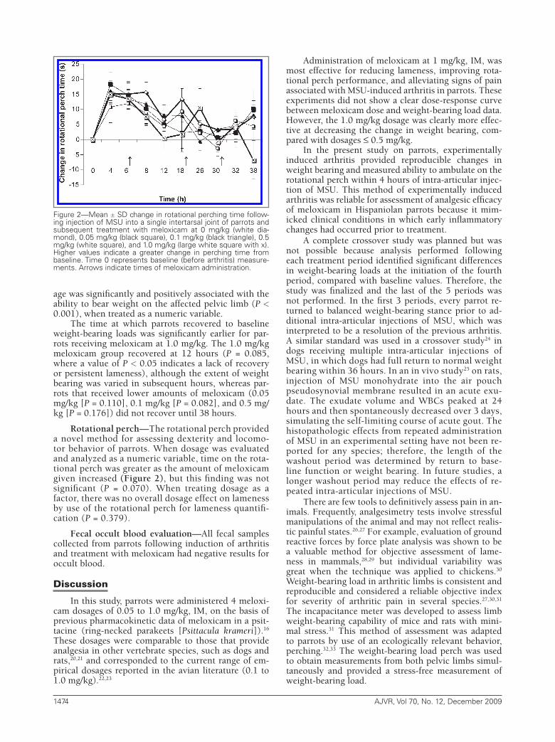
Rotational perch—The rotational perch provided a novel method for assessing dexterity and locomo-tor behavior of parrots. When dosage was evaluated and analyzed as a numeric variable, time on the rota-tional perch was greater as the amount of meloxicam given increased (Figure 2), but this finding was not significant (P = 0.070). When treating dosage as a factor, there was no overall dosage effect on lameness by use of the rotational perch for lameness quantifi-cation (P = 0.379).
Fecal occult blood evaluation—All fecal samples collected from parrots following induction of arthritis and treatment with meloxicam had negative results for occult blood.
Discussion
In this study, parrots were administered 4 meloxi-cam dosages of 0.05 to 1.0 mg/kg, IM, on the basis of previous pharmacokinetic data of meloxicam in a psit-tacine (ring-necked parakeets [Psittacula krameri]).16 These dosages were comparable to those that provide analgesia in other vertebrate species, such as dogs and rats,20,21 and corresponded to the current range of em-pirical dosages reported in the avian literature (0.1 to 1.0 mg/kg).22,23
Administration of meloxicam at 1 mg/kg, IM, was most effective for reducing lameness, improving rota-tional perch performance, and alleviating signs of pain associated with MSU-induced arthritis in parrots. These experiments did not show a clear dose-response curve between meloxicam dose and weight-bearing load data. However, the 1.0 mg/kg dosage was clearly more effec-tive at decreasing the change in weight bearing, com-pared with dosages ≤ 0.5 mg/kg.
In the present study on parrots, experimentally induced arthritis provided reproducible changes in weight bearing and measured ability to ambulate on the rotational perch within 4 hours of intra-articular injec-tion of MSU. This method of experimentally induced arthritis was reliable for assessment of analgesic efficacy of meloxicam in Hispaniolan parrots because it mim-icked clinical conditions in which early inflammatory changes had occurred prior to treatment.
A complete crossover study was planned but was not possible because analysis performed following each treatment period identified significant differences in weight-bearing loads at the initiation of the fourth period, compared with baseline values. Therefore, the study was finalized and the last of the 5 periods was not performed. In the first 3 periods, every parrot re-turned to balanced weight-bearing stance prior to ad-ditional intra-articular injections of MSU, which was interpreted to be a resolution of the previous arthritis. A similar standard was used in a crossover study24 in dogs receiving multiple intra-articular injections of MSU, in which dogs had full return to normal weight bearing within 36 hours. In an in vivo study25 on rats, injection of MSU monohydrate into the air pouch pseudosynovial membrane resulted in an acute exu-date. The exudate volume and WBCs peaked at 24 hours and then spontaneously decreased over 3 days, simulating the self-limiting course of acute gout. The histopathologic effects from repeated administration of MSU in an experimental setting have not been re-ported for any species; therefore, the length of the washout period was determined by return to base-line function or weight bearing. In future studies, a longer washout period may reduce the effects of re-peated intra-articular injections of MSU.
There are few tools to definitively assess pain in an-imals. Frequently, analgesimetry tests involve stressful manipulations of the animal and may not reflect realis-tic painful states.26,27 For example, evaluation of ground reactive forces by force plate analysis was shown to be a valuable method for objective assessment of lame-ness in mammals,28,29 but individual variability was great when the technique was applied to chickens.30 Weight-bearing load in arthritic limbs is consistent and reproducible and considered a reliable objective index for severity of arthritic pain in several species.27,30,31 The incapacitance meter was developed to assess limb weight-bearing capability of mice and rats with mini-mal stress.31 This method of assessment was adapted to parrots by use of an ecologically relevant behavior, perching.32,33 The weight-bearing load perch was used to obtain measurements from both pelvic limbs simul-taneously and provided a stress-free measurement of weight-bearing load.
Figure 2—Mean ± SD change in rotational perching time follow-ing injection of MSU into a single intertarsal joint of parrots and subsequent treatment with meloxicam at 0 mg/kg (white dia-mond), 0.05 mg/kg (black square), 0.1 mg/kg (black triangle), 0.5 mg/kg (white square), and 1.0 mg/kg (large white square with x). Higher values indicate a greater change in perching time from baseline. Time 0 represents baseline (before arthritis) measure-ments. Arrows indicate times of meloxicam administration.
08-11-0382r.indd 1474 11/24/2009 2:42:36 PM
AJVR, Vol 70, No. 12, December 2009 1475
Multiple measurement tools provide greater depth of analgesic assessment.34 The rotational perch is a new measurement tool designed specifically for this study and in this species. Parrots with signs of pain associat-ed with experimentally induced arthritis had decreased dexterity and reduced ambulatory time on the rota-tional perch. Similar techniques are used to evaluate rodents for motor coordination, strength, and dexter-ity.35 Treadmill performance has been used to evaluate lameness in horses and the effectiveness of NSAIDs.36 A treadmill would not be appropriate for a perching bird as they are not well adapted for ambulating on flat surfac-es. Rotational perch data revealed roughly the same pat-tern as the weight-bearing load data, but the results were not significant at the 5% level, attributed primarily to large amounts of individual variability and the number of com-parisons. Our findings suggest that the rotational perch is a useful tool in evaluating lameness in parrots, provided that sample size is adequate.
Control parrots had improvement in the extent of weight bearing over time that may be attributable to simply resting the pelvic limb as well as resolution of the MSU-induced arthritis.31,32 Weight-bearing load was a passive measurement and did not require the use of the MSU-injected pelvic limb. The rotational perch re-quired active use of the arthritic leg, adding repeated stress to the pelvic limbs. Concurrent use of both mea-surement tools evaluated an arthritic pelvic limb in dy-namic motion. It was possible that repeated ambulation during the course of each testing period exacerbated arthritic pain. When 2 testing points are close together, the arthritis may be worse at the second testing, com-pared with parrots having longer periods of rest. For example, although the changes in the extent of weight bearing were minor, weight bearing improved for all parrots between 18 to 26 hours, but most parrots had slight increases in lameness when tested between 26 to 30 hours and 30 to 32 hours.
Gastrointestinal bleeding secondary to NSAID us-age is a known adverse effect in mammals; however, this adverse effect of NSAIDs has not been established for birds. In our study, fecal occult blood test results for all parrots were negative following meloxicam adminis-tration and testing cycles.
In our study, parrots with experimentally induced arthritis that received meloxicam at 1.0 mg/kg were able to bear significantly more weight on the affected pelvic limb than parrots receiving meloxicam at dosages ≤ 0.5 mg/kg. Parrots that received meloxicam at 1.0 mg/kg had better performance on a rotating rod. Results of this study indicate that 1 mg/kg can be considered a thera-peutic dosage of meloxicam for relief of arthritic pain in Hispaniolan parrots. Further study is recommended to evaluate the pharmacokinetics of meloxicam at this dosage as well as evaluate the long-term effects of this dosage on renal function in parrots.
a. Exact, Kaytee Products Inc, Chilton, Wis.b. Paralube, petrolatum ophthalmic ointment sterile ocular lubri-
cant, E. Fougera and Co, Melville, NY.c. Meloxicam 5 mg/mL, Boehringer Ingelheim, St Joseph, Mo.d. 0.9% sodium chloride injection, USP, Hospira, Lake Forest, Ill.e. IITC Incapacitance Meter, model 600, IITC Life Science, Wood-
land Hills, Calif.
f. 24A-D Series parallel Shaft DC Gearmoter, model 0194, serial No. 0194JTE0015, Bodine Electric Co, Chicago, Ill.
g. Filtered PWM DC Basic Speed Control, model 0790, serial No. 0790KNDN0031, Bodine Electric Co, Chicago, Ill.
h. Hemocult Sensa, Bechman Coulter Inc, Fullerton, Calif.i. Mixed Procedure, Statistical Analysis Software, version 9.1.3,
SAS Institute Inc, Cary, NC.
References
1. Kehlet H. Acute pain control and accelerated postoperative sur-gical recovery. Surg Clin North Am 1999;79:431–443.
2. Institute of Laboratory Animal Resources. Guide for the care and use of laboratory animals. Washington, DC: National Academy Press, 1996.
3. Mazario J, Roza C, Herrero JF. The NSAID dexketoprofen tro-metamol is as potent as mu-opioids in the depression of wind-up and spinal cord nociceptive reflexes in normal rats. Brain Res 1999;816:512–517.
4. Urquhart E. Central analgesic activity of nonsteroidal antiin-flammatory drugs in animal and human pain models. Semin Arthritis Rheum 1993;23:198–205.
5. Bergh MS, Budsberg SC. The coxib NSAIDs: potential clinical and pharmacologic importance in veterinary medicine. J Vet In-tern Med 2005;19:633–643.
6. Yamada S, Kawate T, Sakamoto H, et al. Cyclo-oxygenase-2-im-munoreactive neurons in the lumbar dorsal horn in a chicken acute inflammation model. Anat Sci Int 2006;81:164–172.
7. Anhut H, Brune K, Frolich JC, et al. Prostaglandin D2 is the prevailing prostaglandin in the acute inflammatory exudate of urate arthritis in the chicken. Br J Pharmacol 1979;65:357–359.
8. Machin KL, Tellier LA, Lair S. Pharmacodynamics of flunixin and ketoprofen in mallard ducks (Anas platyrhynchos). J Zoo Wildl Med 2001;32:222–229.
9. Machin KL, Livingston A. Assessment of the analgesic effects of ketoprofen in ducks anesthetized with isoflurane. Am J Vet Res 2002;63:821–826.
10. McGeown D, Danbury TC, Waterman-Pearson AE, et al. Ef-fect of carprofen on lameness in broiler chickens. Vet Rec 1999;144:668–671.
11. Danbury TC, Weeks CA, Chambers JP, et al. Self-selection of the analgesic drug carprofen by lame broiler chickens. Vet Rec 2000;146:307–311.
12. Baert K, De Backer P. Comparative pharmacokinetics of three non-steroidal anti-inflammatory drugs in five bird species. Comp Biochem Physiol C Toxicol Pharmacol 2003;134:25–33.
13. Baert K, De Backer P. Disposition of sodium salicylate, flunix-in and meloxicam after intravenous administration in broiler chickens. J Vet Pharmacol Ther 2002;25:449–453.
14. Hughes RA, Sufka KJ. The ontogeny of thermal nociception in domestic fowl: thermal stimulus intensity and isolation effects. Dev Psychobiol 1990;23:129–140.
15. Graham JE, Kollias-Baker C, Craigmill AL, et al. Pharmacoki-netics of ketoprofen in Japanese quail (Coturnix japonica). J Vet Pharmacol Ther 2005;28:399–402.
16. Wilson HG, Hernandez-Divers S, Budsberg SC, et al. Pharma-cokinetics and use of meloxicam in psittacine birds, in Proceed-ings. Annu Conf Assoc Avian Vet 2004;7–9.
17. Naidoo V, Wolter K, Cromart AD, et al. The pharmacokinetics of meloxicam in vultures. J Vet Pharmacol Ther 2008;31:128–134.
18. Mandel NS. Structural changes in sodium urate crystals on heat-ing. Arthritis Rheum 1980;23:772–776.
19. AVMA. Use of placebo controls in assessment of new therapies for alleviation of acute pain in client-owned animals. Available at: www.avma.org/issues/policy/placebo_controls.asp. Accessed Aug 27, 2009.
20. Cross AR, Budsberg SC, Keefe TJ. Kinetic gait analysis of meloxi-cam efficacy in a sodium urate-induced synovitis model in dogs. Am J Vet Res 1997;58:626–631.
21. Laird JMA, Herrero JF, Garcia de la Rubia P, et al. Analgesic ac-tivity of the novel COX-2 preferring NSAID, meloxicam in non-arthritic rats: central and peripheral components. Inflamm Res 1997;46:203–210.
22. Marx KL. Therapeutic agents. In: Harrison GJ, Lightfoot TL,
08-11-0382r.indd 1475 11/24/2009 2:42:36 PM
1476 AJVR, Vol 70, No. 12, December 2009
eds. Clinical avian medicine. Vol 1. Palm Beach, Fla: Spix Pub-lishing, 2006;293.
23. Pollock C, Carpenter JW, Antinoff N. In: Carpenter JW, ed. Ex-otic animal formulary. 3rd ed. St Louis: Elsevier Inc, 2001;222.
24. Borer LR, Peel JE, Seewald W, et al. Effect of carprofen, etodolac, meloxicam, or butorphanol in dogs with induced acute synovi-tis. Am J Vet Res 2003;64:1429–1437.
25. Schiltz C, Liote F, Prudhommeaux F, et al. Monosodium urate monohydrate crystal-induced inflammation in vivo quantitative histomorphometric analysis of cellular events. Arthritis Rheum 2002;46:1643–1650.
26. Kobayashi K, Imaizumi R, Sumichica H, et al. Sodium iodoac-etate-induced experimental osteoarthritis and associated pain model in rats. J Vet Med Sci 2003;65:1195–1199.
27. McDougall JJ, Watkins L, Li Z. Vasoactive intestinal peptide (VIP) is a modulator of joint pain in a rat model of osteoarthri-tis. Pain 2006;123:98–105.
28. Hazewinkel HA, van den Brom WE, Theijse LF, et al. Reduced dosage of ketoprofen for the short-term and long-term treat-ment of joint pain in dogs. Vet Rec 2003;152:11–14.
29. Shott E, Berge OG, Angeby-Moller K, et al. Weight bearing as an objective measure of arthritic pain in the rat. J Pharmacol Toxicol Methods 1994;13:79–83.
30. Corr SA, McCorquodale CC, McGovern RE, et al. Evaluation of
ground reaction forces produced by chickens walking on a force plate. Am J Vet Res 2003;64:76–82.
31. Min SS, Han JS, Kim YI, et al. A novel method for convenient assessment of arthritic pain in voluntarily walking rats. Neurosci Lett 2001;308:95–98.
32. Paul-Murphy JR, Krugner-Higby LA, Tourdot RL, et al. Evaluation of liposome-encapsulated butorphanol tartrate for alleviation of experimentally induced arthritic pain in green-cheeked conures (Pyrrhura molinae). Am J Vet Res 2009;70:1211–1219.
33. Paul-Murphy JR, Sladky KK, Krugner-Higby LA, et al. Analge-sic effects of carprofen and liposome-encapsulated butorphanol tartrate in Hispaniolan parrots (Amazona ventralis) with experi-mentally induced arthritis. Am J Vet Res 2009;70:1201–1210.
34. Short CE, Van Poznak A. Animal pain. New York: Churchill Liv-ingstone Inc, 1992.
35. Ortega LF, Herrera JE, Salazar LA, et al. Evaluation of morphine analgesic activity in rats submitted to repetitive intra-articular injections of uric acid. Proc West Pharmacol Soc 2003;46:163–164.
36. Keegan KG, Messer NT, Reed SK, et al. Effectiveness of admin-istration of phenylbutazone alone or concurrent administration of phenylbutazone and flunixin meglumine to alleviate lame-ness in horses. Am J Vet Res 2008;69:167–173.
08-11-0382r.indd 1476 11/24/2009 2:42:36 PM