Pharmacokinetics and effect of intravenous meloxicam in weaned Holstein calves following scoop...
15
RESEARCH ARTICLE Open Access Pharmacokinetics and effect of intravenous meloxicam in weaned Holstein calves following scoop dehorning without local anesthesia Johann F Coetzee 1,3* , Ruby A Mosher 1 , Butch KuKanich 2 , Ronette Gehring 1 , Brad Robert 1 , J Brandon Reinbold 1 and Brad J White 1 Abstract Background: Dehorning is a common practice involving calves on dairy operations in the United States. However, less than 20% of producers report using analgesics or anesthetics during dehorning. Administration of a systemic analgesic drug at the time of dehorning may be attractive to dairy producers since cornual nerve blocks require 10 – 15 min to take effect and only provide pain relief for a few hours. The primary objectives of this trial were to (1) describe the compartmental pharmacokinetics of meloxicam in calves after IV administration at 0.5 mg/kg and (2) to determine the effect of meloxicam (n = 6) or placebo (n = 6) treatment on serum cortisol response, plasma substance P (SP) concentrations, heart rate (HR), activity and weight gain in calves after scoop dehorning and thermocautery without local anesthesia. Results: Plasma meloxicam concentrations were detectable for 50 h post-administration and fit a 2-compartment model with a rapid distribution phase (mean T ½α = 0.22 ± 0.087 h) and a slower elimination phase (mean T ½β = 21.86 ± 3.03 h). Dehorning caused a significant increase in serum cortisol concentrations and HR (P < 0.05). HR was significantly lower in the meloxicam-treated calves compared with placebo-treated calves at 8 h (P = 0.039) and 10 h (P = 0.044) after dehorning. Mean plasma SP concentrations were lower in meloxicam treated calves (71.36 ± 20.84 pg/mL) compared with control calves (114.70 ± 20.84 pg/mL) (P = 0.038). Furthermore, the change in plasma SP from baseline was inversely proportional to corresponding plasma meloxicam concentrations (P = 0.008). The effect of dehorning on lying behavior was less significant in meloxicam-treated calves (p = 0.40) compared to the placebo-treated calves (P < 0.01). Calves receiving meloxicam prior to dehorning gained on average 1.05 ± 0.13 kg bodyweight/day over 10 days post-dehorning compared with 0.40 ± 0.25 kg bodyweight/day in the placebo-treated calves (p = 0.042). Conclusions: To our knowledge, this is the first published report examining the effects of meloxicam without local anesthesia on SP, activity and performance of calves post-dehorning. These findings suggest that administration of meloxicam alone immediately prior to dehorning does not mitigate signs of acute distress but may have long term physiological, behavior and performance effects. Keywords: Analgesia, Meloxicam, Dehorning, Substance P, Cortisol, Heart rate, Accelerometers, Performance * Correspondence: [email protected] 1 Department of Clinical Sciences, College of Veterinary Medicine, Kansas State University, 66506-5601, Manhattan, KS, USA 3 Present address: Veterinary Diagnostic and Production Animal Medicine, College of Veterinary Medicine, Iowa State University, 50014, Ames, IA, USA Full list of author information is available at the end of the article © 2012 Coetzee et al.; licensee BioMed Central Ltd. This is an Open Access article distributed under the terms of the Creative Commons Attribution License (http://creativecommons.org/licenses/by/2.0), which permits unrestricted use, distribution, and reproduction in any medium, provided the original work is properly cited. Coetzee et al. BMC Veterinary Research 2012, 8:153 http://www.biomedcentral.com/1746-6148/8/153
-
Upload
independent -
Category
Documents
-
view
0 -
download
0
Transcript of Pharmacokinetics and effect of intravenous meloxicam in weaned Holstein calves following scoop...

Coetzee et al. BMC Veterinary Research 2012, 8:153http://www.biomedcentral.com/1746-6148/8/153
RESEARCH ARTICLE Open Access
Pharmacokinetics and effect of intravenousmeloxicam in weaned Holstein calves followingscoop dehorning without local anesthesiaJohann F Coetzee1,3*, Ruby A Mosher1, Butch KuKanich2, Ronette Gehring1, Brad Robert1, J Brandon Reinbold1
and Brad J White1
Abstract
Background: Dehorning is a common practice involving calves on dairy operations in the United States. However,less than 20% of producers report using analgesics or anesthetics during dehorning. Administration of a systemicanalgesic drug at the time of dehorning may be attractive to dairy producers since cornual nerve blocks require10 – 15 min to take effect and only provide pain relief for a few hours. The primary objectives of this trial were to(1) describe the compartmental pharmacokinetics of meloxicam in calves after IV administration at 0.5 mg/kg and(2) to determine the effect of meloxicam (n = 6) or placebo (n = 6) treatment on serum cortisol response, plasmasubstance P (SP) concentrations, heart rate (HR), activity and weight gain in calves after scoop dehorning andthermocautery without local anesthesia.
Results: Plasma meloxicam concentrations were detectable for 50 h post-administration and fit a 2-compartmentmodel with a rapid distribution phase (mean T½α= 0.22 ± 0.087 h) and a slower elimination phase (meanT½β= 21.86 ± 3.03 h). Dehorning caused a significant increase in serum cortisol concentrations and HR (P < 0.05). HRwas significantly lower in the meloxicam-treated calves compared with placebo-treated calves at 8 h (P = 0.039) and10 h (P = 0.044) after dehorning. Mean plasma SP concentrations were lower in meloxicam treated calves(71.36 ± 20.84 pg/mL) compared with control calves (114.70 ± 20.84 pg/mL) (P = 0.038). Furthermore, the change inplasma SP from baseline was inversely proportional to corresponding plasma meloxicam concentrations (P = 0.008).The effect of dehorning on lying behavior was less significant in meloxicam-treated calves (p = 0.40) compared tothe placebo-treated calves (P < 0.01). Calves receiving meloxicam prior to dehorning gained on average1.05 ± 0.13 kg bodyweight/day over 10 days post-dehorning compared with 0.40 ± 0.25 kg bodyweight/day in theplacebo-treated calves (p = 0.042).
Conclusions: To our knowledge, this is the first published report examining the effects of meloxicam without localanesthesia on SP, activity and performance of calves post-dehorning. These findings suggest that administration ofmeloxicam alone immediately prior to dehorning does not mitigate signs of acute distress but may have long termphysiological, behavior and performance effects.
Keywords: Analgesia, Meloxicam, Dehorning, Substance P, Cortisol, Heart rate, Accelerometers, Performance
* Correspondence: [email protected] of Clinical Sciences, College of Veterinary Medicine, KansasState University, 66506-5601, Manhattan, KS, USA3Present address: Veterinary Diagnostic and Production Animal Medicine,College of Veterinary Medicine, Iowa State University, 50014, Ames, IA, USAFull list of author information is available at the end of the article
© 2012 Coetzee et al.; licensee BioMed Central Ltd. This is an Open Access article distributed under the terms of the CreativeCommons Attribution License (http://creativecommons.org/licenses/by/2.0), which permits unrestricted use, distribution, andreproduction in any medium, provided the original work is properly cited.

Coetzee et al. BMC Veterinary Research 2012, 8:153 Page 2 of 15http://www.biomedcentral.com/1746-6148/8/153
BackgroundDehorning is one of the most common practices involv-ing calves on dairy operations in the United States [1].Although the American Veterinary Medical Associationrecommends the use of practices which reduce painassociated with dehorning, there are currently no drugsapproved for analgesia in cattle in the United States [2].Meloxicam is a non-steroidal anti-inflammatory drug(NSAID) of the oxicam class that is approved in theEuropean Union for adjunctive therapy in the treatmentof cattle for acute respiratory disease; diarrhea and acutemastitis when administered at 0.5 mg/kg IV or SC [3].Meloxicam has recently been approved in Canada forimproving appetite and weight gains at the onset of diar-rhea (calves > 1 w of age) and relief of pain following de-budding of horn buds in calves less than 3 months ofage. Heinrich et al. (2009) demonstrated that 0.5 mg/kgmeloxicam IM combined with a cornual nerve blockreduced serum cortisol response for 6 hours in 6-12 wkold calves compared with calves receiving only localanesthesia prior to cautery dehorning [4]. Furthermore,calves receiving meloxicam had lower heart rates and re-spiratory rates than placebo treated control calves over24 hours post-dehorning. Stewart et al. (2009) found thatmeloxicam administered IV at 0.5 mg/kg mitigated theonset of pain responses associated with hot-iron dehorn-ing in 33 ± 3 day-old calves compared with administra-tion of a cornual nerve block alone as measured by heartrate variability and eye temperature [5]. Heinrich et al.(2010) reported that meloxicam treated calves were lessactive than controls for 5 hours after dehorning and dis-played less sensitivity to pressure algometry 4 h afterdehorning compared with administration of a cornualnerve block alone [6]. Ingvast-Larsson et al. describedthe pharmacokinetics of meloxicam in goats andobserved fewer signs of distress in treated kids comparedwith controls after dehorning [7]. These findings indicatethat administration of meloxicam at 0.5 mg/kg IV or IMdecreases behavioral and physiological responses linkedto pain and distress associated with cautery dehorning.
In previous studies, a reduction in signs of distress fol-lowing systemic meloxicam administration prior todehorning was achieved in combination with localanesthesia provided with a cornual nerve block [4-6].However, a survey of North-Central and North-EasternUnited States dairy producers found that only 12.4% ofdairy owners use local anesthetic nerve blocks and only1.8% provide systemic analgesia at the time of dehorning[8]. Similarly, only 18% of Wisconsin dairy producers re-port using local anesthetics prior to dehorning [9]. Thesedata are consistent with the results of the recent Na-tional Animal Health Monitoring System (NAHMS) sur-vey that reported that only 13.8 percent of U.Soperations report using analgesics or anesthetics during
hot iron dehorning [1]. Administration of effective sys-temic analgesia at the time of dehorning instead of localanesthesia may be attractive to dairy producers sincecornual nerve blocks delay cattle processing becausethese require 10 – 15 minutes to take effect [10].The effects of meloxicam administration without local
anesthesia on post-surgical behavior and performance inolder calves (> 16 weeks of age) have not been described.In addition, the compartmental pharmacokinetics ofmeloxicam administered intravenously to calves sub-jected to dehorning procedures has not been reported. Ifmeloxicam administration alone mitigates pain and dis-tress and is associated with quantifiable performancebenefits when administered prior to dehorning, thiswould provide producers and veterinarians with a prac-tical and cost-effective way to reduce pain and distressafter dehorning. The primary objectives of this trial wereto (1) describe the compartmental pharmacokinetics ofmeloxicam in calves after IV administration at 0.5 mg/kgand (2) to determine the effect of meloxicam on cortisolresponse, substance P (SP) concentrations, heart rate(HR), activity and weight gain in calves after scoopdehorning and thermocautery without local anesthesia.
ResultsNo calves were determined to require rescue analgesiaduring the course of the study.
Meloxicam pharmacokineticsThe average observed and predicted plasma time-concentration curve of meloxicam following IV adminis-tration at 0.5 mg/kg bodyweight in calves is presented inFigure 1. The data fit a 2-compartment model character-ized by rapid distribution of meloxicam from the centralto the peripheral compartment followed by a slower de-cline in mean plasma meloxicam concentrations governedby metabolism and excretion processes. The mean phar-macokinetic parameters calculated after fitting this modelto the data are summarized in Table 1.
CortisolMean serum cortisol concentrations over time for themeloxicam and placebo treated groups are presented inFigure 2. There were no significant differences in cortisolconcentrations noted between treatment groups at anytime point. Mean cortisol concentrations were 57.62 ±11.62 nmol/L in the control group and 42.10 ± 11.62nmol/L in the meloxicam treated group (P= 0.35). Corti-sol concentrations in both treatment groups were signifi-cantly higher at 10, 15, 20 and 30 minutes afterdehorning compared the other time points (P< 0.01).However, there was no evidence of a time by treatmentinteraction (P=0.85). Mean non-compartmental analysisparameters for cortisol are summarized in Table 2. There

0
1
2
3
4
5
6
0 10 20 30 40 50 60 70
Mel
oxi
cam
pla
sma
con
cen
trat
ion
(µ
g/m
L)
Time (hours)Observed Predicted
0.1
1
10
0 10 20 30 40 50 60 70
Co
nce
ntr
atio
n (
µg
/mL
)
Time (hours)
Figure 1 Average predicted time-concentration curve compared to observed data (± standard deviation; n = 6 calves) afteradministration of meloxicam at 0.5 mg/kg IV. Linear and semi-log scale (inset) plots.
Coetzee et al. BMC Veterinary Research 2012, 8:153 Page 3 of 15http://www.biomedcentral.com/1746-6148/8/153
was no significant difference between Cmax, Tmax andAUEC between treatment groups (P>0.05). There wasalso no association between the log transformed percentchange in cortisol concentrations and the correspondingplasma meloxicam concentrations at each timepoint(P=0.45).
Substance P (SP)Mean (± SEM) SP concentrations were 114.70 ± 20.84pg/mL in the control calves and 71.36 ± 20.84 pg/mLin the meloxicam treated calves (P=0.038) (Figure 3).There was no evidence of an effect of time (P=0.29)or a time by treatment interaction (P=0.16) on log-transformed plasma SP concentrations. The backtransformed estimate of the difference between aver-age SP concentration in meloxicam and placebo-treated calves was 0.50 (95% Confidence interval: 0.26to 0.96). Therefore, the plasma SP concentration isestimated to be 0.5 times less in the presence ofmeloxicam treatment than in the absence of treatmentin calves after dehorning (95% Confidence interval:
0.26 to 0.96 times). Furthermore, there was an inverserelationship between log-transformed meloxicam con-centrations and log-transformed SP percent changefrom baseline (P=0.008) with the regression curvedescribed by the equation Log SP% Change =3.4475533 - 0.9440988*Log Meloxicam (Figure 4).
Activity and behaviorData from one calf (calf number 7, meloxicam group)was not available for analysis due to accelerometer mal-function resulting in loss of data. The effect of dehorningon lying behavior was modified by meloxicam adminis-tration, as evidenced by the significant (p< 0.01) inter-action between time relative to dehorning (pre vs. post)and treatment group (meloxicam vs. control). Calves inthe control group spent a lower proportion (42.7%) oftime lying post-dehorning compared to pre-dehorning(46.1%); however, there were no significant differences(p=0.40) in the proportion of time the meloxicam calvesspent lying pre- or post-dehorning (43.1%, 43.0%, re-spectively) (Figure 5).

Table 1 Mean plasma pharmacokinetic parameters formeloxicam after IV administration at 0.5 mg/kgbodyweight determined using a two- compartmentmodel
Parameter Units Mean Standard error
Vc mL/kg 94.88 9.04
V2 mL/kg 99.07 7.85
Vss mL/kg 193.94 10.34
CL mL/h/kg 6.64 0.76
CLD2 mL/h/kg 225.18 47.43
T½α hr 0.22 0.087
T½β hr 21.86 3.03
AUC hr*ug/mL 81.02 10.58
MRT hr 31.24 4.37
K10 1/h 0.075 0.012
K12 1/h 2.70 0.78
K21 1/h 2.20 0.39
Vc, - volume of the central compartment; V2 - volume of the peripheralcompartment; Vss- apparent volume of distribution at steady-state; CL - totalbody clearance; CLD2: - distributional clearance from the central compartmentto the peripheral compartment; T½α and T½β half-lives for the distributionphases; AUC- area under the time-concentration curve; MRT- mean residencetime of the drug in the body; k12 and k21 - μ-rate constants for the drug’smovement between the central and peripheral compartments; andk10 - elimination rate constant.
Table 2 Mean peak serum cortisol concentrations (Cmax),time to peak concentration (Tmax) and area under thetime-effect curve (AUEC) for cortisol determined usingnon-compartmental analysis
Cortisol parameters Control Meloxicam P value
Tmax (min) 15.83 ± 4.92 19.17 ± 9.17 0.68
Cmax (nmol/L) 159.17 ± 11.032 165.00 ± 14.84 0.76
AUEC minute●nmol/L 133,584 ± 18,039 122,961 ± 20,441 0.70
Cmax Peak cortisol concentrations.Tmax time to peak cortisol concentration.AUEC area under the time-effect curve for cortisol determined using non-compartmental analysis.
Coetzee et al. BMC Veterinary Research 2012, 8:153 Page 4 of 15http://www.biomedcentral.com/1746-6148/8/153
Heart rate (HR)After periods of missing data were removed there were156,398 data points available for analysis. The compari-son of mean heart rate in the period before and after
0
20
40
60
80
100
120
140
160
180
0 5 10 15 20 30
Time category pos
nmol
/L
Placebo
Figure 2 Mean (± SEM) serum cortisol concentrations (nmol/L) in calv30 s) prior to dehorning. There were no significant differences between t
dehorning in meloxicam and placebo treated calves ispresented in Figure 6. The mean HR was not signifi-cantly different between calves assigned to the controlgroup (91.85 ± 3.82 beats/minute) and the meloxicamtreated group (90.27 ± 4.19 beats/minute) prior todehorning (p= 0.79). However, after dehorning, the meanHR in calves in the placebo treated control group in-creasing to 94.83 ± 3.82 beats/minute (p < 0.0001) andin the meloxicam treated calves increased to 97.72 ±4.19 beats/minute (p< 0.0001). There was however nodifference in overall HR between the two treatmentgroups during the entire post-dehorning period (p=0.62).In order to address issues with model stability and con-
vergence when examining hourly changes in HR aftertreatment, data were truncated to 20,000 data points col-lected at 15 sec intervals over 12 h after dehorning(Figure 7). This allowed evaluation of the effect of
60 360 1320 1800 2700 3120
t-dehorning (minutes)
Meloxicam
es receiving 0.5 mg/kg meloxicam or placebo IV immediately (<reatments at any timepoints.

0
20
40
60
80
100
120
140
160
PlaceboMeloxicam
Su
bst
ance
P (
pg
/mL
)
Treatment Group
Figure 3 Mean (± SEM) plasma Substance P concentrations (nmol/L) in calves receiving 0.5 mg/kg meloxicam or placebo IVimmediately (< 30 s) prior to dehorning. Columns not connected by a symbol of the same shape and color are significantly different (P < 0.05).
Figure 4 Linear regression fit (solid line) and 95% confidence interval (dotted line) of log transformed plasma substance P (SP) percentchange from baseline concentrations compared with log meloxicam concentrations. There was a negative correlation between logmeloxicam concentrations and% change in SP (P = 0.008) described by the equation Log SP% Change= 3.4475533 - 0.9440988*Log Meloxicam.
Coetzee et al. BMC Veterinary Research 2012, 8:153 Page 5 of 15http://www.biomedcentral.com/1746-6148/8/153

0%
10%
20%
30%
40%
50%
60%
Control MeloxicamTreatment Group
Mod
el-e
stim
ated
pro
port
ion
of ti
me
lyin
g
Pre-dehorning Post-dehorning
Figure 5 Model estimated proportion of time calves spent lying by treatment group and time relative to dehorning (pre = 48 h,post = 168 h). Model included random effects for calf identification and trial replicate. Columns not connected by a symbol of the same shapeand color are significantly different (P < 0.05).
Coetzee et al. BMC Veterinary Research 2012, 8:153 Page 6 of 15http://www.biomedcentral.com/1746-6148/8/153
meloxicam treatment on hourly HR during the period im-mediately following dehorning. There wasevidence of aneffect of time (P <0.0001) and a time by treatment inter-action (P <0.0001) on HR over this period. Mean HR wassignificantly elevated over the first 6 hours after dehorning(Range: 99.85 – 106.13 beats/minute) (P < 0.05) compared
84
86
88
90
92
94
96
98
100
102
104
lortnoCTreatm
Bea
ts/ m
inu
te
Pre-dehornin
Figure 6 Mean (± SEM) heart rate (beats/min) in calves over 48 h befoimmediately (< 30 s) prior to dehorning. Columns not connected by a s
with the heart rate from 7 to 12 h after dehorning (Range:86.79 to 97.30 beats/minute). The difference in mean HRin calves treated with meloxicam compared to placebo-treated calves approached significance at 7h after dehorning(P=0.0639) and was significantly lower at 8 h (P=0.034) and10 h (P=0.045) after dehorning.
macixoleMent Groups
g Post-dehorning
re and after receiving 0.5 mg/kg meloxicam or placebo IVymbol of the same shape and color are significantly different (P < 0.05).

Figure 7 Mean (± SEM) heart rate (beats/min) in calves collected every 15 s over 12 h after receiving 0.5 mg/kg meloxicam (▲) orplacebo (□) IV immediately (< 30 s) prior to dehorning. Data points not connected by a symbol of the same shape are significantly different(Pthinsp;< 0.05).
Coetzee et al. BMC Veterinary Research 2012, 8:153 Page 7 of 15http://www.biomedcentral.com/1746-6148/8/153
Average daily gain (ADG) in bodyweightDuring the 19 days preceding the current study, therewas no significant difference (P= 0.41) in mean ADG inbody weight (± SD) for calves in the meloxicam group
Figure 8 Mean Average Daily Gain (ADG) (Kg) (+/− SEM) 10 days postmg/kg IV prior to dehorning. Columns not connected by a symbol of th
(0.86 ± 0.19 kg/calf/day) and the control group (1.02 ±0.41 kg/calf/day). The mean ADG in calves in the pla-cebo or meloxicam treated groups 10 days after dehorn-ing is presented in Figure 8. Calves receiving meloxicam
-dehorning after placebo or meloxicam administration at 0.5e same shape and color are significantly different (P < 0.05).

Coetzee et al. BMC Veterinary Research 2012, 8:153 Page 8 of 15http://www.biomedcentral.com/1746-6148/8/153
prior to dehorning gained an average of 1.05 ± 0.13 kg/day over 10 days post dehorning. This was significantlygreater than the mean average daily gain in bodyweightof 0.40 ± 0.25 kg/day in the control group over the sameperiod (p=0.0418).
DiscussionThe primary objectives of this trial were to describe thecompartmental pharmacokinetics of meloxicam in calvesafter IV administration at 0.5 mg/kg and to determine theanalgesic effect of meloxicam after scoop dehorning andthermocautery without local anesthesia. Surgery-inducedpain and central sensitization consist of two phases: an im-mediate incisional phase and a prolonged inflammatoryphase that arises primarily due to tissue damage [11].Demonstrating the adequacy of preemptive analgesia hastwo basic requirements [11]. The first is to demonstratethe direct pharmacological effect of the analgesic. This wasaccomplished in the present study by comparing differ-ences in acute biomarkers of pain and distress includingsubstance P, cortisol response and heart rate between trea-ted and control subjects. The second requirement is todemonstrate the extension of the antinociceptive effectinto the postoperative period when pain due to inflamma-tion becomes the dominant process [11]. In practicalterms, two approaches have been used to demonstrate theefficacy of preemptive analgesic regimens. The first is todemonstrate a reduction in pain intensity beyond the pres-ence of the drug in the biophase in studies involving trea-ted and untreated control subjects. The second approachis to demonstrate that a treatment applied before surgeryis more effective than the treatment applied at the end ofsurgery [11]. In the present study, we used pharmacoki-netic analysis to determine the presence of meloxicam inthe biophase and cortisol, SP and heart rate analysis to de-termine the direct pharmacological effect of meloxicam intreated and untreated calves. Continuous, telemetric as-sessment of posture and activity over 96 hours afterdehorning and weight gain over 10 days was used to deter-mine if meloxicam effects extended into the post-operativeperiod.In the present study, plasma meloxicam concentrations
were still quantifiable in the last sample, at 52 hourspost-injection, and showed a bi-exponential decay fol-lowing administration. The initial steep decline in plasmaconcentrations was likely due to the rapid distribution ofthe drug from the central to the peripheral compart-ment. This was followed by a slower decline in plasmaconcentrations associated with drug metabolism and ex-cretion. Although exceptions due to species and drugcompound exist, NSAIDs in general tend to be highlyprotein-bound in the plasma which limits distributioninto the tissue, leading to low volume of the central com-partment and low volume of distribution [12] as was
seen in the current study. The extended half-life ofmeloxicam in cattle is likely due to a low total bodyclearance representing mostly hepatic clearance sincehigh levels of protein binding tend to limit glomerularfiltration of drug compounds.The clinical implication of the slow elimination of
meloxicam from the body is that infrequent drug adminis-tration (once every few days) may be sufficient to mitigatepain effects due to post-surgical inflammation in calves. Itwas recently reported that generic meloxicam tablets have100% bioavailability following oral administration in ru-minant calves suggesting that this may provide a practicaland cost effective alternative to IV administration in thoseanimals [13]. The pharmacokinetic profile of meloxicamdescribed in the current report along with the associatedeffects on behavior and performance suggest that adminis-tration immediately prior to dehorning may have effectsfor several days post-dehorning. Given that the plasmahalf-life of meloxicam is longer than previously reportedfor ketoprofen (0.42 h) [14], salicylate (0.5 h) [15] and flu-nixin (4- 8 h) [16], these results suggest that meloxicammay have an extended duration of activity compared withother NSAIDs currently available in the U.S. However, fur-ther research is needed to determine if the effect of theseanalgesics is directly related to plasma drug concentra-tions, and if so, to determine the effective range. Althoughthe literature is deficient in studies with cattle, the effectiveplasma concentration (EC50) of meloxicam is estimated tobe 0.73 μg/mL in the horse [17] and 0.36 μg/mL in thedog [18]. If cattle respond to meloxicam as horses anddogs, the effective plasma drug concentration would bemaintained for several days following IV administration ofmeloxicam at 0.5mg/kg.Cortisol, substance P and heart rate analysis was used
as an indicator of the direct pharmacological effect ofmeloxicam on pain and distress associated with dehorn-ing in treated and untreated calves. It has been reportedthat plasma cortisol concentrations reach a peak within30 minutes after dehorning after which levels decrease toa plateau concentration that persists for 5 – 6 hours[19,20]. The results presented here are consistent withprevious reports that demonstrate a significant increasein plasma cortisol concentration after dehorning [4,5,21].However, in contrast with the findings of Heinrich andothers [4], the present study failed to demonstrate thatcalves receiving meloxicam prior to dehorning had lowercortisol concentrations compared with untreated con-trols. This may be due to differences in the blood collec-tion schedule that was designed to minimize the effect ofanimal handling on behavioral assessment. Furthermore,the present study enrolled older calves (<3mo vs. >4 mo)and necessarily employed a different method of dehorn-ing (cautery disbudding vs. amputation). Since the hornbud attaches to the skull of calves at approximately 2

Coetzee et al. BMC Veterinary Research 2012, 8:153 Page 9 of 15http://www.biomedcentral.com/1746-6148/8/153
months of age, then gradually develops a diverticulumcommunicating with the frontal sinus, removal of thehorn tissue is generally considered more invasive andstressful in older animals resulting in higher cortisol con-centrations [22].The most likely explanation for the differences in cortisol
response is that previous reports frequently combinedNSAID administration with local anesthesia. Local anes-thetics mitigate acute incisional pain by blocking voltage-gated sodium channels in the nerves preventing the gener-ation and propagation of nerve impulses or action poten-tials [10]. In the bovine, this effect lasts approximately 2 – 3h after dehorning with the provision of a lidocaine block ofthe cornual nerve [10,20]. Lidocaine local anesthesia com-bined with systemic ketoprofen administration prior todehorning significantly attenuates the plasma cortisol re-sponse compared with the effect of the agents administeredseparately [19,20,23]. The reason why we chose not to ad-minister local anesthesia in the present study was becausesurveys suggest that less that 20% of U.S. dairy producerscurrently use local anesthetics prior to dehorning and ourgoal was to look at the effect of an NSAID without localanesthesia [1,8,9]. The absence of an effect of meloxicam onacute cortisol response suggests that provision of systemicanalgesia alone without local anesthesia is inadequate inproviding adequate preemptive analgesia using the defin-ition provided by Kissin, 2000 [11].Substance P is an 11-amino acid prototypic neuropep-
tide that regulates the excitability of dorsal horn nocicep-tive neurons and is expressed in areas of the neuroaxisinvolved in the integration of pain, stress, and anxiety [24].It has been reported that plasma SP concentrations aresignificantly higher in beef calves after castration com-pared with uncastrated controls [25]. In the present study,mean plasma substance P concentration was estimated tobe reduced by 50% (95% Confidence interval: 26 to 96%)in calves that received meloxicam prior to dehorning com-pared with placebo-treated controls. Furthermore, weobserved that increases in plasma substance P concentra-tions from baseline corresponded with lower log plasmameloxicam concentrations. These findings support the hy-pothesis that meloxicam treatment mitigates SP releaseand therefore potentially reduced pain perception in calvesafter dehorning without local anesthesia. To our know-ledge this is the first study that has demonstrated a rela-tionship between substance P concentrations andanalgesic drug concentrations after dehorning. This sug-gests that SP measurement may have potential to be usedas a biomarker for determining analgesic drug efficacy incalves subjected to painful procedures. Furthermore, theabsence of an effect of meloxicam treatment on serumcortisol concentrations suggests that simultaneous deter-mination of plasma SP and cortisol concentrations duringpainful procedures may assist in differentiating between
acute stress associated with handling and distress asso-ciated with nociception as previously described [25].Normal HR in unstressed cattle range from 70 to 90
bpm [26] but mean HR has been shown to increase by30 to 40 bpm over baseline levels by stressful events suchas branding, electric shock and handling prior to dehorn-ing and castration [21,27,28]. Schwartzkopf-Gensweinand others reported that HR in dehorned calves was sig-nificantly higher that control animals for 120 minutesafter the procedure [21]. Similarly, Grondahl-Nielsenet al. (1999) observed that HR was elevated for 3.5 h indehorned calves receiving no anesthetic or analgesiccompared with calves that were only sham dehorned[29]. Likewise, Stewart et al, 2009 observed that HR wasraised above baseline for 3 h after dehorning withoutlocal anesthesia [5]. Heinrich et al (2009) observed agreater increase in HR, measured by thoracic ausculta-tion at 7 time points over 24 h, in placebo-treated calvescompared with calves that received 0.5 mg/kg meloxicamIM combined with corneal nerve block at 10 minutesprior to dehorning [4]. In the present study, baseline HRwas higher than previously reported and were main-tained above 100 bpm for 6 hours after dehorning inboth treatment groups. This difference could be attribu-ted to the use of older calves (16 – 20 wks vs. < 6 wks)than what had been used in previous experiments. Inour study we observed a significant decrease in meanHR in meloxicam-treated calves at 8 and 10 hours afterdehorning. These results support the conclusions ofHeinrich and others (2009) [4] that meloxicam reducesthe stress response after dehorning as reflected by inchanges in heart rate even in the absence of localanesthetic administration at the time of dehorning.Comparison between calf behavior before and after
dehorning and assessment of weight gain over 10 dayspost-dehorning was used to determine if meloxicameffects extended into the post-operative period. It was re-cently reported that calves that received oral meloxicamat 1 mg/kg spent more time lying down over 4 dayscompared with placebo-treated control calves [30]. Theamount of time cattle exhibit specific behaviors is com-monly used to indicate comfort and/or clinical illness[31-34]. An increase or decrease in lying behavior, how-ever, does not expressly indicate pain or comfort andmust be interpreted within the context of what is normalfor a particular animal. Cattle in pain due to lamenesshave been observed to lie more [35] whereas cattle inpain due to dehorning and castration have been observedto lie less than nonpainful controls [6,30,36,37]. Further-more, lying behavior among individual cattle within aherd has been observed to vary more than lying behaviorbetween herds [34]. Therefore, within-animal compari-sons, such as implicitly occurs with an analysis such asin the current study comparing an individual’s control

Coetzee et al. BMC Veterinary Research 2012, 8:153 Page 10 of 15http://www.biomedcentral.com/1746-6148/8/153
period (pre-procedure) and treatment period (post-pro-cedure) behavior, are likely to be more sensitive thanbetween-animal comparisons in detecting changes dueto a particular treatment. Since meloxicam-treated calveswere intermingled equally with placebo-treated calves,the difference noted between groups in the pre-procedure period is likely due to individual variation inthe normal amount of time spent lying.It has been previously reported that calves that received
meloxicam at 0.5 mg/kg IM combined with corneal nerveblocks were less active than controls during the first 5 hfollowing dehorning compared with placebo-treated con-trols [6]. In the present study, calves receiving meloxicamwithout local anesthesia demonstrated no significant dif-ference in lying activity before and after dehorning. In con-trast, control calves spent less time lying after dehorning,and although the percentage difference was numericallysmall (3.4%), this difference was significant due to theassociated small standard error. This is similar to theresults reported by Theurer and others (2012) thatobserved differences in lying behavior for 5 days afterdehorning in calves that received oral meloxicam at 1 mg/kg [30]. When considering a 3.4% difference over a 24hour period, control calves stood an average of 49 minutesmore per day after dehorning than before. In meloxicamcalves, the absence of an effect of dehorning on lying be-havior may have been due to the analgesic activity of thedrug. These results suggest that meloxicam mitigates be-havioral effects of dehorning even in the absence of localanesthetic administration.Studies reporting a difference in weight gain between
analgesic-treated and unmedicated calves after dehorningare deficient in the published literature. Previously, Faul-kner and Weary (2000) demonstrated that calves receiv-ing ketoprofen prior to dehorning tended to gain moreweight during the 24 h after dehorning than untreatedcalves [38]. However, during the subsequent 24 hourperiod, weight gains were similar between the twogroups. Administration of the NSAID, sodium salicylatein drinking water for 2 d after dehorning and castrationwas found to increase ADG over 13 d [39]. Over the 10day duration of the present study, calves receivingmeloxicam gained significantly more weight than thosein the control group, with a mean difference of 0.65 ±0.28 kg/day (p=0.0418). This finding supports the hy-pothesis that extended exposure to a non-steroidal anti-inflammatory drug (NSAID) may maintain growth andperformance after castration.
Although studies reporting a performance benefit afterNSAID administration 2- 3 weeks following processingand castration are deficient in the published literature,meloxicam administration has been associated withimproved average daily gain in calves suffering from clin-ical bovine respiratory disease [40]. Therefore, in order
to definitively attribute the observed difference in ADGto the effect of meloxicam on pain and inflammationassociated only with dehorning, future studies shouldalso include additional meloxicam treated and untreatedcontrol calves that are processed but not dehorned.The biological explanation for the improved ADG
observed in this study was not investigated. Meloxicamtreated and untreated control calves were co-mingled inpens for the duration of the study precluding assessmentof individual animal feed intake or feed efficiency. Fur-thermore, even though the accelerometer analysisrevealed significant differences in the pre and post-dehorning behavior of control calves, this technology iscurrently not sufficiently sensitive to characterize subtledifferences in feeding and walking behavior that couldcontribute to performance differences. However, Theurerand others [30] recently reported that calves thatreceived oral meloxicam prior to dehorning spent moretime at the grain bunk and less time at the hay feedercompared to the control group which could explain thedifference in weight gain observed in the present study.Another possible contributory factor to the performancedifference is that increased activity of nociceptorsincreases sympathetic tone and adrenal secretory activitywhich may inhibit gastric centers causing decreasedrumen motility [41]. Mellor and others (2002) reported asignificant increase in plasma adrenaline and noradren-aline concentration for 5 and 50 minutes respectivelyafter scoop dehorning in 10 week old calves [42]. Adren-ergic influences on reticuloruminal motility comprise de-pression of the gastric centers resulting in inhibition ofintrinsic and extrinsic rumen motility [43]. The durationof these effects and how these relate to plasma catechol-amine concentrations has not been reported. Futurestudies examining individual or pen level feed intake andfeed efficiency and the association with rumen motilityafter dehorning may help explain the difference in ADGobserved in this report.
ConclusionThe results of this trial support the conclusions of previousreports that observed a significant effect of meloxicam onbehavior and heart rate changes after dehorning with localanesthesia [4,6,30]. However, we failed to detect an effect ofmeloxicam administered without local anesthesia on serumcortisol concentrations. This suggests that systemic admin-istration of an NSAID does not adequately mitigate acutesigns of distress associated with dehorning. This study con-tributes to the body of literature by demonstrating for thefirst time that meloxicam administration significantlyreduces plasma substance P concentrations and that an in-verse relationship exists between meloxicam concentrationsand changes in circulating SP concentration after dehorn-ing. To our knowledge, this is also the first published report
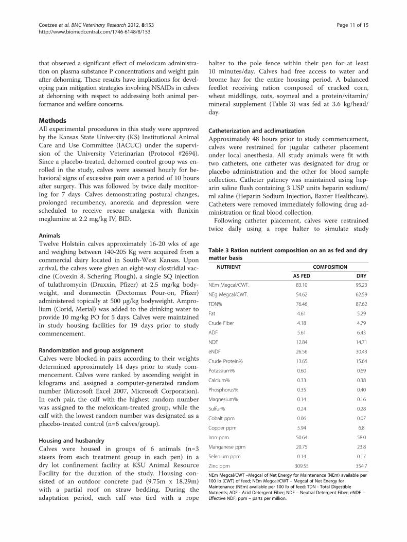
Table 3 Ration nutrient composition on an as fed and drymatter basis
NUTRIENT COMPOSITION
AS FED DRY
NEm Megcal/CWT. 83.10 95.23
NEg Megcal/CWT. 54.62 62.59
TDN% 76.46 87.62
Fat 4.61 5.29
Crude Fiber 4.18 4.79
ADF 5.61 6.43
NDF 12.84 14.71
eNDF 26.56 30.43
Crude Protein% 13.65 15.64
Potassium% 0.60 0.69
Calcium% 0.33 0.38
Phosphorus% 0.35 0.40
Magnesium% 0.14 0.16
Sulfur% 0.24 0.28
Cobalt ppm 0.06 0.07
Copper ppm 5.94 6.8
Iron ppm 50.64 58.0
Manganese ppm 20.75 23.8
Selenium ppm 0.14 0.17
Zinc ppm 309.55 354.7
NEm Megcal/CWT –Megcal of Net Energy for Maintenance (NEm) available per100 lb (CWT) of feed; NEm Megcal/CWT – Megcal of Net Energy forMaintenance (NEm) available per 100 lb of feed; TDN - Total DigestibleNutrients; ADF - Acid Detergent Fiber; NDF – Neutral Detergent Fiber; eNDF –Effective NDF; ppm – parts per million.
Coetzee et al. BMC Veterinary Research 2012, 8:153 Page 11 of 15http://www.biomedcentral.com/1746-6148/8/153
that observed a significant effect of meloxicam administra-tion on plasma substance P concentrations and weight gainafter dehorning. These results have implications for devel-oping pain mitigation strategies involving NSAIDs in calvesat dehorning with respect to addressing both animal per-formance and welfare concerns.
MethodsAll experimental procedures in this study were approvedby the Kansas State University (KS) Institutional AnimalCare and Use Committee (IACUC) under the supervi-sion of the University Veterinarian (Protocol #2694).Since a placebo-treated, dehorned control group was en-rolled in the study, calves were assessed hourly for be-havioral signs of excessive pain over a period of 10 hoursafter surgery. This was followed by twice daily monitor-ing for 7 days. Calves demonstrating postural changes,prolonged recumbency, anorexia and depression werescheduled to receive rescue analgesia with flunixinmeglumine at 2.2 mg/kg IV, BID.
AnimalsTwelve Holstein calves approximately 16-20 wks of ageand weighing between 140-205 Kg were acquired from acommercial dairy located in South-West Kansas. Uponarrival, the calves were given an eight-way clostridial vac-cine (Covexin 8, Schering Plough), a single SQ injectionof tulathromycin (Draxxin, Pfizer) at 2.5 mg/kg body-weight, and doramectin (Dectomax Pour-on, Pfizer)administered topically at 500 μg/kg bodyweight. Ampro-lium (Corid, Merial) was added to the drinking water toprovide 10 mg/kg PO for 5 days. Calves were maintainedin study housing facilities for 19 days prior to studycommencement.
Randomization and group assignmentCalves were blocked in pairs according to their weightsdetermined approximately 14 days prior to study com-mencement. Calves were ranked by ascending weight inkilograms and assigned a computer-generated randomnumber (Microsoft Excel 2007, Microsoft Corporation).In each pair, the calf with the highest random numberwas assigned to the meloxicam-treated group, while thecalf with the lowest random number was designated as aplacebo-treated control (n=6 calves/group).
Housing and husbandryCalves were housed in groups of 6 animals (n=3steers from each treatment group in each pen) in adry lot confinement facility at KSU Animal ResourceFacility for the duration of the study. Housing con-sisted of an outdoor concrete pad (9.75m x 18.29m)with a partial roof on straw bedding. During theadaptation period, each calf was tied with a rope
halter to the pole fence within their pen for at least10 minutes/day. Calves had free access to water andbrome hay for the entire housing period. A balancedfeedlot receiving ration composed of cracked corn,wheat middlings, oats, soymeal and a protein/vitamin/mineral supplement (Table 3) was fed at 3.6 kg/head/day.
Catheterization and acclimatizationApproximately 48 hours prior to study commencement,calves were restrained for jugular catheter placementunder local anesthesia. All study animals were fit withtwo catheters, one catheter was designated for drug orplacebo administration and the other for blood samplecollection. Catheter patency was maintained using hep-arin saline flush containing 3 USP units heparin sodium/ml saline (Heparin Sodium Injection, Baxter Healthcare).Catheters were removed immediately following drug ad-ministration or final blood collection.Following catheter placement, calves were restrained
twice daily using a rope halter to simulate study

Coetzee et al. BMC Veterinary Research 2012, 8:153 Page 12 of 15http://www.biomedcentral.com/1746-6148/8/153
sampling procedures. Furthermore, calves were runthrough the chute handling facilities once daily andmanipulated in the same manner as the proposed studyprocedures. Cattle were also fitted with commerciallymanufactured 3-dimensional accelerometers (GP1SENSR, Reference LLC) at described in previous studies[44]. The accelerometers were left on the calves untilstudy completion, approximately 7 days after placement.
Treatment administrationCalves were subjected to either meloxicam or placebotreatment as outlined below (n=6 steers/treatment). Doseswere calculated based on individual animal bodyweightdetermined 14 hours prior to study commencement. TheIV dose was rounded to the nearest 0.5 ml and adminis-tered using a 20 mL syringe. Meloxicam or the placebowas administered immediately (<30 seconds) prior to com-mencement of the dehorning procedure.
1) Intravenous (IV) injection of 0.5 mg/kg of meloxicam(Metacam 5 mg/ml solution for injection (NADA141-219), Boehringer Ingelheim Vetmedica, Inc; Lot# 118ZN12) was administered as a single bolus in thejugular vein using a designated catheter. The catheterwas flushed with 5 mL of heparin-saline andremoved immediately after administration.
2) Intravenous sodium chloride injection (0.9% SodiumChloride Injection USP, Baxter Healthcare Corp) wasadministered at a volume based on a presumed doseof 0.5 mg/kg meloxicam injection.
Observers and analytical chemists in the study weremasked to treatment group allocation.
DehorningPrior to dehorning, all calves were restrained in a chutewith a head gate and a rope halter. The horn was removedusing a Barnes dehorning instrument (Stone Manufactur-ing & Supply Company). Briefly, the opposing blades ofthe instrument were aligned with the base of the horns atthe skin-horn junction. The handles of the instrumentwere then closed slowly to ensure proper placement of theinstrument. Once optimal positioning was achieved, thehandles were spread quickly apart to engage the bladesand cut off the horn. Hemostasis was achieved throughthermocautery using a pre-heated electric dehorning iron(Stone Manufacturing & Supply Company). All dehorningprocedures were performed by a single experienced veter-inarian (BR). After dehorning, calves remained standingbut unrestrained in the chute for 20 minutes to facilitateintensive blood sampling. Subsequent samples were col-lected in housing pens with calves periodically restrainedusing a rope halter.
Blood sample collectionEighteen milliliters of whole blood for cortisol, substanceP (SP) and meloxicam determination was collected intosyringes using the pre-placed jugular catheter immedi-ately prior to drug or placebo administration, and at 5,10, 15, 20, 30, 60 minutes and again at 6, 22, 30, 45 and52 hours thereafter. Blood was immediately transferredto 6 ml serum and lithium heparin vacutainer tubes (BDDiagnostics) for cortisol and drug determination respect-ively. Blood for SP determination was collected in 6 mlEDTA tubes containing the serine protease inhibitor,aprotonin at 500 KIU/mL of blood. The vacutainer tubeswere stored on ice for no more than 60 minutes pendingsample processing. Thereafter, blood samples were cen-trifuged at 1,600 g for 15 minutes at 4°C. Serum andplasma were pipetted from their respective tubes andplaced in cryovials for storage at -80°C prior to sampleanalysis. All samples were analyzed within 60 days ofsample collection.
Cortisol determinationSerum cortisol concentrations were determined using asolid-phase competitive chemiluminescent enzyme im-munoassay and an automated analyzer system (Immulite1000 Cortisol, Siemens Medical Solutions Diagnostics) aspreviously described using an assay validated in bovineplasma [15,45]. A sample volume of 100 μL was used ineach assay well. The reported calibration range for theassay is 28 to 1,380 nmol/L with an analytical sensitivityof 5.5 nmol/L.
Substance P determinationPlasma substance P concentrations were determined induplicate using a validated method as previouslydescribed [25]. Briefly, Substance P was extracted fromplasma by acidifying with acetic acid and fractionatingwith reverse-phase solid-phase extraction columns. Thepeptide was eluted from the column using an organic-aqueous solvent mixture and concentrated by dryingunder nitrogen. The dried extract was reconstituted andanalyzed according to the manufacturer’s instructions inthe Substance P ELISA kit (Assay designs, Ann Arbor,MI). The coefficient of variation between triplicate bo-vine samples at each fortified SP concentration was <30%. The linear regression line fit between the threepoints at each of three control concentrations had a cor-relation coefficient of 0.99.
AccelerometersActivity data was collected for 48 and 168 hours pre-and post-dehorning, respectively. Accelerometerssampled at 100Hz and summarized values for theselected variables every 5 seconds. Five variables wererecorded by the accelerometers for each 5-second

Coetzee et al. BMC Veterinary Research 2012, 8:153 Page 13 of 15http://www.biomedcentral.com/1746-6148/8/153
interval: average acceleration in each of the three axes(X, Y, and Z), the combined average magnitude for allthree axes, and the maximum combined vector magni-tude. At trial completion, data were imported into com-mercial data mining software (Insightful Miner,Insightful Corporation), and a previously validated deci-sion tree [30,36,44] was used to classify the behavior aslying, standing, or walking for each 5 second interval.
Heart rate determinationHeart rate data were recorded for 48 hours before andafter dehorning for each calf. Heart rate was recordedand analyzed using a commercially available heart ratemonitor and software (RS800 and Polar Pro TrainerEquine Edition, Polar Electro, Inc, Lake Success, NY) aspreviously described [45]. The heart rate monitor con-sisted of a transmitter placed over the heart in the leftforeflank attached to a girth strap placed around theheart girth of the calves, and a wrist unit attached to theelastic strap which received and recorded the signal fromthe transmitter. Appropriate conductance for the electro-des on the strap, one positioned on the sternum and oneover the right scapula, was facilitated by use of ultra-sound gel (Medline Industries Inc. Mundeline, Il). Thetransmitter measured the electric signal (ECG) of theheart every 15 seconds. Prior to study commencement,the heart rate wrist unit time was synchronized with thestopwatches used for all other sample collection. Thecorresponding heart rate within 15 seconds of each timepoint was used for analysis.
Average daily weight gain (ADG)Calves were individually weighed on arrival, approxi-mately 14 hours prior to dehorning and 10 days post-dehorning using a commercial livestock scale (For-MostLivestock Equipment). Food and water were not withheldprior to weighing. Average daily gain (ADG) was calcu-lated by subtracting the arrival from the pre-dehorningweight and the pre-dehorning weight from the post-dehorning weight and dividing this by the number ofdays that passed between weigh dates.
Plasma meloxicam determinationPlasma concentrations of meloxicam (m/z 352.09!114.90)were determined with high-pressure liquid chromatography(Shimadzu Prominence, Shimadzu Scientific Instruments)and mass spectrometry (API 2000, Applied Biosystems) aspreviously described [13,46]. With a limit of quantificationof 0.025 μg/mL, the standard curve was linear from 0.025μg/mL to 10 μg/mL and was accepted if the correlation co-efficient exceeded 0.99 and predicted values were within15% of the actual values. The accuracy of the assay was103 ± 7% of the actual value and the coefficient of
variation was 7% determined on replicates of 5 each at0.025, 0.5, and 5 μg/mL.
Pharmacokinetic analysisCompartmental pharmacokinetic analysis of the meloxi-cam time concentration data was performed using a com-mercially available software program (WinNonlin,Pharsight Corporation) as previously described [15,47].Model selection was conducted using visual inspection ofpredicted versus observed data plots and two measures ofgoodness of fit (Aikaike Information Criterion andSchwarz Bayesian Criterion).
Data analysis and statisticsHypothesis tests were conducted using JMP 5.1.2 analyt-ical software (SAS Institute, INC) unless otherwise speci-fied [48]. For statistical analysis, the calf was consideredthe experimental unit. The mean ± standard error of themeans (SEM) and the mean difference where appropriatewere calculated for each outcome variable at each timepoint. Statistical significance was designated a priori atp<0.05. Model fit was assessed by evaluating the form ofmarginal studentized residuals versus fitted values plot.The model was determined appropriate if the mean ofthe residuals versus fitted values plot was centered onzero.For repeated measures data (heart rate, substance P
and cortisol concentrations at each time point), a ran-dom effects-mixed model was constructed with treat-ment, time and the interaction between time andtreatment designated as fixed effects. In this model, ani-mal nested in treatment was designated as a random ef-fect to account for the between subject effects. Wheresignificant interactions were detected, comparisons be-tween different combinations were conducted using two-sided Student t-tests. The experiment wise significancelevel was protected by employing the Tukey method inexamining comparisons. Substance P data were not nor-mally distributed and therefore these were log trans-formed prior to statistical analysis. For heart rate data,the statistical model failed to converge due to the largenumber of data points relative to the number of animalson trial and periods of missing data especially after 12 hpost-dehorning. Therefore, an initial analysis was con-ducted on the cumulative data collected in the periodsbefore and after dehorning followed by a detailed ana-lysis of the hourly data over 12 h post-dehorning.Accelerometer data were imported into a commercial
data mining software (Insightful Miner, Insightful Cor-poration, Seattle, WA), and a previously validated deci-sion tree [36,44] was used to classify the behavior aslying, standing, or walking for each 5 second interval.Following classification, data were aggregated on anhourly basis by the summing of the counts of 5-second

Coetzee et al. BMC Veterinary Research 2012, 8:153 Page 14 of 15http://www.biomedcentral.com/1746-6148/8/153
intervals spent in each behavior. Hours with known peri-ods of human intervention (feeding, sample collection,animal processing, and treatment administration) wereremoved equally from calves in both treatment groups.The remaining hourly behavioral data from both repli-cates of the trial were imported into a statistical program(SAS 9.1, SAS Institute, Cary, NC) for analysis. The pro-portion of time calves spent in each activity was modeledusing logistic regression (PROC GLIMMIX) to evaluatepotential associations between lying behavior and timerelative to dehorning (pre- or post-), treatment (Meloxi-cam or control), and the interaction between these vari-ables. Random effects were included in the model toaccount for a lack of independence in each sampling dueto multiple calves housed within the same pen andrepeated measures on individual calves. Pairwise com-parisons were performed to determine statistically sig-nificant differences.Summary measures of cortisol including peak
plasma concentration (Cmax), time to peak concentra-tion (Tmax) and area under the time-effect curve(AUEC) were determined as previously described [47]using the linear trapezoidal rule and WinNonlin soft-ware (Pharsight Corporation, Cary, NC). Analysis ofvariance (ANOVA) was employed to evaluate differ-ences in single measurement, normally distributeddata (ADG, Cmax, and AUEC). The Kruskal-WallisTest was used to analyze non-parametric data (Tmax).In order to determine the relationship between
plasma meloxicam concentrations, SP and serum cor-tisol concentrations at the corresponding time pointsafter dehorning, post hoc linear regression curves wereconstructed using the log-transformed percentagechange from baseline. Statistical significance wasdesignated a priori at p<0.05.
Competing interestsA provisional patent application (PCT/US2011/044017) titled “OralAdministration of Meloxicam in Cattle for Improved Growth followingDehorning and Reduction of Bovine Respiratory Disease upon Castration”was filed by Kansas State University on July 14, 2011.
Authors’ contributionJFC conceived the study, assisted with the animal phase of the trial,conducted the statistical analysis and prepared the manuscript forpublication. RAM assisted with the animal phase of the study, statisticalanalysis and manuscript preparation. BK conducted the meloxicam extractionand analysis. RG conducted the pharmacokinetic analysis. BR assisted withthe collection and analysis of the accelerometer data. JBR conducted thedehorning procedures and BJW conducted the statistical analysis of theaccelerometer data. All authors read and approved the final manuscript.
AcknowledgementsThis research was supported by the College of Veterinary Medicine, KansasState University. Dr Coetzee and Dr Mosher were supported by USDA-CSREES, Animal Protection (Animal Well-being), NRI Grant # 2008-35204-19238 and 2009-65120-05729.
Author details1Department of Clinical Sciences, College of Veterinary Medicine, KansasState University, 66506-5601, Manhattan, KS, USA. 2Department of Anatomyand Physiology, Kansas State University, 66506-5601, Manhattan, KS, USA.3Present address: Veterinary Diagnostic and Production Animal Medicine,College of Veterinary Medicine, Iowa State University, 50014, Ames, IA, USA.
Received: 22 May 2012 Accepted: 15 August 2012Published: 1 September 2012
References1. USDA: Dairy 2007, Heifer Calf Health and Management Practices on U.S.
Dairy Operations. 2007. USDA:APHIS:VS:CEAH. Fort Collins, CO #N550.0110 ;2009. Available at http://www.aphis.usda.gov/animal_health/nahms/dairy/downloads/dairy07/Dairy07_ir_CalfHealth.pdf. Accessed 21 May 2012.
2. American Veterinary Medical Association: AVMA Policy: Castration andDehorning of Cattle ; 2008. Available at: http://www.avma.org/issues/policy/animal_welfare/dehorning_cattle.asp. Accessed 21 May 2012.
3. European Agency for the Evaluation of Medicinal Products (EMEA) Web site:Committee for Veterinary Medicinal Products: Meloxicam. Maximum ResidueLimit (MRL) Summary Report (2); Available at http://www.emea.europa.eu/pdfs/vet/mrls/057199en.pdf; accessed on 12 November 2009.
4. Heinrich A, Duffield TF, Lissemore KD, Squires EJ, Millman ST: The impact ofmeloxicam on postsurgical stress associated with cautery dehorning. JDairy Sci 2009, 92(2):540–547.
5. Stewart M, Stookey JM, Stafford KJ, Tucker CB, Rogers AR, Dowling SK,Verkerk GA, Schaefer AL, Webster JR: Effects of local anesthetic andnonsteroidal anti-inflammatory drug on pain responses of dairy calvesto hot-iron dehorning. J Dairy Sci 2009, 92(4):1512–1519.
6. Heinrich A, Duffield TF, Lissemore KD, Millman ST: The effect of meloxicamon behavior and pain sensitivity of dairy calves following cauterydehorning with a local anesthetic. J Dairy Sci 2010, 93(6):2450–2457.
7. Ingvast-Larsson C, Hogberg M, Mengistu U, Olsen L: Pharmacokinetics ofmeloxicam in adult goat and its analgesic effect in disbudded kids. J VetPharm Ther 2010, 34:64–69.
8. Fulwider WK, Grandin T, Rollin BE, Engle TE, Dalsted NL, Lamm WD: Surveyof dairy management practices on one hundred thirteen North Centraland northeastern United States Dairies. J Dairy Sci 2008, 91(4):1686–1692.
9. Hoe FGH, Ruegg PL: Opinions and practices of Wisconsin dairy producersabout biosecurity and animal well-being. J Dairy Sci 2006, 89:2297–2308.
10. Webb AI, Pablo LS: Local Anesthetics. In Veterinary Pharmacology andTherapeutics. 9th edition. Edited by Riviere JM, Papich MG. Ames, IA, USA:Wiley-Blackwell; 2009:381–397.
11. Kissin I: Preemptive analgesia. Anesthesiology 2000, 93:1138–1143.12. Lees P: Analgesic, anti-inflammatory, antipyretic drugs. In Veterinary
pharmacology and therapeutics. 9th edition. Edited by Riviere JM, Papich MG.Ames, IA, USA: Wiley-Blackwell; 457–492.
13. Coetzee JF, KuKanich B, Mosher R, Allen PS: Pharmacokinetics ofintravenous and oral meloxicam in ruminant calves. Vet Ther 2009,10(4):E1–E8.
14. Landoni MF, Cunningham FM, Lees P: Pharmacokinetics andpharmacodynamics of ketoprofen in calves applying PK/PD modeling.J Vet Pharm Therap 1995, 18(5):315–324.
15. Coetzee JF, Gehring R, Bettenhausen AC, Lubbers BL, Thomson DU,KuKanich B, Toerber SE, Apley MD: Mitigation of plasma cortisolresponse in bulls following intravenous sodium salicylateadministration prior to castration. J Vet Pharm Therap 2007,30(4):305–313.
16. Anderson KL, Neff-Davis CALE, Davis VD: Bass: Pharmacokinetics of flunixinmeglumine in lactating cattle after single and multiple intramuscularand intravenous administrations. Amer J Vet Res 1990, 51(9):1464–1467.
17. Toutain PN, Reymond V, Laroute P, Garcia M, Popot Y, Gonnaire A, Hirsch R:Narbe. Pharmacokinetics of meloxicam in plasma and urine of horses.Am J Vet Res 2004, 65:1542–1547.
18. Jeunesse EC, Bargues IA, Toutain CE, Lacroix MZ, Letellier IM, Giraudel JM,Toutain PL: Paw inflammation model in dog. J Pharmacol Exp Ther 2011,338:548–558.
19. Sutherland MA, Mellor DJ, Stafford KJ, Gregory NG, Ward RN, Bruce RA:Cortisol responses to dehorning of calves given a 5-h localanaesthetic regimen plus phenylbutazone, ketoprofen, or

Coetzee et al. BMC Veterinary Research 2012, 8:153 Page 15 of 15http://www.biomedcentral.com/1746-6148/8/153
adrenocorticotropic hormone prior to dehorning. Res Vet Sci 2002,2002(73):115–123.
20. Sylvester SP, Mellor DJ, Stafford KJ, Bruce RA, Ward RN: Acute cortisolresponses of calves to scoop dehorning using local anaesthesia and/orcautery of the wound. Aust Vet J 1998, 76(2):118–122.
21. Schwartzkopf-Genswein KS, Booth-McLean ME, McAllister TA, Mears GJ:Physiological and behavioural changes in Holstein calves during andafter dehorning or castration. Can J Anim Sci 2005, 85:131–138.
22. American Veterinary Medical Association: Welfare implications of thedehorning and disbudding of cattle. 2010. Available at: http://www.avma.org/reference/backgrounders/dehorning_cattle_bgnd.pdf. Accessed 21 May2012.
23. McMeekan CM, Stafford KJ, Mellor DJ, Bruce RA, Ward RN, Gregory N:Effects of a local anaesthetic and a non-steroidal anti-inflammatoryanalgesic on the behavioural responses of calves to dehorning. NZVet J 1999, 47:92–96.
24. Coetzee JF: A review of pain assessment techniques and pharmacologicalapproaches to pain relief after bovine castration in the United States.Invited Review. Appl Anim Behav Sci 2011, 135(4):192–213.
25. Coetzee JF, Lubbers BL, Toerber SE, Gehring R, Thomson DU, White BJ,Apley MD: Plasma concentrations of substance P and cortisol in beefcalves after castration or simulated castration. Am J Vet Res 2008, 69(6):751–762.
26. Hopster H, Blokhuis HJ: Validation of a heart-rate monitor for measuring astress response in dairy cows. Can J Anim Sci 1994, 74:465–474.
27. Lefcourt AM, Kahl S, Akers RM: Correlation of indices of stress withintensity of electrical shock in cows. J Dairy Sci 1986, 69:833–842.
28. Lay DC, Friend TH, Randel RD, Bower CL, Grissom KK, Jenkins OC:Behavioural and physiological effects of freeze or hot-iron branding oncrossbred cattle. J Anim Sci 1992, 70:330–336.
29. Grondahl-Nielsen C, Simonsen HB, Damkjer Lund J, Hesselholt M:Behavioural, endocrine and cardiac responses in young calvesundergoing dehorning without and with the use of sedation andanalgesia. Vet J 1999, 1999(158):14–20.
30. Theurer ME, White BJ, Coetzee JF, Edwards LN, Mosher RA, Cull CA:Assessment of behavioral changes associated with oral meloxicamadministration at time of dehorning in calves using a remotetriangulation device and accelerometers. BMC Vet Res 2012, 8:48.
31. Weary DM, Huzzey JM, von Keyserlingk MAG: Board-Invited Review: Usingbehavior to predict and identify ill health in animals. J Anim Sci 2009,87(2):770–777.
32. Stanton AL, Kelton DF, Leblanc SJ, Millman ST, Wormuth J, Dingwell RT,Leslie KE: The effect of treatment with long-acting antibiotic atpostweaning movement on respiratory disease and on growth incommercial dairy calves. J Dairy Sci 2010, 93(2):574–581.
33. Hanzlicek GA, White BJ, Mosier D, Renter DG, Anderson DE: Serialevaluation of physiologic, pathological, and behavioral changes relatedto disease progression of experimentally induced Mannheimiahaemolytica pneumonia in postweaned calves. Am J Vet Res 2010,71:359–369.
34. Ito K, Weary DM, von Keyerslingk MAG: Lying behavior: Assessing within-and between- herd variation in free-stall-housed dairy cows. J Dairy Sci2009, 92:4412–4420.
35. Ito K, von Keyserlingk MAG, LeBlanc SJ, Weary DM: Lying behavior as anindicator of lameness in dairy cows. J Dairy Sci 2010, 93:3553–3560.
36. White BJ, Coetzee JF, Renter DG, Babcock AH, Thomson DU, Andresen D:Evaluation of two-dimensional accelerometers to monitor beef cattlebehavior post-castration. Amer J Vet Res 2008, 69(8):1005–1012.
37. Ting ST, Earley B, Crowe MA: Effect of repeated ketoprofen administrationduring surgical castration of bulls on cortisol, immunological function,feed intake, growth, and behavior. J Anim Sci 2003, 81:1253–1264.
38. Faulkner PM, Weary DM: Reducing pain after dehorning in dairy calves. JDairy Sci 2000, 83:2037–2041.
39. Baldridge SL, Coetzee JF, Dritz SS, Reinbold JB, Gehring R, Havel J, KukanichB: Pharmacokinetics and physiologic effects of intramuscularlyadministered xylazine hydrochloride-ketamine hydrochloride-butorphanol tartrate alone or in combination with orally administeredsodium salicylate on biomarkers of pain in Holstein calves followingcastration and dehorning. Am J Vet Res 2011, 72(10):1305–1317.
40. Friton GM, Cajal C, Ramirez-Romero R: Long-term effects of meloxicam inthe treatment of respiratory disease in fattening cattle. Vet Rec 2005,156:809–811.
41. Garry FB: Disorders of reticuloruminal motor function. In Large AnimalInternal Medicine. 3rd edition. Edited by Smith BP. St Louis, MO, USA:Mosby; 723–724.
42. Mellor DJ, Stafford KJ, Todd SE, Lowe TE, Gregory NG, Bruce RA, Ward RN: Acomparison of catecholamine and cortisol responses of young lambsand calves to painful husbandry procedures. Aust Vet J 2002,80(4):228–233.
43. Leek BF: Review: reticuloruminal motility–a pharmacological target with adifference? Vet Q 2001, 23(1):26–31.
44. Robert B, White BJ, Renter DG, Larson RL: Evaluation of three dimensionalaccelerometers as a method to categorize and classify behavior patternsin cattle. Comput Electron Agric 2009, 67:80–84.
45. Kotschwar JL, Coetzee JF, Anderson DE, Gehring R, KuKanich B: AnalgesicEfficacy of Sodium Salicylate in an Amphotericin B Induced BovineSynovitis-Arthritis Model. J Dairy Sci 2000, 92(8):3731–3743.
46. Coetzee JF, Mosher RA, Kohake LE, Cull CA, Kelly LL, Mueting SL, KuKanich B:Pharmacokinetics of oral gabapentin alone or co-administered withmeloxicam in ruminant beef calves. Vet J 2011, 190(1):98–102.
47. Gibaldi M, Perrier D: Pharmacokinetics. 2nd edition. New York, USA: MarcelDekker Inc; 1982.
48. Sall J, Creighton L, Lehman A: JMP Start Statistics. 3rd edition. Belmont,California: Thomson Brooks/Cole; 2004:363.
doi:10.1186/1746-6148-8-153Cite this article as: Coetzee et al.: Pharmacokinetics and effect ofintravenous meloxicam in weaned Holstein calves following scoopdehorning without local anesthesia. BMC Veterinary Research 2012 8:153.
Submit your next manuscript to BioMed Centraland take full advantage of:
• Convenient online submission
• Thorough peer review
• No space constraints or color figure charges
• Immediate publication on acceptance
• Inclusion in PubMed, CAS, Scopus and Google Scholar
• Research which is freely available for redistribution
Submit your manuscript at www.biomedcentral.com/submit