Acute Reactogenicity after Intramuscular Immunization with ...
10
Acute Reactogenicity after Intramuscular Immunization with Recombinant Vesicular Stomatitis Virus Is Linked to Production of IL-1b Kathleen Athearn 1,2,3 , Christopher J. Sample 1,2 , Brice E. Barefoot 1,2 , Kristi L. Williams 2,4. , Elizabeth A. Ramsburg 1,2,3 * . 1 Human Vaccine Institute, School of Medicine, Duke University, Durham, North Carolina, United States of America, 2 Department of Medicine, School of Medicine, Duke University, Durham, North Carolina, United States of America, 3 Department of Pathology, School of Medicine, Duke University, Durham, North Carolina, United States of America, 4 School of Nursing, School of Medicine, Duke University, Durham, North Carolina, United States of America Abstract Vaccines based on live viruses are attractive because they are immunogenic, cost-effective, and can be delivered by multiple routes. However, live virus vaccines also cause reactogenic side effects such as fever, myalgia, and injection site pain that have reduced their acceptance in the clinic. Several recent studies have linked vaccine-induced reactogenic side effects to production of the pro-inflammatory cytokine interleukin-1b (IL-1b) in humans. Our objective was therefore to determine whether IL-1b contributed to pathology after immunization with recombinant vesicular stomatitis virus (rVSV) vaccine vectors, and if so, to identify strategies by which IL-1b mediated pathology might be reduced without compromising immunogenicity. We found that an rVSV vaccine induced local and systemic production of IL-1b in vivo, and that accumulation of IL-1b correlated with acute pathology after rVSV immunization. rVSV-induced pathology was reduced in mice deficient in the IL-1 receptor Type I, but the IL-1R2/2 mice were fully protected from lethal rechallenge with a high dose of VSV. This result demonstrated that IL-1 contributed to reactogenicity of the rVSV, but was dispensable for induction of protective immunity. The amount of IL-1b detected in mice deficient in either caspase-1 or the inflammasome adaptor molecule ASC after rVSV immunization was not significantly different than that produced by wild type animals, and caspase- 12/2 and ASC2/2 mice were only partially protected from rVSV-induced pathology. Those data support the idea that some of the IL-1b expressed in vivo in response to VSV may be activated by a caspase-1 and ASC-independent mechanism. Together these results suggest that rVSV vectors engineered to suppress the induction of IL-1b, or signaling through the IL- 1R would be less reactogenic in vivo, but would retain their immunogenicity and protective capacity. Such rVSV would be highly desirable as either vaccine vectors or oncolytic therapies, and would likely be better tolerated in human vaccinees. Citation: Athearn K, Sample CJ, Barefoot BE, Williams KL, Ramsburg EA (2012) Acute Reactogenicity after Intramuscular Immunization with Recombinant Vesicular Stomatitis Virus Is Linked to Production of IL-1b. PLoS ONE 7(10): e46516. doi:10.1371/journal.pone.0046516 Editor: Eliane Namie Miyaji, Instituto Butantan, Brazil Received May 25, 2012; Accepted September 4, 2012; Published October 8, 2012 Copyright: ß 2012 Athearn et al. This is an open-access article distributed under the terms of the Creative Commons Attribution License, which permits unrestricted use, distribution, and reproduction in any medium, provided the original author and source are credited. Funding: Supported by United States National Institutes of Health grants AI089756 (to KLW), and AI51445 (to EAR). A portion of the work was conducted in the Global Health Research Building at Duke University, which receives support from grant number UC6 AI058607. The funders had no role in study design, data collection and analysis, decision to publish, or preparation of the manuscript. Competing Interests: The authors have declared that no competing interests exist. * E-mail: [email protected] . These authors contributed equally to this work. Introduction Vaccines based on attenuated viral vectors (e.g. poxviruses, adenovirus) are highly immunogenic, relatively inexpensive to produce, and can be delivered by multiple routes. These advantages have accelerated the development of virus-vectored vaccines, especially as concerns about emerging infectious diseases such as avian influenza and multi-drug resistant tuberculosis increase. However, pre-existing anti-vector immunity can signif- icantly impair the ability to raise immune responses to vaccination in the pre-immune host. That suggests that it will be advantageous to prioritize development of viral vaccine vectors to which pre- existing immunity is rare in the human population. Vaccine vectors based on recombinant vesicular stomatitis virus (rVSV) are highly immunogenic and can protect small animals and non- human primates against a range of infectious diseases [1,2,3,4,5]. VSV is also a potent oncolytic agent, and has been shown to be superior to nine other viruses tested against multifocal glioma [6,7] and other cancers [8,9]. Natural infection of humans with VSV is extremely uncommon, and therefore pre-existing immunity to rVSV vectors is almost non-existent [10,11,12]. Despite these advantages, development of rVSV for human clinical use continues to be delayed because of concerns about vector- associated pathology. Small laboratory animals immunized with rVSV vaccines display clinical signs of acute illness and lose up to 20% of their pre-immunization body weight in the first four days after immunization [13,14]. Although most animals recover, undesirable side effects such as these would be unacceptable in human vaccinees. Weight loss is less pronounced in non-human primates immunized with rVSVs, but the one human to date therapeutically immunized with a single cycle non-replicating experimental rVSV vaccine against Ebola virus developed a high fever and transient viremia in the first 24 hours after vector PLOS ONE | www.plosone.org 1 October 2012 | Volume 7 | Issue 10 | e46516
-
Upload
khangminh22 -
Category
Documents
-
view
3 -
download
0
Transcript of Acute Reactogenicity after Intramuscular Immunization with ...

Acute Reactogenicity after Intramuscular Immunizationwith Recombinant Vesicular Stomatitis Virus Is Linked toProduction of IL-1bKathleen Athearn1,2,3, Christopher J. Sample1,2, Brice E. Barefoot1,2, Kristi L. Williams2,4.,
Elizabeth A. Ramsburg1,2,3*.
1 Human Vaccine Institute, School of Medicine, Duke University, Durham, North Carolina, United States of America, 2 Department of Medicine, School of Medicine, Duke
University, Durham, North Carolina, United States of America, 3 Department of Pathology, School of Medicine, Duke University, Durham, North Carolina, United States of
America, 4 School of Nursing, School of Medicine, Duke University, Durham, North Carolina, United States of America
Abstract
Vaccines based on live viruses are attractive because they are immunogenic, cost-effective, and can be delivered by multipleroutes. However, live virus vaccines also cause reactogenic side effects such as fever, myalgia, and injection site pain thathave reduced their acceptance in the clinic. Several recent studies have linked vaccine-induced reactogenic side effects toproduction of the pro-inflammatory cytokine interleukin-1b (IL-1b) in humans. Our objective was therefore to determinewhether IL-1b contributed to pathology after immunization with recombinant vesicular stomatitis virus (rVSV) vaccinevectors, and if so, to identify strategies by which IL-1b mediated pathology might be reduced without compromisingimmunogenicity. We found that an rVSV vaccine induced local and systemic production of IL-1b in vivo, and thataccumulation of IL-1b correlated with acute pathology after rVSV immunization. rVSV-induced pathology was reduced inmice deficient in the IL-1 receptor Type I, but the IL-1R2/2 mice were fully protected from lethal rechallenge with a highdose of VSV. This result demonstrated that IL-1 contributed to reactogenicity of the rVSV, but was dispensable for inductionof protective immunity. The amount of IL-1b detected in mice deficient in either caspase-1 or the inflammasome adaptormolecule ASC after rVSV immunization was not significantly different than that produced by wild type animals, and caspase-12/2 and ASC2/2 mice were only partially protected from rVSV-induced pathology. Those data support the idea thatsome of the IL-1b expressed in vivo in response to VSV may be activated by a caspase-1 and ASC-independent mechanism.Together these results suggest that rVSV vectors engineered to suppress the induction of IL-1b, or signaling through the IL-1R would be less reactogenic in vivo, but would retain their immunogenicity and protective capacity. Such rVSV would behighly desirable as either vaccine vectors or oncolytic therapies, and would likely be better tolerated in human vaccinees.
Citation: Athearn K, Sample CJ, Barefoot BE, Williams KL, Ramsburg EA (2012) Acute Reactogenicity after Intramuscular Immunization with RecombinantVesicular Stomatitis Virus Is Linked to Production of IL-1b. PLoS ONE 7(10): e46516. doi:10.1371/journal.pone.0046516
Editor: Eliane Namie Miyaji, Instituto Butantan, Brazil
Received May 25, 2012; Accepted September 4, 2012; Published October 8, 2012
Copyright: � 2012 Athearn et al. This is an open-access article distributed under the terms of the Creative Commons Attribution License, which permitsunrestricted use, distribution, and reproduction in any medium, provided the original author and source are credited.
Funding: Supported by United States National Institutes of Health grants AI089756 (to KLW), and AI51445 (to EAR). A portion of the work was conducted in theGlobal Health Research Building at Duke University, which receives support from grant number UC6 AI058607. The funders had no role in study design, datacollection and analysis, decision to publish, or preparation of the manuscript.
Competing Interests: The authors have declared that no competing interests exist.
* E-mail: [email protected]
. These authors contributed equally to this work.
Introduction
Vaccines based on attenuated viral vectors (e.g. poxviruses,
adenovirus) are highly immunogenic, relatively inexpensive to
produce, and can be delivered by multiple routes. These
advantages have accelerated the development of virus-vectored
vaccines, especially as concerns about emerging infectious diseases
such as avian influenza and multi-drug resistant tuberculosis
increase. However, pre-existing anti-vector immunity can signif-
icantly impair the ability to raise immune responses to vaccination
in the pre-immune host. That suggests that it will be advantageous
to prioritize development of viral vaccine vectors to which pre-
existing immunity is rare in the human population. Vaccine
vectors based on recombinant vesicular stomatitis virus (rVSV) are
highly immunogenic and can protect small animals and non-
human primates against a range of infectious diseases [1,2,3,4,5].
VSV is also a potent oncolytic agent, and has been shown to be
superior to nine other viruses tested against multifocal glioma [6,7]
and other cancers [8,9]. Natural infection of humans with VSV is
extremely uncommon, and therefore pre-existing immunity to
rVSV vectors is almost non-existent [10,11,12]. Despite these
advantages, development of rVSV for human clinical use
continues to be delayed because of concerns about vector-
associated pathology. Small laboratory animals immunized with
rVSV vaccines display clinical signs of acute illness and lose up to
20% of their pre-immunization body weight in the first four days
after immunization [13,14]. Although most animals recover,
undesirable side effects such as these would be unacceptable in
human vaccinees. Weight loss is less pronounced in non-human
primates immunized with rVSVs, but the one human to date
therapeutically immunized with a single cycle non-replicating
experimental rVSV vaccine against Ebola virus developed a high
fever and transient viremia in the first 24 hours after vector
PLOS ONE | www.plosone.org 1 October 2012 | Volume 7 | Issue 10 | e46516
administration [15]. That result demonstrated that even single
cycle vectors can induce significant adverse side effects, and
suggested that alternate strategies to reduce residual viral vector
reactogenicity are required.
For rVSV-based vaccines to move forward to clinical use it is
important to identify ways to eliminate rVSV vector-associated
pathology that do not at the same time compromise vector
immunogenicity or oncolytic properties. It has been shown
previously that TNF-a produced in response to intranasal
immunization with rVSV vaccine vectors contributes to, but is
not the only factor responsible for, rapid weight loss after rVSV
immunization [14]. In those studies, TNF-a deficient mice were
partially protected from acute pathology after VSV challenge, and
TNF-a was not required for the generation of humoral immune
responses to rVSV [14]. We undertook the present study to
expand on those observations, focusing on the importance of IL-1.
We addressed intramuscular immunization, because that is the
most likely route of administration for rVSV vectors in humans.
Results
Mice deficient in the interleukin-1 receptor (IL-1R2/2)are protected from weight loss after immunization withVSV vaccine vectors
Mice immunized with recombinant VSV (rVSV) vaccine
vectors lose weight rapidly after immunization. Weight loss is
usually maximal by the second day after immunization but most
animals recover, with weight returning to normal by five to seven
days after immunization. Interleukin-1b (IL-1b) is a pro-inflam-
matory cytokine that causes acute weight loss and fever in mice
and humans [16,17,18], and which stimulates the production of
other pro-inflammatory mediators such as IL-6 [17,19] and
prostaglandin E2 (PGE2) [20,21]. To determine whether IL-1 can
contribute to the induction of acute pathology after administration
of rVSV vaccine vectors, we immunized groups of C57BL/6 wild
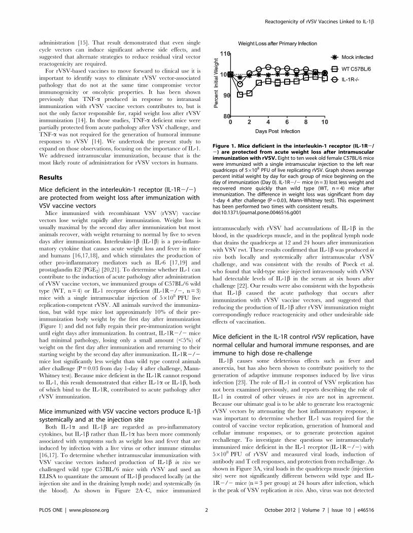
type (WT, n = 4) or IL-1 receptor deficient (IL-1R2/2, n = 3)
mice with a single intramuscular injection of 56108 PFU live
replication-competent rVSV. All animals survived the immuniza-
tion, but wild type mice lost approximately 10% of their pre-
immunization body weight by the first day after immunization
(Figure 1) and did not fully regain their pre-immunization weight
until eight days after immunization. In contrast, IL-1R2/2 mice
had minimal pathology, losing only a small amount (,5%) of
weight on the first day after immunization and returning to their
starting weight by the second day after immunization. IL-1R2/2
mice lost significantly less weight than wild type control animals
after challenge (P = 0.03 from day 1-day 4 after challenge, Mann-
Whitney test). Because mice deficient in the IL-1R cannot respond
to IL-1, this result demonstrated that either IL-1a or IL-1b, both
of which bind to the IL-1R, contributed to acute pathology after
rVSV immunization.
Mice immunized with VSV vaccine vectors produce IL-1bsystemically and at the injection site
Both IL-1a and IL-1b are regarded as pro-inflammatory
cytokines, but IL-1b rather than IL-1a has been more commonly
associated with symptoms such as weight loss and fever that are
induced by infection with a live virus or other immune stimulus
[16,17]. To determine whether intramuscular immunization with
VSV vaccine vectors induced production of IL-1b in vivo we
challenged wild type C57BL/6 mice with rVSV and used an
ELISA to quantitate the amount of IL-1b produced locally (at the
injection site and in the draining lymph node) and systemically (in
the blood). As shown in Figure 2A–C, mice immunized
intramuscularly with rVSV had accumulations of IL-1b in the
blood, in the quadriceps muscle, and in the popliteal lymph node
that drains the quadriceps at 12 and 24 hours after immunization
with VSV rwt. These results confirmed that IL-1b was produced in
vivo both locally and systemically after intramuscular rVSV
challenge, and was consistent with the results of Poeck et al.
who found that wild-type mice injected intravenously with rVSV
had detectable levels of IL-1b in the serum at six hours after
challenge [22]. Our results were also consistent with the hypothesis
that IL-1b caused the acute pathology that occurs after
immunization with rVSV vaccine vectors, and suggested that
reducing the production of IL-1b after rVSV immunization might
correspondingly reduce reactogenicity and other undesirable side
effects of vaccination.
Mice deficient in the IL-1R control rVSV replication, havenormal cellular and humoral immune responses, and areimmune to high dose re-challenge
IL-1b causes some deleterious effects such as fever and
anorexia, but has also been shown to contribute positively to the
generation of adaptive immune responses induced by live virus
infection [23]. The role of IL-1 in control of VSV replication has
not been examined previously, and reports describing the role of
IL-1 in control of other viruses in vivo are not in agreement.
Because our ultimate goal is to be able to generate less reactogenic
rVSV vectors by attenuating the host inflammatory response, it
was important to determine whether IL-1 was required for the
control of vaccine vector replication, generation of humoral and
cellular immune responses, or to generate protection against
rechallenge. To investigate these questions we intramuscularly
immunized mice deficient in the IL-1 receptor (IL-1R2/2) with
56108 PFU of rVSV and measured viral loads, induction of
antibody and T cell responses, and protection from rechallenge. As
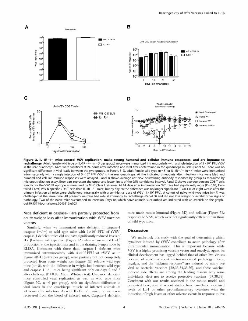
shown in Figure 3A, viral loads in the quadriceps muscle (injection
site) were not significantly different between wild type and IL-
1R2/2 mice (n = 3 per group) at 24 hours after infection, which
is the peak of VSV replication in vivo. Also, virus was not detected
Figure 1. Mice deficient in the interleukin-1 receptor (IL-1R2/2) are protected from acute weight loss after intramuscularimmunization with rVSV. Eight to ten week old female C57BL/6 micewere immunized with a single intramuscular injection to the left rearquadriceps of 56108 PFU of live replicating rVSV. Graph shows averagepercent initial weight by day for each group of mice beginning on theday of immunization (Day 0). IL-1R2/2 mice (n = 3) lost less weight andrecovered more quickly than wild type (WT, n = 4) mice afterimmunization. The difference in weight loss was significant from day1-day 4 after challenge (P = 0.03, Mann-Whitney test). This experimenthas been performed two times with consistent results.doi:10.1371/journal.pone.0046516.g001
Reactogenicity of rVSV Vaccines Linked to IL-1b
PLOS ONE | www.plosone.org 2 October 2012 | Volume 7 | Issue 10 | e46516

in the blood of any infected animal. Those results demonstrated
that IL-1 is not required for the control of VSV replication in vivo.
As shown in Figure 3B, serum neutralizing antibody titers against
VSV were not significantly different in IL-1R2/2 (n = 4 per
group per timepoint) and wild type mice (n = 5 per group per
timepoint) at any time after immunization. When we used an
MHC Class I tetramer to measure CD8 T cell responses to an
immunodominant H-2 Kb-restricted epitope (N-RGYVYQGL-C)
in the VSV N protein [24], IL-1R2/2 mice had slightly fewer
anti-VSV N specific CD8 T cells in the blood than did wild type
animals (Figure 3C) at 14 and 28 days after immunization,
although the difference was only statistically significant at 14 days
after immunization (P = 0.03, Two-tailed T test). Finally, to
determine whether these immune responses were sufficient to
protect wild type and IL-1R2/2 mice from re-challenge with
rVSV, we challenged all animals intranasally with 16108 PFU of
rVSV at eight weeks after the primary infection. As shown in
Figure 3D, pre-immune wild type and pre-immune IL-1R2/2
mice were fully protected from rechallenge, while naı̈ve wild type
animals (n = 7, open triangles, Figure 3D), lost up to 20% of their
pre-infection body weight. Two out of the seven naı̈ve animals
succumbed to infection. Together, these results indicated that IL-1
was not required either for the control of rVSV replication in vivo,
or for the generation of protective anti-VSV immune responses.
The results also supported the idea that suppressing the production
of or response to IL-1b in response to rVSV vector administration
would not render rVSV vectors unsafe or non-immunogenic.
Mice deficient in inflammasome adaptor molecule ASCare partially protected from acute weight loss afterimmunization with rVSV vaccine vectors
Because the ultimate goal of these studies was to devise
strategies by which we could suppress IL-1b production and
thereby reduce the pathology of rVSV vectors in vivo, we sought to
determine the mechanism by which IL-1b was being produced in
response to VSV. IL-1b is synthesized as an inactive precursor
molecule (pro-IL-1b), which must be cleaved either intracellularly
by endogenous protease caspase-1 [25,26,27], or extracellularly by
matrix metalloprotease 9 [28] or other neutrophil [29] and mast
cell-associated proteases [30] to become biologically active. It was
shown recently that murine bone marrow derived dendritic cells
(BMDC) infected with VSV in vitro produce IL-1b via formation of
an inflammasome composed of RNA helicase RIG-I, adaptor
molecule ASC, and caspase-1 [22]. Because the authors did not
test whether RIG-I, ASC, and caspase-1 were required to produce
IL-1b in response to VSV in vivo we used caspase-1-deficient and
ASC-deficient mice to determine the effects of the absence of these
molecules on acute pathology after rVSV immunization. It was
not practical to test RIG-I deficient mice for IL-1b induction,
because RIG-I deficient mice do not produce IFN in response to
VSV and therefore rapidly succumb to infection [31].
We challenged ASC-deficient mice (ASC2/2) and wild type
C57BL/6 mice intramuscularly with 56108 PFU rVSV as in
Figures 1, 2, and 3 and measured production of IL-1b and acute
pathology after immunization. As shown in Figure 4A, the
systemic and local production of IL-1b was not significantly
reduced in ASC2/2 mice relative wild type control mice
immunized in parallel. Consistent with that result, and with our
prediction that IL-1b induces acute weight loss after rVSV
infection, ASC2/2 (n = 5) mice lost slightly less weight after
rVSV challenge than did wild type C57BL/6 (n = 6) mice infected
in parallel, with the difference only reaching statistical significance
only on the second day after challenge (P = 0.004, Mann Whitney
Test, Figure 4B). A second experiment replicated these findings
almost exactly, with ASC2/2 mice (n = 6) showing slightly
enhanced protection relative wild type animals (n = 10, Figure S1).
The difference in weight loss between wild type and ASC2/2
mice was small but highly reproducible, and ASC2/2 mice were
never protected to the same extent as IL-1R2/2 mice. To
confirm that observation, we infected the three groups in parallel
(56108 PFU rVSV intramuscular). As shown in Figure 4C, IL-
1R2/2 mice (n = 4) lost significantly less weight than wild type
(n = 5) mice (days 1–4 after challenge P,0.05 via one way
ANOVA with Bonferroni multiple comparison test) and recovered
their pre-immunization body weight more quickly than either wild
type or ASC2/2 mice. The difference in weight loss between IL-
1R2/2 and ASC2/2 mice was significant on the first and
second day after challenge (P,0.05 via one way ANOVA with
Bonferroni multiple comparison test). Similar to the results
obtained in IL-1R2/2 mice, ASC2/2 mice made equivalent
humoral responses and slightly reduced cellular responses to VSV,
and were fully protected from high dose rechallenge (Figure 4D–
F). Taken together, these results demonstrated that production of
IL-1b in vivo after intramuscular rVSV immunization occurred
independent of inflammasome adaptor molecule ASC. Because
ASC deficient mice were not protected fully from acute pathology
after rVSV immunization, strategies that suppress the function of
ASC would be predicted to partially but not completely abrogate
acute pathology after rVSV immunization.
Figure 2. Wild type mice challenged with rVSV produce IL-1b locally and systemically. Groups of adult female C57BL/6 mice wereimmunized with two injections (56108 PFU per injection) of rVSV in each rear quadriceps, or were sham inoculated with sterile PBS. At 12 and24 hours after infection mice (n = 4 per timepoint), were sacrificed and IL-1b in the blood (Panel A), draining popliteal LN (Panel B), and quadricepsmuscle (Panel C) was quantitated via ELISA. Open and filled bars represent the 12 and 24 hour timepoints respectively. Dotted line on graphs showsthe average amount of IL-1b detected in the blood and tissues of sham-inoculated mice (n = 4). This experiment has been performed twice withconsistent results.doi:10.1371/journal.pone.0046516.g002
Reactogenicity of rVSV Vaccines Linked to IL-1b
PLOS ONE | www.plosone.org 3 October 2012 | Volume 7 | Issue 10 | e46516
Mice deficient in caspase-1 are partially protected fromacute weight loss after immunization with rVSV vaccinevectors
Similarly, when we immunized mice deficient in caspase-1
(caspase-12/2) or wild type mice with 16109 PFU of rVSV,
caspase-1 deficient mice did not have significantly reduced levels of
IL-1b relative wild type mice (Figure 5A) when we measured IL-1bproduction at the injection site and in the draining lymph node by
ELISA. Consistent with those data, caspase-1 deficient mice
immunized intramuscularly with 56108 PFU of rVSV as in
Figure 4B–C (n = 5 per group), were partially but not completely
protected from acute weight loss (Figure 5B) relative wild type
mice (n = 5), with the difference in weight loss between wild type
and caspase-12/2 mice being significant only on days 2 and 3
after challenge (P,0.05, Mann Whitney test). Caspase-1 deficient
mice controlled viral replication as well as wild type mice
(Figure 5C, n = 6 per group), with no significant difference in
viral loads in the quadriceps muscle of infected animals at
24 hours after infection. As with IL-1R2/2 mice, no virus was
recovered from the blood of infected mice. Caspase-1 deficient
mice made robust humoral (Figure 5D) and cellular (Figure 5E)
responses to VSV, which were not significantly different than those
of wild type mice.
Discussion
We undertook this study with the goal of determining which
cytokines induced by rVSV contribute to acute pathology after
intramuscular immunization. This is important because while
VSV is a highly promising vaccine vector and oncolytic agent, its
clinical development has lagged behind that of other live viruses
because of concerns about vector-associated pathology. Fever,
myalgia, and the ‘‘sickness response’’ are induced by many live
viral or bacterial vaccines [32,33,34,35,36], and these vaccine-
induced side effects are among the leading reasons why some
individuals elect not to receive protective vaccines [37,38,39].
Consistent with our results obtained in the mouse model and
presented here, several recent studies have correlated increased
levels of IL-1 or other pro-inflammatory cytokines with the
induction of high fevers or other adverse events in response to live
Figure 3. IL-1R2/2 mice control VSV replication, make strong humoral and cellular immune responses, and are immune torechallenge. Adult female wild type or IL-1R2/2 (n = 3 per group) mice were immunized intramuscularly with a single injection of 56108 PFU rVSVin the rear quadriceps. Mice were sacrificed at 24 hours after infection and viral titers determined in the quadriceps muscle (Panel A). There was nosignificant difference in viral loads between the two groups. In Panels B–D, adult female wild type (n = 5) or IL-1R2/2 (n = 4) mice were immunizedintramuscularly with a single injection of 56108 PFU rVSV in the rear quadriceps. At the indicated timepoints after infection mice were bled andhumoral and cellular immune responses were assayed. Panel B shows average anti-VSV neutralizing antibody responses by group as measured bymicroneutralization assay. Error bars represent the upper and lower limits of the 95% confidence interval. Panel C shows average percent CD8 T cellsspecific for the VSV N1 epitope as measured by MHC Class I tetramer. At 14 days after immunization, WT mice had significantly more (P = 0.03, Two-tailed T test) VSV N specific CD8 T cells than IL-1R2/2 mice, but by day 28 the difference was no longer significant (P = 0.13). At eight weeks after theprimary infection all mice were challenged intranasally with a semi-lethal dose of rVSV (16108 PFU). A cohort of naı̈ve wild type mice (n = 7) waschallenged at the same time. All pre-immune mice had robust immunity to rechallenge (Panel D) and did not lose weight or exhibit other signs ofpathology. Two of the naı̈ve mice succumbed to infection. Days on which naı̈ve animals succumbed are indicated with an asterisk on the graph.doi:10.1371/journal.pone.0046516.g003
Reactogenicity of rVSV Vaccines Linked to IL-1b
PLOS ONE | www.plosone.org 4 October 2012 | Volume 7 | Issue 10 | e46516
Figure 4. Mice deficient in the inflammasome adaptor ASC (ASC2/2) are partially protected from acute weight loss afterintramuscular immunization with rVSV. Groups of adult female C57BL/6 wild type or ASC2/2 mice were immunized with two injections(56108 PFU per injection) of rVSV in each rear quadriceps, or were sham inoculated with sterile PBS. At 12 and 24 hours after infection mice (n = atleast 4 per timepoint), were sacrificed and IL-1b in the blood (Panel A left), draining popliteal LN (Panel A middle), and quadriceps muscle (Panel Aright) was quantitated via ELISA. The amount of IL-1b produced by wild type and ASC2/2 mice was not significantly different at any time or in anyorgan. The comparison of IL-1b production by wild type and ASC2/2 mice has been performed twice with consistent results. Panel B shows averagepercent initial weight for wild type (n = 6) and ASC2/2 (n = 5) mice after intramuscular challenge with 56108 PFU of rVSV. The comparison of WT andASC2/2 mice has been performed four times with consistent results. Panel C shows average percent initial weight for wild type (n = 5), ASC2/2(n = 5), and IL-1R2/2 (n = 4) mice infected with rVSV. IL-1R2/2 mice lost significantly less weight than wild type (days 2–4) or ASC2/2 mice (days 1–2) (P,0.05 via one way ANOVA with Bonferroni test). The comparison of WT, IL-1R2/2, and ASC2/2 mice has been performed twice with consistentresults. Panel D shows average serum neutralizing titers for WT (n = ) and ASC2/2 (n = ) mice after primary immunization with VSV. There were nosignificant differences in neutralizing titer between the two groups. Panel E shows average percent CD8 T cells specific for the VSV N1 epitope asmeasured by MHC Class I tetramer. At 14 days after immunization, WT mice (n = 9) had significantly more (P = 0.001, Two-tailed T test) VSV N specificCD8 T cells than ASC2/2 mice (n = 9), but by day 28 the difference was no longer significant (P = 0.07). At eight weeks after the primary infection pre-immune WT (n = 10) and ASC2/2 (n = 6) mice were challenged intranasally with a semi-lethal dose of rVSV (16108 PFU). A cohort of naı̈ve wild type
Reactogenicity of rVSV Vaccines Linked to IL-1b
PLOS ONE | www.plosone.org 5 October 2012 | Volume 7 | Issue 10 | e46516
mice (n = 5) was challenged at the same time. All pre-immune mice had robust immunity to rechallenge (Panel F) and did not lose weight or exhibitother signs of pathology. One of the naı̈ve mice succumbed to infection.doi:10.1371/journal.pone.0046516.g004
Figure 5. Mice deficient in caspase-1 are partially protected from acute weight loss after intramuscular immunization. Groups ofadult C57BL/6 wild type or caspase 12/2 mice were immunized with two injections (56108 PFU per injection) of rVSV in each rear quadriceps, orwere sham inoculated with sterile PBS. At 24 hours after infection mice (n = 2–3 mice per timepoint), were sacrificed and IL-1b in the drainingpopliteal LN (Panel A left) and quadriceps muscle (Panel A right) was quantitated via ELISA. The amount of IL-1b produced by wild type and caspase12/2 mice was not significantly different in either organ. The comparison of IL-1b production by wild type and caspase 12/2 mice has beenperformed twice with consistent results. Panel B shows average percent initial weight for wild type and caspase 12/2 mice (n = 5 per group) afterintramuscular challenge with 56108 PFU of rVSV. The comparison of WT and caspase 12/2 mice has been performed twice with consistent results.Caspase 12/2 mice lost significantly less weight than wild type controls on days 2 and 3 after challenge (P,0.05 via Mann Whitney test). Panel Cshows average viral loads in the quadriceps muscle of infected mice (n = 6 per group). Data is compiled from two identically performed experiments.Viral loads in WT and caspase 12/2 mice were not significantly different. Panel D shows average serum neutralizing antibody titers for wild type andcaspase 12/2 mice immunized with rVSV (n = 5 per group per timepoint). Error bars represent the upper and lower limits of the 95% confidenceinterval. Panel E shows average percent 6 SEM of CD8 T cells in the blood binding to an MHC Class I tetramer recognizing an immunodominantepitope within VSV N. There were no significant differences in the antibody or CD8 T cell responses between the two groups at any time. Thecomparison of humoral and cellular immune responses in WT and caspase 12/2 mice has been performed twice with consistent results.doi:10.1371/journal.pone.0046516.g005
Reactogenicity of rVSV Vaccines Linked to IL-1b
PLOS ONE | www.plosone.org 6 October 2012 | Volume 7 | Issue 10 | e46516

virus vaccination in humans [36,40], or have identified genetic
polymorphisms in the IL-1 gene which predispose individuals to
severe adverse events after receiving live virus vaccines [41]. For
these reasons it is important to determine the ways in which pro-
inflammatory cytokines contribute to reactogenicity of VSV
vectors in particular, not only because those findings could
advance development of VSV-based therapeutics, but also because
our findings might help to inform the development of other live
virus vaccines and oncotherapies.
Previous studies in which VSV vectors were delivered to mice
intranasally showed that intranasal immunization with VSV
induces the pro-inflammatory cytokine TNF-a, and that TNF-aproduction directly correlated with weight loss and acute
pathology [14]. Although intranasal immunization has many
advantages (needle free delivery, induction of mucosal immunity,
etc), a significant drawback to intranasal immunization with rVSV
is the risk of neurotropic spread of the virus. VSV instilled in the
nose rapidly colonizes the olfactory neurons, and migrates into the
brain [42]. The neurovirulence of VSV vectors has been
significantly reduced by attenuating the capacity of the VSV
vector to replicate [43], but even a remote chance of neurotropic
spread will likely prevent use of the intranasal route for human
inoculations. Therefore, we decided to determine which cytokines
were responsible for acute pathology after intramuscular immu-
nization, which is regarded as a safer route by which to administer
potentially neurotropic agents.
We show here that mice deficient in the interleukin-1 receptor
(IL-1R) are significantly protected from weight loss after
intramuscular challenge with VSV. Although these results do
not preclude the contribution of other inflammatory processes to
pathology, they do positively identify IL-1 as an important target
for intervention.
The IL-1 receptor binds IL-1a and IL-1b. Therefore because
IL-1R2/2 mice were protected from pathology, it was possible
that IL-1a, IL-1b, or both cytokines contributed to VSV-
associated pathology in vivo. IL-1b has been well characterized as
a mediator of acute inflammatory responses in mice and humans,
namely the induction of fever and cachexia, the acute phase
response, and upregulation of other inflammatory mediators such
as IL-6 in response to infectious or non-infectious stimuli [44,45],
reviewed in [46]. IL-1b is more commonly associated with these
pathologies than is IL-1a. For example, IL-1b deficient mice, but
not IL-1a deficient mice, are protected from fever after injection of
turpentine [16,17]. Similarly, IL-1b, but not IL-1a, is the etiologic
agent of hereditary periodic fever syndromes and other ‘‘autoin-
flammatory’’ diseases in humans [47,48]. IL-1a is more commonly
associated with ‘‘sterile’’ inflammation that accompanies apoptotic
cell death [49], and with the exception of adenovirus mediated
inflammation [50] has not been associated with pathology arising
from viral infection. For these reasons, we predict that IL-1b is the
primary mediator of pathology in our system, but further
experiments will be required to formally exclude a role for IL-1a.
Once we had established that IL-1 contributed to acute
pathology after intramuscular VSV inoculation, the most impor-
tant question to pursue was whether IL-1 would be required for
control of VSV replication in vivo, and for the induction of immune
responses to the VSV vector. If IL-1 were not required for control
of virus replication, or for generation of protective immune
responses, then that would suggest that engineering rVSV vectors
to suppress IL-1 production and/or signaling would be a rational
approach to reducing rVSV associated pathology.
Although three separate reports have now described the
induction of IL-1 in response to VSV challenge in vivo
[22,51,52], none of these had examined whether or not the
induced IL-1 contributed positively to or was required for
generation of immune responses or protection. In other viral
infection models where this has been investigated, published
reports are not in agreement. For example, Schmitz et al found
that IL-1R2/2 mice infected with influenza had a significantly
higher rate of mortality than did wild type control animals, but the
increase in mortality did not correlate with a higher virus burden
in the IL-1R2/2 mice [53]. Three additional reports examined
the role of IL-1b, as well as components of the Nlrp3
inflammasome, in control of influenza infection [23,54,55]. In
these, two out of three found transiently elevated viral loads in
mice in which IL-1b production was decreased [23,54], the other
report found no difference in virus burden [55]. Similarly, only
one of the three reports [23] found that a reduction in IL-1bcorrelated with a reduction in cellular immune responses- the
others either did not examine immune responses or did not find a
correlation.
In this study, we found that IL-1R2/2 mice controlled viral
loads as well as wild type control animals. Viral loads at the
injection site were not significantly different between groups, and
neither group had a detectable viremia after challenge. This result
highlights the relative safety of the intramuscular immunization
route. Equally important was that IL-1R2/2 mice challenged
with VSV made robust humoral and cellular responses to VSV
antigens, and were fully protected from a high dose intranasal
rechallenge with VSV. Interestingly, we did observe levels of anti-
VSV CD8 T cells in the IL-1R2/2 mice and in ASC2/2 mice
were transiently but significantly decreased, which is consistent
with the report of Iwasaki et al, who found that anti-influenza CD8
T cell responses were slightly decreased in mice with reduced levels
of IL-1b production [23]. In our studies, the number of anti-VSV
specific CD8 T cells was significantly lower in IL-1R2/2 and
ASC2/2 mice at 14 days after infection, but was not significantly
different at either 7 or 28 days after infection. This finding
warrants further investigation, and follow-up studies will focus on
determining whether CD8 T cells primed in the absence of IL-1
signaling are functionally different (e.g. in cytotoxic capacity, or in
acquisition of central memory phenotype) from those primed in
IL-1 intact animals.
In summary, these results support the idea that IL-1 production
is not required either for control of VSV replication in vivo, or for
the induction of protective immune responses to VSV antigens
after intramuscular immunization. Because the relative magnitude
of immune response to foreign antigens expressed by VSV is
generally similar to the relative magnitude of the immune response
to VSVs own antigens [56], we predict that IL-1 would also not be
required for the induction of immune responses to vaccine
antigens expressed by VSV. Nonetheless, determining whether
IL-1 is important for the response to foreign (vaccine) antigens
expressed by an rVSV will be an important direction for further
study.
The second question arising from our results is how the VSV-
induced production of IL-1 might be suppressed. One means of
suppressing the biological response to IL-1 in humans is via
injection of recombinant IL-1 receptor antagonists. Currently
marketed as the drugs Anakinra or Kineret, these agents effect a
systemic suppression of IL-1a and IL-1b. Although systemic
suppression of IL-1 is an effective treatment for some conditions
(severe rheumatoid arthritis, for example) [57], systemic suppres-
sion of IL-1 also carries the risk of enhanced susceptibility to
infection [57,58]. Therefore we decided to determine whether
there might be a way of suppressing IL-1 only in the cells directly
infected with VSV, or within the focus of VSV-infected cells. To
determine the appropriate target molecule(s) to effect local
Reactogenicity of rVSV Vaccines Linked to IL-1b
PLOS ONE | www.plosone.org 7 October 2012 | Volume 7 | Issue 10 | e46516

suppression of IL-1, it was necessary to determine the mechanism
by which IL-1 was being produced in our model. Several years
ago, Muruve et al reported that adenovirus activated human
monocytes (THP-1 cells) to produce IL-1b in vitro, and that IL-1bproduction was dependent upon NLRP3 and caspase-1 [51]. In
the supplement to that manuscript, the authors reported that
infection of THP-1 cells with VSV did not induce IL-1bproduction, although detailed methods for that study were not
provided. In contrast to that, two more recent studies reported that
VSV-infected THP-1 cells do produce IL-1b in vitro. In the first
study, Poeck et al found that VSV induced production of IL-1bwas dependent upon caspase-1, but not upon NLRP3 [22]. In
contrast to that, Rajan et al found VSV mediated induction of IL-
1b from THP-1 cells to be dependent on NLRP3, and dependent
on caspase-1 [52]. It is likely that the discrepancies in these reports
were due to subtle differences in experimental procedures, but the
different outcomes reported by each of these groups highlight the
complexity of elucidating the precise mechanism(s) by which VSV
may induce IL-1.
In our studies we found that VSV induced robust production of
IL-1b in vivo and that IL-1b was still produced in mice lacking
caspase-1 or ASC. One caveat to those findings is that ELISA
assays such as the one used here do not rigorously discriminate
between detection of pro-IL-1b and detection of the cleaved active
IL-1b. Therefore it is possible that the some of the IL-1b detected
in caspase2/2 mice was not biologically active. Precise determi-
nation of the amount of active IL-1b present in caspase2/2 mice
will require the development of better detection reagents. Despite
this limitation, our results support a model in which there are
multiple pathways by which mature IL-1b is produced in vivo in
response to VSV. The data are also consistent with the idea that
the manner in which mature IL-1b is produced (via caspase-1 or
not) may vary with the cell in which the IL-1b is induced. We
further observed that mice lacking caspase-1 or ASC were not
protected from acute pathology to the same extent that IL-1R2/
2 mice were. That meant that VSV vectors engineered to
suppress components of the RIG-I/ASC/caspase-1 inflamma-
some, or the Nlrp3/ASC/caspase-1 inflammasome, would be
unlikely to be significantly less reactogenic than the parent vector.
On the other hand, rVSV designed to suppress the biological
response to IL-1b (for example via inclusion of a soluble IL-1btrap), or to IL-1a and IL-1b (via inclusion of a soluble IL-1
receptor antagonist) might reduce the biological response to IL-1
in vivo and therefore be less reactogenic.
In summary, we have shown here that IL-1 contributes to acute
pathology after intramuscular immunization with VSV. IL-1 was
not required for control of viral replication, for the induction of
cellular or humoral immune responses, or for development of
protective immunity to rechallenge. These results add to our
understanding of the role for IL-1 in promoting immunity to viral
challenge, and support the notion that the requirement for IL-1bin promoting adaptive immunity may vary according to the type
and dose of pathogen encountered as well as the route of exposure.
Finally, by identifying IL-1b as a major source of reactogenicity for
rVSV vaccines, we are able to propose a novel strategy to
ameliorate side effects without compromising immunogenicity.
Materials and Methods
Ethics StatementAll animal studies were reviewed and approved by the Duke
University Institutional Animal Care and Use Committee.
VirusesVesicular stomatitis virus (Indiana strain) was originally
obtained from Dr. John Rose (Yale University). Virus was
propagated on BHK-21 cells (ATCC CCL-10) and titered using
a standard plaque assay.
Inoculation of miceEight to ten-week-old C57BL/6 wildtype and IL-1 receptor
type 1 deficient (IL-1R2/2, strain name B6.129S7-Il1r1tm1Imx/J)
mice were obtained from Jackson Laboratories. Caspase12/2 on
the C57BL/6 background were generously provided by Dr.
Fayyaz Sutterwala and Dr. Richard Flavell. ASC2/2 and
Nlrp32/2 mice on the C57BL/6 background were generously
provided from Genentech Inc. (San Francisco, CA). Mice obtained
commercially were housed for at least 1 week before experiments
were initiated. Mice were housed in microisolator cages in a
biosafety level 2-equipped animal facility. Viral stocks were diluted
to appropriate titers in serum-free DMEM. For intramuscular
immunization (i.m.), mice were injected with the indicated amount
of virus(es) in 50 ml total volume. For intranasal (i.n.) vaccination,
mice were lightly anesthetized with isoflurane using a vaporizer
and administered the indicated amount of virus in 30 ml total
volume. The Institutional Animal Care and Use Committee of
Duke University approved all animal experiments.
Determination of viral titers by plaque assayMice were sacrificed via anesthetic overdose and organs
removed aseptically. After dissection organs were weighed, and
homogenized in sterile buffer (100 ml buffer per 0.1 g organ
weight). Homogenates were titered by standard plaque assay on
BHK-21 cells (ATCC CCL-10) using a semi-solid overlay to detect
infectious VSV. After 48 hours the overlay was removed and the
cell layer stained with crystal violet to visualize plaques.
IL-1b ELISAMice were bled and then sacrificed via anesthetic overdose and
organs removed aseptically. Organs were homogenized in 500 mL
(100 mL for lymph nodes) of buffer containing 137 mM NaCl,
20 mM Tris-Cl pH 8.0, 5 mM EDTA, 0.05% Triton-X 100, and
protease inhibitor cocktail (Roche). Serum and organ homogenate
supernatants were assayed for IL-1b by ELISA (R&D Systems).
Organ IL-1b amounts were normalized to the amount of protein
in the samples, as determined using a bicinchoninic acid (BCA)
protein assay (Thermo Scientific).
Assay for neutralizing antibody against VSVBlood was obtained from mice on days 7, 14, and 28 after
vaccination via cheek bleed. Heat inactivated serum was diluted in
serum-free DMEM such that the final dilution in the first well of a
96-well plate was 1:10 for day 7 samples and 1:100 for day 14 and
28 samples, with subsequent two-fold dilutions. Samples were
assayed in duplicate. 100 PFU of rVSV diluted in serum-free
DMEM was added to each well and incubated for one hour at
37uC-5%CO2, after which 4,000 BHK-21 cells (ATCC CCL-10)
diluted in 5%FBS-DMEM were added to each well. Plates were
incubated at 37uC-5%CO2 for three days, and cytopathic effect
was observed. The neutralizing titer was defined as the highest
dilution of serum that gave 100% neutralization of rVSV.
Tetramer assayTo obtain peripheral blood lymphocytes blood was collected
into serum free medium (DMEM) containing heparin. Blood was
layered onto a Ficoll gradient and spun, after which lymphocytes
Reactogenicity of rVSV Vaccines Linked to IL-1b
PLOS ONE | www.plosone.org 8 October 2012 | Volume 7 | Issue 10 | e46516
were collected from the interface. Cells were washed and
resuspended in DMEM containing 5% FCS.
Staining was performed on freshly isolated lymphocytes as
previously described [4]. Briefly, approximately 56106 cells were
added to the wells of a 96-well V-bottom plate and were blocked
with unconjugated streptavidin (Molecular Probes) and Fc block
(Pharmingen) for 15 min at room temperature (RT). Following a
5-min centrifugation at 5006g, lymphocytes were labeled with a
FITC-conjugated anti-CD62L antibody, (Pharmingen), an allo-
phycocyanin-conjugated anti-CD8 antibody (Pharmingen), and
tetramer for 30 min at RT. The tetramer was a PE-conjugated
major histocompatibility complex (MHC) class I Kb tetramer
(NIH Tetramer Facility) containing the H-2Kb restricted peptide
VSV N53–59 (N-RGYVYQGL-C). Sham-inoculated control ani-
mals were used to determine background levels of tetramer
binding. Background was routinely less than 0.1% and was
subtracted from all reported percentages.
Statistical AnalysisStatistical comparisons were made using GraphPad Prism
software. Results were considered significant when P,0.05.
Supporting Information
Figure S1 Mice deficient in the inflammasome adaptorASC (ASC2/2) are partially protected from acuteweight loss after intramuscular immunization withrVSV. Average percent initial weight for wild type (n = 10) and
ASC2/2 (n = 6) mice after intramuscular challenge with
56108 PFU of rVSV. The difference in weight loss between wild
type and ASC2/2 mice was significant on the first and second
day after challenge (P,0.05, Mann Whitney test).
(TIF)
Author Contributions
Conceived and designed the experiments: ER KA KW. Performed the
experiments: KA CS BB. Analyzed the data: KA CS BB ER. Contributed
reagents/materials/analysis tools: KW. Wrote the paper: ER.
References
1. Kahn JS, Roberts A, Weibel C, Buonocore L, Rose JK (2001) Replication-competent or attenuated, nonpropagating vesicular stomatitis viruses expressing
respiratory syncytial virus (RSV) antigens protect mice against RSV challenge.J Virol 75: 11079–11087.
2. Kapadia SU, Rose JK, Lamirande E, Vogel L, Subbarao K, et al. (2005) Long-
term protection from SARS coronavirus infection conferred by a singleimmunization with an attenuated VSV-based vaccine. Virology 340: 174–182.
3. Daddario-DiCaprio KM, Geisbert TW, Stroher U, Geisbert JB, Grolla A, et al.
(2006) Postexposure protection against Marburg haemorrhagic fever with
recombinant vesicular stomatitis virus vectors in non-human primates: anefficacy assessment. Lancet 367: 1399–1404.
4. Barefoot BE, Sample CJ, Ramsburg EA (2009) Recombinant vesicular stomatitis
virus expressing influenza nucleoprotein induces CD8 T-cell responses thatenhance antibody-mediated protection after lethal challenge with influenza
virus. Clin Vaccine Immunol 16: 488–498.
5. Iyer AV, Pahar B, Boudreaux MJ, Wakamatsu N, Roy AF, et al. (2009)
Recombinant vesicular stomatitis virus-based west Nile vaccine elicits stronghumoral and cellular immune responses and protects mice against lethal
challenge with the virulent west Nile virus strain LSU-AR01. Vaccine 27: 893–903.
6. Wollmann G, Tattersall P, van den Pol AN (2005) Targeting human
glioblastoma cells: comparison of nine viruses with oncolytic potential. J Virol
79: 6005–6022.
7. Ozduman K, Wollmann G, Piepmeier JM, van den Pol AN (2008) Systemicvesicular stomatitis virus selectively destroys multifocal glioma and metastatic
carcinoma in brain. J Neurosci 28: 1882–1893.
8. Stojdl DF, Lichty BD, tenOever BR, Paterson JM, Power AT, et al. (2003) VSVstrains with defects in their ability to shutdown innate immunity are potent
systemic anti-cancer agents. Cancer Cell 4: 263–275.
9. Hadaschik BA, Zhang K, So AI, Fazli L, Jia W, et al. (2008) Oncolytic vesicular
stomatitis viruses are potent agents for intravesical treatment of high-risk bladdercancer. Cancer Res 68: 4506–4510.
10. Johnson KM, Vogel JE, Peralta PH (1966) Clinical and serological response to
laboratory-acquired human infection by Indiana type vesicular stomatitis virus(VSV). Am J Trop Med Hyg 15: 244–246.
11. Fields BN, Hawkins K (1967) Human infection with the virus of vesicular
stomatitis during an epizootic. N Engl J Med 277: 989–994.
12. Tesh RB, Peralta PH, Johnson KM (1969) Ecologic studies of vesicular
stomatitis virus. I. Prevalence of infection among animals and humans living inan area of endemic VSV activity. Am J Epidemiol 90: 255–261.
13. Roberts A, Kretzschmar E, Perkins AS, Forman J, Price R, et al. (1998)
Vaccination with a recombinant vesicular stomatitis virus expressing aninfluenza virus hemagglutinin provides complete protection from influenza
virus challenge. J Virol 72: 4704–4711.
14. Publicover J, Ramsburg E, Robek M, Rose JK (2006) Rapid pathogenesis
induced by a vesicular stomatitis virus matrix protein mutant: viral pathogenesisis linked to induction of tumor necrosis factor alpha. J Virol 80: 7028–7036.
15. MacKenzie D (2009) Ebola accident puts vaccine to the test. New Scientist: 4.
16. Zheng H, Fletcher D, Kozak W, Jiang M, Hofmann KJ, et al. (1995) Resistance
to fever induction and impaired acute-phase response in interleukin-1 beta-deficient mice. Immunity 3: 9–19.
17. Fantuzzi G, Dinarello CA (1996) The inflammatory response in interleukin-1
beta-deficient mice: comparison with other cytokine-related knock-out mice.
J Leukoc Biol 59: 489–493.
18. Horai R, Asano M, Sudo K, Kanuka H, Suzuki M, et al. (1998) Production of
mice deficient in genes for interleukin (IL)-1alpha, IL-1beta, IL-1alpha/beta,
and IL-1 receptor antagonist shows that IL-1beta is crucial in turpentine-
induced fever development and glucocorticoid secretion. J Exp Med 187: 1463–
1475.
19. Lesnikov VA, Efremov OM, Korneva EA, Van Damme J, Billiau A (1991) Fever
produced by intrahypothalamic injection of interleukin-1 and interleukin-6.
Cytokine 3: 195–198.
20. Higashi S, Ohishi H, Kudo I (2000) Augmented prostaglandin E2 generation
resulting from increased activities of cytosolic and secretory phospholipase A2
and induction of cyclooxygenase-2 in interleukin-1 beta-stimulated rat calvarial
cells during the mineralizing phase. Inflamm Res 49: 102–111.
21. Angel J, Berenbaum F, Le Denmat C, Nevalainen T, Masliah J, et al. (1994)
Interleukin-1-induced prostaglandin E2 biosynthesis in human synovial cells
involves the activation of cytosolic phospholipase A2 and cyclooxygenase-2.
Eur J Biochem 226: 125–131.
22. Poeck H, Bscheider M, Gross O, Finger K, Roth S, et al. (2010) Recognition of
RNA virus by RIG-I results in activation of CARD9 and inflammasome
signaling for interleukin 1 beta production. Nat Immunol 11: 63–69.
23. Ichinohe T, Lee HK, Ogura Y, Flavell R, Iwasaki A (2009) Inflammasome
recognition of influenza virus is essential for adaptive immune responses. J Exp
Med 206: 79–87.
24. Van Bleek GM, Nathenson SG (1990) Isolation of an endogenously processed
immunodominant viral peptide from the class I H-2Kb molecule. Nature 348:
213–216.
25. Thornberry NA, Bull HG, Calaycay JR, Chapman KT, Howard AD, et al.
(1992) A novel heterodimeric cysteine protease is required for interleukin-1 beta
processing in monocytes. Nature 356: 768–774.
26. Kostura MJ, Tocci MJ, Limjuco G, Chin J, Cameron P, et al. (1989)
Identification of a monocyte specific pre-interleukin 1 beta convertase activity.
Proc Natl Acad Sci U S A 86: 5227–5231.
27. Cerretti DP, Kozlosky CJ, Mosley B, Nelson N, Van Ness K, et al. (1992)
Molecular cloning of the interleukin-1 beta converting enzyme. Science 256: 97–
100.
28. Schonbeck U, Mach F, Libby P (1998) Generation of biologically active IL-1
beta by matrix metalloproteinases: a novel caspase-1-independent pathway of
IL-1 beta processing. J Immunol 161: 3340–3346.
29. Coeshott C, Ohnemus C, Pilyavskaya A, Ross S, Wieczorek M, et al. (1999)
Converting enzyme-independent release of tumor necrosis factor alpha and IL-
1beta from a stimulated human monocytic cell line in the presence of activated
neutrophils or purified proteinase 3. Proc Natl Acad Sci U S A 96: 6261–6266.
30. Mizutani H, Schechter N, Lazarus G, Black RA, Kupper TS (1991) Rapid and
specific conversion of precursor interleukin 1 beta (IL-1 beta) to an active IL-1
species by human mast cell chymase. J Exp Med 174: 821–825.
31. Kato H, Takeuchi O, Sato S, Yoneyama M, Yamamoto M, et al. (2006)
Differential roles of MDA5 and RIG-I helicases in the recognition of RNA
viruses. Nature 441: 101–105.
32. Brydon L, Walker C, Wawrzyniak A, Whitehead D, Okamura H, et al. (2009)
Synergistic effects of psychological and immune stressors on inflammatory
cytokine and sickness responses in humans. Brain Behav Immun 23: 217–224.
33. Frey SE, Couch RB, Tacket CO, Treanor JJ, Wolff M, et al. (2002) Clinical
responses to undiluted and diluted smallpox vaccine. N Engl J Med 346: 1265–
1274.
Reactogenicity of rVSV Vaccines Linked to IL-1b
PLOS ONE | www.plosone.org 9 October 2012 | Volume 7 | Issue 10 | e46516

34. Usonis V, Bakasenas V, Kaufhold A, Chitour K, Clemens R (1999)
Reactogenicity and immunogenicity of a new live attenuated combined measles,
mumps and rubella vaccine in healthy children. Pediatr Infect Dis J 18: 42–48.
35. Vestergaard M, Hviid A, Madsen KM, Wohlfahrt J, Thorsen P, et al. (2004)
MMR vaccination and febrile seizures: evaluation of susceptible subgroups and
long-term prognosis. JAMA 292: 351–357.
36. Izurieta HS, Haber P, Wise RP, Iskander J, Pratt D, et al. (2005) Adverse events
reported following live, cold-adapted, intranasal influenza vaccine. JAMA 294:
2720–2725.
37. Wicker S, Rabenau HF, Doerr HW, Allwinn R (2009) Influenza vaccination
compliance among health care workers in a German university hospital.
Infection 37: 197–202.
38. Opstelten W, Hak E, Verheij TJ, van Essen GA (2001) Introducing a
pneumococcal vaccine to an existing influenza immunization program:
vaccination rates and predictors of noncompliance. Am J Med 111: 474–479.
39. Velan B, Kaplan G, Ziv A, Boyko V, Lerner-Geva L (2011) Major motives in
non-acceptance of A/H1N1 flu vaccination: the weight of rational assessment.
Vaccine 29: 1173–1179.
40. Farez MF, Correale J (2011) Yellow Fever Vaccination and Increased Relapse
Rate in Travelers With Multiple Sclerosis. Arch Neurol.
41. Stanley SL, Jr., Frey SE, Taillon-Miller P, Guo J, Miller RD, et al. (2007) The
immunogenetics of smallpox vaccination. J Infect Dis 196: 212–219.
42. Plakhov IV, Arlund EE, Aoki C, Reiss CS (1995) The earliest events in vesicular
stomatitis virus infection of the murine olfactory neuroepithelium and entry of
the central nervous system. Virology 209: 257–262.
43. Clarke DK, Nasar F, Lee M, Johnson JE, Wright K, et al. (2007) Synergistic
attenuation of vesicular stomatitis virus by combination of specific G gene
truncations and N gene translocations. J Virol 81: 2056–2064.
44. Finck BN, Johnson RW (1997) Anorexia, weight loss and increased plasma
interleukin-6 caused by chronic intracerebroventricular infusion of interleukin-
1beta in the rat. Brain Res 761: 333–337.
45. Samad TA, Moore KA, Sapirstein A, Billet S, Allchorne A, et al. (2001)
Interleukin-1beta-mediated induction of Cox-2 in the CNS contributes to
inflammatory pain hypersensitivity. Nature 410: 471–475.
46. Dinarello CA (2011) A clinical perspective of IL-1beta as the gatekeeper of
inflammation. Eur J Immunol 41: 1203–1217.
47. Hoffman HM, Mueller JL, Broide DH, Wanderer AA, Kolodner RD (2001)
Mutation of a new gene encoding a putative pyrin-like protein causes familialcold autoinflammatory syndrome and Muckle-Wells syndrome. Nat Genet 29:
301–305.
48. Lachmann HJ, Quartier P, So A, Hawkins PN (2011) The emerging role ofinterleukin-1beta in autoinflammatory diseases. Arthritis Rheum 63: 314–324.
49. Chen CJ, Kono H, Golenbock D, Reed G, Akira S, et al. (2007) Identification ofa key pathway required for the sterile inflammatory response triggered by dying
cells. Nat Med 13: 851–856.
50. Di Paolo NC, Miao EA, Iwakura Y, Murali-Krishna K, Aderem A, et al. (2009)Virus binding to a plasma membrane receptor triggers interleukin-1 alpha-
mediated proinflammatory macrophage response in vivo. Immunity 31: 110–121.
51. Muruve DA, Petrilli V, Zaiss AK, White LR, Clark SA, et al. (2008) Theinflammasome recognizes cytosolic microbial and host DNA and triggers an
innate immune response. Nature 452: 103–107.
52. Rajan JV, Rodriguez D, Miao EA, Aderem A (2011) The NLRP3 inflamma-some detects encephalomyocarditis virus and vesicular stomatitis virus infection.
J Virol 85: 4167–4172.53. Schmitz N, Kurrer M, Bachmann MF, Kopf M (2005) Interleukin-1 is
responsible for acute lung immunopathology but increases survival of respiratory
influenza virus infection. J Virol 79: 6441–6448.54. Allen IC, Scull MA, Moore CB, Holl EK, McElvania-TeKippe E, et al. (2009)
The NLRP3 inflammasome mediates in vivo innate immunity to influenza Avirus through recognition of viral RNA. Immunity 30: 556–565.
55. Thomas PG, Dash P, Aldridge JR, Jr., Ellebedy AH, Reynolds C, et al. (2009)The intracellular sensor NLRP3 mediates key innate and healing responses to
influenza A virus via the regulation of caspase-1. Immunity 30: 566–575.
56. Ramsburg EA, Publicover JM, Coppock D, Rose JK (2007) Requirement forCD4 T cell help in maintenance of memory CD8 T cell responses is epitope
dependent. J Immunol 178: 6350–6358.57. Furst DE (2004) Anakinra: review of recombinant human interleukin-I receptor
antagonist in the treatment of rheumatoid arthritis. Clin Ther 26: 1960–1975.
58. Nigrovic PA, Mannion M, Prince FH, Zeft A, Rabinovich CE, et al. (2011)Anakinra as first-line disease-modifying therapy in systemic juvenile idiopathic
arthritis: report of forty-six patients from an international multicenter series.Arthritis Rheum 63: 545–555.
Reactogenicity of rVSV Vaccines Linked to IL-1b
PLOS ONE | www.plosone.org 10 October 2012 | Volume 7 | Issue 10 | e46516