An Adeno-Associated Virus-Based Intracellular Sensor of Pathological Nuclear Factor-κB Activation...
15
An Adeno-Associated Virus-Based Intracellular Sensor of Pathological Nuclear Factor-kB Activation for Disease- Inducible Gene Transfer Abdelwahed Chtarto 1,2 *, Olivier Bockstael 1,2. , Elias Gebara 3. , Katia Vermoesen 4 , Catherine Melas 1,2 , Catherine Pythoud 5 , Marc Levivier 5 , Olivier De Witte 1 , Ruth Luthi-Carter 3 , Ralph Clinkers 4 , Liliane Tenenbaum 5 1 Laboratory of Experimental Neurosurgery, Universite ´ Libre de Bruxelles (ULB), Brussels, Belgium, 2 Faculty of Medecine, Institut de Recherche Interdisciplinaire en Biologie Humaine et Mole ´ culaire (I.R.I.B.H.M.), Universite ´ Libre de Bruxelles (ULB), Brussels, Belgium, 3 Brain and Mind Institute, Ecole Polytechnique Fe ´de ´rale de Lausanne (EPFL), Lausanne, Switzerland, 4 Department of Pharmaceutical Chemistry and Drug Analysis, Center for Neurosciences (C4F), Faculty of Medecine and Pharmacy, Vrije Universiteit Brussel, Brussels, Belgium, 5 Department of Clinical Neuroscience, Lausanne University Hospital, Lausanne, Switzerland Abstract Stimulation of resident cells by NF-kB activating cytokines is a central element of inflammatory and degenerative disorders of the central nervous system (CNS). This disease-mediated NF-kB activation could be used to drive transgene expression selectively in affected cells, using adeno-associated virus (AAV)-mediated gene transfer. We have constructed a series of AAV vectors expressing GFP under the control of different promoters including NF-kB -responsive elements. As an initial screen, the vectors were tested in vitro in HEK-293T cells treated with TNF-a. The best profile of GFP induction was obtained with a promoter containing two blocks of four NF-kB -responsive sequences from the human JCV neurotropic polyoma virus promoter, fused to a new tight minimal CMV promoter, optimally distant from each other. A therapeutical gene, glial cell line-derived neurotrophic factor (GDNF) cDNA under the control of serotype 1-encapsidated NF-kB -responsive AAV vector (AAV-NF) was protective in senescent cultures of mouse cortical neurons. AAV-NF was then evaluated in vivo in the kainic acid (KA)-induced status epilepticus rat model for temporal lobe epilepsy, a major neurological disorder with a central pathophysiological role for NF-kB activation. We demonstrate that AAV-NF, injected in the hippocampus, responded to disease induction by mediating GFP expression, preferentially in CA1 and CA3 neurons and astrocytes, specifically in regions where inflammatory markers were also induced. Altogether, these data demonstrate the feasibility to use disease-activated transcription factor-responsive elements in order to drive transgene expression specifically in affected cells in inflammatory CNS disorders using AAV-mediated gene transfer. Citation: Chtarto A, Bockstael O, Gebara E, Vermoesen K, Melas C, et al. (2013) An Adeno-Associated Virus-Based Intracellular Sensor of Pathological Nuclear Factor-kB Activation for Disease-Inducible Gene Transfer. PLoS ONE 8(1): e53156. doi:10.1371/journal.pone.0053156 Editor: Rossella Rota, Ospedale Pediatrico Bambino Gesu ` , Italy Received June 22, 2012; Accepted November 26, 2012; Published January 3, 2013 Copyright: ß 2013 Chtarto et al. This is an open-access article distributed under the terms of the Creative Commons Attribution License, which permits unrestricted use, distribution, and reproduction in any medium, provided the original author and source are credited. Funding: This work was supported by FNRS-Te ´le ´vie: grant number: 7.4.593.08 F. ‘‘Cre ´dit aux chercheurs’’ from the Belgian National Research Foundation: grant number: 1.5.093.08, ‘‘Fonds National de la Recherche Scientifique Me ´dicale’’ grant number 7.4.536.09 F. ‘‘Re ´ gion Bruxelles-Capitale’’ (SOIB) grant REGAVEC 2007– 10, Swiss National Research Foundation (FNS) grant number: FN31003A-127177, Agency for Innovation by Science and Technology (IWT Vlaanderen), and Research Foundation Flanders (FWO Vlaanderen). The funders had no role in study design, data collection and analysis, decision to publish, or preparation of the manuscript. Competing Interests: The authors have declared that no competing interests exist. * E-mail: [email protected] . These authors contributed equally to this work. Introduction The clinical implementation of drug-dependent regulatable viral vectors for gene therapy has been hampered by the adverse effects of the pharmacologic inducer and the immunogenicity of the artificial transactivator generally containing viral and/or bacterial elements [1,2]. Ideally, gene therapy for CNS diseases should be modulated according to the severity and the evolution of the disease. In addition, the transgene should be specifically expressed in the affected brain region and cell types. As an alternative to drug-mediated regulatory systems, we considered the possibility that regulatory DNA sequences binding transcription factors that are activated in pathological conditions could be used to generate pathology-inducible promoters responding to specific endogenous stimuli [3–5]. The NF-kB family of transcription factors consists of 5 members, defined by their Rel homology region, forming homo- and heterodimer complexes [6]. While inactive, NK-kB dimers are bound to IkB proteins and retained in the cytoplasm. During activation, IkB is degraded and the NF-kB transcription factors are released and translocate to the nucleus where they regulate the transcription of numerous genes [7]. Stimulation of the NF-kB pathway is a key pathologic component of numerous CNS disorders [8]. In particular, NFkB activation is a major pathogenic component in the kainic acid (KA)-induced rat model of status epilepticus (SE) [9,10], one of the most widely used and best characterized animal models for temporal lobe epilepsy (TLE) [11–14]. This model displays characteristic neuropathological aspects of the disease including PLOS ONE | www.plosone.org 1 January 2013 | Volume 8 | Issue 1 | e53156
Transcript of An Adeno-Associated Virus-Based Intracellular Sensor of Pathological Nuclear Factor-κB Activation...
An Adeno-Associated Virus-Based Intracellular Sensor ofPathological Nuclear Factor-kB Activation for Disease-Inducible Gene TransferAbdelwahed Chtarto1,2*, Olivier Bockstael1,2., Elias Gebara3., Katia Vermoesen4, Catherine Melas1,2,
Catherine Pythoud5, Marc Levivier5, Olivier De Witte1, Ruth Luthi-Carter3, Ralph Clinkers4,
Liliane Tenenbaum5
1 Laboratory of Experimental Neurosurgery, Universite Libre de Bruxelles (ULB), Brussels, Belgium, 2 Faculty of Medecine, Institut de Recherche Interdisciplinaire en
Biologie Humaine et Moleculaire (I.R.I.B.H.M.), Universite Libre de Bruxelles (ULB), Brussels, Belgium, 3 Brain and Mind Institute, Ecole Polytechnique Federale de Lausanne
(EPFL), Lausanne, Switzerland, 4 Department of Pharmaceutical Chemistry and Drug Analysis, Center for Neurosciences (C4F), Faculty of Medecine and Pharmacy, Vrije
Universiteit Brussel, Brussels, Belgium, 5 Department of Clinical Neuroscience, Lausanne University Hospital, Lausanne, Switzerland
Abstract
Stimulation of resident cells by NF-kB activating cytokines is a central element of inflammatory and degenerative disordersof the central nervous system (CNS). This disease-mediated NF-kB activation could be used to drive transgene expressionselectively in affected cells, using adeno-associated virus (AAV)-mediated gene transfer. We have constructed a series of AAVvectors expressing GFP under the control of different promoters including NF-kB -responsive elements. As an initial screen,the vectors were tested in vitro in HEK-293T cells treated with TNF-a. The best profile of GFP induction was obtained with apromoter containing two blocks of four NF-kB -responsive sequences from the human JCV neurotropic polyoma viruspromoter, fused to a new tight minimal CMV promoter, optimally distant from each other. A therapeutical gene, glial cellline-derived neurotrophic factor (GDNF) cDNA under the control of serotype 1-encapsidated NF-kB -responsive AAV vector(AAV-NF) was protective in senescent cultures of mouse cortical neurons. AAV-NF was then evaluated in vivo in the kainicacid (KA)-induced status epilepticus rat model for temporal lobe epilepsy, a major neurological disorder with a centralpathophysiological role for NF-kB activation. We demonstrate that AAV-NF, injected in the hippocampus, responded todisease induction by mediating GFP expression, preferentially in CA1 and CA3 neurons and astrocytes, specifically in regionswhere inflammatory markers were also induced. Altogether, these data demonstrate the feasibility to use disease-activatedtranscription factor-responsive elements in order to drive transgene expression specifically in affected cells in inflammatoryCNS disorders using AAV-mediated gene transfer.
Citation: Chtarto A, Bockstael O, Gebara E, Vermoesen K, Melas C, et al. (2013) An Adeno-Associated Virus-Based Intracellular Sensor of Pathological NuclearFactor-kB Activation for Disease-Inducible Gene Transfer. PLoS ONE 8(1): e53156. doi:10.1371/journal.pone.0053156
Editor: Rossella Rota, Ospedale Pediatrico Bambino Gesu, Italy
Received June 22, 2012; Accepted November 26, 2012; Published January 3, 2013
Copyright: � 2013 Chtarto et al. This is an open-access article distributed under the terms of the Creative Commons Attribution License, which permitsunrestricted use, distribution, and reproduction in any medium, provided the original author and source are credited.
Funding: This work was supported by FNRS-Televie: grant number: 7.4.593.08 F. ‘‘Credit aux chercheurs’’ from the Belgian National Research Foundation: grantnumber: 1.5.093.08, ‘‘Fonds National de la Recherche Scientifique Medicale’’ grant number 7.4.536.09 F. ‘‘Region Bruxelles-Capitale’’ (SOIB) grant REGAVEC 2007–10, Swiss National Research Foundation (FNS) grant number: FN31003A-127177, Agency for Innovation by Science and Technology (IWT Vlaanderen), andResearch Foundation Flanders (FWO Vlaanderen). The funders had no role in study design, data collection and analysis, decision to publish, or preparation of themanuscript.
Competing Interests: The authors have declared that no competing interests exist.
* E-mail: [email protected]
. These authors contributed equally to this work.
Introduction
The clinical implementation of drug-dependent regulatable
viral vectors for gene therapy has been hampered by the adverse
effects of the pharmacologic inducer and the immunogenicity of
the artificial transactivator generally containing viral and/or
bacterial elements [1,2]. Ideally, gene therapy for CNS diseases
should be modulated according to the severity and the evolution of
the disease. In addition, the transgene should be specifically
expressed in the affected brain region and cell types. As an
alternative to drug-mediated regulatory systems, we considered the
possibility that regulatory DNA sequences binding transcription
factors that are activated in pathological conditions could be used
to generate pathology-inducible promoters responding to specific
endogenous stimuli [3–5].
The NF-kB family of transcription factors consists of 5
members, defined by their Rel homology region, forming homo-
and heterodimer complexes [6].
While inactive, NK-kB dimers are bound to IkB proteins and
retained in the cytoplasm. During activation, IkB is degraded and
the NF-kB transcription factors are released and translocate to the
nucleus where they regulate the transcription of numerous genes
[7]. Stimulation of the NF-kB pathway is a key pathologic
component of numerous CNS disorders [8]. In particular, NFkB
activation is a major pathogenic component in the kainic acid
(KA)-induced rat model of status epilepticus (SE) [9,10], one of the
most widely used and best characterized animal models for
temporal lobe epilepsy (TLE) [11–14]. This model displays
characteristic neuropathological aspects of the disease including
PLOS ONE | www.plosone.org 1 January 2013 | Volume 8 | Issue 1 | e53156
hippocampal sclerosis (neuron loss and gliosis), neuroinflamma-
tion, synaptic reorganization, such as mossy fibre sprouting, and
the chronic recurrence of spontaneous seizures [15–19].
NFkB activation resulting from increases in inflammatory
cytokines has also been involved in pathological processes related
to aging [20] and Alzheimer Disease [21]. Additional data suggest
that NFkB activation is not an autocompensatory response, since
NFkB activation fails to protect neurons against apoptosis
associated with long-term culturing or dual TNFa and amyloid-
beta toxicity [22].
Harnessing disease-mediated NF-kB activation to drive trans-
gene expression in CNS cells could constitute an interesting
intracellular approach to anti-inflammatory intervention [23].
Several lines of transgenic reporter mice using tandem NFkB-
responsive promoter sequences have exhibited heterogeneous
phenotypes. This can likely be explained by the fact that the Rel
family of transcription factors includes numerous variants whose
abundance varies depending on the tissue and which bind with
differential affinities to different variants of the NFkB response
element (NFkB-RE). We therefore reviewed these data carefully
prior to selecting NFkB-REs for testing in our regulatable gene
therapy vectors. Transgenic mice using the NFkB responsive
sequence from the Igk light chain promoter mainly express the
reporter gene in immune organs and intestine and transgene
expression is further inducible by TNFa, IL1b or LPS in other
organs such as lungs and liver [24]. In contrast, the NFkB
responsive element present in the HIV promoter is expressed in
immune organs, but is also constitutively active in the CNS [25].
The regulatory region of human neurotropic polyoma virus JCV
which contains a NFkB-RE variant that differs from the HIV
sequence by only one nucleotide [26] is active in infected
astrocytes and oligodendrocytes presumably due to TNFastimulation of these glial cells [27].
A neuroinflammation-responsive AAV vector based on JCV
NFkB-RE consensus transcriptional sequences was designed. We
first tested this NFkB-inducible vector in an in vitro model of brain
aging [28] and showed that its delivery of glial cell line-derived
neurotrophic factor (GDNF) cDNA under the control of the
NFkB-inducible promoter enhanced survival of aging cortical
neurons in culture. We subsequently showed that hippocampally-
delivered, the NFkB-inducible AAV vector responded to systemic
KA injection in neurons and astrocytes in specific subregions of
the CA1, CA3 hippocampal layers and stratum orens where
inflammatory markers were also induced. Altogether, these results
bode positively for the utilization of our novel pathology-inducible
vector design for anti-inflammatory gene transfer applications.
Materials and Methods
PlasmidsThe pNFkB-d2EGFP plasmid containing 4 NFkB responsive
element (NFkBRE) fused to a minimal thymidine kinase promoter
(mTK) was purchased from Clontech (Palo Alto, CA, USA) The
pHpaI-EGFP self-complementary AAV vector was a kind gift
from D.McCarty and RJ Samulski [29].
We first constructed pSC-NF4-mTK-d2EGFP by replacing the
EcoRI-SalI fragment of pHpaI-EGFP containing the CMV
promoter, EGFP coding sequence and SV40 polyA with a NotI-
SalI fragment from pNFkB-d2EGFP containing a transcription
blocker site, a composite NFkB-responsive promoter (4 copies of
the NFkB-responsive element fused to a minimal thymidine kinase
promoter), destabilized GFP (d2EGFP) and SV40 polyA. Into the
obtained vector, d2EGFP was replaced with EGFP to generate
pSC-NF4-mTK-EGFP (Fig. 1A). The pSC-mCMVD2-EGFP was
derived by replacing the mTK promoter in pSC-NF4-mTK-
EGFP with a new modified minimal CMV promoter (mCMV)
named mCMVD2 in which sequences 59and 39to the TATA box
were deleted. To obtain pSC-NF4-d1-EGFP and pSC-NF4-d3-
EGFP we increased the distance between the NFkBRE and the
mCMVD2 promoter by introducing stuffer sequences of various
lengths. The pSC-NF-Ctrl was obtained by excising a fragment
containing NFkBRE in pSC-NF4-mCMVD2-EGFP. To construct
pSC-NF8-d1-EGFP and pSC-NF12-EGFP, we replaced the NF4
in pSC-NF4-mCMVD2-EGFP respectively by two or three blocks
of 4 NFkBRE, separated by 16 bp. The pSC-NF8-d1-GDNF
vector was obtained by replacing the EGFP coding sequence in
pSC-NF8-d1-EGFP by the human GDNF cDNA (a kind gift from
Dr Nicole Deglon, Lausanne Switzerland).
Cell LineThe HEK-293T cell line was purchased from Q-One Biotech
(Glasgow, UK) and cultured in Dulbecco’s modified Eagle’s
medium (DMEM) supplemented with 10% FCS (Gibco BRL, Life
Technologies, Merelbeke, Belgium).
TransfectionsFor the analysis of the inducibility of the vectors, HEK-293T
cells were transfected using the calcium phosphate coprecipitation
method. Fifty-thousand cells in 6-wells were transfected with
250 ng DNA. Forty-eight hours later and 5 hours before analysis,
transfected cells were treated or not treated with TNFa(Invitrogen) (100 ng/ml). Cells transfected with AAV-NF-EGFP
were analyzed for GFP expression on a FACStar analyser/sorter
(Becton Dickinson). To analyse the inducibility of AAV-NF-
hGDNF vectors in HEK 293T, the supernatants were harvested
4 h after changing the medium to measure GDNF concentration
using a commercial ELISA assay (Human GDNF CytoSets,
catalog #CHC2423, BioSource, Nivelles, Belgium). The transfec-
tion efficiency was normalized using a plasmid constitutively
expressing GFP protein.
Viral ProductionTo produce recombinant AAV2/1 viral stocks, HEK-293T cells
(5.06106 cells seeded on 10 cm plates) were cotransfected, in a 1:1
molar ratio, with the vector plasmid (3 mg/plate) together with the
helper/packaging plasmid pD1rs (10 mg/plate) expressing the
AAV viral genes (rep gene from AAV serotype 2 and cap gene
from AAV serotype 1) and the adenoviral genes required for AAV
replication and encapsidation (Plasmid Factory, Heidelberg). Fifty
hours post-transfection, the medium was discarded and the cells
were harvested by low-speed centrifugation and resuspended in
Tris pH 8.0, NaCl 0.1 M. After three cycles of freezing/thawing,
the lysate was clarified by 30 min centrifugation at 10 000 g,
treated with DNase at 37uC for 30 min, and centrifuged at 10
000 g for 30 min to eliminate the residual debris. The virus was
further purified by iodixanol gradient followed by QXL-sepharose
chromatography, according to a well-established method [30].
Viral genomes (vg) were titrated by quantitative PCR as previously
described [31]. Titers were 1.9561010 and 7.361011 vg/ml
respectively for rAAV1-NF8-d1-EGFP and rAAV1-NF8-d1-
hGDNF.
Cultures and Infection of Cortical NeuronsNeuronal cultures were prepared from embryonic day 16
Sprague-Dawley rat fetuses using methods similar to those
described previously [32].
Nuclear Factor-kB-Inducible Viral Vector
PLOS ONE | www.plosone.org 2 January 2013 | Volume 8 | Issue 1 | e53156
Neurons were plated in 96-well or 48-well dishes (CostarTM)
previously coated with poly-L-lysine (MW 30,000–70,000).
Half of the medium was replaced, weekly, with freshly prepared
NeurobasalTM medium (Invitrogen) supplemented with 2% B27
(Invitrogen), 16 penicillin-streptomycin, 0,5 mM L-glutamine,
and 15 mM KCl. On in vitro Day 3, cells were infected at a
multiplicity of infection of 104 vg/cell.
ImmunocytochemistryPrimary cultures were fixed with 4% paraformaldehyde (PFA)
for 20 min at 4uC. Cultures were washed 3610 min with PBS and
then incubated for 1 h in a blocking solution of PBS supplemented
with 10% Bovine Serum Albumin (PAA laboratories GmbH,
Austria) and 0.1% Triton in PBS. The cells were then incubated
overnight at 4uC in a blocking solution containing mouse
monoclonal anti-NeuN antibody (1:400, Chemicon), goat poly-
clonal anti-PSD95 (1:500, Abcam).
Cells were washed 3610 min with PBS and then incubated for
1 h with a fluorescent secondary Cy3-conjugated goat anti-mouse
antibody (1:1000, Jackson ImmunoResearch Laboratories), Alexa
660 donkey anti-goat antibody (1:500, Invitrogen) followed by
3610 min PBS washes.
Images of immunostained cells were acquired with a BD
pathway 435 Openaccess instrument for cell counting. Image
analysis was performed with ImageJ software (US NIH).
For activated NFkB staining, the same protocol was applied
using a mouse monoclonal IgG3 anti-activated NFkB recognizing
an epitope overlapping the nuclear location signal of the p65
subunit of the NFkB heterodimer thus selectively binding to the
activated form of NFkB [33] (Millipore, catalog # MAB3026).
The antibody was diluted 1:100 as primary antibody and a biotin-
conjugated donkey anti-mouse IgG (Jackson ImmunoResearch,
cat number: 715-065-150) diluted 1:500 was used as secondary
antibody. For nuclear counterstainings, cells were incubated in the
Figure 1. Description of the NF-kB-responsive AAV vectors. A. Schematic diagram of NF-kB-responsive AAV vectors used in this study. ITR:AAV Inverted Terminal Repeat; TB: Transcriptional Blocker; NFkBRE (4X): four copies of the JC virus NFkB consensus sequence; mTK: herpes simplexvirus minimal thymidine kinase promoter; SD/SA: Splice Donor/Splice Acceptor sequence [72]; mCMVD2: improved minimal CMV promoter; d1, d3:distance separating the last NFkB consensus sequence and the minimal promoter, respectively, 31 and 71 bp. B. Sequence analysis of thetranscription factor binding sites within the minimal CMV and the minimal CMV D2 promoters. The mCMV promoter originates from position 252 toposition +77 in the wild-type promoter [45]. Analysis of the mCMV sequences highlighted the presence of several transcription factor binding sites :AP2, LFA-1, CRE-1, IFNc, XRE-1 and GCF. The mCMVD2 was obtained by deleting the sequence upstream of the TATA box (252 to 232) and thesequence downstream of the IFNc site (+17 to +77) resulting in the removal of AP2, XRE-1 and GCF sites.doi:10.1371/journal.pone.0053156.g001
Nuclear Factor-kB-Inducible Viral Vector
PLOS ONE | www.plosone.org 3 January 2013 | Volume 8 | Issue 1 | e53156
Nuclear Factor-kB-Inducible Viral Vector
PLOS ONE | www.plosone.org 4 January 2013 | Volume 8 | Issue 1 | e53156
Hoescht 33258 dye (Sigma-Aldrich) diluted at 1 mg/ml in TBS for
15 min. Three 10 min washes in TBS of were performed between
each step. Images of immunostained cells were obtained by
confocal microscopy (Lasersharp version 3.2 (Biorad, Hercules,
CA) coupled to Axiovert 100 microscope, (Carl Zeiss, Gottingen,
Germany)). The intensity of the staining of individual nuclei was
quantified by measuring optical densities using the Image J
software (NIH, USA).
Glial Cell Line-derived Neurotrophic Factor (GDNF)Determination by ELISA
Medium was harvested from rAAV1-NF8-d1-EGFP- and
rAAV1-NF8-d1-hGDNF-infected cortical neuron cultures at the
indicated time points.GDNF concentrations were measured using
a commercial ELISA assay (Human GDNF CytoSets, catalog
#CHC2423, BioSource, Nivelles, Belgium) and expressed in pg/
ml. Recombinant human GDNF (provided by the manufacturer)
was used to establish the standard curve.
Kainic Acid-induced Post-status Epilepticus Ral Model ofTemporal Lobe Epilepsy
Intracerebral AAV vector injections. Adult male Wistar
rats (Charles River) weighing approx. 200 g were housed and
treated according to the Belgian law. The protocols were in
accordance with national rules on animal experiments and were
approved by the Ethics Committee of the Faculty of Medicine of
the ‘‘Universite Libre de Bruxelles’’. Animals were anesthetized
with a mixture of ketamine (Imalgene 1000, Merial; 100 mg/kg)
and xylazin (Rompun, Bayer; 10 mg/kg) and placed in a Kopf
stereotaxic frame (Kopf Instruments, Tujunga, CA, USA). The
injection coordinates in the hippocampus were 4.8 mm posterior,
4.6 mm lateral to bregma, and 4.4 mm below the dural surface
and in the cerebellum: 11.96 mm posterior, 2.0 mm lateral to
bregma, and 2.5 mm below the dural surface. The injection rate
was 0.2 ml/min. The needle was left in place for 1 min before a
slow withdrawal over an additional minute.Intraperitoneal kainic acid injections. The induction of
status epilepticus was performed as described earlier [34,35].Briefly, consecutive intraperitoneal kainic acid (KA) injections
(5 mg/kg, diluted in PBS, NanocsH) were administered with a one
hour interval. If a rat was nearing SE, half-doses (2.5 mg/kg) were
given in order to reduce mortality. Control rats were injected with
saline (NaCl 0.9%). Based on previous studies in which these
animals were chronically monitored with video-electrocorticogra-
phical recordings, nearly all rats develop spontaneous seizures
(99.95%) with the first unprovoked seizure recorded 11.6866.86
days post-status epilepticus (n = 35).
This is in line with other groups applying the same status
epilepticus induction procedure with individually-dosed kainic
acid [16,36].
Briefly, consecutive intraperitoneal kainic acid (KA) injections
(5 mg/kg, diluted in PBS, NanocsH) were administered with a one
hour interval. If a rat was nearing SE, half-doses (2.5 mg/kg) were
given in order to reduce mortality. Control rats were injected with
saline (NaCl 0.9%).
Animals were sacrificed one week after intraperitoneal KA
injection and perfused intracardiacally first with saline, then with
4% paraformaldehyde (PF4). Brains were postfixed for 24 hours in
PF4.
The experiment was repeated 3 times with similar results.
Immunofluorescence on Brain SectionsGFP labeling. Coronal brain sections (50 mm) obtained using
a vibratome (Leica Microsystems, Wetzlar, Germany) were
sequentially incubated in: i) THST (50 mM Tris, 0.5 M NaCl,
0.5% Triton X100 (Merck, Frankfurter, Germany) pH7.6)
containing 10% horse serum for 2 hours; ii) polyclonal rabbit
anti-GFP (1:3000, Molecular Probes, Invitrogen, Carlsbad, CA)
diluted in THST containing 5% horse serum for 16 hours at 4uC;
iii) donkey anti rabbit IgG conjugated with biotin (Amersham, GE
Healthcare, Munich, Germany) diluted 1:600 in THST containing
5% horse serum, 2 hours at room temperature; iv) streptavidin
conjugated to cyanine 2 (1:300; Jackson ImmunoResearch, West
Grove, PA) in THST containing 5% horse serum, 2 hours at room
temperature. Three washings in TBS (Tris 10 mM, NaCl 0.9%,
pH7.6) of 10 min. were performed between each step.
Sections were mounted using FluorSave mounting fluid for
fluorescence (Calbiochem, Merck, Frankfurter, Germany) and
photographed using a Zeiss Axiophot 2 microscope equipped with
FITC and TRITC filters (Car Zeiss, Gottingen, Germany) as well
as an AxioCam digital camera (Carl Zeiss, Gottingen, Germany).
Images were acquired as jpeg files using the KS300 software (Car
Zeiss, Gottingen, Germany).
The number of GFP-positive cells was evaluated by stereological
procedures based on the Cavalieri principle (Sterio, 1984). For
each animal, serial sections with an interval of 500 mm were
analyzed by means of the optical fractionator of the Stereo-
investigator software (MBF Bioscience, Williston, VT) connected
to the microscope with a CCD video camera (Leica Microsystems,
Wetzlar, Germany).
GFP:NeuN and GFP:GFAP co-labelings. For double im-
munofluorescence, these incubations were combined with mouse
monoclonal antibodies (anti-NeuN (1:200, Chemicon, Millipore,
Billerica, MA) or anti-glial fibrillary acid protein (GFAP, 1:200,
Chemicon, Millipore, Billerica, MA)) (step ii); and donkey anti-
mouse IgG coupled to cyanine 3 (1:200; Jackson ImmunoRe-
search, West Grove PA) in THST containing 5% horse serum)
(step iv).
Sections were mounted using FluorSave mounting fluid for
fluorescence (Calbiochem, Merck, Frankfurter, Germany).
Co-labeling analysis were performed by confocal microscopy on
pictures taken on at least three different sections within the
transduction zone using an automatic image analysis system
(Lasersharp version 3.2 (Biorad, Hercules, CA) coupled to
Axiovert 100 microscope, (Carl Zeiss, Gottingen, Germany)).
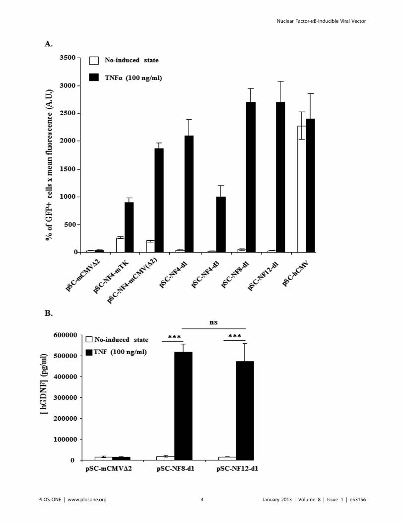
Figure 2. In vitro TNFa-mediated induction of NF-kB-responsive AAV vectors. HEK-293T cells were transfected with AAV-NF-EGFP (A) orwith AAV-NF-hGDNF (B) vectors using the calcium phosphate coprecipitation method. 56105 cells in 6-wells were transfected with 250 ng DNA.Forty-eight hrs later, transfected cells were treated or not with TNFa (100 ng/ml) for 5 hrs. HEK-293T transfected with AAV-NF-EGFP were analyzed forGFP expression on a FACStar analyser/sorter (Becton Dickinson) (A). To analyse the inducibility of AAV-NF-hGDNF vectors in HEK-293T, thesupernatant were harvested 4 h after changing the medium to measure secreted GDNF concentrations by a commercial ELISA assay (Human GDNFCytoSets, catalog #CHC2423, BioSource, Nivelles,Belgium) (B). For both AAV-NF8-d1-hGDNF and AAV-NF12-d1-hGDNF vectors, the differencesbetween the TNFa-treated and untreated cultures were highly significant. The differences between the basal levels and the induced levels for AAV-NF8-d1-hGDNF versus AAV-NF12-d1-hGDNF are not significant (B). (***, p,0.001; ns, p.0,05, one-way ANOVA Newman-Keuls Multiple ComparisonTest). Data are from one representative out of three experiments (3 separate transfections). Bars represent means 6 standard deviations (SD) (A) ormeans 6 standard error of the means (SEM) (B) from triplicate wells. A.U., arbitrary units.doi:10.1371/journal.pone.0053156.g002
Nuclear Factor-kB-Inducible Viral Vector
PLOS ONE | www.plosone.org 5 January 2013 | Volume 8 | Issue 1 | e53156
Nuclear Factor-kB-Inducible Viral Vector
PLOS ONE | www.plosone.org 6 January 2013 | Volume 8 | Issue 1 | e53156
Pictures were then processed and analysed with the Image J
software (NIH, USA).
GFP:IbaI co-labeling. For GFP:IbaI double immunofluo-
rescence, the above described GFP labeling was combined with
goat anti-IbaI (Abcam, cat number: ab 5076) followed by a donkey
anti-goat A.568 antibody (Molecular probes, cat number A-11057)
diluted 1:500.
GFP:Olig2 co-labeling. For GFP:Olig2 double immunoflu-
orescence, a chicken monoclonal anti-GFP antibody (Abcam,
Cambridge, UK) at a 1:1000 dilution was combined with rabbit
polyclonal anti-Olig2 IgG (diluted 1:500; Chemicon/EMD
Millipore).
In order to better visualize the structure of the tissue, cerebellar
sections were incubated in the Hoescht 33258 dye (Sigma-Aldrich)
diluted at 1 mg/ml in TBS for 30 min. Three washings in TBS of
10 min were performed between each step.
Sections were mounted using Glycergel Mounting Medium for
fluorescence (Dako, Belgium, cat number: C0563).
Confocal MicroscopyCo-labeling analysis was performed on pictures taken on at least
three different sections using a LSM510 NLO multiphoton
confocal microscope fitted on an Axiovert M200 inverted
microscope equipped with C-Apochromat 406/1.2 N.A. and
636/1.2 N.A. water immersion objectives (Zeiss, Iena, Germany).
The 488 nm excitation wavelength of the Argon/2 laser, a main
dichroic HFT 488 and a band-pass emission filter (BP500–
550 nm) were used for selective detection of the green fluoro-
chrome (Cy2, Alexa 488).
The 543 nm excitation wavelength of the HeNe1 laser, a main
dichroic HFT 488/543/633 and a long-pass emission filter
(BP565–615 nm) were used for selective detection of the red
fluorochrome (Cy3).
The nuclear stain Hoechst was excited in multiphotonic mode
at 760 nm with a Mai Tai tunable broad-band laser (Spectra-
Physics, Darmstad, Germany) and detected using a main dichroic
HFT KP650 and a band-pass emission filter (BP435–485 nm).
Optical sections, 2 microns thick, 512 by 512 pixels, were
collected sequentially for each fluorochrome. Z-stacks with a focus
step of 1 micron were collected.
The data-sets generated were merged and displayed with the
Zeiss Zen 2009 software and exported in LSM image format.
Countings were performed with the ImageJ 1.46 a software
(NIH, USA). Figures were prepared with Adobe Photoshop CS3
software.
Chromogenic Immunohistochemistry on Brain SectionsFor CD11b staining, vibratome coronal brain sections (50 mm)
were sequentially incubated in: i) 3% H2O2 in TBS (Tris 10 mM,
0.9% NaCl, Ph 7.6) for 30 min.; ii) THST (50 mM Tris,
0.5 M NaCl, 0.5% Triton X100 pH 7.6) containing 10% horse
serum for 1 hour; iii) mouse monoclonal anti-CD11b (1:500,
Serotec, MorphoSys, Dusseldorf, Germany) in THST containing
5% horse serum overnight a 4uC; (step iii), then goat anti-mouse
IgG conjugated with HRP (Molecular Probes, Invitrogen,
Carlsbad, CA, from TSA kit) diluted in Blocking Reagent
provided with the TSA kit (Molecular Probes, Invitrogen,
Carlsbad, CA) was added for 2 hours at room temperature (step
iv); and v) diaminobenzidine (Vector, NTL Laboratories, Brussels,
Belgium), according to the manufacturer’s protocol. Sections were
photographed using a Zeiss Axiophot 2 (Carl Zeiss, Gottingen,
Germany) microscope.
For activated NFkB staining, the same protocol was applied
using a mouse monoclonal IgG3 anti-activated NFkB recognizing
an epitope overlapping the nuclear location signal of the p65
subunit of the NFkB heterodimer thus selectively binding to the
activated form of NFkB [33] (Millipore, catalog # MAB3026).
The antibody was diluted 1:100 as primary antibody (step iv) and a
goat anti-mouse IgG conjugated with biotin diluted 1:200 was
used as secondary antibody (step v). Densitometric analysis of the
stainings was performed using the image J software (NIH, US).
Statistical AnalysisAll the statistical analysis was performed using the GraphPad
Software.
Results were expressed as mean 6SEM and statistical
significance was evaluated with one-way ANOVA Newman-Keuls
or student T-test. Differences were considered as significant when
p,0.05. Correlation analysis was evaluated with Pearson’s
correlation test.
Results
1. Design and in vitro Evaluation of NFkB-inducible AAVVectors
NFkB-inducible reporter cassettes were constructed by fusing a
minimal promoter with several repeats of the NFkB responsive
elements (NFkB–RE) from the non-coding regulatory region of JC
virus [26] upstream to an EGFP reporter gene. The promoter-
reporter cassettes were then introduced in a self-complementary
AAV vector which allows a rapid onset of transgene expression
[37,38]. In order to avoid influence of the AAV ITR promoter/
enhancer activity on the NFkB-RE-containing promoter [39,40],
a transcriptional blocker site [41] was placed between the left ITR
and the test promoters and a bidirectional SV40 polyA was placed
between the transgene cDNA and the right ITR [42] (see Fig. 1).
Two different minimal promoters fused to four repeats of the
NFkB-RE were compared. These consisted of a minimal
thymidine kinase promoter [43] and a minimal CMV promoter
[42] from which other putative transcription factor binding sites
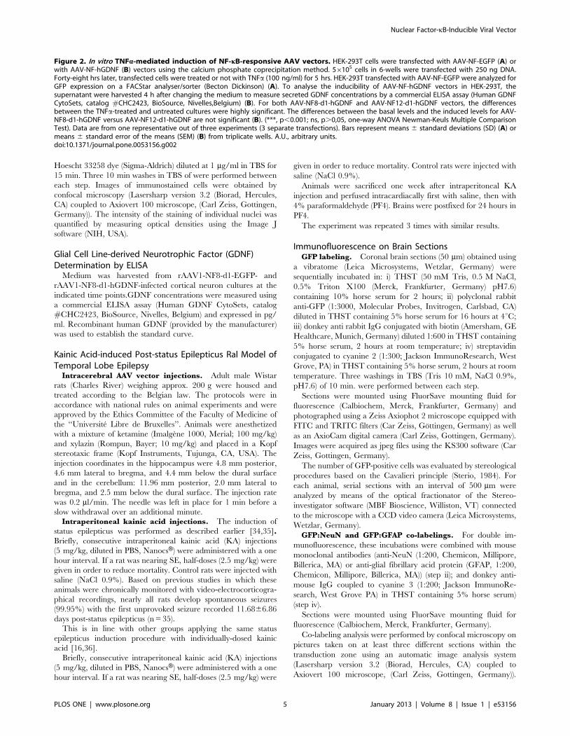
Figure 3. Enhanced neuronal survival in senescent cultures of cortical neurons mediated by rAAV2/1-NF8d1-driven GDNFtransgene expression. (A) Embryonic day 16 cortical neurons were maintained in 96 wells plates for 60 days. Cultures were fixed and stained withNeuN (a neuronal specific nuclear protein) or PSD95 (a neuronal postsynaptic density protein) a class of anchoring proteins which serve to localizevarious neuronal ion channels to post-synaptic densities antibodies. The number of NeuN-positive or PSD95-positive neurons is significantly higher incultures treated with rAAV2/1-NF8d1-GDNF as compared to rAAV2/1-NF8d1-EGFP (CtrL) (p,0.05; student test, n = 10). Bars represent means 6 SEM.(B) Similar cultures seeded on coated glass coverslips were fixed at day 10, day 21 and day 47 and stained with antibodies directed against activatedNFkB followed by a secondary antibody coupled with cyanine 2. Shown are confocal microscopy photographs. Bars represent 50 mM. The meanoptical density of the nuclei was measured using the Image J software. Differences between day 10 (n = 39) and day 21 (n = 59) or day 47 (n = 57)were respectively highly significant (**; p,0.001) and very highly significant (###; p,0.0001). The experiment was repeated twice with similarresults. (C) Culture media from rAAV1-NF8-d1-GDNF- and rAAV1-NF8-d1-EGFP-infected cultures were harvested from day 5 to day 42 post-infectionand GDNF concentrations (pg/ml) were measured using ELISA assay. For rAAV1-NF8-d1-GDNF-infected cultures, all values, except at day 5, weresignificantly higher (***; p,0.0001; n = 10 for rAAV1-NF8-d1-hGDNF and n = 8 for rAAV1-NF8-d1-EGFP) than the basal level (medium from rAAV1-NF8-d1-EGFP-infected cultures). The experiment was repeated twice with similar results.doi:10.1371/journal.pone.0053156.g003
Nuclear Factor-kB-Inducible Viral Vector
PLOS ONE | www.plosone.org 7 January 2013 | Volume 8 | Issue 1 | e53156
Nuclear Factor-kB-Inducible Viral Vector
PLOS ONE | www.plosone.org 8 January 2013 | Volume 8 | Issue 1 | e53156
upstream and downstream of the TATA box were deleted,
retaining only 3 downstream consensus sequences (CRE-1, LFA-1
and IFNc). This minimal promoter will be hereafter designated as
mCMVD2 (see Fig. 1A and Fig. 1B). The pSC-NF-Ctrl-EGFP
plasmid containing mCMVD2, TB and the SV40 polyA but
devoid of NFkB-responsive sequences was used as a control.
The test vectors were transfected into HEK-293T cells, which
were left untreated or exposed to TNFa (100 ng/ml). HEK-293T
cells were chosen for these studies based on previous data
suggesting that they have little or no NFkB activation under basal
conditions [44]. Fig. 2A shows that the use of the mCMVD2
promoter resulted in a higher cytokine-induced level of GFP
expression than the minimal TK promoter (p,0.001, one way
ANOVA, Newman-Keuls multiple comparison Test) without
increasing basal level and thus it was selected for the following
constructions. As expected, the control (pSC-NF-CtrL-EGFP and
pSC-NF-CtrL-hGDNF) vectors were not inducible by TNFa(p.0.05) (Fig. 2A and 2B).
i) Optimisation of the distance between NFkB-RE and
mCMVD2. The distance between enhancers and the transcrip-
tion initiation site is known to be important for the interaction
between the proteins involved in the trancriptional complex [45].
In order to optimize the inducibility of the AAV-NF vector, we
tested various spacings between the NF-kB repeats and the TATA
box of the mCMVD2 promoter.
Increasing the distance from 6 bp (pSC-NF4-mCMVD2) to
31 bp (pSC-NF4-d1) or 71 bp (pSC-NF4-d3) resulted in significant
(p,0.01) distance-dependent decreases of the basal reporter level.
The profile was different for the induced levels however.
Inducibility was maintained up to a distance of 31 bp (p.0.05,
pSC-NF4-mCMVD2 versus pSC-NF4-d1) and decreased at a
distance of 71 bp (p,0.001, pSC-NF4-mCMVD2 versus pSC-
NF4-d3). On the basis that a spacing distance of 31 bps
maintained high inducible expression levels while decreasing basal
expression (compared to the shorter spacing of 6 bp) the 31 bp
spacing incorporated in pSC-NF4-d1 was carried forward for
further development.
ii) Optimisation of the number of NFkB-RE. Starting
from pSC-NF4-d1 (with 4 NFkB-REs), the number of repeats was
increased to 8 and 12 (corresponding to pSC-NF8-d1 and pSC-
NF12-d1; see Fig. 1A). pSC-NF8- d1 and pSC-NF12-d1 showed
basal levels of GFP expression similar to pSC-NF4-d1 whereas the
TNFa-induced levels were higher than pSC-NF4-d1 for both
plasmids (p,0.01, pSC-NF4-d1 versus pSC-NF8-d1 and pSC-
NF4-d1 versus pSC-NF12-d1) (Fig. 2A). Using two reporter genes
(EGFP and hGDNF), no significant difference in TNFa-induced
levels was observed between pSC-NF8-d1 and pSC-NF12-d1
(Fig. 2A and 2B). Based on its retaining a larger residual cloning
capacity (approx. 900 bp), pSC-NF8-d1 vector was selected for
further testing.
2. Enhanced Survival of Senescent Cortical NeuronsMediated by NF-kB-inducible GDNF Expression
We then wanted to test whether an effect of a therapeutic
transgene could be conveyed by the rAAV2-NF8-d1 recombinant
virus. To address this question, the rAAV2-NF8-d1 vector
expressing the human GDNF cDNA was evaluated in an in vitro
model of brain aging.
To determine which capsid serotype would be suitable for our
primary cells in culture, we evaluated control cultures infected
with AAV vectors expressing EGFP under the control of the CMV
promoter transencapisdated into serotype 1, 2 and 5 capsids (5,000
cells/well; 104 vg/cell). This relatively low multiplicity of infection
was chosen to avoid previously described AAV vectors-related
toxicity in primary neurons cultures [46]. GFP-positive cells were
observed from day 3 after infection and their detectable numbers
reached 170 (rAAV2/1), 40 (rAAV2/2) and 92 (rAAV2/5) at 5
days post-infection (data not shown). Therefore, serotype 1 was
selected for further experiments.
We then proceeded to test the efficacy of our new vector in
long-term cultures of E16 cortical neurons and astrocytes in
serum-free medium. In this model, neurons gradually mature and
form synapses but eventually undergo apoptosis starting at
approximately 35–40 days in vitro. 5,000 cells were infected with
rAAV2/1-NF8-d1-EGFP (n = 10) as a control or rAAV2/1-NF8-
d1-GNDF (n = 10) at a multiplicity of 104 vg/cell.
After 60 days, cells infected with the rAAV2/1-NF8-d1 vectors
were fixed and expression of NeuN (a neuronal specific nuclear
protein also known as Fox3a) and PSD95 (a neuronal postsynaptic
density protein) were evaluated. The number of NeuN-positive
and PSD95-positive structures was significantly higher in the
rAAV2/1-NF8-d1-GDNF-infected wells compared to rAAV2/1-
NF8-EGFP-infected wells (see Fig. 3A). We then tested whether
NFkB was activated in aging culture. Parallel cultures on coated
glass coverslips in 48 well cultures dishes were fixed at different
time points and processed for immunofluorescence using an
antibody recognizing the activated form of NFkB. The intensity of
the nuclear labeling was quantified and shown to increase with
time from day 10 to day 47 after seeding (Fig. 3B).
We further measured the concentrations of secreted GDNF in
the culture media. As shown in Figure 3C, the GDNF
concentration increased over time until 26 days post-infection
(corresponding to 31 days post-seeding), then decreased. The
GDNF decrease from 33 days post-infection (38 days post-seeding)
was presumably due to a reduced number of neurons in aging
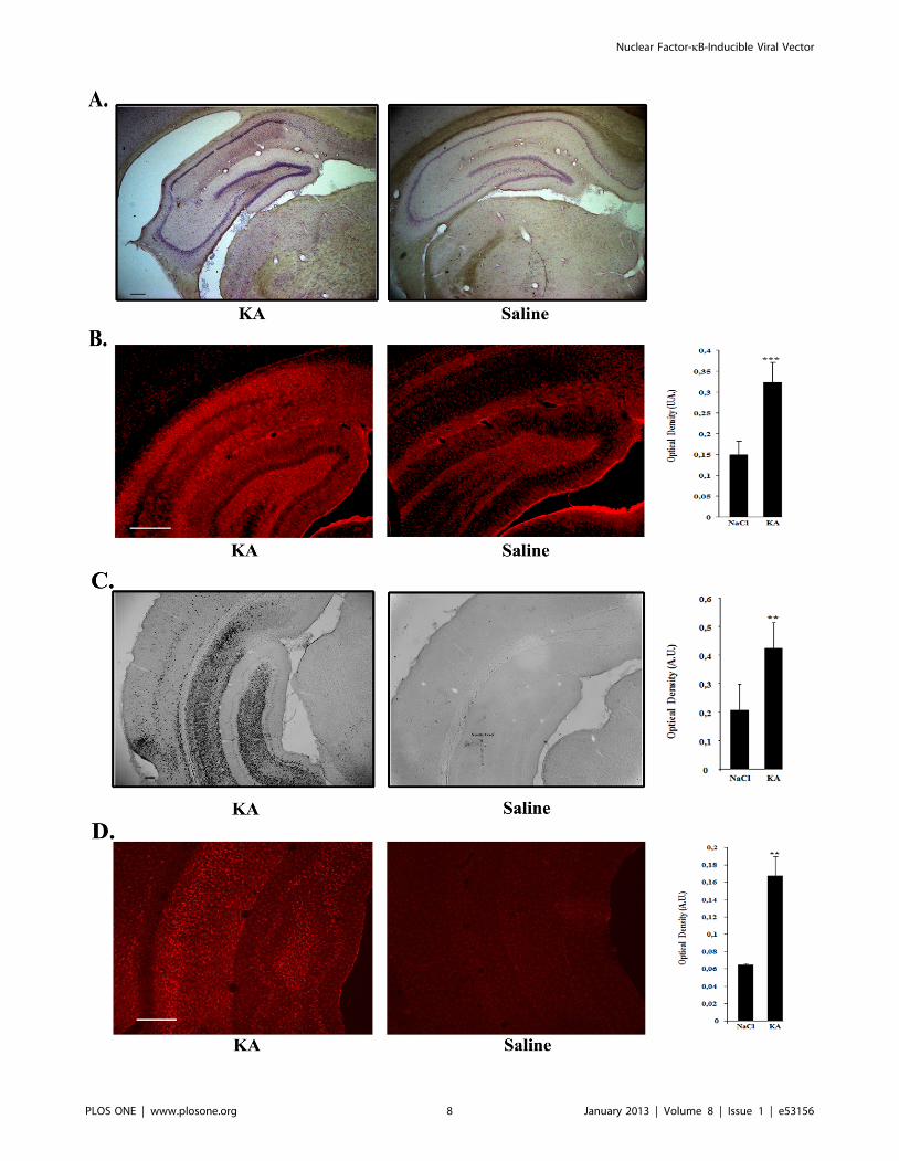
Figure 4. Hippocampal sclerosis and upregulation of inflammatory markers in the hippocampus of kainic acid-treated rats. A.Haematoxylin-eosin staining showing typical hippocampal sclerosis in kainic acid-treated rats (KA) (left) as compared to controls (right). Barrepresents 200 mm. B. Comparison of the intensity of the GFAP astrocytic marker staining in kainic acid- and saline-treated rats. Vibratome brainsections (50 mm) were immunolabeled using mouse anti-GFAP followed by a secondary antibody coupled to Cy3 fluorophore. The intensity of thestaining of three random areas in the hipocampal layers on every section was quantified using the Image J program. Data are expressed as the meanoptical density 6 SEM (n = 8 for each group of animals). The difference between kainic acid and saline-treated groups was highly significant (studenttest, P = 0,00018). Bar represents 500 mm. C. Comparison of the intensity of the CD11b staining of activated microglia in kainic acid- and saline-treatedrats. Vibratome brain sections (50 mm) were immunolabeled using mouse anti-CD11b followed by streptavidin-biotin-peroxidase staining. Theintensity of the staining of three random areas within the hippocampus on every section was quantified using the Image J program. Arrow showslocalized staining at the level of the needle tract. Data are expressed as the mean optical density 6 SEM (n = 4 for each group of animals). Thedifference between kainic acid and saline-treated groups was significant (student test, P = 0,0068). Bar represents 200 mm. D. Comparison of theintensity of the IbaI microglial staining in kainic acid- and saline-treated rats. Vibratome brain sections (50 mm) were immunolabeled using goat anti-IbaI followed by anti-goat antibody coupled with cyanine 3. The intensity of the staining of three random areas within the hippocampus on everysection was quantified using the Image J program. Data are expressed as the mean optical density 6 SEM (n = 6 for each group of animals). Thedifference between kainic acid and saline-treated groups was significant (student test, p = 0,00121). Bar represents 500 mm.doi:10.1371/journal.pone.0053156.g004
Nuclear Factor-kB-Inducible Viral Vector
PLOS ONE | www.plosone.org 9 January 2013 | Volume 8 | Issue 1 | e53156
cultures which were previously shown to undergo apoptosis (data
not shown).
Altogether, these data suggest that GDNF expressed in response
to NFkB activation exert a protective effect on aging neurons.
3. Selective Induction of rAAV1-NF-mediated GeneExpression in Hippocampal Neurons and Astrocytes in anin vivo Model of Epilepsy
In order to show proof of principle of pathology-induced
expression from our new gene transfer vector in vivo, we tested its
activity in a KA-induced rat SE model for temporal lobe epilepsy
[16,36].
The rAAV2/1-NF8-d1 recombinant virus with an EGFP
reporter gene (86106 viral genomes in 2 ml) was injected in the
right hippocampus one month prior to SE induction. Eight
animals received KA and 8 control animals received an equivalent
number of saline injections.
Haematoxylin-eosin staining of coronal brain sections of KA-
treated rats showed typical hippocampal sclerosis whereas
hippocampus from saline-treated rats had a normal morphology
(Fig. 4A). Activated astrocytes and microglia were evidenced in
these regions as demonstrated by GFAP (Fig. 4B) CD11b (Fig. 4C)
and IbaI (Fig. 4D) stainings. Quantification of the staining by
optical density showed that GFAP, CD11b and IbaI expression
was significantly stronger in KA-treated versus saline treated
animals (for GFAP: p = 0.0003, n = 8 for each group; for CD11b:
p = 0.0135; n = 4 for each group; for IbaI: p = 0.0012; n = 6 for
each group).
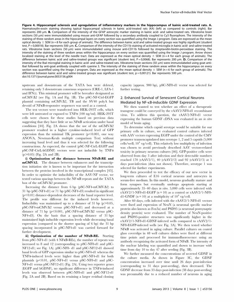
Prominent gene expression was evidenced in the hippocampus
of KA-injected rats whereas only few dispersed GFP-positive cells
were detected in saline-injected animals (Fig. 5A). The regional
pattern of GFP expression in the SE animals consisted of a
preferential transduction of the CA1 and CA3 layers as well as an
occasional labeling of cells in the stratum oriens (see Fig. 5A).
Figure 5. Disease-inducible rAAV1-NF-mediated transgeneexpression in the hippocampus of kainic acid-treated rats.Recombinant AAV2/1-NF8-d1-EGFP (86106 vg) was stereotaxicallyinjected in the right hippocampus of male Wistar rats. Animals werekept in two groups: one group was intraperitoneally-injected withkainic acid (n = 8) one month post stereotaxy and the other groupreceived only saline (n = 8). The animals were sacrificed one week afterinjection. Vibratome brain sections (50 mm) were immunolabeled usingan anti-GFP antibody, biotin-streptavidin amplification and Cy2fluorophore. A. Representative examples of GFP-labeling of the CAhippocampal layers dentate gyrus post-kainic acid injection. Note themassive presence of GFP-positive cells in the CA1 and CA3 layers.
Stereological counts of GFP-positive cell numbers per animal demon-strates a highly significant induction of the AAV-NF vector by kainic acid(P = 0,000875; student test; n = 8 for the saline group; n = 6 for the kainicacid group). KA, kainic acid; saline, 0.9% NaCl. The experiment wasrepeated 3 times with similar results. Bars represent 500 mm and 100mmin lower and higher (1,2,3 subpannels) magnification pictures,respectively. B. Comparison of the staining intensity for activated NFkBin kainic acid- and saline-treated rats. Vibratome brain sections (50 mm)were immunolabeled using anti-activated NFkB antibody followed bystreptavidin-biotin-peroxidase staining. Note, in particular, the localizedincreased staining in the CA hippocampal layers. The intensity of thestaining of three random areas in the hipocampal layers on everysection was quantified using the Image J program. Data are expressedas the mean optical density 6 SEM (n = 8 for each group of animals).The difference between KA and saline-treated groups was significant(student test, P = 0.0010). Bars represent 500 mm. C. Correlationbetween NFkB activation and GFP expression mediated by rAAV2/1-NF8-d1-EGFP in hippocampal layers. A group of 16 rats was injectedwith a rAAV1/2-NF8-d1-EGFP in the hippocampus. 8 rats were injectedwith KA and 8 other animals were injected with the saline solution. TwoKA-treated rats were removed from the analysis since they eithercontained no GFP-positive cells (presumably due to a failure to injectthe virus) or had a totally different profile of GFP-expression (no GFP-positive cells in the hippocampal layers, presumably due to wrongstereotaxic coordinates). The total number of GFP-immunoreactive cellsper animal (as evaluated by stereology was correlated with the meanoptical density of the NFkB stainings in the region containing thetransduced cells. There was a significant correlation between the twoparameters with a coefficient of 0.7001 (p = 0.0002; n = 6 for KA-treatedrats and n = 8 for saline-treated rats). The experiment was repeated asecond time (n = 8 for KA-treated rats and n = 7 for saline-treated rats)with similar results.doi:10.1371/journal.pone.0053156.g005
Nuclear Factor-kB-Inducible Viral Vector
PLOS ONE | www.plosone.org 10 January 2013 | Volume 8 | Issue 1 | e53156
Nuclear Factor-kB-Inducible Viral Vector
PLOS ONE | www.plosone.org 11 January 2013 | Volume 8 | Issue 1 | e53156
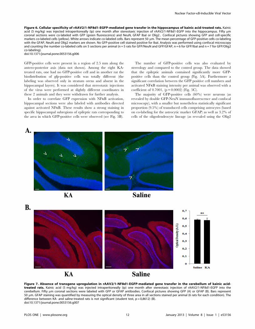
GFP-positive cells were present in a region of 2.5 mm along the
antero-posterior axis (data not shown). Among the eight KA-
treated rats, one had no GFP-positive cell and in another rat the
biodistribution of gfp-positive cells was totally different (the
labeling was observed only in stratum orens and absent in the
hippocampal layers). It was considered that stereotaxic injections
of the virus were performed at slightly different coordinates in
these 2 animals and they were withdrawn for further analysis.
In order to correlate GFP expression with NFkB activation,
hippocampal sections were also labeled with antibodies directed
against activated NFkB. These results show a strong staining in
specific hippocampal subregions of epileptic rats corresponding to
the area in which GFP-positive cells were observed (see Fig. 5B).
The number of GFP-positive cells was also evaluated by
stereology and compared to the control group. The data showed
that the epileptic animals contained significantly more GFP-
positive cells than the control group (Fig. 5A). Furthermore a
significant correlation between the GFP positive cell numbers and
activated NFkB staining intensity per animal was observed with a
coefficient of 0.7001, (p = 0.0002) (Fig. 5C).
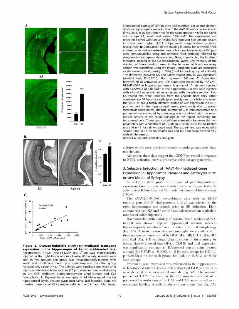
The majority of GFP-positive cells (60%) were neurons (as
revealed by double GFP-NeuN immunofluorescence and confocal
microscopy), with a smaller but nonetheless statistically significant
proportion (9.5%) of transduced cells comprising astrocytes (based
on co-labeling for the astrocytic marker GFAP) as well as 3.2% of
cells of the oligodendrocyte lineage (as revealed using the Olig2
Figure 6. Cellular specificity of rAAV2/1-NF8d1-EGFP-mediated gene transfer in the hippocampus of kainic acid-treated rats. Kainicacid (5 mg/kg) was injected intraperitoneally (ip) one month after stereotaxic injection of rAAV2/1-NF8d1-EGFP into the hippocampus. Fifty mmcoronal sections were co-labeled with GFP (green fluorescence) and NeuN, GFAP IbaI or Olig2. Confocal pictures showing GFP and cell-specificmarkers co-labeled cells (yellow). White arrows indicate co-labeled cells. Bars represent 50 mm. The mean percentage of GFP-positive cells co-labelingwith the GFAP, NeuN and Olig2 markers are shown. No GFP-positive cell stained positive for IbaI. Analysis was performed using confocal microscopyand counting the number co-labeled cells on 5 sections per animal (n = 5 rats for GFP/NeuN and GFP/GFAP, n = 6 for GFP/IbaI and n = 7 for GFP/Olig2co-labeling).doi:10.1371/journal.pone.0053156.g006
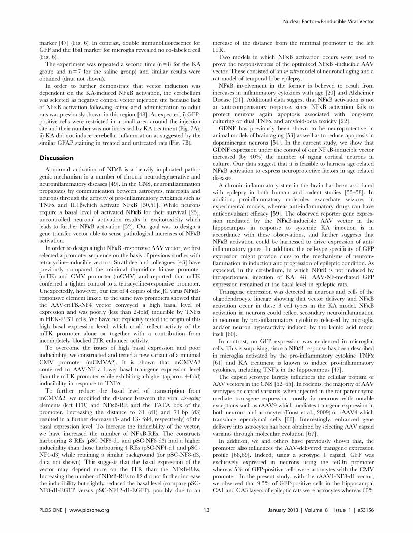
Figure 7. Absence of transgene upregulation in rAAV2/1-NF8d1-EGFP-mediated gene transfer in the cerebellum of kainic acid-treated rats. Kainic acid (5 mg/kg) was injected intraperitoneally (ip) one month after stereotaxic injection of rAAV2/1-NF8d1-EGFP into thecerebellum. Fifty mm coronal sections were labeled with GFP or GFAP antibodies. Confocal pictures showing GFP (A) or GFAP (B). Bars represent50 mm. GFAP staining was quantified by measuring the optical density of three area in all sections stained per animal (6 rats for each condition). Thedifference between KA- and saline-treated rats is not significant (student test, p = 0,8612) (B).doi:10.1371/journal.pone.0053156.g007
Nuclear Factor-kB-Inducible Viral Vector
PLOS ONE | www.plosone.org 12 January 2013 | Volume 8 | Issue 1 | e53156
marker [47] (Fig. 6). In contrast, double immunofluorescence for
GFP and the IbaI marker for microglia revealed no co-labeled cell
(Fig. 6).
The experiment was repeated a second time (n = 8 for the KA
group and n = 7 for the saline group) and similar results were
obtained (data not shown).
In order to further demonstrate that vector induction was
dependent on the KA-induced NFkB activation, the cerebellum
was selected as negative control vector injection site because lack
of NFkB activation following kainic acid administration to adult
rats was previously shown in this region [48]. As expected, i) GFP-
positive cells were restricted in a small area around the injection
site and their number was not increased by KA treatment (Fig. 7A);
ii) KA did not induce cerebellar inflammation as suggested by the
similar GFAP staining in treated and untreated rats (Fig. 7B).
Discussion
Abnormal activation of NFkB is a heavily implicated patho-
genic mechanism in a number of chronic neurodegenerative and
neuroinflammatory diseases [49]. In the CNS, neuroinflammation
propagates by communication between astrocytes, microglia and
neurons through the activity of pro-inflammatory cytokines such as
TNFa and IL1bwhich activate NFkB [50,51]. While neurons
require a basal level of activated NFkB for their survival [25],
uncontrolled neuronal activation results in excitotoxicity which
leads to further NFkB activation [52]. Our goal was to design a
gene transfer vector able to sense pathological increases of NFkB
activation.
In order to design a tight NFkB -responsive AAV vector, we first
selected a promoter sequence on the basis of previous studies with
tetracycline-inducible vectors. Strathdee and colleagues [43] have
previously compared the minimal thymidine kinase promoter
(mTK) and CMV promoter (mCMV) and reported that mTK
conferred a tighter control to a tetracycline-responsive promoter.
Unexpectedly, however, our test of 4 copies of the JC virus NFkB-
responsive element linked to the same two promoters showed that
the AAV-mTK-NF4 vector conveyed a high basal level of
expression and was poorly (less than 2-fold) inducible by TNFain HEK-293T cells. We have not explicitly tested the origin of this
high basal expression level, which could reflect activity of the
mTK promoter alone or together with a contribution from
incompletely blocked ITR enhancer activity.
To overcome the issues of high basal expression and poor
inducibility, we constructed and tested a new variant of a minimal
CMV promoter (mCMVD2). It is shown that mCMVD2
conferred to AAV-NF a lower basal transgene expression level
than the mTK promoter while exhibiting a higher (approx. 4-fold)
inducibility in response to TNFa.
To further reduce the basal level of transcription from
mCMVD2, we modified the distance between the viral cis-acting
elements (left ITR) and NFkB-RE and the TATA box of the
promoter. Increasing the distance to 31 (d1) and 71 bp (d3)
resulted in a further decrease (5- and 13- fold, respectively) of the
basal expression level. To increase the inducibility of the vector,
we have increased the number of NFkB-REs. The constructs
harbouring 8 REs (pSC-NF8-d1 and pSC-NF8-d3) had a higher
inducibility than those harbouring 4 REs (pSC-NF4-d1 and pSC-
NF4-d3) while retaining a similar background (for pSC-NF8-d3,
data not shown). This suggests that the basal expression of the
vector may depend more on the ITR than the NFkB-REs.
Increasing the number of NFkB-REs to 12 did not further increase
the inducibility but slightly reduced the basal level (compare pSC-
NF8-d1-EGFP versus pSC-NF12-d1-EGFP), possibly due to an
increase of the distance from the minimal promoter to the left
ITR.
Two models in which NFkB activation occurs were used to
prove the responsiveness of the optimized NFkB -inducible AAV
vector. These consisted of an in vitro model of neuronal aging and a
rat model of temporal lobe epilepsy.
NFkB involvement in the former is believed to result from
increases in inflammatory cytokines with age [20] and Alzheimer
Disease [21]. Additional data suggest that NFkB activation is not
an autocompensatory response, since NFkB activation fails to
protect neurons again apoptosis associated with long-term
culturing or dual TNFa and amyloid-beta toxicity [22].
GDNF has previously been shown to be neuroprotective in
animal models of brain aging [53] as well as to reduce apoptosis in
dopaminergic neurons [54]. In the current study, we show that
GDNF expression under the control of our NFkB-inducible vector
increased (by 40%) the number of aging cortical neurons in
culture. Our data suggest that it is feasible to harness age-related
NFkB activation to express neuroprotective factors in age-related
diseases.
A chronic inflammatory state in the brain has been associated
with epilepsy in both human and rodent studies [55–58]. In
addition, proinflammatory molecules exacerbate seizures in
experimental models, whereas anti-inflammatory drugs can have
anticonvulsant efficacy [59]. The observed reporter gene expres-
sion mediated by the NFkB-inducible AAV vector in the
hippocampus in response to systemic KA injection is in
accordance with these observations, and further suggests that
NFkB activation could be harnessed to drive expression of anti-
inflammatory genes. In addition, the cell-type specificity of GFP
expression might provide clues to the mechanisms of neuroin-
flammation in induction and progression of epileptic condition. As
expected, in the cerebellum, in which NFkB is not induced by
intraperitoneal injection of KA [48] AAV-NF-mediated GFP
expression remained at the basal level in epileptic rats.
Transgene expression was detected in neurons and cells of the
oligodendrocyte lineage showing that vector delivery and NFkB
activation occur in these 3 cell types in the KA model. NFkB
activation in neurons could reflect secondary neuroinflammation
in neurons by pro-inflammatory cytokines released by microglia
and/or neuron hyperactivity induced by the kainic acid model
itself [60].
In contrast, no GFP expression was evidenced in microglial
cells. This is surprising, since a NFkB response has been described
in microglia activated by the pro-inflammatory cytokine TNFa[61] and KA treatment is known to induce pro-inflammatory
cytokines, including TNFa in the hippocampus [47].
The capsid serotype largely influences the cellular tropism of
AAV vectors in the CNS [62–65]. In rodents, the majority of AAV
serotypes or capsid variants, when injected in the rat parenchyma
mediate transgene expression mostly in neurons with notable
exceptions such as rAAV9 which mediates transgene expression in
both neurons and astrocytes (Foust et al., 2009) or rAAV4 which
transduce ependymal cells [66]. Interestingly, enhanced gene
delivery into astrocytes has been obtained by selecting AAV capsid
variants through molecular evolution [67].
In addition, we and others have previously shown that, the
promoter also influences the AAV-delivered transgene expression
profile [68,69]. Indeed, using a serotype 1 capsid, GFP was
exclusively expressed in neurons using the tetOn promoter
whereas 5% of GFP-positive cells were astrocytes with the CMV
promoter. In the present study, with the rAAV1-NF8-d1 vector,
we observed that 9.5% of GFP-positive cells in the hippocampal
CA1 and CA3 layers of epileptic rats were astrocytes whereas 60%
Nuclear Factor-kB-Inducible Viral Vector
PLOS ONE | www.plosone.org 13 January 2013 | Volume 8 | Issue 1 | e53156
were neurons. Whether these data reflect the proportion of
astrocytes and neurons in which NFkB has been activated by the
KA treatment or are biased by the tropism of the AAV serotype 1
capsid and mCMV promoter cannot be concluded from our data.
It remains to be determined if the use of other AAV serotypes
would modify the proportion of neurons, astrocytes and possibly
microglial cells expressing a transgene driven by the AAV-NF
vector. Furthermore, the optimal targeting of glial versus neuronal
cells may also vary by disease indication.
The onset of AAV-mediated transgene expression in the brain is
fairly slow [38]. However, serotype 1 AAV vectors are character-
ized by remarkably rapid kinetics in several regions of the brain
with maximal expression reached at 2 to 4 days post-injection [70];
[71]. In addition, self-complementary vectors that bypass the
limitation of second-strand viral DNA synthesis further reduce the
delay for transgene expression [70]; [29].
In conclusion, this study constitutes a proof-of-concept step
toward the use of NFkB-inducible AAV vectors for disease-
inducible transgene delivery to treat disorders of the nervous
system. Nonetheless, more data will be required to establish
whether transgene expression tightly follows the temporal course
of disease and whether pathology-appropriate cellular targeting of
the inflammatory response can be reliably achieved.
Acknowledgments
We thank the vector core of the University Hospital of Nantes supported
by the Association Francaise contre les Myopathies (AFM) for preparing
the large-scale rAAV2/1-NF8d1 stock.
We thank Dr Veerle Baekelandt (Lab. Molecular Virology and Gene
Therapy, Katholieke Universiteit Leuven, Belgium) for her help for the
stereological analysis.
We would like to acknowledge Dr Frederic Bollet-Quivogne, PhD
(Logistic Scientist F.R.S.- F.N.R.S.) of the Light Microscopy Facility
(LiMiF), Universite Libre de Bruxelles, Brussels, Belgium, for his support
with the confocal imaging presented herein.
Author Contributions
Conceived and designed the experiments: AC RLC RC LT. Performed the
experiments: AC RLC KV EG OB CM CP LT. Analyzed the data: AC
ML ODW RLC RC LT. Contributed reagents/materials/analysis tools:
AC KM EG OB CM CP. Wrote the paper: AC RLC RC LT.
References
1. Fluri DA, Baba MD, Fussenegger M (2007) Adeno-associated viral vectorsengineered for macrolide-adjustable transgene expression in mammalian cells
and mice. BMC Biotechnol 7: 75.
2. Gossen M, Freundlieb S, Bender G, Muller G, Hillen W, et al. (1995)Transcriptional activation by tetracyclines in mammalian cells. Science 268:
1766–1769.
3. Adriaansen J, Khoury M, de Cortie CJ, Fallaux FJ, Bigey P, et al. (2007)
Reduction of arthritis following intra-articular administration of an adeno-
associated virus serotype 5 expressing a disease-inducible TNF-blocking agent.Ann Rheum Dis 66: 1143–1150.
4. Jakobsson J, Georgievska B, Ericson C, Lundberg C (2004) Lesion-dependentregulation of transgene expression in the rat brain using a human glial fibrillary
acidic protein-lentiviral vector. Eur J Neurosci 19: 761–765.
5. Phillips MI, Tang Y, Schmidt-Ott K, Qian K, Kagiyama S (2002) Vigilantvector: heart-specific promoter in an adeno-associated virus vector for
cardioprotection. Hypertension 39: 651–655.
6. Siebenlist U, Franzoso G, Brown K (1994) Structure, regulation and function of
NF-kappa B. Annu Rev Cell Biol 10: 405–455.
7. Karin M, Delhase M (2000) The I kappa B kinase (IKK) and NF-kappa B: key
elements of proinflammatory signalling. Semin Immunol 12: 85–98.
8. Kaltschmidt B, Widera D, Kaltschmidt C (2005) Signaling via NF-kappaB in thenervous system. Biochim Biophys Acta 1745: 287–299.
9. Lubin FD, Johnston LD, Sweatt JD, Anderson AE (2005) Kainate mediatesnuclear factor-kappa B activation in hippocampus via phosphatidylinositol-3
kinase and extracellular signal-regulated protein kinase. Neuroscience 133: 969–
981.
10. Lubin FD, Ren Y, Xu X, Anderson AE (2007) Nuclear factor-kappa B regulates
seizure threshold and gene transcription following convulsant stimulation.J Neurochem 103: 1381–1395.
11. Ben Ari Y (1985) Limbic seizure and brain damage produced by kainic acid:
mechanisms and relevance to human temporal lobe epilepsy. Neuroscience 14:375–403.
12. Cavalheiro EA, Leite JP, Bortolotto ZA, Turski WA, Ikonomidou C, et al. (1991)Long-term effects of pilocarpine in rats: structural damage of the brain triggers
kindling and spontaneous recurrent seizures. Epilepsia 32: 778–782.
13. Lothman EW, Bertram EH, Kapur J, Stringer JL (1990) Recurrent spontaneous
hippocampal seizures in the rat as a chronic sequela to limbic status epilepticus.
Epilepsy Res 6: 110–118.
14. Vincent P, Mulle C (2009) Kainate receptors in epilepsy and excitotoxicity.
Neuroscience 158: 309–323.
15. Bouilleret V, Ridoux V, Depaulis A, Marescaux C, Nehlig A, et al. (1999)
Recurrent seizures and hippocampal sclerosis following intrahippocampal
kainate injection in adult mice: electroencephalography, histopathology andsynaptic reorganization similar to mesial temporal lobe epilepsy. Neuroscience
89: 717–729.
16. Hellier JL, Patrylo PR, Buckmaster PS, Dudek FE (1998) Recurrent spontaneous
motor seizures after repeated low-dose systemic treatment with kainate:
assessment of a rat model of temporal lobe epilepsy. Epilepsy Res 31: 73–84.
17. Majores M, Eils J, Wiestler OD, Becker AJ (2004) Molecular profiling of
temporal lobe epilepsy: comparison of data from human tissue samples andanimal models. Epilepsy Res 60: 173–178.
18. Ravizza T, Balosso S, Vezzani A (2011) Inflammation and prevention ofepileptogenesis. Neurosci Lett 497: 223–230.
19. Stafstrom CE, Thompson JL, Holmes GL (1992) Kainic acid seizures in the
developing brain: status epilepticus and spontaneous recurrent seizures. Brain
Res Dev Brain Res 65: 227–236.
20. Gemma C, Mesches MH, Sepesi B, Choo K, Holmes DB, et al. (2002) Diets
enriched in foods with high antioxidant activity reverse age-induced decreases in
cerebellar beta-adrenergic function and increases in proinflammatory cytokines.
J Neurosci 22: 6114–6120.
21. Tarkowski E, Liljeroth AM, Minthon L, Tarkowski A, Wallin A, et al. (2003)
Cerebral pattern of pro- and anti-inflammatory cytokines in dementias. Brain
Res Bull 61: 255–260.
22. Patel JR, Brewer GJ (2008) Age-related differences in NFkappaB translocation
and Bcl-2/Bax ratio caused by TNFalpha and Abeta42 promote survival in
middle-age neurons and death in old neurons. Exp Neurol 213: 93–100.
23. Mattson MP, Meffert MK (2006) Roles for NF-kappaB in nerve cell survival,
plasticity, and disease. Cell Death Differ 13: 852–860.
24. Carlsen H, Moskaug JO, Fromm SH, Blomhoff R (2002) In vivo imaging of NF-
kappa B activity. J Immunol 168: 1441–1446.
25. Bhakar AL, Tannis LL, Zeindler C, Russo MP, Jobin C, et al. (2002)
Constitutive nuclear factor-kappa B activity is required for central neuron
survival. J Neurosci 22: 8466–8475.
26. Ranganathan PN, Khalili K (1993) The transcriptional enhancer element, kappa
B, regulates promoter activity of the human neurotropic virus, JCV, in cells
derived from the CNS. Nucleic Acids Res 21: 1959–1964.
27. Wollebo HS, Safak M, Del VL, Khalili K, White MK (2011) Role for tumor
necrosis factor-alpha in JC virus reactivation and progressive multifocal
leukoencephalopathy. J Neuroimmunol 233: 46–53.
28. Lesuisse C, Martin LJ (2002) Immature and mature cortical neurons engage
different apoptotic mechanisms involving caspase-3 and the mitogen-activated
protein kinase pathway. J Cereb Blood Flow Metab 22: 935–950.
29. McCarty DM, Fu H, Monahan PE, Toulson CE, Naik P, et al. (2003) Adeno-
associated virus terminal repeat (TR) mutant generates self-complementary
vectors to overcome the rate-limiting step to transduction in vivo. Gene Ther 10:
2112–2118.
30. Zolotukhin S, Byrne BJ, Mason E, Zolotukhin I, Potter M, et al. (1999)
Recombinant adeno-associated virus purification using novel methods improves
infectious titer and yield. Gene Ther 6: 973–985.
31. Lock M, McGorray S, Auricchio A, Ayuso E, Beecham EJ, et al. (2010)
Characterization of a recombinant adeno-associated virus type 2 Reference
Standard Material. Hum Gene Ther 21: 1273–1285.
32. Zala D, Benchoua A, Brouillet E, Perrin V, Gaillard MC, et al. (2005)
Progressive and selective striatal degeneration in primary neuronal cultures using
lentiviral vector coding for a mutant huntingtin fragment. Neurobiol Dis 20:
785–798.
33. Zabel U, Henkel T, Silva MS, Baeuerle PA (1993) Nuclear uptake control of NF-
kappa B by MAD-3, an I kappa B protein present in the nucleus. EMBO J 12:
201–211.
34. Vermoesen K, Smolders I, Massie A, Michotte Y, Clinckers R (2010) The
control of kainic acid-induced status epilepticus. Epilepsy Res 90: 164–166.
35. Vermoesen K, Massie A, Smolders I, Clinckers R (2012) The antidepressants
citalopram and reboxetine reduce seizure frequency in rats with chronic
epilepsy. Epilepsia 53: 870–878. 10.1111/j.1528-1167.2012.03436.x [doi].
Nuclear Factor-kB-Inducible Viral Vector
PLOS ONE | www.plosone.org 14 January 2013 | Volume 8 | Issue 1 | e53156
36. Williams PA, White AM, Clark S, Ferraro DJ, Swiercz W, et al. (2009)
Development of spontaneous recurrent seizures after kainate-induced statusepilepticus. J Neurosci 29: 2103–2112.
37. Fu H, Muenzer J, Samulski RJ, Breese G, Sifford J, et al. (2003) Self-
complementary adeno-associated virus serotype 2 vector: global distribution andbroad dispersion of AAV-mediated transgene expression in mouse brain. Mol
Ther 8: 911–917.38. McCarty DM, Monahan PE, Samulski RJ (2001) Self-complementary
recombinant adeno-associated virus (scAAV) vectors promote efficient trans-
duction independently of DNA synthesis. Gene Ther 8: 1248–1254.39. Afione SA, Conrad CK, Kearns WG, Chunduru S, Adams R, et al. (1996) In
vivo model of adeno-associated virus vector persistence and rescue. J Virol 70:3235–3241.
40. Haberman RP, McCown TJ, Samulski RJ (2000) Novel transcriptionalregulatory signals in the adeno-associated virus terminal repeat A/D junction
element. J Virol 74: 8732–8739.
41. Eggermont J, Proudfoot NJ (1993) Poly(A) signals and transcriptional pause sitescombine to prevent interference between RNA polymerase II promoters.
EMBO J 12: 2539–2548.42. Chtarto A, Bender HU, Hanemann CO, Kemp T, Lehtonen E, et al. (2003)
Tetracycline-inducible transgene expression mediated by a single AAV vector.
Gene Ther 10: 84–94.43. Strathdee CA, McLeod MR, Hall JR (1999) Efficient control of tetracycline-
responsive gene expression from an autoregulated bi-directional expressionvector. Gene 229: 21–29.
44. Channavajhala PL, Rao VR, Spaulding V, Lin LL, Zhang YG (2005) hKSR-2inhibits MEKK3-activated MAP kinase and NF-kappaB pathways in inflam-
mation. Biochem Biophys Res Commun 334: 1214–1218.
45. Boshart M, Weber F, Jahn G, Dorsch-Hasler K, Fleckenstein B, et al. (1985) Avery strong enhancer is located upstream of an immediate early gene of human
cytomegalovirus. Cell 41: 521–530.46. Howard DB, Powers K, Wang Y, Harvey BK (2008) Tropism and toxicity of
adeno-associated viral vector serotypes 1, 2, 5, 6, 7, 8, and 9 in rat neurons and
glia in vitro. Virology 372: 24–34. S0042-6822(07)00658-7 [pii];10.1016/j.virol.2007.10.007 [doi].
47. Somera-Molina KC, Nair S, Van Eldik LJ, Watterson DM, Wainwright MS(2009) Enhanced microglial activation and proinflammatory cytokine upregula-
tion are linked to increased susceptibility to seizures and neurologic injury in a‘two-hit’ seizure model. Brain Res 1282: 162–172. S0006-8993(09)01093-2
[pii];10.1016/j.brainres.2009.05.073 [doi].
48. Rong Y, Baudry M (1996) Seizure activity results in a rapid induction of nuclearfactor-kappa B in adult but not juvenile rat limbic structures. J Neurochem 67:
662–668.49. Verma IM (2004) Nuclear factor (NF)-kappaB proteins: therapeutic targets. Ann
Rheum Dis 63 Suppl 2: ii57–ii61.
50. Depino AM, Earl C, Kaczmarczyk E, Ferrari C, Besedovsky H, et al. (2003)Microglial activation with atypical proinflammatory cytokine expression in a rat
model of Parkinson’s disease. Eur J Neurosci 18: 2731–2742.51. Tansey MG, Goldberg MS (2010) Neuroinflammation in Parkinson’s disease: its
role in neuronal death and implications for therapeutic intervention. NeurobiolDis 37: 510–518.
52. Caba E, Bahr BA (2004) Biphasic NF-kappaB activation in the excitotoxic
hippocampus. Acta Neuropathol 108: 173–182.53. Emborg ME, Moirano J, Raschke J, Bondarenko V, Zufferey R, et al. (2009)
Response of aged parkinsonian monkeys to in vivo gene transfer of GDNF.Neurobiol Dis 36: 303–311.
54. Lin LF, Doherty DH, Lile JD, Bektesh S, Collins F (1993) GDNF: a glial cell
line-derived neurotrophic factor for midbrain dopaminergic neurons. Science260: 1130–1132.
55. Jankowsky JL, Derrick BE, Patterson PH (2000) Cytokine responses to LTP
induction in the rat hippocampus: a comparison of in vitro and in vivotechniques. Learn Mem 7: 400–412.
56. Oprica M, Eriksson C, Schultzberg M (2003) Inflammatory mechanisms
associated with brain damage induced by kainic acid with special reference tothe interleukin-1 system. J Cell Mol Med 7: 127–140.
57. Turrin NP, Rivest S (2004) Innate immune reaction in response to seizures:implications for the neuropathology associated with epilepsy. Neurobiol Dis 16:
321–334.
58. Vezzani A, Granata T (2005) Brain inflammation in epilepsy: experimental andclinical evidence. Epilepsia 46: 1724–1743.
59. Vezzani A, Aronica E, Mazarati A, Pittman QJ (2011) Epilepsy and braininflammation. Exp Neurol.
60. Lacaille JC, Mueller AL, Kunkel DD, Schwartzkroin PA (1987) Local circuitinteractions between oriens/alveus interneurons and CA1 pyramidal cells in
hippocampal slices: electrophysiology and morphology. J Neurosci 7: 1979–
1993.61. Sheppard PW, Sun X, Emery JF, Giffard RG, Khammash M (2011)
Quantitative characterization and analysis of the dynamic NF-kappaB responsein microglia. BMC Bioinformatics 12: 276. 1471–2105–12–276 [pii];10.1186/
1471-2105-12-276 [doi].
62. Blits B, Derks S, Twisk J, Ehlert E, Prins J, et al. (2010) Adeno-associated viralvector (AAV)-mediated gene transfer in the red nucleus of the adult rat brain:
comparative analysis of the transduction properties of seven AAV serotypes andlentiviral vectors. J Neurosci Methods 185: 257–263.
63. Burger C, Gorbatyuk OS, Velardo MJ, Peden CS, Williams P, et al. (2004)Recombinant AAV viral vectors pseudotyped with viral capsids from serotypes
1, 2, and 5 display differential efficiency and cell tropism after delivery to
different regions of the central nervous system. Mol Ther 10: 302–317.64. Taymans JM, Vandenberghe LH, Haute CV, Thiry I, Deroose CM, et al. (2007)
Comparative analysis of adeno-associated viral vector serotypes 1, 2, 5, 7, and 8in mouse brain. Hum Gene Ther 18: 195–206.
65. Van der PA, Toelen J, Carlon M, Van den HC, Coun F, et al. (2011) Efficient
and stable transduction of dopaminergic neurons in rat substantia nigra byrAAV 2/1, 2/2, 2/5, 2/6.2, 2/7, 2/8 and 2/9. Gene Ther 18: 517–527.
66. Davidson BL, Stein CS, Heth JA, Martins I, Kotin RM, et al. (2000)Recombinant adeno-associated virus type 2, 4, and 5 vectors: transduction of
variant cell types and regions in the mammalian central nervous system. ProcNatl Acad Sci U S A 97: 3428–3432. 10.1073/pnas.050581197 [doi];050581197
[pii].
67. Koerber JT, Klimczak R, Jang JH, Dalkara D, Flannery JG, et al. (2009)Molecular evolution of adeno-associated virus for enhanced glial gene delivery.
Mol Ther 17: 2088–2095. mt2009184 [pii];10.1038/mt.2009.184 [doi].68. Bockstael O, Chtarto A, Wakkinen J, Yang X, Melas C, et al. (2008) Differential
transgene expression profiles from rAAV2/1 vectors using the tetON and CMV
promoters in the rat brain. Hum Gene Ther.69. Haberman R, Criswell H, Snowdy S, Ming Z, Breese G, et al. (2002)
Therapeutic liabilities of in vivo viral vector tropism: adeno-associated virusvectors, NMDAR1 antisense, and focal seizure sensitivity. Mol Ther 6: 495–500.
70. Bockstael O, Melas C, Pythoud C, Levivier M, McCarty D, et al. (2012) RapidTransgene Expression in Multiple Precursor Cell Types of Adult Rat
Subventricular Zone Mediated by Adeno-Associated Type 1 Vectors. Hum
Gene Ther. 10.1089/hum.2011.216 [doi].71. Reimsnider S, Manfredsson FP, Muzyczka N, Mandel RJ (2007) Time course of
transgene expression after intrastriatal pseudotyped rAAV2/1, rAAV2/2,rAAV2/5, and rAAV2/8 transduction in the rat. Mol Ther 15: 1504–1511.
72. Zolotukhin S, Potter M, Hauswirth WW, Guy J, Muzyczka N (1996) A
‘‘humanized’’ green fluorescent protein cDNA adapted for high-level expressionin mammalian cells. J Virol 70: 4646–4654.
Nuclear Factor-kB-Inducible Viral Vector
PLOS ONE | www.plosone.org 15 January 2013 | Volume 8 | Issue 1 | e53156