Aloe vera gel alleviates cardiotoxicity in streptozocin?induced ...
9
JPP 2010, 62: 115–123 ß 2010 The Authors. Journal compilation ß 2010 Royal Pharmaceutical Society of Great Britain Received March 3, 2009 Accepted October 14, 2009 DOI 10.1211/jpp/62.01.0013 ISSN 0022-3573 Correspondence: Neeti Jain, Department of Pharmacology and Toxicology, Defence Research and Development Establishment, Jhansi Road, Gwalior, M.P., India. E-mail: [email protected] Research Paper Aloe vera gel alleviates cardiotoxicity in streptozocin-induced diabetes in rats Neeti Jain a , Rajagopalan Vijayaraghavan a , Satish Chandra Pant a , Vinay Lomash a and Mohammed Ali b a Pharmacology and Toxicology Division, Defence Research and Development Establishment (DRDE), Jhansi Road Gwalior, M.P. and b Department of Phytochemistry and Pharmacognosy, Faculty of Pharmacy, Jamia Hamdard, New Delhi, India Abstract Objectives Persistent hyperglycaemia results in oxidative stress along with the generation of oxygen free radicals and appears to be an important factor in the production of secondary complications in diabetes. The aim of this work was to evaluate markers of oxidative stress in heart tissue along with the protective, antioxidant and antidiabetic activity of 30% Aloe vera gel in diabetic rats. Methods Streptozocin was given as a single intravenous injection and 30% Aloe vera gel was given in two doses for 20 days, orally. Blood glucose, glycosylated haemoglobin, blood reduced glutathione, serum lactate dehydrogenase and serum creatine kinase levels were measured on day 21 after drug treatment. Heart rate and mean blood pressure were recorded at the end of the study. Different biochemical variables were evaluated in the heart tissue, including thiobarbituric acid reactive substance (TBARS), reduced glutathione, superoxide dismutase and catalase in diabetic and in Aloe vera-treated diabetic rats. Key findings In streptozocin diabetic rats, the TBARS level was increased significantly, superoxide dismutase and reduced glutathione significantly decreased, and the catalase level was significantly increased. Aloe vera 30% gel (200 mg/kg) treatment in diabetic rats reduced the increased TBARS and maintained the superoxide dismutase and catalase activity up to the normal level. Aloe vera gel increased reduced glutathione by four times in diabetic rats. Conclusions Aloe vera gel at 200 mg/kg had significant antidiabetic and cardioprotective activity. Keywords Aloe vera gel; antidiabetic; cardioprotection; streptozocin Introduction Active oxygen metabolism plays an important role in the normal functioning of cells. Blood glucose level, free oxygen radicals and oxidative stress appear to be important factors in the production of secondary complications in diabetes. [1,2] Hyperglycaemia generates abnormally high levels of free radicals by autoxidation of glucose and protein glycation. The goal of treatment should be glycaemic control and oxidative stress reduction to control the risk of complications. [3,4] It is well established that the major reason for mortality occurring in diabetic patients is cardiovascular disease, because reactive oxygen species (ROS) are increased in various tissues which are involved in the development of diabetic complications. [5,6] Lipid peroxidation is a free radical mediated event. The primary products of such damage are complex mixtures of peroxides that break down to produce carbonyl compounds, e.g. malondialdehyde. There is evidence that acute elevation in the blood glucose level destroys the body’s natural antioxidant defence such as superoxide dismutase (SOD), catalase and reduced glutathione, which results in reduced protection against the damaging effect of free radicals. The circulating level of malondialdehyde is higher in plasma of diabetic patients as compared with those in nondiabetic individuals. [7] Modern drugs, including insulin and other oral hypoglycaemic agents such as sulfonylureas and biguanides, control blood glucose level but they also produce side effects. [8] Recently there has been renewed interest in natural products as potential thera- peutic agents. Many plants synthesize a variety of phenolic compounds with antioxidant 115 Downloaded from https://academic.oup.com/jpp/article/62/1/115/6135776 by guest on 21 July 2022
-
Upload
khangminh22 -
Category
Documents
-
view
1 -
download
0
Transcript of Aloe vera gel alleviates cardiotoxicity in streptozocin?induced ...
JPP 2010, 62: 115–123� 2010 The Authors.Journal compilation � 2010Royal Pharmaceutical Societyof Great BritainReceived March 3, 2009Accepted October 14, 2009DOI 10.1211/jpp/62.01.0013ISSN 0022-3573
Correspondence: Neeti Jain,Department of Pharmacologyand Toxicology,Defence Research andDevelopment Establishment,Jhansi Road, Gwalior,M.P., India.E-mail: [email protected]
Research Paper
Aloe vera gel alleviates cardiotoxicity in streptozocin-induced
diabetes in rats
Neeti Jaina, Rajagopalan Vijayaraghavana, Satish Chandra Panta,
Vinay Lomasha and Mohammed Alib
aPharmacology and Toxicology Division, Defence Research and Development Establishment (DRDE),Jhansi Road Gwalior, M.P. and bDepartment of Phytochemistry and Pharmacognosy,Faculty of Pharmacy, Jamia Hamdard, New Delhi, India
Abstract
Objectives Persistent hyperglycaemia results in oxidative stress along with the generationof oxygen free radicals and appears to be an important factor in the production ofsecondary complications in diabetes. The aim of this work was to evaluate markers ofoxidative stress in heart tissue along with the protective, antioxidant and antidiabeticactivity of 30% Aloe vera gel in diabetic rats.Methods Streptozocin was given as a single intravenous injection and 30% Aloe vera gelwas given in two doses for 20 days, orally. Blood glucose, glycosylated haemoglobin,blood reduced glutathione, serum lactate dehydrogenase and serum creatine kinase levelswere measured on day 21 after drug treatment. Heart rate and mean blood pressurewere recorded at the end of the study. Different biochemical variables were evaluated inthe heart tissue, including thiobarbituric acid reactive substance (TBARS), reducedglutathione, superoxide dismutase and catalase in diabetic and in Aloe vera-treateddiabetic rats.Key findings In streptozocin diabetic rats, the TBARS level was increased significantly,superoxide dismutase and reduced glutathione significantly decreased, and the catalaselevel was significantly increased. Aloe vera 30% gel (200 mg/kg) treatment in diabetic ratsreduced the increased TBARS and maintained the superoxide dismutase and catalaseactivity up to the normal level. Aloe vera gel increased reduced glutathione by four times indiabetic rats.Conclusions Aloe vera gel at 200 mg/kg had significant antidiabetic and cardioprotectiveactivity.Keywords Aloe vera gel; antidiabetic; cardioprotection; streptozocin
Introduction
Active oxygen metabolism plays an important role in the normal functioning of cells.Blood glucose level, free oxygen radicals and oxidative stress appear to be importantfactors in the production of secondary complications in diabetes.[1,2] Hyperglycaemiagenerates abnormally high levels of free radicals by autoxidation of glucose and proteinglycation. The goal of treatment should be glycaemic control and oxidative stress reductionto control the risk of complications.[3,4] It is well established that the major reason formortality occurring in diabetic patients is cardiovascular disease, because reactive oxygenspecies (ROS) are increased in various tissues which are involved in the development ofdiabetic complications.[5,6] Lipid peroxidation is a free radical mediated event. The primaryproducts of such damage are complex mixtures of peroxides that break down to producecarbonyl compounds, e.g. malondialdehyde. There is evidence that acute elevation in theblood glucose level destroys the body’s natural antioxidant defence such as superoxidedismutase (SOD), catalase and reduced glutathione, which results in reduced protectionagainst the damaging effect of free radicals. The circulating level of malondialdehyde ishigher in plasma of diabetic patients as compared with those in nondiabetic individuals.[7]
Modern drugs, including insulin and other oral hypoglycaemic agents such assulfonylureas and biguanides, control blood glucose level but they also produce sideeffects.[8] Recently there has been renewed interest in natural products as potential thera-peutic agents. Many plants synthesize a variety of phenolic compounds with antioxidant
115
Dow
nloaded from https://academ
ic.oup.com/jpp/article/62/1/115/6135776 by guest on 21 July 2022

activity that can play a role in protection against moleculardamage induced by ROS.[9] In traditional medicine, severalmedicinal plants or their extracts are widely used in manycountries for the prevention and treatment of a variety ofdiseases including diabetes.[10] Aloe plants have been usedmedicinally for centuries. There are more than 200 verities ofAloe vera found worldwide. Among them, we have used Aloebarbadensis, commonly called Aloe vera. It belongs to thefamily Liliaceae and is one of the most widely used healingplants in the history of mankind.[11]
The plant has stiff gray-green lance-shaped leavescontaining a clear gel in a central mucilaginous pulp. Twodistinct preparations of Aloe plants are mostly usedmedicinally. The leaf exudates (aloe) are used as a laxativeand the mucilaginous gel (Aloe vera) is used as a remedyagainst a variety of skin disorders.[12] Aloe leaf exudates alsopossess antidiabetic properties.[13]
Countless studies have demonstrated the healing power ofAloe vera gel. Acemannan, the major carbohydrate fractionin the gel, is a water-soluble long chain mannose polymerwhich accelerates wound healing, modulates immune func-tion (particularly macrophage activation and production ofcytokines) and demonstrates antineoplastic and antiviraleffects.[14–16] The gel also contains bradykininase, an anti-inflammatory component, magnesium lactate, which helps inthe prevention of itching, and salicylic acid and other anti-prostaglandin compounds which relieve inflammation.[17]
There have been several reports on the hypoglycaemicand antioxidant activity of Aloe. The effect varies with regardto the plant species, the part of the plant used, and in thepreparation of extracts as well as the animal models.[18–20]
However, the antioxidant and cardioprotective activities indiabetic patients have not yet been reported. This work wasundertaken to study the cardioprotective effect of A. vera gelin diabetic rats.
Materials and Methods
Chemicals
Streptozocin and bovine serum albumin (BSA) were obtainedfrom Sigma Chemicals (St Louis, MO, USA). All otherchemicals used in the study were from Merck, (India).Commercially available kits were used for lactate dehydro-genase (LDH) and creatine kinase assay (Ecoline, Merck,India).
Plant material
Aloe vera was collected from the botanical garden of JamiaHamdard and authenticated by Dr M. P. Sharma (Professorand taxonomist, Department of Botany, Jamia Hamdard,New Delhi, India). A specimen was retained in theDepartment of Pharmacognosy and Phytochemistry, Facultyof Pharmacy, Jamia Hamdard (voucher no. PRL/JH/06/19).Mucilage of Aloe vera (Aloe vera gel) was obtained byincision on the leaves. It was stored at 4oC underrefrigeration. For preparing the 30% gel, leaf mucilage washomogenized in distilled water freshly before use.
For experimental study, doses of 100 and 200 mg/kg ofeach were calculated and the weighed amount of residue wasdissolved in distilled water.
Experimental animals
Male Wistar albino rats (150–200 g) were procured from theCentral Animal House Facility, DRDE, Gwalior, India. Theanimals were kept in polypropylene cages (three in eachcage) under standard laboratory conditions. They had freeaccess to a pellet diet (Ashirwad feed, India) and water wasfreely available. The animal house temperature was main-tained at 25 ± 2°C and relative humidity maintained at50 ± 15%. The study was approved by Animal EthicsCommittee (IAEC) of the institute.
Experimental induction of diabetes
After 18-h fasting, the rats were injected intravenously throughthe tail vein with a single dose of 40 mg/kg streptozocin,freshly dissolved in 0.1 M citrate buffer (pH 4.5). Streptozo-cin-treated animals were allowed to drink 5% glucosesolution overnight to overcome hypoglycaemic shock.Diabetes in rats was observed by moderate polydipsia andmarked polyurea. Three days after streptozocin injection,blood glucose level was estimated by the orthotoludinemethod.[21] Animals having a blood glucose level of morethan 200 mg/dl were selected for study. These rats wereobserved daily for any mortality or morbidity.
Treatment protocol
The animals were randomly divided into five groups of sixanimals each. Group I received distilled water, 1 ml/kg(healthy rats); group II, the streptozocin group, receiveddistilled water (1 ml/kg); group III, the streptozocin-diabeticanimals, received 30% Aloe vera gel (100 mg/kg); group IV,the streptozocin-diabetic animals, received 30% Aloe vera gel(200 mg/kg); and group V, the streptozocin-diabetic animals,received gliclazide (25 mg/kg). The drugs were administeredto all rats by oral gavage, once a day for 20 days.
Blood collection and biochemicalstudies in serum
On day 21, fasting blood samples were collected from theretro-orbital plexus of all rat groups. Blood was used forassay of blood glucose, glycosylated haemoglobin (HbA1c),and reduced glutathione levels.[21–23]
Serum was separated for assay of specific serum markerenzymes, namely lactate dehydrogenase (LDH) and creatinekinase.[24–26] Assays were performed using commercial kits(Ecoline, Merck, India), as per the manufacturer’sinstructions.
Study of cardioprotective activity
Heart rate and blood pressure was measured by usinga plethysmograph (Letica Scientific Instrument, USA).Animals were trained for one week in a restrainer beforetaking heart rate and blood pressure.
With this method rats were placed in a constanttemperature (32oC) chamber for 90 min and were then putinto a rat holder. The tail-cuff and piezoelectric pulse sensor
116 Journal of Pharmacy and Pharmacology 2010; 62: 115–123
Dow
nloaded from https://academ
ic.oup.com/jpp/article/62/1/115/6135776 by guest on 21 July 2022
were placed at the base of the tail and were connected to afully automatic blood pressure analyser (Letica ScientificInstruments, USA). The tail-cuff was inflated and deflatedautomatically at 1 min intervals. The pressure in the cuff wasdisplayed on the screen of the plethysmograph. For each rat,eight individual readings were obtained. The highest and thelowest measurement were discarded, and the average of theremaining six readings was taken as the individual meanblood pressure (MBP). Heart rate was calculated in thesame way.
All the animals were killed by decapitation under etheranaesthesia on day 21 post Aloe vera treatment and heartswere dissected out. Heart tissues were washed with ice-coldnormal saline for biochemical assay and histopathologicalevaluation.
Biochemical studies
The thiobarbituric acid reactive substance assay was used tomeasure lipid peroxidation. A 10% suspension of homo-genized myocardial tissue was prepared in 0.15 M KCl,pH 7.4, and a sample was used for the assay according tothe method by Ohkawa et al.[27] In brief, to 1 ml suspension,0.2 ml SDS, 1.5 ml 20% acetic acid followed by 0.5 ml 0.8%thiobarbituric acid were added. The absorbance of super-natants was read at 540 nm at room temperature against anappropriate blank.
Heart tissues were homogenized in 50 mM potassiumphosphate buffer (1 : 10) and homogenate was used for assayof superoxide dismutase (SOD) activity by following theinhibition of pyrogallol autoxidation at 420 nm according tothe method of Marklund.[28] For catalase assay, 50 µl super-natant was added to 19 mMH2O2. Disappearance of H2O2 wasmonitored at 240 nm for 3 min at 1-min intervals.[29]
For the reduced glutathione assay, heart homogenate wasprepared separately in 0.02 M EDTA solution. Totalglutathione was measured according to modified methodsof Sedlak and Lindsay[30] and Ellman[31] at 410 nm.
Histopathological studies
Rat heart tissue was removed, washed with normal saline andfixed in 10% natural buffered formalin solution (pH 7.0–7.2).After proper fixation, tissue was processed for dehydration inascending grades of ethanol, clearing with toluene, followedby impregnation in paraffin wax; 5-µm sections thick werecut with the help of a semiautomatic rotary microtome.Sections were stained with haematoxylin and eosin. Stainedparaffin sections of heart were examined under a lightmicroscope (Leica DMLB, Leica Microsystems Ltd,Germany) and photomicrographs were taken. Representativeareas in the images were captured and analysed with the helpof a Leica Qwin V3 digital image processing and analysissystem.
HPTLC analysis
Aloe vera gel was dissolved in 100% methanol and sonicatedfor 20 min. The solution was filtered to obtain a clearsolution and enough volume of the solution was spotted onthe TLC with an auto-spotting machine. The mobile phaseused for the development of the chromatogram was ethylacetate : methanol : water (10 : 1.7 : 1.3).
The plate was developed on a CAMAG HPTLC ScorinUnit and dried. A CAMAG HPTLC plate scanner was usedto record the chromatograms on the plates. The plates werescanned at 254 nm.
Statistical analysis
Statistical analysis was carried out using Sigma Stat 2.03(Sigma Stat software, USA). All data were expressed asmean ± SEM. Groups of data were compared with analysis ofvariance followed by Dunnett’s t-test. Values were consi-dered statistically significant when P < 0.05.
Results
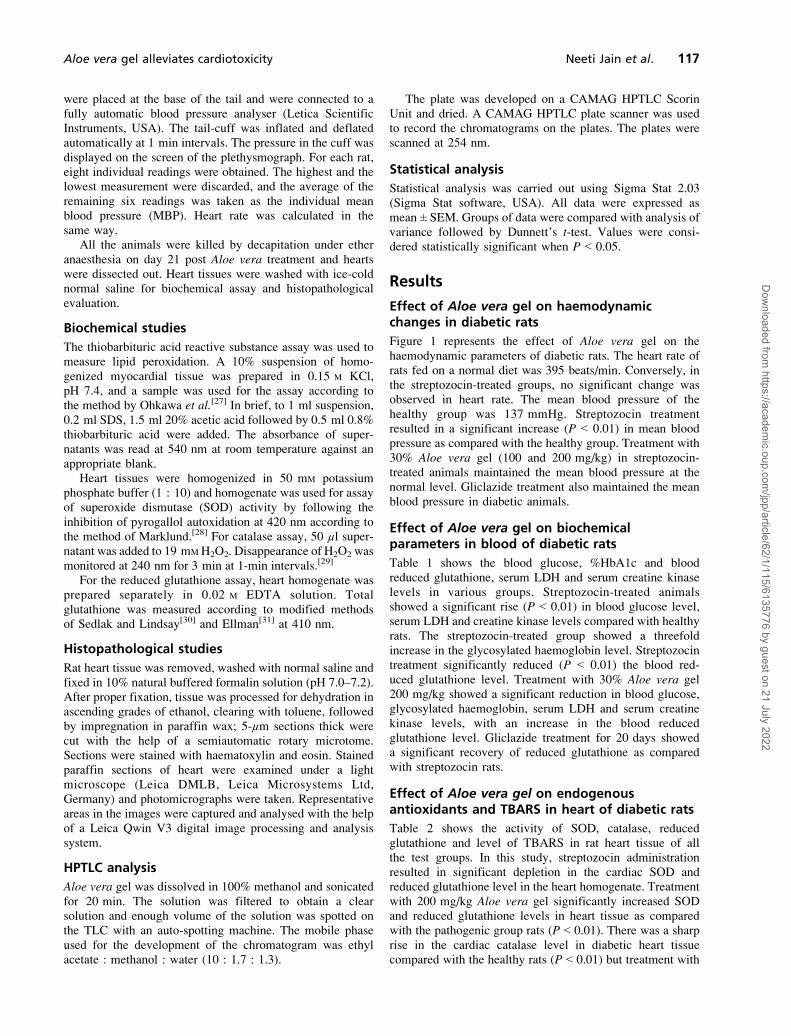
Effect of Aloe vera gel on haemodynamicchanges in diabetic rats
Figure 1 represents the effect of Aloe vera gel on thehaemodynamic parameters of diabetic rats. The heart rate ofrats fed on a normal diet was 395 beats/min. Conversely, inthe streptozocin-treated groups, no significant change wasobserved in heart rate. The mean blood pressure of thehealthy group was 137 mmHg. Streptozocin treatmentresulted in a significant increase (P < 0.01) in mean bloodpressure as compared with the healthy group. Treatment with30% Aloe vera gel (100 and 200 mg/kg) in streptozocin-treated animals maintained the mean blood pressure at thenormal level. Gliclazide treatment also maintained the meanblood pressure in diabetic animals.
Effect of Aloe vera gel on biochemicalparameters in blood of diabetic rats
Table 1 shows the blood glucose, %HbA1c and bloodreduced glutathione, serum LDH and serum creatine kinaselevels in various groups. Streptozocin-treated animalsshowed a significant rise (P < 0.01) in blood glucose level,serum LDH and creatine kinase levels compared with healthyrats. The streptozocin-treated group showed a threefoldincrease in the glycosylated haemoglobin level. Streptozocintreatment significantly reduced (P < 0.01) the blood red-uced glutathione level. Treatment with 30% Aloe vera gel200 mg/kg showed a significant reduction in blood glucose,glycosylated haemoglobin, serum LDH and serum creatinekinase levels, with an increase in the blood reducedglutathione level. Gliclazide treatment for 20 days showeda significant recovery of reduced glutathione as comparedwith streptozocin rats.
Effect of Aloe vera gel on endogenousantioxidants and TBARS in heart of diabetic rats
Table 2 shows the activity of SOD, catalase, reducedglutathione and level of TBARS in rat heart tissue of allthe test groups. In this study, streptozocin administrationresulted in significant depletion in the cardiac SOD andreduced glutathione level in the heart homogenate. Treatmentwith 200 mg/kg Aloe vera gel significantly increased SODand reduced glutathione levels in heart tissue as comparedwith the pathogenic group rats (P < 0.01). There was a sharprise in the cardiac catalase level in diabetic heart tissuecompared with the healthy rats (P < 0.01) but treatment with
Aloe vera gel alleviates cardiotoxicity Neeti Jain et al. 117
Dow
nloaded from https://academ
ic.oup.com/jpp/article/62/1/115/6135776 by guest on 21 July 2022
either dose of Aloe vera gel prevented this rise in the cardiaccatalase level.
The myocardial lipid peroxidation marker TBARS wassignificantly elevated in diabetic rat heart tissue (P < 0.01).Aloe vera gel (200 mg/kg) treatment significantly reducedthe activity of lipid peroxides as compared with diabetic rats(P < 0.01). Gliclazide treatment did not show any significant
reduction in lipid peroxide levels in streptozocin rats after20 days treatment.
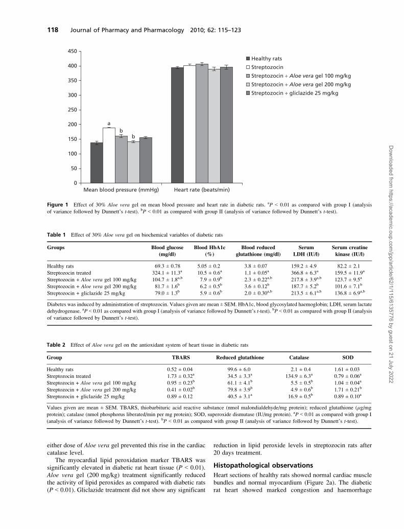
Histopathological observations
Heart sections of healthy rats showed normal cardiac musclebundles and normal myocardium (Figure 2a). The diabeticrat heart showed marked congestion and haemorrhage
450
350
250
150
50
Mean blood pressure (mmHg) Heart rate (beats/min)0
400
300
200
100
a
bb
Healthy rats
Streptozocin
Streptozocin + Aloe vera gel 100 mg/kg
Streptozocin + Aloe vera gel 200 mg/kg
Streptozocin + gliclazide 25 mg/kg
Figure 1 Effect of 30% Aloe vera gel on mean blood pressure and heart rate in diabetic rats. aP < 0.01 as compared with group I (analysis
of variance followed by Dunnett’s t-test). bP < 0.01 as compared with group II (analysis of variance followed by Dunnett’s t-test).
Table 1 Effect of 30% Aloe vera gel on biochemical variables of diabetic rats
Groups Blood glucose
(mg/dl)
Blood HbA1c
(%)
Blood reduced
glutathione (mg/dl)
Serum
LDH (IU/l)
Serum creatine
kinase (IU/l)
Healthy rats 69.3 ± 0.78 5.05 ± 0.2 3.8 ± 0.07 159.2 ± 4.9 82.2 ± 2.1
Streptozocin treated 324.1 ± 11.3a 10.5 ± 0.6a 1.1 ± 0.05a 366.8 ± 6.3a 159.5 ± 11.9a
Streptozocin + Aloe vera gel 100 mg/kg 104.7 ± 1.8a,b 7.9 ± 0.9b 2.3 ± 0.22a,b 217.8 ± 3.9a,b 123.7 ± 9.5a
Streptozocin + Aloe vera gel 200 mg/kg 81.7 ± 1.6b 6.2 ± 0.5b 3.6 ± 0.12b 187.7 ± 5.2b 101.6 ± 7.1b
Streptozocin + gliclazide 25 mg/kg 79.0 ± 1.3b 5.9 ± 0.6b 2.0 ± 0.30a,b 213.5 ± 6.1a,b 136.8 ± 6.9a,b
Diabetes was induced by administration of streptozocin. Values given are mean ± SEM. HbA1c, blood glycosylated haemoglobin; LDH, serum lactate
dehydrogenase. aP < 0.01 as compared with group I (analysis of variance followed by Dunnett’s t-test). bP < 0.01 as compared with group II (analysis
of variance followed by Dunnett’s t-test).
Table 2 Effect of Aloe vera gel on the antioxidant system of heart tissue in diabetic rats
Group TBARS Reduced glutathione Catalase SOD
Healthy rats 0.52 ± 0.04 99.6 ± 6.0 2.1 ± 0.4 1.61 ± 0.03
Streptozocin treated 1.73 ± 0.32a 34.5 ± 3.3a 134.9 ± 6.3a 0.79 ± 0.06a
Streptozocin + Aloe vera gel 100 mg/kg 0.95 ± 0.23b 61.1 ± 4.1b 5.5 ± 0.5b 1.04 ± 0.04a
Streptozocin + Aloe vera gel 200 mg/kg 0.41 ± 0.02b 79.8 ± 3.9b 4.9 ± 0.6b 1.71 ± 0.21b
Streptozocin + gliclazide 25 mg/kg 0.89 ± 0.12 40.5 ± 3.1a 16.9 ± 0.5b 0.89 ± 0.10a
Values given are mean ± SEM. TBARS, thiobarbituric acid reactive substance (nmol malondialdehyde/mg protein); reduced glutathione (µg/mg
protein); catalase (nmol phosphorus liberated/min per mg protein); SOD, superoxide dismutase (IU/mg protein). aP < 0.01 as compared with group I
(analysis of variance followed by Dunnett’s t-test). bP < 0.01 as compared with group II (analysis of variance followed by Dunnett’s t-test).
118 Journal of Pharmacy and Pharmacology 2010; 62: 115–123
Dow
nloaded from https://academ
ic.oup.com/jpp/article/62/1/115/6135776 by guest on 21 July 2022
(a) (b)
(c) (d)
(e)
Figure 2 Typical photomicrographs of the normal rat heart and diabetic rat heart after treatment with and without Aloe vera. The heart tissue was
stained with haematoxylin and eosin. (a) Normal control rats. (b) Diabetic rats treated with streptozocin only. (c) Diabetic rats treated with Aloe vera
gel 100 mg/kg. (d) Diabetic rats treated with Aloe vera gel 200 mg/kg. (e) Diabetic rats treated with gliclazide 25 mg/kg. Magnification ×20.
Aloe vera gel alleviates cardiotoxicity Neeti Jain et al. 119
Dow
nloaded from https://academ
ic.oup.com/jpp/article/62/1/115/6135776 by guest on 21 July 2022
(Figure 2b). The heart section of diabetic rats treated with30% Aloe vera gel (100 mg/kg) showed congested bloodvessels with mild haemorrhage (Figure 2c). Administrationof 200 mg/kg 30% gel to diabetic rats resulted in heartsections showing normal myocardium (Figure 2d). Heartsections of diabetic rats treated with gliclazide showedcongestion with patchy haemorrhage (Figure 2e).
Discussion
Diabetes mellitus is one of the most common chronicdiseases and is associated with hyperglycaemia and comor-bidities such as obesity and hypertension. The use of a lowerdose of streptozocin (40 mg/kg) produced a partial destruc-tion of β-cells, but the rats become permanently diabetic.[32]
Since the β-cells are not completely destroyed the rats do notrequire insulin to survive.[33] In diabetes, hyperglycaemia-induced oxidative stress is caused by free radical generationor by overproduction of superoxide anion. Hyperglycaemiacan simply inactivate existing enzymes by glycating theirprotein, leading to DNA cleavage.[34]
The increased level of blood glucose in streptozocin-induced diabetic rats was lowered by both doses of 30% Aloevera gel (100 and 200 mg/kg). Aloe has long been used all overthe world for its various medicinal properties. Reports areavailable regarding the antidiabetic activity of differentextracts of Aloe vera gel. Okyar et al.[35] reported thehypoglycaemic activity of the alcoholic extract of Aloe veraleaf pulp and gel on diabetic rats. Our findings matched thoseof Can et al.,[36] who reported the antidiabetic, antioxidant andhepatoprotective activity of Aloe vera leaf pulp and gel extractin type 2 diabetic rats. Glycosylated haemoglobin (HbA1c)indicates the percentage of haemoglobin bound to glucose. It isused as a measurement of the mean blood glucose level over aperiod of six to eight weeks i.e. during the life span of red bloodcells.[37] This glycation itself gives rise to oxygen free radicalformation, which leads to peroxidation of lipids and furthergeneration of free radicals.[38,39] The measurement ofglycosylated haemoglobin is supposed to be a very sensitiveindex for glycaemic control. In our short study we found asignificant rise in the level of HbA1c in diabetic control rats.Fuji and Nomoto[40] reported a significant change in theHbA1c level after two weeks of streptozocin administration inrats. Treatment with Aloe vera gel significantly reduced theHbA1c level, thus showing regulation of the blood glucoselevel for 20 days in streptozocin-treated rats. It has beenreported that each per cent reduction in the HbA1c levelresulted in a 35% reduction in the risk of microvascularcomplications, including myocardial infarction, in patientswith type 2 diabetes.[4,41]
In diabetic patients, uncontrolled elevation in the bloodsugar level gives rise to the formation of free radicals. Thereare many ways by which hyperglycaemia may increase thegeneration of free radicals, such as glycoxidation, polyolpathway, prostanoid biosynthesis and protein glycation.[42]
Reduced glutathione is an important inhibitor of free radicalmediated lipid peroxidation and it protects the cellularsystem against the toxic effects of lipid peroxidation.[43]
Hence, a drug that could prevent the generation of free
radicals or increase the free radical scavenging enzymes maybe effective in streptozocin-induced diabetes.[44] It has beenreported that the ethanolic extract of Aloe vera gel possessesantioxidant properties.[36] In this study, administration of30% Aloe vera gel at either dose maintained the blood andheart levels of reduced glutathione.
The cytotoxicity of xenobiotics can be evaluated using theserum activity of marker enzymes. Serum LDH and creatinekinase, which are distributed throughout the body, possessisoenzymes that are recognized as markers for liver muscleand heart lesion.[45] Contradictory reports are available in theliterature on the relationship between diabetes and creatinekinase activity.[46–50] The quantity of enzyme released fromthe damaged tissue is a measure of the number of the necroticcells. Treatment with 30% Aloe vera gel reduced the leakageof LDH and creatine kinase from the tissue bed to serum,thus showing muscle integrity and a reduction in cytotoxicityin diabetic rats. Aloe vera gel showed a reduction in theleakage of these enzymes in serum, providing the protectionagainst streptozocin-induced muscular damage.[51,52]
Oxidative stress leads to cardiac fibrosis and one of themost important pathogenetic factors of the heart’s impairedfunctional integrity in diabetes.[53] Lipid peroxidation (LPO)is a key marker of oxidative stress. The peroxidation ofpolyunsaturated fatty acids of the cell membrane mayproduce tissue damage and finally causes various diabetes-induced complications.[54,55] The heart is a vital organ fordiabetes-induced secondary complications, such as diabetescardiomyopathy, which generally occurs due to hypergly-caemia and oxygen free radicals. Several reports areavailable stating that cardiomyopathy has resulted due toaccumulation of myocardial collagen, and early diastolic andsystolic dysfunction.[56–58] Reports are available regardingthe antioxidant property of gel extracts along with itsantidiabetic activity, and these factors could be responsiblefor the cardioprotection as shown by histological observa-tion.[20,59] Interestingly, gliclazide (a second generationsulfonylurea derivative) by controlling the blood glucoselevel showed similar results against oxidative stress.[60]
SOD and catalase are principally antioxidant enzymes thatscavenge superoxide anion (O.-) formed as the intermediateproduct of H2O2 breakdown and catalysed reduction of H2O2,thus protecting tissue from highly reactive hydroxyl radi-cals.[61–63] However, there is variation on the status of thisenzyme in the diabetic state. Some studies have reporteddecreased SOD and catalase activity in vital organs duringdiabetes, or increased activity, or no change in theenzyme.[64–70] SOD and catalase play vital roles in cardio-protection and muscular integrity during diabetes. It has beenreported that impairment of endothelium-dependent vasodi-latation by the coronay bed could be abolished by perfusionwith SOD and persistent with α-tocopherol.[71]
In our study, administration of streptozocin decreased theactivity of SOD in heart.[72] The observed decrease in SODactivity could result from inactivation of SOD by H2O2 or byglycation of the enzyme, which has been reported to occur indiabetes.[73–75] A sharp rise in heart catalase activity was foundin diabetic rats thatmatched the findings of others.[54,76–81] Thisresult suggested a compensatory response to oxidative stressdue to an increase in endogenous H2O2 production.
120 Journal of Pharmacy and Pharmacology 2010; 62: 115–123
Dow
nloaded from https://academ
ic.oup.com/jpp/article/62/1/115/6135776 by guest on 21 July 2022
Treatment of diabetic rats with 30% Aloe vera gel for20 days increased the activity of non-enzymatic antioxidantssuch as reduced glutathione in heart and blood. The activity ofSOD and catalase in diabetic hearts was retained with thistreatment. Thus, the antioxidant activity of Aloe vera gel couldbe either due to inhibition of glycation of these antioxidantenzymes or protection of β-cells from streptozocin-induceddamage. Glucose, which forms a Schiff’s base with protein,has been reported to have affinity for proteins, especially thosecontaining transition metal ions.[82] This increased level ofglucose was maintained by Aloe vera gel, thus reducing theglycation of the enzyme. The elevated level of SOD indiabetic rats after treatment with Aloe vera gel predicts thatAloe vera gel may contain free radical scavenging activity,which could exert a beneficial action against the pathologicalalteration caused by the presence of O2
.- and .OH.[83]
Hypertension is generally believed to be more prevalentamong diabetic than nondiabetic subjects, and it isknown that hypertension is an independent risk factor forcardiovascular mortality in patients with diabetes.[84–87]
In diabetes, it seems that increased oxidative stress dueto hyperglycaemia or reduced availability of nitric oxideto vascular tissue leads to hypertension.[88] Consequently,treatment which can reduce oxidative stress may be effectivein preventing hypertension observed in diabetes.
In this study, we observed a significant increase in themean blood pressure and found normal heart rate after 20 daysof streptozocin administration. These results were in agree-ment with the conclusions of studies which have used thedirect technique for measurement of blood pressure.[89–91]
Gliclazide has been reported also to reduce blood pressure indiabetic rats for a longer duration.[92]
Hamman[93] reported that the mechanism of action of Aloevera extracts to reduce blood glucose levels was by enhancingglucose metabolism. They proposed that the glucose loweringeffect could have been due to an antioxidant mechanism.
Aloe vera contains approximately 75 potentially activeconstituents.[94] It is a rich source of antioxidants such asvitamins A and C, and also contains minerals, sugars, ligninand other chemicals.[95] Tanaka et al.[96] have identified fivephytosterols from the leaves which are responsible for itsantidiabetic activity. HPTLC analysis has shown the presenceof two major phytochemicals, which may be phenolic com-pounds, and polysaccharide, which are reported to haveantioxidant and antidiabetic properties.[94,97]
Conclusions
The effect of Aloe vera gel on heart antioxidants could havebeen due to tight regulation of the blood glucose level indiabetic rats, which prevented excessive formation of freeradicals through various biochemical pathways and reducedthe glycation of potential antioxidant enzymes.
Declarations
Conflict of interest
The Author(s) declare(s) that they have no conflicts ofinterest to disclose.
Funding
This research received no specific grant from any fundingagency in the public, commercial, or not-for-profit sectors.
References
1. Okhubo Y et al. Intensive insulin therapy prevents progression
of diabetic microvascular complications in Japanese patients
with non-insulin dependent diabetes mellitus; a randomized
prospective 6-years study. Diabetes Res Clin Pract 1995; 28:
103–117.
2. Thornalley PJ et al. Negative association between erythrocyte
reduced glutathione concentration and diabetic complications.
Clin Sci (Lond) 1996; 91: 575–582.
3. Ohkubo Y et al. Intensive treatment of diabetes on the
development and progression of long-term complications in
insulin-dependent diabetes mellitus. N Engl J Med 1993; 329:
977–986.
4. UKPDS group. Intensive blood glucose control with sulphonyl-
urea or insulin compared with conventional treatment and risk
of complications in patients with type 2 diabetes. Lancet 1998;
352: 837–853.
5. Dandona P et al. Oxidative damage to DNA in diabetes
mellitus. Lancet 1996; 347: 444–445.
6. Baynes JW, Thorpe SR. Role of oxidative stress in diabetic
complications: a new perspective on an old paradigm. Diabetes
1999; 48: 1–9.
7. Nishigaki I et al. Lipid peroxides levels of serum lipo-
protein fractions of diabetic patients. Biochem Med 1981; 25:
373–358.
8. Nissen SE, Wolski K. Effects of rosiglitazone on the risk of
myocardial infarction and death from cardiovascular cause.
N Engl J Med 2007; 356: 2457–2471.
9. Vaya J et al. Antioxidant constituents from licorice roots:
isolation, structure elucidation and antioxidative capacity
toward LDL oxidation. Free Radic Biol Med 1997; 23:
302–313.
10. Akhtar FM, Ali MR. Study of antidiabetic effect of a compound
medicinal plants prescription in normal and diabetic rabbits.
J Pakistan Med Assoc 1984; 45: 408–420.
11. Moon EJ et al. A novel angiogenic factor derived from Aloe
vera gel: beta-sitosterol, a plant sterol. Angiogenesis 1999; 3:
117–123.
12. Capasso F, Gaginella TS. Laxatives: A Practice Guide. Milan:
Springer Italia, 1997.
13. Ghannam N et al. The antidiabetic activity of aloes: preliminary
clinical and experimental observations. Horm Res 1986; 24:
288–294.
14. Peng SY et al. Decreased mortality of Norman murine sarcoma
in mice treated with the immunomodulator, Acemannan. Mol
Bio Ther 1991; 3: 79–87.
15. Zhang L, Tizard IR. Activation of a mouse macrophage cell line
by acemannan: the major carbohydrate fraction from Aloe vera
gel. Immunopharmacology 1996; 35: 119–128.
16. Ramamoorthy L et al. Acemannan, a beta-(1,4)-acetylated
mannan, induces nitric oxide production in macrophage cell line
RAW 264.7. Mol Pharmacol 1996; 50: 878–884.
17. Yagi A et al. Radical scavenging glycoprotein inhibiting
cyclooxygenase-2 and thromboxane A synthase from aloe
vera gel. Planta Med 2003; 69: 269–271.
18. Rajendran A et al. Evaluation of therapeutic efficacy of Aloe
vera sap in diabetes and treating wounds and inflammation in
animals. J Appl Sci Res 2007 3: 1434–1436.
Aloe vera gel alleviates cardiotoxicity Neeti Jain et al. 121
Dow
nloaded from https://academ
ic.oup.com/jpp/article/62/1/115/6135776 by guest on 21 July 2022
19. Afaf IA et al. Effects of Aloe vera (Elsabar) ethanolic extract on
blood glucose level in Wistar albino rats. J Appl Sci Res 2008;
4: 1841–1845.
20. Rajasekaran S et al. Antioxidant effect of Aloe vera gel extract
in streptozotocin-induced diabetes in rats. Pharmacol Rep 2005;
57: 90–96.
21. Hyavarina A, Nikkita E. Specific determination of blood
glucose with ortho-toludine. Clin Chim Acta 1962; 7: 140–143.
22. Trivalli LA et al. Glycated haemoglobin assay. N Engl J Med
1971; 284: 353–354.
23. Beutler IR et al. Improved method of determination of
glutathione. J Lab Clin Med 1963; 61: 882–888.
24. Bergmeyer HU, Brent E. Lactate dehydrogenase UV assay with
pyruvate kinase and NADH. In: Bergmeyer HU, ed. Methods in
Enzymatic Analysis, 2nd edn. London: Academic Press, 1974:
574–579.
25. Lum SR, Gambino G. A comparision of serum as heparinised
plasma for routine chemistry tests. Am J Clin Pathol 1974; 61:
108–112.
26. Guglielmo CG et al. A sport physiological perspective on bird
migration: evidence for flight-induced muscle damage. J Exp
Biol 2001; 204: 2683–2690.
27. Ohkawa H et al. Assay of lipid peroxides in animal tissues by
thiobarbituric reaction. Anal Biochem 1979; 95: 351–358.
28. Marklund SL. Pyrogallol autooxidation. In: Greenwald RA, ed.
Handbook of Methods for Oxygen Radical Research. Boca
Raton: CRC Press, 1985: 243–247.
29. Clairborne A. Catalase activity. In: Greenwald RA, ed. Hand-
book of Methods for Oxygen Radical Research. Boca Raton:
CRC Press, 1985: 283–284.
30. Sedlak J, Lindsay RH. Assay of total protein bound and
nonprotein sulfhydral group in tissue with Ellman’s reagent.
Anal Biochem 1968; 25: 192–205.
31. Ellman GL. Tissue sulfhydryl groups. Arch Biochem Biophys
1959; 82: 70–77.
32. Ayber MJ et al. Hypoglycemic effects of water extract of
Smallanthus soncifolius (yacan) leaves in normal and diabetic
rats. J Ethanopharmacol 2001; 74: 125–132.
33. Hayden MR, Tyagi SC. Intimal redox stress: accelerated
atherosclerosis in metabolic syndrome and type 2 diabetes
mellitus. Atheroscleropathy. Cardiovasc Diabetol 2002; 1: 3–8.
34. Wiernsperger NF. Oxidative stress as a therapeutic target in
diabetes: revisiting the controversy. Diabetes Metab 2003; 29:
579–585.
35. Okyar A et al. Effect of aloe vera leaves on blood glucose level
in type 1 and type 2 diabetic rats model. Phytother Res 2001;15: 157–161.
36. Can A et al. Effect of Aloe vera leaf gel and pulp extracts on the
liver in type-II diabetic rat model. Biol Pharmaceut Bull 2004
27: 694.
37. Ghacha R et al. HbA1c and serum fructosamine as marker of
chronic glycemic state in type 2 diabetic hemodialysis patients.
Dialysis Transplant 2001; 30: 214–217.
38. Gupta BL et al. Effect of experimental diabetes on the activities
of hexokinase, glucose-6-phosphate dehydrogenase and cate-
cholamines in rat erythrocytes of different ages. Indian J Exp
Biol 1997; 35: 792–795.
39. Jain SK et al. Erythrocyte membrane lipid peroxidation and
glycosylated hemoglobin in diabetes. Diabetes 1989; 38:
1539–1543.
40. Fuji E, Nomoto T. Changes in glycosylated hemoglobin in short
and semi long term streptozotocin-diabetic mice and rats. Japan
J Pharmacol 1984; 34: 113–115.
41. Krapek K et al. Medication adherence and associated
hemoglobin A1c in type 2 diabetes. Ann Pharmacother 2004;
38: 1357–1362.
42. Armstrong AM, Young IS. The effect of dietary treatment on
lipid peroxidation and antioxidant status in newly diagnosed
non-insulin dependent diabetes. Free Radic Biol Med 1996; 21:
719–726.
43. Garg MC, Bansal DD. Antioxidant status of streptozotocin
diabetic rats. Indian J Exp Biol 1996; 34: 264–266.
44. Paolisso G, Giugliano D. Oxidative stress and insulin action: is
there a relationship? Diabetologia 1996; 39: 357–363.
45. Aldrich JE. Clinical enzymology. In: Anderson SC,
Cockayne S, eds. Clinical Chemistry: Concept and Applica-
tions. New York: McGraw Hill, 2003: 261–284.
46. Pepato MT et al. Evaluation of toxicity after one-months
treatment with bauhinia forficate decoction in streptozotocin-
induced diabetic rats. BMC Compl Alt Medi 2004; 4: 7.
47. Scott FW et al. Serum enzymes in the BB rat before and after onset
of the overt diabetic syndrome. Clin Chem 1984; 17: 270–275.
48. Lazarov G et al. Creatine kinase in patients with diabetes
mellitus. Vutr Bolesv 1990; 29: 77–83.
49. Zhao X et al. Effect of diabetes on creatine kinase activity
in streptozotocin diabetic rats. Clin Med 1999; 112: 1028–1030.
50. Al-Shabanah AO et al. Effect of streptozotocin-induced hypergly-
cemia on intravenous pharmacokinetics and acute cardiotoxicity of
doxorubicin in rats. Pharmacol Res 2000; 41: 31–37.
51. Stanely P et al. Hypoglycemic and other related action of
tinospora cordiofolia roots in alloxan induced diabetic rats.
J Ethanopharmacol 2000; 70: 9–15.
52. Mansour HA et al. Biochemical study of the effects of some
Egyptian herbs in alloxan-induced diabetic rats. Toxicology
2002; 170: 221–228.
53. Aragno M et al. Oxidative stress triggers cardiac fibrosis in the
heart of diabetic rats. Endocrinology 2007; 149: 380–388.
54. Tatsuki R et al. Lipid peroxidation in the pancreas and other
organ in streptozotocin diabetic rats. Jpn J Pharmacol 1997; 75:
267–273.
55. Pari L, Latha M. Antidiabetic effect of Scoparia dulcis: effect
on lipid peroxidation in streptozotocin diabetes. Gen Physiol
Biophys 2005; 24: 13–26.
56. Modrak J. Collagen metabolism in the myocardium from
streptozotocin-diabetic rats. Diabetes 1980; 29: 547–550.
57. Shimizu M et al. Collagen remodeling in myocardia of patients
with diabetes. J Clin Pathol 1993; 46: 32–36.
58. Tschope C et al. Prevention of cardiac fibrosis and left
ventricular dysfunction in diabetic cardiomyopathy in rats by
transgenic expression of the human tissue kallikrein gene.
FASEB J 2004; 18: 828–835.
59. Beppu H et al. Antidiabetic effects of dietary administration of
Aloe arborescens Miller components on multiple low-dose
streptozotocin-induced diabetes in mice: investigation on
hypoglycemic action and systemic absorption dynamics of
aloe components. J Ethnopharmacol 2006; 103: 468–477.
60. Saravanan R, Pari L. Antihyperlipidemic and antiperoxi-
dative effect of Diasulin, a poly herbal formulation in alloxan
induced hyperglycemic rats. BMC Complement Altern Med
2005; 5: 14.
61. Kaleem M et al. Antidiabetic and antioxidant activity of
Annona squamosa extract in streptozotocin-induced diabetic
rats. Singapore Med J 2006; 47: 670–675.
62. Chance B et al. The mechanism of catalase action. I. Steady
state analysis. Arch Biochem 1952; 37: 301–321.
63. Uchigata Y et al. Protection by superoxide dismutase, catalase
and play (ADP-ribose) synthetase inhibitors against alloxan and
streptozotocin induced islet DNA strand breaks and against the
inhibition of proinsulin synthesis. J Biol Chem 1982; 251:
6084–6088.
64. Kedziora-Kornatowska K et al. Effect of vitamin E and vitamin
C supplementation of antioxidative state renal glomerular
122 Journal of Pharmacy and Pharmacology 2010; 62: 115–123
Dow
nloaded from https://academ
ic.oup.com/jpp/article/62/1/115/6135776 by guest on 21 July 2022
basement membrane thickness in diabetic kidney. Nephron Exp
Nephrol 2003; 95: 134–143.
65. Obrosova I et al. Early changes in lipid peroxidation and
antioxidative defence in rat retina. Eur J Pharmcol 2000; 398:
139–146.
66. Rauscher F et al. Effect of coenzyme Q10 treatment on
antioxidant pathways in normal and streptozotocin-induced
diabetic rats. J Biochem Mol Toxicol 2001; 15: 41–46.
67. Otsyula M et al. Oxidative stress in rats after 60 days of
hypergalactosemia or hyperglycemia. Int J Toxicol 2003; 5:
423–427.
68. Mekinova D et al. Effect of intake of exogenous vitamins C, E
and B carotene on the antioxidant status in kidney of rats with
streptozotocin-induced diabetes. Nahrung 1995; 39: 257–261.
69. Maritima AC et al. Effect of α-lipoic acid on biomarkers of
oxidative stress in streptozotocin-induced diabetic rats. J Nutr
Biochem 2003; 14: 288–294.
70. Godin DV et al. Antioxidant enzyme alterations in experimental
and clinical diabetes. Mol Cell Biochem 1988; 84: 223–231.
71. Rosen P et al. Endothelial relaxation is disturbed by oxidative
stress in the diabetic rat heart: influence of tocopherol as
antioxidant. Diabetologia 1995; 38: 1157–1168.
72. Kediziora KK et al. Effect of vitamin E and vitamin C
supplementation of antioxidative state renal glomerular base-
ment membrane thickness in diabetic kidney. Exp Nephrol
2003; 95: 134–143.
73. Sozmen EY et al. Catalase/superoxide dismutase (SOD) and
catalase/paraoxonase (PON) ratios may implicate poor glycemic
control. Arch Med Res 2001; 32: 283–287.
74. Soon VY, Tan BKH. Evaluation of the hypoglycemic
antioxidant activities of morinda officinalis in streptozotocin
induced diabetic rats. Singapore Med J 2002; 43: 77–85.
75. Ravi K et al. Protective effect of Eugenia jambolana seed
kernel on tissue antioxidants in streptozotocin induced diabetic
rats. Biol Phar Bull 2004; 27: 1212–1217.
76. Kakkar R et al. Time course study of oxidative stress in aorta
and heart of diabetic rat. Clin Sci 1996; 91: 441–448.
77. Noyan T et al. The oxidant and antioxidant effect of 25-
hydroxyvitamin D3 in liver, kidney and heart tissues of diabetic
rats. Clin Exp Med 2005; 5: 31–36.
78. Wohaieb SA, Godin DV. Alteration in free radical tissue
defence mechanism in streptozotocin induced diabetes in rats.
Effect of insulin treatment. Diabetes 1987; 36: 1014–1018.
79. Matkovics B et al. Further prove on oxidative stress in alloxan
diabetic rat tissue. Acta Physiol Hung 1997/98; 85: 183–192.
80. Stefek M et al. Effect of dietary supplementation with the
pyridoindole antioxidant stobadine on antioxidant state and
ultrastructure of diabetic rat myocardium. Acta Diabetol 2000;
37: 111–117.
81. Judith AH et al. Superoxide and respiratory coupling in
mitochondria of insulin deficient diabetic rats. Endocrinology
2008; 150: 146–155.
82. Taniguchi N et al. Involvement of glycation and oxidative stress in
diabetic macroangiopathy. Diabetes 1996; 45: S81–83.
83. Pushparaj P, Tan BKH. Effect of Averrhoa bilimbi leaf extract
on blood glucose and lipids in streptozotocin-induced diabetic
rats. J Ethanopharmacol 2000; 72: 69–76.
84. Dubrey SW et al. Risk factors for cardiovascular disease
in IDDM. A study of identical twins. Diabetes 1994; 43:
831–835.
85. Tomlinson KC et al. Functional consequences of streptozotocin-
induced diabetes mellitus, with particular reference to the
cardiovascular system. Pharmacol Rev 1992; 44: 103–150.
86. Rodrigues B, McNeill JH. Cardiac function in sponta-
neously hypertensive diabetic rats. Am J Physiol 1986 251:
H571–H580.
87. Litwin SE et al. Abnormal cardiac function in the streptozo-
tocin-diabetic rat. Changes in active and passive properties of
the left ventricle. J Clin Invest 1990; 86: 481–488.
88. Ozcelikay AT et al. Reversal effect of L-arginine treatment on
vascular responsiveness of streptozotocin-diabetic rats. Phar-
macol Res 2000; 41: 201–209.
89. Maeda CY et al. Streptozotocin diabetes modifies arterial
pressure and baroreflex sensitivity in rats. Braz J Med Biol Res
1995; 28: 497–501.
90. Brooks DP et al. Vasopressin in rats with genetic and
streptozotocin induced diabetes. Diabetes 1989; 38: 54–57.
91. Dai S et al. Improvement in cardiac function in streptozotocin-
diabetic rats by salt loading. Can J Physiol Pharmacol 1994; 72:
1288–1293.
92. De Mattia G et al. Diabetic endothelial dysfunction: effect of
free radicals scavenging in type 2 diabetes patients. J Diabetes
Complications 2003; 17: 30–35.
93. Hamman JH. Composition and applications of Aloe vera leaf
gel. Molecules 2008; 13: 1599–1616.
94. Surjushe A et al. Aloe vera: a short review. Indian J Dermatol
2008; 53: 163–166.
95. Athetron P. Aloe vera revisited. Br J Phytother 1998; 4: 76–83.
96. Tanaka M et al. Identification of five phytosterols from Aloe
vera gel as anti-diabetic compounds. Biol Pharm Bull 2006; 29:
1418–1422.
97. Hikino H et al. Isolation and hypoglycemic activity of arborans
A and B, glycans of Aloe vera arborescens var. natalensis
leaves. Int J Crude Drug Res 1986; 24: 183–186.
Aloe vera gel alleviates cardiotoxicity Neeti Jain et al. 123
Dow
nloaded from https://academ
ic.oup.com/jpp/article/62/1/115/6135776 by guest on 21 July 2022