Limb-girdle muscular dystrophy type 2D gene therapy restores α-sarcoglycan and associated proteins

Rev Port Cardiol. 2013;32(5):395---409
Revista Portuguesa de
CardiologiaPortuguese Journal of Cardiology
www.revportcardiol.org
REVIEW ARTICLE
Cardiotoxicity associated with cancer therapy: Pathophysiologyand prevention�
Rui Adãoa, Gilles de Keulenaerb, Adelino Leite-Moreiraa, Carmen Brás-Silvaa,∗
a Departamento de Fisiologia e Cirurgia Cardiotorácica, Faculdade de Medicina, Universidade do Porto, Porto, Portugalb Laboratory of Physiology, University of Antwerp, Antwerp, Belgium
Received 21 September 2012; accepted 1 November 2012Available online 10 June 2013
KEYWORDSCardiotoxicity;Cardiomyopathy;Chemotherapy;Anthracyclines;Radiotherapy;Trastuzumab;HER2
Abstract Cardiotoxicity is one of the most significant adverse effects of cancer treatment,and is responsible for considerable morbidity and mortality. Among the effects of chemothera-peutic agents on the cardiovascular system, the most frequent and serious is heart failure withventricular systolic dysfunction. Other toxic effects include hypertension, thromboembolic dis-ease, pericardial disease, arrhythmias and myocardial ischemia. For several decades, cancertherapy-induced cardiomyopathy was almost exclusively associated with the use of cumulativedoses of anthracyclines, which cause permanent damage at the cellular level. However, newtherapeutic agents, such as the monoclonal antibody trastuzumab, induce transient reversiblemyocyte dysfunction which is unrelated to the dose used. Early identification of potential car-diovascular injury, accurate diagnosis of cardiotoxic events and implementation of appropriatemonitoring plans are essential in patients with cancer. Close cooperation between cardiologistsand oncologists is thus crucial, in order to balance the risks and benefits of cardiotoxic anti-cancer therapy. In this article we review the various responses to cardiotoxic cancer treatmentsand their relationship with the main antineoplastic drugs used in clinical practice. In addition,we discuss the main guidelines on detection and monitoring of cardiotoxicity in patients withcancer.© 2012 Sociedade Portuguesa de Cardiologia. Published by Elsevier España, S.L. All rightsreserved.
PALAVRAS-CHAVEEfeitos cardiotóxicos;
Cardiotoxicidade associada à terapêutica oncológica: mecanismos fisiopatológicos eestratégias de prevencão
Cardiomiopatia;Quimioterapia;Antraciclinas;Radioterapia;Trastuzumab;erbB
Resumo A cardiotoxicidade é um dos efeitos adversos mais significativos do tratamentooncológico, responsável por uma considerável morbimortalidade. Entre os eventos lesivos dosagentes/fármacos quimioterápicos no sistema cardiovascular, destaca-se, pela sua maior fre-quência e gravidade, a ocorrência de insuficiência cardíaca com disfuncão ventricular sistólica.Outros efeitos tóxicos cardiovasculares incluem hipertensão arterial, doenca tromboembólica,
� Please cite this article as: Adão R, et al. Cardiotoxicidade associada à terapêutica oncológica: mecanismos fisiopatológicos e estratégiasde prevencão. Rev Port Cardiol. 2013. http://dx.doi.org/10.1016/j.repc.2012.11.002.
∗ Corresponding author.E-mail address: [email protected] (C. Brás-Silva).
2174-2049/$ – see front matter © 2012 Sociedade Portuguesa de Cardiologia. Published by Elsevier España, S.L. All rights reserved.

396 R. Adão et al.
doencas pericárdicas, arritmias e isquemia miocárdica. Durante várias décadas, a cardiomiopa-tia induzida por terapêutica oncológica era quase exclusivamente associada ao uso de dosescumulativas de antraciclinas, que promovem lesões permanentes a nível celular. No entanto,o uso de novos agentes terapêuticos, como o anticorpo monoclonal trastuzumab, induz umadisfuncão transitória reversível dos miócitos sem que haja relacão com a dose utilizada. Atual-mente, é essencial para os doentes com cancro a identificacão precoce da lesão cardiovascular,o diagnóstico preciso de eventos cardiotóxicos e a implementacão de planos de monitorizacãoadequados. Neste contexto, é fulcral na prática clínica uma cooperacão estreita entre cardiolo-gistas e oncologistas, de forma a equilibrar os riscos cardiotóxicos com os benefícios da terapiaantineoplásica em doentes oncológicos. Neste artigo revimos as diversas respostas cardiotóxicasao uso de tratamentos oncológicos e a sua relacão com os principais fármacos antineoplásicosusados na prática clínica. Além disso, serão abordadas as principais linhas de orientacão noque respeita às estratégias de detecão/monitorizacão da cardiotoxicidade em indivíduos comcancro.© 2012 Sociedade Portuguesa de Cardiologia. Publicado por Elsevier España, S.L. Todos osdireitos reservados.
I
Cyapfiias
dom
roitcs
ciewpiab
ftvtvmdrko
dtaac
D
AcnCmrca
A
The most common form of cardiotoxicity in cancertreatment is anthracycline-related cardiomyopathy.7 Ini-tial evidence indicated that left ventricular (LV) systolicdysfunction was closely related to cumulative doses of
Table 1 Criteria to confirm or revise a preliminary diagno-sis of cardiac dysfunction.
(1) Cardiomyopathy characterized by a decrease in LVEFthat was either global or more severe in the septum
(2) Symptoms of CHF(3) Associated signs of CHF, including S3 gallop,
tachycardia, or both(4) Decline in LVEF of at least 5% to less than 55% with signs
or symptoms of CHF, or a decline in LVEF of at least 10%to below 55% without signs or symptoms
ntroduction
ancer treatment has made dramatic advances in recentears; the development of intensive antineoplastic ther-pies has substantially improved the prognosis of canceratients.1 However, despite its unquestionable bene-ts, the safety aspects of cancer therapy cannot be
gnored, not least because these drugs’ mechanisms ofction can have harmful effects on the cardiovascularystem.2,3
Cancer is currently the second leading cause ofeath in Portugal after cardiovascular disease,4 and col-rectal cancer is the leading cause of cancer-relatedortality.5
For several decades, cancer therapy-induced cardiotoxiceactions were almost exclusively associated with the usef anthracyclines, but a new dimension appeared whent was recognized that drugs targeting the action of cer-ain tyrosine kinase receptors or tumor (estrogen) receptorsan have undesirable clinical effects on the cardiovascularystem.3,6
A large part of the literature on the cardiotoxicity ofancer therapy deals solely with cardiomyopathy, but thiss only one of several forms of cardiac dysfunction.7 Oth-rs may result in ischemia or changes in blood pressure,8
hile antineoplastic agents can lead to thickening of theericardium or disrupt pericardial fluid balance, resultingn effusion.9 Such treatment can also increase the risk ofrrhythmias in patients predisposed to ventricular ectopiceats.10
Protection of cardiac function is a constant challengeor the pharmaceutical industry, regulatory authorities andhe physicians who have to deal with adverse reactions toarious anticancer agents.3 Assessment of patients exposedo such drugs, analysis of the risks they pose to indi-iduals and patient groups with cancer, prevention anditigation of cardiac injury, monitoring of cardiac function
uring and after therapy, and treatment of chemotherapy-elated cardiotoxicity, have all generated a vast quantity ofnowledge and data that is collectively known as ‘‘cardio-ncology’’.7,11In this article we review the various responses to car-iotoxic cancer treatments, and their relationship withhe main antineoplastic drugs used in clinical practice. Inddition, we analyze the latest guidelines on detectionnd monitoring of cardiotoxicity in patients with can-er.
efinition of cardiotoxicity
standard definition of cardiotoxicity is essential for bothlinicians and researchers.3 One of the most precise defi-itions was proposed by the Cardiac Review and Evaluationommittee, as part of its oversight of clinical trials of theonoclonal antibody trastuzumab (Table 1).12 The crite-
ia do not include subclinical cardiovascular lesions whichan occur as an initial response to some chemotherapeuticgents.13
ntineoplastic drugs and cardiomyopathy
Adapted from Seidman et al.12
CHF: congestive heart failure; LVEF: left ventricular ejectionfraction.
Cardiotoxicity associated with cancer therapy: Pathophysiology and prevention strategies 397
Table 2 Proposed classification of chemotherapy-related cardiomyopathy.
Type of drug Prototype Cumulative dose relationship Reversibility
Type I DoxorubicinCyclophosphamide Yes No
tp
angptlstd
d
PVtaapmthesis of DNA, RNA and proteins, as well as of importanttranscription factors that regulate cardiospecific genes.24,25
Reduced protein expression, together with degradationof myofilaments, results in disruption of sarcomeric pro-
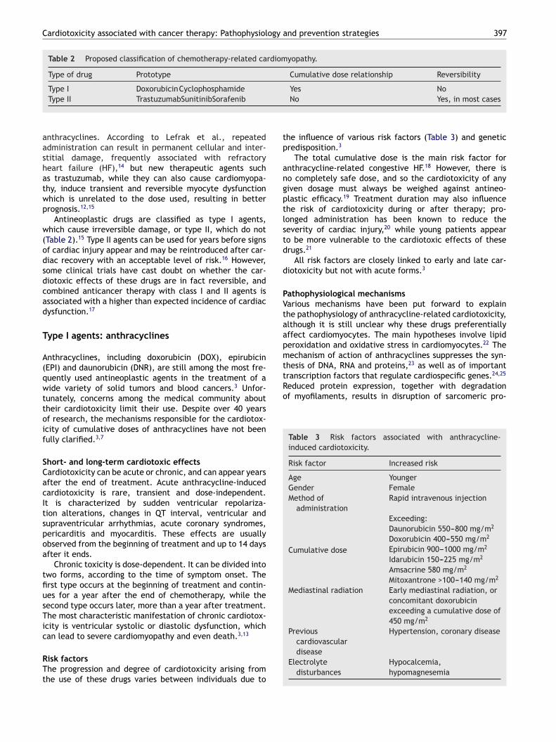
Table 3 Risk factors associated with anthracycline-induced cardiotoxicity.
Risk factor Increased risk
Age YoungerGender FemaleMethod of
administrationRapid intravenous injection
Cumulative dose
Exceeding:Daunorubicin 550---800 mg/m2
Doxorubicin 400---550 mg/m2
Epirubicin 900---1000 mg/m2
Idarubicin 150---225 mg/m2
Amsacrine 580 mg/m2
Mitoxantrone >100---140 mg/m2
Mediastinal radiation Early mediastinal radiation, orconcomitant doxorubicinexceeding a cumulative dose of450 mg/m2
Previouscardiovascular
Hypertension, coronary disease
Type II TrastuzumabSunitinibSorafenib
anthracyclines. According to Lefrak et al., repeatedadministration can result in permanent cellular and inter-stitial damage, frequently associated with refractoryheart failure (HF),14 but new therapeutic agents suchas trastuzumab, while they can also cause cardiomyopa-thy, induce transient and reversible myocyte dysfunctionwhich is unrelated to the dose used, resulting in betterprognosis.12,15
Antineoplastic drugs are classified as type I agents,which cause irreversible damage, or type II, which do not(Table 2).15 Type II agents can be used for years before signsof cardiac injury appear and may be reintroduced after car-diac recovery with an acceptable level of risk.16 However,some clinical trials have cast doubt on whether the car-diotoxic effects of these drugs are in fact reversible, andcombined anticancer therapy with class I and II agents isassociated with a higher than expected incidence of cardiacdysfunction.17
Type I agents: anthracyclines
Anthracyclines, including doxorubicin (DOX), epirubicin(EPI) and daunorubicin (DNR), are still among the most fre-quently used antineoplastic agents in the treatment of awide variety of solid tumors and blood cancers.3 Unfor-tunately, concerns among the medical community abouttheir cardiotoxicity limit their use. Despite over 40 yearsof research, the mechanisms responsible for the cardiotox-icity of cumulative doses of anthracyclines have not beenfully clarified.3,7
Short- and long-term cardiotoxic effectsCardiotoxicity can be acute or chronic, and can appear yearsafter the end of treatment. Acute anthracycline-inducedcardiotoxicity is rare, transient and dose-independent.It is characterized by sudden ventricular repolariza-tion alterations, changes in QT interval, ventricular andsupraventricular arrhythmias, acute coronary syndromes,pericarditis and myocarditis. These effects are usuallyobserved from the beginning of treatment and up to 14 daysafter it ends.
Chronic toxicity is dose-dependent. It can be divided intotwo forms, according to the time of symptom onset. Thefirst type occurs at the beginning of treatment and contin-ues for a year after the end of chemotherapy, while thesecond type occurs later, more than a year after treatment.The most characteristic manifestation of chronic cardiotox-icity is ventricular systolic or diastolic dysfunction, whichcan lead to severe cardiomyopathy and even death.3,13
Risk factorsThe progression and degree of cardiotoxicity arising fromthe use of these drugs varies between individuals due to
No Yes, in most cases
he influence of various risk factors (Table 3) and geneticredisposition.3
The total cumulative dose is the main risk factor fornthracycline-related congestive HF.18 However, there iso completely safe dose, and so the cardiotoxicity of anyiven dosage must always be weighed against antineo-lastic efficacy.19 Treatment duration may also influencehe risk of cardiotoxicity during or after therapy; pro-onged administration has been known to reduce theeverity of cardiac injury,20 while young patients appearo be more vulnerable to the cardiotoxic effects of theserugs.21
All risk factors are closely linked to early and late car-iotoxicity but not with acute forms.3
athophysiological mechanismsarious mechanisms have been put forward to explainhe pathophysiology of anthracycline-related cardiotoxicity,lthough it is still unclear why these drugs preferentiallyffect cardiomyocytes. The main hypotheses involve lipideroxidation and oxidative stress in cardiomyocytes.22 Theechanism of action of anthracyclines suppresses the syn-
23
diseaseElectrolyte
disturbancesHypocalcemia,hypomagnesemia
3
tsmabrah
pbchldbr
SAohltaStwtvarr
aocaam
ciaieTdtfigmifiwt
cV
cscbCpleraodilihoiucrA
T
MogirdCccad
ROwaipaw
opdSu
PTtts
98
eins such as titin in cardiac cells, leading to cardiacarcopenia.26 At the same time, destruction of myofila-ents may be worsened by combined therapies such as
nthracyclines with trastuzumab,16 which can also affectioenergetics27 and damage DNA.28 Anthracyclines also dis-upt the dynamic regulation of cardiac function, alteringdrenergic and adenylyl cyclase activity29 and calciumomeostasis.27,30
Clinical and experimental evidence suggests that anotherossible mechanism for cardiotoxicity is cardiac cell deathy apoptosis or necrosis after each exposure to anthra-yclines. Given the limited regenerative capacity of theeart, the number of cardiomyocytes progressively falls,eading to ventricular remodeling.22 De Angelis et al. haveemonstrated that DOX-induced cardiomyopathy can alsoe mediated by loss of cardiac stem cells and reversed byestoration of progenitor cell function.31
trategies to limit cardiotoxicitylthough there is no generally accepted way to limitr prevent anthracycline cardiotoxicity, various strategiesave been adopted, most notably the synthesis of ana-ogues of the natural compounds, the development ofumor-specific formulations, and the use of cardioprotectivegents.32
ynthetic analogues of natural compounds. Changes inhe structure of anthracyclines have produced compoundsith low levels of cardiotoxicity, allowing the adminis-
ration of higher doses.22 EPI33 and idarubicin (IDA)34 areiable alternatives to DOX and DNR, respectively. Othernthracyclines such as pirarubicin and aclarubicin, althoughegistered in some countries, do not yet play a significantole in global terms.35
Epirubicin. EPI is a semi-synthetic epimer of DOX withsimilar oncological spectrum.36 Although its mechanism
f action is similar to that of DOX, some of its physical,hemical and pharmacokinetic properties are different,32
nd it is significantly less cardiotoxic than DOX whendministered in doses that result in the same level ofyelosuppression.36
Idarubicin. IDA is a structural analogue of DNR that inter-alates into DNA, interacting with topoisomerase II andnhibiting nucleic acid synthesis.32 It is highly lipophilicnd thus is easily absorbed into cells, and can be admin-stered intravenously or orally. However, doubts have beenxpressed as to whether it is in fact any less cardiotoxic.35,37
umor-specific formulations. New techniques have beeneveloped to deliver anthracyclines in ways that reduceheir absorption in cardiac tissue.38 The current methodor passive delivery of DOX and DNR to the target tumors by incorporating them into liposomes,39 which offers aood degree of cardioprotection by increasing the drug’solecular size through encapsulation and extending its elim-
nation time. This enables the drug to remain in the organismor longer and with fewer adverse effects, and also keepst away from organs that have normal capillary junctions,hile easily penetrating areas with immature vascular sys-
ems, such as tumors.38
By contrast, anthracycline prodrugs, unlike liposomalompounds, can reach the tumor by an active pathway.arious prodrugs have been developed by conjugating anti-
ogdp
R. Adão et al.
ancer agents with peptides, carbohydrates, antibodies,erum proteins and synthetic polymers.35 These conjugatesannot penetrate normal cells and are activated specificallyy cancer cells.35,40
ardioprotective agents. It is a priority to developharmacological therapies that protect the cardiovascu-ar system without interfering with the antineoplasticffects of anthracyclines.32,41 Given the importance ofeactive oxygen species (ROS) and oxidative stress innthracycline-induced cardiotoxicity, research has focusedn drugs and natural compounds that improve the antioxi-ant defenses of cardiomyocytes.41 Various types of drug,ncluding antioxidants, iron-chelating agents and lipid-owering drugs, have been tested in animal models andn humans,41---43 but the cardioprotective effect of some isighly questionable.42,43 Dexrazoxane (Cardioxane®) is thenly drug approved for clinical use to prevent anthracycline-nduced cardiotoxicity,44 but controversy surrounds itsse due to possible compromise of antineoplastic effi-acy and increase in secondary tumors, and it has beenestricted to adult patients by the US Food and Drugministration.45
ype II agents: trastuzumab
onoclonal antibodies are among the best-known examplesf targeted cancer therapy and are widely used. The HER2ene is overexpressed in 15---25% of breast cancers, result-ng in overproduction of human epidermal growth factoreceptor 2 (HER2). Trastuzumab binds to the extracellularomain of HER2, thereby inhibiting its signal transduction.7
hemotherapeutic regimens that do not include anthra-yclines result in lower rates of cardiac dysfunction thanombined trastuzumab and anthracyclines, but trastuzumabssociated with vinorelbine, gemcitabine or liposomal DOXo not present significant risk of cardiotoxicity.46
isk factorsne of the main risk factors for cardiotoxicity associatedith trastuzumab is the use of high cumulative doses ofnthracyclines (>300 mg/m2).47 Other important risk factorsnclude LV dysfunction, irrespective of anthracycline use,re-existing systemic hypertension, body mass index >25 anddvanced age. However, chest radiotherapy concomitantlyith trastuzumab is clinically viable.48
Recent evidence shows that elderly cancer patients (agedver 70) with a history of heart disease and/or diabetesresent a higher incidence of trastuzumab-related car-iotoxic effects in breast cancer treatment. According toerrano et al., this population should be closely and contin-ously monitored.49
athophysiological mechanismshe precise pathophysiological mechanisms by whichrastuzumab acts within cells are not known, but they arehought to be closely linked to inhibition of HER2 cardiacignaling. Studies have demonstrated the important role
f HER2 in cardiomyocyte survival and development50,51;enetically modified mice with reduced HER2 levelsevelop dilated cardiomyopathy, reduced adaptation toressure overload and greater sensitivity to anthracycline
Cardiotoxicity associated with cancer therapy: Pathophysiology and prevention strategies 399
Anthracyclines, hypoxia, oxidative stress
Neuregulin
A B
Trastuzumab
Anthracyclines, hypoxia, oxidative stress
Neuregulin
HER2 HER2 HER2 HER4Signaling pathways
Compensatorymechanisms
HER2 HER2 HER2
Heart failure
Signaling pathways
HER4
Apoptosis
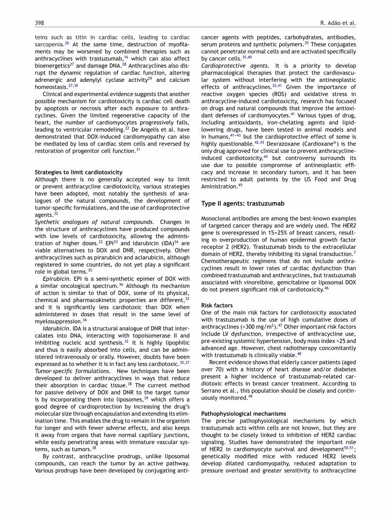
Figure 1 In response to oxidative stress, neuregulin activates compensatory mechanisms via HER2 receptors (A); in the presenceof trastuzumab, HER2/HER2 and HER2/HER4 dimers are blocked and the compensatory mechanisms are inactivated, leading toapoptosis and heart failure (B).
Adapted from Di Cosimo et al.57.
pttwdatlp
CaTtitldiINt
toxicity,52,53 while at the cellular level overexpressionof HER2 and/or neuregulin (NRG)-mediated activation ofthe HER2/HER4 signaling pathway confers greater pro-tection against oxidative stress and prevents apoptosis.54
High serum levels of HER2 have been detected in indi-viduals with chronic HF,55 and clinical trials have shownthat administration of recombinant human NRG-1 improvescardiac function in chronic HF and is well tolerated.56
Thus, while cardiac stress leads to increased HER2 expres-sion and HER2/HER4 activation by NRG, inhibition ofHER2 by trastuzumab induces ventricular dysfunction(Figure 1).57
However, trastuzumab’s pathophysiological mechanism isprobably more complex and does not only involve HER2 inhi-bition. Studies have revealed low toxicity after the use oflapatinib, a tyrosine kinase inhibitor with dual action againstHER2 and epidermal growth factor (EGF).58 There may bevarious reasons for the differences in toxicity betweenthese two drugs. Studies analyzing the effect on cardiomy-ocytes of cytotoxic immune reactions triggered by theIgG1 domain of trastuzumab show that antibody-dependentcell-mediated cytotoxicity in tumor cells is closely linked
to cardiotoxicity.59 Another suggested mechanism involvesa single intracellular response after activation of HER2in cardiomyocytes. After binding to HER2, trastuzumabmodulates mitochondrial integrity via the BCL-X family oftwoc
roteins, depleting ATP and leading to contractile dysfunc-ion. Curiously, some studies indicate that lapatinib hashe opposite effect, reducing trastuzumab’s cytotoxicityhen the two drugs are administered together.60,61 This isue to their different effect on adenosine monophosphate-ctivated protein kinase (AMPK): while this is reduced byrastuzumab, reducing intracellular ATP, it is enhanced byapatinib, leading to increased ATP production by oxidativeathways.61
ardiotoxicity of concomitant trastuzumab andnthracyclineshe mechanisms of cardiotoxicity associated with concomi-ant use of trastuzumab and anthracyclines are of particularmportance in view of the frequency with which combinedherapy is used to treat breast cancer.62 Such therapyeads to increased ROS levels and reductions in antioxi-ant compounds, promoting oxidative stress that resultsn cardiac dysfunction and overexpression of angiotensinI (Ang II), high levels of which inhibit the action ofRG, preventing it from binding to HER receptors andhereby blocking antiapoptotic signaling pathways. Inhibi-
ion of these pathways can also increase ROS production,hile Ang II also affects the activation and upregulationf NADPH oxidase63,64 by interacting with the G protein-oupled AT1 receptor, which activates NADPH oxidase
400 R. Adão et al.
Trastuzumab
Neuregulin
HER2
HER4
↓ Dimerization
↓ Cell survival
AT1
NADPHoxidase
↑ Oxidative stress
ASK-1
P38/JNK
Apoptosis Heart failure
Anthracyclines
Angiotensin II
Figure 2 Interaction of the cardiotoxic mechanisms of anthracyclines with those of trastuzumab.
vsAaMd
aAa
SVcmcdTIaawlataiTt
tdcgsfcetSigHvcdveabmI(pgo
ia protein kinase C.63 NADPH oxidase in turn producesuperoxide anion radicals, which are potent ROS. Finally,T1 receptor signaling is linked to the activation ofpoptosis signaling kinase 1 (ASK1), a member of theAPK family, and thus leads to cell death and cardiacysfunction.65
To summarize, trastuzumab directly inhibits anti-poptotic signaling pathways and causes overexpression ofng II, which stimulates ROS production and inhibits thection of NRG (Figure 2).66
trategies to limit cardiotoxicityarious strategies have been developed to reduce theardiotoxicity of trastuzumab without significantly compro-ising its therapeutic efficacy, including optimization of
hemotherapeutic combinations, shortening of treatmenturation and careful monitoring of patients.57
rastuzumab without anthracyclines. The Breast Cancernternational Research Group 006 trial was the first to testn adjuvant chemotherapeutic regimen with trastuzumabnd without anthracyclines to treat breast cancer, whichas shown to have equivalent antineoplastic efficacy with a
ower incidence of cardiotoxic events than combined ther-py with trastuzumab and anthracyclines.67 Although otherrials have confirmed the efficacy of this approach,68 there
re conflicting opinions regarding the role of anthracyclinesn combined cancer treatments.69rastuzumab-toxin conjugates. Monoclonal antibodieshat are tumor-specific but insufficiently cytotoxic in
icpg
hemselves can be chemically bound to cytotoxic agents toirect them to specific antigens on target tumors, whichonfers more control over apoptosis in tumor cells andreater selectivity in their action.70 Phillips et al. demon-trated that trastuzumab conjugated with DM1, derivedrom the fungal toxin maytansine, is associated with lowardiotoxicity,71 and clinical trials have confirmed thefficacy and tolerability of the trastuzumab-DM1 conjugateo treat breast cancer.72
hort-term treatment regimens. There is considerablenterest in short-term treatment regimens due to theirreater safety and better cost-benefit ratio.3 The Finlanderceptin (FinHER) trial compared adjuvant docetaxel andinorelbine with or without trastuzumab for early breastancer. Trastuzumab was administered before other car-iotoxic drugs and in combination with docetaxel andinorelbine for only nine weeks, in order to test the hypoth-sis that this would reduce cardiotoxicity while maintainingntineoplastic efficacy. There was a strong associationetween low cardiotoxicity and short trastuzumab treat-ent regimens.73
ndividualized anthracycline therapy. Topoisomerase II-�TOP2A) is a target for anthracyclines, and is considered aossible predictor of response to these agents. The TOP2Aene, which is located close to the HER2 gene, is moreften overexpressed in HER2-positive tumors (34---90%) than
n other cancers (5---10%). Several reports and retrospectivelinical analyses have found that treatment for HER2-ositive breast cancer that included anthracyclines hasreater efficacy, suggesting that anthracycline use should be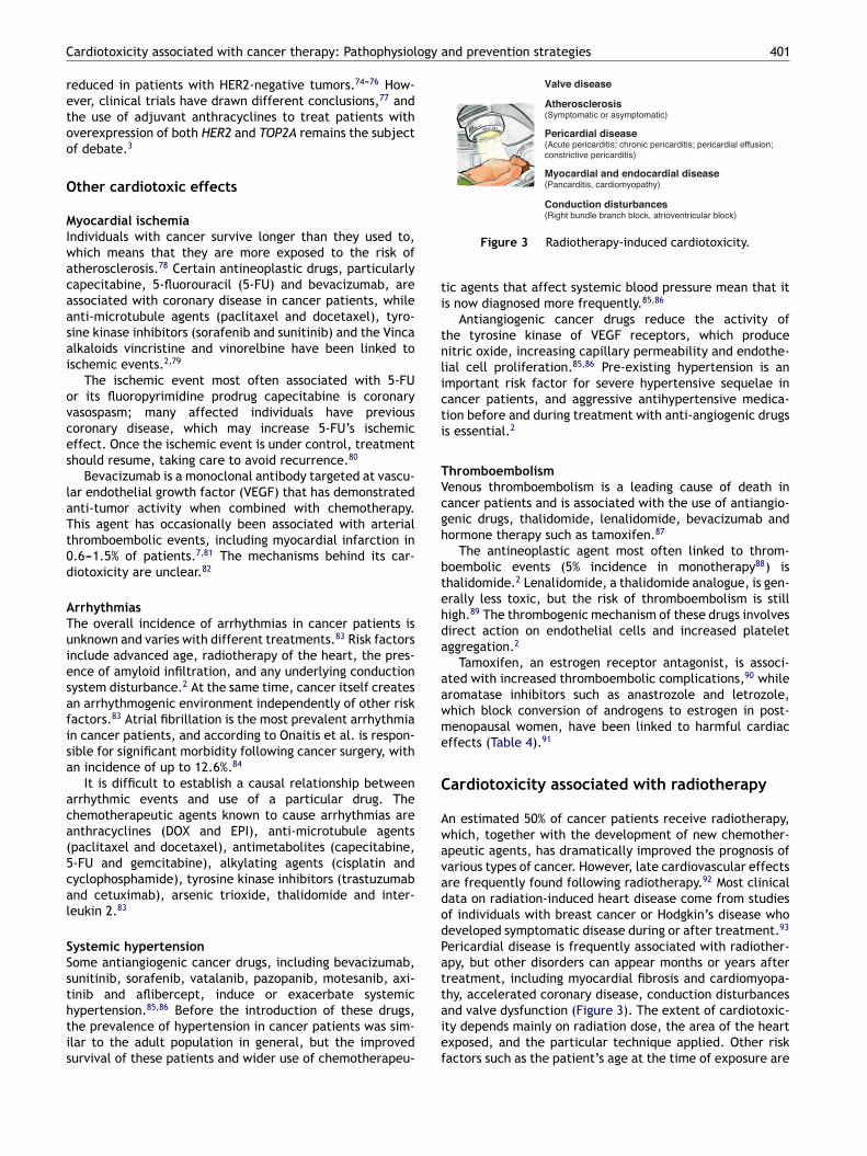
ogy and prevention strategies 401
Valve disease
Atherosclerosis(Symptomatic or asymptomatic)
Pericardial disease(Acute pericarditis; chronic pericarditis; pericardial effusion;constrictive pericarditis)
Myocardial and endocardial disease(Pancarditis, cardiomyopathy)
Conduction disturbances(Right bundle branch block, atrioventricular block)
ti
tnlicti
TVcgh
btehda
aawme
C
AwavadodPatt
Cardiotoxicity associated with cancer therapy: Pathophysiol
reduced in patients with HER2-negative tumors.74---76 How-ever, clinical trials have drawn different conclusions,77 andthe use of adjuvant anthracyclines to treat patients withoverexpression of both HER2 and TOP2A remains the subjectof debate.3
Other cardiotoxic effects
Myocardial ischemiaIndividuals with cancer survive longer than they used to,which means that they are more exposed to the risk ofatherosclerosis.78 Certain antineoplastic drugs, particularlycapecitabine, 5-fluorouracil (5-FU) and bevacizumab, areassociated with coronary disease in cancer patients, whileanti-microtubule agents (paclitaxel and docetaxel), tyro-sine kinase inhibitors (sorafenib and sunitinib) and the Vincaalkaloids vincristine and vinorelbine have been linked toischemic events.2,79
The ischemic event most often associated with 5-FUor its fluoropyrimidine prodrug capecitabine is coronaryvasospasm; many affected individuals have previouscoronary disease, which may increase 5-FU’s ischemiceffect. Once the ischemic event is under control, treatmentshould resume, taking care to avoid recurrence.80
Bevacizumab is a monoclonal antibody targeted at vascu-lar endothelial growth factor (VEGF) that has demonstratedanti-tumor activity when combined with chemotherapy.This agent has occasionally been associated with arterialthromboembolic events, including myocardial infarction in0.6---1.5% of patients.7,81 The mechanisms behind its car-diotoxicity are unclear.82
ArrhythmiasThe overall incidence of arrhythmias in cancer patients isunknown and varies with different treatments.83 Risk factorsinclude advanced age, radiotherapy of the heart, the pres-ence of amyloid infiltration, and any underlying conductionsystem disturbance.2 At the same time, cancer itself createsan arrhythmogenic environment independently of other riskfactors.83 Atrial fibrillation is the most prevalent arrhythmiain cancer patients, and according to Onaitis et al. is respon-sible for significant morbidity following cancer surgery, withan incidence of up to 12.6%.84
It is difficult to establish a causal relationship betweenarrhythmic events and use of a particular drug. Thechemotherapeutic agents known to cause arrhythmias areanthracyclines (DOX and EPI), anti-microtubule agents(paclitaxel and docetaxel), antimetabolites (capecitabine,5-FU and gemcitabine), alkylating agents (cisplatin andcyclophosphamide), tyrosine kinase inhibitors (trastuzumaband cetuximab), arsenic trioxide, thalidomide and inter-leukin 2.83
Systemic hypertensionSome antiangiogenic cancer drugs, including bevacizumab,sunitinib, sorafenib, vatalanib, pazopanib, motesanib, axi-tinib and aflibercept, induce or exacerbate systemic
hypertension.85,86 Before the introduction of these drugs,the prevalence of hypertension in cancer patients was sim-ilar to the adult population in general, but the improvedsurvival of these patients and wider use of chemotherapeu-aief
Figure 3 Radiotherapy-induced cardiotoxicity.
ic agents that affect systemic blood pressure mean that its now diagnosed more frequently.85,86
Antiangiogenic cancer drugs reduce the activity ofhe tyrosine kinase of VEGF receptors, which produceitric oxide, increasing capillary permeability and endothe-ial cell proliferation.85,86 Pre-existing hypertension is anmportant risk factor for severe hypertensive sequelae inancer patients, and aggressive antihypertensive medica-ion before and during treatment with anti-angiogenic drugss essential.2
hromboembolismenous thromboembolism is a leading cause of death inancer patients and is associated with the use of antiangio-enic drugs, thalidomide, lenalidomide, bevacizumab andormone therapy such as tamoxifen.87
The antineoplastic agent most often linked to throm-oembolic events (5% incidence in monotherapy88) ishalidomide.2 Lenalidomide, a thalidomide analogue, is gen-rally less toxic, but the risk of thromboembolism is stilligh.89 The thrombogenic mechanism of these drugs involvesirect action on endothelial cells and increased plateletggregation.2
Tamoxifen, an estrogen receptor antagonist, is associ-ted with increased thromboembolic complications,90 whileromatase inhibitors such as anastrozole and letrozole,hich block conversion of androgens to estrogen in post-enopausal women, have been linked to harmful cardiac
ffects (Table 4).91
ardiotoxicity associated with radiotherapy
n estimated 50% of cancer patients receive radiotherapy,hich, together with the development of new chemother-peutic agents, has dramatically improved the prognosis ofarious types of cancer. However, late cardiovascular effectsre frequently found following radiotherapy.92 Most clinicalata on radiation-induced heart disease come from studiesf individuals with breast cancer or Hodgkin’s disease whoeveloped symptomatic disease during or after treatment.93
ericardial disease is frequently associated with radiother-py, but other disorders can appear months or years afterreatment, including myocardial fibrosis and cardiomyopa-hy, accelerated coronary disease, conduction disturbances
nd valve dysfunction (Figure 3). The extent of cardiotoxic-ty depends mainly on radiation dose, the area of the heartxposed, and the particular technique applied. Other riskactors such as the patient’s age at the time of exposure are
402R.
Adãoet
al.
Table 4 Cardiotoxicity of the main classes of cancer drugs used in clinical practice.
Class Examples Cancers treated Cardiotoxicity Mechanism of action
Anthracyclines Doxorubicin, epirubicin,daunorubicin,idarubicin, mitoxantrone
Acute leukemias, Hodgkin’s andnon-Hodgkin’s lymphomas, breastcancer
Acute: HF, arrhythmias, QT intervalchanges, ventricular repolarizationabnormalitiesChronic (dose-dependent): LV dysfunction(non-reversible)
Damage tocardiomyocytes:- Oxidative stress (ROSproduction)- Apoptosis
Alkylating agentsCyclophosphamide Bladder, endometrial, breast,
ovarian, lung and blood cancersAcute HF (usually reversible), pericardialeffusion, arrhythmias
ROS production
Cisplatin Myocardial ischemia ---
Antimetabolites5-Fluorouracil Solid tumors, including lung, colon
and breastMyocardial ischemia, MI, arrhythmias
Coronary spasm,cardiomyocyte toxicityCapecitabine
Anti-microtubuleagents
Paclitaxel Breast and ovarian cancer, Kaposi’ssarcoma, non-small-cell lung cancer
Bradycardia, syncope, LV dysfunction,ventricular arrhythmias, myocardialischemia
Damage tocardiomyocytes
Docetaxel Breast cancer, non-small-cell lungcancer, head and neck cancers,prostate, bladder and ovarian cancer,gastric adenocarcinoma
Vinca alkaloids Vincristine, vinblastineand vinorelbine
Leukemias and lymphomas Myocardial ischemia ---
Tyrosine kinaseinhibiting antibodies
Trastuzumab Breast cancer LV dysfunction (reversible) HER2 receptor inhibitionBevacizumab Metastatic colorectal cancer, lung
cancerSystemic hypertension, venous thrombosis,LV dysfunction
VEGF inhibition
Rituximab Lymphomas, leukemias, transplantrejection, some autoimmunedisorders
Orthostatic hypotension Associated with allergiesand angioedema
Alemtuzumab Chronic lymphocytic leukemia,cutaneous T-cell lymphoma, T-celllymphoma
Hypotension, myocardial ischemia, LVdysfunction
---

Cardiotoxicityassociated
with
cancertherapy:
Pathophysiologyand
preventionstrategies
403
Table 4 (Continued)
Class Examples Cancers treated Cardiotoxicity Mechanism of action
Small-moleculetyrosine kinaseinhibitors
Imatinib Chronic myeloid leukemia,lymphoblastic leukemia,gastrointestinal stromal tumors
LV dysfunction Inhibition ofmitochondrial protectionpathways
Sunitinib Renal cell carcinoma,imatinib-resistant gastrointestinalstromal tumors
LV dysfunction, HF, myocardial ischemia,hypertension, ECG alterations
Mitochondrial damage
Sorafenib Hepatocellular carcinoma, renal cellcarcinoma
LV dysfunction ---
Erlotinib Lung and pancreatic cancer Myocardial ischemia, MI ---Lapatinib Breast cancer LV dysfunction, HF ---Dasatinib Chronic myeloid leukemia after
imatinib treatment, acutelymphoblastic leukemia
LV dysfunction, HF ---
Unclassified
Gemcitabine Non-small-cell lung cancer,pancreatic, breast and bladdercancer
Pericardial effusion ---
Retinoic acid Acute promyelocytic leukemia Pericardial effusion, LV dysfunction Retinoic acid syndromeArsenic trioxide Relapsing acute promyelocytic
leukemiaProlonged QT interval, torsades de pointes Interaction with ion
channelsTamoxifen Breast cancer Deep vein thrombosis, pulmonary
embolism, stroke---
Thalidomide andlenalidomide
Multiple myeloma Edema and sinus bradycardia, deep veinthrombosis
---
Interleukin 2 Melanoma, metastatic renal cellcarcinoma
Hypotension, arrhythmias, myocardialischemia, cardiomyopathy, myocarditis
Septic shock
ECG: electrocardiographic; HER2: human epidermal growth factor receptor 2; HF: heart failure; LV: left ventricular; MI: myocardial infarction; ROS: reactive oxygen species.
404 R. Adão et al.
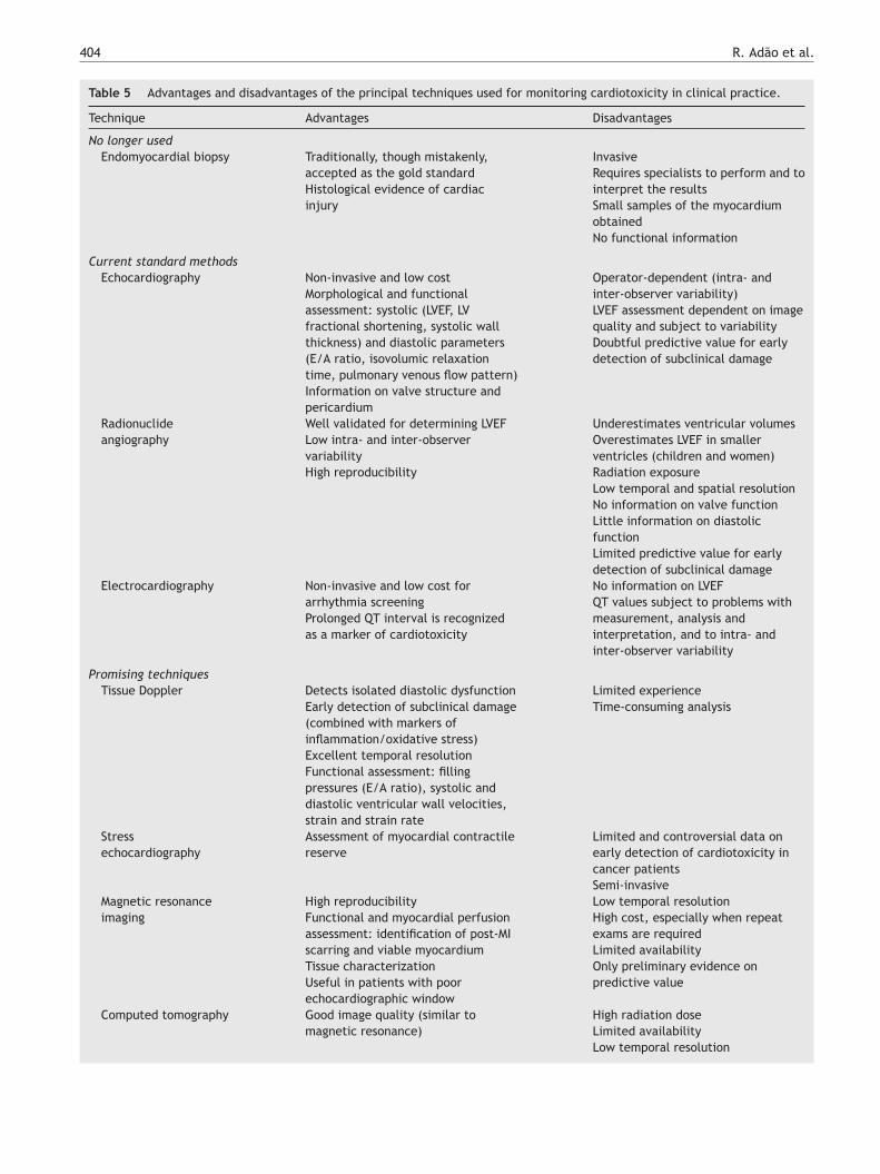
Table 5 Advantages and disadvantages of the principal techniques used for monitoring cardiotoxicity in clinical practice.
Technique Advantages Disadvantages
No longer usedEndomyocardial biopsy Traditionally, though mistakenly,
accepted as the gold standardHistological evidence of cardiacinjury
InvasiveRequires specialists to perform and tointerpret the resultsSmall samples of the myocardiumobtainedNo functional information
Current standard methodsEchocardiography Non-invasive and low cost
Morphological and functionalassessment: systolic (LVEF, LVfractional shortening, systolic wallthickness) and diastolic parameters(E/A ratio, isovolumic relaxationtime, pulmonary venous flow pattern)Information on valve structure andpericardium
Operator-dependent (intra- andinter-observer variability)LVEF assessment dependent on imagequality and subject to variabilityDoubtful predictive value for earlydetection of subclinical damage
Radionuclideangiography
Well validated for determining LVEFLow intra- and inter-observervariabilityHigh reproducibility
Underestimates ventricular volumesOverestimates LVEF in smallerventricles (children and women)Radiation exposureLow temporal and spatial resolutionNo information on valve functionLittle information on diastolicfunctionLimited predictive value for earlydetection of subclinical damage
Electrocardiography Non-invasive and low cost forarrhythmia screeningProlonged QT interval is recognizedas a marker of cardiotoxicity
No information on LVEFQT values subject to problems withmeasurement, analysis andinterpretation, and to intra- andinter-observer variability
Promising techniquesTissue Doppler Detects isolated diastolic dysfunction
Early detection of subclinical damage(combined with markers ofinflammation/oxidative stress)Excellent temporal resolutionFunctional assessment: fillingpressures (E/A ratio), systolic anddiastolic ventricular wall velocities,strain and strain rate
Limited experienceTime-consuming analysis
Stressechocardiography
Assessment of myocardial contractilereserve
Limited and controversial data onearly detection of cardiotoxicity incancer patientsSemi-invasive
Magnetic resonanceimaging
High reproducibilityFunctional and myocardial perfusionassessment: identification of post-MIscarring and viable myocardiumTissue characterizationUseful in patients with poorechocardiographic window
Low temporal resolutionHigh cost, especially when repeatexams are requiredLimited availabilityOnly preliminary evidence onpredictive value
Computed tomography Good image quality (similar tomagnetic resonance)
High radiation doseLimited availabilityLow temporal resolution
Cardiotoxicity associated with cancer therapy: Pathophysiology and prevention strategies 405
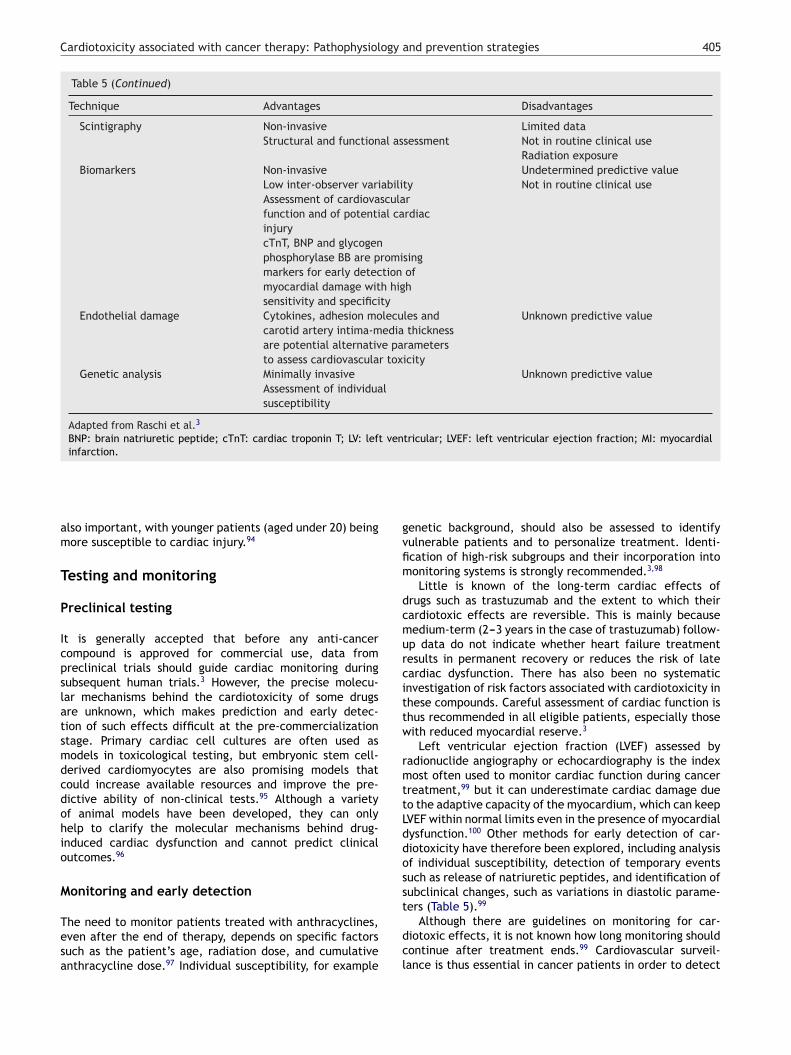
Table 5 (Continued)
Technique Advantages Disadvantages
Scintigraphy Non-invasiveStructural and functional assessment
Limited dataNot in routine clinical useRadiation exposure
Biomarkers Non-invasiveLow inter-observer variabilityAssessment of cardiovascularfunction and of potential cardiacinjurycTnT, BNP and glycogenphosphorylase BB are promisingmarkers for early detection ofmyocardial damage with highsensitivity and specificity
Undetermined predictive valueNot in routine clinical use
Endothelial damage Cytokines, adhesion molecules andcarotid artery intima-media thicknessare potential alternative parametersto assess cardiovascular toxicity
Unknown predictive value
Genetic analysis Minimally invasiveAssessment of individualsusceptibility
Unknown predictive value
Adapted from Raschi et al.3
BNP: brain natriuretic peptide; cTnT: cardiac troponin T; LV: left ventricular; LVEF: left ventricular ejection fraction; MI: myocardialinfarction.
gvfim
dcmurcittw
rmttLddosst
also important, with younger patients (aged under 20) beingmore susceptible to cardiac injury.94
Testing and monitoring
Preclinical testing
It is generally accepted that before any anti-cancercompound is approved for commercial use, data frompreclinical trials should guide cardiac monitoring duringsubsequent human trials.3 However, the precise molecu-lar mechanisms behind the cardiotoxicity of some drugsare unknown, which makes prediction and early detec-tion of such effects difficult at the pre-commercializationstage. Primary cardiac cell cultures are often used asmodels in toxicological testing, but embryonic stem cell-derived cardiomyocytes are also promising models thatcould increase available resources and improve the pre-dictive ability of non-clinical tests.95 Although a varietyof animal models have been developed, they can onlyhelp to clarify the molecular mechanisms behind drug-induced cardiac dysfunction and cannot predict clinicaloutcomes.96
Monitoring and early detection
The need to monitor patients treated with anthracyclines,even after the end of therapy, depends on specific factorssuch as the patient’s age, radiation dose, and cumulativeanthracycline dose.97 Individual susceptibility, for example
dcl
enetic background, should also be assessed to identifyulnerable patients and to personalize treatment. Identi-cation of high-risk subgroups and their incorporation intoonitoring systems is strongly recommended.3,98
Little is known of the long-term cardiac effects ofrugs such as trastuzumab and the extent to which theirardiotoxic effects are reversible. This is mainly becauseedium-term (2---3 years in the case of trastuzumab) follow-
p data do not indicate whether heart failure treatmentesults in permanent recovery or reduces the risk of lateardiac dysfunction. There has also been no systematicnvestigation of risk factors associated with cardiotoxicity inhese compounds. Careful assessment of cardiac function ishus recommended in all eligible patients, especially thoseith reduced myocardial reserve.3
Left ventricular ejection fraction (LVEF) assessed byadionuclide angiography or echocardiography is the indexost often used to monitor cardiac function during cancer
reatment,99 but it can underestimate cardiac damage dueo the adaptive capacity of the myocardium, which can keepVEF within normal limits even in the presence of myocardialysfunction.100 Other methods for early detection of car-iotoxicity have therefore been explored, including analysisf individual susceptibility, detection of temporary eventsuch as release of natriuretic peptides, and identification ofubclinical changes, such as variations in diastolic parame-ers (Table 5).99
Although there are guidelines on monitoring for car-iotoxic effects, it is not known how long monitoring shouldontinue after treatment ends.99 Cardiovascular surveil-ance is thus essential in cancer patients in order to detect

4
tp
C
Tidiiadpc
rbolsdgqt
maa
E
Pda
Cd
Rd
C
T
A
TfFFtcr
R
06
he cardiotoxic effects of antineoplastic therapy as early asossible.
onclusion
he extensive use of chemotherapy and radiotherapyn clinical practice has led to considerable controversyue to their potential adverse cardiovascular effectsn surviving cancer patients, including cardiomyopathy,schemia, arrhythmias, hypertension, pericardial diseasend thromboembolism. There is no consensus on stan-ardizing the monitoring of cardiac function in theseatients, and no reliable models able to predict the risk ofardiotoxicity.
With regard to cancer drugs, specialists in the areaecognize the need to understand the mechanisms responsi-le for cardiotoxicity. Careful management and assessmentf cardiac risk is essential in these patients, and theong-term effects of different treatments require furthertudy. To this end, it is increasingly important to establishynamic partnerships between oncologists and cardiolo-ists in order to reduce mortality and improve patients’uality of life, while maintaining the efficacy of thereatment.
To summarize, cardiotoxicity induced by cancer treat-ent should be considered a multidisciplinary problem, the
pproach to which should include basic science, oncologynd cardiovascular medicine.
thical disclosures
rotection of human and animal subjects. The authorseclare that no experiments were performed on humans ornimals for this study.
onfidentiality of data. The authors declare that no patientata appear in this article.
ight to privacy and informed consent. The authorseclare that no patient data appear in this article.
onflicts of interest
he authors have no conflicts of interest to declare.
cknowledgements
his work was funded by the Portuguese Foundationor Science and Technology (project no. FCOMP-01-0124-EDER-011051, FEDER, COMPETE, Ref. FCT-PTDC/SAU-CF/100442/2008), the João Porto 2008 Study Grant, and byhe University of Porto/Santander Totta (Projectos pluridis-iplinares IJUP 2009 and 2010). Carmen Brás-Silva is aesearcher for Programa Ciência 2008.
R. Adão et al.
eferences
1. Minami M, Matsumoto S, Horiuchi H. Cardiovascularside-effects of modern cancer therapy. Circ J. 2010;74:1779---86.
2. Yeh ET, Bickford CL. Cardiovascular complications of cancertherapy: incidence, pathogenesis, diagnosis, and manage-ment. J Am Coll Cardiol. 2009;53:2231---47.
3. Raschi E, Vasina V, Ursino MG, et al. Anticancer drugs and car-diotoxicity: insights and perspectives in the era of targetedtherapy. Pharmacol Ther. 2010;125:196---218.
4. Araújo A, Barata F, Barroso S, et al. Cost of cancer care inPortugal. Acta Med Port. 2009;22:525---36.
5. Ferlay J, Parkin DM, Steliarova-Foucher E. Estimates of can-cer incidence and mortality in Europe in 2008. Eur J Cancer.2010;46:765---81.
6. Viale PH, Yamamoto DS. Cardiovascular toxicity associ-ated with cancer treatment. Clin J Oncol Nurs. 2008;12:627---38.
7. Ewer MS, Ewer SM. Cardiotoxicity of anticancer treatments:what the cardiologist needs to know. Nat Rev Cardiol.2010;7:564---75.
8. López Medrano F, Sánchez Munoz A, Sánchez Sánchez V, et al.Cardiotoxicity of 5-fluorouracil: ischemia or myocardial toxic-ity? Rev Clin Esp. 2001;201:106---7.
9. Lestuzzi C. Neoplastic pericardial disease: old and currentstrategies for diagnosis and management. World J Cardiol.2010;2:270---9.
10. Barbey JT, Soignet S. Prolongation of the QT interval andventricular tachycardia in patients treated with arsenic tri-oxide for acute promyelocytic leukemia. Ann Intern Med.2001;135:842---3.
11. Eschenhagen T, Force T, Ewer MS, et al. Cardiovascular sideeffects of cancer therapies: a position statement from theHeart Failure Association of the European Society of Cardi-ology. Eur J Heart Fail. 2011;13:1---10.
12. Seidman A, Hudis C, Pierri MK, et al. Cardiac dysfunctionin the trastuzumab clinical trials experience. J Clin Oncol.2002;20:1215---21.
13. Albini A, Pennesi G, Donatelli F, et al. Cardiotoxicityof anticancer drugs: the need for cardio-oncology andcardio-oncological prevention. J Natl Cancer Inst. 2010;102:14---25.
14. Lefrak EA, Pitha J, Rosenheim S, et al. A clinicopatho-logic analysis of adriamycin cardiotoxicity. Cancer. 1973;32:302---14.
15. Ewer MS, Lippman SM. Type II chemotherapy-related cardiacdysfunction: time to recognize a new entity. J Clin Oncol.2005;23:2900---2.
16. Ewer MS, Vooletich MT, Durand JB, et al. Reversibility oftrastuzumab-related cardiotoxicity: new insights based onclinical course and response to medical treatment. J ClinOncol. 2005;23:7820---6.
17. Telli ML, Hunt SA, Carlson RW, et al. Trastuzumab-related car-diotoxicity: calling into question the concept of reversibility.J Clin Oncol. 2007;25:3525---33.
18. Ryberg M, Nielsen D, Skovsgaard T, et al. Epirubicin cardiotoxi-city: an analysis of 469 patients with metastatic breast cancer.J Clin Oncol. 1998;16:3502---8.
19. Lipshultz SE, Lipsitz SR, Sallan SE, et al. Chronic progres-sive cardiac dysfunction years after doxorubicin therapyfor childhood acute lymphoblastic leukemia. J Clin Oncol.2005;23:2629---36.
20. Hortobagyi GN, Frye D, Buzdar AU, et al. Decreased cardiactoxicity of doxorubicin administered by continuous intravenousinfusion in combination chemotherapy for metastatic breastcarcinoma. Cancer. 1989;63:37---45.

ogy a
Cardiotoxicity associated with cancer therapy: Pathophysiol21. Kremer LC, van Dalen EC, Offringa M, et al. Anthracycline-induced clinical heart failure in a cohort of 607 chil-dren: long-term follow-up study. J Clin Oncol. 2001;19:191---6.
22. Sawyer DB, Peng X, Chen B, et al. Mechanisms of anthracyclinecardiac injury: can we identify strategies for cardioprotection?Prog Cardiovasc Dis. 2010;53:105---13.
23. Olson RD, Mushlin PS. Doxorubicin cardiotoxicity: analysis ofprevailing hypotheses. FASEB J. 1990;4:3076---86.
24. Ito H, Miller SC, Billingham ME, et al. Doxorubicin selec-tively inhibits muscle gene expression in cardiac muscle cellsin vivo and in vitro. Proc Natl Acad Sci U S A. 1990;87:4275---9.
25. Jeyaseelan R, Poizat C, Wu HY, et al. Molecular mech-anisms of doxorubicin-induced cardiomyopathy. Selectivesuppression of Reiske iron-sulfur protein, ADP/ATP translo-case, and phosphofructokinase genes is associated with ATPdepletion in rat cardiomyocytes. J Biol Chem. 1997;272:5828---32.
26. Lim CC, Zuppinger C, Guo X, et al. Anthracyclines inducecalpain-dependent titin proteolysis and necrosis in cardiomy-ocytes. J Biol Chem. 2004;279:8290---9.
27. Takahashi S, Denvir MA, Harder L, et al. Effects ofin vitro and in vivo exposure to doxorubicin (adriamycin)on caffeine-induced Ca2+ release from sarcoplasmic reticu-lum and contractile protein function in ‘chemically-skinned’rabbit ventricular trabeculae. Jpn J Pharmacol. 1998;76:405---13.
28. Lebrecht D, Kokkori A, Ketelsen UP, et al. Tissue-specificmtDNA lesions and radical-associated mitochondrial dysfunc-tion in human hearts exposed to doxorubicin. J Pathol.2005;207:436---44.
29. Fu M, Matoba M, Liang QM, et al. Properties of G-proteinmodulated receptor-adenylyl cyclase system in myocardiumof spontaneously hypertensive rats treated with adriamycin.Int J Cardiol. 1994;44:9---18.
30. Dodd DA, Atkinson JB, Olson RD, et al. Doxorubicin car-diomyopathy is associated with a decrease in calcium releasechannel of the sarcoplasmic reticulum in a chronic rabbitmodel. J Clin Invest. 1993;91:1697---705.
31. De Angelis A, Piegari E, Cappetta D, et al. Anthracycline car-diomyopathy is mediated by depletion of the cardiac stem cellpool and is rescued by restoration of progenitor cell function.Circulation. 2010;121:276---92.
32. Mordente A, Meucci E, Silvestrini A, et al. New devel-opments in anthracycline-induced cardiotoxicity. Curr MedChem. 2009;16:1656---72.
33. Ganzina F. 4′-epi-doxorubicin, a new analogue of doxorubicin:a preliminary overview of preclinical and clinical data. CancerTreat Rev. 1983;10:1---22.
34. Ganzina F, Pacciarini MA, di Pietro N. Idarubicin (4-demethoxydaunorubicin). A preliminary overview of pre-clinical and clinical studies. Invest New Drugs. 1986;4:85---105.
35. Kratz F, Warnecke A, Schmid B, et al. Prodrugs ofanthracyclines in cancer chemotherapy. Curr Med Chem.2006;13:477---523.
36. van Dalen EC, Michiels EM, Caron HN, et al. Different anthracy-cline derivates for reducing cardiotoxicity in cancer patients.Cochrane Database Syst Rev. 2010:CD005006.
37. Cortés-Funes H, Coronado C. Role of anthracyclines in the eraof targeted therapy. Cardiovasc Toxicol. 2007;7:56---60.
38. Slingerland M, Guchelaar HJ, Gelderblom H. Liposomal drugformulations in cancer therapy: 15 years along the road. DrugDiscov Today. 2012;17:160---6.
39. Abraham SA, Waterhouse DN, Mayer LD, et al. Theliposomal formulation of doxorubicin. Methods Enzymol.2005;391:71---97.
nd prevention strategies 407
40. Sessa C, Valota O, Geroni C. Ongoing phase I and II studies ofnovel anthracyclines. Cardiovasc Toxicol. 2007;7:75---9.
41. Wouters KA, Kremer LC, Miller TL, et al. Protecting againstanthracycline-induced myocardial damage: a review of themost promising strategies. Br J Haematol. 2005;131:561---78.
42. Myers C, Bonow R, Palmeri S, et al. A randomized controlledtrial assessing the prevention of doxorubicin cardiomyopathyby N-acetylcysteine. Semin Oncol. 1983;10:53---5.
43. Ladas EJ, Jacobson JS, Kennedy DD, et al. Antioxidantsand cancer therapy: a systematic review. J Clin Oncol.2004;22:517---28.
44. Schuchter LM, Hensley ML, Meropol NJ, et al. 2002 update ofrecommendations for the use of chemotherapy and radiothe-rapy protectants: clinical practice guidelines of the AmericanSociety of Clinical Oncology. J Clin Oncol. 2002;20:2895---903.
45. Swain SM. Adult multicenter trials using dexrazoxane to pro-tect against cardiac toxicity. Semin Oncol. 1998;25:43---7.
46. Martin M, Esteva FJ, Alba E, et al. Minimizing cardiotoxicitywhile optimizing treatment efficacy with trastuzumab: reviewand expert recommendations. Oncologist. 2009;14:1---11.
47. Swain SM, Whaley FS, Gerber MC, et al. Delayed administrationof dexrazoxane provides cardioprotection for patients withadvanced breast cancer treated with doxorubicin-containingtherapy. J Clin Oncol. 1997;15:1333---40.
48. Piccart-Gebhart MJ, Procter M, Leyland-Jones B, et al.Trastuzumab after adjuvant chemotherapy in HER2-positivebreast cancer. N Engl J Med. 2005;353:1659---72.
49. Serrano C, Cortés J, de Mattos-Arruda L, et al. Trastuzumab-related cardiotoxicity in the elderly: a role for cardiovascularrisk factors. Ann Oncol. 2011;23:897---902.
50. Lee KF, Simon H, Chen H, et al. Requirement for neuregulinreceptor erbB2 in neural and cardiac development. Nature.1995;378:394---8.
51. Zhao YY, Sawyer DR, Baliga RR, et al. Neuregulins promote sur-vival and growth of cardiac myocytes. Persistence of ErbB2 andErbB4 expression in neonatal and adult ventricular myocytes.J Biol Chem. 1998;273:10261---9.
52. Ozcelik C, Erdmann B, Pilz B, et al. Conditional mutation ofthe ErbB2 (HER2) receptor in cardiomyocytes leads to dilatedcardiomyopathy. Proc Natl Acad Sci U S A. 2002;99:8880---5.
53. Crone SA, Zhao YY, Fan L, et al. ErbB2 is essential inthe prevention of dilated cardiomyopathy. Nat Med. 2002;8:459---65.
54. Lemmens K, Doggen K, de Keulenaer GW. Role of neuregulin-1/ErbB signaling in cardiovascular physiology and disease:implications for therapy of heart failure. Circulation.2007;116:954---60.
55. Perik PJ, de Vries EG, Gietema JA, et al. Serum HER2 levelsare increased in patients with chronic heart failure. Eur J HeartFail. 2007;9:173---7.
56. Jabbour A, Hayward CS, Keogh AM, et al. Parenteral admin-istration of recombinant human neuregulin-1 to patientswith stable chronic heart failure produces favourable acuteand chronic haemodynamic responses. Eur J Heart Fail.2011;13:83---92.
57. Di Cosimo S. Heart to heart with trastuzumab: a review oncardiac toxicity. Target Oncol. 2011;6:189---95.
58. Geyer CE, Forster J, Lindquist D, et al. Lapatinib pluscapecitabine for HER2-positive advanced breast cancer. N EnglJ Med. 2006;355:2733---43.
59. Sliwkowski MX, Lofgren JA, Lewis GD, et al. Nonclinical studiesaddressing the mechanism of action of trastuzumab (her-ceptin). Semin Oncol. 1999;26:60---70.
60. Shell SA, Lyass L, Trusk PB, et al. Activation of AMPK is nec-essary for killing cancer cells and sparing cardiac cells. Cell
Cycle. 2008;7:1769---75.61. Spector NL, Yarden Y, Smith B, et al. Activation of AMP-activated protein kinase by human EGF receptor 2/EGF

4
08receptor tyrosine kinase inhibitor protects cardiac cells. ProcNatl Acad Sci U S A. 2007;104:10607---12.
62. Albini A, Cesana E, Donatelli F, et al. Cardio-oncologyin targeting the HER receptor family: the puzzle of dif-ferent cardiotoxicities of HER2 inhibitors. Future Cardiol.2011;7:693---704.
63. Nakagami H, Takemoto M, Liao JK. NADPH oxidase-derivedsuperoxide anion mediates angiotensin II-induced cardiachypertrophy. J Mol Cell Cardiol. 2003;35:851---9.
64. Cardinale D, Colombo A, Sandri MT, et al. Prevention ofhigh-dose chemotherapy-induced cardiotoxicity in high-riskpatients by angiotensin-converting enzyme inhibition. Circu-lation. 2006;114:2474---81.
65. Lemarié CA, Paradis P, Schiffrin EL. New insights on signalingcascades induced by cross-talk between angiotensin II andaldosterone. J Mol Med (Berl). 2008;86:673---8.
66. Zeglinski M, Ludke A, Jassal DS, et al. Trastuzumab-induced cardiac dysfunction: a ‘dual-hit’. Exp Clin Cardiol.2011;16:70---4.
67. Robert N, Leyland-Jones B, Asmar L, et al. Randomizedphase III study of trastuzumab, paclitaxel, and carboplatincompared with trastuzumab and paclitaxel in women withHER-2-overexpressing metastatic breast cancer. J Clin Oncol.2006;24:2786---92.
68. Glück S, McKenna Jr EF, Royce M. XeNA: capecitabine plusdocetaxel, with or without trastuzumab, as preoperativetherapy for early breast cancer. Int J Med Sci. 2008;5:341---6.
69. Castrellon AB, Glück S. Adjuvant therapy for HER2 positivebreast cancer: are anthracyclines still necessary? Clin AdvHematol Oncol. 2008;6:666---72.
70. Chari RV. Targeted cancer therapy: conferring specificity tocytotoxic drugs. Acc Chem Res. 2008;41:98---107.
71. Lewis Phillips GD, Li G, Dugger DL, et al. Targeting HER2-positive breast cancer with trastuzumab-DM1, an antibody-cytotoxic drug conjugate. Cancer Res. 2008;68:9280---90.
72. Burris 3rd HA, Rugo HS, Vukelja SJ, et al. Phase II study of theantibody drug conjugate trastuzumab-DM1 for the treatmentof human epidermal growth factor receptor 2 (HER2)-positivebreast cancer after prior HER2-directed therapy. J Clin Oncol.2011;29:398---405.
73. Joensuu H, Kellokumpu-Lehtinen PL, Bono P, et al. Adju-vant docetaxel or vinorelbine with or without trastuzumab forbreast cancer. N Engl J Med. 2006;354:809---20.
74. Muss HB, Thor AD, Berry DA, et al. C-erbB-2 expression andresponse to adjuvant therapy in women with node-positiveearly breast cancer. N Engl J Med. 1994;330:1260---6.
75. Di Leo A, Gancberg D, Larsimont D, et al. HER-2 amplifica-tion and topoisomerase IIalpha gene aberrations as predictivemarkers in node-positive breast cancer patients randomlytreated either with an anthracycline-based therapy or withcyclophosphamide, methotrexate, and 5-fluorouracil. ClinCancer Res. 2002;8:1107---16.
76. Villman K, Sjöström J, Heikkilä R, et al. TOP2A and HER2 geneamplification as predictors of response to anthracycline treat-ment in breast cancer. Acta Oncol. 2006;45:590---6.
77. Bartlett JM, Munro A, Cameron DA, et al. Type 1receptor tyrosine kinase profiles identify patients withenhanced benefit from anthracyclines in the BR9601 adju-vant breast cancer chemotherapy trial. J Clin Oncol. 2008;26:5027---35.
78. Anderson JL, Adams CD, Antman EM, et al. ACC/AHA 2007guidelines for the management of patients with unstableangina/non ST-elevation myocardial infarction: a report of
the American College of Cardiology/American Heart Associ-ation Task Force on Practice Guidelines (Writing Committee toRevise the 2002 Guidelines for the Management of PatientsWith Unstable Angina/Non ST-Elevation Myocardial Infarc-R. Adão et al.
tion): developed in collaboration with the American Collegeof Emergency Physicians, the Society for CardiovascularAngiography and Interventions, and the Society of ThoracicSurgeons: endorsed by the American Association of Car-diovascular and Pulmonary Rehabilitation and the Societyfor Academic Emergency Medicine. Circulation. 2007;116:e148---304.
79. Yeh ET, Tong AT, Lenihan DJ, et al. Cardiovascularcomplications of cancer therapy: diagnosis, pathogenesis, andmanagement. Circulation. 2004;109:3122---31.
80. Kosmas C, Kallistratos MS, Kopterides P, et al. Cardiotoxicityof fluoropyrimidines in different schedules of administra-tion: a prospective study. J Cancer Res Clin Oncol. 2008;134:75---82.
81. Scappaticci FA, Skillings JR, Holden SN, et al. Arterial throm-boembolic events in patients with metastatic carcinomatreated with chemotherapy and bevacizumab. J Natl CancerInst. 2007;99:1232---9.
82. Zambelli A, della Porta MG, Eleuteri E, et al. Predicting andpreventing cardiotoxicity in the era of breast cancer targetedtherapies. Novel molecular tools for clinical issues. Breast.2011;20:176---83.
83. Guglin M, Aljayeh M, Saiyad S, et al. Introducing anew entity: chemotherapy-induced arrhythmia. Europace.2009;11:1579---86.
84. Onaitis M, D’Amico T, Zhao Y, et al. Risk factors for atrialfibrillation after lung cancer surgery: analysis of the Societyof Thoracic Surgeons general thoracic surgery database. AnnThorac Surg. 2010;90:368---74.
85. Izzedine H, Ederhy S, Goldwasser F, et al. Management ofhypertension in angiogenesis inhibitor-treated patients. AnnOncol. 2009;20:807---15.
86. Scartozzi M, Galizia E, Chiorrini S, et al. Arterial hyperten-sion correlates with clinical outcome in colorectal cancerpatients treated with first-line bevacizumab. Ann Oncol.2009;20:227---30.
87. Khorana AA, Francis CW, Culakova E, et al. Thromboembolismis a leading cause of death in cancer patients receiv-ing outpatient chemotherapy. J Thromb Haemost. 2007;5:632---4.
88. Rodeghiero F, Elice F. Thalidomide and thrombosis. Pathophys-iol Haemost Thromb. 2003;33:15---8.
89. Hirsh J. Risk of thrombosis with lenalidomide and its preven-tion with aspirin. Chest. 2007;131:275---7.
90. Deitcher SR, Gomes MP. The risk of venous thromboem-bolic disease associated with adjuvant hormone therapy forbreast carcinoma: a systematic review. Cancer. 2004;101:439---49.
91. Ewer MS, Glück S. A woman’s heart: the impact of adju-vant endocrine therapy on cardiovascular health. Cancer.2009;115:1813---26.
92. Yusuf SW, Sami S, Daher IN. Radiation-induced heart dis-ease: a clinical update. Cardiol Res Pract. 2011;2011:317659.
93. Veinot JP, Edwards WD. Pathology of radiation-induced heartdisease: a surgical and autopsy study of 27 cases. Hum Pathol.1996;27:766---73.
94. Hancock SL, Tucker MA, Hoppe RT. Factors affecting late mor-tality from heart disease after treatment of Hodgkin’s disease.JAMA. 1993;270:1949---55.
95. Kettenhofen R, Bohlen H. Preclinical assessment of cardiactoxicity. Drug Discov Today. 2008;13:702---7.
96. Barros TP, Alderton WK, Reynolds HM, et al. Zebrafish: anemerging technology for in vivo pharmacological assessment
to identify potential safety liabilities in early drug discovery.Br J Pharmacol. 2008;154:1400---13.97. Shankar SM, Marina N, Hudson MM, et al. Monitoringfor cardiovascular disease in survivors of childhood can-

ogy a
Cardiotoxicity associated with cancer therapy: Pathophysiolcer: report from the Cardiovascular Disease Task Forceof the Children’s Oncology Group. Pediatrics. 2008;121:
e387---96.98. Trachtenberg BH, Landy DC, Franco VI, et al. Anthracycline-associated cardiotoxicity in survivors of childhood cancer.Pediatr Cardiol. 2011;32:342---53.
1
nd prevention strategies 409
99. Altena R, Perik PJ, van Veldhuisen DJ, et al. Cardiovascu-lar toxicity caused by cancer treatment: strategies for early
detection. Lancet Oncol. 2009;10:391---9.00. Lester SJ, Tajik AJ, Nishimura RA, et al. Unlocking the mys-teries of diastolic function: deciphering the Rosetta Stone 10years later. J Am Coll Cardiol. 2008;51:679---89.
Copyright © 2022 FDOKUMEN