Cardioprotective Effects of Fullerenol C60(Oh)24 on a Single Dose Doxorubicin-induced Cardiotoxicity...
12
Technology in Cancer Research and Treatment ISSN 1533-0346 Volume 7, Number 1, February 2008 ©Adenine Press (2008) Cardioprotective Effects of Fullerenol C60(Oh)24 on a Single Dose Doxorubicin-induced Cardiotoxicity in Rats With Malignant Neoplasm www.tcrt.org The therapeutic utility of the anthracycline antibiotic doxorubicin is limited due to its cardio- toxicity. Our aim was to investigate the efficacy of fullerenol C 60 (OH) 24 in preventing single, high-dose doxorubicin-induced cardiotoxicity in rats with malignant neoplasm. Experiment was performed on adult female Sprague Dawley rats with chemically induced mammary car- cinomas. The animals were sacrificed two days after the application of doxorubicin and/or fullerenol, and the serum activities of CK, LDH and α-HBDH, as well as the levels of MDA, GSH, GSSG, GSH-Px, SOD, CAT, GR, and TAS in the heart, were determined. The results obtained from the enzymatic activity in the serum show that the administration of a single dose of 8 mg/kg in all treated groups induces statistically significant damage. There are significant changes in the enzymes of LDH and CK (p < 0.05), after an i.p. administration of doxorubicin/fullerenol and fullerenol. Comparing all groups with untreated control group, point to the conclusion that in the case of a lower α-HBDH/LDH ratio, results in more serious the liver parenchymal damage. The results revealed that doxorubicin induced oxidative dam- age and that the fullerenol antioxidative influence caused significant changes in MDA, GSH, GSSG, GSH-Px, SOD, CAT, GR, and TAS level in the heart (p < 0.05). Therefore, it is sug- gested that fullerenol might be a potential cardioprotector in doxorubicin-treated individuals. Key words: Cardioprotection; Doxorubicin; Fullerenol; Oxidative stress; Heart; Rats; and Mammary carcinomas. Introduction Doxorubicin (Dox), an anthracycline antibiotic, is a potent broad-spectrum an- tineoplastic agent that is highly effective in treating patients with leukaemia’s, lymphomas and many solid tumors (1). Conversely, the clinical use of Dox has been seriously limited by its undesirable side effects, particularly a dose-depen- dent myocardial injury, leading to a potentially congestive heart failure (2, 3). Dox can induce acute (arrhythmias, hypotension, activity of some enzymatic bio- markers, and oxidative tissue damage) and chronic (dilative cardiomiopathy and congestive heart failure) cardiotoxicity (4, 5). It is commonly believed that the formation of free radicals plays a crucial role in the mechanism of Dox-induced cardiotoxicity. There is significant evidence indicating that mitochondria are the principal targets in this pathological process. The induction of oxidative stress can be explained by the chemical structure of Dox, which is prone to generate reactive oxygen species (ROS). Dox is an aglycone con- sisting of a tetracyclic ring with adjacent quinine-hydroquinone groups, which ac- cumulates in the mitochondria. In vitro investigations showed that the intracellular, Rade Injac, M.D. 1,* Martina Perse, M.D. 2 Marija Boskovic, M.D. 3 Vukosava Djordjevic-Milic, M.D. 4 Aleksandar Djordjevic, Ph.D. 5 Anastazija Hvala, Ph.D. 6 Anton Cerar, Ph.D. 2 Borut Strukelj. Ph.D. 1 1 Institute of Pharmaceutical Biology University of Ljubljana Askerceva 7, 1000 Ljubljana, Slovenia 2 Institute of Pathology Medical Experimental Centre Medical Faculty, University of Ljubljana Korytkova 2, 1000 Ljubljana Slovenia 3 Institute of Pharmacokinetics and Biopharmaceutics University of Ljubljana Askerceva 7, 1000 Ljubljana, Slovenia 4 Department of Pharmacy University of Novi Sad, Hajduk Veljkova 3 21000 Novi Sad, Serbia 5 Department of Chemistry University of Novi Sad Trg Dositeja Obradovica 3 21000 Novi Sad, Serbia 6 Institute of Pathology University of Ljubljana Korytkova 2, 1000 Ljubljana, Slovenia * Corresponding Author: Rade Injac, M.D. Email: [email protected]
-
Upload
independent -
Category
Documents
-
view
1 -
download
0
Transcript of Cardioprotective Effects of Fullerenol C60(Oh)24 on a Single Dose Doxorubicin-induced Cardiotoxicity...

Technology in Cancer Research and TreatmentISSN 1533-0346Volume 7, Number 1, February 2008©Adenine Press (2008)
Cardioprotective Effects of Fullerenol C60(Oh)24 on a Single Dose Doxorubicin-induced Cardiotoxicity
in Rats With Malignant Neoplasm
www.tcrt.org
The therapeutic utility of the anthracycline antibiotic doxorubicin is limited due to its cardio-
toxicity. Our aim was to investigate the efficacy of fullerenol C60(OH)24 in preventing single,
high-dose doxorubicin-induced cardiotoxicity in rats with malignant neoplasm. Experiment
was performed on adult female Sprague Dawley rats with chemically induced mammary car-
cinomas. The animals were sacrificed two days after the application of doxorubicin and/or
fullerenol, and the serum activities of CK, LDH and α-HBDH, as well as the levels of MDA,
GSH, GSSG, GSH-Px, SOD, CAT, GR, and TAS in the heart, were determined. The results
obtained from the enzymatic activity in the serum show that the administration of a single
dose of 8 mg/kg in all treated groups induces statistically significant damage. There are
significant changes in the enzymes of LDH and CK (p < 0.05), after an i.p. administration
of doxorubicin/fullerenol and fullerenol. Comparing all groups with untreated control group,
point to the conclusion that in the case of a lower α-HBDH/LDH ratio, results in more serious
the liver parenchymal damage. The results revealed that doxorubicin induced oxidative dam-
age and that the fullerenol antioxidative influence caused significant changes in MDA, GSH,
GSSG, GSH-Px, SOD, CAT, GR, and TAS level in the heart (p < 0.05). Therefore, it is sug-
gested that fullerenol might be a potential cardioprotector in doxorubicin-treated individuals.
Key words: Cardioprotection; Doxorubicin; Fullerenol; Oxidative stress; Heart; Rats; and
Mammary carcinomas.
Introduction
Doxorubicin (Dox), an anthracycline antibiotic, is a potent broad-spectrum an-tineoplastic agent that is highly effective in treating patients with leukaemia’s, lymphomas and many solid tumors (1). Conversely, the clinical use of Dox has been seriously limited by its undesirable side effects, particularly a dose-depen-dent myocardial injury, leading to a potentially congestive heart failure (2, 3). Dox can induce acute (arrhythmias, hypotension, activity of some enzymatic bio-markers, and oxidative tissue damage) and chronic (dilative cardiomiopathy and congestive heart failure) cardiotoxicity (4, 5). It is commonly believed that the formation of free radicals plays a crucial role in the mechanism of Dox-induced cardiotoxicity. There is significant evidence indicating that mitochondria are the principal targets in this pathological process.
The induction of oxidative stress can be explained by the chemical structure of Dox, which is prone to generate reactive oxygen species (ROS). Dox is an aglycone con-sisting of a tetracyclic ring with adjacent quinine-hydroquinone groups, which ac-cumulates in the mitochondria. In vitro investigations showed that the intracellular,
Rade Injac, M.D.1,*
Martina Perse, M.D.2Marija Boskovic, M.D.3Vukosava Djordjevic-Milic, M.D.4Aleksandar Djordjevic, Ph.D.5Anastazija Hvala, Ph.D.6Anton Cerar, Ph.D.2Borut Strukelj. Ph.D.1
1Institute of Pharmaceutical BiologyUniversity of LjubljanaAskerceva 7, 1000 Ljubljana, Slovenia2Institute of PathologyMedical Experimental CentreMedical Faculty, University of LjubljanaKorytkova 2, 1000 Ljubljana Slovenia3Institute of Pharmacokinetics and BiopharmaceuticsUniversity of LjubljanaAskerceva 7, 1000 Ljubljana, Slovenia4Department of PharmacyUniversity of Novi Sad, Hajduk Veljkova 321000 Novi Sad, Serbia5Department of ChemistryUniversity of Novi SadTrg Dositeja Obradovica 321000 Novi Sad, Serbia6Institute of PathologyUniversity of LjubljanaKorytkova 2, 1000 Ljubljana, Slovenia
*Corresponding Author:Rade Injac, M.D.Email: [email protected]
�

�
Technology in Cancer Research & Treatment, Volume 7, Number 1, February 2008
as well as intra-mitochondrial, concentration of Dox is nearly twice higher than the extra-cellular concentration. The NADH dehydrogenase (mitochondrial enzymes) acts on Dox in a way that the quinone ring undergoes redox cycling between qui-nine and semiquinone states. At some point in this process, the generated electrons are captured by oxidizing agents, in-cluding oxygen, which then set off a sequence reaction leading to the production of ROS (5, 6). Cardiomyocytes express only low levels of catalase. Selenium-dependent GSH-peroxidae-1 is readily inactivated when expoused to Dox (7, 8), which, in addition, reduces the levels and activity of cytosolic CuZn su-peroxide dismutase, signifying that reductions in the levels of various antioxidant enzymes characterize a common response to Dox treatment (9). In the same manner, Dox has also been shown to depress the levels of a variety of antioxidants, which further contributes to oxidative stress (7, 10). In addition, in-direct support is presented by studies showing that Dox in-creases levels of tissue malondialdehyde (MDA), which is a product of lipid peroxidation (11, 12).
Dox was shown to down-regulate cardiac-specific transcrip-tional regulatory proteins (cardiac adriamycinresponsive protein, CARP), possibly causing reduced expression of specific genes, such as the Ca2+-gated Ca2+-release channel (ryanodin receptor-2), α-actin, myosin light chain 2 slow, and others (13-15). Moreover, Dox was shown to bind to cardiolipin, which is highly abundant in the heart mitochon-dria, altering the normal assembly of components that con-stitute the respiratory chain (4).
It is also important to note the differences in the abilities of iron chelators, antioxidants, and free radical scavengers to protect animals or patients from Dox-induced cardiomy-opathy. Although dexrazoxane has been proven effective, antioxidants do not always prevent cardiotoxicity. Impor-tantly, antioxidants such as probucol, melatonin, garlic, vi-tamins (A, E, and C), flavonoids, Centella asiatica extract, and thiol-containing reduction agents (GSH, N-acetylcyste-ine, S-acetylcysteine, amifostine) (3, 16-22), neither prevent nor delay the development of cardiomyopathy in patients treated with anthracyclines (23).
In vitro and in vivo studies have showna that the water soluble fullerenol (Full) C60(OH)24, a polyhydroxylated derivative of fullerene C60, has strong antioxidative potential (24-30). It may function as a free radical scavenger and it strongly sup-presses cytotoxicity of Dox in animal models. The scope of this experiment was to investigate the potential cardioprotec-tive effects of fullerenol C60(OH)24 on the hearts of rats with mammary carcinomas after a single dose of doxorubicin.
Materials And Methods
Chemicals and Preparation
Fullerenol C60(OH)24 was synthesized and characterized from the polybromine derivative C60Br24, which was synthesized in the reaction of C60 in Br2 with FeBr3 as a catalyst (31). Fullere-nol C60(OH)24 (Novi Sad, Serbia) was dissolved in a sterilized and apyrogenic 0.9% NaCl : Tween 80 (80:20; w/w) solution (20 mg/mL) in a laminar flow cabin immediately before use.
Doxorubicin (Adriablastina®, Pharmacia & Upjohn, Milan, Italy), was dissolved in a sterilized and apyrogenic 0.9% NaCl solution (3 mg/mL) in a laminar flow cabin just before use.
MNU (1-methyl-1-nitrosourea) was obtained from Sigma (Deisenhofen, Germany). It was always dissolved just be-fore use in a sterilized and apyrogenic 0.9% NaCl solution (14 mg/mL) in a laminar flow cabine.
Animals and Treatment
Female Sprague-Dawley outbred rats (Harlan, Italy) were obtained at 3 weeks of age, quarantined and were housed 3-4 per cage at 22-23 ºC room temperature, 70 ± 10% humidity, and 12 h light : dark cycle. They had free access to standard laboratory diet (Altromin, Germany) and water.
All experiments were approved by the National Animal Ethical Committee of Republic of Slovenia (licence num-ber 3440-138/2006) and were conducted in accordance with the European Convention for the protection of ver-tebrate animals used for experimental and other scientific purposes (ETS 123).
All 32 rats received i.p. applications of MNU (50 mg/kg body weight) (carcinogenicity was induced chemically) on the 50th and 113th day of age (32). The first palpable tumor of mam-mary gland was manifested one week after the second treat-ment with MNU. On the 160th day of age rats were treated with Dox and/or Full. The most effective dose of Full was determined in a preliminary study, which had shown signifi-cant differences for blood and tissue markers, as well as for pathohistological and oxidative stress after two days of Dox administration at a dose of 100 mg/kg of Full (unpublished data). The animals of both control groups received sterilized and apyrogenic 0.9% NaCl solution. Two days after Dox and/or Full application rats were sacrificed by CO2.
Animals were randomly divided into five groups as follows:
I. Untreated control group – rats received saline only;II. Cancer control group – rats received MNU and saline;III. Dox group – rats received MNU and Dox 8 mg/kg;IV. Dox/Full group – rats received MNU and Full 100
mg/kg 30 min before Dox 8 mg/kg;V. Full group – rats received MNU and Full 100 mg/kg
(29).

Technology in Cancer Research & Treatment, Volume 7, Number 1, February 2008
RunningTitleNeeded �
Histopathological examination revealed all treated rats de-veloped mammary tumors. Tumor incidence was 4.94 ± 0.58 per rat.
Blood for analysis was taken via heart puncture after open-ing the thoracic region. Venous blood samples were divided into two portions. 200 μL-portion was put into eppendorfs with K-EDTA (15%), carefully mixed for 20 min and used for blood cell counting. The second portion of the blood samples (8 ± 2 mL per animal) was kept at room tempera-ture for approximately 2 h and then centrifugated. Serums were used for analysis of enzymatic activity. Samples were stored at -80 ºC before the use.
Each heart was quickly removed from the sacrificed rat, placed in an ice-cold solution and adipose tissues was trimmed off. Each heart was finally minced and homog-enized in a Tris-buffer solution (pH 7.4; organ : buffer 1:10; w/w) and divided into two portions. One was used for malondialdehyde (MDA) determination, and the second was centrifugated at 13,000x g for 20 min at 4 ºC (Beckman refrigerated, Ultracentrifuge). Supernatant was used for the assays of total protein (TP) concentration, glutathione (GSH), glutathione peroxidase (GSH-Px), total antioxidant status (TAS), glutathione reductase (GR), catalase (CAT) and superoxide dismutase (SOD).
Coefficient of Heart
The coefficient of heart to body weight was calculated as the ratio of tissues (wet weight, mg) to body weight (g).
Electron Microscopy of Cardiac Tissue
For electron microscopy, the tissue samples taken from the left ventricles were cut into 1 mm3 blocks, fixed in neutral buffered formaldehyde and postfixed in 1% osmium tetrox-ide in 0.1 M phosphate buffer (pH 7.4), dehydrated in graded ethanol series and then embedded in Epon. Semi thin sec-tions were stained with Azur II. Ultra thin sections (40-60 nm thick) were placed on grids (200 meshes) and contrasted with uranyl acetate and lead citrate. Sections were examined with Jeol 1200 Ex II transmission electron microscope.
Blood Biomarker Assay
In the present study, creatine kinase (CK), lactate dehydroge-nase (LDH) and alpha-hydroxybutyrate dehydrogenase (α-HBDH) activity were assayed for evaluating cardiac damage, using Tecan Saffire (Tecan UK, Milton Keynes, UK).
The assay for CK and LDH was carried out according to the methods described in the commercial kits Chema Diagnosti-ca (Jesi, Italy). α-HBDH was determined with a commercial
kit obtained from Dialab (Vienna, Austria). The results for all enzymes were expressed as U/L.
MDA, free GSH and GSSG levels were measured with a Chromsystems Diagnostic commercial kit for HPLC analy-sis (Munchen, Germany) and were expressed as μg/L and mg/L, respectively. Free GSH/GSSG ratio was also calcu-lated. An HPLC Agilent HP 1100-model system equipped with an autosampler and a fluorescence detector (Waldbronn, Germany), was used. Randox commercial systems: Ransod (SOD), Ransel (GSH-Px), total antioxidant status (TAS) and glutathione reductase (GR) (Crumlin, United Kingdom), and Sentinel Diagnostics (Milan, Italy), were used to determine the levels of SOD, GSH-Px, TAS, GR, and TP, respectively. Catalase activity in the tissue homogenate was quantified according to the method by Aebi (33) in which H2O2 was reacted with the cell lysates (obtained as described above). The initial rate of H2O2 disappearance (0 to 60 seconds) was recorded spectrophotometrically at a wavelength of 240 nm. The results for SOD and CAT were expressed as U/mg TP, for GSH-Px and GR as U/g TP, and for TAS as mM.
Statistical Analysis
Results were expressed as mean ± standard deviation (SD). For comparison between the groups, ANOVA (single factor) and Student’s t test (two-sample assuming equal variances) were used to compare the groups. The statistical significance for all tests was set at p < 0.05.
Results
Preliminary Study
The preliminary study was performed on healthy adult male Wistar rats, weighing 180-220 g. The animals were divided into six groups, each containing eight individuals. Doxo-rubicin was administrated i.p. in a single dose of 8 mg/kg. Fullerenol C60(OH)24 in treated animals was administrated i.p. (in doses 50, 100, 200 mg/kg) 30 minutes before the application of doxorubicin. The control group (intact ani-mals) was given saline (1 mL/kg). One group was treated only with fullerenol (100 mg/kg i.p.). The experiment was performed in two replications, and the animals were sacri-ficed after two and 14 days of doxorubicin administration. A significant difference in blood and tissue markers, as well as in pathohistological and oxidative stress experiments was found after two days of doxorubicin administration. Pre-liminary results indicate that 50 mg/kg is not sufficient to protect from Dox toxicity, while 200 mg/kg might be toxic and even lethal for animals.
An effective dose of fullerenol was established at 100 mg/kg and it was used for further investigation.

�
Technology in Cancer Research & Treatment, Volume 7, Number 1, February 2008
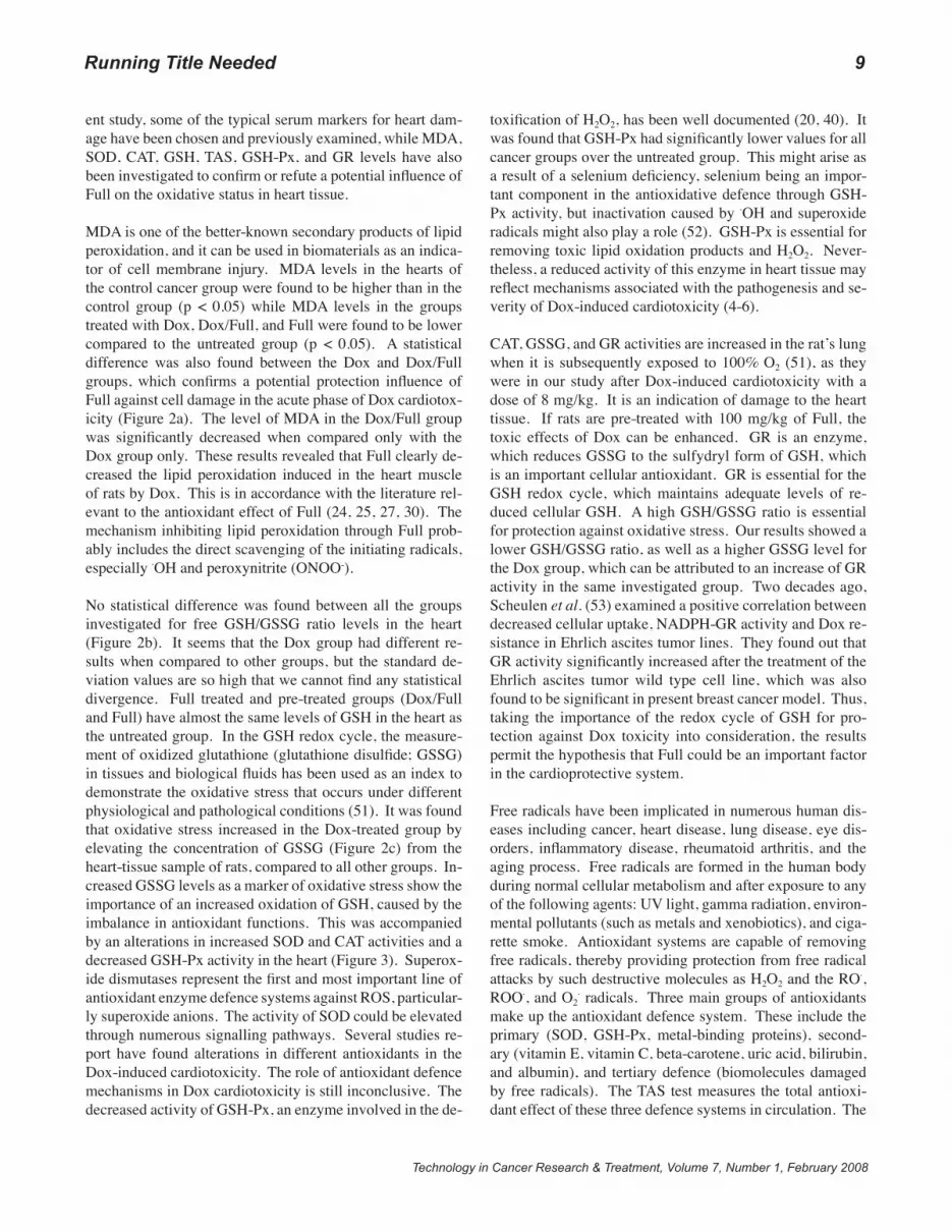
Coefficients of Hearts, Body weights, and Ultra Structural Pathology Results
We investigated the potential of Full protective activity to-wards Dox toxicity using a MNU-induced breast cancer rat model. Final body weights of MNU treated animals were approximately 17% less compared to the untreated group of rats (Table I). However, no body weight loss was observed among the MNU treated groups of rats (cancer groups). The difference in the weight of hearts or in the coefficients of hearts was not significant either. No definitive pathological changes were found in the heart tissues of rats treated with Dox and/or Full when examined by hematoxylin and eosin staining at the light microscopic level (not shown). How-ever, when ultra structural studies were performed, the ven-tricular tissues from Dox-treated rats showed marked myo-cardial damage. These changes consisted of mitochondrial damage, the accumulation of intracytoplasmic vacuoles, and focal myofilament disarray (Figure 1b). The ventricles of control rats showed normal ultra structure of cardiomyo-cytes (Figure 1a). Ultra structural analysis of heart tissues from rats treated with Dox and Full indicated that the hearts of the rats were protected from Dox-induced subcellular damage. Dox/Full rats did not appear to show significant cardiac damage although occasional focal loss of cristae in the mitochondria was observed (Figure 1c).
Heart Marker Enzymes and Hematological Parameters
Table II shows the activities of marker enzymes for car-diac function (CK, LDH, α-HBDH) in the serum of all the groups of rats.
A statistically significant increase of CK and LDH levels in the serum was found in in Dox group compared to the can-cer control group. At the same time the level of CK was significantly lower in the cancer control and the Full groups compared to the untreated group. The α-HBDH levels in the serum of all groups were without statistically important dif-ferences (Table II). To determine potential levels of liver or heart damage the α-HBDH/LDH ratio was calculated. Statit-stically significant difference was shown comparing the un-treated group (and all the other groups). Significant variation between all treated groups (Dox, Dox/Full, Full) compared to the cancer control group was observed.
The results of measuring the white blood cells (WBC) number, red blood cells (RBC), hemoglobin (Hb), hematocrite (Ht),
Table I Bodyweights of rats, hearts, and coefficients of heart after sacrificing.
Group Body weight (g) After sacrificing
Heart weight (g)
Coefficient of heart (mg/g)
I 271.2 ± 12.8 0.98 ± 0.17 3.6 ± 0.6 II 262.5 ± 22.0 0.89 ± 0.09 3.4 ± 0.4 III 257.0 ± 15.8 0.92 ± 0.11 3.6 ± 0.4 IV 262.0 ± 13.8 0.85 ± 0.06 3.2 ± 0.1 V 251.5 ± 14.4 0.84 ± 0.08 3.3 ± 0.2
Figure 1: Mitochondria showing (A) normal morphology, the double mem-brane envelope and lamellar cristae in the control rats; (B) irregular shape with lucent matrix and disorganised cristae in the Dox rats; and (C) amor-phous material and rare disintegrated cristae in the Dox/Full rats.

mean corpuscular volume (MCV), mean corpuscular hemoglobin (MCH), and mean corpuscular hemoglo-bin concentrations (MCHC) in animals sacrificed after two days of Dox and/or Full application, showed statisti-cally significant changes of some hematological param-eters (Table II).
A significant increase of neu-trofiles and a significant de-crease of lymphocytes was detected in the Dox, Dox/Full, and Full treated groups over both control groups (I and II). Percentage of neu-trophils was approximately five times higher in the Dox group compared to the un-treated group (57.8 ± 13.6% versus 8.7 ± 6.5%). On the other hand, the level of lym-phocytes was almost twice lower for the Dox group com-pared to both control groups (38.8 ± 12.8% versus 80.0 ± 7.4% and 88.6 ± 28.8%).
A significantly higher (p < 0.05) Ht concentration was observed in the Dox treated group. MCV and MCHC values of the stud-ied serums were found to be significantly lower in the Dox/Full treated group and higher in the Dox and the Dox/Full treated groups compared to the cancer con-trol group, respectively.
Oxidative Status in Heart
In order to explore the ef-fect of fullerenol C60(OH)24 on the cardiac function of the rats with mammary car-cinomas, lipid peroxidation, antioxidant defence system capabilities, and protein oxidation were evaluated
according to the appropriate methods reported in the section Materials and Methods. Figure 2 shows the level of the peroxidation marker MDA, as well as glutathione status and free GSH/GSSG ratio. There was a significant elevation in the MDA concentration in the cancer group compared to the untreated group. In addition, the free GSH/GSSG ratio in the heart tissue was not significantly changeed compared to the corresponding untreated and cancer control groups in the acute phase of Dox toxicity (Figure 2b). On the other hand, MDA levels in the heart tissue in the Dox, the Dox/Full and the Full groups were significantly more decreased (Figure 2a) compared to the cancer control group (p < 0.05).
Figure 3 depicts the activities of mitochondrial antioxidant en-zymes (GR, CAT, GSH-Px, and SOD). A significant (p < 0.05) increase in the activities of GR, CAT, and SOD, and decrease
Tabl
e II
Hem
atol
ogic
al an
d se
rum
enzy
mat
ic fi
ndin
gs.
Gro
up
WBC
x109
/L
RBC
x101
2/L
Hb
g/dL
H
t %M
CV
flM
CH
pgM
CHC
g/dL
N
eutro
phil
%Ly
mph
ocyt
e %
CK
LDH
-H
BDH
-HBD
H/
LDH
ratio
I
6.9±
2.4
6.7±
2.1
13.2
±4.6
37
.7±1
2.3
56.6
±18.
5 20
.6±6
.6
36.4
±12.
0 8.
7±6.
5 88
.6±2
8.8
206.
1±27
.6
861.
5±13
2.0
265.
9±26
.8
0.31
±0.0
4II
14.1
±5.3
5.
1±1.
7 11
.0±3
.3
31.9
±9.8
63
.2±5
.21
22.0
±1.7
34
.7±0
.5
17.0
±3.2
1 80
.0±7
.4
153.
5±20
.21
817.
4±25
3.6
207.
2±44
.91
0.25
±0.0
51
III
4.3±
0.71
.2
6.8±
0.42
14
.7±0
.22
39.4
±1.2
2 58
.2±1
.51
21.7
±1.0
37
.3±0
.72
57.8
±13.
61.2
38
.8±1
2.81
.220
0.5±
38.7
2 11
27.5
±107
.21.
2 24
3.3±
48.3
0.
22±0
.041
.2
IV
4.3±
1.11
.2
6.8±
0.52
14
.8±1
.12
39.7
±3.0
58
.0±0
.81.
2 21
.7±0
.4
37.3
±0.6
2 52
.7±1
9.11
.2
44.7
±19.
71.2
154.
4±46
.03
818.
7±22
3.53
27
2.0±
89.9
0.
32±0
.022
.3
V
10.6
±4.3
7.
2±1.
5 13
.9±2
.9
36.1
±8.5
58
.7±1
.81.
2 26
.9±1
3.4
36.1
±5.6
41
.2±8
.91.
2.3
48.7
±21.
71.2
146.
4±44
.71.
378
9.5±
155.
13
254.
8±74
.6
0.32
±0.0
32.3
1,2,
3 - R
epre
sent
s sig
nific
ant d
iffer
ence
from
the
corre
spon
ding
gro
up (p
< 0
.05)
.
Technology in Cancer Research & Treatment, Volume 7, Number 1, February 2008
MDA heart
0
200
400
600
800
1000
1200
1400
1600
I II III IV V
group
microg/L
#1
*2 +2 *3
+2
A
GSH free/GSSG - heart
0.0
0.5
1.0
1.5
2.0
2.5
I II III IV V
group
ratio
*1 *1 *1,2
B
Glutathione disulfide (GSSG) heart
05
1015202530354045
I II III IV V
group
mg/L
#1 *2 *3 *3
C
Figure 2: Level of (A) MDA, (B) free GSH/GSSG ratio, and (C) GSSG in heart samples. Significant difference from the control (I) healthy group1, control (II) cancer group2, and DOX (III) treated group3 (* p < 0.05; # p < 0.01; + p < 0.001).

�
Technology in Cancer Research & Treatment, Volume 7, Number 1, February 2008
of GSH-Px activity were observed in the Dox intoxicated rats. Activities of these enzymes in mitochondria (CAT, GR, and SOD) were maintained to almost normal (p < 0.05) levels upon Full treatment in both the Full and the Dox/Full groups and were significantly lower compared to the Dox group.
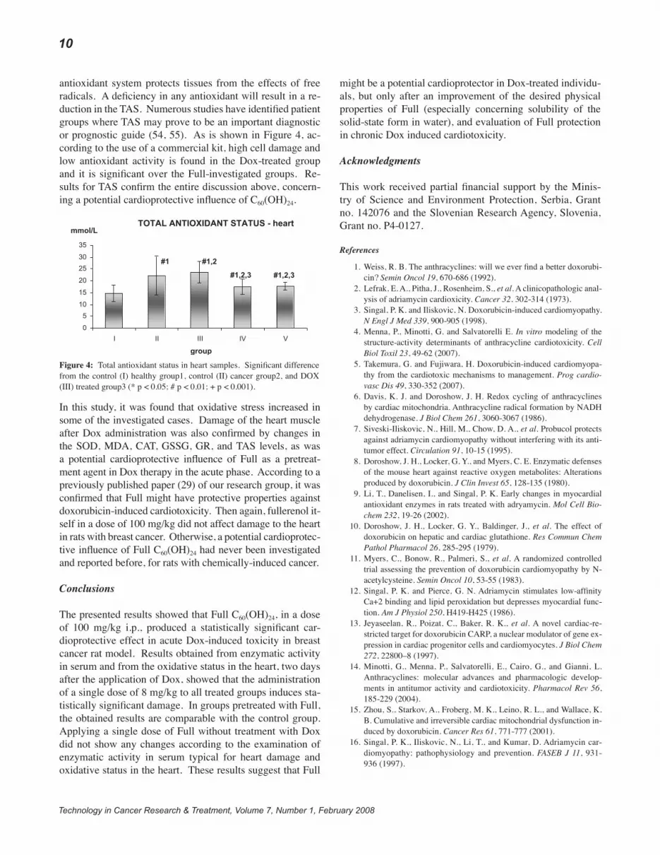
We analyzed the total antioxidant capacity of the heart in or-der to evaluate the total antioxidant status (TAS) of the rats with mammary carcinomas, treated and pre-treated with Dox and/or Full, respectively. We used a kit to measure TAS, wherein 2,2’-azinobis-(3-ethylbenzthiazoline sulphonate) (ABTS) was incubated with a peroxidase (metmyoglobin) and H2O2 to produce the radical cation ABTS·+, which has a relatively stable blue-green color and can be measured at 600 nm. Antioxidants in the sample caused the suppression in the production of this color to a degree that was proportional to their concentration. A statistically significant increase of TAS level was found in the Dox-treated group (Figure 4) compared to all the other groups (p < 0.05). Groups pre-treated with 100 mg/kg of Full had similar levels of TAS as the corresponding untreated group, and a significant differ-ence compared to the Dox treated group.
Discussion
There has not been any specific treatment for Dox cardio-myopathy demonstrated and usually Dox cardiomyopathy and heart failure are refractory to conventional therapy. β-blockers and ACE inhibitors may be useful in patients with
congestive heart failure. Malignant arrhythmias may also require treatment. Cardiac transplantation remains a vital option for patients with an end-stage heart failure due to Dox cardiomyopathy (5).
For more than two decades, researchers have investigated the role of iron in Dox-induced cardiomyopathy. The iron-chela-tor dexrazoxane (ICRF-187) was initially shown to protect dogs against Dox’s cardiotoxicity. Dexrazoxane was subse-quently shown to exert a cardioprotective effect in women undergoing Dox chemotherapy for advanced breast cancer, while not appreciably interfering with Dox’s antitumor activ-ity (34, 35). Dexrazoxane is a prodrug that is activated by in-tracellular hydrolysis (36), therefore it should be administered to patients before the administration of Dox (37) to achieve maximal therapeutic efficacy against Dox cardiotoxicity.
In the present in vivo study, a single dose effect of Dox and Full on the antioxidative status in the heart (acute phase) has been investigated. We used very similar model, as was men-tioned above, for women with breast cancer. Adult female rats with mammary carcinomas were pre-treated with 100 mg/kg of Full 30 min prior to being administered 8 mg/kg of Dox as a chemotherapeutic, to improve a potential cardiopro-tective influence of Full, according to known Dox-induced cardiotoxicity and Full antioxidative properties (24-30).
Chronic anthracycline treatment induces a progressive and se-vere deterioration of the repolarization phase in rat ECG, heart
CAT heart
02468
1012141618
I II III IV V
group
U/mg TP
#1 *2 *3 *3
A GR heart
02468
101214161820
I II III IV V
group
U/g TP
#1 *1
*2,3 *2,3
B
GSH-Px heart
0
50
100
150
200
250
300
I II III IV V
group
U/g TP
*1 #1 #1 *2
#1 *2,3
C SOD heart
0
5
10
15
20
25
30
35
I II III IV V
group
U/mg TP
*1,2
*3 #3
D
Figure 3: Level of (A) CAT, (B) GR, (C) GSH-Px, and (D) SOD in heart samples. Significant difference from the control (I) healthy group1, control (II) cancer group2, and DOX (III) treated group3 (* p < 0.05; # p < 0.01; + p < 0.001).

Technology in Cancer Research & Treatment, Volume 7, Number 1, February 2008
RunningTitleNeeded �
failure, myocardial infarction, and other pathological changes of the heart muscle and function (38). According to our results, after the administration of single dose of Dox in acute phase of cardiotoxicity significant changes of heart have been observed only at the ultrastructural level of pathologic examination.
CK, LDH, and α-HBDH are commonly used serum enzymat-ic markers for detection of hepatic and cardiac injury caused by Dox (20, 39, 40). CK is an enzyme used as an important marker of various heart, brain, and skeletal muscle diseases. Thus, an increase of the circulating level of CK may be as-sociated with myocardial infarction, acute cerebrovascular disease, trauma, or a disease of skeletal muscles. It is well-known that after a myocardial infarction (4 and 6 hours af-ter the onset of acute symptoms) CK levels start to raise and reach the peak 18 to 30 hours after the onset of acute symp-toms. Normal values are reached again during the third day.
As it can be seen from Table II, the Full group as well as the Dox group, which was pretreated with Full (30 min before Dox treatment), has lower circulating levels of CK compared to the Dox group, indicating protective activity of Full against Dox cardiotoxicity. There are some previously published papers (24, 29) with potential cardioprotective in-fluence of Full in Dox induced cardiotoxicity. Nevertheless, these studies were performed strictly in healty rats, while our was performed in rats, affected with cancer.
It is also well-known that both LDH and α-HBDH are often used as markers of cardiovascular damage. Their elevated levels indicate that heart function might be impaired after the administration of Dox (Table II). An increase in the cir-culating level of LDH is an index of myocardial infarction, renal failure, hepatitis, anaemia, malignancies, and dam-aged skeletal muscles. However, in our experiment signifi-cant increase of LDH level was observed only in the Dox group. In a recently published paper (20), Dox administra-tion revealed significant change in the activities of cardiac marker enzymes in myocardial tissue of rats. Activities of these enzymes in the heart were restored to near normal conditions in the group pretreated with an Centella asiatica extract, which reduced the extent of myocardial damage in-duced by Dox. The same occurred in the experiment using fullerenol as a pretreatment agent.
The levels of α-HBDH in the serum of all groups were very similar and without statistically important differences (Table II). Bjelogrlic et al. (41) confirmed that the activity of all investigated parameters (CK, LDH, AST, and α-HBDH), which were elevated at 24 h, remained high at 48 h, with the exception of α-HBDH. The α-HBDH activity dropped at 48 h after a single bolus dose of Dox (10 mg/kg; i.v.) in mice, which could explain our non-significant results for this parameter after two days of treatment.
Even though this study was not aimed at investigating the side effects of Dox on renal function or the renal protec-tion of Full, it is important to discuss the possibility that α-HBDH leakage could also have come from the kidneys. In that case, lower α-HBDH activity in animals treated with Dox needs an additional explanation. There is no literature data about the time-dependent changes in serum α-HBDH activity caused by Dox nephrotoxicity. Therefore, more ex-periments measuring the enzyme activity from cardiac and renal tissue are necessary to determine the exact origin of serum α-HBDH (heart, kidneys or both) (42).
To differentiate between liver and heart disease, the α-HBDH/LDH ratio can be calculated. Comparing all groups with the untreated group, indicates the conclusion that in the case of a lower α-HBDH/LDH ratio, parenchymal liver damage is greater (< 0.25). On the other hand, the groups pre-treated with Full pointed to almost normal level of the α-HBDH/LDH ratio, taking into account that the untreated group serves as a reference group for the calculation of α-HBDH/LDH ratio. According to the literature Full exhibits very strong antioxidant activity (24, 25, 27, 30), as well as a potential cardioprotective (29) effect. In some cases it is also liver-protective, exhibiting for example radioprotection (26, 30). On the other hand its weak solubility and low distribu-tion after i.p. administration could pose high risk for abdomi-nal cavity and organs (peritonitis).
An LDH test serum is often used to detect tissue alterations and diagnose heart attack, anaemia, and liver diseases. Gen-erally, a high LDH level shows a myocardial lesion when combined with CK and α-HBDH, while indicating hepatocel-lular damage when combined with AST and ALT enzymes.
The results of measuring the number of red blood cells (RBC) (indicator of anaemia) in the blood samples two days after treatment did not show a statistically significant differ-ence between the treated animals and the untrated group. In contrast, there was significant difference in the Dox and the Dox/Full groups compared to the cancer control group, due to the high number of ulcerating mammary carcinomas in the cancer control group, which resulted in anaemia.
Lowered white blood cell (WBC) count with a higher risk of infection is one of the general side effects of Dox. In adult healthy female Sprague-Dawley rats usually 80% of all WBC are lymphocytes, followed by 14% of neutrophils, 3% of ep-osinophils, 2% of monocytes, and 1% of basophils (43). Our results show (Table II) significant differences in proportion of neutrophils and lymphocytes in the Dox, the Dox/Full and the Full treated groups compared to both control groups. The reduced number of lymphocytes usually appears after steroid treatment or uraemia, systemic lupus erythematosus, Legion-naire’s disease, marrow infiltration, AIDS, chemotherapy, and

�
Technology in Cancer Research & Treatment, Volume 7, Number 1, February 2008
radiotherapy. It is obvious that in our case chemotherapeutic treatment caused decreased number of lymphocytes. How-ever, neutrophils are usually first responders to microbial in-fection or foreign materials, therefore the increased number of neutrophils is usually the hallmark of acute inflammatory processes. Both, Full and Dox are xenobiotics for rat, but level of producing neutrophils was lower in the Full group than in the Dox group. Therefore, Full could have anti-in-flammatory effect on the organism after treatment with Dox (the Dox and the Dox/Full groups).
The interactions of Dox and its metabolites (alcohol and agly-cone) with Hb, which caused chemical modifications on the cysteine residues of Hb by anthracycline drugs, may repre-sent another important metabolic event, which is affected by increase in the oxygen affinity of Hb treated with Dox (14).
A high Ht may be due to dehydration (burns, diarrhea), erythrocytosis, and polycythemia vera (44). Diarrhea may occur during chemotherapy treatment with Dox. It is usu-ally a mild form of diarrhea and can usually be easily con-trolled with medication. In our case the Dox treated groups (the Dox and the Dox/Full), had diarrhea and consequently secondary dehydration was developed which influenced the values of Ht (Table II). MCV measures the mean or average size of individual red blood cells. Microcytic red blood cells are seen in iron deficiency anaemia, lead poisoning, and the genetic diseases, i.e., thalassemia major and thalassemia mi-nor. Macrocytic red blood cells (observed in the cancer con-trol group) are associated with pernicious anemia and folic acid deficiencies. MCH measures the amount, or mass, of Hb present in one RBC. MCHC represents the average pro-portion of Hb in red blood cells. The MCH and the MCHC are used to assess whether the red blood cells are normo-chromic, hypochromic, or hyperchromic. Increased MCV, variable MCH and MCHC values could indicate macrocytic anemia, which is most often caused by a vitamin B12 defi-ciency (due to pernicious anemia) and folic acid deficiency. An increase of MCV and MCH was found in the Full-treat-ed group, but the MCHC level was normal according to the literature data (44) and the untreated group. On the other hand, MCV and MCH values were normal for the Dox/Full and Dox-treated groups whereas MCHC was significantly different compared to the cancer control group.
The mechanisms involved in anthracycline-mediated cardio-toxicity are well known, and numerous investigations have indicated a role of iron in this process. Anthracyclines inhibit iron mobilization from the iron storage protein, ferritin, result-ing in a marked accumulation of ferritin-iron (45). According to a previous hypothesis, levels of iron could be normal but the other biomarkers could show the potential anemia.
The ultra structural changes demonstrated microscopically in
Dox intoxicated rats were similar to those found in the litera-ture (46). Pretreatment with 100 mg/kg of Full showed con-siderable prevention of the ultra structural alterations includ-ing disorders of mitochondrial fine structure. This indicates that administration of Full scavenges the free radicals thereby preventing the cellular damage in the membrane. Previously reports have shown that supplementation of antioxidants in Dox induced myocardial injury has a favorable effect (47). Cardiac myocytes are the likely targets of ROS attack in the failing heart. It is possible that free radicals cause damage at or near the site of their formation. Consequently, as a major source of ROS production, mitochondria could also be the major targets susceptible to ROS attack (48). The defects in mitochondrial architecture would lead to the alteration of the mitochondrial metabolism, resulting in decreased activities of mitochondrial enzymes, in the Dox intoxicated hearts; thus, becoming a key contributor to intrinsic cell dysfunction (49).
Oxidative stress is a disturbance in the prooxidant-anti-oxidant balance in favor of the former, leading to potential damage. Such damage is often, again a bit loosely, called oxidative damage. In principle, oxidative stress can result from diminished amounts of antioxidants, an increased pro-duction of ROS/RNS or even excessive of natural ROS/RNS systems. Oxidative stress can cause damage to all types of biomolecules, including DNA, proteins and lipids (lipid per-oxidation). In many situations it is unclear which is the most important target, since injury mechanisms overlap widely. A cell exposed to severe oxidative stress may die. Cell death can essentially occur by two mechanisms, necrosis and apoptosis, although death by mechanisms with features overlapping these pathways is sometimes seen. Necrosis and apoptosis can both result from oxidative stress. Molecular mechanisms of cell death can also be found as useful targets in many cancer therapies. The objective of cancer treatment is to kill cancer cells with as little damage to normal cells as possible. Anthracyclines are widely used chemotherapeutics in the treatment of leukaemia, breast cancer, Hodgkin’s dis-ease, and sarcomas. The best known are daunorubicin and doxorubicin. Like all antitumor drugs, the anthacyclines pro-duce a number of side-effects, the most serious being damage to the heart. Acute cardiotoxicity occurs within minutes of drug administration and manifests as arrhythmias (29) and non-specific electrocardiographic changes (38) (this was also checked in a preliminary study on healthy rats). More serious side-effect is a chronic irreversible congestive heart failure, which becomes a significant problem once a certain total dose of anthracycline has been administered (further investigation of a potential Full cardioprotective influence in chronic therapy of Dox will be conducted). This cardio-toxicity limits the doses of Dox that can be safely given to cancer patients (50). Nevertheless, it is not clear why the heart should be a particular target, although the levels of its antioxidant defence enzymes are only moderate. In the pres-
Technology in Cancer Research & Treatment, Volume 7, Number 1, February 2008
RunningTitleNeeded �
ent study, some of the typical serum markers for heart dam-age have been chosen and previously examined, while MDA, SOD, CAT, GSH, TAS, GSH-Px, and GR levels have also been investigated to confirm or refute a potential influence of Full on the oxidative status in heart tissue.
MDA is one of the better-known secondary products of lipid peroxidation, and it can be used in biomaterials as an indica-tor of cell membrane injury. MDA levels in the hearts of the control cancer group were found to be higher than in the control group (p < 0.05) while MDA levels in the groups treated with Dox, Dox/Full, and Full were found to be lower compared to the untreated group (p < 0.05). A statistical difference was also found between the Dox and Dox/Full groups, which confirms a potential protection influence of Full against cell damage in the acute phase of Dox cardiotox-icity (Figure 2a). The level of MDA in the Dox/Full group was significantly decreased when compared only with the Dox group only. These results revealed that Full clearly de-creased the lipid peroxidation induced in the heart muscle of rats by Dox. This is in accordance with the literature rel-evant to the antioxidant effect of Full (24, 25, 27, 30). The mechanism inhibiting lipid peroxidation through Full prob-ably includes the direct scavenging of the initiating radicals, especially ·OH and peroxynitrite (ONOO-).
No statistical difference was found between all the groups investigated for free GSH/GSSG ratio levels in the heart (Figure 2b). It seems that the Dox group had different re-sults when compared to other groups, but the standard de-viation values are so high that we cannot find any statistical divergence. Full treated and pre-treated groups (Dox/Full and Full) have almost the same levels of GSH in the heart as the untreated group. In the GSH redox cycle, the measure-ment of oxidized glutathione (glutathione disulfide; GSSG) in tissues and biological fluids has been used as an index to demonstrate the oxidative stress that occurs under different physiological and pathological conditions (51). It was found that oxidative stress increased in the Dox-treated group by elevating the concentration of GSSG (Figure 2c) from the heart-tissue sample of rats, compared to all other groups. In-creased GSSG levels as a marker of oxidative stress show the importance of an increased oxidation of GSH, caused by the imbalance in antioxidant functions. This was accompanied by an alterations in increased SOD and CAT activities and a decreased GSH-Px activity in the heart (Figure 3). Superox-ide dismutases represent the first and most important line of antioxidant enzyme defence systems against ROS, particular-ly superoxide anions. The activity of SOD could be elevated through numerous signalling pathways. Several studies re-port have found alterations in different antioxidants in the Dox-induced cardiotoxicity. The role of antioxidant defence mechanisms in Dox cardiotoxicity is still inconclusive. The decreased activity of GSH-Px, an enzyme involved in the de-
toxification of H2O2, has been well documented (20, 40). It was found that GSH-Px had significantly lower values for all cancer groups over the untreated group. This might arise as a result of a selenium deficiency, selenium being an impor-tant component in the antioxidative defence through GSH-Px activity, but inactivation caused by ·OH and superoxide radicals might also play a role (52). GSH-Px is essential for removing toxic lipid oxidation products and H2O2. Never-theless, a reduced activity of this enzyme in heart tissue may reflect mechanisms associated with the pathogenesis and se-verity of Dox-induced cardiotoxicity (4-6).
CAT, GSSG, and GR activities are increased in the rat’s lung when it is subsequently exposed to 100% O2 (51), as they were in our study after Dox-induced cardiotoxicity with a dose of 8 mg/kg. It is an indication of damage to the heart tissue. If rats are pre-treated with 100 mg/kg of Full, the toxic effects of Dox can be enhanced. GR is an enzyme, which reduces GSSG to the sulfydryl form of GSH, which is an important cellular antioxidant. GR is essential for the GSH redox cycle, which maintains adequate levels of re-duced cellular GSH. A high GSH/GSSG ratio is essential for protection against oxidative stress. Our results showed a lower GSH/GSSG ratio, as well as a higher GSSG level for the Dox group, which can be attributed to an increase of GR activity in the same investigated group. Two decades ago, Scheulen et al. (53) examined a positive correlation between decreased cellular uptake, NADPH-GR activity and Dox re-sistance in Ehrlich ascites tumor lines. They found out that GR activity significantly increased after the treatment of the Ehrlich ascites tumor wild type cell line, which was also found to be significant in present breast cancer model. Thus, taking the importance of the redox cycle of GSH for pro-tection against Dox toxicity into consideration, the results permit the hypothesis that Full could be an important factor in the cardioprotective system.
Free radicals have been implicated in numerous human dis-eases including cancer, heart disease, lung disease, eye dis-orders, inflammatory disease, rheumatoid arthritis, and the aging process. Free radicals are formed in the human body during normal cellular metabolism and after exposure to any of the following agents: UV light, gamma radiation, environ-mental pollutants (such as metals and xenobiotics), and ciga-rette smoke. Antioxidant systems are capable of removing free radicals, thereby providing protection from free radical attacks by such destructive molecules as H2O2 and the RO·, ROO·, and O2
· radicals. Three main groups of antioxidants make up the antioxidant defence system. These include the primary (SOD, GSH-Px, metal-binding proteins), second-ary (vitamin E, vitamin C, beta-carotene, uric acid, bilirubin, and albumin), and tertiary defence (biomolecules damaged by free radicals). The TAS test measures the total antioxi-dant effect of these three defence systems in circulation. The
10
Technology in Cancer Research & Treatment, Volume 7, Number 1, February 2008
antioxidant system protects tissues from the effects of free radicals. A deficiency in any antioxidant will result in a re-duction in the TAS. Numerous studies have identified patient groups where TAS may prove to be an important diagnostic or prognostic guide (54, 55). As is shown in Figure 4, ac-cording to the use of a commercial kit, high cell damage and low antioxidant activity is found in the Dox-treated group and it is significant over the Full-investigated groups. Re-sults for TAS confirm the entire discussion above, concern-ing a potential cardioprotective influence of C60(OH)24.
In this study, it was found that oxidative stress increased in some of the investigated cases. Damage of the heart muscle after Dox administration was also confirmed by changes in the SOD, MDA, CAT, GSSG, GR, and TAS levels, as was a potential cardioprotective influence of Full as a pretreat-ment agent in Dox therapy in the acute phase. According to a previously published paper (29) of our research group, it was confirmed that Full might have protective properties against doxorubicin-induced cardiotoxicity. Then again, fullerenol it-self in a dose of 100 mg/kg did not affect damage to the heart in rats with breast cancer. Otherwise, a potential cardioprotec-tive influence of Full C60(OH)24 had never been investigated and reported before, for rats with chemically-induced cancer.
Conclusions
The presented results showed that Full C60(OH)24, in a dose of 100 mg/kg i.p., produced a statistically significant car-dioprotective effect in acute Dox-induced toxicity in breast cancer rat model. Results obtained from enzymatic activity in serum and from the oxidative status in the heart, two days after the application of Dox, showed that the administration of a single dose of 8 mg/kg to all treated groups induces sta-tistically significant damage. In groups pretreated with Full, the obtained results are comparable with the control group. Applying a single dose of Full without treatment with Dox did not show any changes according to the examination of enzymatic activity in serum typical for heart damage and oxidative status in the heart. These results suggest that Full
might be a potential cardioprotector in Dox-treated individu-als, but only after an improvement of the desired physical properties of Full (especially concerning solubility of the solid-state form in water), and evaluation of Full protection in chronic Dox induced cardiotoxicity.
Acknowledgments
This work received partial financial support by the Minis-try of Science and Environment Protection, Serbia, Grant no. 142076 and the Slovenian Research Agency, Slovenia, Grant no. P4-0127.
References
TOTAL ANTIOXIDANT STATUS - heart
0
5
10
15
20
25
30
35
I II III IV V
group
mmol/L
#1 #1,2
#1,2,3 #1,2,3
Figure 4: Total antioxidant status in heart samples. Significant difference from the control (I) healthy group1, control (II) cancer group2, and DOX (III) treated group3 (* p < 0.05; # p < 0.01; + p < 0.001).
1.
2.
3.
4.
5.
6.
7.
8.
9.
10.
11.
12.
13.
14.
15.
16.
Weiss, R. B. The anthracyclines: will we ever find a better doxorubi-cin? Semin Oncol 19, 670-686 (1992).Lefrak, E. A., Pitha, J., Rosenheim, S., et al. A clinicopathologic anal-ysis of adriamycin cardioxicity. Cancer 32, 302-314 (1973).Singal, P. K. and Iliskovic, N. Doxorubicin-induced cardiomyopathy. N Engl J Med 339, 900-905 (1998).Menna, P., Minotti, G. and Salvatorelli E. In vitro modeling of the structure-activity determinants of anthracycline cardiotoxicity. Cell Biol Toxil 23, 49-62 (2007).Takemura, G. and Fujiwara, H. Doxorubicin-induced cardiomyopa-thy from the cardiotoxic mechanisms to management. Prog cardio-vasc Dis 49, 330-352 (2007).Davis, K. J. and Doroshow, J. H. Redox cycling of anthracyclines by cardiac mitochondria. Anthracycline radical formation by NADH dehydrogenase. J Biol Chem 261, 3060-3067 (1986).Siveski-Iliskovic, N., Hill, M., Chow, D. A., et al. Probucol protects against adriamycin cardiomyopathy without interfering with its anti-tumor effect. Circulation 91, 10-15 (1995).Doroshow, J. H., Locker, G. Y., and Myers, C. E. Enzymatic defenses of the mouse heart against reactive oxygen metabolites: Alterations produced by doxorubicin. J Clin Invest 65, 128-135 (1980).Li, T., Danelisen, I., and Singal, P. K. Early changes in myocardial antioxidant enzymes in rats treated with adryamycin. Mol Cell Bio-chem 232, 19-26 (2002).Doroshow, J. H., Locker, G. Y., Baldinger, J., et al. The effect of doxorubicin on hepatic and cardiac glutathione. Res Commun Chem Pathol Pharmacol 26, 285-295 (1979).Myers, C., Bonow, R., Palmeri, S., et al. A randomized controlled trial assessing the prevention of doxorubicin cardiomyopathy by N-acetylcysteine. Semin Oncol 10, 53-55 (1983). Singal, P. K. and Pierce, G. N. Adriamycin stimulates low-affinity Ca+2 binding and lipid peroxidation but depresses myocardial func-tion. Am J Physiol 250, H419-H425 (1986).Jeyaseelan, R., Poizat, C., Baker, R. K., et al. A novel cardiac-re-stricted target for doxorubicin CARP, a nuclear modulator of gene ex-pression in cardiac progenitor cells and cardiomyocytes. J Biol Chem 272, 22800–8 (1997).Minotti, G., Menna, P., Salvatorelli, E., Cairo, G., and Gianni, L. Anthracyclines: molecular advances and pharmacologic develop-ments in antitumor activity and cardiotoxicity. Pharmacol Rev 56, 185-229 (2004).Zhou, S., Starkov, A., Froberg, M. K., Leino, R. L., and Wallace, K. B. Cumulative and irreversible cardiac mitochondrial dysfunction in-duced by doxorubicin. Cancer Res 61, 771-777 (2001).Singal, P. K., Iliskovic, N., Li, T., and Kumar, D. Adriamycin car-diomyopathy: pathophysiology and prevention. FASEB J 11, 931-936 (1997).

Technology in Cancer Research & Treatment, Volume 7, Number 1, February 2008
RunningTitleNeeded 11
17.
18.
19.
20.
21.
22.
23.
24.
25.
26.
27.
28.
29.
30.
31.
32.
33.34.
35.
36.
Singal, P. K., Li, T., Kumar, D., Danelisen, I., and Iliskovic, N. Adria-mycin-induced heart failure: mechanism and modulation. Mol Cell Biochem 207, 77-85 (2000).Ahmed, H. H., Mannaa, F., Elmegeed, G. A., and Doss, S. H. Cardio-protective activity of melatonin and its novel synthesized derivatives on doxorubicin-induced cardiotoxicity. Bioorg Med Chem 13, 1847-1857 (2005).Mukherjee, S., Banerjee, S. K., Maulik, M., Dinda, A. K., Talwar, K. K., and Maulik, S. K. Protection against acute adriamycin-induced cardiotoxicity by garlic: Role of endogenous antioxidants and inhibi-tion of TNF-α expression. BMC Pharmacol 3, 16-24 (2003). Gnanapragasam, A., Yogeeta, S., Subhashini, R., Ebenezar, K. K., Sathish, V., and Devaki, T. Adriamycin induced myocardial failure in rats: Protective role of Centella asiatica. Mol Cell Biochem 294, 55-63 (2007).Minotti, G., Cairo, G., and Monti, E. Role of iron in anthracycline car-diotoxicity: new tunes for an old song? FASEB J 13, 199-212 (1993).Zucchi, R. and Danesi, R. Cardiac toxicity of antineoplastic anthracy-clines. Curr Med Chem 3, 151-71 (2003).Ladas, E. J., Jacobson, J. S., Kennedy, D. D., et al. Antioxidants and cancer therapy: A systematic review. J Clin Oncol 22, 517-28 (2004).Bogdanovic, G., Kojic, V., Djordjevic, A., Canadanovic-Brunet, J., Vojinovic-Miloradov, M., and Baltic, V. Modulating activity of fullerenol C60(OH)22 on doxorubicin induced cytotoxicity. Toxicol-ogy in vitro 18, 629-637 (2004). Mirkov, S., Djordjevic, A., Andric, N., Andric, S., Kostic, T., Bogda-novic, G., Vojinovic-Miloradov, M., and Kovacevic, R. Nitric oxide-scavenging activity of polyhydroxylated fullerenol. Nitric Oxide 11, 201-207 (2004).Trajkovic, S., Dobric, S., Djordjevic, A., Dragojevic-Simic, V., and Milovanovic, V. Radioprotective efficiency of fullerenol in irradiated mice. Mater Sci Forum 494, 549-554 (2005).Djordjevic, A., Canadanovic-Brunet, J., Vojinovic-Miloradov, M., and Bogdanovic, G. Antioxidant properties and hypothetical radical mechanism of fullerenol C60(OH)24. Oxidation Communications 27, 806-812 (2005). Djordjevic, A., Bogdanovic, G., and Dobric, S. Fullerenes in biomed-icine. J BUON 11, 391-404 (2006).Djordjevic-Milic, V., Djordjevic, A., Dobric, S., Injac, R., Vuckovic, D., Stankov, K., Dragojevic-Simic, V., and Suvajdzic, Lj. Influence of fullerenol C60(OH)24 on doxorubicin induced cardiotoxicity in rats. Mater Sci Forum 518, 525-529 (2006).Trajkovic, S., Dobric, S., Jacevic, V., Dragojevic-Simic, V., Milova-novic, Z., and Djordjevic, A. Tissue-protective effects of fullerenol C60(OH)24 and amifostine. Colloids and Suferfaces B: Biointerfaces 58, 39-43 (2007).Djordjevic, A., Vojinovic-Miloradov, M., Petranovic, N., Devecerski, A., Lazar, D., and Ribar, B. Catalytic preparation and characterization of C60Br24. Fullerene Sci Technol 6, 689-94 (1998).Liska, J., Galbavy, S., Macejova, D., Zlatos, J. and Brtko, J. Histopa-thology of mammary tumors in female rats treated with 1-methyl-1-nitrosourea. Endocr Regul 34, 91-96 (2000).Aebi, H. Catalase in vitro. Methods Enzymol 105, 121-126 (1984).Speyer, J. L., Green, M. D., Kramer, E., et al. Protecive effect of the bispiperazinedione ICRF-187 against doxorubicin-induced cardia toxicity in women with advanced breast cancer. N Engl J Med 319, 745-752 (1988).Curran, C. F., Narang, P. K., and Reynolds, R. D. Toxicity profile of dexrazoxane (Zinecard, ICRF-187, ADR-529, NSC-169780), a modulator of doxorubicin cardiotoxicity. Cancer Treat Rev 18, 241-252 (1991).Seifert, C. F., Nesser, M. E., and Thompson, D. F. Dexrazoxane in the prevention of doxorubicin-induced cardiotoxicity. Ann Pharmacother 28, 1063-1072 (1994).
37.
38.
39.
40.
41.
42.
43.
44.
45.
46.
47.
48.
49.
50.
51.
52.
53.
54.
55.
Tetef, M. L., Synold, T. W., Chow, W., et al. Phase I trial of 96-hour continuous infusion of dexrazone in patients with advanced malig-nancies. Clin Cancer Res 7, 1569-1576 (2001).Sacco, G., Bigioni, M., Evangelista, S., Goso, C., et al. Cardiopro-tective effects of zofenopril, a new angiotension-converting enzyme inhibitor, on doxorubicin-induced cardiotoxicity in the rat. Eur J Pharmacol 414, 71-8 (2001).Van Acker, F. A. A., Hulshof, J. W., Haenen, G. R. M. M., et al. New synthetic flavonoids as potent protectors against doxorubicin-induced cardiotoxicity. Free Radic Biol Med 31, 31-37 (2001).Khan, M., Varadharaj, S., Shobha, J. C., Naidu, M. U., et al. C-phycocyanin ameliorates doxorubicin-induced oxidative stress and apoptosis in adult rat cardiomyocytes. J Cardiovasc Pharmacol 47, 9-20 (2006).Bjelogrlic, S. K., Radic, J., Jovic, V., and Radulovic, S. Activity of d,l-alpha-tocopherol (vitamin E) against cardiotoxicity induced by doxorubicin and doxorubicin with cyclophosphamide in mice. Basic Clin Pharmacol Toxicol 97, 311-319 (2005).Mizushima, Y., Sassa, K., Hamayaki, T., Fujishita, T., Oosaki, R., and Kobayashi, M. Diuretic response to cyclophosphamide in rats bear-ing a matrix metalloproteinase-9-producing tumor. Brit J Cancer 78, 1030-1034 (1998).Bellhorn, R. W., Korte, G. E., and Abrutyn, D. Spontaneous corneal degeneration in the rat. Lab Anim Sci 38, 46-50 (1988).Hoffman, R., Benz Jr., E. J., Shattil, S. J., et al., eds. In: Hematology: Basic Principles and Practice. 4th ed. Philadelphia, Pa: Churchill Livingston, p. 2674 (2005).Kwok, J. C. and Richardson, D. R. Examination of the mechanism in-volved in doxorubicin-mediated iron accumulation in ferritin: Studies using metabolic inhibitors, protein synthesis inhibitors and lysosomo-tropic agents. Mol Pharmacol 65, 181-95 (2004).Oliveira, P. J., Bjork, A. J., Santos, S. M., Leino, R. L., Kent Froberg, M., Moreno, A. J., and Wallace, K.B. Carvidilol mediated antioxdant protection against doxorubicin induced cardiac mitochondrial toxic-ity. Toxicol Appl Pharmacol 200, 159-168 (2004).Balanehru, S. and Nagarajan, B. Intervention of adriamycin induced free radical damage. Biochem Int 28, 735-744 (1992).Stadtman, E. R. Protein oxidation and aging. Science 257, 1220-1224 (1992).Mimnaugh, E. G., Trush, M. A., and Mohit Bhatnagar Gram F. E. Enhancement of reactive oxygen dependant mitochondrial membrane lipid peroxidation by the anti cancer drug adriamyin. Biochem Phar-macol 34, 847-856 (1985).Halliwell, B. and Gutteridge, J. M. C., eds. In: Free radicals in biol-ogy and medicine. 3rd edition. Oxford science press Inc. New York, pp 126 and 717 (2005).Mills, B. J., Richie, J. P., and Jr. Lang, C. A. Glutathione disulfide vari-ability in normal human blood. Anal Biochem 222, 95-101 (1994). Blum, J. and Fridovich, I. Inactivation of glutathione peroxidase by superoxide radical. Arch Biochem Biophys 249, 500-508 (1985).Scheulen, M. E., Hoensch, H., Kappus, H., et al. Positive correlation between decreased cellular uptake, NADPH-glutathione reductase activity and adriamycin resistance in Ehrlich ascites tumor lines. Arch Toxicol 60, 154-7 (1987).Lindeman, J. H. N., Van Zoeren-Grobbenm, D., Schrijver, J., et al. The total free radical trapping ability of cord blood plasma in preterm and term babies. Pediatr Res 26, 20-24 (1989).Miller, N. J., Rice-Evans, C., Davies, M. J., et al. A new method for measuring antioxidant activity. Biochem Soc Trans 21, 95S (1993).
Received: Needed; Revised: Needed; Accepted: Needed