drift capacity of reinforced concrete columns subjected to ...
Upload
khangminh22Category
view
0download
0
HAL Id: hal-02623721https://hal.inrae.fr/hal-02623721
Submitted on 31 Mar 2021
HAL is a multi-disciplinary open accessarchive for the deposit and dissemination of sci-entific research documents, whether they are pub-lished or not. The documents may come fromteaching and research institutions in France orabroad, or from public or private research centers.
L’archive ouverte pluridisciplinaire HAL, estdestinée au dépôt et à la diffusion de documentsscientifiques de niveau recherche, publiés ou non,émanant des établissements d’enseignement et derecherche français ou étrangers, des laboratoirespublics ou privés.
Distributed under a Creative Commons Attribution| 4.0 International License
EPA is cardioprotective in male rats subjected to sepsis,but ALA is not beneficial
Thibault Léger, Chrystele Jouve, Isabelle Hininger-Favier, Jean-PaulRigaudière, Frédéric Capel, Vincent Sapin, Clarisse Moreau, Alice Charrier,
Luc Demaison
To cite this version:Thibault Léger, Chrystele Jouve, Isabelle Hininger-Favier, Jean-Paul Rigaudière, Frédéric Capel, etal.. EPA is cardioprotective in male rats subjected to sepsis, but ALA is not beneficial. Antioxidants, MDPI, 2020, 9 (5), pp.371. �10.3390/antiox9050371�. �hal-02623721�

antioxidants
Article
EPA Is Cardioprotective in Male Rats Subjected toSepsis, but ALA is Not Beneficial
Thibault Leger 1 , Chrystèle Jouve 1, Isabelle Hininger-Favier 2 , Jean-Paul Rigaudiere 1 ,Frédéric Capel 1 , Vincent Sapin 3 , Clarisse Moreau 1, Alice Charrier 1 and Luc Demaison 1,*
1 UNH, Unité de Nutrition Humaine, UMR 1019, Université Clermont Auvergne, INRAE, CRNH Auvergne,63000 Clermont-Ferrand, France; [email protected] (T.L.); [email protected] (C.J.);[email protected] (J.-P.R.); [email protected] (F.C.); [email protected] (C.M.);[email protected] (A.C.)
2 LBFA U1055, Université Grenoble Alpes, INSERM, 38058 Grenoble, France;[email protected]
3 Department of Medical Biochemistry and Molecular Biology, CHU Clermont-Ferrand,63000 Clermont-Ferrand, France; [email protected]
* Correspondence: [email protected]
Received: 24 March 2020; Accepted: 28 April 2020; Published: 29 April 2020�����������������
Abstract: It has been proven that dietary eicosapentaenoic acid (C20:5 n-3 or EPA) protects the heartagainst the deleterious effects of sepsis in female rats. We do not know if this is the case for malerodents. In this case, the efficiency of other n-3 polyunsaturated fatty acids (PUFAs) remains tobe determined in both female and male rats. This study aimed at (i) determining whether dietaryEPA is cardioprotective in septic male rats; (ii) evaluating the influence of dietary α-linolenic (C18:3n-3 or ALA) on cardiac function during this pathology; and (iii) finding out the physiological andmolecular mechanisms responsible for the observed effects. Sixty male rats were divided intothree dietary groups. The animals were fed a diet deficient in n-3 PUFAs (DEF group), a dietenriched with ALA (ALA group) or a diet fortified with EPA (EPA group) for 6 weeks. Thereafter,each group was subdivided into 2 subgroups, one being subjected to cecal ligation and puncture(CLP) and the other undergoing a fictive surgery. Cardiac function was determined in vivo andex vivo. Several parameters related to the inflammation process and oxidative stress were determined.Finally, the fatty acid compositions of circulating lipids and cardiac phospholipids were evaluated.The results of the ex vivo situation indicated that sepsis triggered cardiac damage in the DEF group.Conversely, the ex vivo data indicated that dietary ALA and EPA were cardioprotective by resolvingthe inflammation process and decreasing the oxidative stress. However, the measurements of thecardiac function in the in vivo situation modulated these conclusions. Indeed, in the in vivo situation,sepsis deteriorated cardiac mechanical activity in the ALA group. This was suspected to be due toa restricted coronary flow which was related to a lack of cyclooxygenase substrates in membranephospholipids. Finally, only EPA proved to be beneficial in sepsis. Its action necessitates bothresolution of inflammation and increased coronary perfusion. In that sense, dietary ALA, which doesnot allow the accumulation of vasodilator precursors in membrane lipids, cannot be protective duringthe pathology.
Keywords: sepsis; heart; n-3 polyunsaturated fatty acids; inflammation; oxidative stress
1. Introduction
Sepsis due to microorganism infection is defined as a state of general systemic inflammationwhich can degenerate into septic shock, possibly leading to death. In Western societies with high
Antioxidants 2020, 9, 371; doi:10.3390/antiox9050371 www.mdpi.com/journal/antioxidants

Antioxidants 2020, 9, 371 2 of 22
financial incomes, incidences of sepsis are constantly increasing, but lethality is regressing probablydue to the improvements in health care in intensive care units [1]. However, enhanced survival canbe accompanied by comorbidity. Analysis of billings from 2013 revealed that sepsis accounts fornearly $24 billion in annual costs, making it the most expensive condition to treat in the entire U.S.healthcare system [2].
Systemic hypotension is one of the hallmarks of sepsis and partly results from cyclooxygenase 2(COX2) overexpression and eicosanoid release [3]. Crucial treatment after antibiotic therapy is fluidadministration to restore physiological blood pressure [4]. Septic shock is defined as a sepsis resistantto fluid resuscitation [5]. It is characterized by a mortality rate ranging from 20 to 45% and nearly 50%of the deaths occur as a consequence of heart failure [6,7].
Sepsis-related depression of myocardial function is due to bacterial lipopolysaccharides [8].Endotoxins bind to cardiac Toll-like receptors (TLRs) and activate the NF-κB pathway, leading topro-inflammatory cytokine overexpression [9]. These last (tumor necrosis factor alpha (TNF-α),interleukin-1β (IL-1β), interleukin-6 (IL-6), etc.) belong to the immune response system and lead to thedestruction of the microorganism. However, they are also able to damage host cells and to triggerheart failure [10].
Polyunsaturated fatty acids (PUFAs) of the n-3 families have been recognized as cardioprotectivein several pathological situations including arrhythmias [11], myocardial stunning [12] andhypertrophy [13]. Regrettably, the diet in Western societies is too rich in n-6 PUFAs and doesnot contain enough n-3 PUFAs [14]. Yet, numerous studies report the beneficial effects of n-3 PUFAsduring sepsis [15–17] due to the n-3 PUFA-related anti-inflammatory properties which contrast withthe pro-inflammatory actions of n-6 PUFAs [18]. In a recent study performed in female rats subjectedto cecal ligation and puncture (CLP), we partially highlighted the mechanism of the eicosapentaenoicacid (C20:5 n-3 or EPA)-related cardioprotective effect [19]. It is triggered by an improved oxygenmetabolism preconditioning the heart against the deleterious effects of excessive inflammation.However, females may also be protected by their hormonal status. Dietary EPA is protective duringsepsis, but we do not know whether other n-3 PUFAs such as α-linolenic (C18:3 n-3 or ALA) acid isefficient. Indeed, in contrast with dietary EPA, ALA does not allow the easy accumulation of EPA inmembrane phospholipids [20]. Phospholipid EPA is directly accessible to the cyclooxygenase enzymevia the action of phospholipase A2. This leads to the formation of vasoactive eicosanoids which canincrease the coronary flow [21]. Dietary ALA though favors the accumulation of docosahexaenoic acid(C22:6 n-3 or DHA) which blocks the cyclooxygenase enzyme [22], thus inhibiting the formation ofvasoactive compounds and reducing the coronary flow. The heart acts like an engine. Reducing thecoronary flow decreases cardiac contraction resulting in depressed cardiac function despite normalcardiomyocyte health. Therefore, dietary ALA could protect the cardiomyocytes while reducingmyocardial contractility.
This study set out to (i) determine whether dietary EPA is cardioprotective in male rats madeseptic by cecal ligation and puncture (CLP), (ii) estimate the influence of dietary ALA in septic malerats, (iii) and evaluate the physiological and molecular mechanisms underlying the effects of thesePUFAs. For this purpose, three groups of male Wistar rats were fed a diet each differing in theirPUFA composition: the first one mimicking the Western diet was rich in n-6 PUFAs and deficient inn-3 PUFAs, the second one was fortified with EPA and the third one was supplemented with ALA.After 6 weeks of feeding, sepsis was induced by CLP and, exactly 24 h later, cardiac function wasevaluated in in vivo and ex vivo. The data were enhanced by the measurement of several of theparameters involved in the inflammation process and production of oxidative stress as well as thosedescribing the fatty acid profile of circulating and membrane lipids.

Antioxidants 2020, 9, 371 3 of 22
2. Materials and Methods
2.1. Ethical Approval
All experiments followed the European Union recommendations concerning the care and useof laboratory animals for experimental and scientific purposes. All animal work was approvedby the local board of ethics for animal experimentation (Comité d’éthique pour l’expérimentationanimale Auvergne) and notified to the research animal facility of our laboratory (authorizationn◦APAFIS#2213-2016082409264678 v2).
2.2. Experimental Animals and Diet
Sixty 4 month-old male Wistar rats (Janvier, Le Genest Saint Isle, France) were maintained 3–4 percage under controlled lightening, hygrometry and temperature conditions. Each rat received a n-3PUFAs-deficient diet for six weeks. After this period, the animals were randomly allocated to threedietary groups for 6 weeks. The three diets differed in their PUFA composition. They all contained 5%of lipids and approximately 2.25% of linoleic acid (C18:2 n-6 or LA). In each of the different diets, 0.625%of their monounsaturated fatty acid (MUFAs) were substituted with a specific PUFA: (i) LA for the n-3PUFA-deficient diet (DEF); (ii) ALA for the ALA-rich diet; and (iii) EPA for the EPA-rich diet. The fattyacid composition of the 3 diets is presented in Table 1. The diets with n-3 PUFAs displayed a n-6/n-3PUFA ratio close to 3. At the end of this period, half of the rats from each group was subjected to CLPto induce severe sepsis (Sept) and the other half were sham-operated (Sham). Thus, the study dealtwith six sub-groups according to the diet and surgery: (i) DEF-Sham; (ii) DEF-Sept; (iii) ALA-Sham;(iv) ALA-Sept; (v) EPA-Sham; and (vi) EPA-Sept.
Table 1. Fatty acid composition of the 3 diets.
Fatty acid DEF ALA EPA
C14:0 0.1 0.1 0.1C16:0 6.5 6.5 5.7C18:0 2.7 2.8 3.0C20:0 0.1 0.1 0.1C24:0 0.1 0.1 0.1SFAs 9.5 9.6 9.0
C16:1 n-7 0.1 0,1 0.1C18:1 n-9 40.8 30.0 26.6C18:1 n-7 0.9 0.8 0.8C20:1 n-9 0.1 0.1 0.4MUFAs 42.0 31.1 27.9
C18:2 n-6 47.1 44.4 44.7C20:2 n-6 Tr. Tr. 0.2C20:3 n-6 Tr. Tr. 0.1C20:4 n-6 Tr. Tr. 0.8
n-6 PUFAs 48.1 44.4 46.9C18:3 n-3 0.1 13.4 0.1C20:4 n-3 Tr. nd 0.4C20:5 n3 nd nd 15.5
n-3 PUFAs 0.1 13.4 15.6n-6/n-3 481 3.3 3
DEF: n-3 PUFA deficient diet; ALA: diet enriched with α-linolenic acid; EPA: diet enriched with eicosapentaenoicacid; SFAs: saturated fatty acids; MUFAs: monounsaturated fatty acids; PUFAs: polyunsaturated fatty acids;n-6/n-3: ratio between n-6 and n-3 PUFAs; Tr.: trace amount; nd: not detected.
2.3. Surgical Procedure
CLP was performed according to Toscano et al. [23]. Briefly, the animals were anesthetized withisoflurane (induction 4%, maintenance 2%). After shaving the fur, the external abdominal wall wasdisinfected with alcoholic betadine and a horizontal incision was performed in the ventral wall at the

Antioxidants 2020, 9, 371 4 of 22
level of the caecum in order to reach the abdominal cavity. The caecum was externalized and placedonto the disinfected external abdominal wall. A ligation of the caecum was carried out at 2 cm fromthe apex of the organ. Afterwards, two perforations were performed in the caecum lining at 1 cminterval on the upper face of the organ with a 20-gauge needle and soft pressure was applied to thecaecum to facilitate externalization of digestive matter to the outer surface of the organ. The caecumwas reinserted immediately after into the abdominal cavity and buprenorphine (0.05 mg/kg of bodyweight) was injected subcutaneously into the neck. Thereafter, the peritoneum was closed with 6.0 silksutures and the skin with sutures and metal clips. Sham-operated animals were treated identicallyas compared to septic animals except that the caecum was not subjected to ligation and puncture.Following the operation, anesthesia was stopped and the rats were placed one per cage in the animalfacility where the waking occurred between 3 and 5 min after isoflurane arrest. Each rat was then keptin a separate cage for 24 h. One rat died during anesthesia and two septic animals did not survive the24 h post-surgery period.
2.4. Evaluation of Body Composition
The animals body weight along with the body composition were determined by nuclear magneticresonance imaging with an adequate spectrometer (Echo MRI LL, Houston, TX, USA) just beforethe surgical procedure and at the end of the 24 h postoperative period. The data thus enabledthe measurement of the lean body mass, fat mass and total body water in the animals. After thepostoperative period, the weights of abdominal (perirenal plus visceral adipose tissues) and epididymalfats were measured.
2.5. Appraisal of In Vivo Cardiac Function
Twenty-four hours after the CLP, body composition was assessed and the in vivo cardiac functionwas appraised using a Millar pressure probe (Harvard Apparatus, Les Ulis, France) which wasintroduced into the aorta and left ventricle cavity. After anesthesia with ketamine (100 mg/kg of bodyweight) and xylazine (20 mg/kg of body weight), heparinization of the rats was carried out via thesaphenous vein (500 UI/kg of body weight). Then, the probe was introduced into the right carotidartery and immediately inserted into the cavity of the left ventricle. In order to ensure the stability ofthe cardiac function, the measurements were carried out for 10 min. They allowed the measurement ofthe left ventricular developed pressure (LVDP), contraction and relaxation (dP/dt max and dP/dt min,respectively) and heart rate. The systolic, diastolic and mean aortic pressure was then determined bypulling out the probe from the left ventricle into the aorta. Then, the probe was removed and the rightcarotid was bound. Blood was collected from the abdominal aorta and centrifuged (1800 g, 10 min,4 ◦C) for plasma preparation. Once obtained, the plasma was immediately frozen at the temperatureof liquid nitrogen and stored at −80 ◦C to await the performance of biochemical analyses.
2.6. Assessment of Ex Vivo Cardiac Function
Directly after the evaluation of the in vivo cardiac function (24 h after the surgery), the ex vivoperfusion was performed in standardized conditions according to the non-recirculating Langendorffmethod. A rapid thoracotomy was carried out and the heart was immediately collected and placedin 4 ◦C saline buffer until cessation of heartbeat. It was then immediately (in the first minuteafter the removal of the heart in order to avoid cellular damages and preconditioning) perfusedby the aorta at 37 ◦C with a Krebs-Henseleit buffer composed of (in mM) NaCl (119), KCl (4.8),MgSO4 (1.6), NaHCO3 (22), KH2PO4 (1.2), CaCl2 (1.8), D-glucose (11) and sodium hexanoate(0.5), pH 7.4. The perfusion buffer was maintained at 37 ◦C with a water-bath and constantlyoxygenated with carbogen (95% O2, 5% CO2). Afterwards, the pulmonary artery was cannulatedfor the collection of coronary effluents. The heart was constantly perfused with a peristaltic pump(Gilson, Middleton, WI, USA) to keep the perfusion flow constant at 12 mL/min until the end ofperfusion. From the fifth minute of perfusion, the heart was electrically paced at a rate of 370 beats/min.

Antioxidants 2020, 9, 371 5 of 22
To evaluate heart contraction, a latex balloon attached to a pressure probe and an amplifier was insertedinto the left ventricle. It was inflated until the diastolic pressure reached 10 mmHg. This systemallowed the assessment of the systolic, diastolic and developed pressure as well as the heart rate;contraction (dP/dt max) and relaxation (dP/dt min). The evaluation of these two last parameters waspossible by the use of perfectly standardized perfusion conditions. The perfusion pressure was alsoevaluated with a pressure probe inserted just before the aortic cannula. In conditions of perfusionat fixed coronary flow, the perfusion pressure corresponds to the coronary pressure. Changes inthe coronary pressure reveal modifications of coronary volume (coronary dilatation, constrictionand/or obliteration of coronary vessels). All the parameters of cardiac function were measured at the30th minute of perfusion and the data were recorded and analyzed with HSE software (Hugo SachsElektronik, March-Hugstetten, Germany). Just before the end of the recording, samples of arterial andvenous perfusion fluids were collected anaerobically for determination of oxygen partial pressureswith a blood gas analyzer (Radiometer, Neuilly-Plaisance, France). Cardiac oxygen consumption wascalculated by subtracting the venous oxygen partial pressure to the arterial one and by multiplying thedifference by the coronary flow. The cardiac metabolic efficiency was also calculated (ratio between therate pressure product and oxygen consumption). Finally, the heart was freeze-clamped at −196 ◦C andstored at −80 ◦C for subsequent analyses.
2.7. Lipid Profile
Fatty acid composition of plasma lipids and cardiac phospholipids. The measurements wereperformed according to a previously described method (Demaison et al., 1994). The total lipids wereextracted according to Folch et al. (1957) and the phospholipids were separated from non-phosphoruslipids using a Sep-Pak® Cartridge. After transmethylation with boron trifluoride methanol, the fattyacid methyl esters were separated and analyzed by gas chromatography. The determination of the fattyacid profile of the plasma lipids was performed similarly except that the phospholipids separation stepwas omitted.
Plasma triglycerides. The concentration of plasma triglycerides was determined by a colorimetricmethod. Triglycerides were hydrolyzed by lipase to free fatty acids and glycerol. The glycerolphosphorylation produced hydrogen peroxides (H2O2) which reacted with the reagents of thecommercially available kit (Thermo Fisher Scientific, Asnières-sur-Seine, France) to generatequinone-imine, a colored compound measurable at 510 nm.
2.8. Fluid Biochemistry
The inflammatory cytokine tumor necrosis factor- α (TNF-α) was analyzed in the plasma usinga commercially available enzyme-linked immunosorbent assay (ELISA) kit (Abcam, Paris, France).Lactate and pyruvate were assessed in the venous perfusion fluid according to Bergmeyer [24].
2.9. Gene Expression
Gene expression of TNF-α, interleukin-1β (IL-1β), interleukin-6 (IL-6), mitochondrial superoxidedismutase 2 (SOD2) and sirtuin 3 (SIRT3) proteins was measured in the cardiac homogenates.RNA extraction was performed using TRIzol® (Thermo Scientific, Waltham, MA, USA) according tothe manufacturer’s instructions. Chloroform was added (0.2 mL/mL of TRIzol®), and samples weremixed and centrifuged (12,000 g, 15 min, 4 ◦C). The aqueous phase containing total RNAs was collected,mixed with isopropanol to precipitate RNAs and centrifuged (12,000 g, 15 min, 4 ◦C). The resulting pelletwas washed with ethanol 70% (v/v), dried and suspended in water. RNA quantification and integritywere controlled by measuring the ratio of optical density at 260 and 280 nm and agarose gel migration.For each sample, 2 µg of total RNAs were used to perform reverse transcription. The resulting productswere used for reverse transcription quantitative polymerase chain reaction (RT-qPCR) to appraise geneexpression. Gene expression was determined using specific primers (Table 2) and Rotor-Gene SYBRGreen PCR master mix on a Rotor-Gene Q System (Qiagen, Courtaboeuf, France). Finally, mRNA

Antioxidants 2020, 9, 371 6 of 22
quantification was calculated using the ddCT method. β-actin was used as the housekeeping gene dueto its intergroup stability.
Table 2. Real-time Polymerase Chain Reaction (PCR) primers used in this study.
Gene Sequence (5′-3′)
β-Actin (NM_031144.3) (F) TCTGTGTGGATTGGTGGCTCTA(R) CTGCTTGCTGATCCACATCTG
TNF-α (NM_012675.3) (F) GCC TCT TCT CAT TCC TGC TC(R) GAG CCC ATT TGG GAA CTT CT
IL-1β (NM_031512.2) (F) AAATGCCTCGTGCTGTCTGA(R) GGTGTGCCGTCTTTCATCAC
IL-6 (NM_012589.2) (F) AGCGATGATGCACTGTCAGA(R) GGAACTCCAGAAGACCAGAGC
SOD2 (NM_017051.2) (F) TGAACAATCTGAACGTCACCG(R) CCTTAGGGCTCAGGTTTGTC
SIRT3 (NM_001106313.2) (F) TGCTACTCATTCTTGGGACCTC(R) CTGTACCGATTCAGACAAGCTG
(F): forward; (R): reverse; TNF-α: tumor necrosis factor alpha; IL-1β: interleukin-1β; IL-6: interleukin-6;SOD2: superoxide dismutase 2; SIRT3: sirtuin 3.
2.10. Western Blot Analysis
Tissues were ground three times in a mini bead beater in the presence of a lysis bufferconstituted of 4-(2-hydroxyethyl)-1-piperazine ethane sulfonic acid (HEPES) 50 mM, sodiumchloride 150 mM, ethylene diamine tetraacetic acid (EDTA) 10 mM, anhydrous sodium tetrabasicpyrophosphate 10 mM, β-glycerophosphate 25 mM, sodium fluoride 100 mM and anhydrousglycerol 1.086 M supplemented with phosphatase inhibitors (Sigma Aldrich, Saint-Quentin-Fallavier,France). Successive centrifugations were performed and the supernatants collected. Protein wasquantified using a bicinchoninic acid assay kit (Thermo Fisher Scientific, Asnières-sur-Seine, France).For protein immunoblotting, 25 µg of proteins were loaded for separation by sodium dodecyl sulfatepolyacrylamide gel electrophoresis (SDS-PAGE) and transferred onto polyvinylidene fluoride (PVDF)membranes. The membranes were then immunoblotted with the appropriate antibody to detectacetylated-superoxide dismutase 2 (Ac-SOD2, 24 kDa, 1:1000, Abcam #ab137037), nuclear factor ofkappa light polypeptide gene enhancer in B-cell inhibitor alpha (IκBα, 39 kDa, 1:1000, Cell Signaling#9242), peroxisome proliferator-activated receptor gamma coactivator 1-alpha (PGC-1α, 90 kDa, 1:500,Santa Cruz sc-13067), superoxide dismutase 2 (SOD2, 22 kDa, 1:1000, Cell Signaling #13194), uncouplingprotein-3 (UCP3, 34 kDa, 1:1000, Abcam #ab10985), and voltage-dependent anion-selective channel(VDAC, 32 kDa, 1:1000, Cell Signaling #4866). Antibody binding was detected using horse raddishperoxidase (HRP)-conjugated secondary antibodies and the ECL Western Blotting Substrate (ThermoFisher Scientific, Asnières-sur-Seine, France).
Immunoblots were visualized using a chemiluminescence imaging system (MF ChemiBIS,DNR bio-imaging systems, Jerusalem, Israel) and quantified using Multi Gauge V3.2 software.The assessments were performed on myocardial tissue. Myocardial proteins were referred todensitometry after protein coloration with Red Ponceau S stain for its intergroup stability.
2.11. Oxidative Stress Status
Several markers were used to determine the oxidative stress status in the plasma and heart tissuesamples: (i) protein thiol residues whose disappearance reflects an increased oxidative stress weredetermined according to Faure and Lafond [25]; (ii) the antioxidant status was evaluated according toa global marker of the antioxidant power (Ferric Reducing Antioxidant Power, FRAP); (iii) the activityof glutathione peroxidase, a selenoenzyme involved in the protection against H2O2, was measured bythe modified procedure of Gunzler [26] using tert-butyl hydroperoxide solution as a substrate instead
Antioxidants 2020, 9, 371 7 of 22
of hydrogen peroxide; (iv) glutathione levels in the heart were evaluated (total, GSH and GSSG) usinga one-step fluorimetric method with a commercially available kit (Abcam, Paris, France).
2.12. Statistical Analysis
Results are presented as mean ± SEM. The data were subjected to a 2-way analysis of variancedescribing the effects of the diet, those of sepsis and the cross-interaction between these two factors.When it was necessary, the means were compared using Duncan’s test. A probability lower than 0.05was considered significant. All the calculations were performed using the NCSS (Number CruncherStatistical System, 2010) software (NCSS, LLC, Kaysville, UT).
3. Results
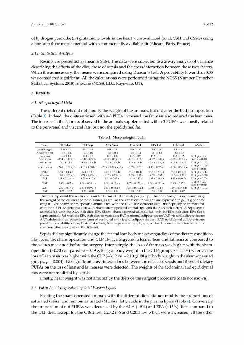
3.1. Morphological Data
The different diets did not modify the weight of the animals, but did alter the body composition(Table 3). Indeed, the diets enriched with n-3 PUFA increased the fat mass and reduced the lean mass.The increase in the fat mass observed in the animals supplemented with n-3 PUFAs was mostly relatedto the peri-renal and visceral fats, but not the epididymal fat.
Table 3. Morphological data.
Tissue DEF Sham DEF Sept ALA Sham ALA Sept EPA Fict EPA Sept p-Value
Body weight 552 ± 22 549 ± 15 581 ± 24 565 ± 18 590 ± 22 578 ± 20∆ body weight −3.3 ± 0.6 −2.0 ± 0.8 −3.0 ± 0.4 −2.5 ± 0.3 −2.1 ± 0.3 −2.2 ± 0.3
Fat mass 12.5 ± 1.1 12.4 ± 0.9 14.6 ± 0.8 15.5 ± 0.9 15.9 ± 1.1 14.6 ± 1.2 D ef: p = 0.021∆ fat mass −0.34 ± 0.19 a, b −0.17 ± 0.31 b −0.87 ± 0.15 a, c −0.01 ± 0.12 b −0.97 ± 0.08 a −0.39 ± 0.17 b, c S ef: p = 0.001Lean mass 79.0 ± 1.1 a 79.6 ± 0.9 a, b 77.5 ± 0.9 a, b 76.4 ± 1.0 b 75.7 ± 1.0 a, b 76.9 ± 1.3 a, b D ef: p = 0.032
∆ lean mass −2.61 ± 0.50 a, b −3.10 ± 0.68 b, c −2.35 ± 0.32 a, c, d, e −3.59 ± 0.26 b −1.33 ± 0.37 a, d −2.66 ± 0.36 b, e D ef: p = 0.025S ef: p = 0.003
Water 57.3 ± 1.0 a, b 57.1 ± 0.6 a 55.5 ± 0.6 a, b 55.0 ± 0.8 b 54.3 ± 0.9 a, b 55.0 ± 0.9 a, b D ef: p = 0.014∆ water −1.89 ± 0.63 a, b −0.71 ± 0.49 a, b −1.15 ± 0.29 a, b −2.05 ± 0.37 a −0.39 ± 0.37 b −0.34 ± 0.38 b D ef: p = 0.020
PAT 1.28 ± 0.11 a, b 1.25 ± 0.10 a 1.31 ± 0.07 a 1.61 ± 0.10 b 1.65 ± 0.08 ab 1.49 ± 0.10 ab D ef: p = 0.024
VAT 1.43 ± 0.05 a 1.64 ± 0.10 a, c 1.68 ± 0.10 a, c 1.85 ± 0.13 b, c 1.86 ± 0.10 b, c 2.09 ± 0.15 b D ef: p < 0.001S ef: p = 0.048
AAT 2.77 ± 0.17 a 2.89 ± 0.19 a, b 2.99 ± 0.15 a, b 3.46 ± 0.19 a, b 3.60 ± 0.11 b 3.49 ± 0.27 a, b D ef: p = 0.002EAT 1.25 ± 0.12 1.29 ± 0.08 1.33 ± 0.09 1.48 ± 0.08 1.36 ± 0.07 1. 44 ± 0.07
The data represent the mean and standard error of 10 animals per group. The body weight is expressed in g,the weight of the different adipose tissues, as well as the variations in weight, are expressed in g/100 g of bodyweight. DEF Sham: sham-operated animals fed with the n-3 PUFA deficient diet; DEF Sept: septic animals fedwith the n-3 PUFA deficient diet; ALA Sham: sham-operated animals fed with the ALA-rich diet; ALA Sept: septicanimals fed with the ALA-rich diet; EPA Sham: sham-operated animals fed with the EPA-rich diet; EPA Sept:septic animals fed with the EPA-rich diet; ∆: variation; PAT: perirenal adipose tissue; VAT: visceral adipose tissue;AAT: abdominal adipose tissue (sum of peri-renal and visceral adipose tissues); EAT: epididymal adipose tissue;p-value: probability value; D ef: diet effects; S ef: sepsis effects; a, b, c, d, e: the data on a same line without acommon letter are significantly different.
Sepsis did not significantly change the fat and lean body masses regardless of the dietary conditions.However, the sham-operation and CLP always triggered a loss of lean and fat masses compared tothe values measured before the surgery. Interestingly, the loss of fat mass was higher with the sham-operation (−0.73 compared to −0.19 g/100 g of body weight in the CLP group, p = 0.003) whereas theloss of lean mass was higher with the CLP (−3.12 vs. −2.10 g/100 g of body weight in the sham-operatedgroups, p = 0.004). No significant cross-interactions between the effects of sepsis and those of dietaryPUFAs on the loss of lean and fat masses were detected. The weights of the abdominal and epididymalfats were not modified by sepsis.
Finally, heart weight was not affected by the diets or the surgical procedure (data not shown).
3.2. Fatty Acid Composition of Total Plasma Lipids
Feeding the sham-operated animals with the different diets did not modify the proportions ofsaturated (SFAs) and monounsaturated (MUFAs) fatty acids in the plasma lipids (Table 4). Conversely,the proportion of n-6 PUFAs was decreased by the ALA (−8%) and EPA (−13%) diets compared tothe DEF diet. Except for the C18:2 n-6, C20:2 n-6 and C20:3 n-6 which were increased, all the other

Antioxidants 2020, 9, 371 8 of 22
individual n-6 PUFAs were decreased by the n-3 PUFA-rich diets. The changes were more obvious forthe EPA diet compared to the ALA diet. By contrast, the proportions of n-3 PUFAs were increased bythe n-3 PUFA-rich diets (+459% and +775% for the ALA and EPA diets compared to the DEF diet).This was particularly strong for the C18:3 n-3, C20:5 n-3, C22:5 n-3 and C22:6 n-3 in the ALA group(+∞, +943, +563 and +361% compared to the DEF group) and C20:5 n-3, C22:5 n-3 and C22:6 n-3 in theEPA group (+5300, +1388 and +384%). All these modifications contributed to reduce the n-6 to n-3PUFA ratio of plasma lipids (−84 and −90% for the ALA and EPA diets compared to the DEF diet).
Table 4. Fatty acid composition of plasma lipids.
Fatty acid DEF Sham DEF Sept ALA Sham ALA Sept EPA Sham EPA Sept p-Value
C14:0 0.41 ± 0.04 1.22 ± 0.77 0.39 ± 0.03 0.52 ± 0.10 0.38 ± 0.04 0.45 ± 0.04C15:0 0.16 ± 0.01 0.14 ± 0.01 0.16 ± 0.01 0.16 ± 0.02 0.14 ± 0.02 0.15 ± 0.01 S ef: p < 0.001C16:0 16.53 ± 0.56 a 18.59 ± 0.48 b 17.05 ± 0.32 a, c 17.79 ± 0.48 b, c, d 16.99 ± 0.39 a, d 18.43 ± 0.47 b, c S ef: p < 0.05C17:0 0.23 ± 0.01 a 0.21 ± 0.01 a, b 0.21 ± 0.01 a, b 0.20 ± 0.01 a, b 0.22 ± 0.01 a, b 0.20 ± 0.01 b S ef: p = 0.035C18:0 13.57 ± 0.59 13.95 ± 0.65 13.84 ± 0.43 14.96 ± 0.62 12.44 ± 1.42 14.39 ± 0.44C20:0 0.08 ± 0.03 0.14 ± 0.03 0.08 ± 0.02 0.12 ± 0.02 0.12 ± 0.03 0.15 ± 0.03C22:0 0.20 ± 0.02 0.21 ± 0.02 0.22 ± 0.01 0.24 ± 0.01 0.24 ± 0.01 0.22 ± 0.02C24:0 0.49 ± 0.02 a, b 0.50 ± 0.01 a, b 0.51 ± 0.02 a, b 0.50 ± 0.02 a, b 0.53 ± 0.02 a 0.47 ± 0.02 b
DMA 16:0 0.14 ± 0.01 a, b 0.11 ± 0.01 a, b 0.13 ± 0.02 a, b 0.10 ± 0.01 a 0.13 ± 0.02 b 0.13 ± 0.01 abDMA 18:0 0.07 ± 0.02 a, c 0.01 ± 0.01 b, d 0.06 ± 0.02 a, d, e 0.01 ± 0.01 b, c, e 0.09 ± 0.02 a 0.02 ± 0.01 b, c, e S ef: p < 0.001
SFA 32.10 ± 0.45 a 35.3 ± 1.21 b, c 32.89 ± 0.37 a 34.86 ± 0.51 b 31.51 ± 1.29 a, c 34.83 ± 0.28 b S ef: p < 0.001
C16:1 n-7 1.20 ± 0.18 a 1.19 ± 0.06 a, c 1.29 ± 0.12 a, c 1.44 ± 0.12 b, c 1.07 ± 0.01 a 1.55 ± 0.17 b, c D ef: p = 0.048S ef: p < 0.001
C18:1 n-7 2.10 ± 0.09 a 2.06 ± 0.10 a 2.06 ± 0.08 a 2.09 ± 0.10 a 1.88 ± 0.10 b 1.93 ± 0.07 a D ef: p = 0.003
C18:1 n-9 12.60 ± 1.03 a, c 14.67 ± 0.86 b 10.85 ± 0.43 a 12.86 ± 0.80 c 10.08 ± 0.52 a 12.62 ± 0.60 b, c D ef: p = 0.009S ef: p < 0.001
C20:1 n-9 0.15 ± 0.01 a, b 0.17 ± 0.01 a 0.13 ± 0.01 b, c 0.16 ± 0.01 a, d 0.15 ± 0.01 b, d, e 0.15 ± 0.01 a, c, e S ef: p = 0.004C22:1 n-9 0.06 ± 0.02 a, b 0.09 ± 0.02 b, c 0.03 ± 0.02 a 0.09 ± 0.02 b 0.05 ± 0.02 a, c 0.11 ± 0.01 b S ef: p < 0.001
C24:1 n-9 0.36 ± 0.02 a 0.42 ± 0.02 a, b 0.41 ± 0.02 a, b 0.46 ± 0.02 b 0.46 ± 0.02 b 0.44 ± 0.03 a, b D ef: p = 0.040CI: p = 0.035
MUFA 16.66 ± 1.24 a, c 18.76 ± 0.95 b 14.94 ± 0.47 a, d 17.23 ± 0.99 b, c, d 15.29 ± 1.83 a 16.94 ± 0.76 b, c, d S ef: p < 0.001
C18:2 n-6 11.96 ± 0.92 a 13.11 ± 0.77 b, d, e 13.05 ± 0.55 a, e 15.20 ± 0.74 b, f 14.06 ± 0.53 b, e 15.53 ± 0.39 c, d, f D ef: p < 0.001S ef: p < 0.001
C18:3 n-6 0.25 ± 0.02 a 0.14 ± 0.01 b 0.22 ± 0.01 a 0.14 ± 0.01 b 0.15 ± 0.02 b 0.12 ± 0.02 bD ef: p = 0.002S ef: p < 0.001CI: p = 0.015
C20:2 n-6 0.17 ± 0.01 a, c 0.18 ± 0.01 a, c 0.19 ± 0.01 a, c 0.21 ± 0.01 b 0.20 ± 0.01 b, c 0.19 ± 0.01 b, c D ef: p = 0.047
C20:3 n-6 0.28 ± 0.02 a 0.21 ± 0.02 a 0.44 ± 0.03 b, c 0.42 ± 0.04 b 0.52 ± 0.03 c 0.47 ± 0.04 b, c D ef: p < 0.001S ef: p = 0.048
C20:4 n-6 34.34 ± 1.90 a 27.62 ± 0.74 d, e 31.81 ± 0.97 b 25.71 ± 1.26 d 28.48 ± 1.04 e 22.16 ± 0.86 c D ef: p < 0.001S ef: p < 0.001
C22:4 n-6 0.74 ± 0.02 a 0.81 ± 0.06 a 0.32 ± 0.02 b 0.30 ± 0.01 b 0.23 ± 0.02 b, d 0.20 ± 0.02 c, d D ef: p < 0.001C22:5 n-6 2.58 ± 0.22 a 2.85 ± 0.15 a 0.22 ± 0.01 b 0.27 ± 0.02 b 0.13 ± 0.02 b 0.15 ± 0.01 b D ef: p < 0.001
n-6 PUFA 50.32 ± 1.29 a 44.91 ± 0.85 c, d 46.24 ± 0.62 c 42.25 ± 0.97 d 43.78 ± 0.64 c, d 38.83 ± 0.71 b D ef: p < 0.001S ef: p < 0.001
C18:3 n-3 nd a nd a 0.51 ± 0.04 b 0.77 ± 0.09 c 0.02 ± 0.02 a 0.01 ± 0.01 aD ef: p < 0.001S ef: p = 0.011CI: p = 0.001
C20:5 n-3 0.07 ± 0.02 a 0.09 ± 0.02 a 0.73 ± 0.06 b 0.63 ± 0.04 b, a 3.78 ± 0.32 c 3.75 ± 0.26 c D ef: p < 0.001
C22:5 n-3 0.08 ± 0.02 a 0.10 ± 0.02 a 0.53 ± 0.02 b 0.48 ± 0.02 b 1.19 ± 0.08 c 1.33 ± 0.05 dD ef: p < 0.001S ef: p = 0.007CI: p < 0.001
C22:6 n-3 0.95 ± 0.03 a 0.99 ± 0.03 a 4.38 ± 0.12 b 3.99 ± 0.16 c 4.60 ± 0.12 b 4.51 ± 0.15 b D ef: p < 0.001n-3 PUFA 1.10 ± 0.04 a 1.20 ± 0.04 a 6.15 ± 0.13 b 5.87 ± 0.11 b 9.63 ± 0.30 c 9.60 ± 0.29 c D ef: p < 0.001
PUFA 51.42 ± 1.29 a, d, f 46.10 ± 0.83 b, e 52.39 ± 0.56 c, f 48.11 ± 0.98 b, d, e 53.41 ± 0.67 c, f 48.43 ± 0.88 b, d S ef: p < 0.001
n-6/n-3 46.3 ± 2.16 a 38.1 ± 1.8 b 7.6 ± 0.2 c 7.2 ± 0.2 c 4.6 ± 0.1 c 4.1 ± 0.1 cD ef: p < 0.001S ef: p = 0.002CI: p = 0.002
The results include 10 animals per group. DEF Sham: sham-operated animals fed with the n-3 PUFA deficient diet;DEF Sept: septic animals fed with the n-3 PUFA deficient diet; ALA Sham: sham-operated animals fed with theALA-rich diet; ALA Sept: septic animals fed with the ALA-rich diet; EPA Sham: sham-operated animals fed withthe EPA-rich diet; EPA Sept: septic animals fed with the EPA-rich diet; DMA: dimethyl acetal; SFA: saturated fattyacids; MUFA: monounsaturated fatty acids; PUFA: polyunsaturated fatty acids; n-6/n-3: ratio between n-6 and n-3PUFA; nd: not detected; D ef: diet effects; S ef: sepsis effects; CI: cross-interaction. a, b, c, d, e, f: the data printed ona same line without a common letter are significantly different.
Sepsis also induced changes in the fatty acid profile of plasma lipids. It increased the proportionsof SFAs, namely C16:0 (+12, +4 and +8% in the DEF, ALA and EPA groups, p < 0.001, NS, p = 0.029compared to the sham-operated groups) and those of MUFAs. By contrast, it strongly reduced theamount of n-6 PUFAs (−11, −9 and −11% for the DEF, ALA and EPA groups). The main fatty acidsconcerned were the C18:3 n-6 (−44, −36 and −20% for the DEF, ALA and EPA groups) and C20:4 n-6(arachidonic acid (ARA), −20, −19 and −22% for the DEF, ALA and EPA groups). These decreases inn-6 PUFAs were not offset by augmented proportions of n-3 PUFAs. The n-6 to n-3 PUFA ratio was

Antioxidants 2020, 9, 371 9 of 22
thus significantly reduced in the DEF group (−18%), but not in the ALA and EPA groups (−5 and−11%, not significant).
Two significant cross-interactions between the effects of the diets and those of sepsis were noticed.Firstly, a sepsis-induced increase in the C18:3 n-3 was observed in the ALA group, but not in the twoother groups in which the C18:3 n-3 proportion was almost null. Secondly, the n-6 to n-3 PUFA ratiowas significantly reduced by sepsis in the DEF group, but not in the ALA and EPA groups.
3.3. Plasma Triglycerides
The plasma concentrations of triglycerides were similar in the different dietary groups after thesham-operation (Figure 1). Sepsis increased their concentrations (+82, +50 and +77% in the DEF,ALA and EPA groups). No differences were observed between the 3 dietary groups.
Figure 1. Plasma triglyceride concentrations. The means represent a consortium of 10 animals pergroup. DEF: n-3 polyunsaturated fatty acids (PUFA) deficient diet; ALA: diet enriched with α-linolenicacid; EPA: diet enriched with eicosapentaenoic acid; Sham: fictive operation; Sept: cecal ligation andpuncture (CLP) performed to induce sepsis; S ef: sepsis effect; a, b: means without a common letter aresignificantly different.
3.4. In Vivo Cardiac Function
The cardiac function was estimated in anesthetized animals with a pressure gauge introducedinto the aorta and left ventricular cavity via the right carotid. In sham-operated animals, the changes indietary PUFA did not alter the systolic, diastolic and mean aortic pressure (Table 5). No changes in theparameters of the left ventricle contraction and relaxation were also observed. The LVDP, heart rate,contraction and relaxation were unchanged by the dietary lipids.
Table 5. In vivo cardiac function.
Parameter DEF Sham DEF Sept ALA Sham ALA Sept EPA Sham EPA Sept p-Value
Aorta
ASP 88.3 ± 6.5 86.0 ± 3.1 91.9 ± 5.2 84.3 ± 4.0 91.1 ± 6.1 82.8 ± 3.8ADP 63.8 ± 5.4 58.6 ± 3.7 58.7 ± 3.5 55.1 ± 4.1 64.7 ± 6.5 52.6 ± 6.5AMP 76.1 ± 5.9 72.3 ± 3.4 72.2 ± 3.6 69.7 ± 3.9 77.9 ± 6.3 67.7 ± 5.1
Heart
LVDP 93 ± 2 ab 99 ± 3 ab 107 ± 5 a 87 ± 4 b 103 ± 6 ac 102 ± 3 ab CI: p = 0.032
HR 246 ± 7 ab 271 ± 11 ab 259 ± 8 ab 257 ± 16 ab 233 ± 7 a 289 ± 10 b S ef: p = 0.004CI: p = 0.027
dP/dt max 5183 ± 189 ab 5504 ± 238 ab 5579 ± 355 a 4244 ± 569 b 5034 ± 410 ab 5533 ± 341 ab CI: p < 0.045dP/dt min −4549 ± 455 −4364 ± 204 −4294 ± 361 −3416 ± 349 −4244 ± 463 −4664 ± 130
The data represent the means and SEM of 10 samples per group. DEF Sham: sham-operated animals fed with then-3 PUFA deficient diet; DEF Sept: septic animals fed with the n-3 PUFA deficient diet; ALA Sham: sham-operatedanimals fed with the ALA-rich diet; ALA Sept: septic animals fed with the ALA-rich diet; EPA Sham: sham-operatedanimals fed with the EPA-rich diet; EPA Sept: septic animals fed with the EPA-rich diet; ASP: aortic systolicpressure; ADP: aortic diastolic pressure; AMP: aortic mean pressure; LVDP: left ventricular developed pressure;HR: heart rate; dp/dt max: contraction; dP/dt min: relaxation. Systolic and diastolic pressures, heart rate andcontraction/relaxation are expressed in mmHg, beats/min and mmHg/s. vp-value: probability value; S ef: Sepsiseffects; CI: cross interaction between the effects of the diets and those of sepsis. a, b, c: the data on a same linewithout a common letter are significantly different.

Antioxidants 2020, 9, 371 10 of 22
Sepsis tended to decrease the aortic pressures, but this was not significant. In contrast,our observations suggested that it stimulated the cardiac function. It increased the heart rate(+11%, p = 0.004), but this was significant only in the EPA group (+24 vs. +10 and −1% in the DEF andALA groups). It also tended to increase the contraction in the DEF and EPA groups (+6 and +10%,respectively), but it reduced this parameter in the ALA group (−24%, p = 0.027). The relaxation wasunchanged by sepsis in the DEF and EPA groups, but tended to be deteriorated in the ALA group(−20% p = 0.12).
3.5. Plasma Oxidative Stress in the In Vivo Situation
The changes in cardiac function were not explained by the modifications of the oxidative stressparameters in the plasma (Figure 2). The amounts of thiol residues, inversely proportional tothe oxidative stress, were unaffected by the dietary PUFA and sepsis. Conversely, a significantcross-interaction was observed: although the amount of thiol residues were not modified by sepsis inthe DEF and ALA groups, it was increased in the EPA group (+10%). Lipid peroxidation was evaluatedby measuring thiobarbituric acid reactive substances (TBARS) levels and was not affected by thedietary PUFAs nor the sepsis. No significant cross-interaction was observed with the 2-way analysisof variance. However, when the effect of sepsis was evaluated using a 1-way analysis of variance inthe ALA group only, a decrease in the TBARS level was noticed (−12%, p < 0.006). The anti-oxidativedefenses (FRAP) were not affected by the treatments, but glutathione peroxidase activity was reducedby sepsis, especially in the DEF and EPA groups (−17 and −12%, respectively).
Figure 2. Parameters of the plasma oxidative stress. A: amounts of thiol residues; B: amounts ofthiobarbituric acid reactive substances (TBARS); C: anti-oxidative defenses (FRAP); D: glutathioneperoxidase (GPX) activities. The means represent a consortium of 10 animals per group. DEF: n-3 PUFAdeficient diet; ALA: diet enriched with α-linolenic acid; EPA: diet enriched with eicosapentaenoicacid; Sham: fictive operation; Sept: operation performed to induce sepsis; TBARS: thiobarbituric acidreactive substances; FRAP: ferric reducing antioxidant power; GPX: glutathione peroxidase activity;S ef: sepsis effect; CI: cross interaction; a, b, c, d, e: means without a common letter in a same figure aresignificantly different.
Antioxidants 2020, 9, 371 11 of 22
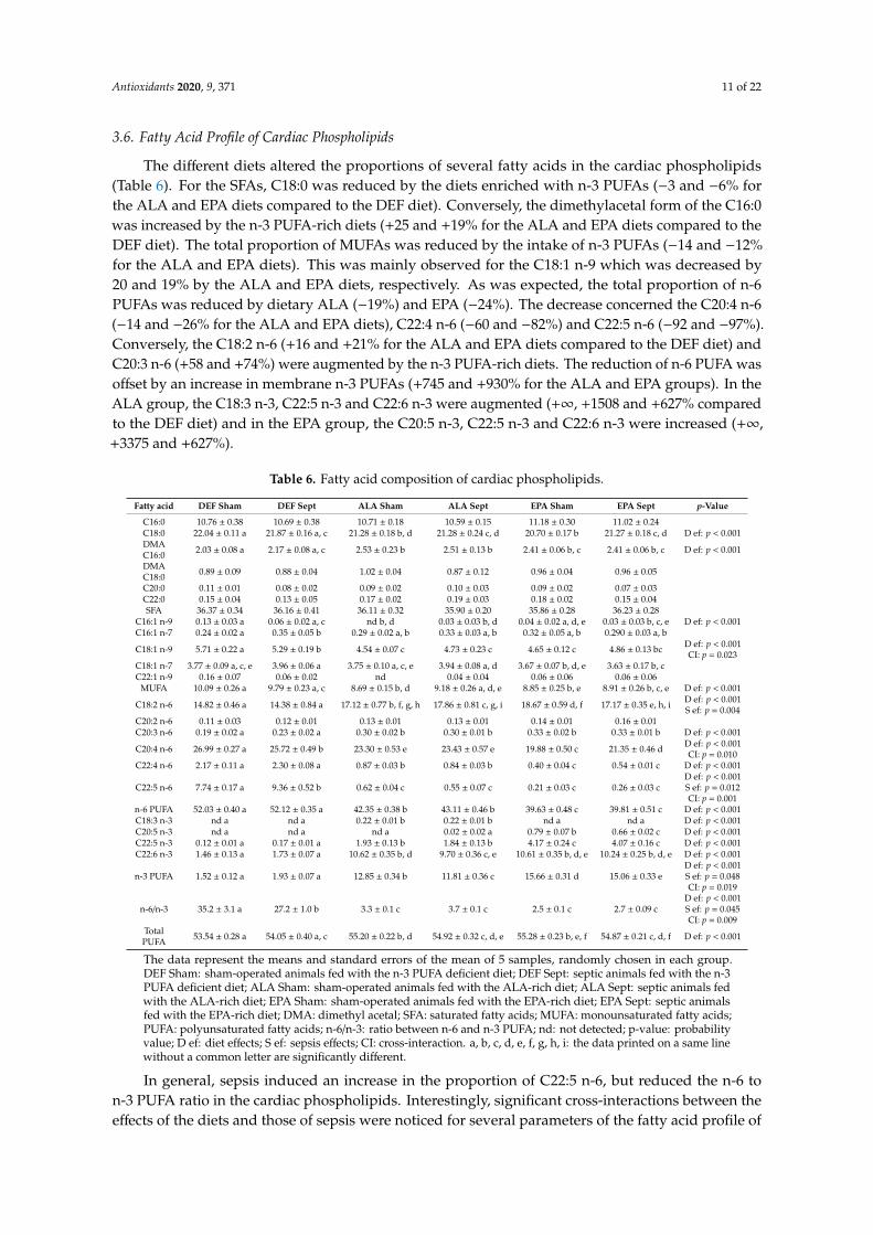
3.6. Fatty Acid Profile of Cardiac Phospholipids
The different diets altered the proportions of several fatty acids in the cardiac phospholipids(Table 6). For the SFAs, C18:0 was reduced by the diets enriched with n-3 PUFAs (−3 and −6% forthe ALA and EPA diets compared to the DEF diet). Conversely, the dimethylacetal form of the C16:0was increased by the n-3 PUFA-rich diets (+25 and +19% for the ALA and EPA diets compared to theDEF diet). The total proportion of MUFAs was reduced by the intake of n-3 PUFAs (−14 and −12%for the ALA and EPA diets). This was mainly observed for the C18:1 n-9 which was decreased by20 and 19% by the ALA and EPA diets, respectively. As was expected, the total proportion of n-6PUFAs was reduced by dietary ALA (−19%) and EPA (−24%). The decrease concerned the C20:4 n-6(−14 and −26% for the ALA and EPA diets), C22:4 n-6 (−60 and −82%) and C22:5 n-6 (−92 and −97%).Conversely, the C18:2 n-6 (+16 and +21% for the ALA and EPA diets compared to the DEF diet) andC20:3 n-6 (+58 and +74%) were augmented by the n-3 PUFA-rich diets. The reduction of n-6 PUFA wasoffset by an increase in membrane n-3 PUFAs (+745 and +930% for the ALA and EPA groups). In theALA group, the C18:3 n-3, C22:5 n-3 and C22:6 n-3 were augmented (+∞, +1508 and +627% comparedto the DEF diet) and in the EPA group, the C20:5 n-3, C22:5 n-3 and C22:6 n-3 were increased (+∞,+3375 and +627%).
Table 6. Fatty acid composition of cardiac phospholipids.
Fatty acid DEF Sham DEF Sept ALA Sham ALA Sept EPA Sham EPA Sept p-Value
C16:0 10.76 ± 0.38 10.69 ± 0.38 10.71 ± 0.18 10.59 ± 0.15 11.18 ± 0.30 11.02 ± 0.24C18:0 22.04 ± 0.11 a 21.87 ± 0.16 a, c 21.28 ± 0.18 b, d 21.28 ± 0.24 c, d 20.70 ± 0.17 b 21.27 ± 0.18 c, d D ef: p < 0.001DMAC16:0 2.03 ± 0.08 a 2.17 ± 0.08 a, c 2.53 ± 0.23 b 2.51 ± 0.13 b 2.41 ± 0.06 b, c 2.41 ± 0.06 b, c D ef: p < 0.001
DMAC18:0 0.89 ± 0.09 0.88 ± 0.04 1.02 ± 0.04 0.87 ± 0.12 0.96 ± 0.04 0.96 ± 0.05
C20:0 0.11 ± 0.01 0.08 ± 0.02 0.09 ± 0.02 0.10 ± 0.03 0.09 ± 0.02 0.07 ± 0.03C22:0 0.15 ± 0.04 0.13 ± 0.05 0.17 ± 0.02 0.19 ± 0.03 0.18 ± 0.02 0.15 ± 0.04SFA 36.37 ± 0.34 36.16 ± 0.41 36.11 ± 0.32 35.90 ± 0.20 35.86 ± 0.28 36.23 ± 0.28
C16:1 n-9 0.13 ± 0.03 a 0.06 ± 0.02 a, c nd b, d 0.03 ± 0.03 b, d 0.04 ± 0.02 a, d, e 0.03 ± 0.03 b, c, e D ef: p < 0.001C16:1 n-7 0.24 ± 0.02 a 0.35 ± 0.05 b 0.29 ± 0.02 a, b 0.33 ± 0.03 a, b 0.32 ± 0.05 a, b 0.290 ± 0.03 a, b
C18:1 n-9 5.71 ± 0.22 a 5.29 ± 0.19 b 4.54 ± 0.07 c 4.73 ± 0.23 c 4.65 ± 0.12 c 4.86 ± 0.13 bc D ef: p < 0.001CI: p = 0.023
C18:1 n-7 3.77 ± 0.09 a, c, e 3.96 ± 0.06 a 3.75 ± 0.10 a, c, e 3.94 ± 0.08 a, d 3.67 ± 0.07 b, d, e 3.63 ± 0.17 b, cC22:1 n-9 0.16 ± 0.07 0.06 ± 0.02 nd 0.04 ± 0.04 0.06 ± 0.06 0.06 ± 0.06
MUFA 10.09 ± 0.26 a 9.79 ± 0.23 a, c 8.69 ± 0.15 b, d 9.18 ± 0.26 a, d, e 8.85 ± 0.25 b, e 8.91 ± 0.26 b, c, e D ef: p < 0.001
C18:2 n-6 14.82 ± 0.46 a 14.38 ± 0.84 a 17.12 ± 0.77 b, f, g, h 17.86 ± 0.81 c, g, i 18.67 ± 0.59 d, f 17.17 ± 0.35 e, h, i D ef: p < 0.001S ef: p = 0.004
C20:2 n-6 0.11 ± 0.03 0.12 ± 0.01 0.13 ± 0.01 0.13 ± 0.01 0.14 ± 0.01 0.16 ± 0.01C20:3 n-6 0.19 ± 0.02 a 0.23 ± 0.02 a 0.30 ± 0.02 b 0.30 ± 0.01 b 0.33 ± 0.02 b 0.33 ± 0.01 b D ef: p < 0.001
C20:4 n-6 26.99 ± 0.27 a 25.72 ± 0.49 b 23.30 ± 0.53 e 23.43 ± 0.57 e 19.88 ± 0.50 c 21.35 ± 0.46 d D ef: p < 0.001CI: p = 0.010
C22:4 n-6 2.17 ± 0.11 a 2.30 ± 0.08 a 0.87 ± 0.03 b 0.84 ± 0.03 b 0.40 ± 0.04 c 0.54 ± 0.01 c D ef: p < 0.001
C22:5 n-6 7.74 ± 0.17 a 9.36 ± 0.52 b 0.62 ± 0.04 c 0.55 ± 0.07 c 0.21 ± 0.03 c 0.26 ± 0.03 cD ef: p < 0.001S ef: p = 0.012CI: p = 0.001
n-6 PUFA 52.03 ± 0.40 a 52.12 ± 0.35 a 42.35 ± 0.38 b 43.11 ± 0.46 b 39.63 ± 0.48 c 39.81 ± 0.51 c D ef: p < 0.001C18:3 n-3 nd a nd a 0.22 ± 0.01 b 0.22 ± 0.01 b nd a nd a D ef: p < 0.001C20:5 n-3 nd a nd a nd a 0.02 ± 0.02 a 0.79 ± 0.07 b 0.66 ± 0.02 c D ef: p < 0.001C22:5 n-3 0.12 ± 0.01 a 0.17 ± 0.01 a 1.93 ± 0.13 b 1.84 ± 0.13 b 4.17 ± 0.24 c 4.07 ± 0.16 c D ef: p < 0.001C22:6 n-3 1.46 ± 0.13 a 1.73 ± 0.07 a 10.62 ± 0.35 b, d 9.70 ± 0.36 c, e 10.61 ± 0.35 b, d, e 10.24 ± 0.25 b, d, e D ef: p < 0.001
n-3 PUFA 1.52 ± 0.12 a 1.93 ± 0.07 a 12.85 ± 0.34 b 11.81 ± 0.36 c 15.66 ± 0.31 d 15.06 ± 0.33 eD ef: p < 0.001S ef: p = 0.048CI: p = 0.019
n-6/n-3 35.2 ± 3.1 a 27.2 ± 1.0 b 3.3 ± 0.1 c 3.7 ± 0.1 c 2.5 ± 0.1 c 2.7 ± 0.09 cD ef: p < 0.001S ef: p = 0.045CI: p = 0.009
TotalPUFA 53.54 ± 0.28 a 54.05 ± 0.40 a, c 55.20 ± 0.22 b, d 54.92 ± 0.32 c, d, e 55.28 ± 0.23 b, e, f 54.87 ± 0.21 c, d, f D ef: p < 0.001
The data represent the means and standard errors of the mean of 5 samples, randomly chosen in each group.DEF Sham: sham-operated animals fed with the n-3 PUFA deficient diet; DEF Sept: septic animals fed with the n-3PUFA deficient diet; ALA Sham: sham-operated animals fed with the ALA-rich diet; ALA Sept: septic animals fedwith the ALA-rich diet; EPA Sham: sham-operated animals fed with the EPA-rich diet; EPA Sept: septic animalsfed with the EPA-rich diet; DMA: dimethyl acetal; SFA: saturated fatty acids; MUFA: monounsaturated fatty acids;PUFA: polyunsaturated fatty acids; n-6/n-3: ratio between n-6 and n-3 PUFA; nd: not detected; p-value: probabilityvalue; D ef: diet effects; S ef: sepsis effects; CI: cross-interaction. a, b, c, d, e, f, g, h, i: the data printed on a same linewithout a common letter are significantly different.
In general, sepsis induced an increase in the proportion of C22:5 n-6, but reduced the n-6 ton-3 PUFA ratio in the cardiac phospholipids. Interestingly, significant cross-interactions between theeffects of the diets and those of sepsis were noticed for several parameters of the fatty acid profile of

Antioxidants 2020, 9, 371 12 of 22
the cardiac phospholipids. Cross-interactions were observed for the C20:4 n-6, C22:5 n-6, total n-3PUFAs and ratio between n-6 and n-3 PUFAs. The proportion of C20:4 n-6 was increased by sepsis inthe EPA group (+7%), but not in the other dietary groups. The proportion of C22:5 n-6 was augmentedin the DEF group (+21%), but not in the ALA and EPA groups. The proportion of total n-3 PUFAs wasreduced by sepsis in both the ALA and EPA groups (−8 and −4%), but not in the DEF group. Finally,the n-6 to n-3 PUFA ratio was not changed by sepsis in the ALA and EPA groups, but was reduced bythe pathological event in the DEF group (−23%).
3.7. Ex Vivo Cardiac Function
The cardiac function was measured in the ex vivo situation under standard conditions (Figure 3)when the hearts were paced at 370 beats/min. The LVDP, contraction, perfusion pressure, oxygenconsumption and metabolic efficiency were unaffected by the diets in the sham-operated animals.Conversely, the relaxation rate was reduced by the diet enriched with ALA compared to that deficientin n-3 PUFAs (−21%). This was not the case for that measured with the diet enriched with EPA whosevalue ranged between those of the DEF and ALA groups.
Figure 3. Activities of the isolated perfused hearts. A: left ventricular developed pressure; B: contraction;C: relaxation; D: perfusion pressure; E: oxygen consumption; F: metabolic efficiency. The meansrepresent a consortium of 10 animals per group. DEF: n-3 PUFA deficient diet; ALA: diet enriched withα-linolenic acid; EPA: diet enriched with eicosapentaenoic acid; Sham: fictive operation; Sept: operationperformed to induce sepsis; LVDP: left ventricular developed pressure; dP/dt max: contraction; dP/dtmin: relaxation; S ef: sepsis effect; CI: cross interaction; a, b: means without a common letter in a samefigure are significantly different.

Antioxidants 2020, 9, 371 13 of 22
Sepsis significantly decreased the LVDP in the DEF group (−21%). This was due to reductionsin the rates of contraction and relaxation. By contrast, in the ALA and EPA groups, the LVDP aswell as the rates of contraction and relaxation tended to be improved by sepsis. The sepsis-induceddecrease in the LVDP in the DEF group was associated with a reduction in the perfusion pressure,whereas the upholding of the LVDP in the ALA and EPA groups was associated with maintainedperfusion pressures.
3.8. Appraisal of the Mitochondrial Function
Lactate release in the venous effluent, pyruvate washout and glycolytic rate (sum of lactate pluspyruvate releases) were not affected by the diets and sepsis (data not shown). Conversely, the lactaterelease to oxygen consumption ratio, reflecting the part of anaerobic glycolysis in energy production,was increased by sepsis in the DEF group (+57%, p < 0.05) without being significantly altered in theother two groups (Figure 4A). This suggests sepsis-induced alterations of mitochondrial function inthe DEF group. This was confirmed by VDAC expression (Figure 4D), which was only reduced bysepsis (−29%, p < 0.003 by 1-way analysis of variance) in the DEF group. This decreased mitochondrialdensity can be partly explained by reduced mitochondrial biogenesis as evidenced by the PGC1-αexpression (Figure 4G) which only tended to be reduced (−16%, p = 0.085) in this group.
Figure 4. Appraisal of the mitochondrial function. (A) Lactate release in the coronary effluents of theisolated hearts to oxygen consumption ratio; (B) and (E) Voltage-dependent anion channel (VDAC)and peroxisome proliferator activated receptor gamma coactivator 1 alpha (PGC1-α) representativeimmunoblots; (C) and (F) Corresponding Ponceau red-colored total proteins; (D) and (G) CorrespondingVDAC and PGC1-α expressions. The means represent a consortium of 10 animals per group. DEF: n-3PUFA deficient diet; ALA: diet enriched with α-linolenic acid; EPA: diet enriched with eicosapentaenoicacid; Sham: fictive operation; Sept: operation performed to induce sepsis; a, b, c: means without acommon letter in a same figure are significantly different.

Antioxidants 2020, 9, 371 14 of 22
3.9. Inflammation
Systemic inflammation was evaluated by measuring the concentration of TNF-α in the plasma(Figure 5). Low in the sham-operated groups, the concentration of this inflammatory cytokine wasstrongly increased by sepsis in the DEF group (+205%), but not in the ALA group. In the EPA group,its value ranged between those measured in the DEF and ALA groups.
Figure 5. Inflammation markers. (A) Representative immunoblots of nuclear factor of kappa lightpolypeptide gene enhancer in B-cells inhibitor, alpha (IκBα); (B) Densitometry to red Ponceau S stain.The means represent a consortium of 10 animals per group. TNF-a: tumor necrosis factor a; IL1-β:interleukin 1-β; IL-6: interleukin 6; DEF: n-3 PUFA deficient diet; ALA: diet enriched with α-linolenicacid; EPA: diet enriched with eicosapentaenoic acid; Sham: fictive operation; Sept: operation performedto induce sepsis; a, b, c, d: means without a common letter in a same figure are significantly different.
Cardiac inflammation was determined by evaluating the activation of the NFκB pathway andmeasuring the gene expression of IL-1β, IL-6 and TNF-α. Activation of the NFκB pathway wasestimated by the decrease in IκBα protein expression. Except for the low value encountered in thesham-operated ALA-fed animals (−8 and −8% compared to the DEF and EPA groups), the other datawere high and did not differ from one another. The activated NFκB pathway in the sham-operatedanimals of the ALA group did not increase the expression of TNF-α and IL-6. The expression of IL-1βwas even reduced compared to that of the other two groups (−49 and 62% compared to the DEF andEPA groups).
However, sepsis increased the gene expression of IL-1β in all the dietary groups (+61, +189and +102%, respectively) compared to the sham-operated animals of the DEF, ALA and EPA groups.For TNF-αmRNA, sepsis tended to induce similar effects, but the difference was significant only for

Antioxidants 2020, 9, 371 15 of 22
the ALA and EPA groups (+169 and +103%, respectively). Finally, sepsis increased IL-6 mRNA only inthe ALA group (+278% compared to the sham-operated group with the ALA substitution).
3.10. Cardiac Oxidative Stress in the Ex Vivo Situation
The levels of reduced and oxidized glutathione were appraised to measure the rate of reactiveoxygen species (ROS) detoxification (Figure 6). If the amount of reduced glutathione was unalteredby the dietary manipulations and sepsis, those of oxidized glutathione depended on both of thesefactors. In the sham-operated animals, oxidized glutathione was higher in the DEF and ALA groupscompared to the EPA group (+136 and +76%, respectively). Sepsis maintained a high level of oxidizedglutathione in the DEF group, but strongly reduced it in the ALA group (−45%) and maintained it at alow level in the EPA group. Those changes reverberated logically on the GSH to GSSG ratio which isinversely proportional to the oxidative stress: it was low in the hearts of sham-operated animals fedwith the DEF and ALA diets, as well as in those of septic rats fed with the DEF diet, but it was high inthe other three subgroups; the sham-operated EPA group and the ALA and EPA sepsis groups.
Figure 6. Parameters of cardiac oxidative stress. (A) Representative immunoblots of uncouplingprotein 3 (UCP3); (B) Representative immunoblots of mitochondrial superoxide dismutase (SOD2);(C) Representative immunoblots of mitochondrial superoxide dismutase acetylated on lysine moieties(K-SOD2); (D) Densitometry to red Ponceau S stain. The means represent a consortium of 10 animalsper group. GSH: reduced glutathione; GSSG: oxidized glutathione; SIRT3: sirtuin 3; DEF: n-3 PUFAdeficient diet; ALA: diet enriched with α-linolenic acid; EPA: diet enriched with eicosapentaenoic acid;Sham: fictive operation; Sept: operation performed to induce sepsis; a, b, c, d, e: means without acommon letter in a same figure are significantly different.
The sepsis-induced decrease in oxidative stress in the ALA rats was apparently related to anincrease in cardiac SOD2 content (+15% compared to the sham-operated animals of the same dietary
Antioxidants 2020, 9, 371 16 of 22
group) without any change in SOD2 acetylation which is responsible for a reduced superoxidedetoxification capacity. This was confirmed by a low SOD2 mRNA expression (−10% compared tothe sham-operated animals of the same dietary group). These changes were not observed in the EPAgroup. Rather, the low oxidative stress observed in the septic EPA-fed rats was obviously due to anincreased UCP3 content (+45% compared to the sham-operated animals of the same dietary group)deemed to reduce lipid-related ROS production. This last phenomenon was associated with a highSIRT3 mRNA content in the EPA-fed subgroups (+67% compared to the other two dietary groups).
4. Discussion
This study aimed at (i) verifying the cardioprotective effects of EPA in male rats made septic byCLP; (ii) evaluating the effects of ALA on the cardiac consequences of early sepsis; and (iii) determiningprobable physiological and molecular mechanisms responsible for the observed effects. Severalcharacteristics indicate that the rats subjected to CLP were septic: (i) the concentrations of plasmatriglycerides were significantly higher in the CLP groups compared to the sham-operated ones.Inflammation inhibits skeletal muscle β-oxidation in favor of glucose degradation, leading toaccumulation of plasma triglycerides [27], (ii) the plasma TNF-α concentration was high in theDEF animals subjected to CLP, confirming the occurrence of a systemic inflammation; (iii) although theplasma concentrations of TNF-αwere low in the CLP-subjected animals fed the ALA and EPA diets,the gene expressions of TNF-α and IL-1β in the heart were high, suggesting cardiac inflammation;(iv) the plasma ARA content was considerably reduced in the CLP groups. This progression is knownto occur with sepsis and has already been demonstrated in humans [28]; (v) finally, the lean body masswas decreased in the septic groups, and this cachexic event is a hallmark of inflammation [29].
4.1. Ex Vivo Cardiac Function
The cardiac function was monitored in the ex vivo situation to get rid of all hormonal, nervous,systemic, but also bacterial, influences. It was determined in standardized conditions (compositionof the perfusion fluid, temperature, left ventricular diastolic pressure) at constant flow (12 mL/min)with electrical pacing at 370 impulsions/min. Under these circumstances, cardiac mechanical activityreflects the health status of the cardiomyocytes and the perfusion pressure mirrors the dilatation statusof the coronary microvessels.
In our study, we observed that the LVDP was reduced significantly by sepsis in the DEF group,whereas it tended to be increased in the other two groups. The sepsis-induced decrease in the LVDP ofthe DEF group was due to concomitant alterations of the contractility and relaxation, and it was stronglyassociated with a reduction of the coronary pressure which did not occur in the other 2 dietary groups.It is known that sepsis increases the expression of the COX-2 enzyme [3] which favors the synthesis ofvasodilator agents (i.e., prostacyclin) from ARA. Since the DEF group contained the highest amount ofARA in the cardiac phospholipids, and the lowest content of COX2 inhibitor DHA, it is said that thehighest prostacyclin production occurred in this group. Sepsis reduced thus the coronary pressure.However, the coronary flow was fixed to the value of 12 mL/min, and the oxygen and substrate supplieswere the same for all the hearts whose weight did not differ between groups. It is thus unlikely thatthe reduced coronary pressure in this group was responsible for the decreased mechanical function.In contrast, it is more likely that the reduced mechanical activity was a consequence of mitochondrialdamage. This hypothesis was strengthened by the sepsis-induced reduction of the myocardial oxygenconsumption observed in the DEF group (−10%, p < 0.01). It was sustained by the increased lactaterelease to oxygen consumption ratio suggesting an increased part of anaerobic glycolysis in energyproduction and mitochondrial damage. It is asserted by the reduced VDAC content, which suggestsa lower mitochondrial density partly due to a reduced expression of PGC1-α and to mitochondrialbiogenesis. Finally, the hypothesis was also supported by the results of the cardiac GSH to GSSG ratio,which displayed a low value in the DEF diet-fed animals subjected to CLP. The high level of cellular ROSwas probably responsible for an inhibition of some respiratory chain complexes [30]. In all likelihood,

Antioxidants 2020, 9, 371 17 of 22
the ROS overproduction probably increased cellular damage, disrupted the mitochondria and reducedthe cardiac mechanical function. In the other two dietary groups (ALA and EPA), the ex vivo cardiacfunction was not significantly altered and even tended to be increased. This clearly indicated thatno cellular damage occurred in these last two groups. These conclusions are substantiated by thelevels of circulating TNF-α which were high in the DEF diet-fed rats subjected to CLP and low intheir homologues supplemented with n-3 PUFAs. Inflammatory cytokines, such as TNF-α and IL-1β,increase the calcium spike of the cardiomyocytes [31]. Therefore, the high circulating TNF-α levelencountered in the septic DEF subgroup probably prompted a sharp increase in myocardial calciumto a high level and triggered mitochondrial dysfunctions and cardiomyocyte apoptosis. By contrast,the low level of TNF-α in the ALA and EPA groups reflected the anti-inflammatory effects of thesePUFAs and their role in the preservation of cardiomyocyte health. ALA and EPA cardioprotectiveeffects were related to a low oxidative stress: in the ALA group, this was associated with an increasedmitochondrial SOD2 content and, thus, an increased capacity for superoxide detoxification. In theEPA group, this was associated with a high protein expression of UCP3, an uncoupling protein whichreduces lipid-related ROS overproduction [32].
4.2. In Vivo Cardiac Function
When the cardiac function was determined in the in vivo situation, early sepsis led to a stimulationof the heart rate and contractility in the DEF and EPA groups, but not in the ALA group wherethe pathology triggered a significant decrease in the LVDP and contractility and a status quo forthe heart rate. These data contrast with the results of the isolated hearts which displayed a strongsepsis-induced negative inotropic effect only in the DEF group. The major difference between thein vivo and ex vivo conditions was the coronary flow. In the ex vivo situation, the coronary flowwas fixed and similar for all of the hearts. This was not the case in vivo, since the coronary flowdepends on both the systolic aortic pressure and dilatation status of the coronary arterioles. After CLP,the lowest systolic aortic pressure was observed in the ALA group. On the other hand, the strongestcoronary constriction occurred in the same group, as was indicated by the high ex vivo coronarypressure. Thus, it can be said that the in vivo coronary flow was the lowest in the ALA-CLP group.In a parallel study, we determined, ex vivo, the LVDP under conditions of increasing coronary flows,and we demonstrated a positive linear relation between the two factors: doubling the coronary flowmultiplied the LVDP by a factor 2 (data not shown). Since we suspected that the in vivo coronary flowin the ALA group was the lowest among the groups of CLP-subjected animals, this would explainwhy the LVDP and contraction, in the ALA group, were also the most subdued. This could explainthe low contractility observed, in vivo, in this group despite the fact that no cellular damage wasnoticed ex vivo. The in vivo coronary flow depends partly on the amount of PUFAs in the membranephospholipids. PUFAs, mainly ARA and EPA, can be converted into vasodilator agents through themembrane-located COX activity. Since COX2 is induced during inflammation, we observed that thecoronary pressure was reduced by sepsis. However, this was true for only the DEF and EPA groups.In the ALA group, this parameter tended to be increased. Analysis of the PUFA composition of themembrane phospholipids indicated that ARA and EPA were reduced in these animals and that the highproportion of DHA can inhibit COX2 [22]. In contrast, either ARA or EPA was high in the littermatesfed the DEF and EPA diets. Sufficient amounts of vasodilation agents were apparently produced inthese last 2 groups. This was not the case in the ALA group.
The high coronary flow expected in vivo in the DEF and EPA groups subjected to CLP enabled agood supply of oxygen to the myocardium and stimulated the heart rate. The lack of stimulation in theALA group probably resulted from the suspected low in vivo coronary flow.
4.3. Efficiency of Oxygen Metabolism and Membrane PUFAs
In a previous study [19], we reported that feeding female rats with EPA improved the oxygenmetabolism and was cardioprotective during sepsis. The phenomenon was characterized by
Antioxidants 2020, 9, 371 18 of 22
an increased mitochondrial uncoupling protein 3 (UCP3) level which was responsible for lowermitochondrial oxidative stress. It was also associated with an increased mitochondrial SIRT3 contentwhich is cardioprotective and allows the maintenance of energy metabolism. Our study suggests thatthe mechanism is probably similar in male rats.
In the present study, improved oxygen metabolism was also observed in the ALA group, but thecause was different: the oxidative stress was reduced due to the improvement in SOD2 activity (increasedcontent with a maintained acetylation rate). The increased SOD2 protein expression probably resultedfrom a high IL-6 production as suggested by the high level of cardiac IL-6 mRNA. Indeed, high IL-6has been shown to favor SOD2 expression in prostate, myeloma and brain cells [33–35] affordingprotection against interventions inducing a cellular oxidative stress. The high cardiac IL-6 expressionprobably helped to fight against oxidative stress by favoring SOD2 expression and ROS detoxification.
An improvement in oxygen metabolism was not observed with the DEF diet since the ratio ofreduced to oxidized glutathione was extremely low in the heart after CLP, and this underlines thenoxious action of excess TNF-α.
The improved oxygen metabolism in the EPA group was probably related to cellular EPAaccumulation. Indeed, the increased SIRT3 mRNA expression as well as UCP3 and EPA contentsoccurred only in the EPA group. By contrast, the improved SOD2 activity after the ALA diet wasnot related to membrane EPA, since this PUFA was only present in trace amounts in this dietarygroup. The improved SOD2 activity can result from a high proportion of membrane docosapentaenoicacid (C22:5 n-3 or DPA) and/or a decreased long-chain n-6 PUFAs proportion (ARA, C22:4 n-6 andC22:5 n-6).
Finally, sepsis-induced vasodilation was partially triggered by ARA and EPA in both the DEF andEPA groups. Conversely, the sepsis-induced depression of vasodilation and contractile activity in theALA group resulted from a deficiency in both ARA and EPA in the membrane phospholipids.
4.4. Inflammation, Oxidative Stress and Bactericidal Activity
Since the animals were fed diets with different PUFA compositions for several weeks beforeCLP induction, the fatty acid composition of lipopolysaccharides was most likely different whenthe pathology was triggered. This might result in altered toxicity against the organism. Our results,however, suggested that an inflammatory response was present in the three groups, although it wasmodulated by the dietary PUFAs. In the n-6 PUFA-rich group, a huge systemic TNF-α release wasobserved and explained myocardial toxicity. The release was not observed in the n-3 PUFA-enrichedgroups although the inflammation occurred through an increased TNF-α and IL-1β gene expression inthe myocardium. By contrast, TNF-α and IL-1β mRNA expressions were not increased in the DEFgroup, probably because of a rapid mRNA translation. Thus, the fate of the inflammatory cytokinemRNA differed according to the characteristics of the membrane lipids. In the n-3 PUFA-rich heart,mRNA translation in proteins appeared slowed, perhaps aborted via the activity of DHA-relatedresolvins, protectins and/or maresins. This protected the heart against the noxious effects of CLP.
The low cytokine translation in the n-3 PUFA-rich hearts reduced systemic oxidative stress.It took different forms in the ALA and EPA groups: in the first one, a decreased lipid peroxidationwas observed, whereas in the last one, an increased protein protection was noticed as suggested bythe increased thiol content. These differential behaviors could be explained by the changes in themembrane lipid composition: the increased in vivo coronary flow suspected in the EPA group and thesepsis-induced stimulation of mechanical function suggest an activation of oxidative phosphorylation.In all likelihood, it resulted in a lower mitochondrial membrane potential which was associated with adecreased mitochondrial ROS release [36], protecting proteins against free-radical attack. The case ofthe ALA-rich hearts is less obvious. The diet did not increase the coronary flow and did not triggersepsis-induced stimulation of the mechanical function. Rather, the cardiac mechanical function wasdepressed in vivo, which should reduce the energy demand, increase the mitochondrial membranepotential and increase the generation of ROS. The high ratio of reduced to oxidized glutathione observed

Antioxidants 2020, 9, 371 19 of 22
after CLP, associated with increased SOD2 activity, suggested that the mitochondrial ROS detoxificationwas high and resulted in reduced lipid peroxidation. The phenomenon was also associated with a lowplasma TNF-α level explaining the reduced oxidative stress.
TNF-α is a strong bactericidal agent [37]. It is produced during sepsis to kill the infectious bacteria.Of course, its toxic activity can turn against the host cells and trigger organ failure. In the DEF group,the high circulating TNF-α level probably prevented bacterial proliferation, but triggered cardiacfailure. In the ALA and EPA groups, mRNA translation to TNF-α protein was partially or totallyprevented. Taken together, all these data indicate that the sepsis-induced inflammatory response wasattenuated by dietary ALA and EPA. The bactericidal capacities were thus low, and this may favorbacterial survival and/or proliferation. This could be detrimental for the organism in the long term.
4.5. Limitations of the Study
In the present study, we performed our measurements 24 h after sepsis induction to satisfy therequirements of the local committee of ethics for the animal experimentation and animal welfarestructure of our laboratory in terms of animal comfort. Thus, we cannot certify that the long-termeffects of EPA would be beneficial. However, data from the literature support this hypothesis. Indeed,numerous animal studies [15–17] report the long-term beneficial effects of dietary EPA-rich fish oils onthe survival rate during sepsis.
Additionally, the parameters that we determined do not include measurements of apoptosis andfibrosis. It would have been interesting to obtain data describing these cellular phenomena to bettercharacterize the beneficial effects of EPA. Similarly, the determination of the mechanisms of ALAwould have been more complete with measurements of apoptosis and fibrosis since ex vivo cardiacfunction was high but in vivo contractility was low. Unfortunately, we did not have sufficient tissue todetermine the different processes of cellular death in our study.
5. Conclusions
Our results show that LA-rich diets, which are characteristic of the Western societies,favor myocardial damage during sepsis. In contrast, EPA-fortified diets are cardioprotective inmale rats, as has already been observed in female rats. This beneficial action is due to a doublemechanism: (i) EPA has anti-inflammatory action, reduces the oxidative stress and preserves theenergy metabolism through an increase in UCP3; (ii) incorporation of EPA in membrane phospholipidsincreases the vasodilator reserve of the coronary microvessels. Regarding diets enriched with ALA,the conclusion remains equivocal since the cardiac mechanical function was reduced in the in vivosituation. This last fatty acid displays anti-oxidative and protective effects through an increase inmitochondrial SOD2, but it does not allow the same increase in the vasodilator reserve as is observedwith EPA. In conclusion, supplementing the Western diet with EPA can contribute to reducingsepsis-related mortality and to decreasing the health expenditures linked to this pathology. Our resultsunderline the prime importance of dietary prevention in the management of sepsis. Dietary n-3 PUFAsseem to afford cardioprotection, but this effect is not sufficient to be beneficial for the heart in the in vivosituation. Membranes must be enriched with the vasodilator EPA to afford a real cardioprotectionduring sepsis. The association of EPA and ALA appears as a relevant strategy since it can increase theexpression of UCP3 and SOD2, contributing thus to reducing ROS production and, at the same time,helping to increase the detoxification of superoxide radicals.
Author Contributions: All the authors contributed to the technical part of the study. T.L. and C.M. determinedthe in vivo cardiac function. A.C. and L.D. evaluated the ex vivo cardiac function. C.J. performed the Western Blotanalyses and the measurement of plasma triglycerides. I.H.-F. determined the parameters of the oxidative stress.J.-P.R. and F.C. performed the lipid analyses. V.S. measured plasma TNF-α. L.D. performed the statistical analyses.T.L., and L.D. contributed to the study conception and design, analysis and interpretation of data. L.D. wrote themanuscript, and T.L. revisited it critically and gave final approval of the version to be published. All authors haveread and agreed to the published version of the manuscript.

Antioxidants 2020, 9, 371 20 of 22
Funding: This work was supported by the French Federation of Cardiology (FFC) and the Surgical Associationfor the Development and Enhancement of the Techniques of Screening and Treatment of Cardiovascular Diseases(ADETEC).
Acknowledgments: The authors warmly thank the staff of the Experimental Unit of Animal Nutrition of theNational Institute of Agronomical Research of Theix/Saint-Genès-Champanelle for animal care. They also wish tothank Patricia Mabrut for editing the manuscript.
Conflicts of Interest: The authors declare that they have no conflict of interest with the contents of this article.The content is solely the responsibility of the authors and does not necessarily represent the official views of theNational Institute of Agronomical Research, the French Federation of Cardiology (FFC) or the society ADETEC.
Abbreviations
ARA: arachidonic acid; ADETEC: Surgical Association for the Development and Enhancement of theTechniques of Screening and Treatment of Cardiovascular Diseases; ALA: α-linolenic acid; CaCl2: calciumchloride; CLP: cecal ligation and puncture; CO2: carbon dioxide; COX: cyclooxygenase; DEF:ω3 polyunsaturatedfatty acid-deficient diet; DHA: docosahexaenoic acid; DPA: docosapentaenoic acid; dP/dt max: maximal rateof rise in the left ventricular developed pressure; dP/dt min: maximal rate of decrease in the left ventriculardeveloped pressure; EPA: eicosapentaenoic acid; FFC: French Federation of Cardiology; FRAP: ferric reducingantioxidant power; GSH: reduced glutathione; GSSG: oxidized glutathione; HIF-1: hypoxia-inducible factor;H2O2: hydrogen peroxide; HSE: Hugo Sachs Elektronik; IL-1β: interleukin-1β; IL-6: interleukin-6; KCl: potassiumchloride; KH2PO4: potassium dihydrogen phosphate; LA: linoleic acid; LSD: least significant difference;LVDP: left ventricular developed pressure; MgSO4: magnesium sulfate; mRNA: messenger ribonucleic acid;MUFAs: monounsaturated fatty acids; NaCl: sodium chloride; NaHCO3: sodium bicarbonate; NFκB: nuclearfactor kappa-B; O2: oxygen dioxide; PCR: polymerase chain reaction; PUFA: polyunsaturated fatty acid;RT-qPCR: quantitative real time polymerase chain reaction; RNA: ribonucleic acid; Sept: septic; SEM: standarderror of the mean; SFAs: saturated fatty acids; Sham: fictive operation; SIRT-3: sirtuin-3; TBARS: thiobarbituricacid reactive substances; TNF-α: tumor necrosis factor alpha; UCP3: uncoupling protein 3.
References
1. Mayr, F.B.; Yende, S.; Angus, D.C. Epidemiology of severe sepsis. Virulence 2014, 5, 4–11. [CrossRef][PubMed]
2. Torio, C.M.; Moore, B.J. National inpatient hospital costs: The most expensive conditions by payer, 2013:Statistical Brief #204. In Healthcare Cost and Utilization Project (HCUP) Statistical Briefs; Agency for HealthcareResearch and Quality (US): Rockville, MD, USA, 2006.
3. Suzuki, T.; Hashimoto, S.I.; Toyoda, N.; Nagai, S.; Yamazaki, N.; Dong, H.Y.; Sakai, J.; Yamashita, T.;Nukiwa, T.; Matsushima, K. Comprehensive gene expression profile of LPS-stimulated human monocytes bySAGE. Blood 2000, 96, 2584–2591. [CrossRef] [PubMed]
4. Marik, P.; Bellomo, R. A rational approach to fluid therapy in sepsis. Br. J. Anaesth. 2016, 116, 339–349.[CrossRef] [PubMed]
5. Corrêa, T.D.; Rocha, L.L.; Pessoa, C.M.; Silva, E.; Assuncao, M.S. Fluid therapy for septic shock resuscitation:Which fluid should be used? Einstein (Sao Paulo) 2015, 13, 462–468. [CrossRef]
6. Blanco, J.; Muriel-Bombín, A.; Sagredo, V.; Taboada, F.; Gandía, F.; Tamayo, L.; Collado, J.; García-Labattut, Á.;Carriedo, D.; Valledor, M.; et al. Incidence, organ dysfunction and mortality in severe sepsis: A Spanishmulticentre study. Crit. Care 2008, 12, R158. [CrossRef]
7. Fernandes, C.J., Jr.; Akamine, N.; Knobel, E. Cardiac troponin: A new serum marker of myocardial injury insepsis. Intensive Care Med. 1999, 25, 1165–1168. [CrossRef]
8. Opal, S.M. Endotoxins and other sepsis triggers. Contrib. Nephrol. 2010, 167, 14–24.9. Yeagley, D.; Lang, C.H. Endotoxin-Induced IL-6 Promoter Activation in Skeletal Muscle Requires an
NF-kappaB Site. Int. J. Interferon Cytokine Mediat. Res. 2010, 2010, 9–21.10. Lecour, S.; James, R.W. When are pro-inflammatory cytokines SAFE in heart failure? Eur. Heart J. 2011, 32,
680–685. [CrossRef]11. Lombardi, F.; Terranova, P. Anti-arrhythmic properties of n-3 polyunsaturated fatty acids (n-3 PUFA).
Curr. Med. Chem. 2007, 14, 2070–2080. [CrossRef]12. Demaison, L.; Sergiel, J.P.; Moreau, D.; Grynberg, A. Influence of the phospholipid n-6/n-3 polyunsaturated
fatty acid ratio on the mitochondrial oxidative metabolism before and after myocardial ischemia.Biochim. Biophys. Acta 1994, 1227, 53–59. [CrossRef]

Antioxidants 2020, 9, 371 21 of 22
13. McLennan, P.L.; Abeywardena, M.Y.; Dallimore, J.A.; Raederstorff, D. Dietary fish oil preserves cardiacfunction in the hypertrophied rat heart. Br. J. Nutr. 2012, 108, 645–654. [CrossRef] [PubMed]
14. Simopoulos, A.P. n-3 fatty acids and human health: Defining strategies for public policy. Lipids 2001, 36,S83–S89. [CrossRef] [PubMed]
15. Haworth, O.; Levy, B.D. Endogenous lipid mediators in the resolution of airway inflammation. Eur. Respir. J.2007, 30, 980–992. [CrossRef]
16. Khaper, N.; Bryan, S.; Dhingra, S.; Singal, R.; Bajaj, A.; Pathak, C.M.; Singal, P.K. Targeting the viciousinflammation-oxidative stress cycle for the management of heart failure. Antioxid. Redox Signal. 2010, 13,1033–1049. [CrossRef]
17. Seki, H.; Fukunaga, K.; Arita, M.; Arai, H.; Nakanishi, H.; Taguchi, R.; Miyasho, T.; Takamiya, R.; Asano, K.;Ishizaka, A.; et al. The anti-inflammatory and proresolving mediator resolvin E1 protects mice from bacterialpneumonia and acute lung injury. J. Immunol. 2010, 184, 836–843. [CrossRef]
18. Maroon, J.C.; Bost, J.W. Omega-3 fatty acids (fish oil) as an anti-inflammatory: An alternative to nonsteroidalanti-inflammatory drugs for discogenic pain. Surg. Neurol. 2006, 65, 326–331. [CrossRef]
19. Leger, T.; Azarnoush, K.; Traoré, A.; Cassagnes, L.; Rigaudière, J.P.; Jouve, C.; Bouvier, D.; Sapin, V.; Pereira, B.;Bonny, J.M.; et al. Antioxidant and cardioprotective effects of EPA on early low-severity sepsis through UCP3and SIRT3 upholding of the mitochondrial redox potential. Oxid. Med. Cell. Longev. 2019, 2019, 9710352.[CrossRef]
20. Léger, T.; Hininger-Favier, I.; Capel, F.; Geloen, A.; Rigaudière, J.P.; Jouve, C.; Pitois, E.; Pineau, G.; Vaysse, C.;Chardigny, J.M.; et al. Dietary canolol protects the heart against the deleterious effects induced by theassociation of rapeseed oil, vitamin E and coenzyme Q10 in the context of a high-fat diet. Nutr. Metab. (Lond)2018, 15, 15. [CrossRef]
21. Limbu, R.; Cottrell, G.S.; McNeish, A.J. Characterisation of the vasodilation effects of DHA and EPA,n-3 PUFAs (fish oils), in rat aorta and mesenteric resistance arteries. PLoS ONE 2018, 13, e0192484. [CrossRef]
22. Massaro, M.; Habib, A.; Lubrano, L.; Del Turco, S.; Lazzerini, G.; Bourcier, T.; Weksler, B.B.; De Caterina, R.The omega-3 fatty acid docosahexaenoate attenuates endothelial cyclooxygenase-2 induction through bothNADP(H) oxidase and PKC epsilon inhibition. Proc. Natl. Acad. Sci. USA 2006, 103, 15184–15189. [CrossRef]
23. Toscano, M.G.; Ganea, D.; Gamero, A.M. Cecal ligation puncture procedure. J. Vis. Exp. 2011, 51, e2860.[CrossRef]
24. Bergmeyer, H.U. Methods of Enzymatic Analysis; Bergmeyer, H.U., Ed.; Academic Press: London, UK, 1974.25. Faure, P.; Lafond, J.L. Measurement of plasma sulfhydryl and carbonyl groups as a possible indicator of
protein oxidation. In Analysis of Free Radicals in Biological Systems; Favier, A.E., Cadet, J., Kalyanamaran, B.,Fontecave, M., Pierre, J.L., Eds.; Birkhäuser: Basel, Switzerland, 1995.
26. Günzler, W.A.; Kremers, H.; Flohé, L. An improved coupled test procedure for glutathione peroxidase(EC-1-11-1-9-) in blood. Z. Clin. Chem. Klin. Biochem. 1974, 12, 444–448.
27. Feingold, K.R.; Grunfeld, C. The effect of inflammation and infection on lipids and lipoproteins. In Endotext;Feingold, K.R., Anawalt, B., Boyce, A., Chrousos, G., Dungan, K., Grossman, A., Hershman, J.M., Kaltsas, G.,Koch, C., Kopp, P., et al., Eds.; MDText.com, Inc.: South Dartmouth, MA, USA, 2000. Available online:https://pubmed.ncbi.nlm.nih.gov/26561701/ (accessed on 8 January 2019).
28. Novak, F.; Borovska, J.; Vecka, M.; Rychlikova, J.; Vavrova, L.; Petraskova, H.; Zak, A.; Novakova, O. Plasmaphospholipid fatty acid profile is altered in both septic and non-septic critically ill: A correlation withinflammatory markers and albumin. Lipids 2017, 52, 245–254. [CrossRef] [PubMed]
29. Solheim, T.S.; Laird, B.J.; Balstad, T.R.; Bye, A.; Stene, G.; Baracos, V.; Strasser, F.; Griffiths, G.; Maddocks, M.;Fallon, M.; et al. Cancer cachexia: Rationale for the MENAC (Multimodal-Exercise, Nutrition andAnti-inflammatory medication for Cachexia) trial. BMJ Support. Palliat. Care 2018, 8, 258–265. [CrossRef][PubMed]
30. Brown, G.C.; Borutaite, V. Inhibition of mitochondrial respiratory complex I by nitric oxide, peroxynitriteand S-nitrosothiols. Biochim. Biophys. Acta 2004, 1658, 44–49. [CrossRef] [PubMed]
31. Monnerat, G.; Alarcón, M.L.; Vasconcellos, L.R.; Hochman-Mendez, C.; Brasil, G.; Bassani, R.A.; Casis, O.;Malan, D.; Travassos, L.H.; Sepúlveda, M.; et al. Macrophage-dependent IL-1beta production induces cardiacarrhythmias in diabetic mice. Nat. Commun. 2016, 7, 13344. [CrossRef] [PubMed]

Antioxidants 2020, 9, 371 22 of 22
32. Goglia, F.; Skulachev, V.P. A function for novel uncoupling proteins: Antioxidant defense of mitochondrialmatrix by translocating fatty acid peroxides from the inner to the outer membrane leaflet. FASEB J. 2003, 17,1585–1591. [CrossRef] [PubMed]
33. Brown, C.O.; Salem, K.; Wagner, B.A.; Bera, S.; Singh, N.; Tiwari, A.; Choudhury, A.; Buettner, G.R.; Goel, A.Interleukin-6 counteracts therapy-induced cellular oxidative stress in multiple myeloma by up-regulatingmanganese superoxide dismutase. Biochem. J. 2012, 444, 515–527. [CrossRef] [PubMed]
34. Jung, J.E.; Kim, G.S.; Chan, P.H. Neuroprotection by interleukin-6 is mediated by signal transducer andactivator of transcription 3 and antioxidative signaling in ischemic stroke. Stroke 2011, 42, 3574–3579.[CrossRef]
35. Zhang, D.; Cui, Y.; Niu, L.; Xu, X.; Tian, K.; Young, C.Y.; Lou, H.; Yuan, H. Regulation of SOD2 andbeta-arrestin1 by interleukin-6 contributes to the increase of IGF-1R expression in docetaxel resistant prostatecancer cells. Eur. J. Cell Biol. 2014, 93, 289–298. [CrossRef] [PubMed]
36. Suski, J.M.; Lebiedzinska, M.; Bonora, M.; Pinton, P.; Duszynski, J.; Wieckowski, M.R. Relation betweenmitochondrial membrane potential and ROS formation. Methods Mol. Biol. 2012, 810, 183–205. [PubMed]
37. Perfettini, J.L.; Hospital, V.; Stahl, L.; Jungas, T.; Verbeke, P.; Ojcius, D.M. Cell death and inflammationduring infection with the obligate intracellular pathogen, Chlamydia. Biochimie 2003, 85, 763–769. [CrossRef][PubMed]
© 2020 by the authors. Licensee MDPI, Basel, Switzerland. This article is an open accessarticle distributed under the terms and conditions of the Creative Commons Attribution(CC BY) license (http://creativecommons.org/licenses/by/4.0/).
Copyright © 2022 FDOKUMEN