Assessing Fluid Resuscitation in Adults with Sepsis Who Are ...
11
University of Birmingham Assessing fluid resuscitation in adults with sepsis who are not mechanically ventilated Seccombe, Adam; McCluskey, Lauren; Moorey, Hannah; Lasserson, Daniel; Sapey, Elizabeth DOI: 10.1007/s11606-019-05073-9 License: Creative Commons: Attribution (CC BY) Document Version Publisher's PDF, also known as Version of record Citation for published version (Harvard): Seccombe, A, McCluskey, L, Moorey, H, Lasserson, D & Sapey, E 2019, 'Assessing fluid resuscitation in adults with sepsis who are not mechanically ventilated: a systematic review of diagnostic test accuracy studies', Journal of General Internal Medicine, vol. 34, no. 9, pp. 1874-1883. https://doi.org/10.1007/s11606-019-05073-9 Link to publication on Research at Birmingham portal General rights Unless a licence is specified above, all rights (including copyright and moral rights) in this document are retained by the authors and/or the copyright holders. The express permission of the copyright holder must be obtained for any use of this material other than for purposes permitted by law. • Users may freely distribute the URL that is used to identify this publication. • Users may download and/or print one copy of the publication from the University of Birmingham research portal for the purpose of private study or non-commercial research. • User may use extracts from the document in line with the concept of ‘fair dealing’ under the Copyright, Designs and Patents Act 1988 (?) • Users may not further distribute the material nor use it for the purposes of commercial gain. Where a licence is displayed above, please note the terms and conditions of the licence govern your use of this document. When citing, please reference the published version. Take down policy While the University of Birmingham exercises care and attention in making items available there are rare occasions when an item has been uploaded in error or has been deemed to be commercially or otherwise sensitive. If you believe that this is the case for this document, please contact [email protected] providing details and we will remove access to the work immediately and investigate. Download date: 08. Jul. 2022
-
Upload
khangminh22 -
Category
Documents
-
view
6 -
download
0
Transcript of Assessing Fluid Resuscitation in Adults with Sepsis Who Are ...

University of Birmingham
Assessing fluid resuscitation in adults with sepsiswho are not mechanically ventilatedSeccombe, Adam; McCluskey, Lauren; Moorey, Hannah; Lasserson, Daniel; Sapey,ElizabethDOI:10.1007/s11606-019-05073-9
License:Creative Commons: Attribution (CC BY)
Document VersionPublisher's PDF, also known as Version of record
Citation for published version (Harvard):Seccombe, A, McCluskey, L, Moorey, H, Lasserson, D & Sapey, E 2019, 'Assessing fluid resuscitation in adultswith sepsis who are not mechanically ventilated: a systematic review of diagnostic test accuracy studies',Journal of General Internal Medicine, vol. 34, no. 9, pp. 1874-1883. https://doi.org/10.1007/s11606-019-05073-9
Link to publication on Research at Birmingham portal
General rightsUnless a licence is specified above, all rights (including copyright and moral rights) in this document are retained by the authors and/or thecopyright holders. The express permission of the copyright holder must be obtained for any use of this material other than for purposespermitted by law.
•Users may freely distribute the URL that is used to identify this publication.•Users may download and/or print one copy of the publication from the University of Birmingham research portal for the purpose of privatestudy or non-commercial research.•User may use extracts from the document in line with the concept of ‘fair dealing’ under the Copyright, Designs and Patents Act 1988 (?)•Users may not further distribute the material nor use it for the purposes of commercial gain.
Where a licence is displayed above, please note the terms and conditions of the licence govern your use of this document.
When citing, please reference the published version.
Take down policyWhile the University of Birmingham exercises care and attention in making items available there are rare occasions when an item has beenuploaded in error or has been deemed to be commercially or otherwise sensitive.
If you believe that this is the case for this document, please contact [email protected] providing details and we will remove access tothe work immediately and investigate.
Download date: 08. Jul. 2022

Assessing Fluid Resuscitation in Adults with Sepsis Who AreNot Mechanically Ventilated: a Systematic Reviewof Diagnostic Test Accuracy StudiesAdam Seccombe, MBChB1, Lauren McCluskey, MBChB2, Hannah Moorey, MBChB1,Daniel Lasserson, MBBS MD1, and Elizabeth Sapey, MBBS PhD1
1Birmingham Acute Care Research Group, University of Birmingham, Birmingham, UK; 2Worcestershire Acute Hospitals NHS Trust, Redditch, UK.
BACKGROUND: Fluid resuscitation is a widely used in-tervention that is mandated in themanagement of sepsis.While its use can be life-saving, its overuse is associatedwith harm. Despite this, the best means of assessing aneed for fluid resuscitation in an acute medical setting isunclear.OBJECTIVE: To assess studies of diagnostic tests thatidentify the need for fluid resuscitation in adults withsepsis, as defined by the presence of fluid responsiveness.DESIGN: Protocol registration was performed in advance(PROSPERO:CRD42017048651). Research databasesearches were performed alongside additional searchesto identify grey literature. Diagnostic test accuracy stud-ies that assessed any fluid assessment tool were identifiedindependently by two authors, before data extraction andquality assessments were performed.PARTICIPANTS: Adults with sepsis, without intensivecare organ support, who would be appropriate for admis-sion to an acute medical unit.KEY RESULTS: Of the 26,841 articles that werescreened, 14 studies were identified for inclusion, involv-ing a combined total of 594 patients. Five categories ofindex test were identified: inferior vena cava collapsibilityindex (IVCCI), haemodynamic change with passive legraise, haemodynamic change with respiration, haemody-namic change with intravenous fluid administration, andstatic assessment tools. Due to the high level of clinicalheterogeneity affecting all aspects of study design, quan-titative analysis was not feasible. There was a lack ofconsensus on reference tests to determine fluidresponsiveness.CONCLUSION: While fluid resuscitation is considered akey part of themanagement of sepsis, evidence to supportfluid assessment in awake adults is lacking. This reviewhas highlighted a number of research recommendationsthat should be addressed as a matter of urgency if patientharm is to be avoided.
KEY WORDS: sepsis; fluid assessment; fluid responsiveness; intravenous
fluid; acute medicine.
J Gen Intern Med
DOI: 10.1007/s11606-019-05073-9
© The Author(s) 2019
INTRODUCTION
Fluid resuscitation is integral to the management of the hypo-volaemic, acutely unwell patient,1 but assessing whether pa-tients require intravenous fluid is complex as multiple factorscan affect fluid balance.2 The process currently relies onrecognising clinical features of hypovolaemia andhypervolaemia but these features are non-specific.3 Whilecompetence in fluid assessment is a training requirement forall grades of clinicians,4 preventable morbidity due to intrave-nous fluid mismanagement is commonly identified, with onein five patients suffering complications.5 An effective assess-ment tool must strike a balance that limits such harms withoutimpacting upon or delaying the life-saving benefits of fluidresuscitation.The National Institute of Health and Care Excellence
(NICE) intravenous fluid guidelines5 for the UK advocatefluid assessment using a combination of clinical features andbedside tests. Although there is no formal definition of theterm Bhypovolaemia^, the guidelines list an approach to itsidentification. However, all but one of the suggested measuresare non-specific and two, respiratory rate and the NationalEarly Warning Score,6 do not reflect fluid status but simplyidentify acutely unwell patients. Of note, the NICE guidelinedevelopment group stated that their recommendations werebased on consensus opinion, extrapolated from systematicreviews within older guidelines7 that focused on acute illnessrather than hypovolaemia. There are no equivalent guidelinesfrom American or European healthcare bodies.Fluid assessment is particularly challenging in sepsis as
haemodynamic compromise is primarily caused by three dif-ferent factors: vasodilatation, increased vascular permeability,and cardiac dysfunction. Ensuring the dose of IV fluid isoptimised is important as excess intravenous fluid has beenshown to cause harm in patients with sepsis,8–10 with furthertrials ongoing.11, 12 The Surviving Sepsis guidelines advise adose of fluid over the first 3 h equating to > 2 L in a 70-kgpatient.13 Following this initial dose, they recommend
Prior Presentation The findings of this review were presented as aposter at the Society of Acute Medicine international conference inBournemouth, UK, in September 2018.
Received November 19, 2018Revised March 15, 2019Accepted April 19, 2019
additional fluid use is guided by frequent reassessment ofhemodynamic status. However, they are unable to specifyhow this should be performed. Furthermore, they acknowl-edge that these recommendations are supported by a lowquality of evidence.13
Over the last two decades, studies and systematic re-views14–17 have focused on the ability of fluid assessmenttools to predict fluid responsiveness: a haemodynamic im-provement following fluid bolus. It is a dynamic test, i.e.describes change over two measurements, in contrast to statictests that use a single measurement. The presence of fluidresponsiveness can be estimated through indirect means suchas performing a passive leg raise18 which increases cardiacvenous return, or by observing changes in measurementsduring respiration.19
The vast majority of published studies of fluid responsive-ness are based in the intensive care unit (ICU), using patientswho are sedated and ventilated. Because differences in accu-racy between mechanically ventilated and non-mechanicallyventilated patients have been observed,20 these findings maynot translate to acute medical patients.This systematic review assessed studies of adults with sep-
sis who would be appropriate for admission to the acutemedical unit (AMU) and compared index tests designed todetermine the need for fluid resuscitation with reference stan-dards that identify the presence of fluid responsiveness.
METHODOLOGY
This systematic reviewwas reported using PRISMA (PreferredReporting Items for Systematic Reviews and Meta-analyses)guidelines.21 Prior to commencing the review, the protocol waspublished (PROSPERO:CRD42017048651).22
Search Strategy
An electronic database search was undertaken up to 14June 2018, following consultation with an information spe-cialist, using keywords and subject headings that encompassedthree domains: sepsis, intravenous fluid, and patient location.Search strategies were piloted prior to use. Appendix 1(online) contains a sample search strategy for MEDLINE.The following bibliographic databases were searched:MEDLINE (Ovid) from 1946; Embase (Ovid) from 1947;CINAHL (Ebsco) from 1937; and the Cochrane Library(Wiley) from 1996. No restrictions on publication languageor date were applied.References of included articles and relevant systematic
reviews were hand-searched, forward citation searching ofincluded articles was performed via Web of Science, andsimplified search strategies were completed in the Zetoc data-base (The British Library) and the Conference ProceedingsCitation Index (Web of Science) to identify grey literature. Asearch of research registers (ClinicalTrials.gov, UK ClinicalResearch Network Study Portfolio Database, and World
Health Organization International Clinical Trials RegistryPlatform) was undertaken to identify relevant, ongoingstudies.Results from these searches were entered into reference
management software (Endnote version 7.3.1 [Clarivate Ana-lytics, Philadelphia, PA, USA]). Duplicate records were re-moved using an automated algorithm and subsequent manualsearching.
Study Selection
Studies were considered for inclusion based on pre-definedeligibility criteria (Appendix Table 5, online). In brief, thesewere diagnostic test accuracy studies incorporating an indextext used to assess hypovolaemia or the need for fluid resus-citation, including adults with sepsis who were not sedated oranaesthetised. If a study included multiple presenting diagno-ses, the authors were contacted to obtain data for patients withsepsis. If > 50% of participants had sepsis or confirmed infec-tion, the study was included regardless. As per the protocol,studies set in ICU were included because of a paucity ofevidence. During the search process, it was noted that themajority of studies did not report the proportion of patientswho were sedated or anaesthetised. Therefore, the eligibilitycriteria were changed to exclude patients who were mechan-ically ventilated.An initial screening process was completed independently
by two reviewers using titles and abstracts. Selected studieswere reviewed in full according to the above criteria. Dis-agreements were resolved through discussion, supported bya third reviewer. Results were recorded using Microsoft Excel2010 (Microsoft Corporation, Redmond, WA, USA).
Data Extraction and Evidence Synthesis
Data were extracted using a piloted, standardised form, fol-lowing translation of non-English language articles if required.The following data were extracted: study characteristics (in-cluding setting and sample size); patient characteristics (in-cluding age, gender, acuity score, blood pressure, heart rate,preceding intravenous fluid volume, concurrent use of vaso-pressors/inotropes, admission diagnoses); and details of theindex test(s), reference standard, and target condition. Studyauthors were contacted directly to request missing data. Studyquality and risk of bias were assessed using a modified versionof QUADAS-2.23 The heterogeneity of study design andoutcomes precluded a meta-analysis of quantitative results,so a narrative overview of the studies is presented.
RESULTS
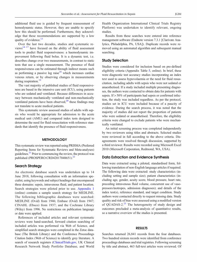
Searches returned 26,841 records from the four databases.Two hundred sixteen records were identified from conferenceproceedings databases and trial registries. Following screeningby title and abstract, 463 full-text articles were reviewed. Of
Seccombe et al.: Assessment for Fluid Resuscitation in Sepsis JGIM

the 124 that were diagnostic test accuracy studies, 83 wereexcluded because the study population was ventilated. Four-teen articles were identified for inclusion.24–37 Figure 1 detailsthe selection process and reasons for exclusion.
Study Characteristics
The 14 included studies had a median sample size of 33 (range14–116) and were primarily single-centre. Three were pub-lished as a conference abstract and two were set outside theICU, both in emergency departments (ED).Nine studies based inclusion on the presence of a composite
definition of shock, most commonly referred to as acutecirculatory failure, defined by the presence of one or morehaemodynamic markers, clinical signs, and blood tests. Bloodpressure was the key driver for study inclusion. In 13 studies,patients met the inclusion criteria if they were hypotensivealone (Table 1). The most common reason for patient exclu-sion was an arrhythmia (including irregular heart rhythm),used in seven studies. Another excluded all patients with anyform of cardiac disease.
Patient Population
Eight studies exclusively included patients with sepsis. Twoauthors from the remaining six studies provided individualpatient data for septic patients. Of note, patients with an
admission diagnosis of heart failure were present in two stud-ies. The recording of preceding intravenous fluid use waslimited, mentioned in five studies. In two of these, patientsreceived a median of 4 L prior to participation, double theminimum initial fluid dose for sepsis according to the Surviv-ing Sepsis guidelines13 (Table 2).
Target Condition
The target condition for all studies was fluid responsiveness.However, its relationship to hypovolaemia was poorly articu-lated. Nine studies failed to use the term Bhypovolaemia^ atany point in the rationale, methods, or interpretation of find-ings (Table 1).
Reference Standard
Table 3 shows the significant heterogeneity in the referencestandard, the test used to determine the presence or absence offluid responsiveness. Only two studies used the same param-eter.32, 34 There was marked variation in the proportion ofpatients meeting the reference standard: median 50% (range17.4–65.4%).Two studies used static tests to identify fluid responsive-
ness, both from the Surviving Sepsis guidelines.13 The re-maining 12 used a dynamic test. One study used absolute (>10 mmHg) rise in non-invasive systolic blood pressure.
Figure 1 Flowchart of study selection. Legend: flowchart summarising study selection and inclusion processes in this systematic review,including the reasons for exclusion of all full-text articles that were reviewed.
Seccombe et al.: Assessment for Fluid Resuscitation in SepsisJGIM

Eleven studies used a relative rise in cardiac function; the mostcommonmeasurements were cardiac index and stroke volume(SV), used by four studies each. The most common measure-ment tool was echocardiography, used in eight studies.Of the 12 studies that used a dynamic test, all used a fluid
bolus. Nine gave a fixed 500 mL bolus. The other three gave aweight-based bolus that ranged between 490 and 2100 mL fora 70-kg patient. The rate of the bolus ranged between 15 and30 min, although one study gave 500 mL using a pressure bag
and noted a variable infusion rate. Five studies used crystal-loid, six used colloid, and one did not specify the type of fluid.
Index Tests
Five categories of index test (i.e. the diagnostic test underevaluation) were identified in the studies and are summarisedin Table 4. They included inferior vena cava (IVC) measure-ments, change following passive leg raise (PLR), change with
Table 1 Main Characteristics of Included Studies
Author Year Setting No. ofpatients
Patientpopulation
Primaryindex test(s)
Additionalindextest(s)
Referencestandard
Targetcondition
Hypovolaemiamentioned?
de Valk24 2014 ED,Netherlands
23* Shock (SBP < 90, SBP > 40less than normal, HR > 100,CRT > 2 s, or lactate > 2)
IVCCI None Rise in SBPafter fluidbolus
Fluidresponsiveness
Yes
Corl25 2017 ED/ICU,USA
55* ACF (SBP < 90/MAP < 65for > 30 min, UO< 0.5,HR> 120 for > 30 min, pH< 7.3, or lactate > 2)
IVCCI Change inIVCCI afterfluid and PLR,IVCDi/e
Rise in CIafter fluidbolus
Fluidresponsiveness
No
Muller26 2012 ICU, France 40 ACF (MAP< 65, UO< 0.5,tachycardia, mottled skin, orlactate > 2)
IVCCI E wavevelocity, LVOTVTI, E/A ratio,E/Ea ratio
Rise inLVOT VTIafter
Fluidresponsiveness
No
Preau27 2017 ICU, France 90 Sepsis and ACF (SBP < 90,SBP > 40 less than normal,UO < 0.5, HR > 100, ormottled skin)
IVCCI ±standardisedrespiration
IVCD, SVI Rise in SVIafter fluidbolus
Fluidresponsiveness
Yes
Lanspa28 2013 ED/ICU,USA
14 Sepsis and refractoryhypotension (SBP < 90 after> 20 mL/kg of IV fluid)
IVVCI, AoVV,and SVV
None Rise in CIafter fluidbolus
Fluidresponsiveness
No
Abodorra29
(Poster)2014 ICU, Egypt 40 Sepsis and ACF (undefined) IVCCI (after
fluid bolus)Change inIVCCI afterfluid
Rise inLVOT VTIafter fluidbolus
Fluidresponsiveness
No
Dutta30
(poster)2014 ED/ICU,
India116 Sepsis and hypotension
(undefined)Change in SVafter PLR
None Rise in SVafter fluidbolus
Fluidresponsiveness
Yes
Klarer31
(poster)2010 ICU,
Switzerland27 Hypotension (MAP<
60 mmHg) and/or reducedCI (CI < 2.7 L/min/m2)
Change in CI,SVI, and MAPafter PLR
None Rise in CIafter fluidbolus
Fluidresponsiveness
No
Preau32 2010 ICU, France 34 Sepsis or acute pancreatitisand ACF (SBP < 90, SBP> 40 less than normal, UO< 0.5, HR > 100, or mottledskin)
Change in SV,PP, and VFafter PLR
None Rise in SVafter fluidbolus
Fluidresponsiveness
Yes
Soubrier33 2007 ICU, France 32 Haemodynamic instability(SBP < 90, MAP < 75, SBP> 40 less than normal, UO< 0.5 over 3 h, HR > 100, ormottled skin)
PPV and SBPV± standardisedrespiration
None Rise in CIafter fluidbolus
Fluidresponsiveness
Yes
Preau34 2012 ICU, France 23 ACF (SBP < 90, SBP > 40less than normal, UO < 0.5for > 1 h, HR > 100, ormottled skin)
PPVand VFV ±standardisedrespiration
None Rise in SVafter fluidbolus
Fluidresponsiveness
No
Jung35 2012 ED, SouthKorea
26 Sepsis and hypotension(SBP < 90, MAP < 70, SBP> 40 less than normal in theabsence of another cause)
FTc CVP, IVCD Rise in SVafter fluidbolus
Fluidresponsiveness
No
Keller36 2009 ICU, USA 44 Any admission to ICU witha plan to insert a CVC
IJV aspect ratio None CVP <8 mmHg
Fluidresponsiveness
No
Soliman37 2017 ICU, Egypt 30 Sepsis and hypotension(MAP < 65) or impairedtissue perfusion (lactate > 4)
Change in COafter fluid
None MAP > 65and lactate <4
Fluidresponsiveness
No
Summary of the 14 included studies and illustrating the wide heterogeneity. Primary index tests are those mentioned in the study’s aim.ACF acute circulatory failure, AoVV aortic velocity variation, CI cardiac index, CO cardiac output, CRT capillary refill time, CVP central venouspressure, ED emergency department, FTc corrected flow time, HR heart rate, ICU intensive care unit, IJV internal jugular vein, IVCCI inferior venacava collapsibility index, IVCDi/e end-inspiratory/expiratory inferior vena cava diameter, LVOT VTI left ventricular outflow tract velocity time integral,MAP mean arterial pressure, PLR passive leg raise, PP pulse pressure, PPV pulse pressure variation, SBP systolic blood pressure, SBPV systolic bloodpressure variation, SV stroke volume, SVI stroke volume index, SVV stroke volume variation, UO urine output, VF femoral artery velocity, VFV femoralartery velocity variation*Individual patient data
Seccombe et al.: Assessment for Fluid Resuscitation in Sepsis JGIM

respiration, change following intravenous fluid, and staticmeasurements.Six studies assessed inferior vena cava collapsibility index
(IVCCI), calculated using the difference in IVC diameterduring respiration divided by end-expiratory IVC diameter.Four studies measured IVCCI before a fluid bolus was givenand used a measure of left ventricular function to identify fluidresponsiveness. The area under the curve of the receiveroperating characteristic (AUROC) for IVCCI was similar foreach of these studies (0.77, 0.82, 0.82, 0.83). A fifth used
systolic blood pressure to identify fluid responsiveness, givingan AUROC of 0.68. The final study looked at IVCCI after afluid bolus was given and reported an AUROC of 0.91.Three studies, two published as conference abstracts only,
explored haemodynamic change after a PLR. Three separatestudies reported haemodynamic change during respiration. Anumber of different haemodynamic measurements were ex-plored in both categories with SVand pulse pressure the mostcommon. Two studies explored a static tool as part of theirprimary aim, although others included results for additional
Table 2 Main Patient Characteristics
Author Year Male(%)
Age(years)
MAP(mmHg)
HR (bpm) Diagnoses Additionaltreatment(e.g. inotropes)
Preceding IVfluid (L)
de Valk24* 2014 23 (48) 55 ± 18 75 ± 15 117 ± 8 Sepsis (100) – M 100 (Q 0–325)Corl25* 2017 23 (42) 68 ± 19 99 ± 19 115 ± 30 Sepsis (100) Vasopressors (58) M 4000 (Q 3350–6000)Muller26 2012 – M 63
(5P 56,95P 70)
M 71(5P 66,95P 77)
M 101(5P 91,95P 116)
Sepsis (60), bleeding (28),dehydration (13)
– –
Preau27 2017 58 (64) 55 ± 29 Unknown 102 ± 33 Sepsis (100) Vasopressors (16) (within 24 h) M 1000(0–2500)
Lanspa28 2013 5 (36) M 62(Q 46–81)
M 65 (Q 61–70)
M 102 (Q 80–112)
Sepsis (100) Vasopressors (57) M 4600 (Q 3000–5900)
Abodorra29 2014 – 54 ± 14 58 ± 12 108 ± 12 Sepsis (100) – –Dutta30 2014 – – Unknown Unknown Sepsis (100) – –Klarer31 2010 – M 60
(R 29–82)M 61(R 48–104)
M 104 (R 53–145)
Sepsis (52), heart failure (19),respiratory failure (15), other (14)
Vasopressors/inotropes (100)
–
Preau32 2010 19 (56) 53 ± 19 77 ± 14 101 ± 22 Sepsis (82), acute pancreatitis (18) Vasopressors (18) –Soubrier33 2007 9 (28) 61 ± 13 89 ± 14 103 ± 16 Sepsis (13), pneumonia (75),
haematological disease (3),trauma (6), abdominal surgery (3)
Vasopressors (9) 25% received IV fluid inpreceding 24 h
Preau34 2012 16 (70) 50 ± 5 79 ± 11 104 ± 19 Sepsis (87), acute pancreatitis (13) – –Jung35 2012 17 (65) M 74 (Q
58–83)M 57(Q 50–66)
94 (83–114) Sepsis (100) No –
Keller36 2009 22 (50) 66 ± 14 67 ± 12 92 ± 22 Sepsis (46), GI bleed (14), heartfailure (9), not recorded (32)
– –
Soliman37 2017 43.3 48 ± 20 53 ± 8 – Sepsis (100) Vasopressors (notrecorded)
–
Summary of the patient characteristics for the included studies. B–^ means data not available. Data are presented as means ± SD or as medians(indicated by BM^) with a measure of spread in brackets (preceded by BQ^ if quartiles, BR^ if range, and B5P^ or B95P^ if 5th and 95th percentilesrespectively). Number of patients, names of diagnoses, and use of vasopressors are presented with percentages in parenthesis.*Individual patient data
Table 3 Summary of Reference Standards
Author Year Met reference standard (%) Measurement Fluid bolus
Parameter Threshold rise (%) Measurement tool Volume Fluid type Rate (min)
de Valk24 2014 17.4 Systolic blood pressure > 10 mmHg NIBP 500 mL 0.9% saline 15Corl25 2017 56.4 Cardiac index > 10 Bioreactance 500 mL 0.9% saline Pressure bagMuller26 2012 50 LVOT VTI > 15 Echocardiography 500 mL 6% starch 15Preau27 2017 55.6 Stroke volume index > 10 Echocardiography 500 mL 4% gelatine 30Lanspa28 2013 35.7 Cardiac index > 15 Echocardiography 10 mL/kg Crystalloid < 20Abodorra29 2014 50 LVOT VTI > 15 Echocardiography 500 mL Not recorded 15Dutta30 2014 62.9 Stroke volume > 10 Echocardiography 30 mL/kg Crystalloid Not recordedKlarer31 2010 Not recorded Cardiac index > 15 Pulse contour analysis 500 mL 0.9% saline 15Preau32 2010 41.1 Stroke volume > 15 Echocardiography 500 mL 6% starch 30Soubrier33 2007 59.4 Cardiac index > 15 Echocardiography 500 mL 6% starch 20Preau34 2012 43.5 Stroke volume > 15 Echocardiography 500 mL 6% starch 30Jung35 2012 65.4 Stroke volume > 10 Oesophageal Doppler 7 mL/kg 6% starch 30Keller36 2009 59.1 Central venous pressure < 8 mmHg via central venous catheter N/A—static testSoliman37 2017 33.3 MAP < 65 mmHg or lactate < 4 mmol/L (measurement tool unclear) N/A—static test
Summary of the reference standards used by each study described according to the method of measuring the physiological parameter and, if a dynamicassessment tool was used, the means by which the fluid bolus was given.LVOT VTI left ventricular outflow tract velocity time integral, NIBP non-invasive blood pressure
Seccombe et al.: Assessment for Fluid Resuscitation in SepsisJGIM

index tests (Table 1). Finally, one study assessed haemody-namic change after a fluid bolus, the reference standard in 12of the other included studies.
Risk of Bias
Several factors contributed to a high overall risk of bias (seeFig. 2). Two studies used static measurements as referencestandards (central venous pressure35 and blood pressure/lactate levels36) whilst acknowledging they would not effec-tively identify fluid responsiveness. This would bias estimatesof sensitivity and specificity. The remaining 12 used fluidresponsiveness; however, none provided satisfactory evidenceto support their choice of reference standard.Twelve studies failed to report acceptable observer variation
data: nine provided no data for the index test, one reportedintra-observer variation only, one reported observer variationin a healthy cohort, and one reported inter-observer variationgreater than the index test’s threshold values. Twelve studiescalculated the index test’s optimal threshold using post hocanalysis with no a priori definition of a diagnostic cut-off. As aresult, in the six studies assessing IVCCI, thresholds rangedfrom > 15 to >50% (median > 37%). Finally, only five studiesreported the proportion of patients with uninterpretable results:three for echocardiography (11.5%, 12.8%, and 16.1%) andtwo for IVCCI (12.5%, 13.5%).
DISCUSSION
This is the first systematic review which explores diagnostictests that determine the need for fluid resuscitation in adults withsepsis who are notmechanically ventilated. Despite this, all buttwo studies were based solely in ICU as opposed to a ward orAMU, where the majority of medical patients are managed.The characteristics of included patients varied between
studies and were often poorly described. Six studies combinedseptic shock with shock arising from very different pathophys-iologies, and seven excluded patients with arrhythmia withoutjustification. This exclusion criterion is especially relevantgiven our ageing population and the increased prevalence ofarrhythmia in this cohort (up to 17% of adults > 80 years haveatrial fibrillation38).
The most common inclusion criterion was hypotension,which was enough to warrant inclusion by itself in 13 studies.All studies identified fluid responsiveness as the target condi-tion, which was universally seen as equivalent to a benefitfrom fluid resuscitation.Fluid responsiveness, the target condition in all studies, was
dynamically assessed in 12 studies. However, it was defined in11 different ways, including variations in the delivery of thefluid bolus, the threshold value, and the measurement tool. Adirect measure of cardiac function was used in 11 studies andwas assessed by echocardiography in eight studies.
Table 4 Summary of Studied Index Tests
Category of index test Author Year Primary index tests(measurement tool)
Threshold AUROC Sn Sp PPV NPV
Inferior vena cava de Valk24* 2014 IVCCI (US) ≥ 36.5% 0.68 (0.37–0.98) 75 57.9 27.3 97.7Corl25* 2017 IVCCI (US) ≥ 25% 0.82 (0.68–0.95) 83.9 79.2 83.9 79.2Muller26 2012 IVCCI (US) ≥ 40% 0.77 (0.60–0.88) 70 80 77.8 72.7Preau27 2017 IVCCI (US) ≥ 48% 0.82 (0.73–0.91) 76 88 88 75
IVCCI with standardised respiration (US) ≥ 31% 0.89 (0.82–0.97) 84 90 91 82Lanspa28 2013 IVCCI (US) ≥ 15% 0.83 (0.58–1.00) 100 67 62 100Abodorra29 2014 IVCCI after 100 mL (US) > 45% 0.91 90 65 72 88.7
PLR Dutta30 2014 Change in SV (Echo) ≥ 15% – 87.7 100 100 82.7Klarer31 2010 Change in CI (PC) > 15% – – – 50 86
Change in SVI (PC) > 15% – – – 20 77Change in MAP (PC) > 10% – – – 12 80
Preau32 2010 Change in SV (Echo) ≥ 10% 0.94 (0.90–0.98) 86 90 86 90Change in PP (PC) ≥ 9% 0.86 (0.78–0.94) 79 85 79 85Change in VF (Echo) ≥ 8% 0.93 (0.89–0.97) 86 80 75 89
Respiration Lanspa28 2013 AoVV (Echo) ≥ 25% 0.67 (0.32–1.00) 75 66.7 50 85.7SVV (PC) ≥ 17% 0.92 (0.73–1.00) 60 100 100 81.8
Soubrier33 2007 PPVa (PC) ≥ 12% 0.81 (0.73–0.89) 63 92 92 63SBPV (PC) ≥ 9% 0.82 (0.74–0.90) 47 92 90 54PPVa with standardised respiration (PC) ≥ 33% 0.72 (0.63–0.81) 21 92 80 44SBPV with standardised respiration (PC) ≥ 30% 0.69 (0.59–0.79) 26 92 80 83
Preau34 2012 PPVa (PC) ≥ 10% 0.71 (0.59–0.83) 60 100 100 76VFV (US) ≥ 10% 0.74 (0.63–0.85) 60 100 100 76PPVa with standardised respiration (PC) ≥ 12% 0.95 (0.90–1.00) 90 100 100 93VFV with standardised respiration (US) ≥ 12% 0.95 (0.90–1.00) 90 100 100 93
Static Jung35 2012 FTc (oesophageal Doppler) < 301 ms 0.87 (0.71–0.98) 88.2 88.8 93.7 79.9Keller36 2009 IJV aspect ratio (US) < 0.83 0.84 (0.72–0.96) 78 77 83 71
Fluid Soliman37 2017 Change in CO (bioimpedance)after 30 mL/kg 0.9% saline over 2 h
> 12.5% 0.9 90 70 80 90
Summary of the primary index tests for included studies. B–^ means data not available.AoVV aortic velocity variation, AUROC area under the curve of the receiver operating characteristic, CI cardiac index, CO cardiac output, FTccorrected flow time, IJV internal jugular vein, IVCCI inferior vena cava collapsibility index, MAP mean arterial pressure, NPV negative predictivevalue, PC pulse contour analysis, PLR passive leg raise, PP pulse pressure, PPV positive predictive value, PPVa pulse pressure variation, SBP systolicblood pressure, SBPV systolic blood pressure variation, Sn sensitivity, Sp specificity, SV stroke volume, SVI stroke volume index, SVV stroke volumevariation, US ultrasound, VF femoral artery velocity, VFV femoral artery velocity variation*Individual patient data
Seccombe et al.: Assessment for Fluid Resuscitation in Sepsis JGIM

Five categories of index test were described. However, thesmall size, risk of bias, and clinical heterogeneity of includedstudies prevented meaningful comparisons. Index test thresh-olds were chosen post hoc in all but two studies; 12 studiesfailed to report adequate observer variability data; and none ofthe included studies specified how their index test should beused in practice, i.e. replace, be added to, or triage for thecurrent diagnostic process. Comparisons were further limitedby the inadequate reporting of patient characteristics whichimpact upon the accuracy of the included index tests. Increasesin respiratory rate and tidal volume, frequently seen in criti-cally ill patients, have been shown to affect the IVCCI andrespiration variation measurements, for example.39
In summary, there was a small number of heterogeneousstudies available to guide the provision of intravenous fluid inadults with sepsis who are not mechanically ventilated. Thisheterogeneity, and a lack of consensus regarding associateddefinitions, is found throughout the literature.Given that fluid responsiveness is widely assumed to be the
most effective way of guiding fluid resuscitation, a singlemeasurement strategy with a pre-defined threshold would be
of great benefit to the medical community. Currently, however,the heterogeneity surrounding the definition of fluid respon-siveness limits our ability to learn from previous research andplan for future studies. Others have noted such heterogeneity40
and have recognised the need for a consensus definition.41
Cardiac output is widely regarded as the best means of deter-mining fluid responsiveness42 and is supported by Starling’sprinciples.43 However, its use makes the assumption that anincrease in cardiac output is always beneficial. In addition, theability of cardiac output rises to predict benefit from fluidresuscitation has never been tested against more commonlyused haemodynamic markers (e.g. blood pressure) and thereare concerns about the practicalities of monitoring cardiacoutput in a medical ward.Despite excluding mechanically ventilated patients, the el-
igibility criteria of this systematic review (Appendix Table 5,online) allowed inclusion of studies that are more limited intheir generalisability to an acute medical population. Moststudies were set in ICU andmany participants had been treatedfor days before inclusion. The absence of patients who are notcritically unwell would have artificially increased the
Figure 2 Risk of bias assessment. Legend: table summarising a risk of bias assessment performed using a modified version of QUADAS-2.23
B+^: low risk of bias; B?^: unclear risk of bias; B-^: high risk of bias.
Seccombe et al.: Assessment for Fluid Resuscitation in SepsisJGIM

sensitivity of all index tests.44 This risk was acknowledged inthe protocol22 after scoping identified a lack of evidenceoutside of ICU.The wide clinical heterogeneity meant statistical tests to
exclude publication bias were not feasible. However, the iden-tification of three unpublished conference proceedings reflectsa robust search strategy and suggests that the low number ofincluded studies is due to limited evidence rather than meth-odological shortcomings.It should also be noted that the diagnostic test accuracy
methodology itself limits the analysis of the index tests. Thismethodology assumes a dichotomous status for an individual,e.g. hypovolaemic or not hypovolaemic. In reality, a spectrumexists between euvolaemia and hypovolaemia. All describedindex tests provide the clinician with continuous data andtherefore would support a nuanced analysis. Simplifying mea-surement tools to provide a binary answer limits a clinician’sability to optimise fluid provision.This systematic review has highlighted multiple research
opportunities that should be pursued. Given the variety ofassumptions surrounding fluid responsiveness and its frequen-cy in the literature, it should be explored as a priority. Afterobserver variability data has been gathered, the proportion ofwell and acutely unwell adults who are fluid responsive shouldbe assessed using several haemodynamic measurements. Con-sideration should be given to the impact of factors such as age,disease severity, and comorbidities on these proportions. Toensure feasibility outside of ICU, non-invasive measurementtools should be used.Appropriately powered observational studies should then
examine potential associations between fluid responsivenessand commonly used outcome measures. Fluid responsivenesshas been noted in health and may simply be a marker ofcardiac function, and so should also be explored as a prognos-tic indicator. Each approach should be tested in differentaetiologies of shock.To support the integration of these diagnostic tests into
clinical practice, an understanding of the current decision-making process is needed, which will clarify the exact purposeof the chosen diagnostic test. Beyond this point, a randomisedcontrolled trial will be able to measure its impact in a clinicalsetting.
CONCLUSION
Intravenous fluid remains a central recommendation in guide-lines for the management of sepsis (including the SurvivingSepsis guidelines13 and NICE CG1745) but evidence to sup-port these recommendations is lacking. There is no consensusdefinition of hypovolaemia. While recent studies have usedfluid responsiveness to identify when fluid resuscitation isrequired, there is no agreement on which reference standardtest should define fluid responsiveness. Finally, if fluid respon-siveness is identified, there is no evidence to clarify how it
should be treated (i.e. which fluid, volume, and rate), orwhether treatment is indicated at all.While this systematic review highlights a lack of evidence,
it identifies research recommendations that, if met, wouldbuild an evidence base for the provision of fluids in acutelyunwell adults outside of ICU. Intravenous fluids are a commontreatment across all medical specialities. Prompt administra-tion can be life-saving, but excessive use is associated withpatient harm. Research in this field should, therefore, be apriority for the research community.
Acknowledgements: The authors would like to acknowledge SueBayliss (Information Specialist at the University of Birmingham, UK)who helped to develop the search strategies.
Corresponding Author: Adam Seccombe, MBChB; BirminghamAcute Care Research GroupUniversity of Birmingham, Birmingham,UK (e-mail: [email protected]).
Funders This study was not funded by grants from funding agenciesin the public, commercial or not-for-profit sectors. This research wassupported by the National Institute for Health Research (NIHR)Community Healthcare MedTech and In Vitro Diagnostics Co-operative at Oxford Health NHS Foundation Trust (through partfunding to Prof Lasserson). The views expressed are those of theauthors and not necessarily those of the NHS, the NIHR or theDepartment of Health and Social Care.
Compliance with Ethical Standards:
Conflict of Interest: The authors declare that they do not have aconflict of interest.
Open Access This article is distributed under the terms of theCreative Commons Attribution 4.0 International License (http://creativecommons.org/licenses/by/4.0/), which permits unrestricteduse, distribution, and reproduction in any medium, provided you giveappropriate credit to the original author(s) and the source, provide alink to the Creative Commons license, and indicate if changes weremade.
REFERENCES1. Nolan JP, Soar J. Advanced Life Support. 7th ed. UK: Resuscitation
Council; 2015.2. Seccombe A, Sapey E. What is the evidence base for fluid resuscitation
in acute medicine? Clin Med (Lond). 2018;18(3):225–230.3. Pacagnella RC, Souza JP, Durocher J, et al. A systematic review of the
relationship between blood loss and clinical signs. PLoS One.2013;8(3):e57594.
4. The UK Foundation Programme Office. The Foundation ProgrammeCurriculum. London: The UK Foundation Programme Office; 2016.
5. National Institute for Health and Care Excellence. Intravenous fluidtherapy in adults in hospital. CG174. London: NICE; 2013.
6. Royal College of Physicians. National Early Warning Score (NEWS):standardising the assessment of acute-illness severity in the NHS.London: RCP; 2012.
7. National Institute for Health and Care Excellence. Acutely ill adults inhospital: recognising and responding to deterioration. CG50. London:NICE; 2007.
8. Maitland K, Kiguli S, Opoka RO, et al. Mortality after fluid bolus inAfrican children with severe infection. N Engl J Med. 2011;364(26):2483–2495.
9. Vincent JL, Sakr Y, Sprung CL, et al. Sepsis in European intensive careunits: results of the SOAP study. Crit Care Med. 2006;34(2):344–353.
10. Marik PE, Linde-Zwirble WT, Bittner EA, Sahatjian J, Hansell D. Fluidadministration in severe sepsis and septic shock, patterns and outcomes:an analysis of a large national database. Intensive Care Med.2017;43(5):625–632.
Seccombe et al.: Assessment for Fluid Resuscitation in Sepsis JGIM

11. Macdonald SPJ, Taylor DM, Keijzers G, et al. REstricted FluidREsuscitation in Sepsis-associated Hypotension (REFRESH): studyprotocol for a pilot randomised controlled trial. Trials. 2017;18(1):399.
12. Hjortrup PB, Haase N, Bundgaard H, et al. Restricting volumes ofresuscitation fluid in adults with septic shock after initial management:the CLASSIC randomised, parallel-group, multicentre feasibility trial.Intensive Care Med. 2016;42(11):1695–1705.
13. Rhodes A, Evans LE, Alhazzani W, et al. Surviving Sepsis Campaign:International Guidelines for Management of Sepsis and Septic Shock:2016. Intensive Care Med. 2017;43(3):304–377.
14. Rameau A, de With E, Boerma EC. Passive leg raise testing effectivelyreduces fluid administration in septic shock after correction of non-compliance to test results. Ann Intensive Care. 2017;7(1):2.
15. Messina A, Longhini F, Coppo C, et al. Use of the Fluid Challenge inCritically Ill Adult Patients: A Systematic Review. Anesth Analg.2017;125(5):1532–1543.
16. Glassford NJ, Eastwood GM, Bellomo R. Physiological changes afterfluid bolus therapy in sepsis: a systematic review of contemporary data.Crit Care. 2014;18(6):696.
17. Barbier C, Loubieres Y, Schmit C, et al. Respiratory changes in inferiorvena cava diameter are helpful in predicting fluid responsiveness inventilated septic patients. Intensive Care Med. 2004;30(9):1740–1746.
18. Monnet X, Marik P, Teboul JL. Passive leg raising for predicting fluidresponsiveness: a systematic review and meta-analysis. Intensive CareMed. 2016;42(12):1935–1947.
19. Morgan BC, Martin WE, Hornbein TF, Crawford EW, Guntheroth WG.Hemodynamic effects of intermittent positive pressure respiration. Anes-thesiology. 1966;27(5):584–590.
20. Lamia B, Ochagavia A, Monnet X, Chemla D, Richard C, Teboul JL.Echocardiographic prediction of volume responsiveness in critically illpatients with spontaneously breathing activity. Intensive Care Med.2007;33(7):1125–1132.
21. Stewart LA, Clarke M, Rovers M, et al. Preferred Reporting Items forSystematic Review and Meta-Analyses of individual participant data: thePRISMA-IPD Statement. JAMA. 2015;313(16):1657–1665.
22. Seccombe A, McCluskey L, Moorey H, Sapey E. Diagnostic testaccuracy of assessments for intravenous fluid resuscitation in septicadults: a systematic review. 2017; PROSPERO: CRD42017048651:http://www.crd.york.ac.uk/PROSPERO/display_record.asp?ID=CRD42017048651. Accessed 20 May 2019
23. Whiting PF, Rutjes AW, Westwood ME, et al. QUADAS-2: a revised toolfor the quality assessment of diagnostic accuracy studies. Ann InternMed. 2011;155(8):529–536.
24. de Valk S, Olgers TJ, Holman M, Ismael F, Ligtenberg JJ, Ter MaatenJC. The caval index: an adequate non-invasive ultrasound parameter topredict fluid responsiveness in the emergency department? BMCAnesthesiol. 2014;14:114.
25. Corl KA, George NR, Romanoff J, et al. Inferior vena cava collapsibilitydetects fluid responsiveness among spontaneously breathing critically-illpatients. J Crit Care. 2017;41:130–137.
26. Muller L, Bobbia X, Toumi M, et al. Respiratory variations of inferiorvena cava diameter to predict fluid responsiveness in spontaneouslybreathing patients with acute circulatory failure: need for a cautious use.Crit Care. 2012;16(5):R188.
27. Preau S, Bortolotti P, Colling D, et al. Diagnostic Accuracy of theInferior Vena Cava Collapsibility to Predict Fluid Responsiveness inSpontaneously Breathing Patients With Sepsis and Acute CirculatoryFailure. Crit Care Med. 2017;45(3):e290-e297.
28. Lanspa MJ, Grissom CK, Hirshberg EL, Jones JP, Brown SM. Applyingdynamic parameters to predict hemodynamic response to volumeexpansion in spontaneously breathing patients with septic shock. Shock.2013;39(2):155–160.
29. Abodorra MEM, El Awady SM, Fayed AM, El Badawy TH. A comparisonbetween left ventricular outflow tract velocity time integral and inferiorvena cava collapsibility index as a predictor to fluid responsiveness incritically ill septic patients. Intensive Care Med. 2014;40 Suppl 1:202–203.
30. Dutta S, Chandrasekharan VP. Passive leg raising: An indicator to fluidresponsiveness in sepsis. Indian J Crit Care Med. 2014;18:S7.
31. Klarer A, Rudiger A, Maggiorini M. Passive leg raising detects patientswho will not benefit from fluid loading. Intensive Care Med.2010;36:S336.
32. Preau S, Saulnier F, Dewavrin F, Durocher A, Chagnon JL. Passive legraising is predictive of fluid responsiveness in spontaneously breathingpatients with severe sepsis or acute pancreatitis. Crit Care Med.2010;38(3):819–825.
33. Soubrier S, Saulnier F, Hubert H, et al. Can dynamic indicators help theprediction of fluid responsiveness in spontaneously breathing critically illpatients? Intensive Care Med. 2007;33(7):1117–1124.
34. Preau S, Dewavrin F, Soland V, et al. Hemodynamic changes during adeep inspiration maneuver predict fluid responsiveness in spontaneouslybreathing patients. Cardiol Res Pract. 2012;2012:191807.
35. Jung SM, Ryu S, Cho YC, et al. Validity of corrected flow time (FTC) as apredictor of fluid responsiveness in patients with sepsis-induced hypo-tension. Intensive Care Med. 2012;38:S200-S201.
36. Keller AS, Melamed R, Malinchoc M, John R, Tierney DM, Gajic O.Diagnostic accuracy of a simple ultrasound measurement to estimatecentral venous pressure in spontaneously breathing, critically ill patients.J Hosp Med. 2009;4(6):350–355.
37. Soliman R. Prediction of fluid status and survival by electricalcardiometry in acute circulatory failure. Eur Heart J Acute CardiovascCare. 2016;5:58–59.
38. Zoni-Berisso M, Lercari F, Carazza T, Domenicucci S. Epidemiology ofatrial fibrillation: European perspective. Clin Epidemiol. 2014;6:213–220.
39. Monnet X, Marik PE, Teboul JL. Prediction of fluid responsiveness: anupdate. Ann Intensive Care. 2016;6(1):111.
40. Cecconi M, Hofer C, Teboul JL, et al. Fluid challenges in intensive care:the FENICE study: A global inception cohort study. Intensive Care Med.2015;41(9):1529–1537.
41. Cherpanath TG, Aarts LP, Groeneveld JA, Geerts BF. Defining fluidresponsiveness: a guide to patient-tailored volume titration. JCardiothorac Vasc Anesth. 2014;28(3):745–754.
42. Marik PE. Fluid Responsiveness and the Six Guiding Principles of FluidResuscitation. Crit Care Med. 2016;44(10):1920–1922.
43. Starling EH, Visscher MB. The regulation of the energy output of theheart. J Physiol. 1926;62:243–261.
44. Reitsma JB, Rutjes AWS, Whiting P, Vlassov VV, Leeflang MMG,Deeks JJ. Chapter 9: Assessing methodological quality. In: Deeks JJ,Bossuyt PM, Gatsonis C, eds. Cochrane Handbook for SystematicReviews of Diagnostic Test Accuracy Version 1.0.0: The CochraneCollaboration; 2009.
Publisher’s Note Springer Nature remains neutral with regard tojurisdictional claims in published maps and institutional affiliations.
APPENDIX 1. SEARCH STRATEGY FOR MEDLINE
1. sepsis.ti,ab2. septic.ti,ab3. septicaemia.ti,ab4. septicemia.ti,ab5. mods.ti,ab6. multiple organ dysfunction syndrome.ti,ab7. mof.ti,ab8. multiple organ failure.ti,ab9. sirs.ti,ab10. systemic inflammatory response syndrome.ti,ab11. exp Systemic Inflammatory Response Syndrome/12. exp Multiple Organ Failure/13. OR/1-12 (196,454 on 7/2/17; Search repeated on 14/
6/18)14. fluid* ADJ3 replace*.ti,ab15. fluid* ADJ3 resuscitat*.ti,ab16. fluid* ADJ3 infus*.ti,ab17. fluid* ADJ3 administrat*.ti,ab18. fluid* ADJ3 restor*.ti,ab19. volume ADJ3 replace*.ti,ab20. volume ADJ3 resuscitat*.ti,ab21. volume ADJ3 infus*.ti,ab22. volume ADJ3 adminstrat*.ti,ab23. volume ADJ3 restor*.ti,ab
Seccombe et al.: Assessment for Fluid Resuscitation in SepsisJGIM

24. intravenous* ADJ3 fluid*.ti,ab25. IV fluid*.ti,ab26. colloid*.ti,ab27. crystalloid*.ti,ab28. hypertonic solution*.ti,ab29. hypertonic saline.ti,ab30. isotonic solution*.ti,ab31. isotonic saline.ti,ab32. ringer*.ti,ab33. hartman*.ti,ab34. albumin*.ti,ab35. gelatin*.ti,ab36. dextran*.ti,ab37. starch*.ti,ab38. exp Fluid Therapy/39. exp Plasma Substitutes/40. exp Infusions, Intravenous/41. exp Colloids/42. exp Hypertonic solutions/43. OR/14-42 (489,435 on 7/2/17; Search repeated on 14/
6/18)44. inpatient*.ti,ab45. in-patient*.ti,ab46. patient ADJ3 admiss*.ti,ab47. patient ADJ3 admit*.ti,ab48. hospital*.ti,ab49. intensive treatment unit*.ti,ab
50. ITU.ti,ab51. intensive care.ti,ab52. ICU.ti,ab53. critical care.ti,ab54. “accident and emergency”.ti,ab55. emergency department*.ti,ab56. emergency room*.ti,ab57. exp Inpatients/58. exp Hospitalization/59. exp Intensive Care Units/60. exp Critical Care/61. exp Emergency Service, Hospital/62. exp Hospital Departments/63. exp Internal Medicine/64. OR/44-63 (2,591,320 on 7/2/17; Search repeated on
14/6/18)65. AND/13,43,64 (4231 on 7/2/17; Search repeated on
14/6/18, 221 records between 2017 to Current)
APPENDIX 2
Table 5 Study Selection Criteria
Study design Diagnostic test accuracy studies
Participants Adults (aged ≥ 18 years) with sepsis of any severityor confirmed infection.Studies were excluded if they involved children,pregnant women, burns patients, trauma patients,perioperative patients, or patients who weremechanically ventilated.
Index test Any history question, examination technique, ordiagnostic test
Referencestandard
Any
Target condition Hypovolaemia or a need for fluid resuscitation
Seccombe et al.: Assessment for Fluid Resuscitation in Sepsis JGIM