A Pragmatic Neurological Screen for Patients With Suspected Cord Compressive Myelopathy
Upload
khangminh22Category
view
4download
0
1
http://dx.doi.org/10.5588/pha.21.0038
AMR PHA SUPPLEMENT
Antimicrobial resistance in neonates with suspected sepsis
S. K. Yadav,1 S. K. Agrawal,2 S. K. Singh,1 A. Giri,1 G. K. Singh,1 R. Ghimire,1 A. G. Stewart,3
K. L. Show,4 F. L. Moses5,6
1 Nobel Medical College and Teaching Hospital, Biratnagar, Nepal
2 B. P. Koirala Institute of Health Sciences, Dharan, Nepal
3 College of Life and Environmental Science, University of Exeter, Exeter, UK
4 Department of Medical Research, Yangon, Myanmar
5 Sierra Leone Ministry of Health and Sanitation, Freetown, Sierra Leone
6 College of Medicine and Allied Health Sciences, University of Sierra Leone, Freetown,
Sierra Leone
Correspondence to: Santosh Kumari Agrawal, Department of Public Health Dentistry, CODS,
B. P. Koirala Institute of Health Sciences, Dharan, Nepal. email:
Running title: AMR pattern and exit outcomes
Article submitted 21 April 2021. Final version accepted 8 June 2021.

2
SUMMARY
SETTING: Nobel Medical College and Teaching Hospital, Biratnagar, Nepal.
OBJECTIVE: To determine the pattern of antimicrobial resistance and hospital exit
outcomes in neonates with suspected sepsis in a tertiary neonatal intensive care unit (NICU).
DESIGN: This hospital-based cohort study was conducted to follow patients from January
to December 2019. All identified cases of suspected sepsis were enlisted from hospital records.
RESULTS: Sepsis was suspected in 177 (88%) of the 200 cases admitted in the NICU; 52
(29%) were culture-positive. Pseudomonas was the predominant organism isolated (n = 40;
78%), followed by coagulase negative staphylococcus (n = 12, 23%). Nine (17%) of the 52
isolates were resistant to the Access and Watch group of antibiotics, including some resistance
to Reserve group drugs such as imipenem and linezolid. Most treated cases (n = 170, 96%)
improved, although 7 (4%) left against medical advice.
CONCLUSION: Most of the pathogens were resistant to WHO Access and Watch
antibiotics and occasional resistance was observed to Reserve group drugs. Most sepsis was
caused by Gram-negative bacilli. Improving turnaround times for antibiotic sensitivity testing
using point-of-care testing, and a greater yield of culture-positive results are needed to enhance
the management of neonatal sepsis.
KEY WORDS: antimicrobial resistance; neonatal sepsis; NICU

3
Neonatal sepsis refers to generalised bacterial infection with systemic signs, and is diagnosed
using a positive blood culture in the first 4 weeks of life.1,2 It is classified by time of occurrence
of infection as early onset sepsis, i.e., bacteraemia in the first 72 h of life, and thereafter, as late
onset sepsis.3
It is a life-threating illness and a major cause of neonatal morbidity and mortality
worldwide.3 Global incidence is estimated to be 1.3 million cases annually, resulting in about
203,000 attributable deaths. Estimates that 2,202 neonates per 100,000 live births develop
neonatal sepsis globally, with a mortality rate of 11–19% (242–418 deaths/100,000 live births),
are derived from high-income country data.4 However, low- and middle-income countries,
where data are limited, are most affected due to the frequency of infections and limited access
to good healthcare.
Due to the non-specific nature of the signs and symptoms in the new-born, most cases
of sepsis are clinically diagnosed, then treated with empirical broad-spectrum antibiotics. This
use of antibiotics promotes antimicrobial resistance (AMR). Bedside tests are not available and
empirical antibiotic treatment contributes to the increased prevalence of antibiotic-resistant
organisms. Examples include methicillin-resistant Staphylococcus aureus, vancomycin-
resistant Enterococcus and multidrug-resistant Gram-negative rods.5,6 At least 700,000 deaths
occur annually due to drug-resistant diseases, which is projected to rise to more than 10 million
deaths globally by 2050 if action is not taken.7
The spectrum of multidrug-resistant bacteria varies. In neonates, sepsis <48 h after birth
is most often caused by Group B streptococcus (43%) and Escherichia coli (16–29%).8,9 For
very low birth weight infants with early onset sepsis, the E. coli rate exceeds that of Group B
streptococcus (5.1 vs. 2.1/1,000 live births).9 Late onset sepsis is mainly caused by Gram-
positive bacteria (49%), most often coagulase-negative Staphylococcus (45%). Gram-negative
late onset sepsis is less common (23%), but is associated with greater mortality in neonatal
intensive care units (NICUs) (19–36%).9 Both forms of sepsis are related to birth weight and
the location where the infection was acquired.10
The neonatal mortality rate in Nepal is 21/1,000 live births.11 Neonatal infection is the
third most common cause of neonatal mortality (17%; 3.6/1,000 live births), after preterm birth
complications (33%) and intrapartum-related events (23%).11
In Nepal, 10–42% of all the patients are prescribed antibiotics for therapeutic or
prophylactic purposes without bacterial confirmation or susceptibility testing. Further data on
AMR in Nepal are limited, possibly due to a lack of prioritisation and the absence of routine

4
surveillance.12 An appropriate antimicrobial agent should be used for sepsis to prevent the
development of multidrug resistance and reduce morbidity and mortality.13
Nepal has significantly more multidrug-resistant bacterial strains closer to health care
sites.12 In one tertiary hospital, coagulase-negative Staphylococcus was the predominant Gram-
positive organism isolated from both early and late onset sepsis cases (46.6%), followed by S.
aureus (14.6%). Other Gram-negative multidrug-resistant bacteria were associated with early
onset sepsis: Acinetobacter species (9.5%) and Klebsiella pneumoniae (7.7%).1
Antibiotics used in Nepali hospitals were found to be effective in curing only 50% of
infections.14 Furthermore, AMR contributes to increased treatment costs, hospital stay,
morbidity and mortality.15
In Nobel Medical College and Teaching Hospital (NMCTH), Biratnagar, Nepal, a
public-private partnership tertiary care referral centre, sepsis is the third most common cause
of admission in the NICU, after asphyxia and meconium aspiration.16 However, there is no
information about the AMR pattern, particularly in relation to patient outcomes. In the present
study, we described the bacterial profile and determined the antimicrobial resistance pattern
and hospital exit outcomes in neonates with suspected sepsis in the NMCTH NICU.
METHODS
Study design
This was a hospital-based, cohort study conducted from January to December 2019.
General setting
Nepal, in South Asia, is mainly located in the Himalayas, with an estimated population of 30.2
million.17 NMCTH is in Biratnagar, the second most densely populated and third most populous
city of Nepal, with a population of 242,548 (2011 census;
https://unstats.un.org/unsD/demographic/sources/census/wphc/Nepal/Nepal-Census-2011-
Vol1.pdf).
Nepal is moving towards an integrated response to AMR. The National Action Plan, led
by the AMR Steering Committee at the Curative Division of Ministry of Health and Population,
recognises the National Public Health Laboratory (under the Department of Health Services) as
the focal point for AMR activities.
Specific setting

5
NMCTH, affiliated to Kathmandu University, has 911 beds, <40,000 admissions per year and
a bed occupancy rate of ~80%. The average outpatient flow is ~1000/day. The NICU is well
equipped, with 17 beds, a nursing staff: patient ratio of 1:2–3 and the highest volume referral
for tertiary care in eastern Nepal.
Laboratory testing
Blood samples from the neonatal ward/emergency department were collected in brain heart
infusion (BHI) broth (1 ml blood in 5 ml of BHI broth). After 24 h culture, the sample was sub-
cultured onto blood and MacConkey agar (aerobic, 35oC) and chocolate agar (anaerobic). Any
organism grown was identified using standard biochemical tests, followed by antimicrobial
susceptibility testing (Clinical and Laboratory Standard Institute guidelines).18 The timing of
the reporting of culture results was noted (standard: within 24–48 h), as was timing of the
reporting of the resistance pattern (standard: <72 h).
Study population/participants
All neonates admitted to the NICU of NMCTH, Biratnagar, Nepal, between 1 January and 31
December 2019 were ascertained from hospital records. We identified cases of suspected sepsis
using criteria adapted from All India Institute of Medical Sciences Protocols in Neonatology
(Table 1).19
Sources of data and data collection
From the clinical and laboratory records and discharge summary, we collected
sociodemographic characteristics, risk factors associated with sepsis, microorganism growth
and antibiotics susceptibility patterns, and hospital exit outcome in relation to septicaemia
treatment using a structured pro forma.
Data validation
All data were double-entered and validated using EpiData database v3.1 for entry (EpiData
Association, Odense, Denmark). Data were analysed using SPSS v21 (IBM SPSS, Armonk,
NY, USA).
Analysis and statistics
The number and proportion of neonates admitted for suspected septicaemia and with samples
sent for blood culture were calculated, along with median and interquartile ranges for the time

6
taken for laboratory procedure, timing of changes in regimen from the issuing of the laboratory
report and duration of empirical treatment. Poisson regression modelled any association
between the demographic and clinical characteristics, hospital exit outcome, potential
confounders and antibiotic resistance. The level of significance was set at P < 0.05.
Ethics approval
Permission for the study was obtained from Institutional Review Committee (IRC), Nobel
Medical College and Teaching Hospital (NMCTH) (Ref: IRC-NMCTH 162/076 of
29/01/2020). The study was also approved by the Ethics Advisory Group of the International
Union Against Tuberculosis and Lung Disease, Paris, France (EAG number: 08/20, date 2020-
02-05). As this was a record review with no patient identifiers, the issue of informed patient
consent did not apply.
RESULTS
Social and clinical characteristics
In total, 200 relevant cases were identified. Around two thirds of these cases were inborn (n =
146, 73.0%), male (n = 130, 65.0%), born at full-term gestation (n = 140, 70.0%) and with
normal birth weight (n = 141, 70.5%).
Sepsis
Sepsis was clinically suspected in 177 (88.5%) cases. More than two thirds of them presented
with features of early onset sepsis, while many also had a range of comorbidities. Most of the
sepsis cases were inborn, admitted within first week of life, males, born at term or of normal
birth weight (Table 2). Among inborn neonates, early onset sepsis (n = 100, 88.7%) was more
common, whereas late onset sepsis (n = 38, 71.7%) was more common in outborn neonates.
Out of the total samples tested (n = 177), 52 (29.3%) were culture-positive, of which in
36 (69.2%) the empirical antibiotic regimen was revised as per blood culture sensitivity pattern.
Almost all cases admitted with sepsis improved with the given regimen (Table 2). Gram-
negative bacterial infection was the main cause of septicaemia among neonates aged 1 week at
admission, predominantly affecting males, inborn babies and those with low birth weight.
Gram-negative sepsis was more common in babies of all gestational ages, and was most
common in both early and late onset neonatal sepsis (Table 3).
Antimicrobial resistance

7
Pseudomonas predominated, followed by coagulase-negative Staphylococcus (Table 4).
Overall, 9 (17.0%) culture-positive cases of septicaemia were resistant to the AWaRe (Access,
Warch, Reserve) group of antibiotics. The most common isolate was sensitive to Access and
Watch group antibiotics, whereas occasional resistance was observed with the Reserve group
of drugs (Table 5).
Turnaround times
The median turnaround time for laboratory culture was 3 days (range 2.0–4.0). The median
time to change empirical therapy after a positive culture and sensitivity report was 3 days (range
3.0–6.0).
Associations with culture-proven sepsis
Of culture-positive cases, two characteristics showed a statistically significant effect on positive
culture results: presence of comorbidities reduced the risk of a positive culture (adjusted relative
risk [aRR] 0.23, 95% confidence interval [CI] 0.09–0.57), while being post-term increased the
risk (aRR 1.33, 95% CI 1.05–1.69) (Table 6). None of the factors which affected the risk of a
positive culture were significantly associated with single drug resistance.
Hospital exit outcomes
Of culture-positive cases, 94.2% (n = 49) improved, but 3 (5.8%) left against medical advice.
DISCUSSION
Demographic and clinical characteristics
In a tertiary NICU in Nepal, over 88% of cases were diagnosed with clinically suspected sepsis,
of which two thirds presented with features of early onset sepsis. However, under one third had
culture-proven septicaemia, mainly caused by Gram-negative bacteria. Risk factors for sepsis
were not identified; AMR was noted but not statistically associated with risk factors or exit
outcome of the neonate from NICU. The presence of comorbidities was associated with a
reduced risk of being culture-positive.
Neonatal sepsis
Clinical sepsis was confirmed in under one third of neonates. Our confirmation rate is in the
middle of other studies in Nepal and elsewhere.20–25 Empirical treatment may have started
before blood was taken for culture or before admission to the NICU. Early use of antibiotics

8
before taking blood for culture is known to lead to sterile culture reports. This needs further
investigation, possible remedial action within the NICU and obstetric unit and consideration if
the integrated preventive and protective measures against infections have failed, and why, or if
there are other factors influencing the high rate of unconfirmed sepsis.
Among suspected sepsis cases, more than two thirds presented with features of early
onset sepsis, which is higher than other nearby reports.24–26 Probable explanations include 1)
most of our new-borns were inborn with subsequent early identification of clinical sepsis and
prompt admission to NICU; and 2) the causative agents for early onset sepsis were acquired
from the mother during birth.1 Further investigation into the possible acquisition of sepsis at
birth is needed to protect vulnerable new-borns.
The predominance of male new-borns has been reported elsewhere.2,24,27,28 This may be
due to the prevalent societal preference given to males in seeking medical care.26 However,
males may also be vulnerable to neonatal sepsis due to an X-linked immune regulatory gene
factor.29 With improved confirmation of sepsis, it would be possible to investigate this sex
difference and begin to counter any bias against female infants.
We found that comorbidities (meconium aspiration syndrome, bowel obstruction, etc.)
significantly reduce the risk of having a positive culture. However, there was a lot of missing
data, possibly biasing this unexpected finding. Comorbidities could be expected to predispose
to the overuse of broad-spectrum antibiotics, leading to sterile cultures. In addition, NMCTH
receives referral cases, many with comorbidities and previous treatment, from different parts of
Nepal.
There are at least two approaches to sterile cultures. First, any use of empirical
antibiotics needs to be kept to a minimum by more rapid turnaround times from the laboratory,
or (preferably) quick and accurate bedside point-of-use diagnostic tests for sepsis, thereby
reducing the need for empirical treatment, as in other infections.30,31 Second, enhanced
professional and continuing education of all health care providers, wherever situated, would
ensure proper understanding and awareness regarding antimicrobials, including the collection
of blood samples for culture before starting empirical therapy.
Antimicrobial resistance profile
Gram-negative organisms are the most common agent causing neonatal sepsis,2,25,32 as we were
able to confirm (Table 4). However, few studies have reported Gram-positive bacterial
septicaemia among neonates in the hospital setting. Among these, methicillin-resistant S.

9
aureus was a critically important pathogen in the NICU population, associated with both
endemic and epidemic infections.20,28
Host and bacterial properties unite to cause sepsis in new-borns. The organism varies
according to the environmental conditions of the hospital, e.g., contamination during sampling,
care of both in-born and out-born cases, and surgical cases mixed with medical cases.26
Contamination during sampling or during birth from the mother’s birth canal could
account for coagulase-negative Staphylococcus. The latter is more life-threatening, and further
investigation to distinguish between the two sources is important. In our study, the
predominance of Pseudomonas suggests transmission of infection from healthcare workers to
patients via contaminated hands. Accordingly, re-enforcement of infection control guidelines
and education of healthcare staff are important. Our study further prompts the need for the
implementation of the WHO AMR action plan in Nepal.
The most common antibiotic resistance was in Pseudomonas, unlike a previous report
from Nepal,26 which showed that Gram-negative organisms were resistant to ampicillin. Other
isolates in our study were, not surprisingly, resistant to first-line and second-line therapy. A
potential way to curb this would be increased and timely surveillance of antibiotic sensitivity
patterns and revision of policies regarding empirical therapy based on AMR, along with the
implementation of the aforementioned point-of-use diagnostic testing for sepsis.
Turnaround time for samples and empirical therapy
The longer the administration of broad-spectrum antibiotics, the greater the progression of
severe sepsis to septic shock, with increased mortality.33 The median turnaround time taken for
laboratory culture plus the median time to changing to an appropriate antibiotic regime means
that it is possible for a septic neonate to wait ≥6 days for a necessary change to appropriate
treatment. This again indicates the need for rapid diagnostic tests.
Strengths
The study is one of the few which report AMR and the turnaround time for the reporting of
culture results and consequent change in antibiotic management.
Limitations
The small numbers of positive blood cultures meant that none of the risks likely to be associated
with resistance to at least one antibiotic or to a hospital exit outcome were fully assessed.

10
Second, as critically ill patients who left against medical advice were likely to have died, our
estimate of the mortality rate may have been an underestimate.
CONCLUSION
Pseudomonas was the most common isolate in culture-proven sepsis. Gram-negative bacilli
were susceptible to Access and Watch group antibiotics, whereas, occasional resistance was
observed with last resort (Reserve) antibiotics. Rapid diagnostic tools for sepsis, which would
also identify AMR and conform to accepted standards and guidelines, are urgently needed in
NICUs to enable the antibiotic regime to be appropriate from the earliest time possible.
As sepsis is a major health risk for neonates, a standardised national and global
surveillance programme collecting neonatal AMR data, including risk factors and clinical
outcomes, is critical. This would enable benchmarking, the identification of remedial risks, the
design and implementation of successful interventions, and allow review of NICU programmes.
Acknowledgements
This research was conducted through the Structured Operational Research and Training
Initiative (SORT IT), a global partnership coordinated by Tropical Disease Research (TDR),
the Special Programme for Research and Training in Tropical Diseases at the WHO. The
specific SORT IT programme that led to these publications included a partnership of TDR with
the WHO Country Office of Nepal and was implemented along with the Tuberculosis Research
and Prevention Centre Non-Governmental Organisation, Yerevan, Armenia; the International
Union Against Tuberculosis and Lung Diseases, Paris, France, and South East Asia, New Delhi,
India offices; the Damien Foundation, Brussels, Belgium; the Narotam Sekhsaria Foundation,
Mumbai, India; Sustainable Health Systems, Freetown, Sierra Leone; the Ministry of Health
and Sanitation, Freetown, Sierra Leone; School of Public Health and Community Medicine, B
P Koirala Institute of Health Sciences, Dharan, Nepal; the Institute of Medical Research,
Bangalore, India; University of Exeter, Exeter, UK; and the University of Washington, Seattle,
WA, USA. Special thanks to the data collector, the staff, and the hospital management team of
the Dhulikhel Hospital, Kathmandu, Nepal, for their help in conducting this study.
This SORT IT AMR Programme was funded by the National Institute of Health
Research, Department of Health & Social Care of the United Kingdom and supported by
implementing partners.
Conflict of interests: none declared.

11
Open access statement and disclaimer: In accordance with WHO’s open-access
publication policy for all work funded by WHO or authored/co-authored by WHO staff
members, WHO retains the copyright of this publication through a Creative Commons
Attribution IGO license (http://creativecommons.org/licenses/by/3.0/igo/legalcode) which
permits unrestricted use, distribution and reproduction in any medium provided the original
work is properly cited.
There should be no suggestion that WHO endorses any specific organization, products
or services. The views expressed in this article are those of the authors and do not necessarily
reflect those of their affiliated institutions. The use of the WHO logo is not permitted. This
notice should be preserved along with the article’s original URL.
Data management statement: The metadata record of the data used in this paper is
available at DOI https://doi.org/10.6084/m9.figshare.14315438. Requests to access these data
should be sent to the corresponding author.

12
References
1 Ansari S, et al. Neonatal septicemia in Nepal: early-onset versus late-onset. Int J Pediatr
2015; 2015: 379806.
2 Shrestha S, Shrestha A. A study of clinico-pathological profile of suspected and
confirmed neonatal sepsis at Kathmandu Medical College. Nepal Med Coll J 2020; 22:
82–87.
3 Simonsen KA, et al. Early-onset neonatal sepsis. Clin Microbiol Rev 2014; 27(1): 21–
47.
4 Fleischmann-Struzek C, et al. The global burden of paediatric and neonatal sepsis: a
systematic review. Lancet Respir Med 2018; 6: 223–230. .
5 Folgori L, et al. Antimicrobial-resistant Gram-negative infections in neonates: burden
of disease and challenges in treatment. Curr Opin Infect Dis 2017; 30: 281–288.
6 Sawhney N, Shinu P, Singh VA. Bacteriological profile and antibiotic susceptibility
pattern of neonatal septicaemia in a tertiary Care Hospital. Int J Curr Microbiol App Sci
2015; 4: 977–984.
7 World Health Organization. Antimicrobial resistance. No time to wait: securing the
future from drug-resistant infections. Report to the Secretary-General of the United
Nations. Geneva, Switzerland: WHO, 2019. https://www.who.int/antimicrobial-
resistance/interagency-coordination-group/final-report/en/ Accessed April 2021.
8 Tzialla C, et al. Antimicrobial therapy in neonatal intensive care unit. Ital J Pediatr 2015;
41: 27.
9 Stoll BJ, et al. Early onset neonatal sepsis: the burden of group B Streptococcal and E.
coli disease continues. Pediatrics 2011; 127: 817–826.
10 Dong Y, Speer CP. Late-onset neonatal sepsis: recent developments. Arch Dis Child
Fetal Neonatal Ed 2015; 100: F257–263.
11 Ministry of Health, Nepal; New ERA; and ICF. Nepal Demographic and Health Survey,
2016. Kathmandu, Nepal: Ministry of Health, Nepal, 2017.
https://www.dhsprogram.com/pubs/pdf/fr336/fr336.pdf Accessed April 2021.
12 Acharya KP, Wilson RT. Antimicrobial resistance in Nepal: a review. Front Med
(Lausanne) 2019; 6: 105.
13 Shrestha R, et al. Bacteriological study of neonatal sepsis and antibiotic susceptibility
pattern of isolates in Kathmandu, Nepal. Nepal Med Coll J 2013; 15: 71–73.
14 Ghimire S, et al. A prospective surveillance of drug prescribing and dispensing in a
teaching hospital in western Nepal. J Pak Med Assoc 2009; 59: 726–731.

13
15 Kafle K, Pokhrel B. Antimicrobial resistance at different levels of health-care services
in Nepal. Regional Health Forum 2011; 15(1): 9–17.
16 Shah L, et al. Clinical profile and outcome of neonates admitted to neonatal intensive
care unit (NICU) at a tertiary care centre in eastern Nepal. J Nepal Paediatr Soc 2013;
33: 177–181.
17 Government of Nepal. National Planning Commission Central Bureau of Statistics.
National Census, 2021. https://censusnepal.cbs.gov.np/Home/Index/EN Accessed
March 2021.
18 Clinical and Laboratory Standards Institute. CLSI Performance standards for
antimicrobial susceptibility testing. 23rd ed. Wayne, PA, USA: CLSI, 2017.
https://clsi.org/standards/products/microbiology/documents/m100/ Accessed March
2021.
19 Agarwal R, Deorari A, Paul VK. AIIMS protocols in Neonatology. New Delhi, India:
CBS Publishers & Distributors, 2015.
20 Kanodia P, et al. Bacteriological profile of blood culture positive sepsis in newborn at
BPKIHS, Dharan Nepal. J Coll Med Sci Nepal 2017; 13: 193–196.
21 Shakya H, Lakhey A. Role of sepsis screening in early diagnosis of neonatal sepsis. J
Pathol Nepal 2017; 7: 1103–1110.
22 Yadav SK, Giri A. Bacteriological profile of neonatal sepsis in a neonatal intensive care
unit of a tertiary care hospital of Eastern Nepal. J Coll Med Sci Nepal 2019; 15: 93–97.
23 Sorsa A, et al. Blood culture result profile and antimicrobial resistance pattern: a report
from neonatal intensive care unit (NICU), Asella teaching and referral hospital, Asella,
south East Ethiopia. Antimicrob Resist Infect Control 2019; 8: 42.
24 Bhattarai S, et al. Bacteriological profile and antibiotic sensitivity pattern of neonatal
sepsis in central paediatric referral hospital in Nepal. J Nepal Paediatric Soc 2019; 39:
1–5.
25 Jatsho J, et al. Clinical and bacteriological profile of neonatal sepsis: a prospective
hospital-based study. Int J Pediatr 2020; 2020: 1835945.
26 Pandit BR, Vyas A. Clinical symptoms, pathogen spectrum, risk factors and
antibiogram of suspected neonatal sepsis cases in tertiary care hospital of southern part
of Nepal: A descriptive cross-sectional study. J Nepal Med Assoc 2020; 58(32): 976–
982.

14
27 Nepal D, et al. Bacteriological profile and antibiotic susceptibility pattern of neonatal
septicemia in Kanti Children Hospital, Nepal. J Gandaki Med Coll Nepal 2020; 13: 97–
103.
28 Shaikh M, et al. Spectrum and Spectrum and antimicrobial susceptibility pattern of
micro-organisms associated with neonatal sepsis in a hospital in Karachi, Pakistan.
Cureus 2020; 12: e10924.
29 Chandna A, et al. Rapid diagnostic tests in neonatal septicemia. Indian J Pediatr 1988;
55: 947–953.
30 Garcia-Basteiro A, et al. Point of care diagnostics for tuberculosis. Pulmonology 2017;
24(2): 73–85.
31 Drain PK, Heichman KA, Wilson D. A new point-of-care test to diagnose tuberculosis.
Lancet Infect Dis 2019; 19: 794–796.
32 Basnyat B, et al. Antibiotic use, its resistance in Nepal and recommendations for action:
a situation analysis. J Nepal Health Res Counc 2015; 13(30): 102–111.
33 Whiles BB, Deis AS, Simpson SQ. Increased time to initial antimicrobial administration
is associated with progression to septic shock in severe sepsis patients. Crit Care Med
2017; 45: 623–629.
34 World Health Organization. Essential medicines and health products. WHO releases the
2019 AWaRe Classification Antibiotics. Geneva, Switzerland: WHO, 2019.
https://www.who.int/medicines/news/2019/WHO_releases2019AWaRe_classification
_antibiotics/en/ Accessed March 2021.

15
Table 1 Definition of neonatal suspected sepsis
Clinically suspected neonatal sepsis: neonates with clinically suspected sepsis having a
constellation of signs and symptoms that can include fever, poor feeding, respiratory
distress, cyanosis, cold clammy skin, tachycardia, seizures, hyperreflexia, jaundice and
temperature or blood pressure instability; all neonates in whom sepsis was suspected
were included in the study
Confirmed neonatal sepsis: defined as the presence of a clinically significant pathogen
isolated from blood cultures with associated signs and symptoms
16
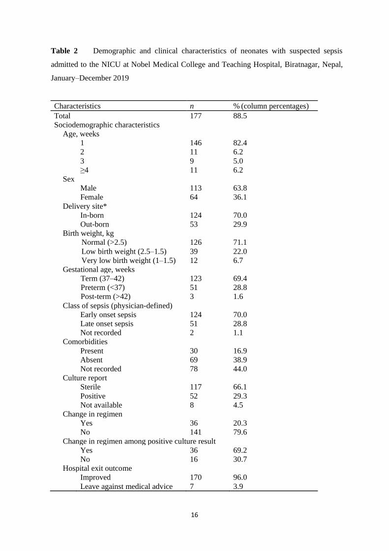
Table 2 Demographic and clinical characteristics of neonates with suspected sepsis
admitted to the NICU at Nobel Medical College and Teaching Hospital, Biratnagar, Nepal,
January–December 2019
Characteristics n % (column percentages)
Total 177 88.5
Sociodemographic characteristics
Age, weeks
1 146 82.4
2 11 6.2
3 9 5.0
≥4 11 6.2
Sex
Male 113 63.8
Female 64 36.1
Delivery site*
In-born 124 70.0
Out-born 53 29.9
Birth weight, kg
Normal (>2.5) 126 71.1
Low birth weight (2.5–1.5) 39 22.0
Very low birth weight (1–1.5) 12 6.7
Gestational age, weeks
Term (37–42) 123 69.4
Preterm (<37) 51 28.8
Post-term (>42) 3 1.6
Class of sepsis (physician-defined)
Early onset sepsis 124 70.0
Late onset sepsis 51 28.8
Not recorded 2 1.1
Comorbidities
Present 30 16.9
Absent 69 38.9
Not recorded 78 44.0
Culture report
Sterile 117 66.1
Positive 52 29.3
Not available 8 4.5
Change in regimen
Yes 36 20.3
No 141 79.6
Change in regimen among positive culture result
Yes 36 69.2
No 16 30.7
Hospital exit outcome
Improved 170 96.0
Leave against medical advice 7 3.9

17
* Patients admitted from another hospital, primary health care centre, health posts and home
deliveries from same or different province.
NICU = neonatal intensive care unit.

18
Table 3 Demographic and clinical characteristic according to culture positivity in
neonates with suspected sepsis admitted to the NICU at Nobel Medical College and
Teaching Hospital, Biratnagar, Nepal, January–December 2019
Total Gram positive bacteria Gram negative bacteria
N n % (Row percentage) n % (Row percentage)
Total 52 7 13.4 45 86.5
Age, weeks
1 45 6 13.3 39 86.6
2 3 0 0.0 3 100
3 2 1 50.0 1 50.0
≥4 2 0 0.0 2 100
Sex
Male 38 2 5.2 36 94.7
Female 14 5 35.7 9 64.2
Delivery site
Inborn 40 6 15.0 34 85.0
Outborn 12 1 8.3 11 91.6
Birth weight
Low birth weight (2.5–1.5) 6 2 33.3 4 66.6
Very low birth weight (1–1.5) 4 2 50.0 2 50.0
Normal 42 3 7.1 39 92.8
Gestation age, weeks
Preterm (<37) 12 3 25.0 9 75.0
Term (37–42) 38 4 10.5 34 89.4
Post-term (>42) 2 0 0.0 2 100
Class of sepsis
Early onset sepsis 39 5 12.8 34 87.1
Late onset sepsis 13 2 15.3 11 84.6
Not recorded
Comorbidities
Present 4 1 25.0 3 75.0
Absent 45 6 13.3 39 86.6
Not recorded 3 0 0.0 3 100
* Patients admitted from another hospital, primary health care centre, health posts and home
deliveries from same or different province.
NICU = neonatal intensive care unit.

19
Table 4 Culture-positive pattern among neonates with suspected sepsis admitted to the
NICU at Nobel Medical College and Teaching Hospital, Biratnagar, Nepal, January–
December 2019
Organisms (n = 71)
Culture-positive
n %
Pseudomonas 40 78.4
Coagulase negative staphylococcus 12 23.0
Klebsiella 5 9.8
Citrobacter 5 9.8
Escherichia coli 4 7.8
Acinetobacter species 2 3.9
Enterococcus 3 5.8
Others* 0 0.0
* Includes Neisseria gonorrhoea.
NICU = neonatal intensive care unit.

20
Table 5 Antimicrobial resistance of the isolated microorganism among neonates with
suspected sepsis admitted to the NICU at Nobel Medical College and Teaching Hospital,
Biratnagar, Nepal, January–December 2019
NICU = neonatal intensive care unit.
Organism
Isolates
N
Drug
n (%)
AWaRe Group of
antibiotics34
Pseudomonas 40 Imipenem: 1 (2.5)
Linezolid: 1 (2.5)
Reserve
Coagulase-negative Staphlococcus 12 ―
Citrobactor spp. 5 Ampicillin: 3 (60)
Amikacin: 2 (40)
Gentamicin: 2 (40)
Cefotaxime: 3 (60)
Azithromycin: 1 (20)
Access
Watch
E. coli 4 Amikacin: 3 (75)
Ampicillin: 2 (50)
Cefotaxime: 2 (50)
Gentamicin: 1 (25)
Access
K. pneumoniae 3 Ampicillin: 1 (20)
Cefotaxime: 1 (20)
Gentamycin: 1 (20)
Access
Enterococcus 3 ―
Acinectobactor 2 Ampicillin: 1 (50) Access

21
Table 6 Factors associated with culture positivity among neonates with suspected sepsis
admitted to the NICU at Nobel Medical College and Teaching Hospital, Biratnagar, Nepal,
January–December 2019
Characteristics
Total
n
Culture-positive
RR P value* aRR 95% CI n %
Total 52 29.3
Age, weeks
1 146 45 30.8 1.69 0.41 1.32 0.53–3.31
2 11 3 27.2 1.50 0.61 1.41 0.46–4.27
3 9 2 22.2 1.22 0.82 0.93 0.24–3.61
≥4 11 2 18.1 Reference Reference
Sex
Male 113 38 33.6 1.54 0.11 1.27 0.80–2.00
Female 64 14 21.8 Reference Reference
Delivery site†
Inborn 124 40 32.2 Reference Reference
Out-born 53 12 22.6 0.70 0.21 0.88 0.48–1.62
Birth weight, kg
Normal (>2.5) 126 42 33.3 Reference Reference
Low birth weight (2.5–1.5) 39 6 15.3 0.46 0.05 0.77 0.36–1.62
Very low birth weight (1–1.5) 12 4 33.3 1 1.00 1.17 0.39–3.49
Gestational age, weeks
Term (37–42) 123 38 30.8 Reference Reference
Preterm (<37) 51 12 23.5 0.76 0.34 0.78 0.43–1.42
Post-term (>42) 3 2 66.6 2.16 0.07 1.33 1.05–1.69
Class of sepsis (physician defined)
Early onset sepsis 124 39 31.4 1.23 0.44 0.76 0.43–1.36
Late onset sepsis 51 13 25.4 Reference Reference
Comorbidities
Yes 30 4 13.3 0.20 0.00 0.23 0.09–0.57
No 69 45 65.2 Reference Reference
*P < 0.05, statistically significant.
† Patients admitted from another hospital, primary health care centre, health posts and
home deliveries from same or different province.
NICU = neonatal intensive care unit; RR = risk ratio, aRR = adjusted RR; CI =
confidence interval.
Copyright © 2022 FDOKUMEN