Anti-Allergic Effect of 3,4-Dihydroxybenzaldehyde Isolated ...
Upload
independentCategory
view
2download
0
The
Journ
al o
f Exp
erim
enta
l M
edic
ine
ARTICLE
JEM © The Rockefeller University Press $30.00
Vol. 205, No. 3, March 17, 2008 699-710 www.jem.org/cgi/doi/
699
10.1084/jem.20071840
The activation, proliferation, and recruitment of T cells to the lungs of allergic asthmatic pa-tients have been shown to promote the im-mune responses generally associated with this disease, such as airway Th2 cytokine produc-tion, the recruitment/accumulation of eosino-phils, and induction of pulmonary pathologies ( 1 ). The importance of T lymphocytes in mouse models of allergic respiratory infl ammation is also highlighted in studies showing that deple-tion of T cells in allergen-provoked mice in-hibits asthma pathologies, including eosinophil recruitment to the lungs ( 2 ). Interestingly, al-though eosinophil recruitment to the lung has been a defi ning characteristic of allergic respi-ratory infl ammation, occurring, for example, even in mild forms of asthma ( 3 ), causative links between pathologies and specifi c eosinophil-mediated activities have remained unresolved (e.g., reference 4 vs. reference 5 ), with some
clinical studies even discounting signifi cant roles for eosinophils ( 6 ). Instead, most papers have implied a nearly unidirectional immune regu-latory mechanism by which T cells promote the infl ammation of asthma with the role of eosinophils remaining ambiguous and the sub-ject of debate. This ambiguity results, in part, because the defi nition and signifi cance of unique eosinophil eff ector functions have also remained debatable, with the primary role of eosinophils restricted to destructive eff ector cells mediating tissue damage ( 7 ). Specifi cally, the dominant eff ector functions ascribed to eosinophils have been hypothesized to result from the produc-tion of reactive oxygen species (i.e., respiratory burst) and the release of cationic granule pro-teins (i.e., eosinophil degranulation) ( 8 ). This perspective is so pervasive that it often excludes discussion of other eff ector functions (for re-view see reference 9 ).
Recent studies of eosinophils and their as-sociated activities have nonetheless revealed a
CORRESPONDENCE
James J. Lee:
Abbreviations used: BAL, bron-
choalveolar lavage; i.n.,
intranasal(ly); i.t.,
intratracheal(ly); MBP, major
basic protein; MDC, macro-
phage-derived chemokine;
TARC, thymus- and activation-
regulated chemokine.
The online version of this article contains supplemental material.
Allergic pulmonary infl ammation in mice is dependent on eosinophil-induced recruitment of eff ector T cells
Elizabeth A. Jacobsen , 1 Sergei I. Ochkur , 1 Ralph S. Pero , 2 Anna G. Taranova , 1 Cheryl A. Protheroe , 2 Dana C. Colbert , 2 Nancy A. Lee , 2 and James J. Lee 1
1 Division of Pulmonary Medicine and 2 Division of Hematology/Oncology, Department of Biochemistry and Molecular Biology,
Mayo Clinic Arizona, Scottsdale, AZ 85259
The current paradigm surrounding allergen-mediated T helper type 2 (Th2) immune re-
sponses in the lung suggests an almost hegemonic role for T cells. Our studies propose an
alternative hypothesis implicating eosinophils in the regulation of pulmonary T cell re-
sponses. In particular, ovalbumin (OVA)-sensitized/challenged mice devoid of eosinophils
(the transgenic line PHIL ) have reduced airway levels of Th2 cytokines relative to the OVA-
treated wild type that correlated with a reduced ability to recruit effector T cells to the
lung. Adoptive transfer of Th2-polarized OVA-specifi c transgenic T cells (OT-II) alone into
OVA-challenged PHIL recipient mice failed to restore Th2 cytokines, airway histopatholo-
gies, and, most importantly, the recruitment of pulmonary effector T cells. In contrast, the
combined transfer of OT-II cells and eosinophils into PHIL mice resulted in the accumula-
tion of effector T cells and a concomitant increase in both airway Th2 immune responses
and histopathologies. Moreover, we show that eosinophils elicit the expression of the Th2
chemokines thymus- and activation-regulated chemokine/CCL17 and macrophage-derived
chemokine/CCL22 in the lung after allergen challenge, and blockade of these chemokines
inhibited the recruitment of effector T cells. In summary, the data suggest that pulmonary
eosinophils are required for the localized recruitment of effector T cells.
700 EOSINOPHILS INDUCE ALLERGEN-MEDIATED T CELL RECRUITMENT TO THE LUNG | Jacobsen et al.
unequivocally show the relationship between eosinophils and the development of pulmonary Th2 immune responses in a mouse model of acute respiratory infl ammation. Specifi cally, PHIL mice and C57BL/6J wild-type controls were sensitized (days 0 and 14) with OVA/Alum and challenged with an OVA aerosol on days 24, 25, and 26 (control animals re-ceived saline alone), and endpoints of pathology were ana-lyzed on day 28 (Fig. S1 A, available at http://www.jem.org/cgi/content/full/jem.20071840/DC1). In contrast to wild-type controls, PHIL mice were unable to induce a pul-monary eosinophilia ( Fig. 1 A ) or elevate bronchoalveolar la-vage (BAL) Th2 cytokines (IL-4, IL-5, and IL-13) in response to allergen ( Fig. 1 B ). Previous hypotheses characterizing the immune responses in the lung after allergen provocation have implicated T cells in the production of Th2 cytokines ( 1 ). Furthermore, mouse models of chronic allergic asthma have correlated reduced airway eosinophilia in CCR3 knockout mice and eosinophil-defi cient mice with reduced lympho-cyte infi ltration and hindered Th2 pathologies ( 12 ). This led us to determine the numbers of lymphocytes accumulating in the airways of allergen-provoked PHIL mice relative to wild-type animals as a possible explanation of the reduced Th2 responses in the absence of eosinophils. These assessments showed that lymphocyte numbers in the airways of OVA-treated PHIL mice were signifi cantly reduced as compared with similarly treated wild-type controls ( Fig. 1 C ). Our studies also demonstrated that the decreased lymphocyte in-fi ltration into the airways of PHIL mice was not transient and that the loss of eosinophils had consequences for the extended kinetics of lymphocyte accumulation. Specifi cally, PHIL and wild-type controls were allergen challenged and analyzed at diff erent time points after allergen challenge (Fig. S1 B). BAL cell accumulation data demonstrated that the eosinophil infi l-tration of the airways in wild-type mice occurred maximally 4 d after allergen challenge and were resolved (i.e., returned to baseline levels) within 2 wk ( Fig. 1 D ). In contrast, al-though lymphocyte infi ltration of the lungs in wild-type mice followed a similar kinetic pattern early (i.e., achieving a maximum at � 4 d after challenge), these cells remained ele-vated during the subsequent 15-d period after allergen chal-lenge ( Fig. 1 E ). However, these data also showed that in the absence of eosinophils, the kinetics of lymphocyte accumula-tion in the airways are disrupted with neither the maximal increase at 4 d after allergen challenge nor the sustained ele-vation of lymphocytes occurring in OVA-treated PHIL mice ( Fig. 1 E ). Thus, allergen-mediated eosinophil recruitment to the lungs of mice appears to be necessary for the pulmonary recruitment and accumulation of lymphocytes, and the cor-responding production of BAL Th2 cytokines.
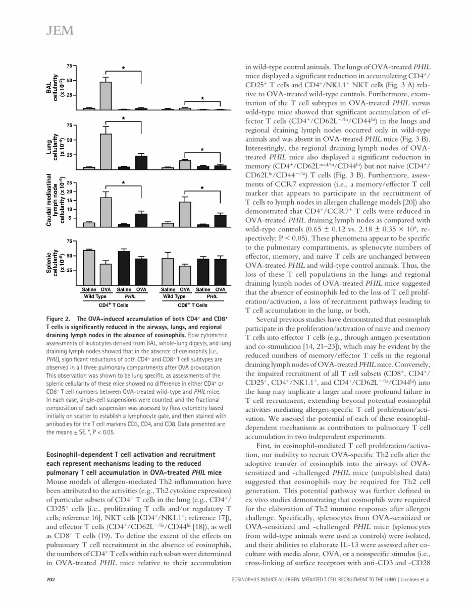
OVA-treated PHIL mice fail to accumulate both CD4 +
and CD8 + T cells in the airways, lungs, and regional
draining lymph nodes
The lack of signifi cant accumulation of total lymphocytes and the reduced Th2 cytokine expression suggested the potential that both CD4 + and CD8 + T cell accumulation in the lungs
wealth of complexity that extends beyond the notion that these leukocytes are primarily a delivery system for their cat-ionic and toxic granule proteins. Two areas of research are primarily responsible for this changing paradigm. First, studies in the mouse have overwhelmingly shown both the absence of extensive eosinophil degranulation in most Th2 infl amma-tory models and the lack of any signifi cant contribution by eosinophil granule proteins to the disease pathologies asso-ciated with pulmonary allergen provocation ( 10, 11 ). Yet, in studies using mice congenitally defi cient of eosinophils, the unique absence of these granulocytes leads to a diminution of Th2 immune responses in both acute and chronic models of allergen provocation ( 5, 12 ). Second, studies assessing immune responses associated with allergen provocation or parasite infection have implicated eosinophils as potential im-mune regulatory cells capable of modulating T cell responses and, in turn, local tissue infl ammation. For example, eosino-phils have been shown to express key mediators of T cell inhibition, activation, and polarization, such as IL-2, IL-4, indoleamine 2,3-dioxygenase, TGF- � , and IL-10 ( 9 ). In addi-tion, recent studies have highlighted the potential role of eosinophils as antigen-presenting cells as part of immune re-sponses leading to T cell proliferation/activation in response to allergen provocation in the lung ( 13 ), as well as in response to parasite infection ( 14 ). Collectively, these studies suggest that eosinophils may be critical cells regulating Th2 immune responses in the lung after allergen provocation, and their role may even supercede the functions of T cells (for review see reference 15 ).
In this study, we provide support for the novel hypothesis that a primary role of eosinophils in allergic pulmonary re-sponses is to elicit the localized recruitment/accumulation of eff ector T cells in the lung. In particular, our data using the eosinophil-defi cient transgenic strain of mice known as PHIL ( 5 ) and two independent models of allergic respiratory infl am-mation showed that the attenuation of Th2 immune responses in the absence of eosinophils (i.e., relative to eosinophil- suffi cient wild-type animals) was accompanied by a concomitant and specifi c loss of T cells from the lung and regional draining lymph nodes. Moreover, using adoptive cell transfer of both eosinophils and allergen-specifi c activated eff ector T cells, we were able to show that eosinophil-mediated activities were required for the recruitment of T cells to the lung, and that one of the mechanisms of this recruitment is likely through the induced production of the Th2-chemoattracting chemo-kines thymus- and activation-regulated chemokine (TARC)/CCL17 and macrophage-derived chemokine (MDC)/CCL22 in the lung.
RESULTS
The reduction of allergen-induced pulmonary pathologies
in OVA-treated PHIL mice is accompanied by a corresponding
decrease in pulmonary Th2 immune responses
We used a novel eosinophil-defi cient transgenic line ( PHIL ), which is congenitally devoid of eosinophils without disturb-ing the production of other hematopoietic lineages ( 5 ), to
JEM VOL. 205, March 17, 2008
ARTICLE
701
mice failed to display allergen-mediated increases in either CD4 + or CD8 + T cells in the airways, lungs, and regional draining lymph nodes. This lack of T cell accumulation was restricted to the lungs and lymph nodes, as spleens from these mice did not statistically diff er in their total numbers of CD4 + and CD8 + T cells, suggesting an inability to accumu-late these cells specifi cally to pulmonary compartments after OVA provocation.
may be altered in OVA-treated PHIL mice ( 15 ). Leukocytes from the airways (i.e., BAL), whole-lung digests, caudal mediastinal lymph nodes, and splenocytes from OVA-sensitized/aerosol-challenged PHIL and wild-type animals were ana-lyzed by fl ow cytometry for populations of CD4 + and CD8 + T cells ( Fig. 2 ). In contrast to OVA-treated wild-type con-trols, which displayed signifi cant increases in both T cell sub-types relative to saline control groups, OVA-treated PHIL
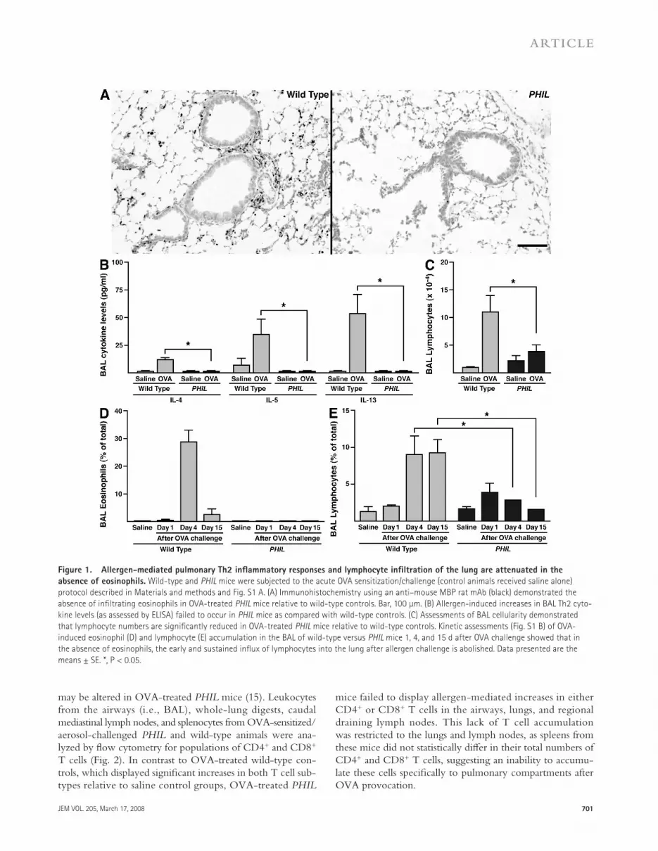
Figure 1. Allergen-mediated pulmonary Th2 infl ammatory responses and lymphocyte infi ltration of the lung are attenuated in the
absence of eosinophils. Wild-type and PHIL mice were subjected to the acute OVA sensitization/challenge (control animals received saline alone)
protocol described in Materials and methods and Fig. S1 A. (A) Immunohistochemistry using an anti – mouse MBP rat mAb (black) demonstrated the
absence of infi ltrating eosinophils in OVA-treated PHIL mice relative to wild-type controls. Bar, 100 μ m. (B) Allergen-induced increases in BAL Th2 cyto-
kine levels (as assessed by ELISA) failed to occur in PHIL mice as compared with wild-type controls. (C) Assessments of BAL cellularity demonstrated
that lymphocyte numbers are signifi cantly reduced in OVA-treated PHIL mice relative to wild-type controls. Kinetic assessments (Fig. S1 B) of OVA-
induced eosinophil (D) and lymphocyte (E) accumulation in the BAL of wild-type versus PHIL mice 1, 4, and 15 d after OVA challenge showed that in
the absence of eosinophils, the early and sustained infl ux of lymphocytes into the lung after allergen challenge is abolished. Data presented are the
means ± SE. *, P < 0.05.
702 EOSINOPHILS INDUCE ALLERGEN-MEDIATED T CELL RECRUITMENT TO THE LUNG | Jacobsen et al.
in wild-type control animals. The lungs of OVA-treated PHIL mice displayed a signifi cant reduction in accumulating CD4 + /CD25 + T cells and CD4 + /NK1.1 + NKT cells ( Fig. 3 A ) rela-tive to OVA-treated wild-type controls. Furthermore, exam-ination of the T cell subtypes in OVA-treated PHIL versus wild-type mice showed that signifi cant accumulation of ef-fector T cells (CD4 + /CD62L � /lo /CD44 hi ) in the lungs and regional draining lymph nodes occurred only in wild-type animals and was absent in OVA-treated PHIL mice ( Fig. 3 B ). Interestingly, the regional draining lymph nodes of OVA-treated PHIL mice also displayed a signifi cant reduction in memory (CD4 + /CD62L med/hi /CD44 hi ) but not naive (CD4 + /CD62L hi /CD44 � /lo ) T cells ( Fig. 3 B ). Furthermore, assess-ments of CCR7 expression (i.e., a memory/eff ector T cell marker that appears to participate in the recruitment of T cells to lymph nodes in allergen challenge models [ 20 ]) also demonstrated that CD4 + /CCR7 + T cells were reduced in OVA-treated PHIL draining lymph nodes as compared with wild-type controls (0.65 ± 0.12 vs. 2.18 ± 0.35 × 10 5 , re-spectively; P < 0.05). These phenomena appear to be specifi c to the pulmonary compartments, as splenocyte numbers of eff ector, memory, and naive T cells are unchanged between OVA-treated PHIL and wild-type control animals. Thus, the loss of these T cell populations in the lungs and regional draining lymph nodes of OVA-treated PHIL mice suggested that the absence of eosinophils led to the loss of T cell prolif-eration/activation, a loss of recruitment pathways leading to T cell accumulation in the lung, or both.
Several previous studies have demonstrated that eosinophils participate in the proliferation/activation of naive and memory T cells into eff ector T cells (e.g., through antigen presentation and co-stimulation [ 14, 21 – 23 ]), which may be evident by the reduced numbers of memory/eff ector T cells in the regional draining lymph nodes of OVA-treated PHIL mice. Conversely, the impaired recruitment of all T cell subsets (CD8 + , CD4 + /CD25 + , CD4 + /NK1.1 + , and CD4 + /CD62L � /lo /CD44 hi ) into the lung may implicate a larger and more profound failure in T cell recruitment, extending beyond potential eosinophil activities mediating allergen-specifi c T cell proliferation/acti-vation. We assessed the potential of each of these eosinophil-dependent mechanisms as contributors to pulmonary T cell accumulation in two independent experiments.
First, in eosinophil-mediated T cell proliferation/activa-tion, our inability to recruit OVA-specifi c Th2 cells after the adoptive transfer of eosinophils into the airways of OVA-sensitized and -challenged PHIL mice (unpublished data) suggested that eosinophils may be required for Th2 cell generation. This potential pathway was further defi ned in ex vivo studies demonstrating that eosinophils were required for the elaboration of Th2 immune responses after allergen challenge. Specifi cally, splenocytes from OVA-sensitized or OVA-sensitized and -challenged PHIL mice (splenocytes from wild-type animals were used as controls) were isolated, and their abilities to elaborate IL-13 were assessed after co-culture with media alone, OVA, or a nonspecifi c stimulus (i.e., cross-linking of surface receptors with anti-CD3 and -CD28
Eosinophil-dependent T cell activation and recruitment
each represent mechanisms leading to the reduced
pulmonary T cell accumulation in OVA-treated PHIL mice
Mouse models of allergen-mediated Th2 infl ammation have been attributed to the activities (e.g., Th2 cytokine expression) of particular subsets of CD4 + T cells in the lung (e.g., CD4 + /CD25 + cells [i.e., proliferating T cells and/or regulatory T cells; reference 16 ], NKT cells [CD4 + /NK1.1 + ; reference 17 ]), and eff ector T cells (CD4 + /CD62L � /lo /CD44 hi [ 18 ]), as well as CD8 + T cells ( 19 ). To defi ne the extent of the eff ects on pulmonary T cell recruitment in the absence of eosinophils, the numbers of CD4 + T cells within each subset were determined in OVA-treated PHIL mice relative to their accumulation
Figure 2. The OVA-induced accumulation of both CD4 + and CD8 +
T cells is signifi cantly reduced in the airways, lungs, and regional
draining lymph nodes in the absence of eosinophils. Flow cytometric
assessments of leukocytes derived from BAL, whole-lung digests, and lung
draining lymph nodes showed that in the absence of eosinophils (i.e.,
PHIL ), signifi cant reductions of both CD4 + and CD8 + T cell subtypes are
observed in all three pulmonary compartments after OVA provocation.
This observation was shown to be lung specifi c, as assessments of the
splenic cellularity of these mice showed no difference in either CD4 + or
CD8 + T cell numbers between OVA-treated wild-type and PHIL mice.
In each case, single-cell suspensions were counted, and the fractional
composition of each suspension was assessed by fl ow cytometry based
initially on scatter to establish a lymphocyte gate, and then stained with
antibodies for the T cell markers CD3, CD4, and CD8. Data presented are
the means ± SE. *, P < 0.05.

JEM VOL. 205, March 17, 2008
ARTICLE
703
in OVA-treated PHIL mice and is part of our ongoing studies further characterizing the role of eosinophils in activation/co-stimulation of T cells (unpublished data).
Second, in eosinophil-dependent recruitment/accumulation of T cells to the lung, the impaired accumulation of multiple T cell subsets in OVA-treated PHIL mice (i.e., CD8 + , CD4 + /CD25 + , CD4 + /NK1.1 + , and CD4 + /CD62L � /lo /CD44 hi T cells) suggested that eosinophils may be required for generalized pathways that promote localized T cell recruitment. The ability of all of these T cell subtypes to express CCR4 ( 24 – 26 ) also provides a potential mechanism by which eosinophils would be able to mediate the recruitment of T cells to sites of Th2 infl ammation. Several lines of evidence further suggested that
antibodies; Fig. 3 C ). These studies demonstrated that the absence of eosinophils (i.e., PHIL mice) leads to a defect in OVA-specifi c Th2 cytokine production. That is, this ob-served defect was restricted to OVA exposure ex vivo, as T cell responses to a nonspecifi c stimulus were equivalent between splenocytes isolated from PHIL versus wild-type mice. Signifi cantly, this OVA-specifi c defect in IL-13 production was limited to animals that were both OVA-sensitized and aerosol-challenged, suggesting an impairment in the activa-tion/proliferation of OVA-specifi c memory T cells into polarized Th2 eff ector T cells in OVA-treated PHIL mice. This immunoregulatory role for eosinophils represents a likely mechanism contributing to the attenuated immune responses
Figure 3. OVA-treated PHIL mice display reduced accumulation of CD4 + T cell subtypes, intrinsic defects in allergen-specifi c T cell
activities, and the loss of allergen-induced TARC and MDC expression. (A) Determinations of T cell subsets for whole-lung digests, regional draining
lymph nodes, and spleen were completed by examining total cell numbers between OVA-treated wild-type versus PHIL mice. OVA-treated wild-type versus
PHIL mice show that CD4 + /CD25 + (T regulatory/proliferation) and CD4 + /NK1.1 + (NKT) cells were elevated in the lungs of OVA-treated wild-type mice but
not in OVA-treated PHIL mice. (B) Effector T cells (CD4 + /CD62L � /lo /CD44 hi ) were reduced in lungs and caudal mediastinal (i.e., regional draining) lymph
nodes. Furthermore, caudal mediastinal lymph nodes also had reduced numbers of memory T cells (CD4 + /CD62L med/hi /CD44 hi ). Naive T cell (CD4 + /CD62L hi /
CD44 � /lo ) numbers remained unchanged between wild-type and PHIL mice in both the spleen and pulmonary compartments. (C) Splenocytes from sensi-
tized and sensitized/challenged PHIL and wild-type mice were cultured for 96 h in the presence of media alone, 200 μ g/ml OVA, or cross-linked anti-
CD3/-CD28. Measurement of IL-13 in the supernatant revealed an inability for sensitized/challenged PHIL splenocytes to generate wild-type levels of this
cytokine. (D) BAL chemokine levels were determined by ELISA from wild-type and PHIL mice subjected to the acute OVA sensitization/challenge protocol
described in Fig. S1 A. Signifi cantly, although substantive increases in MDC and TARC were observed in the BAL of OVA-treated eosinophil-suffi cient wild-
type animals (i.e., relative to saline control mice), increased levels of these Th2 cell chemokines did not occur in the lungs of OVA-treated PHIL mice (i.e., in
the absence of eosinophils). Kinetic assessments (Fig. S1 B) of MDC and TARC in the BAL of post – OVA-challenged wild-type mice showed that the appear-
ance of these Th2 cell chemokines correlated with the levels of OVA-induced airway eosinophilia. Data presented are the means ± SE. *, P < 0.05.
704 EOSINOPHILS INDUCE ALLERGEN-MEDIATED T CELL RECRUITMENT TO THE LUNG | Jacobsen et al.
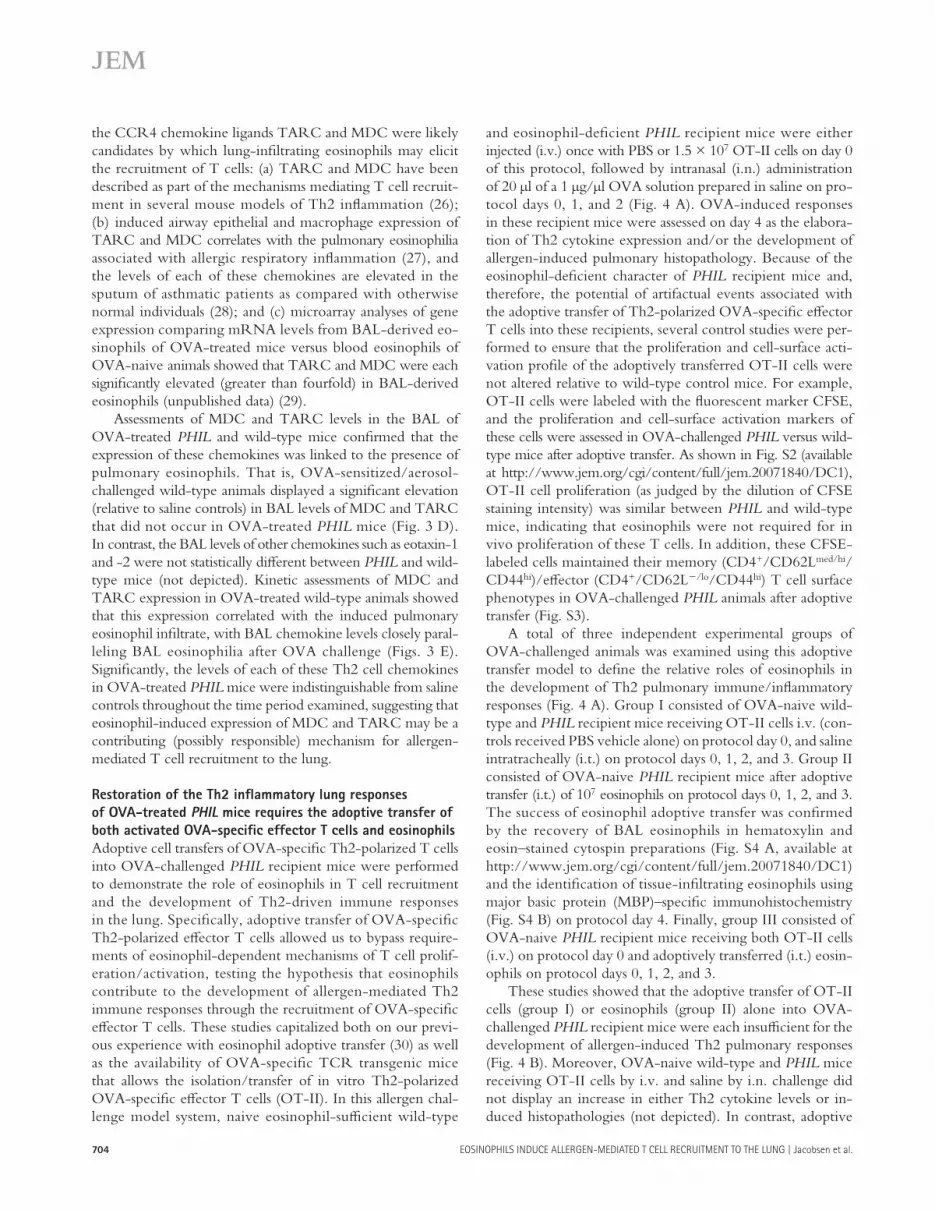
and eosinophil-defi cient PHIL recipient mice were either injected (i.v.) once with PBS or 1.5 × 10 7 OT-II cells on day 0 of this protocol, followed by intranasal (i.n.) administration of 20 μ l of a 1 μ g/ μ l OVA solution prepared in saline on pro-tocol days 0, 1, and 2 ( Fig. 4 A ). OVA-induced responses in these recipient mice were assessed on day 4 as the elabora-tion of Th2 cytokine expression and/or the development of allergen-induced pulmonary histopathology. Because of the eosinophil-defi cient character of PHIL recipient mice and, therefore, the potential of artifactual events associated with the adoptive transfer of Th2-polarized OVA-specifi c eff ector T cells into these recipients, several control studies were per-formed to ensure that the proliferation and cell-surface acti-vation profi le of the adoptively transferred OT-II cells were not altered relative to wild-type control mice. For example, OT-II cells were labeled with the fl uorescent marker CFSE, and the proliferation and cell-surface activation markers of these cells were assessed in OVA-challenged PHIL versus wild-type mice after adoptive transfer. As shown in Fig. S2 (available at http://www.jem.org/cgi/content/full/jem.20071840/DC1), OT-II cell proliferation (as judged by the dilution of CFSE staining intensity) was similar between PHIL and wild-type mice, indicating that eosinophils were not required for in vivo proliferation of these T cells. In addition, these CFSE-labeled cells maintained their memory (CD4 + /CD62L med/hi /CD44 hi )/eff ector (CD4 + /CD62L � /lo /CD44 hi ) T cell surface phenotypes in OVA-challenged PHIL animals after adoptive transfer (Fig. S3).
A total of three independent experimental groups of OVA-challenged animals was examined using this adoptive transfer model to defi ne the relative roles of eosinophils in the development of Th2 pulmonary immune/infl ammatory responses ( Fig. 4 A ). Group I consisted of OVA-naive wild-type and PHIL recipient mice receiving OT-II cells i.v. (con-trols received PBS vehicle alone) on protocol day 0, and saline intratracheally (i.t.) on protocol days 0, 1, 2, and 3. Group II consisted of OVA-naive PHIL recipient mice after adoptive transfer (i.t.) of 10 7 eosinophils on protocol days 0, 1, 2, and 3. The success of eosinophil adoptive transfer was confi rmed by the recovery of BAL eosinophils in hematoxylin and eosin – stained cytospin preparations (Fig. S4 A, available at http://www.jem.org/cgi/content/full/jem.20071840/DC1) and the identifi cation of tissue-infi ltrating eosinophils using major basic protein (MBP) – specifi c immunohistochemistry (Fig. S4 B) on protocol day 4. Finally, group III consisted of OVA-naive PHIL recipient mice receiving both OT-II cells (i.v.) on protocol day 0 and adoptively transferred (i.t.) eosin-ophils on protocol days 0, 1, 2, and 3.
These studies showed that the adoptive transfer of OT-II cells (group I) or eosinophils (group II) alone into OVA-challenged PHIL recipient mice were each insuffi cient for the development of allergen-induced Th2 pulmonary responses ( Fig. 4 B ). Moreover, OVA-naive wild-type and PHIL mice receiving OT-II cells by i.v. and saline by i.n. challenge did not display an increase in either Th2 cytokine levels or in-duced histopathologies (not depicted). In contrast, adoptive
the CCR4 chemokine ligands TARC and MDC were likely candidates by which lung-infi ltrating eosinophils may elicit the recruitment of T cells: (a) TARC and MDC have been described as part of the mechanisms mediating T cell recruit-ment in several mouse models of Th2 infl ammation ( 26 ); (b) induced airway epithelial and macrophage expression of TARC and MDC correlates with the pulmonary eosinophilia asso ciated with allergic respiratory infl ammation ( 27 ), and the levels of each of these chemokines are elevated in the sputum of asthmatic patients as compared with otherwise normal individuals ( 28 ); and (c) microarray analyses of gene expression comparing mRNA levels from BAL-derived eo-sinophils of OVA-treated mice versus blood eosinophils of OVA-naive animals showed that TARC and MDC were each signifi cantly elevated (greater than fourfold) in BAL-derived eosinophils (unpublished data) ( 29 ).
Assessments of MDC and TARC levels in the BAL of OVA-treated PHIL and wild-type mice confi rmed that the expression of these chemokines was linked to the presence of pulmonary eosinophils. That is, OVA-sensitized/aerosol- challenged wild-type animals displayed a signifi cant elevation (relative to saline controls) in BAL levels of MDC and TARC that did not occur in OVA-treated PHIL mice ( Fig. 3 D ). In contrast, the BAL levels of other chemokines such as eotaxin-1 and -2 were not statistically diff erent between PHIL and wild-type mice (not depicted). Kinetic assessments of MDC and TARC expression in OVA-treated wild-type animals showed that this expression correlated with the induced pulmonary eosinophil infi ltrate, with BAL chemokine levels closely paral-leling BAL eosinophilia after OVA challenge ( Figs. 3 E ). Signifi cantly, the levels of each of these Th2 cell chemokines in OVA-treated PHIL mice were indistinguishable from saline controls throughout the time period examined, suggesting that eosinophil-induced expression of MDC and TARC may be a contributing (possibly responsible) mechanism for allergen-mediated T cell recruitment to the lung.
Restoration of the Th2 infl ammatory lung responses
of OVA-treated PHIL mice requires the adoptive transfer of
both activated OVA-specifi c effector T cells and eosinophils
Adoptive cell transfers of OVA-specifi c Th2-polarized T cells into OVA-challenged PHIL recipient mice were performed to demonstrate the role of eosinophils in T cell recruitment and the development of Th2-driven immune responses in the lung. Specifi cally, adoptive transfer of OVA-specifi c Th2-polarized eff ector T cells allowed us to bypass require-ments of eosinophil-dependent mechanisms of T cell prolif-eration/activation, testing the hypothesis that eosinophils contribute to the development of allergen-mediated Th2 immune responses through the recruitment of OVA-specifi c eff ector T cells. These studies capitalized both on our previ-ous experience with eosinophil adoptive transfer ( 30 ) as well as the availability of OVA-specifi c TCR transgenic mice that allows the isolation/transfer of in vitro Th2-polarized OVA-specifi c eff ector T cells (OT-II). In this allergen chal-lenge model system, naive eosinophil-suffi cient wild-type

JEM VOL. 205, March 17, 2008
ARTICLE
705
mice (group III) resulted in goblet cell metaplasia/airway epithelial cell mucin accumulation that was comparable to that observed in OVA-challenged wild-type mice receiving OT-II cells ( Fig. 4 C ). These observations suggest that the eosinophil activities required for the development of Th2 re-sponses extend beyond the potential eff ects on the proliferation/activation of OVA-specifi c eff ector T cells.
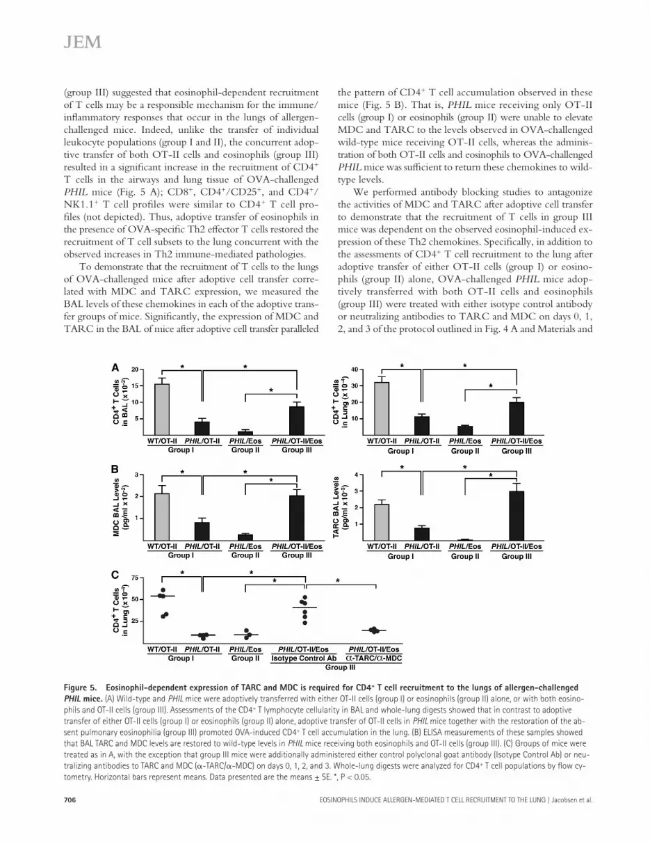
Eosinophils mediate T cell recruitment to the lung
after adoptive transfer into OVA-challenged PHIL mice
by an MDC/TARC-dependent mechanism
The allergen-induced pulmonary pathologies resulting from the combined adoptive transfer of OT-II cells and eosinophils
transfer of both OT-II cells and eosinophils into PHIL mice (group III) led to signifi cant increases in BAL levels of the Th2 cytokines (e.g., IL-5 and IL-13; Fig. 4 B ); IL-4 was not signifi cantly elevated above baseline. The development of Th2-driven histopathologies in each of these groups of mice paralleled the pattern of induced Th2 cytokine expression. That is, administration of either OT-II cells (group I) or eo-sinophils (group II) alone into OVA-challenged PHIL mice failed to result in the development of histopathologies such as the peribronchial aggregation of eosinophils (Fig. S4 B) and goblet cell metaplasia/airway epithelial cell mucin accumu-lation ( Fig. 4 C ). However, the adoptive transfer of both OT-II cells and eosinophils into OVA-challenged PHIL
Figure 4. Adoptive transfer of both OVA-specifi c Th2-polarized effector T cells (OT-II) and eosinophils are required to generate Th2
pulmonary pathologies in PHIL mice. (A) Schematic timeline of the acute OVA challenge protocol associated with the adoptive transfer studies of Th2-
polarized OVA-specifi c T cells (OT-II) and eosinophils. (B) Th2 BAL levels of the cytokines IL-13 and IL-5 (IL-4 was undetectable) were assessed by ELISA.
PHIL mice receiving either OT-II cells (group I) or eosinophils (group II) alone showed no pathologies, whereas adoptive transfer of OT-II cells in PHIL mice
together with the restoration of the absent pulmonary eosinophilia (group III) promoted signifi cant increases in the BAL levels of the Th2 cytokines IL-13
and IL-5. Data presented are the means ± SE. *, P < 0.05. (C) Assessments of goblet cell metaplasia and airway epithelial cell mucin accumulation by PAS
staining of lung sections (purple) revealed that only the adoptive transfer of both OT-II cells and eosinophils to PHIL mice was capable of reconstituting
this allergen-induced histopathology. PHIL /Eos, OVA-provoked PHIL mice after i.t. transfer of eosinophils; PHIL /OT-II, OVA-provoked PHIL mice after
OT-II cell transfer; PHIL /OT-II/Eos, OVA-provoked PHIL mice after adoptive transfer of both OT-II cell (i.v.) and eosinophils (i.t.); WT/OT-II, OVA-provoked
wild-type mice after OT-II cell transfer. Bar, 100 μ m.
706 EOSINOPHILS INDUCE ALLERGEN-MEDIATED T CELL RECRUITMENT TO THE LUNG | Jacobsen et al.
the pattern of CD4 + T cell accumulation observed in these mice ( Fig. 5 B ). That is, PHIL mice receiving only OT-II cells (group I) or eosinophils (group II) were unable to elevate MDC and TARC to the levels observed in OVA-challenged wild-type mice receiving OT-II cells, whereas the adminis-tration of both OT-II cells and eosinophils to OVA-challenged PHIL mice was suffi cient to return these chemokines to wild-type levels.
We performed antibody blocking studies to antagonize the activities of MDC and TARC after adoptive cell transfer to demonstrate that the recruitment of T cells in group III mice was dependent on the observed eosinophil-induced ex-pression of these Th2 chemokines. Specifi cally, in addition to the assessments of CD4 + T cell recruitment to the lung after adoptive transfer of either OT-II cells (group I) or eosino-phils (group II) alone, OVA-challenged PHIL mice adop-tively transferred with both OT-II cells and eosinophils (group III) were treated with either isotype control antibody or neutralizing antibodies to TARC and MDC on days 0, 1, 2, and 3 of the protocol outlined in Fig. 4 A and Materials and
(group III) suggested that eosinophil-dependent recruitment of T cells may be a responsible mechanism for the immune/infl ammatory responses that occur in the lungs of allergen-challenged mice. Indeed, unlike the transfer of individual leukocyte populations (group I and II), the concurrent adop-tive transfer of both OT-II cells and eosinophils (group III) resulted in a signifi cant increase in the recruitment of CD4 + T cells in the airways and lung tissue of OVA-challenged PHIL mice ( Fig. 5 A ); CD8 + , CD4 + /CD25 + , and CD4 + /NK1.1 + T cell profi les were similar to CD4 + T cell pro-fi les (not depicted). Thus, adoptive transfer of eosinophils in the presence of OVA-specifi c Th2 eff ector T cells restored the recruitment of T cell subsets to the lung concurrent with the observed increases in Th2 immune-mediated pathologies.
To demonstrate that the recruitment of T cells to the lungs of OVA-challenged mice after adoptive cell transfer corre-lated with MDC and TARC expression, we measured the BAL levels of these chemokines in each of the adoptive trans-fer groups of mice. Signifi cantly, the expression of MDC and TARC in the BAL of mice after adoptive cell transfer paralleled
Figure 5. Eosinophil-dependent expression of TARC and MDC is required for CD4 + T cell recruitment to the lungs of allergen-challenged
PHIL mice. (A) Wild-type and PHIL mice were adoptively transferred with either OT-II cells (group I) or eosinophils (group II) alone, or with both eosino-
phils and OT-II cells (group III). Assessments of the CD4 + T lymphocyte cellularity in BAL and whole-lung digests showed that in contrast to adoptive
transfer of either OT-II cells (group I) or eosinophils (group II) alone, adoptive transfer of OT-II cells in PHIL mice together with the restoration of the ab-
sent pulmonary eosinophilia (group III) promoted OVA-induced CD4 + T cell accumulation in the lung. (B) ELISA measurements of these samples showed
that BAL TARC and MDC levels are restored to wild-type levels in PHIL mice receiving both eosinophils and OT-II cells (group III). (C) Groups of mice were
treated as in A, with the exception that group III mice were additionally administered either control polyclonal goat antibody (Isotype Control Ab) or neu-
tralizing antibodies to TARC and MDC ( � -TARC/ � -MDC) on days 0, 1, 2, and 3. Whole-lung digests were analyzed for CD4 + T cell populations by fl ow cy-
tometry. Horizontal bars represent means. Data presented are the means ± SE. *, P < 0.05.

JEM VOL. 205, March 17, 2008
ARTICLE
707
tions, as well as their requirement for a threshold level of eo-sinophils, are responsible for many of the diffi culties associated with the interpretation of results from both earlier mouse-model studies of allergic infl ammation and clinical studies of asthma patients that target eosinophils. For example, the partial eosinophil defi ciency of IL-5 � / � mice after OVA sensitization/challenge ( � 90% reduction relative to wild-type mice) is likely only to attenuate and not abolish eosinophil-mediated eff ects on T cells. This, in part, may explain the diff erences in allergen-induced pulmonary infl ammation that occurred in IL-5 � / � mice among diff erent studies (e.g., refer-ence 34 vs. reference 35 ). Indeed, a requirement for a thresh-old level of recruited eosinophils was suggested in studies examining the allergic respiratory infl ammation in OVA-treated IL-5 � / � mice relative to OVA-treated IL-5/eotaxin-1 double-knockout (IL-5 � / � /eotaxin-1 � / � ) animals. That is, although the partial loss of eosinophils failed to prevent OVA-induced infl ammatory responses in IL-5 � / � , these re-sponses were abolished in IL-5 � / � /eotaxin-1 � / � mice that had a complete ablation of pulmonary eosinophils ( 36 ). The demonstrated inability to completely eliminate lung tissue eosinophils among human subjects receiving anti – IL-5 thera-peutics may also represent a confounding issue that has com-plicated conclusions from clinical studies of asthma patients ( 6, 37, 38 ). These observations suggest that together with potential activities on T cell proliferation/activation and de-structive eff ector activities mediated by granule protein re-lease, the additional capability of eosinophils to mediate eff ector T cell accumulation in the lung may provide yet another mechanism by which eosinophils initiate and maintain the immune/infl ammatory responses associated with allergic asthma. Interestingly, two unrelated studies ( 12, 39 ) using an-other eosinophil-defi cient strain of mice (i.e., � dblGATA-1 [ 40 ]) also provide relevant insights supporting the immuno-regulative roles of eosinophils. Fulkerson et al. ( 12 ) reported that microarray assessments of lung gene expression in Asper-gillus allergen-challenged � dblGATA-1 mice (relative to allergen-challenged wild-type animals) showed that the absence of eosinophils was associated with a signifi cant reduction in pulmonary Th2 gene expression (e.g., IL-4 and IL-13). In addition, Voehringer et al. ( 39 ), using � dblGATA eosinophil-defi cient mice and a Nocardia brasiliensis mouse model of lung infl ammation, concluded that eosinophils were not required for pulmonary T cell recruitment during the primary infection of mice. However, eosinophils in this study may be linked with a failure to activate/recruit CD4 + T cells to the lung and, in turn, infl ammatory responses associated with secondary challenges with this parasite. Thus, data from both of these studies highlight a complexity of pulmonary immune responses but again imply a potentially dual role for eosinophils asso-ciated with the elaboration of secondary immune responses in the lung, leading to the proliferation/activation of antigen-specifi c T cells and/or subsequent recruitment of these T cells to the lung.
Finally, there is no reason to assume that either of the eosinophil-mediated eff ects on T cells outlined in this paper
methods. The antibody-mediated blockade of TARC and MDC in group III mice returned CD4 + T cell recruitment in these PHIL animals to the baseline levels observed in PHIL mice from groups I and II ( Fig. 5 C ). These observations ex-tended to multiple T cell subsets (e.g., CD4 + /CD62L � /lo /CD44 hi eff ector T cells and CD8 + T cells) that were also dra-matically reduced in the lungs of anti-TARC/-MDC – treated group III mice (not depicted). These studies showed that T cells are recruited to the lung by the eosinophil-dependent production of TARC and MDC and that this induced ex-pression after allergen challenge is mechanistically linked to the T cell recruitment occurring in these mice.
DISCUSSION
The observation that eosinophils accumulating in the lungs after allergen provocation modulate local tissue immune re-sponses through the specifi c recruitment of T cells suggests a novel mechanism of eosinophil eff ector function whose sig-nifi cance was previously underappreciated. This mechanism represents an expansion/extension of earlier studies investi-gating the potential importance of eosinophils and also pro-vides an alternative interpretation of data initially noted by Lloyd et al. ( 26 ). Specifi cally, we suggest that the expression of eotaxins and perhaps other eosinophil agonist factors (e.g., leukotriene B 4 ) ( 31 ) immediately after allergen challenge elicits the recruitment of eosinophils and the expression of cytokines and growth factors that also promote eosinophil survival and activation. In turn, if and when these accumulat-ing eosinophils achieve a critical level, their activities pro-mote the local expression of Th2 chemokines (e.g., TARC and MDC) that leads to and/or amplifi es the recruitment of T cells to the lungs and allergen-mediated Th2 infl ammation. Thus, either by a direct (i.e., cell autonomous) or an indirect (i.e., induced from a third party cell type) mechanism, infi l-trating lung eosinophils may mediate the induction of TARC and MDC that elicits the T cell recruitment necessary for the infl ammatory responses associated with mouse models of asthma. Interestingly, studies assessing the initial leukocyte recruitment to the lungs in both human subjects and mouse models of allergic respiratory infl ammation demonstrated similar kinetic patterns of eosinophil and lymphocyte accu-mulation into the airways ( 32, 33 ), implying that eosinophil-dependent accumulation of lymphocytes leading to pulmonary Th2 infl ammatory responses may represent a larger, more signifi cant mechanism with implications for asthma patients.
Our larger hypothesis linking eosinophils, T cells, and al-lergic respiratory infl ammation is that eosinophils mediate the recruitment of eff ector T cells by two apparently indepen-dent mechanisms: (a) activation and co-stimulation of T cells promoting proliferation and/or increased survival, and (b) pulmonary production of chemokines that promote the re-cruitment and/or accumulation of T cells in the lung. It is also noteworthy that these eosinophil-dependent mechanisms are not mutually exclusive, and each may contribute to the loss of Th2 immune responses and pulmonary pathologies in OVA-treated PHIL mice. We suggest that these dual func-

708 EOSINOPHILS INDUCE ALLERGEN-MEDIATED T CELL RECRUITMENT TO THE LUNG | Jacobsen et al.
digested for 45 min at 37 ° C, as described previously ( 47 ). Single-cell suspen-
sions of lung, spleens, and caudal mediastinal lymph nodes (regional draining
lymph nodes) were obtained by homogenization with frosted glass slides fol-
lowed by passing through a 40- μ m nylon fi lter to remove larger aggregates
of cells per tissue. Red blood cells were lysed by ammonium chloride
(PharmLyse; BD Biosciences). Viability of the cells was > 95%, as determined
by Trypan blue exclusion. Cell counts were completed after the last wash
before staining for fl ow cytometry.
Cytokine assays. Mouse IL-4, IL-5, IL-13, MDC, and TARC levels were
assessed using immunoassay kits (R & D Systems) according to the manufac-
turer ’ s instructions. The limits of detection for each cytokine assay were
5 – 10 pg/ml.
Splenocyte culture. Single-cell suspensions were obtained from sensitized
and sensitized/challenged PHIL and wild-type mice using the protocol
shown in Fig. S1 A. 10 6 cells/well of splenocytes were cultured in a 96-well
plate with either media alone, with 200 μ g/ml OVA (Sigma-Aldrich), or
with plates precoated with anti-CD3 and -CD28 (BD Biosciences) for 96 h.
Supernatants were analyzed by ELISA.
Flow cytometry analysis. Single-cell suspensions were stained for 25 min
on ice with cell type – specifi c antibodies after blockade of Fc receptors using
1 μ g/ μ l of Fc blocker (CD16/32; BD Biosciences). Antibodies used for
staining specifi c cell types were obtained from BD Biosciences and include
the T cell subtype – specifi c antibodies CD3, CD4, CD8, and CD62L, and
the NK/NKT cell – specifi c marker NK1.1. Additional antibodies represent-
ing other T cell markers (i.e., CD44 and CD25) were purchased from eBio-
science. Propidium iodide staining was used to identify and “ gate out ”
dead cells. Flow cytometry was performed on a cytofl uorimeter (FACScan;
Becton Dickinson). Data acquisition and analysis were performed using
CellQuest Pro (Becton Dickinson) and Sumit (version 4.3; Dako) software.
Isolation and polarization of OT-II cells. Naive OT-II cells were iso-
lated from splenocytes and lymph nodes of 5 – 12-wk-old mice by two
rounds of positive selection using magnetic beads coupled to the T cell –
specifi c marker CD90 + (Thy1.2; MACS [Miltenyi Biotec]). 0.5 × 10 6 puri-
fi ed CD90 + T cells/ml were diff erentiated to the Th2 eff ector phenotype
by incubating the cells in RPMI 1640 media (supplemented with 15% FBS
[HyClone Technologies], 2 × 10 � 5 M 2-mercaptoethanol, 1 mM sodium
pyruvate, 1 × nonessential amino acids, 10 μ g/ml penicillin, 10 μ g/ml strep-
tomycin, and 2 mM l- glutamine; all from Invitrogen) in the presence of
50 μ g/ml OVA 323-339 peptide, 8 ng/ml mouse IL-4 (R & D Systems),
5 μ g/ml anti – IL-12 mAb (C17.8; BD Biosciences), and 5 μ g/ml anti – IFN- �
(XMG1.2; BD Biosciences). Irradiated (30 Gy) splenic APCs (depleted of
CD 90 + cells) were also added to these cultures (2 × 10 6 cells/ml). After
2 d of culture, cells were given 10 ng/ml IL-2 (R & D Systems), and on days
4 and 6 cells were given fresh media with 10 ng/ml of IL-2. After culture
for a total of 7 d, the recovered Th2-polarized eff ector T cells were used for
i.v. injection (1.5 × 10 7 cells/mouse) into recipient mice. In some experi-
ments, cells were labeled with CFSE, as described by the manufacturer
(Invitrogen), before injection. FACS analysis reveals > 98% of cells using
this method are CD3 + , with a composition of � 85% CD4 + and � 15%
CD8 + . Trypan blue exclusion demonstrated > 98% cell viability. Th2 polar-
ization of this cell population was confi rmed by ELISA assessments for the
presence of the Th2 cytokines (i.e., IL-4, IL-5, and IL-13) of 7-d cell-
culture supernatants.
Isolation of mouse eosinophils. Eosinophils were isolated from IL-5 –
expressing transgenic mice (NJ.1638) ( 30 ), as previously described. In brief,
peripheral blood pooled from four mice was layered onto single-step Percoll
gradients. Eosinophils from the buff y coat at the blood – Percoll interface were
subsequently isolated by removing the contaminating lymphocytes using
MACS with magnetic beads conjugated with CD45R/B220 (B cells) and
CD90/Thy1.2 (T cells), according to the manufacturer ’ s recommendations.
are necessarily mutually exclusive, and each is likely to con-tribute to the induced pulmonary immune responses observed in eosinophil-suffi cient wild-type mice. These eosinophil- dependent immunoregulatory mechanisms are also not likely to be limited to the lung and/or allergen-mediated infl amma-tion. That is, the accumulation of tissue eosinophils in a variety of settings may represent provocative examples in which eosinophil-mediated expression of Th2 chemokines leads to the recruitment of eff ector T cell recruitment and the modulation of local tissue immune cascades. In particular, we suggest that the T cell – induced immune responses associated with acute rejec-tion of transplanted organs ( 41 ), the onset/growth of tumors ( 42 ), the accumulation/diff erentiation of thymic T cell popu-lations ( 43 ), and the immune modulation of the gastrointesti-nal tract mucosa ( 44 ) may ultimately each be causatively linked to tissue-specifi c recruitment of activated eff ector T cells by eosinophils accumulating at these sites.
MATERIALS AND METHODS Mice. All studies were performed with either C57BL/6J wild-type or
eosinophil-defi cient PHIL mice ( 5 ) crossed to C57BL/6J for > 10 generations.
C57BL/6J mice and TCR OVA-transgenic animals on a C57BL/6J back-
ground (OT-II) were purchased from the Jackson Laboratory. The mice
were maintained in ventilated microisolator cages housed in the specifi c
pathogen-free animal facility at the Mayo Clinic Arizona. The sentinel cages
within the animal colony surveyed negative for the presence of known
mouse pathogens. Protocols and studies involving animals were performed
in accordance with National Institutes of Health and Mayo Foundation in-
stitutional guidelines.
Acute OVA sensitization and challenge protocols. Mice were sensi-
tized and challenged with OVA as previously described ( 5 ) and outlined in
Fig. S1 A. The kinetics of leukocyte recruitment to the lungs of sensitized
and challenged mice were performed as previously described ( 33 ) and out-
lined in Fig. S1 B. Studies involving adoptive transfer of T cells derived from
OVA-specifi c TCR transgenic mice (OT-II cells) were performed using
a modifi cation of a protocol previously described ( 45 ) and outlined in
Fig. 4 A . Specifi cally, on day 0 of this protocol, either PBS or 1.5 × 10 7
in vitro Th2-polarized eff ector T cells (OT-II) were injected i.v. into OVA-naive
wild-type or PHIL mice. Within 3 h of i.v. OT-II cell transfer, the mice
were gently anesthetized and received an i.n. administration of 20 μ g OVA
(1 μ g/ μ l in saline). Mice were additionally challenged by i.n. administration
of OVA on days 1 and 2 of this protocol (i.e., mice were OVA challenged
via the nasal passages on three consecutive days). In some groups of mice,
10 7 eosinophils in 20 μ l of saline were adoptively transferred (control animals
received saline vehicle alone) via the trachea (i.t.) on days 0, 1, 2, and 3
of this protocol, and within 1 h after OVA administration on the allergen-
challenge days. OVA-induced responses in these recipient mice were assessed
on day 4.
Collection and diff erentials of BAL fl uid – derived cells. BAL assess-
ments were completed as described previously ( 5, 46 ).
Histology. Histopathologic changes of the airways were assessed as de-
scribed previously ( 5 ). 4- μ m formalin-fi xed, paraffi n-embedded sections of
mouse lungs were stained for epithelial cell mucin accumulation using peri-
odic acid Schiff or, for eosinophils, rabbit polyclonal anti – mouse eosinophil
MBP antiserum ( 46 ).
Splenocyte, lymph node, and lung cell isolation. Total pulmonary
leukocytes were recovered from lungs perfused with PBS-EDTA before
removal, placed in complete media with 175 U/ml collagenase IV, and

JEM VOL. 205, March 17, 2008
ARTICLE
709
Gerard . 2004 . A critical role for eosinophils in allergic airways re-modeling. Science . 305 : 1776 – 1779 .
5 . Lee , J.J. , D. Dimina , M.P. Macias , S.I. Ochkur , M.P. McGarry , K.R. O ’ Neill , C. Protheroe , R. Pero , T. Nguyen , S.A. Cormier , et al . 2004 . Defi ning a link with asthma in mice congenitally defi cient in eosino-phils. Science . 305 : 1773 – 1776 .
6 . Leckie , M.J. 2003 . Anti-interleukin-5 monoclonal antibodies: pre-clinical and clinical evidence in asthma models. Am. J. Respir. Med. 2 : 245 – 259 .
7 . Trivedi , S.G. , and C.M. Lloyd . 2007 . Eosinophils in the pathogenesis of allergic airways disease. Cell. Mol. Life Sci. 64 : 1269 – 1289 .
8 . Abu-Ghazaleh , R.I. , H. Kita , and G.J. Gleich . 1992 . Eosinophil activa-tion and function in health and disease. Immunol. Ser. 57 : 137 – 167 .
9 . Jacobsen , E.A. , A.G. Taranova , N.A. Lee , and J.J. Lee . 2007 . Eosinophils: singularly destructive eff ector cells or purveyors of immunoregulation? J. Allergy Clin. Immunol. 119 : 1313 – 1320 .
10 . Denzler , K.L. , S.C. Farmer , J.R. Crosby , M.T. Borchers , G. Cieslewicz , K.A. Larson , S. Cormier-Regard , N.A. Lee , and J.J. Lee . 2000 . Eosinophil major basic protein-1 does not contribute to allergen-induced airway patho-logies in mouse models of asthma. J. Immunol. 165 : 5509 – 5517 .
11 . Denzler , K.L. , M.T. Borchers , J.R. Crosby , G. Cieslewicz , E.M. Hines , J.P. Justice , S.A. Cormier , K.A. Lindenberger , W. Song , W. Wu , et al . 2001 . Extensive eosinophil degranulation and peroxidase-mediated oxi-dation of airway proteins do not occur in a mouse ovalbumin-challenge model of pulmonary infl ammation. J. Immunol. 167 : 1672 – 1682 .
12 . Fulkerson , P.C. , C.A. Fischetti , M.L. McBride , L.M. Hassman , S.P. Hogan , and M.E. Rothenberg . 2006 . A central regulatory role for eo-sinophils and the eotaxin/CCR3 axis in chronic experimental allergic airway infl ammation. Proc. Natl. Acad. Sci. USA . 103 : 16418 – 16423 .
13 . Duez , C. , A. Dakhama , A. Tomkinson , P. Marquillies , A. Balhorn , A.-B. Tonnel , D.L. Bratton , and E.W. Gelfand . 2004 . Migration and accumulation of eosinophils toward regional lymph nodes after airway allergen challenge. J. Allergy Clin. Immunol. 114 : 820 – 825 .
14 . Padigel , U.M. , J.J. Lee , T.J. Nolan , G.A. Schad , and D. Abraham . 2006 . Eosinophils can function as antigen-presenting cells to induce primary and secondary immune responses to Strongyloides stercoralis . Infect. Immun. 74 : 3232 – 3238 .
15 . Lee , N.A. , E.W. Gelfand , and J.J. Lee . 2001 . Pulmonary T cells and eo-sinophils: coconspirators or independent triggers of allergic respiratory pathology? J. Allergy Clin. Immunol. 107 : 945 – 957 .
16 . Taylor , A. , J. Verhagen , C.A. Akdis , and M. Akdis . 2005 . T regulatory cells and allergy. Microbes Infect. 7 : 1049 – 1055 .
17 . Akbari , O. , P. Stock , E. Meyer , M. Kronenberg , S. Sidobre , T. Nakayama , M. Taniguchi , M.J. Grusby , R.H. DeKruyff , and D.T. Umetsu . 2003 . Essential role of NKT cells producing IL-4 and IL-13 in the development of allergen-induced airway hyperreactivity. Nat. Med. 9 : 582 – 588 .
18 . Bingaman , A.W. , D.S. Patke , V.R. Mane , M. Ahmadzadeh , M. Ndejembi , S.T. Bartlett , and D.L. Farber . 2005 . Novel phenotypes and migratory properties distinguish memory CD4 T cell subsets in lym-phoid and lung tissue. Eur. J. Immunol. 35 : 3173 – 3186 .
19 . Gelfand , E.W. , and A. Dakhama . 2006 . CD8+ T lymphocytes and leu-kotriene B4: novel interactions in the persistence and progression of asthma. J. Allergy Clin. Immunol. 117 : 577 – 582 .
20 . Bromley , S.K. , S.Y. Thomas , and A.D. Luster . 2005 . Chemokine re-ceptor CCR7 guides T cell exit from peripheral tissues and entry into aff erent lymphatics. Nat. Immunol. 6 : 895 – 901 .
21 . Shi , H.Z. 2004 . Eosinophils function as antigen-presenting cells. J. Leukoc. Biol. 76 : 520 – 527 .
22 . MacKenzie , J.R. , J. Mattes , L.A. Dent , and P.S. Foster . 2001 . Eosinophils promote allergic disease of the lung by regulating CD4 + Th2 lympho-cyte function. J. Immunol. 167 : 3146 – 3155 .
23 . Wang , H.-B. , I. Ghiran , K. Matthaei , and P.F. Weller . 2007 . Airway eosinophils: allergic infl ammation recruited professional antigen-presenting cells. J. Immunol. 179 : 7585 – 7592 .
24 . Kim , C.H. , B. Johnston , and E.C. Butcher . 2002 . Traffi cking machinery of NKT cells: shared and diff erential chemokine receptor expression among V alpha 24(+)V beta 11(+) NKT cell subsets with distinct cytokine-producing capacity. Blood . 100 : 11 – 16 .
Visual examination of Diff Quick-stained (Dade Behring Inc.) cytospin prep-
arations and by fl ow cytometry analysis with staining for anti-CCR3 (R & D
Systems) revealed that the purity of these eosinophil preparations was > 99%.
i.t. transfer of eosinophils. The adoptive transfer of eosinophils and/or
vehicle control (saline) into the lungs of mice was accomplished in subject
animals lightly anesthetized with 1% isofl uorane before i.t. instillation, as de-
scribed previously ( 30 ). The transfer of eosinophils (or saline vehicle) was
completed 1 h after i.n. administration of OVA on days 0, 1, and 2, and at the
same time of day without OVA on protocol day 3 ( Fig. 4 A ).
Antibody neutralization of MDC and TARC. Mice were treated with
antibodies to MDC and TARC with minor modifi cations of a protocol de-
scribed previously ( 48 ). Specifi cally, 1 h before the OVA challenges, the
PHIL mice received an i.p. injection of either control polyclonal goat IgG
antibody or 20 μ g each of polyclonal IgG goat anti-TARC and anti-MDC
(R & D Systems) on days 0, 1, 2, and 3 of the protocol outlined in Fig. 4 A .
Statistical analysis. All data were derived from at least three independent
experiments, each with cohort sizes of two to fi ve mice. Data presented are
the means ± SE. Statistical analyses were performed using the Student ’ s t test
with diff erences between means considered signifi cant when P < 0.05.
Online supplemental material. Fig. S1 presents schematic timelines of the
acute OVA sensitization/aerosol challenge protocols used to elicit allergic re-
spiratory infl ammation, including the recruitment/accumulation of eosinophils
and lymphocytes to the lung. Fig. S2 demonstrates that OT-II cells displayed
the same proliferative capacity after adoptive transfer into eosinophil-suffi cient
wild-type and PHIL mice. Fig. S3 shows that OT-II cells displayed the same
T cell surface phenotype after adoptive transfer into eosinophil-suffi cient
wild-type and PHIL mice. Fig. S4 demonstrates that OVA provocation of
PHIL mice after adoptive transfer of both OT-II cells and eosinophils resulted
in the development of a BAL eosinophilia and proinfl ammatory aggregates in
the peribronchial regions of the lung. Online supplemental material is avail-
able at http://www.jem.org/cgi/content/full/jem.20071840/DC1.
The authors wish to thank the invaluable contribution of numerous individuals
not listed as authors, including Teresa Tinder, Katie O ’ Neill, Alfred Doyle, and
Dr. Michael McGarry. Moreover, we also wish to acknowledge the tireless efforts
of the Mayo Clinic Arizona medical graphic artist Marv Ruona, Nichole Boruff, and
Joseph Esposito of Research Library Services. In addition, we wish to express our
gratitude to the Lee Laboratories administrative staff (Linda Mardel and Margaret
McGarry), without whom we could not function as an integrated group.
The work presented was supported by the Mayo Foundation and grants from
the National Institutes of Health to J.J. Lee (HL065228 and K26-RR019709), N.A. Lee
(HL058723), and E.A. Jacobsen (HL08514). Additional support was provided by grants
from the American Heart Association to J.J. Lee (045580Z) and N.A. Lee (0555639Z).
The authors have no confl icting fi nancial interests.
Submitted: 27 August 2007
Accepted: 8 February 2008
REFERENCES 1 . Larche , M. , D.S. Robinson , and A.B. Kay . 2003 . The role of T
lymphocytes in the pathogenesis of asthma. J. Allergy Clin. Immunol. 111 : 450 – 463 .
2 . Gavett , S.H. , X. Chen , F. Finkelman , and M. Wills-Karp . 1994 . Depletion of murine CD4+ T lymphocytes prevents antigen-induced airway hyperreactivity and pulmonary eosinophilia. Am. J. Respir. Cell Mol. Biol. 10 : 587 – 593 .
3 . Bousquet , J. , P. Chanez , J.Y. Lacoste , G. Barneon , N. Ghavanian , I. Enander , P. Venge , S. Ahlstedt , J. Simony-Lafontaine , P. Godard , and M. Francois-Bernard . 1990 . Eosinophilic infl ammation in asthma . N. Engl. J. Med. 323 : 1033 – 1039 .
4 . Humbles , A.A. , C.M. Lloyd , S.J. McMillan , D.S. Friend , G. Xanthou , E.E. McKenna , S. Ghiran , N.P. Gerard , C. Yu , S.H. Orkin , and C.

710 EOSINOPHILS INDUCE ALLERGEN-MEDIATED T CELL RECRUITMENT TO THE LUNG | Jacobsen et al.
25 . Iellem , A. , M. Mariani , R. Lang , H. Recalde , P. Panina-Bordignon , F. Sinigaglia , and D. D ’ Ambrosio . 2001 . Unique chemotactic response profi le and specifi c expression of chemokine receptors CCR4 and CCR8 by CD4 + CD25 + regulatory T cells. J. Exp. Med. 194 : 847 – 853 .
26 . Lloyd , C.M. , T. Delaney , T. Nguyen , J. Tian , A.C. Martinez , A.J. Coyle , and J.C. Gutierrez-Ramos . 2000 . CC chemokine receptor (CCR) 3/eotaxin is followed by CCR4/monocyte- derived chemo-kine in mediating pulmonary T helper lymphocyte type 2 recruitment after serial antigen challenge in vivo. J. Exp. Med. 191 : 265 – 274 .
27 . Berin , M.C. , L. Eckmann , D.H. Broide , and M.F. Kagnoff . 2001 . Regulated production of the T helper 2-type T-cell chemoattractant TARC by human bronchial epithelial cells in vitro and in human lung xenografts. Am. J. Respir. Cell Mol. Biol. 24 : 382 – 389 .
28 . Sekiya , T. , H. Yamada , M. Yamaguchi , K. Yamamoto , A. Ishii , O. Yoshie , Y. Sano , A. Morita , K. Matsushima , and K. Hirai . 2002 . Increased levels of a TH2-type CC chemokine thymus and activation-regulated chemokine (TARC) in serum and induced sputum of asth-matics. Allergy . 57 : 173 – 177 .
29 . Voehringer , D. , K. Shinkai , and R.M. Locksley . 2004 . Type 2 immu-nity refl ects orchestrated recruitment of cells committed to IL-4 pro-duction. Immunity . 20 : 267 – 277 .
30 . Shen , H.H. , S.I. Ochkur , M.P. McGarry , J.R. Crosby , E.M. Hines , M.T. Borchers , H. Wang , T.L. Biechele , K.R. O ’ Neill , T.L. Ansay , et al . 2003 . A causative relationship exists between eosinophils and the development of allergic pulmonary pathologies in the mouse. J. Immunol. 170 : 3296 – 3305 .
31 . Tager , A.M. , J.H. Dufour , K. Goodarzi , S.D. Bercury , U.H. von Andrian , and A.D. Luster . 2000 . BLTR mediates leukotriene B 4 – induced chemo-taxis and adhesion and plays a dominant role in eosinophil accumulation in a murine model of peritonitis. J. Exp. Med. 192 : 439 – 446 .
32 . Lommatzsch , M. , P. Julius , M. Kuepper , H. Garn , K. Bratke , S. Irmscher , W. Luttmann , H. Renz , A. Braun , and J.C. Virchow . 2006 . The course of allergen-induced leukocyte infi ltration in human and ex-perimental asthma. J. Allergy Clin. Immunol. 118 : 91 – 97 .
33 . Ohkawara , Y. , X.F. Lei , M.R. Stampfl i , J.S. Marshall , Z. Xing , and M. Jordana . 1997 . Cytokine and eosinophil responses In the lung, pe-ripheral blood, and bone marrow compartments in a murine model of allergen-induced airways infl ammation. Am. J. Respir. Cell Mol. Biol. 16 : 510 – 520 .
34 . Foster , P.S. , S.P. Hogan , A.J. Ramsay , K.I. Matthaei , and I.G. Young . 1996 . Interleukin 5 defi ciency abolishes eosinophilia, airways hyperreactivity, and lung damage in a mouse asthma model . J. Exp. Med. 183 : 195 – 201 .
35 . Hogan , S.P. , K.I. Matthaei , J.M. Young , A. Koskinen , I.G. Young , and P.S. Foster . 1998 . A novel T cell-regulated mechanism modulating allergen-induced airways hyperreactivity in BALB/c mice independently of IL-4 and IL-5. J. Immunol. 161 : 1501 – 1509 .
36 . Mattes , J. , M. Yang , S. Mahalingam , J. Kuehr , D.C. Webb , L. Simson , S.P. Hogan , A. Koskinen , A.N. McKenzie , L.A. Dent , et al . 2002 . Intrinsic defect in T cell production of interleukin (IL)-13 in the absence
of both IL-5 and eotaxin precludes the development of eosinophilia and airways hyperreactivity in experimental asthma. J. Exp. Med. 195 : 1433 – 1444 .
37 . Flood-Page , P. , C. Swenson , I. Faiferman , J. Matthews , M. Williams , L. Brannick , D. Robinson , S. Wenzel , W. Busse , T.T. Hansel , and N.C. Barnes . 2007 . A study to evaluate safety and effi cacy of mepolizumab in patients with moderate persistent asthma. Am. J. Respir. Crit. Care Med. 176 : 1062 – 1071 .
38 . Flood-Page , P.T. , A.N. Menzies-Gow , A.B. Kay , and D.S. Robinson . 2003 . Eosinophil ’ s role remains uncertain as anti-interleukin-5 only par-tially depletes numbers in asthmatic airway. Am. J. Respir. Crit. Care Med. 167 : 199 – 204 .
39 . Voehringer , D. , T.A. Reese , X. Huang , K. Shinkai , and R.M. Locksley . 2006 . Type 2 immunity is controlled by IL-4/IL-13 expression in he-matopoietic non-eosinophil cells of the innate immune system. J. Exp. Med. 203 : 1435 – 1446 .
40 . Yu , C. , A.B. Cantor , H. Yang , C. Browne , R.A. Wells , Y. Fujiwara , and S.H. Orkin . 2002 . Targeted deletion of a high-affi nity GATA-binding site in the GATA-1 promoter leads to selective loss of the eosinophil lineage in vivo. J. Exp. Med. 195 : 1387 – 1395 .
41 . Goldman , M. , A. Le Moine , M. Braun , V. Flamand , and D. Abramowicz . 2001 . A role for eosinophils in transplant rejection. Trends Immunol. 22 : 247 – 251 .
42 . Lotfi , R. , J.J. Lee , and M.T. Lotze . 2007 . Eosinophilic granulocytes and damage-associated molecular pattern molecules (DAMPs): role in the infl ammatory response within tumors. J. Immunother. 30 : 16 – 28 .
43 . Throsby , M. , A. Herbelin , J.M. Pleau , and M. Dardenne . 2000 . CD11c+ eosinophils in the murine thymus: developmental regula-tion and recruitment upon MHC class I-restricted thymocyte deletion. J. Immunol. 165 : 1965 – 1975 .
44 . Hogan , S.P. , and M.E. Rothenberg . 2006 . Eosinophil function in eosinophil-associated gastrointestinal disorders. Curr. Allergy Asthma Rep. 6 : 65 – 71 .
45 . Mathew , A. , B.D. Medoff , A.D. Carafone , and A.D. Luster . 2002 . Cutting edge: Th2 cell traffi cking into the allergic lung is dependent on chemoattractant receptor signaling. J. Immunol. 169 : 651 – 655 .
46 . Lee , J.J. , M.P. McGarry , S.C. Farmer , K.L. Denzler , K.A. Larson , P.E. Carrigan , I.E. Brenneise , M.A. Horton , A. Haczku , E.W. Gelfand , et al . 1997 . Interleukin-5 expression in the lung epithelium of transgenic mice leads to pulmonary changes pathognomonic of asthma. J. Exp. Med. 185 : 2143 – 2156 .
47 . Borchers , M.T. , J.R. Crosby , S.C. Farmer , J. Sypek , T.L. Ansay , N.A. Lee , and J.J. Lee . 2001 . Blockade of CD49d inhibits allergic airway pathologies independent of eff ects on leukocyte recruitment. Am. J. Physiol. Lung Cell. Mol. Physiol. 280 : L813 – L821 .
48 . Belperio , J.A. , M. Dy , L. Murray , M.D. Burdick , Y.Y. Xue , R.M. Strieter , and M.P. Keane . 2004 . The Role of the Th2 CC chemokine ligand CCL17 in pulmonary fi brosis. J. Immunol. 173 : 4692 – 4698 .
Copyright © 2022 FDOKUMEN