Age-related macular degeneration is associated with an unstable ARMS2 (LOC387715) mRNA
7
Age-Related Macular Degeneration Is Associated with Increased Proportion of CD56 D T Cells in Peripheral Blood Carsten Faber, MD, 1 Amardeep Singh, MD, 2,3 Mads Krüger Falk, MD, 2,3 Helene Bæk Juel, MSc, PhD, 1 Torben Lykke Sørensen, MD, DMSc, 2,3 Mogens Holst Nissen, MD, DMSc 1 Purpose: To examine the association between age-related changes in the T-cell compartment and preva- lence of age-related macular degeneration (AMD). Design: Case-control study. Participants: A total of 117 AMD cases and 106 controls were included prospectively. Methods: Fresh-drawn peripheral blood samples were processed for flow cytometric analysis of T-cell populations. Plasma samples were analyzed for anti-cytomegalovirus (CMV) immunoglobulin (Ig)G and complement factor H (CFH) Y402H genotype. The diagnosis of AMD was made according to the Clinical Age-Related Maculopathy Staging System. Main Outcome Measures: Association between frequency of aged T cells and prevalence of AMD. Results: The prevalence of AMD was associated with distinct age-related changes in the T-cell compart- ment. Specifically, the patients with AMD had an increased frequency of CD28 T cells that expressed the CD56 surface marker (patients, 34.9% vs. aged controls, 25.8%; P ¼ 0.002). Participants in the highest tertile of CD56 þ CD28 T cells had an odds ratio (OR) for the presence of AMD of 3.2 (95% confidence interval [CI], 1.2e8.8) after adjustment for CFH genotype, anti-CMV IgG positivity, age, sex, and smoking history. The adjusted OR of the presence of AMD for persons having at least 1 CFH H402 risk allele increased from 3.5 (95% CI, 1.5e8.1) to 13.3 (95% CI, 3.3e53.6) for persons with at least 1 CFH H402 risk allele and above the median level of CD56 þ CD28 T cells. Conclusions: We found increased levels of circulating aged CD56 þ CD28 T cells in patients with AMD. Although this supports the notion of AMD as a systemic disease, it also suggests that the adaptive immune system is implicated in its pathogenesis. Financial Disclosure(s): The author(s) have no proprietary or commercial interest in any materials discussed in this article. Ophthalmology 2013;-:1e7 ª 2013 by the American Academy of Ophthalmology. Age-related macular degeneration (AMD), the most common cause of blindness in the developed world, is a late-onset disease. More than 10% of people aged 60 years or older are affected, and the prevalence increases expo- nentially with age, reflecting that age is the strongest risk factor for AMD. 1e3 The pathogenesis of AMD is not known in detail; however, evidence for the involvement of the immune system is accumulating. A pivotal role of the immune system in AMD pathogenesis has most convincingly been established through the identification of complement proteins in the characteristic subretinal deposits, termed drusen, and the risk-altering polymorphisms in complement encoding genes. The Y402H single nucleotide poly- morphism in complement factor H (CFH Y402H ) particularly confers an increased risk of developing AMD. 4,5 In addi- tion, systemically increased plasma levels of complement components 6e8 and altered density of complement regula- tory proteins on monocytes and T cells 9,10 have been demonstrated in patients with AMD. Several studies have reported on the presence of serum retinal autoantibodies in AMD, 11e13 suggesting that the adaptive immune system also is involved in AMD pathogenesis. In vitro, activated T cells or T-cellederived cytokines, such as interferon (IFN)-g or tumor necrosis factor (TNF)-a, have been shown to increase the expression of several complement proteins identified in drusen in cultures of retinal pigment epithelial (RPE) cells. 14 Those findings may suggest a role of T cells in the immunopathogenesis of AMD. Of note, the immune system and in particular the T-cell compartment change with age, a phenomenon known as immunosenescence. This is primarily caused by a rapid decline in the thymic output of naïve T cells with age, which results in an increased ratio of memory to naïve cells. 15 Phenotypically, T cells in the aging individual lose expression of CD28, which provides costimulatory signals for T-cell activation, and gain a diverse set of surface receptors known to be expressed by natural killer (NK) cells. Although its role on the aging T cells is unknown, CD56 represents one of the best characterized of these 1 Ó 2013 by the American Academy of Ophthalmology ISSN 0161-6420/13/$ - see front matter Published by Elsevier Inc. http://dx.doi.org/10.1016/j.ophtha.2013.04.014
Transcript of Age-related macular degeneration is associated with an unstable ARMS2 (LOC387715) mRNA
Age-Related Macular DegenerationIs Associated with Increased Proportionof CD56D T Cells in Peripheral Blood
Carsten Faber MD1 Amardeep Singh MD23 Mads Kruumlger Falk MD23 Helene Baeligk Juel MSc PhD1
Torben Lykke Soslashrensen MD DMSc23 Mogens Holst Nissen MD DMSc1
Purpose To examine the association between age-related changes in the T-cell compartment and preva-lence of age-related macular degeneration (AMD)
Design Case-control studyParticipants A total of 117 AMD cases and 106 controls were included prospectivelyMethods Fresh-drawn peripheral blood samples were processed for flow cytometric analysis of T-cell
populations Plasma samples were analyzed for anti-cytomegalovirus (CMV) immunoglobulin (Ig)G andcomplement factor H (CFH) Y402H genotype The diagnosis of AMD was made according to the ClinicalAge-Related Maculopathy Staging System
Main Outcome Measures Association between frequency of aged T cells and prevalence of AMDResults The prevalence of AMD was associated with distinct age-related changes in the T-cell compart-
ment Specifically the patients with AMD had an increased frequency of CD28 T cells that expressed the CD56surface marker (patients 349 vs aged controls 258 P frac14 0002) Participants in the highest tertile of CD56thorn
CD28 T cells had an odds ratio (OR) for the presence of AMD of 32 (95 confidence interval [CI] 12e88) afteradjustment for CFH genotype anti-CMV IgG positivity age sex and smoking history The adjusted OR of thepresence of AMD for persons having at least 1 CFH H402 risk allele increased from 35 (95 CI 15e81) to 133(95 CI 33e536) for persons with at least 1 CFH H402 risk allele and above the median level of CD56thorn CD28 Tcells
Conclusions We found increased levels of circulating aged CD56thorn CD28 T cells in patients with AMDAlthough this supports the notion of AMD as a systemic disease it also suggests that the adaptive immunesystem is implicated in its pathogenesis
Financial Disclosure(s) The author(s) have no proprietary or commercial interest in any materials discussedin this article Ophthalmology 2013-1e7 ordf 2013 by the American Academy of Ophthalmology
Age-related macular degeneration (AMD) the mostcommon cause of blindness in the developed world isa late-onset disease More than 10 of people aged 60 yearsor older are affected and the prevalence increases expo-nentially with age reflecting that age is the strongest riskfactor for AMD1e3
The pathogenesis of AMD is not known in detailhowever evidence for the involvement of the immunesystem is accumulating A pivotal role of the immunesystem in AMD pathogenesis has most convincingly beenestablished through the identification of complementproteins in the characteristic subretinal deposits termeddrusen and the risk-altering polymorphisms in complementencoding genes The Y402H single nucleotide poly-morphism in complement factor H (CFHY402H) particularlyconfers an increased risk of developing AMD45 In addi-tion systemically increased plasma levels of complementcomponents6e8 and altered density of complement regula-tory proteins on monocytes and T cells910 have beendemonstrated in patients with AMD Several studies have
2013 by the American Academy of OphthalmologyPublished by Elsevier Inc
reported on the presence of serum retinal autoantibodies inAMD11e13 suggesting that the adaptive immune systemalso is involved in AMD pathogenesis In vitro activated Tcells or T-cellederived cytokines such as interferon (IFN)-gor tumor necrosis factor (TNF)-a have been shown toincrease the expression of several complement proteinsidentified in drusen in cultures of retinal pigment epithelial(RPE) cells14 Those findings may suggest a role of T cellsin the immunopathogenesis of AMD
Of note the immune system and in particular the T-cellcompartment change with age a phenomenon known asimmunosenescence This is primarily caused by a rapiddecline in the thymic output of naiumlve T cells with age whichresults in an increased ratio of memory to naiumlve cells15
Phenotypically T cells in the aging individual loseexpression of CD28 which provides costimulatory signalsfor T-cell activation and gain a diverse set of surfacereceptors known to be expressed by natural killer (NK)cells Although its role on the aging T cells is unknownCD56 represents one of the best characterized of these
1ISSN 0161-642013$ - see front matterhttpdxdoiorg101016jophtha201304014
Ophthalmology Volume - Number - Month 2013
receptors16e18 A large proportion of the aged westernpopulation is infected with cytomegalovirus (CMV) whichbecomes latent after the primary infection Infection resultsin the production of CMV-specific immunoglobulin-Gantibodies and a substantial fraction of the T cells iscommitted to keeping infection under control With ageCMV infection has a marked effect on the age-associatedchanges of the T cells19
On the basis of the hypothesis that these changes in theadaptive immune system contribute to the development ofAMD we tested whether age-related phenotypical changesof the T cells were associated with AMD We found anassociation between CD56thorn CD28 T cells and both earlyand late forms of AMD Although the frequency of CD56thorn
T cells was partly dependent on CFHY402H genotype theodds ratio (OR) for the prevalence of AMD of the 2 factorscombined was markedly increased compared with the ORbased on CFHY402H genotype alone Together these resultsassociate systemic cellular changes in the adaptive immunesystem with AMD which may support the understanding ofAMD as a systemic disease
Materials and Methods
Cases and Controls
Participants were recruited from the Department of Ophthal-mology Copenhagen University Hospital Roskilde Denmark andgraded according to the Clinical Age-Related Maculopathy StagingSystem (CARMS)20 Participants without AMD were divided intogroups of young (lt60 years) or aged controls (60 years) Clinicalinvestigations included ophthalmoscopic fundus examinationdigital fundus photography (Carl Zeiss Jena Germany)autofluorescence imaging (scanning laser ophthalmoscopyHeidelberg Engineering Heidelberg Germany) and spectral-domain optical coherence tomography After blood sampling weperformed fluorescein angiography and indocyanine green imaging(Spectralis HRA-OCT Heidelberg Engineering) in patients withsuspected AMD Participants were assessed for medical conditionscurrent medication body mass index (BMI) and smoking habits(current [current smokers and persons reporting quitting smokingwithin the past year] ever smokers [persons reporting to havesmoked gt100 cigarettes during their life but none within the lastyear] or never smokers) Persons with a history of autoimmune orhematologic disease were excluded None had received ranibizu-mab within the past 30 days and all were naiumlve to bevacizumabCases and controls were included in parallel Peripheral bloodsamples were drawn in heparinized tubes between 800 and 1000AM stored at room temperature and processed within 6 hoursBefore enrollment informed consent was obtained from allpersons The protocol adhered to the tenets of the Declaration ofHelsinki and regional ethics committee approval was obtainedPart of the study population has been described previously10
Flow Cytometry
Freshly drawn blood was treated with red blood cell lysis buffer(BioLegend San Diego CA) according to the manufacturerrsquosrecommendations and washed in phosphate-buffered saline with2 fetal calf serum 2 mmoll ethylenediaminetetraacetic acid and001 azide [PFEA]) For the identification of CD56thorn T cells cellswere incubated in the dark for 30 minutes at 4C with mixtures ofthe following dye-conjugated antihuman antibodies (all from BD
2
Biosciences Franklin Lakes NJ except when stated otherwise)Pacific Blue-CD3 PerCP-Cy55-CD4 APC-Cy7-CD8 FITC-CD28 PE-Cy7-CD56 or appropriate isotype controls conjugatedwith the same fluorochromes After washing in PFEA acquisitionof data was performed on an LSR II instrument (BD Biosciences)and analyzed with FlowJo version 765 for Windows (Tree StarAshland OR) T-cell subsets were gated from single cells identi-fied from the light scatter profiles in the forwardside scatterdetectors as shown in Figure 1 (available at httpaaojournalorg) In addition mixtures of APC-Cy7-CD8 APC-CD27(BioLegend) PerCP-Cy55-CD28 (BioLegend) Pacific Blue-CD45RA (BioLegend) FITC-CD57 and PE-CCR7 (RampDSystems Minneapolis MN) were used for the identification ofnaiumlve and memory subsets of T cells
Plasma Measurements
Blood samples were centrifuged at 1000g for 15 minutes plasmawas isolated and stored at 80C before analysis according to themanufacturerrsquos recommendations for the following commercialkits Genotyping for the CFHY402H single nucleotide poly-morphism was carried out with the CFH H402 and Y402 variantdetection ELISA kit (Hycult Biotech Uden Holland) Cytomeg-alovirus serotyping was carried out with the anti-CMV immuno-globulin-G ELISA kit (Sekisui Virotech Ruumlsselsheim Germany)Antibody levels above the included cutoff standard were consid-ered seropositive
Statistics
Categoric demographic and clinical data were compared using thePearson chi-square test Continuous demographics clinical dataand frequencies of CD56thorn T-cell subsets were compared using theManneWhitney U test Furthermore we divided the populationinto tertiles of CD56thorn T-cell frequency according to the tertilecutoffs in the aged control group On this basis we calculated ORsof AMD prevalence and adjusted for potential confounding factorsusing multinominal logistic regression To evaluate the effect ofCFHY402H genotype we performed additional multinominallogistic regression to determine the ORs for the prevalence ofAMD in 4 subgroups defined by zero or at least 1 risk allele ofCFH and frequency of CD56thorn CD28 T cells below or above themedian frequency in the aged control group All data wereanalyzed with SPSS version 20 for Windows (SPSS Inc ChicagoIL) Scatterplots were prepared in GraphPad Prism 4 for Windows(GraphPad Software Inc San Diego CA) Two-sided P valueslt005 were considered statistically significant
Literature Review Strategy
We performed broad searches of Google and PubMed usingthe search term CD56 and any of the following age-relatedmacular degeneration AMD ARMD macula macular orretina This strategy allowed identification of articles notindexed in MEDLINE and articles using an alternative classifi-cation of AMD
Results
Study Groups
We included 117 persons with AMD and 106 controls Of thecontrols 52 were aged 60 years or older The AMD group included25 persons with early AMD (CARMS grade 2 or 3) 11 personswith geographic atrophy (CARMS grade 4) and 81 persons withexudative AMD (CARMS grade 5) Because frequencies of
Table 1 Demographic and Clinical Characteristics
Young Controls (n [ 54) Aged Controls (n [ 52) All AMD (n [ 117) P value
Age median years (IQR) 475 (380e550) 680 (645e745) 760 (700e810) lt0001Female 630 615 564 0533Smoking status Never 481 442 368Former 389 385 427Current 130 173 205 0649
BMI median (IQR) 247 (223e283) 260 (225e278) 260 (232e291) 0485CFH Y402H genotype YY 500 404 171YH 370 462 453HH 130 135 376 0001
CMV-seropositive 593 635 709 0333
BMI frac14 body mass index CFH frac14 complement factor H CMV frac14 cytomegalovirus IQR frac14 interquartile rangeP value age-related macular degeneration (AMD) vs aged controls Mann-Whitney U test or Pearson Chi square test
Faber et al Circulating CD56+ T Cells in AMD
CD56thorn T cells did not differ between these subgroups all personswith AMD were analyzed as 1 group Compared with the agedcontrols the persons with AMD were older and had increasedfrequency of the CFH H402 risk allele (Table 1)
Increased Levels of CD56D T Cells in Age-RelatedMacular Degeneration
To study the aged T cells we identified the CD28 subsets of Tcells using flow cytometry because CD28 is lost on the aged T cellWe examined the expression of CD28 on the whole population ofT cells (CD28thorn T cells) and on the CD4thorn (CD4thorn CD28thorn Tcells) and CD8thorn (CD8thorn CD28thorn T cells) subsets Hereafter westudied the expression of CD56 on the identified subsets We foundthat patients with AMD displayed increased frequency of CD56thorn Tcells within the CD28 T-cell subset (ManneWhitney P frac14 0002)and within the CD8thorn CD28 T-cell subset (P frac14 0007) We did notobserve differences in the frequency of CD56thorn cells within theCD4thorn CD28 T-cell subset (P frac14 0142) (Table 2) or in thefrequencies of CD28 cells within any of the examined subsets(data not shown)
Figure 2 displays the distribution of CD56thorn CD28 T cells andCD56thorn CD8thorn CD28 T cells in the 3 study groups Althoughexpression of CD56 on T cells has been associated with age-related changes in the T-cell compartment neither subset wassignificantly different between the young and aged controls whencompared as groups However when the subset frequencies of both
Table 2 Median Frequency () of CD56thorn
Young Controls Aged C
T cells 191 (165e241) 207 (17CD28thorn 176 (144e201) 184 (15CD28 260 (158e339) 258 (19
CD4thorn T cells 210 (190e244) 218 (18CD28thorn 212 (188e238) 214 (19CD28 318 (250e500) 321 (18
CD8thorn T cells 95 (65e177) 150 (87CD28thorn 61 (39e89) 76 (45CD28 142 (94e266) 205 (14
IQR frac14 interquartile rangeData are median (interquartile range)P value age-related macular degeneration (AMD) vs aged controls Mann-W
young and aged controls were correlated with age the distributionof CD56thorn CD8thorn CD28 T cells correlated significantly(Spearman P frac14 0025) and the distribution of CD56thorn CD28 Tcells tended to correlate (P frac14 0134) with age (Table 3 available athttpaaojournalorg)
We also determined the frequencies of naiumlve and memorysubsets the ratios of CD4thorn to CD8thorn T cells and the expression ofother NK-associated markers such as CD16 and NKG2D None ofthese were associated with AMD status in our study (data notshown)
Confounding Factors
To quantify the effect of the increased levels of CD56thorn T cells andcontrol for confounders we divided the aged control group andpersons with AMD into tertiles according to the frequency ofCD56thorn T cells within the CD28 T-cell and CD8thorn CD28 T-cellsubsets (Table 4) By comparing the highest and lowest tertilespersons with a level of CD56thorn CD28 T cells in the highesttertile had a crude OR for AMD prevalence of 40 (95confidence interval [CI] 17e91) whereas persons with a levelof CD56thorn CD8thorn CD28 T cells in the highest tertile hada crude OR of 29 (95 CI 13e66) After adjusting forCFHY402H genotype age sex CMV seropositivity BMI andsmoking history only the level of CD56thorn CD28 T cellsremained a significant predictor for the prevalence of AMD withan OR of 32 (95 CI 12e88) Age and CFH genotype each
Cells within the Stated T cell Subsets
ontrols AMD P value
6e242) 222 (187e278) 0102
9e217) 180 (153e208) 0574
7e356) 349 (244e472) 0002
4e240) 212 (181e258) 0950
0e243) 210 (178e235) 0379
2e419) 333 (220e530) 0142e203) 190 (121e289) 0016e113) 75 (43e123) 08454e291) 279 (176e424) 0007
hitney U test
3
Figure 2 Distribution of CD56thorn CD28 (A) CD56thorn CD4thorn CD28 (B) and CD56thorn CD8thorn CD28 T cells (C) between young (lt60 years nfrac1454) andaged (60 years nfrac1452) controls or patients with any form of age-related macular degeneration (AMD) (nfrac14117) The level of CD56thorn CD28 and CD56thorn
CD8thorn CD28 T cells was associated with AMD status Each square represents 1 participant Data derived from flow cytometric analyses of freshly obtainedperipheral blood P values ManneWhitney U test line denotes the median
Ophthalmology Volume - Number - Month 2013
had a marked influence on the ORs whereas ORs univariatelyadjusted for sex CMV seropositivity BMI and smoking historywere similar to the crude OR
Association between CFHY402H Genotype andCD56D CD28L T Cells
The CFHY402H genotype represents one of the strongest risk factorsfor the development of AMD45 Table 5 (available at httpaaojournalorg) displays the OR for AMD according to the copynumber of the CFH H402 risk allele Persons with 1 or 2 riskalleles had a multivariate-adjusted OR of 35 (95 CI 15e81)
Because this genotype has been associated with a state of low-grade chronic inflammation6821 which may drive the develop-ment of CD56thorn T cells16 we analyzed the association betweenthese 2 factors In a univariate analysis the frequency of CD56thorn
cells within both the CD28 T-cell and the CD8thorn CD28 T-cellsubset increased with the number of CFH H402 risk alleles (Fig3 available at httpaaojournalorg) Next we explored theassociation between the level of CD56thorn CD28 T cells and theCFHY402H genotype in relation to AMD In an effort to avoidlow numbers in each group persons were divided into 4 groups
Table 4 Odds Ratios for Prevalence of Age-related Macular
Tertiles of CD56 frequency
CD28L T cells
1(38e216)
2(gt216e305) (
Sample size nCases 19 27Aged controls 18 17
OR (95 CI)Crude 10 15 (06e37)Adjusted for sex 10 15 (06e37)Adjusted for smoking history 10 15 (06e37)Adjusted for BMI 10 15 (06e37)Adjusted for CMVeseropositive 10 15 (06e37)Adjusted for age 10 11 (04e28)Adjusted for CFH Y402H 10 15 (06e37)Adjusted for age and CFH Y402H 10 10 (04e28)Multivariate adjustedy 10 09 (03e27)
BMI frac14 body mass index CFH frac14 complement factor H CI frac14 confidence interCD56 frequency reported as CD56thorn cells within the CD28 T or the CD8yOR adjusted for CFH Y402H genotype age sex CMV-seropositivity smokin
4
according to the median level of CD56thorn CD28 T cells andcopies of CFH H402 risk allele (zero or at least 1 copy)Compared with the reference group persons with either level ofCD56thorn CD28 T cells above median or at least 1 copy of theCFH H402 risk allele had a multivariate-adjusted OR of 59(95 CI 12e290) or 73 (95 CI 17e310) respectivelyPersons with both the high level of CD56thorn CD28 T cells and atleast 1 copy of the CFH H402 risk allele had a multivariate-adjusted OR of 133 (95 CI 33e536) thus demonstratingthat the frequency of CD56thorn cells within the CD28 T-cell subsetwas partly independent of CFHY402H genotype in predicting theprevalence of AMD (Table 6)
Discussion
To test whether the marked age-related changes in the adap-tive immune system were associated with AMD we exam-ined T cells from patients with AMD for such changesCompared with age-matched controls the persons with AMDdisplayed increased expression of the CD56 surface markeron the CD28 T-cell and CD8thorn CD28 T-cell subsets This
Degeneration According to Frequency of CD56thorn T Cells
CD8D CD28L T cells
3gt305e809)
1(27e155)
2(gt155e268)
3(gt268e761)
71 23 31 6317 18 17 17
40 (17e91) 10 14 (06e34) 29 (13e66)40 (17e92) 10 14 (06e33) 29 (13e65)41 (18e94) 10 14 (06e34) 29 (13e65)40 (17e91) 10 14 (06e34) 29 (13e66)39 (16e92) 10 14 (06e34) 28 (12e64)36 (15e87) 10 13 (05e34) 27 (11e65)36 (15e87) 10 15 (06e36) 26 (11e60)34 (13e87) 10 14 (05e37) 23 (09e58)32 (12e88) 10 13 (05e36) 21 (08e54)
val CMV frac14 cytomegalovirus OR frac14 odds ratiothorn CD28 T cell subsetg history and BMI
Table 6 Odds Ratios for Prevalence of Age-related Macular Degeneration According to Frequency of CD56thorn CD28 T cells andCFHY402H Genotype
Frequency of CD56D T cells
CFH Y402H genotypeLowYY
HighYY
LowYHHH
HighYHHH
Sample size nCases 5 15 31 66Aged controls 12 9 15 16
OR (95 CI)Crude 10 40 (11e151) 50 (15e166) 99 (31e321)Adjustedy 10 59 (12e290) 73 (17e310) 133 (33e536)
CFH frac14 complement factor H CI frac14 confidence interval OR frac14 odds ratioLow below high above median frequency of CD56thorn cells within the CD28 T cell subsetyOR adjusted for age sex cytomegalovirus-seropositivity smoking history and body mass index
Faber et al Circulating CD56+ T Cells in AMD
was partly independent of the CFHY402H genotype Inclusionof the level of CD56thorn CD28 T cells markedly increased thepredictive value of the CFHY402H genotype in predicting theprevalence of AMD in this selected cohort of persons from anophthalmologic referral center
CD56 also is known as the neural cell adhesion moleculeand is the principal surface marker of NK cells whenexpressed on T cells it is a stable marker of immunose-nescence1622 Changes in frequency of peripheral CD56thorn Tcells have been associated with various inflammatorydiseases Coronary artery disease22 rheumatoid arthritis23
and Behccediletrsquos uveitis24 have been associated with increasedfrequency whereas psoriasis25 and systemic lupuserythematosus26 have been associated with decreasedfrequency of CD56thorn T cells CD56thorn T cells have notbeen studied in patients with AMD To the best of ourknowledge this is the first report of an associationbetween systemic age-related changes in the adaptiveimmune system and AMD
The combined downregulation of CD28 and upregulationof CD56 on T cells has been ascribed to previous clonalexpansions of activated T cells16e18 CD56 expression on Tcells has been observed when the T cells were activatedin vitro172327 especially when costimulated with proin-flammatory cytokines28 Thus the increased frequency ofCD56 on CD28 T cells in AMD reported in this studymay indicate features of immunosenescence generated ina proinflammatory environment Although studies of labileplasma cytokines and AMD are scarce29e31 severalstudies support that AMD is associated with a systemicproinflammatory environment Thus AMD has been asso-ciated with elevated white blood cell counts3233 andincreased levels of complement split-products6e8 anda recent meta-analysis has corroborated the association oflate AMD with elevated serum C-reactive protein34
Our study design does not allow us to draw conclusionson the causal relationship between CD56thorn T cells andAMD However choroidal infiltrating immune cells havebeen described in AMD35e37 and the CD56thorn T cellsrepresent a highly reactive subset Thus the CD56thorn
T cells are capable of exerting human leukocyteantigen-unrestricted cytotoxicity and have been shownto produce IFN-g and TNF-a on various stimuli inclu-ding interleukin 1817182227 Interleukin 18 is a T-cell
stimulatory cytokine which recently has been shown to beproduced locally in the eye in 2 different models ofAMD3839 In cultures of RPE cells IFN-g and TNF-a havebeen shown to induce the expression of complement che-mokines and other proteins implicated in AMD pathogen-esis including vascular endothelial growth factor4041 Inthis way the CD56thorn T cells may participate in AMDpathogenesis through the secretion of TNF-a and IFN-g inclose proximity to the RPE resulting in high local cytokineconcentrations
A level of CD56thorn CD28 T cells in the highest tertilecorresponded to an OR of 32 for the prevalence of AMDwhich was similar to the OR of 35 for persons having 1 or 2copies of the CFH H402 risk allele in our study This valueis comparable to a recent meta-analysis of 16 613 personsreporting an OR of 23 per copy of the CFH H402 risk allelefor the presence of late AMD42 Furthermore in our studythe combination of the 2 factors had an additive effectreflected in an OR for the prevalence of AMD of 133 forcarriers of 1 or more copies of the risk allele and a levelof CD56thorn CD28 T cells above the median Thus thelevel of CD56thorn CD28 T cells represented anindependent risk factor for the presence of AMD
Further research is needed to determine whether theincreased level of CD56thorn T cells represents a response to oris a contributor to the development of AMD Longitudinalstudies may determine whether the frequency of CD56thorn Tcells identifies persons at risk of developing AMD and maybe used to increase the discriminative ability of existingmodels Clinically these data expand the understanding ofAMD as an ocular disease with a systemic contribution fromthe adaptive immune system This may have implicationsfor the design of causal treatment
In conclusion we have identified increased levels ofCD56thornCD28 T cells in persons with AMD This associationwas independent of various confounding factors includingage sex and smoking habits Although persons with the risk-conferring CFHY402H genotype had higher levels of CD56thorn
CD28T cells inclusion of the frequency ofCD56thornCD28Tcells markedly increased the predictive value of the CFHY402H
genotype Although AMD clearly is a multifactorial diseaseour results have important implications for understandingthat systemic changes in the adaptive immune system maycontribute to the pathogenesis
5
Ophthalmology Volume - Number - Month 2013
References
1 la Cour M Kiilgaard JF Nissen MH Age-related maculardegeneration epidemiology and optimal treatment DrugsAging 200219101ndash33
2 Coleman HR Chan CC Ferris FL III Chew EY Age-relatedmacular degeneration Lancet 20083721835ndash45
3 Klein R Chou CF Klein BE et al Prevalence of age-relatedmacular degeneration in the US population Arch Ophthalmol201112975ndash80
4 Anderson DH Radeke MJ Gallo NB et al The pivotal role ofthe complement system in aging and age-related maculardegeneration hypothesis re-visited Prog Retin Eye Res20092995ndash112
5 Zipfel PF Lauer N Skerka C The role of complement inAMD Adv Exp Med Biol 20107039ndash24
6 Scholl HP Charbel Issa P Walier M et al Systemiccomplement activation in age-related macular degenerationPLoS One [serial online] 20083e2593 Available at httpwwwplosoneorgarticleinfo3Adoi2F1013712Fjournalpone0002593 Accessed April 7 2013
7 Reynolds R Hartnett ME Atkinson JP et al Plasmacomplement components and activation fragments associa-tions with age-related macular degeneration genotypes andphenotypes Invest Ophthalmol Vis Sci 2009505818ndash27
8 Smailhodzic D Klaver CC Klevering BJ et al Risk alleles inCFH and ARMS2 are independently associated with systemiccomplement activation in age-related macular degenerationOphthalmology 2012119339ndash46
9 Haas P Aggermann T Nagl M et al Implication of CD21CD35 and CD55 in the pathogenesis of age-related maculardegeneration Am J Ophthalmol 2011152396ndash9
10 Singh A Faber C Falk M et al Altered expression of CD46and CD59 on leukocytes in neovascular age-related maculardegeneration Am J Ophthalmol 2012154193ndash9
11 Penfold PL Provis JM Furby JH et al Autoantibodies toretinal astrocytes associated with age-related macular degen-eration Graefes Arch Clin Exp Ophthalmol 1990228270ndash4
12 Patel N Ohbayashi M Nugent AK et al Circulating anti-retinal antibodies as immune markers in age-related maculardegeneration Immunology 2005115422ndash30
13 Morohoshi K Ohbayashi M Patel N et al Identification ofanti-retinal antibodies in patients with age-related maculardegeneration Exp Mol Pathol 201293193ndash9
14 Juel HB Kaestel C Folkersen LW et al Retinal pigmentepithelial cells upregulate expression of complement factorsafter co-culture with activated T cells Exp Eye Res 201192180ndash8
15 Aw D Silva AB Palmer DB Immunosenescence emergingchallenges for an ageing population Immunology 2007120435ndash46
16 Vallejo AN Mueller RG Hamel DL Jr et al Expansions ofNK-like alphabetaT cells with chronologic aging novellymphocyte effectors that compensate for functional deficits ofconventional NK cells and T cells Ageing Res Rev 201110354ndash61
17 Lemster BH Michel JJ Montag DT et al Induction of CD56and TCR-independent activation of T cells with agingJ Immunol 20081801979ndash90
18 Pittet MJ Speiser DE Valmori D et al Cutting edge cytolyticeffector function in human circulating CD8thorn T cells closelycorrelates with CD56 surface expression J Immunol20001641148ndash52
19 Chidrawar S Khan N Wei W et al Cytomegalovirus-sero-positivity has a profound influence on the magnitude of major
6
lymphoid subsets within healthy individuals Clin ExpImmunol 2009155423ndash32
20 Seddon JM Sharma S Adelman RA Evaluation of the clinicalage-related maculopathy staging system Ophthalmology2006113260ndash6
21 Guymer RH Tao LW Goh JK et al Identification of urinarybiomarkers for age-related macular degeneration InvestOphthalmol Vis Sci 2011524639ndash44
22 Bergstrom I Backteman K Lundberg A et al Persistentaccumulation of interferon-gamma-producing CD8thornCD56thornT cells in blood from patients with coronary artery diseaseAtherosclerosis 2012224515ndash20
23 Michel JJ Turesson C Lemster B et al CD56-expressing Tcells that have features of senescence are expanded in rheu-matoid arthritis Arthritis Rheum 20075643ndash57
24 Ahn JK Chung H Lee DS et al CD8brightCD56thorn T cells arecytotoxic effectors in patients with active Behcetrsquos uveitisJ Immunol 20051756133ndash42
25 Koreck A Suranyi A Szony BJ et al CD3thornCD56thorn NK Tcells are significantly decreased in the peripheral blood ofpatients with psoriasis Clin Exp Immunol 2002127176ndash82
26 Li WX Pan HF Hu JL et al Assay of T- and NK-cell subsetsand the expression of NKG2A and NKG2D in patients withnew-onset systemic lupus erythematosus Clin Rheumatol201029315ndash23
27 Kelly-Rogers J Madrigal-Estebas L OrsquoConnor TDoherty DG Activation-induced expression of CD56 by Tcells is associated with a reprogramming of cytolytic activityand cytokine secretion profile in vitro Hum Immunol 200667863ndash73
28 Cookson S Reen D IL-15 drives neonatal T cells to acquireCD56 and become activated effector cells Blood 20031022195ndash7
29 Mo FM Proia AD Johnson WH et al Interferon gamma-inducible protein-10 (IP-10) and eotaxin as biomarkers inage-related macular degeneration Invest Ophthalmol Vis Sci2010514226ndash36
30 Seddon JM George S Rosner B Rifai N Progression of age-related macular degeneration prospective assessment of C-reactive protein interleukin 6 and other cardiovascularbiomarkers Arch Ophthalmol 2005123774ndash82
31 Liu B Wei L Meyerle C et al Complement component C5apromotes expression of IL-22 and IL-17 from human T cellsand its implication in age-related macular degenerationJ Transl Med 201191ndash12
32 Blumenkranz MS Russell SR Robey MG et al Risk factorsin age-related maculopathy complicated by choroidal neo-vascularization Ophthalmology 198693552ndash8
33 Shankar A Mitchell P Rochtchina E et al Associationbetween circulating white blood cell count and long-termincidence of age-related macular degeneration the BlueMountains Eye Study Am J Epidemiol 2007165375ndash82
34 Hong T Tan AG Mitchell P Wang JJ A review and meta-analysis of the association between C-reactive protein andage-related macular degeneration Surv Ophthalmol 201156184ndash94
35 Penfold PL Killingsworth MC Sarks SH Senile maculardegeneration the involvement of immunocompetent cellsGraefes Arch Clin Exp Ophthalmol 198522369ndash76
36 Mullins RF Johnson MN Faidley EA et al Choriocapillarisvascular dropout related to density of drusen in human eyeswith early age-related macular degeneration Invest Oph-thalmol Vis Sci 2011521606ndash12
37 Ezzat MK Hann CR Vuk-Pavlovic S Pulido JS Immunecells in the human choroid Br J Ophthalmol 200892976ndash80
Faber et al Circulating CD56+ T Cells in AMD
38 Tarallo V Hirano Y Gelfand BD et al DICER1 loss and AluRNA induce age-related macular degeneration via the NLRP3inflammasome and MyD88 Cell 2012149847ndash59
39 Doyle SL Campbell M Ozaki E et al NLRP3 has a protectiverole in age-related macular degeneration through the inductionof IL-18 by drusen components Nat Med 201218791ndash8
40 JuelHBFaberCUdsenMSet alChemokine expression in retinalpigment epithelial ARPE-19 cells in response to coculture withactivated T cells Invest Ophthalmol Vis Sci 2012538472ndash80
41 Liu B Faia L Hu M Nussenblatt RB Pro-angiogenic effect ofIFNgamma is dependent on the PI3KmTORtranslationalpathway in human retinal pigmented epithelial cells Mol Vis[serial online] 201016184ndash93 Available at httpwwwmolvisorgmolvisv16a23 Accessed April 7 2013
42 Sofat R Casas JP Webster AR et al Complement factor Hgenetic variant and age-related macular degeneration effectsize modifiers and relationship to disease subtype Int J Epi-demiol 201241250ndash62
Footnotes and Financial Disclosures
Originally received January 30 2013Final revision April 11 2013Accepted April 12 2013Available online --- Manuscript no 2013-1761 University of Copenhagen Faculty of Health Sciences Department ofInternational Health Immunology and Microbiology CopenhagenDenmark2 Department of Ophthalmology Copenhagen University Hospital Ros-kilde Denmark3 University of Copenhagen Copenhagen Denmark
Financial Disclosure(s)The author(s) have no proprietary or commercial interest in any materialsdiscussed in this article
Supported by Lundbeckfonden The Danish Eye Research FoundationFabrikant Einar Willumsens Mindelegat Grosserer Chr Andersens LegatMarie og Boslashrge Krogs Fond Dagmar Marshalls Fond Fight for SightDenmark and Augustinus Fonden The sponsor or funding organizationhad no role in the design or conduct of this research
CorrespondenceCarsten Faber MD University of Copenhagen Department of InternationalHealth Immunology and Microbiology Faculty of Health SciencesBlegdamsvej 3 Copenhagen N DK-2200 Denmark E-mailcarstenfabergmailcom
7

Ophthalmology Volume - Number - Month 2013
receptors16e18 A large proportion of the aged westernpopulation is infected with cytomegalovirus (CMV) whichbecomes latent after the primary infection Infection resultsin the production of CMV-specific immunoglobulin-Gantibodies and a substantial fraction of the T cells iscommitted to keeping infection under control With ageCMV infection has a marked effect on the age-associatedchanges of the T cells19
On the basis of the hypothesis that these changes in theadaptive immune system contribute to the development ofAMD we tested whether age-related phenotypical changesof the T cells were associated with AMD We found anassociation between CD56thorn CD28 T cells and both earlyand late forms of AMD Although the frequency of CD56thorn
T cells was partly dependent on CFHY402H genotype theodds ratio (OR) for the prevalence of AMD of the 2 factorscombined was markedly increased compared with the ORbased on CFHY402H genotype alone Together these resultsassociate systemic cellular changes in the adaptive immunesystem with AMD which may support the understanding ofAMD as a systemic disease
Materials and Methods
Cases and Controls
Participants were recruited from the Department of Ophthal-mology Copenhagen University Hospital Roskilde Denmark andgraded according to the Clinical Age-Related Maculopathy StagingSystem (CARMS)20 Participants without AMD were divided intogroups of young (lt60 years) or aged controls (60 years) Clinicalinvestigations included ophthalmoscopic fundus examinationdigital fundus photography (Carl Zeiss Jena Germany)autofluorescence imaging (scanning laser ophthalmoscopyHeidelberg Engineering Heidelberg Germany) and spectral-domain optical coherence tomography After blood sampling weperformed fluorescein angiography and indocyanine green imaging(Spectralis HRA-OCT Heidelberg Engineering) in patients withsuspected AMD Participants were assessed for medical conditionscurrent medication body mass index (BMI) and smoking habits(current [current smokers and persons reporting quitting smokingwithin the past year] ever smokers [persons reporting to havesmoked gt100 cigarettes during their life but none within the lastyear] or never smokers) Persons with a history of autoimmune orhematologic disease were excluded None had received ranibizu-mab within the past 30 days and all were naiumlve to bevacizumabCases and controls were included in parallel Peripheral bloodsamples were drawn in heparinized tubes between 800 and 1000AM stored at room temperature and processed within 6 hoursBefore enrollment informed consent was obtained from allpersons The protocol adhered to the tenets of the Declaration ofHelsinki and regional ethics committee approval was obtainedPart of the study population has been described previously10
Flow Cytometry
Freshly drawn blood was treated with red blood cell lysis buffer(BioLegend San Diego CA) according to the manufacturerrsquosrecommendations and washed in phosphate-buffered saline with2 fetal calf serum 2 mmoll ethylenediaminetetraacetic acid and001 azide [PFEA]) For the identification of CD56thorn T cells cellswere incubated in the dark for 30 minutes at 4C with mixtures ofthe following dye-conjugated antihuman antibodies (all from BD
2
Biosciences Franklin Lakes NJ except when stated otherwise)Pacific Blue-CD3 PerCP-Cy55-CD4 APC-Cy7-CD8 FITC-CD28 PE-Cy7-CD56 or appropriate isotype controls conjugatedwith the same fluorochromes After washing in PFEA acquisitionof data was performed on an LSR II instrument (BD Biosciences)and analyzed with FlowJo version 765 for Windows (Tree StarAshland OR) T-cell subsets were gated from single cells identi-fied from the light scatter profiles in the forwardside scatterdetectors as shown in Figure 1 (available at httpaaojournalorg) In addition mixtures of APC-Cy7-CD8 APC-CD27(BioLegend) PerCP-Cy55-CD28 (BioLegend) Pacific Blue-CD45RA (BioLegend) FITC-CD57 and PE-CCR7 (RampDSystems Minneapolis MN) were used for the identification ofnaiumlve and memory subsets of T cells
Plasma Measurements
Blood samples were centrifuged at 1000g for 15 minutes plasmawas isolated and stored at 80C before analysis according to themanufacturerrsquos recommendations for the following commercialkits Genotyping for the CFHY402H single nucleotide poly-morphism was carried out with the CFH H402 and Y402 variantdetection ELISA kit (Hycult Biotech Uden Holland) Cytomeg-alovirus serotyping was carried out with the anti-CMV immuno-globulin-G ELISA kit (Sekisui Virotech Ruumlsselsheim Germany)Antibody levels above the included cutoff standard were consid-ered seropositive
Statistics
Categoric demographic and clinical data were compared using thePearson chi-square test Continuous demographics clinical dataand frequencies of CD56thorn T-cell subsets were compared using theManneWhitney U test Furthermore we divided the populationinto tertiles of CD56thorn T-cell frequency according to the tertilecutoffs in the aged control group On this basis we calculated ORsof AMD prevalence and adjusted for potential confounding factorsusing multinominal logistic regression To evaluate the effect ofCFHY402H genotype we performed additional multinominallogistic regression to determine the ORs for the prevalence ofAMD in 4 subgroups defined by zero or at least 1 risk allele ofCFH and frequency of CD56thorn CD28 T cells below or above themedian frequency in the aged control group All data wereanalyzed with SPSS version 20 for Windows (SPSS Inc ChicagoIL) Scatterplots were prepared in GraphPad Prism 4 for Windows(GraphPad Software Inc San Diego CA) Two-sided P valueslt005 were considered statistically significant
Literature Review Strategy
We performed broad searches of Google and PubMed usingthe search term CD56 and any of the following age-relatedmacular degeneration AMD ARMD macula macular orretina This strategy allowed identification of articles notindexed in MEDLINE and articles using an alternative classifi-cation of AMD
Results
Study Groups
We included 117 persons with AMD and 106 controls Of thecontrols 52 were aged 60 years or older The AMD group included25 persons with early AMD (CARMS grade 2 or 3) 11 personswith geographic atrophy (CARMS grade 4) and 81 persons withexudative AMD (CARMS grade 5) Because frequencies of
Table 1 Demographic and Clinical Characteristics
Young Controls (n [ 54) Aged Controls (n [ 52) All AMD (n [ 117) P value
Age median years (IQR) 475 (380e550) 680 (645e745) 760 (700e810) lt0001Female 630 615 564 0533Smoking status Never 481 442 368Former 389 385 427Current 130 173 205 0649
BMI median (IQR) 247 (223e283) 260 (225e278) 260 (232e291) 0485CFH Y402H genotype YY 500 404 171YH 370 462 453HH 130 135 376 0001
CMV-seropositive 593 635 709 0333
BMI frac14 body mass index CFH frac14 complement factor H CMV frac14 cytomegalovirus IQR frac14 interquartile rangeP value age-related macular degeneration (AMD) vs aged controls Mann-Whitney U test or Pearson Chi square test
Faber et al Circulating CD56+ T Cells in AMD
CD56thorn T cells did not differ between these subgroups all personswith AMD were analyzed as 1 group Compared with the agedcontrols the persons with AMD were older and had increasedfrequency of the CFH H402 risk allele (Table 1)
Increased Levels of CD56D T Cells in Age-RelatedMacular Degeneration
To study the aged T cells we identified the CD28 subsets of Tcells using flow cytometry because CD28 is lost on the aged T cellWe examined the expression of CD28 on the whole population ofT cells (CD28thorn T cells) and on the CD4thorn (CD4thorn CD28thorn Tcells) and CD8thorn (CD8thorn CD28thorn T cells) subsets Hereafter westudied the expression of CD56 on the identified subsets We foundthat patients with AMD displayed increased frequency of CD56thorn Tcells within the CD28 T-cell subset (ManneWhitney P frac14 0002)and within the CD8thorn CD28 T-cell subset (P frac14 0007) We did notobserve differences in the frequency of CD56thorn cells within theCD4thorn CD28 T-cell subset (P frac14 0142) (Table 2) or in thefrequencies of CD28 cells within any of the examined subsets(data not shown)
Figure 2 displays the distribution of CD56thorn CD28 T cells andCD56thorn CD8thorn CD28 T cells in the 3 study groups Althoughexpression of CD56 on T cells has been associated with age-related changes in the T-cell compartment neither subset wassignificantly different between the young and aged controls whencompared as groups However when the subset frequencies of both
Table 2 Median Frequency () of CD56thorn
Young Controls Aged C
T cells 191 (165e241) 207 (17CD28thorn 176 (144e201) 184 (15CD28 260 (158e339) 258 (19
CD4thorn T cells 210 (190e244) 218 (18CD28thorn 212 (188e238) 214 (19CD28 318 (250e500) 321 (18
CD8thorn T cells 95 (65e177) 150 (87CD28thorn 61 (39e89) 76 (45CD28 142 (94e266) 205 (14
IQR frac14 interquartile rangeData are median (interquartile range)P value age-related macular degeneration (AMD) vs aged controls Mann-W
young and aged controls were correlated with age the distributionof CD56thorn CD8thorn CD28 T cells correlated significantly(Spearman P frac14 0025) and the distribution of CD56thorn CD28 Tcells tended to correlate (P frac14 0134) with age (Table 3 available athttpaaojournalorg)
We also determined the frequencies of naiumlve and memorysubsets the ratios of CD4thorn to CD8thorn T cells and the expression ofother NK-associated markers such as CD16 and NKG2D None ofthese were associated with AMD status in our study (data notshown)
Confounding Factors
To quantify the effect of the increased levels of CD56thorn T cells andcontrol for confounders we divided the aged control group andpersons with AMD into tertiles according to the frequency ofCD56thorn T cells within the CD28 T-cell and CD8thorn CD28 T-cellsubsets (Table 4) By comparing the highest and lowest tertilespersons with a level of CD56thorn CD28 T cells in the highesttertile had a crude OR for AMD prevalence of 40 (95confidence interval [CI] 17e91) whereas persons with a levelof CD56thorn CD8thorn CD28 T cells in the highest tertile hada crude OR of 29 (95 CI 13e66) After adjusting forCFHY402H genotype age sex CMV seropositivity BMI andsmoking history only the level of CD56thorn CD28 T cellsremained a significant predictor for the prevalence of AMD withan OR of 32 (95 CI 12e88) Age and CFH genotype each
Cells within the Stated T cell Subsets
ontrols AMD P value
6e242) 222 (187e278) 0102
9e217) 180 (153e208) 0574
7e356) 349 (244e472) 0002
4e240) 212 (181e258) 0950
0e243) 210 (178e235) 0379
2e419) 333 (220e530) 0142e203) 190 (121e289) 0016e113) 75 (43e123) 08454e291) 279 (176e424) 0007
hitney U test
3
Figure 2 Distribution of CD56thorn CD28 (A) CD56thorn CD4thorn CD28 (B) and CD56thorn CD8thorn CD28 T cells (C) between young (lt60 years nfrac1454) andaged (60 years nfrac1452) controls or patients with any form of age-related macular degeneration (AMD) (nfrac14117) The level of CD56thorn CD28 and CD56thorn
CD8thorn CD28 T cells was associated with AMD status Each square represents 1 participant Data derived from flow cytometric analyses of freshly obtainedperipheral blood P values ManneWhitney U test line denotes the median
Ophthalmology Volume - Number - Month 2013
had a marked influence on the ORs whereas ORs univariatelyadjusted for sex CMV seropositivity BMI and smoking historywere similar to the crude OR
Association between CFHY402H Genotype andCD56D CD28L T Cells
The CFHY402H genotype represents one of the strongest risk factorsfor the development of AMD45 Table 5 (available at httpaaojournalorg) displays the OR for AMD according to the copynumber of the CFH H402 risk allele Persons with 1 or 2 riskalleles had a multivariate-adjusted OR of 35 (95 CI 15e81)
Because this genotype has been associated with a state of low-grade chronic inflammation6821 which may drive the develop-ment of CD56thorn T cells16 we analyzed the association betweenthese 2 factors In a univariate analysis the frequency of CD56thorn
cells within both the CD28 T-cell and the CD8thorn CD28 T-cellsubset increased with the number of CFH H402 risk alleles (Fig3 available at httpaaojournalorg) Next we explored theassociation between the level of CD56thorn CD28 T cells and theCFHY402H genotype in relation to AMD In an effort to avoidlow numbers in each group persons were divided into 4 groups
Table 4 Odds Ratios for Prevalence of Age-related Macular
Tertiles of CD56 frequency
CD28L T cells
1(38e216)
2(gt216e305) (
Sample size nCases 19 27Aged controls 18 17
OR (95 CI)Crude 10 15 (06e37)Adjusted for sex 10 15 (06e37)Adjusted for smoking history 10 15 (06e37)Adjusted for BMI 10 15 (06e37)Adjusted for CMVeseropositive 10 15 (06e37)Adjusted for age 10 11 (04e28)Adjusted for CFH Y402H 10 15 (06e37)Adjusted for age and CFH Y402H 10 10 (04e28)Multivariate adjustedy 10 09 (03e27)
BMI frac14 body mass index CFH frac14 complement factor H CI frac14 confidence interCD56 frequency reported as CD56thorn cells within the CD28 T or the CD8yOR adjusted for CFH Y402H genotype age sex CMV-seropositivity smokin
4
according to the median level of CD56thorn CD28 T cells andcopies of CFH H402 risk allele (zero or at least 1 copy)Compared with the reference group persons with either level ofCD56thorn CD28 T cells above median or at least 1 copy of theCFH H402 risk allele had a multivariate-adjusted OR of 59(95 CI 12e290) or 73 (95 CI 17e310) respectivelyPersons with both the high level of CD56thorn CD28 T cells and atleast 1 copy of the CFH H402 risk allele had a multivariate-adjusted OR of 133 (95 CI 33e536) thus demonstratingthat the frequency of CD56thorn cells within the CD28 T-cell subsetwas partly independent of CFHY402H genotype in predicting theprevalence of AMD (Table 6)
Discussion
To test whether the marked age-related changes in the adap-tive immune system were associated with AMD we exam-ined T cells from patients with AMD for such changesCompared with age-matched controls the persons with AMDdisplayed increased expression of the CD56 surface markeron the CD28 T-cell and CD8thorn CD28 T-cell subsets This
Degeneration According to Frequency of CD56thorn T Cells
CD8D CD28L T cells
3gt305e809)
1(27e155)
2(gt155e268)
3(gt268e761)
71 23 31 6317 18 17 17
40 (17e91) 10 14 (06e34) 29 (13e66)40 (17e92) 10 14 (06e33) 29 (13e65)41 (18e94) 10 14 (06e34) 29 (13e65)40 (17e91) 10 14 (06e34) 29 (13e66)39 (16e92) 10 14 (06e34) 28 (12e64)36 (15e87) 10 13 (05e34) 27 (11e65)36 (15e87) 10 15 (06e36) 26 (11e60)34 (13e87) 10 14 (05e37) 23 (09e58)32 (12e88) 10 13 (05e36) 21 (08e54)
val CMV frac14 cytomegalovirus OR frac14 odds ratiothorn CD28 T cell subsetg history and BMI
Table 6 Odds Ratios for Prevalence of Age-related Macular Degeneration According to Frequency of CD56thorn CD28 T cells andCFHY402H Genotype
Frequency of CD56D T cells
CFH Y402H genotypeLowYY
HighYY
LowYHHH
HighYHHH
Sample size nCases 5 15 31 66Aged controls 12 9 15 16
OR (95 CI)Crude 10 40 (11e151) 50 (15e166) 99 (31e321)Adjustedy 10 59 (12e290) 73 (17e310) 133 (33e536)
CFH frac14 complement factor H CI frac14 confidence interval OR frac14 odds ratioLow below high above median frequency of CD56thorn cells within the CD28 T cell subsetyOR adjusted for age sex cytomegalovirus-seropositivity smoking history and body mass index
Faber et al Circulating CD56+ T Cells in AMD
was partly independent of the CFHY402H genotype Inclusionof the level of CD56thorn CD28 T cells markedly increased thepredictive value of the CFHY402H genotype in predicting theprevalence of AMD in this selected cohort of persons from anophthalmologic referral center
CD56 also is known as the neural cell adhesion moleculeand is the principal surface marker of NK cells whenexpressed on T cells it is a stable marker of immunose-nescence1622 Changes in frequency of peripheral CD56thorn Tcells have been associated with various inflammatorydiseases Coronary artery disease22 rheumatoid arthritis23
and Behccediletrsquos uveitis24 have been associated with increasedfrequency whereas psoriasis25 and systemic lupuserythematosus26 have been associated with decreasedfrequency of CD56thorn T cells CD56thorn T cells have notbeen studied in patients with AMD To the best of ourknowledge this is the first report of an associationbetween systemic age-related changes in the adaptiveimmune system and AMD
The combined downregulation of CD28 and upregulationof CD56 on T cells has been ascribed to previous clonalexpansions of activated T cells16e18 CD56 expression on Tcells has been observed when the T cells were activatedin vitro172327 especially when costimulated with proin-flammatory cytokines28 Thus the increased frequency ofCD56 on CD28 T cells in AMD reported in this studymay indicate features of immunosenescence generated ina proinflammatory environment Although studies of labileplasma cytokines and AMD are scarce29e31 severalstudies support that AMD is associated with a systemicproinflammatory environment Thus AMD has been asso-ciated with elevated white blood cell counts3233 andincreased levels of complement split-products6e8 anda recent meta-analysis has corroborated the association oflate AMD with elevated serum C-reactive protein34
Our study design does not allow us to draw conclusionson the causal relationship between CD56thorn T cells andAMD However choroidal infiltrating immune cells havebeen described in AMD35e37 and the CD56thorn T cellsrepresent a highly reactive subset Thus the CD56thorn
T cells are capable of exerting human leukocyteantigen-unrestricted cytotoxicity and have been shownto produce IFN-g and TNF-a on various stimuli inclu-ding interleukin 1817182227 Interleukin 18 is a T-cell
stimulatory cytokine which recently has been shown to beproduced locally in the eye in 2 different models ofAMD3839 In cultures of RPE cells IFN-g and TNF-a havebeen shown to induce the expression of complement che-mokines and other proteins implicated in AMD pathogen-esis including vascular endothelial growth factor4041 Inthis way the CD56thorn T cells may participate in AMDpathogenesis through the secretion of TNF-a and IFN-g inclose proximity to the RPE resulting in high local cytokineconcentrations
A level of CD56thorn CD28 T cells in the highest tertilecorresponded to an OR of 32 for the prevalence of AMDwhich was similar to the OR of 35 for persons having 1 or 2copies of the CFH H402 risk allele in our study This valueis comparable to a recent meta-analysis of 16 613 personsreporting an OR of 23 per copy of the CFH H402 risk allelefor the presence of late AMD42 Furthermore in our studythe combination of the 2 factors had an additive effectreflected in an OR for the prevalence of AMD of 133 forcarriers of 1 or more copies of the risk allele and a levelof CD56thorn CD28 T cells above the median Thus thelevel of CD56thorn CD28 T cells represented anindependent risk factor for the presence of AMD
Further research is needed to determine whether theincreased level of CD56thorn T cells represents a response to oris a contributor to the development of AMD Longitudinalstudies may determine whether the frequency of CD56thorn Tcells identifies persons at risk of developing AMD and maybe used to increase the discriminative ability of existingmodels Clinically these data expand the understanding ofAMD as an ocular disease with a systemic contribution fromthe adaptive immune system This may have implicationsfor the design of causal treatment
In conclusion we have identified increased levels ofCD56thornCD28 T cells in persons with AMD This associationwas independent of various confounding factors includingage sex and smoking habits Although persons with the risk-conferring CFHY402H genotype had higher levels of CD56thorn
CD28T cells inclusion of the frequency ofCD56thornCD28Tcells markedly increased the predictive value of the CFHY402H
genotype Although AMD clearly is a multifactorial diseaseour results have important implications for understandingthat systemic changes in the adaptive immune system maycontribute to the pathogenesis
5
Ophthalmology Volume - Number - Month 2013
References
1 la Cour M Kiilgaard JF Nissen MH Age-related maculardegeneration epidemiology and optimal treatment DrugsAging 200219101ndash33
2 Coleman HR Chan CC Ferris FL III Chew EY Age-relatedmacular degeneration Lancet 20083721835ndash45
3 Klein R Chou CF Klein BE et al Prevalence of age-relatedmacular degeneration in the US population Arch Ophthalmol201112975ndash80
4 Anderson DH Radeke MJ Gallo NB et al The pivotal role ofthe complement system in aging and age-related maculardegeneration hypothesis re-visited Prog Retin Eye Res20092995ndash112
5 Zipfel PF Lauer N Skerka C The role of complement inAMD Adv Exp Med Biol 20107039ndash24
6 Scholl HP Charbel Issa P Walier M et al Systemiccomplement activation in age-related macular degenerationPLoS One [serial online] 20083e2593 Available at httpwwwplosoneorgarticleinfo3Adoi2F1013712Fjournalpone0002593 Accessed April 7 2013
7 Reynolds R Hartnett ME Atkinson JP et al Plasmacomplement components and activation fragments associa-tions with age-related macular degeneration genotypes andphenotypes Invest Ophthalmol Vis Sci 2009505818ndash27
8 Smailhodzic D Klaver CC Klevering BJ et al Risk alleles inCFH and ARMS2 are independently associated with systemiccomplement activation in age-related macular degenerationOphthalmology 2012119339ndash46
9 Haas P Aggermann T Nagl M et al Implication of CD21CD35 and CD55 in the pathogenesis of age-related maculardegeneration Am J Ophthalmol 2011152396ndash9
10 Singh A Faber C Falk M et al Altered expression of CD46and CD59 on leukocytes in neovascular age-related maculardegeneration Am J Ophthalmol 2012154193ndash9
11 Penfold PL Provis JM Furby JH et al Autoantibodies toretinal astrocytes associated with age-related macular degen-eration Graefes Arch Clin Exp Ophthalmol 1990228270ndash4
12 Patel N Ohbayashi M Nugent AK et al Circulating anti-retinal antibodies as immune markers in age-related maculardegeneration Immunology 2005115422ndash30
13 Morohoshi K Ohbayashi M Patel N et al Identification ofanti-retinal antibodies in patients with age-related maculardegeneration Exp Mol Pathol 201293193ndash9
14 Juel HB Kaestel C Folkersen LW et al Retinal pigmentepithelial cells upregulate expression of complement factorsafter co-culture with activated T cells Exp Eye Res 201192180ndash8
15 Aw D Silva AB Palmer DB Immunosenescence emergingchallenges for an ageing population Immunology 2007120435ndash46
16 Vallejo AN Mueller RG Hamel DL Jr et al Expansions ofNK-like alphabetaT cells with chronologic aging novellymphocyte effectors that compensate for functional deficits ofconventional NK cells and T cells Ageing Res Rev 201110354ndash61
17 Lemster BH Michel JJ Montag DT et al Induction of CD56and TCR-independent activation of T cells with agingJ Immunol 20081801979ndash90
18 Pittet MJ Speiser DE Valmori D et al Cutting edge cytolyticeffector function in human circulating CD8thorn T cells closelycorrelates with CD56 surface expression J Immunol20001641148ndash52
19 Chidrawar S Khan N Wei W et al Cytomegalovirus-sero-positivity has a profound influence on the magnitude of major
6
lymphoid subsets within healthy individuals Clin ExpImmunol 2009155423ndash32
20 Seddon JM Sharma S Adelman RA Evaluation of the clinicalage-related maculopathy staging system Ophthalmology2006113260ndash6
21 Guymer RH Tao LW Goh JK et al Identification of urinarybiomarkers for age-related macular degeneration InvestOphthalmol Vis Sci 2011524639ndash44
22 Bergstrom I Backteman K Lundberg A et al Persistentaccumulation of interferon-gamma-producing CD8thornCD56thornT cells in blood from patients with coronary artery diseaseAtherosclerosis 2012224515ndash20
23 Michel JJ Turesson C Lemster B et al CD56-expressing Tcells that have features of senescence are expanded in rheu-matoid arthritis Arthritis Rheum 20075643ndash57
24 Ahn JK Chung H Lee DS et al CD8brightCD56thorn T cells arecytotoxic effectors in patients with active Behcetrsquos uveitisJ Immunol 20051756133ndash42
25 Koreck A Suranyi A Szony BJ et al CD3thornCD56thorn NK Tcells are significantly decreased in the peripheral blood ofpatients with psoriasis Clin Exp Immunol 2002127176ndash82
26 Li WX Pan HF Hu JL et al Assay of T- and NK-cell subsetsand the expression of NKG2A and NKG2D in patients withnew-onset systemic lupus erythematosus Clin Rheumatol201029315ndash23
27 Kelly-Rogers J Madrigal-Estebas L OrsquoConnor TDoherty DG Activation-induced expression of CD56 by Tcells is associated with a reprogramming of cytolytic activityand cytokine secretion profile in vitro Hum Immunol 200667863ndash73
28 Cookson S Reen D IL-15 drives neonatal T cells to acquireCD56 and become activated effector cells Blood 20031022195ndash7
29 Mo FM Proia AD Johnson WH et al Interferon gamma-inducible protein-10 (IP-10) and eotaxin as biomarkers inage-related macular degeneration Invest Ophthalmol Vis Sci2010514226ndash36
30 Seddon JM George S Rosner B Rifai N Progression of age-related macular degeneration prospective assessment of C-reactive protein interleukin 6 and other cardiovascularbiomarkers Arch Ophthalmol 2005123774ndash82
31 Liu B Wei L Meyerle C et al Complement component C5apromotes expression of IL-22 and IL-17 from human T cellsand its implication in age-related macular degenerationJ Transl Med 201191ndash12
32 Blumenkranz MS Russell SR Robey MG et al Risk factorsin age-related maculopathy complicated by choroidal neo-vascularization Ophthalmology 198693552ndash8
33 Shankar A Mitchell P Rochtchina E et al Associationbetween circulating white blood cell count and long-termincidence of age-related macular degeneration the BlueMountains Eye Study Am J Epidemiol 2007165375ndash82
34 Hong T Tan AG Mitchell P Wang JJ A review and meta-analysis of the association between C-reactive protein andage-related macular degeneration Surv Ophthalmol 201156184ndash94
35 Penfold PL Killingsworth MC Sarks SH Senile maculardegeneration the involvement of immunocompetent cellsGraefes Arch Clin Exp Ophthalmol 198522369ndash76
36 Mullins RF Johnson MN Faidley EA et al Choriocapillarisvascular dropout related to density of drusen in human eyeswith early age-related macular degeneration Invest Oph-thalmol Vis Sci 2011521606ndash12
37 Ezzat MK Hann CR Vuk-Pavlovic S Pulido JS Immunecells in the human choroid Br J Ophthalmol 200892976ndash80
Faber et al Circulating CD56+ T Cells in AMD
38 Tarallo V Hirano Y Gelfand BD et al DICER1 loss and AluRNA induce age-related macular degeneration via the NLRP3inflammasome and MyD88 Cell 2012149847ndash59
39 Doyle SL Campbell M Ozaki E et al NLRP3 has a protectiverole in age-related macular degeneration through the inductionof IL-18 by drusen components Nat Med 201218791ndash8
40 JuelHBFaberCUdsenMSet alChemokine expression in retinalpigment epithelial ARPE-19 cells in response to coculture withactivated T cells Invest Ophthalmol Vis Sci 2012538472ndash80
41 Liu B Faia L Hu M Nussenblatt RB Pro-angiogenic effect ofIFNgamma is dependent on the PI3KmTORtranslationalpathway in human retinal pigmented epithelial cells Mol Vis[serial online] 201016184ndash93 Available at httpwwwmolvisorgmolvisv16a23 Accessed April 7 2013
42 Sofat R Casas JP Webster AR et al Complement factor Hgenetic variant and age-related macular degeneration effectsize modifiers and relationship to disease subtype Int J Epi-demiol 201241250ndash62
Footnotes and Financial Disclosures
Originally received January 30 2013Final revision April 11 2013Accepted April 12 2013Available online --- Manuscript no 2013-1761 University of Copenhagen Faculty of Health Sciences Department ofInternational Health Immunology and Microbiology CopenhagenDenmark2 Department of Ophthalmology Copenhagen University Hospital Ros-kilde Denmark3 University of Copenhagen Copenhagen Denmark
Financial Disclosure(s)The author(s) have no proprietary or commercial interest in any materialsdiscussed in this article
Supported by Lundbeckfonden The Danish Eye Research FoundationFabrikant Einar Willumsens Mindelegat Grosserer Chr Andersens LegatMarie og Boslashrge Krogs Fond Dagmar Marshalls Fond Fight for SightDenmark and Augustinus Fonden The sponsor or funding organizationhad no role in the design or conduct of this research
CorrespondenceCarsten Faber MD University of Copenhagen Department of InternationalHealth Immunology and Microbiology Faculty of Health SciencesBlegdamsvej 3 Copenhagen N DK-2200 Denmark E-mailcarstenfabergmailcom
7
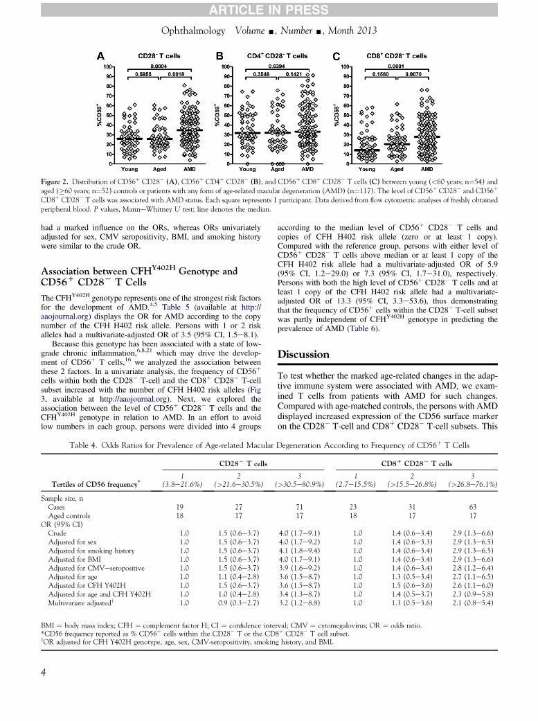
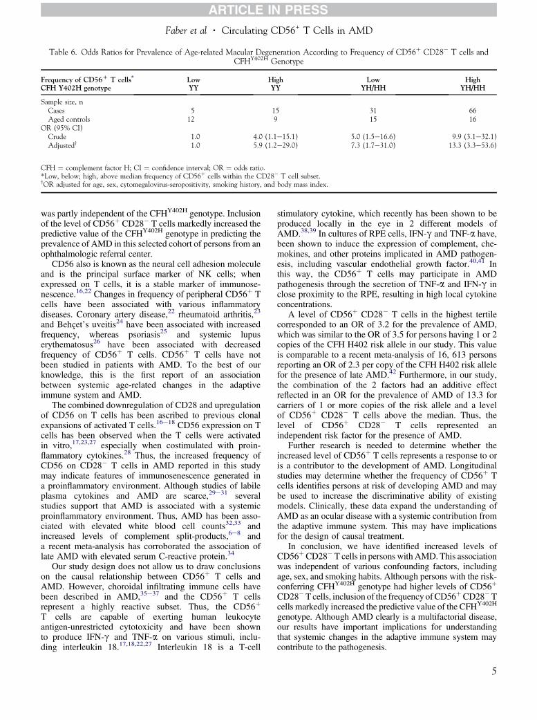
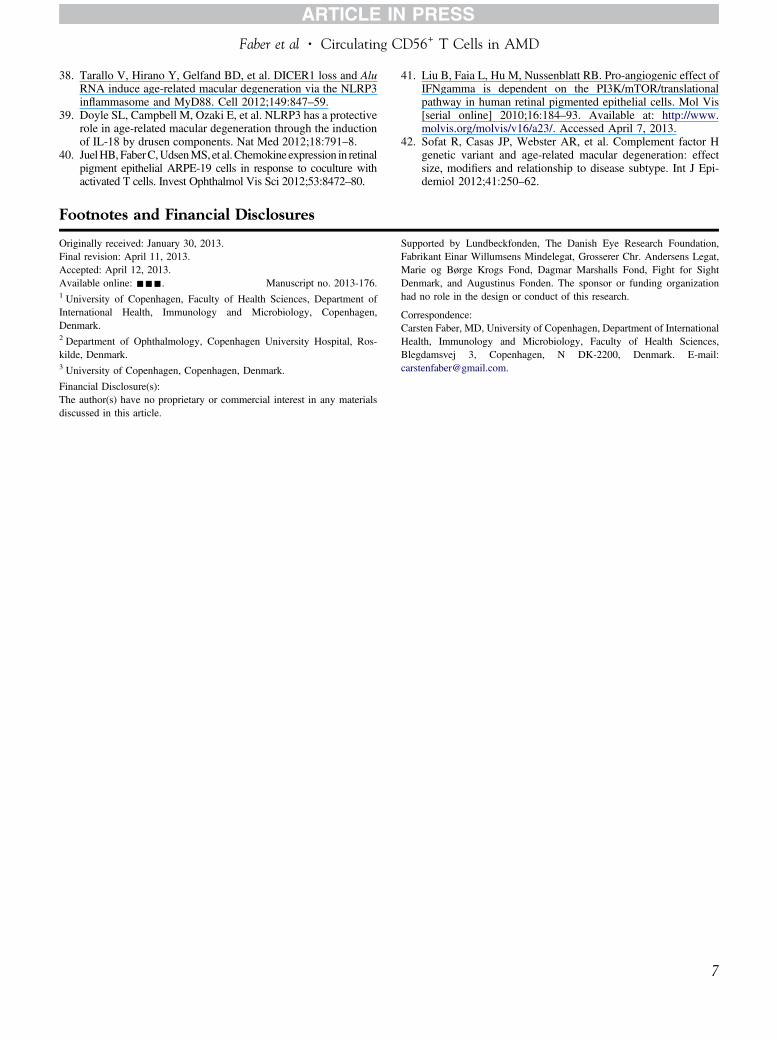