The Role of Abnormal Vitreomacular Adhesion in Age-related Macular Degeneration: Spectral Optical...
20
The Role of Abnormal Vitreomacular Adhesion in Age-related Macular Degeneration: Spectral Optical Coherence Tomography and Surgical Results Francesca Mojana, Lingyun Cheng, Dirk-Uwe G. Bartsch, Gabriel A. Silva, Igor Kozak, Nitin Nigam, and William R. Freeman Jacobs Retina Center at the Shiley Eye Center, Department of Ophthalmology (F.M., L.C., D.- U.G.B., G.A.S., I.K., N.N., W.R.F.) and the Department of Bioengineering, University of California San Diego (G.A.S.), La Jolla, California Abstract Purpose—To assess the incidence of vitreomacular adhesion and traction in age-related macular degeneration (AMD), and to evaluate surgical treatment in a subset of patients with choroidal neovascularization (CNV) nonresponsive to anti-neovascular growth factor (anti-VEGF) treatment. Design—Retrospective observational case-control and interventional case series. Methods—Spectral optical coherence tomography, combined with simultaneous scanning laser ophthalmoscope (Spectral OCT/SLO), was performed in 170 eyes of 94 elderly patients, 61 with exudative AMD, 59 with nonexudative AMD, and 50 control eyes. The presence of hyaloid adhesion to the posterior pole, and vitreomacular traction (VMT) were determined. Five patients with VMT underwent surgical hyaloid removal. Best-corrected visual acuity (BCVA) and retinal thickness were evaluated as outcomes. Results—Hyaloid adhesion was present in 17 eyes with exudative AMD (27.8%), 15 eyes with nonexudative AMD (25.4%), and eight control eyes (16%). Significant difference was found among the groups (P = .002). Among the eyes with hyaloid adhesion, VMT was shown in 10 eyes (59%) with exudative AMD, two eyes (13%) with nonexudative AMD, and one control eye (12%). VMT was associated with the severity of AMD (P = .0082). The area of hyaloid adhesion was significantly smaller than and concentric to the area of CNV complex in eyes with exudative AMD. Eyes with VMT that underwent surgery experienced a modest improvement of BCVA and decrease of retinal thickness. Conclusions—Hyaloid adhesion to the macula is associated with AMD, and frequently causes VMT in eyes with CNV. Tractional forces may antagonize the effect of anti-VEGF treatment, and cause pharmacological resistance in a subpopulation of patients. Future studies are needed to define the role of vitreoretinal surgery in such cases. Spectral OCT/SLO allows careful diagnosis and follow- up. Age-related macular degeneration (AMD) IS the leading cause of severe visual acuity (VA) loss in industrialized countries. 1–3 Although the pathogenesis of the disease is still under investigation, it is clear that the defect lies in the outer retina and retinal pigment epithelium (RPE), and that genetic predisposition has been shown to play a major role in its development. 4–6 The identified genes mainly control the immune responses in the posterior segment, directly or secondarily affecting the RPE environment and function. 7 Although AMD primarily Inquiries to William R. Freeman, University of California San Diego Department of Ophthalmology, Shiley Eye Center, 0946, Joan and Irwin Jacobs Retina Center, 9415 Campus Point Drive, La Jolla, CA 92037; e-mail: [email protected]. NIH Public Access Author Manuscript Am J Ophthalmol. Author manuscript; available in PMC 2009 September 1. Published in final edited form as: Am J Ophthalmol. 2008 August ; 146(2): 218–227. doi:10.1016/j.ajo.2008.04.027. NIH-PA Author Manuscript NIH-PA Author Manuscript NIH-PA Author Manuscript
-
Upload
independent -
Category
Documents
-
view
4 -
download
0
Transcript of The Role of Abnormal Vitreomacular Adhesion in Age-related Macular Degeneration: Spectral Optical...

The Role of Abnormal Vitreomacular Adhesion in Age-relatedMacular Degeneration: Spectral Optical Coherence Tomographyand Surgical Results
Francesca Mojana, Lingyun Cheng, Dirk-Uwe G. Bartsch, Gabriel A. Silva, Igor Kozak, NitinNigam, and William R. FreemanJacobs Retina Center at the Shiley Eye Center, Department of Ophthalmology (F.M., L.C., D.-U.G.B., G.A.S., I.K., N.N., W.R.F.) and the Department of Bioengineering, University of CaliforniaSan Diego (G.A.S.), La Jolla, California
AbstractPurpose—To assess the incidence of vitreomacular adhesion and traction in age-related maculardegeneration (AMD), and to evaluate surgical treatment in a subset of patients with choroidalneovascularization (CNV) nonresponsive to anti-neovascular growth factor (anti-VEGF) treatment.
Design—Retrospective observational case-control and interventional case series.
Methods—Spectral optical coherence tomography, combined with simultaneous scanning laserophthalmoscope (Spectral OCT/SLO), was performed in 170 eyes of 94 elderly patients, 61 withexudative AMD, 59 with nonexudative AMD, and 50 control eyes. The presence of hyaloid adhesionto the posterior pole, and vitreomacular traction (VMT) were determined. Five patients with VMTunderwent surgical hyaloid removal. Best-corrected visual acuity (BCVA) and retinal thickness wereevaluated as outcomes.
Results—Hyaloid adhesion was present in 17 eyes with exudative AMD (27.8%), 15 eyes withnonexudative AMD (25.4%), and eight control eyes (16%). Significant difference was found amongthe groups (P = .002). Among the eyes with hyaloid adhesion, VMT was shown in 10 eyes (59%)with exudative AMD, two eyes (13%) with nonexudative AMD, and one control eye (12%). VMTwas associated with the severity of AMD (P = .0082). The area of hyaloid adhesion was significantlysmaller than and concentric to the area of CNV complex in eyes with exudative AMD. Eyes withVMT that underwent surgery experienced a modest improvement of BCVA and decrease of retinalthickness.
Conclusions—Hyaloid adhesion to the macula is associated with AMD, and frequently causesVMT in eyes with CNV. Tractional forces may antagonize the effect of anti-VEGF treatment, andcause pharmacological resistance in a subpopulation of patients. Future studies are needed to definethe role of vitreoretinal surgery in such cases. Spectral OCT/SLO allows careful diagnosis and follow-up.
Age-related macular degeneration (AMD) IS the leading cause of severe visual acuity (VA)loss in industrialized countries.1–3 Although the pathogenesis of the disease is still underinvestigation, it is clear that the defect lies in the outer retina and retinal pigment epithelium(RPE), and that genetic predisposition has been shown to play a major role in its development.4–6 The identified genes mainly control the immune responses in the posterior segment, directlyor secondarily affecting the RPE environment and function.7 Although AMD primarily
Inquiries to William R. Freeman, University of California San Diego Department of Ophthalmology, Shiley Eye Center, 0946, Joan andIrwin Jacobs Retina Center, 9415 Campus Point Drive, La Jolla, CA 92037; e-mail: [email protected].
NIH Public AccessAuthor ManuscriptAm J Ophthalmol. Author manuscript; available in PMC 2009 September 1.
Published in final edited form as:Am J Ophthalmol. 2008 August ; 146(2): 218–227. doi:10.1016/j.ajo.2008.04.027.
NIH
-PA Author Manuscript
NIH
-PA Author Manuscript
NIH
-PA Author Manuscript

involves the outer retinal layers, several authors have suggested that the vitreous may possiblyplay a role in the pathogenesis and/or progression of AMD. A high incidence of posteriorvitreous attachment was intraoperatively observed by different authors.8 Subsequent studiesof the vitreous in AMD have been performed using ultrasound, which suggest that completeposterior vitreous detachment (PVD) occurs less frequently in AMD than in the age-matchedelder normal population, and that a higher incidence of vitreomacular adhesion is detected inboth exudative and nonexudative AMD.9,10 Conventional time-domain optical coherencetomography (OCT) has also been used to evaluate the vitreous in AMD, and such studies havesuggested that there is a higher rate of vitreomacular adhesion in exudative AMD, while nodifference was found between nonexudative AMD and normal population.11 Differenthypothesis have been formulated to explain the role of vitreomacular adhesion in AMD,focusing on both mechanical and biochemical factors underlying the observed phenomenon.10
We wished to determine the relationship between exudative AMD, nonexudative AMD, andelder normal eyes in terms of the presence and extent of the hyaloid attachment to the retina.We elected to evaluate this using a new technology, Spectral domain OCT (SD-OCT), whichprovides dramatic improvements in the speed and resolution of imaging of the vitreoretinalinterface and other macular diseases.12–15 Here, we evaluated a novel such instrument, whichcouples SD-OCT with the scanning laser ophthalmoscope (SLO), allowing a highly preciseco-localization of optically detected lesion in the 2-dimensional image of the posterior pole(OTI-Spectral OCT/SLO; OTI Inc, Toronto, Canada). With this dual simultaneous imaging, itis possible to correlate fundus details with ultrahigh resolution OCT, while the high sensitivityof the instrument allows observation of subtle details of the vitreous that time-domain OCTcannot visualize.
The purpose of our study was to determine the frequency of persistent hyaloid adherence tothe posterior pole in patients with AMD, and to determine whether this phenomenon divergesfrom the physiological PVD process. The hypothesis is that a local inflammation induced byAMD is causing focal corresponding adhesion, potentially inducing tractional forces on theretina. This is important because the unresponsive chronic macular edema overlying thechoroidal neovascularization (CNV) process could in theory be related to vitreomaculartraction (VMT) and might explain the resistance of some eyes to new anti-vascular endothelialgrowth factor (anti-VEGF) treatments.
We chose to use OTI-Spectral OCT/SLO because we have previously demonstrated that thisdevice allows excellent detection of subtle inner retina pathology, and in some eyes it evenallows visualization of the intact vitreous cortex (Nigam N, et al. IOVS 2006;116:ARVO E-Abstract B225).
MethodsA consecutive case series of nonexudative and exudative AMD patients, who were referred tothe Jacobs Retina Center at the University of California San Diego, in La Jolla, California andwho agreed to Spectral OCT scanning, was reviewed. All patients had AMD and underwent acomplete ophthalmologic examination, VA testing with standardized refraction using EarlyTreatment Diabetic Retinopathy Study (ETDRS) charts, and slit-lamp biomicroscopy wasperformed in mydriasis with either a 78 or a 90 diopters (D) indirect lens. The finding of theWeiss ring, or a clearly visible posterior vitreous cortex that moved freely in the vitreous cavity,indicated the presence of a complete PVD.16 The diagnosis of AMD was based on the clinicalpresence of drusen with RPE changes, geographic atrophy or CNV. All eyes underwentfluorescein angiography (FA) exam and fundus color photographs. We excluded eyes withcoexisting diseases involving the posterior pole or that would affect the presence of
Mojana et al. Page 2
Am J Ophthalmol. Author manuscript; available in PMC 2009 September 1.
NIH
-PA Author Manuscript
NIH
-PA Author Manuscript
NIH
-PA Author Manuscript
vitreomacular adhesion, such as diabetic retinopathy, inflammatory diseases, vascularocclusions, and high myopia. Eyes that had previous vitreoretinal surgery were also excluded,as well as eyes with the presence of media opacities, which would prevent good visualizationof the fundus. A control group of consecutive patients seen in clinic, who did not have AMDor any visible retinopathy, was also analyzed with the same technique to determine the presenceof PVD.
This was a retrospective analysis of a consecutive series of patients. All patients were examinedusing a Spectral OCT (OTI-Spectral OCT/SLO, OTI Inc).
Imaging TechniqueThe light source of the OTI-Spectral OCT/SLO is a modified superluminescent diode, with abandwidth of 40 nm centered at an 830 nm wavelength. The axial resolution is equal to up to5 microns, and the transverse one is 16 microns. The speed is 28,000 A-scans/sec. Every B-scan is composed of 512 A-scans and is acquired in 0.18 seconds. The OCT is coupled to aSLO. In the sensing arm of the OCT, a beam splitter redirects part of the light returned fromthe eye toward a confocal optical receiver. Both OCT and confocal signals carrying theinformation about the reflectivity of the target are collected by a dual-input, which displaystwo images on a screen under computer control. A strict topographic correspondence of theOCT cross-sectional image and the SLO image allows the operator to sweep the cursor all overthe posterior pole, focusing on specific notable areas. Each posterior pole series of B-scanscontains 128 images, covering an area of 8 mm × 8 mm throughout the posterior pole. In thecurrent study, high quality series of scans (horizontally and vertically oriented, respectively)were performed for each eye. The SLO image was centered on the fovea or on the exudativelesion, when present, on the basis of the clinical and angiographic findings. The optic nervewas scanned as well, to evaluate the attachment of the vitreous to its edge. The evidence ofPVD was determined by focusing the instrument on superficial retinal layers and into thevitreous cavity. According to the absence or presence of any adhesion of the hyaloid to theretina surface in the scanned area, the eye was classified into one of the following threecategories (modified from Uchino and Johnson17,18): 1) no evidence of posterior hyaloidadhesion; 2) persistent adhesion of hyaloid only to the optic disk; or 3) persistent adhesion ofhyaloid to the macula (in any of the four quadrants). Statistically, we ordered the categoriesfrom least to most pathology as follows: no adhesion < adhesion to optic disk < persistentadhesion to macula.
Determination of Vitreomacular Traction in Eyes with Hyaloid AdhesionIn those eyes that showed an attachment of the hyaloid to the macula, the adhesion area wascarefully analyzed using Spectral OCT; a VMT was diagnosed when a steep slope of the innermacular surface, or a sharp angulation and localized deformation of the retinal profile, wasdetected at the adhesion site of the hyaloid. Otherwise, VMT was considered to be not present.
Analysis of Hyaloid Adhesion Area, Choroidal Neovascularization Area, and RespectiveLocation in Eyes with Exudative Age-Related Macular Degeneration
Those eyes with exudative AMD and macular attachment of the hyaloid, with well detectedhyaloid in the vertical and horizontal scans, were evaluated for further analysis, as reported.The location and extent of the AMD lesion was identified with the Spectral OCT. All the areasinvolved in the CNV complex were considered: CNV, fibrosis, pigment epithelial detachment(PED), subretinal or intraretinal fluid, diffusely thickened retina, and hard exudates. Thesimultaneous SLO corresponding image allowed the operator to identify their precise location,as related to posterior pole anatomic landmarks. The vertical and horizontal diameters of theaffected region were measured in mm with calipers provided on the OTI-Spectral OCT/SLOsoftware and used to determine the lesion area. The same procedure was performed for the
Mojana et al. Page 3
Am J Ophthalmol. Author manuscript; available in PMC 2009 September 1.
NIH
-PA Author Manuscript
NIH
-PA Author Manuscript
NIH
-PA Author Manuscript

hyaloid area that showed adhesion to the inner retina. The formula for an ellipse was used toestimate both area measurements. The anatomic location of the exudative lesion and the hyaloidadhesion were then analyzed; for this purpose the horizontal scan through the fovea wasreviewed. The distance in between the center of the hyaloid attachment and the center of thefovea was measured, and the same procedure was repeated using the center of the CNVcomplex. The two distances were then compared.
Surgical InterventionA small number of patients, who showed persistent attachment of hyaloid to the macula, withevidence of VMT, were surgically treated. These patients had a history of poorly responsiveCNV despite aggressive anti-VEGF therapy (≥6 intravitreal injections of Bevacizumab orRanibizumab), demonstrable vitreomacular adhesion, and were willing to undergo surgery.One patient had progressive drusenoid RPE detachment with a decrease in VA. All surgerieswere performed by the same surgeon (W.R.F.). A tran pars plana vitrectomy (TPPV) wasperformed with 25-gauge three port instruments (Accurus Surgical System; Alcon Inc, FortWorth, Texas, USA). After core vitrectomy, the posterior hyaloid was engaged with a soft-tipped cannula, or a vitreous cutter, to create posterior hyaloid detachment. Particular care wastaken during this surgical phase of removing the hyaloid adhesion. The surgery was monitoredwith video recording and a subsequent review of the findings was performed by another author(F.M.) to confirm that the hyaloid traction was removed. In some cases SF6 or C3F8 were usedfor tamponade. Intravitreal Bevacizumab was administered at the end of the surgery and on amonthly basis in all of the exudative AMD cases. Neither triamcinolone acetonide nor othertreatments for CNV were used. A complete eye exam was performed at day one, seven, and14 of the follow-up, and at each subsequent month. Best-corrected visual acuity (BCVA) wasmeasured with ETDRS charts at baseline and at each follow-up. Spectral OCT scanning wasrepeated at month one and subsequently every three months.
Statistical AnalysisTo delineate any association between macular abnormality and vitreomacular adhesion, anordinal logistic regression was simulated using the graded vitreomacular adhesion as theresponse variable, and using the macular disease status as a predictor. The eyes were classifiedinto three categories, normal (0), nonexudative AMD (1), and exudative AMD (2) (severityorder). The adhesion of hyaloid to the posterior pole was also graded in order of severity: noadhesion presence < adhesion to the disk < adhesion to the macula. In addition, thevitreomacular adhesion was also classified as binary response (presence of macular hyaloidadhesion or no macular adhesion; eyes with hyaloid adhesion to the disk and no adhesion wereconsidered as part of the same group). Both an ordinary logistic regression and a binary logisticregression were performed. Attributable to the fact that both eyes of the same subject wereused in this study, Generalized Estimating Equations (GEE) was applied to analyze the datato control for intereye dependence; gender, age and lens status were analyzed as the covariates.19,20P values are reported as two-tailed test results.
For the subanalysis of the eyes with hyaloid adhesion to the macula, the adhesion was gradedas tractional or nontractional according the configuration of the retinal profile. The Chi-squarealong with the Cochran-Armitage trend test were utilized.
In addition, within the exudative AMD group, we analyzed the possible correlation betweenvitreomacular adhesion and types of CNV using GEE, and the possible relationship betweentractional vitreoretinal adhesion and the type of CNV using the Fisher exact test. We alsoanalyzed the topographic relationship between the CNV complex and the vitreomacularadhesion by correlating the distance between the fovea and the center of the CNV complexand the hyaloid adhesion, respectively. LogMAR VA was used for the statistic, as the eyes
Mojana et al. Page 4
Am J Ophthalmol. Author manuscript; available in PMC 2009 September 1.
NIH
-PA Author Manuscript
NIH
-PA Author Manuscript
NIH
-PA Author Manuscript
were all tested on the ETDRS chart. All statistical analysis was performed using SAS softwareversion 9.1 (SAS Inc, Cary, North Carolina, USA).
ResultsOTI-Spectraloct/SLO was performed in 170 eyes of 94 patients. Fifty-eight patients werefemales, 36 were males. The average age for the entire group was 76 years (SD ± 8.4). Eighty-three eyes were phakic and 87 were pseudophakic. AMD was diagnosed in 120 eyes (61 eyeshad exudative AMD and 59 nonexudative AMD). Fifty eyes showed no evidence of AMD andconstituted the control group of the study.
The average age of patients with exudative AMD was 79.02 years (SD ± 7.21), while it was75.29 years (SD ± 9.08) in the group of nonexudative AMD and in the control group it was72.12 years (SD ± 7.48). At a careful biomicroscopical exam, 16 eyes showed a complete PVDin the exudative AMD group (26%), 13 in the nonexudative group (22%), and 13 in the controlgroup (26%).
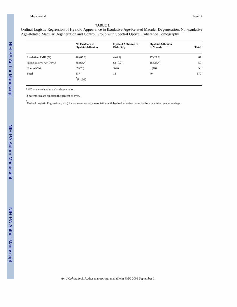
The ordinal logistic regression revealed that age and gender were the significant covariates(P < .0001 and P = .0065). After adjustment for these effects, vitreomacular adhesion wassignificantly associated with macular abnormality (P = .002; Table 1). Further analysis showedthat, when compared with the controls, both exudative and nonexudative AMD eyes weresignificantly more likely to have vitreomacular adhesion (odds ratio (OR), 6.3 for exudativevs control and 3.2 for nonexudative vs control; P = .0012 and P = .0141, respectively). Inanother analysis, eyes with the hyaloid attached only to the optic disk and eyes with no adhesionwere considered to be part of the same group: the binary logistic regression showed similarresults. The significant covariates were age and gender (P = .0018 and P = .0065). Afteradjustment of these effects, vitreomacular adhesion was again associated with macularabnormality (P = .01). The analysis revealed that, when compared with the control eyes,exudative AMD were significantly more likely to have vitreomacular adhesion (OR, 5.3; P = .0052). Compared with control eyes, nonexudative AMD showed a statistically marginalassociation with vitreomacular adhesion (OR, 2.7; P = .06). No statistical significance wasfound when comparing exudative and nonexudative AMD eyes. (OR, 1.9; P = .15) (Table 2).
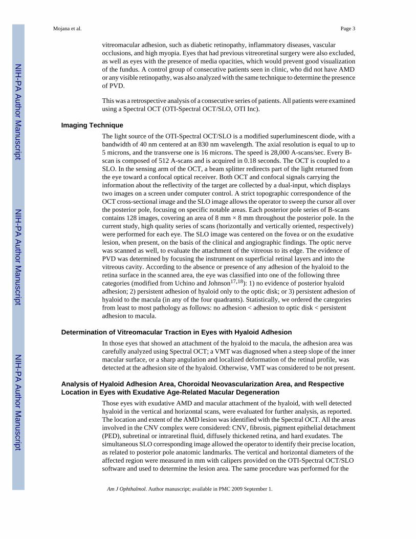
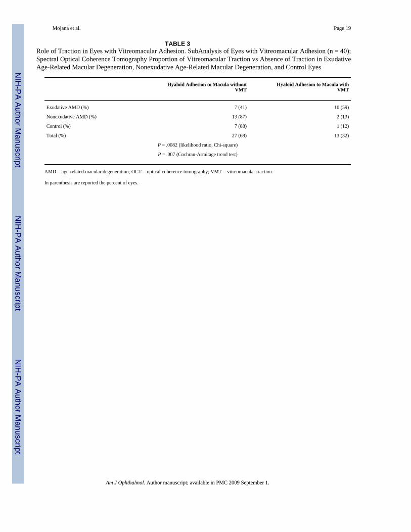
The 40 eyes that had hyaloid adhesion to the macula were further analyzed for the presence ofa tractional component. The analysis showed that VMT was significantly associated with thepresence of AMD (P = .0082 likelihood ratio, Chi-square test) and that there was a significantincrease in VMT when the disease category changed from control to nonexudative AMD toexudative AMD (P = .007 Cochran-Armitage trend test; Table 3; Figure 1).
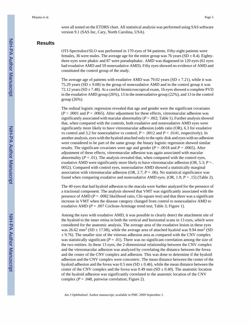
Among the eyes with exudative AMD, it was possible to clearly detect the attachment site ofthe hyaloid to the inner retina in both the vertical and horizontal scans in 13 eyes, which wereconsidered for the anatomic analysis. The average area of the exudative lesion in these eyeswas 26.62 mm2 (SD ± 17.08), while the average area of attached hyaloid was 9.94 mm2 (SD± 9.76). The smaller size of the vitreous adhesion area as compared with the CNV complexwas statistically significant (P = .01). There was no significant correlation among the size ofthe two entities. In these 13 eyes, the 2-dimensional relationship between the CNV complexand the vitreomacular adhesion was analyzed by correlating the distance between the foveaand the center of the CNV complex and adhesion. This was done to determine if the hyaloidadhesion and the CNV complex were concentric. The mean distance between the center of thehyaloid adhesion and the fovea was 0.5 mm (SD ± 0.46), while the mean distance between thecenter of the CNV complex and the fovea was 0.49 mm (SD ± 0.40). The anatomic locationof the hyaloid adhesion was significantly correlated to the anatomic location of the CNVcomplex (P = .048, pairwise correlation; Figure 2).
Mojana et al. Page 5
Am J Ophthalmol. Author manuscript; available in PMC 2009 September 1.
NIH
-PA Author Manuscript
NIH
-PA Author Manuscript
NIH
-PA Author Manuscript
In the subgroup analysis (exudative AMD), we found that minimally classic lesions showedsignificantly higher incidence of local vitreomacular adhesion than classic (OR, 7.7, 1.72 to34.8; P = .0077) and occult (OR, 7.0, 1.17 to 41.6; P = .0327). However, a Chi-square analysisdid not show association between tractional vitreomacular adhesion and types of CNV (classic3/10 vs minimally classic 6/10 vs occult 1/10; P = .4).
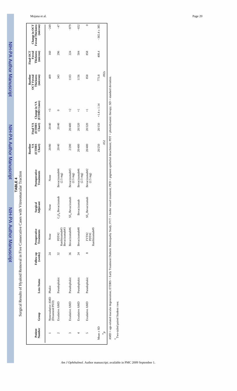
Five patients with evidence of VMT had 25-gauge TPPV with hyaloid removal. Four patientsexperienced an improvement of VA after surgery, as assessed at the end of the follow-up. Inone eye the VA remained stable. The mean VA improvement was 1.4 ETDRS lines (SD ±1.14; P = .052, two-tailed paired Student t test). Four patients had showed a decrease of OCTfoveal thickness, when compared with the preoperative exam. The patient with the shortestfollow-up showed a stable retinal thickness on OCT. The mean retinal thickness decrease onSpectral OCT was −365.4 micron (SD ± 385 micron; P = .092, two-tailed paired Student t test).No complications occurred during the surgeries and the follow-up period. All the patientsaffected by exudative AMD, after surgery, were treated with intravitreal Bevacizumab injection(2.5 mg) every four weeks. The treatment was ongoing at the last follow-up. Details are reportedin Table 4; Figures 3 and 4 show examples of OCT changes in two patients.
One patient with exudative AMD and VMT, caused by a small area of focal hyaloid adhesion,experienced a spontaneous resolution of the attachment, with decrease of macular thickness.Vision did not improve despite ongoing monthly treatments with intravitreal injections ofBevacizumab (Figure 5).
DiscussionOur study clearly shows that eyes with exudative AMD have a significantly higher incidenceof incomplete PVD than elder normal eyes and a similar trend exists also when comparingnonexudative AMD with the control group. We have also shown that there is a higher incidenceof tractional configuration of the attached posterior hyaloid in eyes with exudative AMD. Otherinvestigators have also suggested a possible role of persistent vitreomacular adhesion in eyeswith exudative macular degeneration.9–11,21 Those studies were limited in several ways, andparticularly in the use of ultrasound and/or time domain OCT to evaluate the phenomenon.Our evaluative technique, confocal simultaneous SLO fundus imaging performedsimultaneously with ultra-high speed and ultra-high resolution Spectral OCT, allows theprecise anatomic localization of the vitreoretinal adhesion in relationship to the CNV complex.Our study confirms some of the findings of other authors and gives new and statisticallysignificant information, including the 3-dimensional configuration of the VMT in exudativeAMD cases, the size of the hyaloid adhesion, and its concentricity to the CNV complex. Wefurthermore studied the response of a subpopulation of eyes to surgical intervention.Discrepancies exist in the literature when nonexudative AMD eyes are analyzed. Ondes foundno statistically significant difference between the exudative and nonexudative groups withrespect to PVD.10 In contrast, Krebs reported no statistically significant difference whencomparing nonexudative eyes with a control group.11 Our analysis was conducted with themore advanced instrumentation and showed that the hyaloid adhesion is more frequent innonexudative AMD than in control eyes.
We did not find a correlation between the size of the hyaloid attachment and the area of theCNV complex. This is not surprising, as the duration of the disease and other factors may affectthis finding. It is interesting, and probably physiologically important, that in our study we didfind a concentric configuration of the two processes, suggesting a causal relationship.Nonexudative AMD is mainly represented by multiple lesions variably spread throughout themacula. Being impossible to define the exact anatomical border in these cases, we could justevaluate the topographical relationship existing among the CNV complex in exudative AMD
Mojana et al. Page 6
Am J Ophthalmol. Author manuscript; available in PMC 2009 September 1.
NIH
-PA Author Manuscript
NIH
-PA Author Manuscript
NIH
-PA Author Manuscript
and the incomplete PVD. A trend was nevertheless seen when the group of nonexudative AMDpatients was compared with controls, suggesting that even initial AMD stages might induce areaction at the inner interface of the retina.
It is interesting to speculate on the cause of vitreomacular adhesion, which is seen mostfrequently in exudative AMD eyes, but also more often in nonexudative eyes than elder normaleyes. Furthermore, in exudative AMD patients, minimally classic lesions had a higherincidence of focal vitreomacular adhesion. The results are indicative that a slower, morechronic, and indolent process, and the consequent persistent low grade of local inflammation,are more likely to induce structural changes at the level of the vitreoretinal interface. Themajority of eyes in our study had minimally classic CNV and we therefore do not have anadequate number of eyes to test whether VMT is statistically associated with minimally classicCNV as opposed to other types. The persistent adhesion of the hyaloid to the posterior pole,with or without VMT, is not the underlying cause of AMD. It has become clear, recently, thatthe cause of AMD is one of several genetic defects, which affect the outer retina and RPE.Other authors suggested that vitreomacular adhesion might be considered a risk factor for thenatural history of this disease.11,22–24 This would facilitate in fact the progression fromnonexudative to exudative forms of AMD. The hypotheses are multiple: persistentvitreomacular adhesion might induce chronic low-grade inflammation, prevent normal oxygenand nutrients diffusion to the macula, and/or confine proangiogenic cytokines in the macula.Thus, there is no suggestion that vitrectomy may be useful in preventing the progression ofAMD. We know that both inflammation and disruption of the photoreceptor-RPE interface,resulting in cellular apoptosis in the outer retina, can induce the recruitment of Müller cellsand possibly astrocytes to become reactive. Reactive gliosis is associated with physical changessuch as hypertrophy as well as biochemical changes, including the secretion of variousinflammatory cytokines and reversal of ion pumps and homeostatic buffering.25–28 Thesechanges could directly affect retinal-vitreal adhesion points that induce firmer fibrousattachments between the retina and vitreous. Nonexudative AMD precedes exudative AMDby years.29,30 We have shown that nonexudative AMD eyes also have a higher incidence ofvitreomacular adhesions than control eyes, but not as much as in exudative AMD eyes. Theadditional insult of CNV in eyes with exudative AMD likely increases the VMT throughinteractions with the Müller cell footplates and posterior hyaloid membrane. Our study clearlyshows that anterior-posterior forces, present at the surface of the retina, may exacerbate themacular edema, which originally occurs secondary to the presence of leaking vessels in theCNV complex. Some authors have hypothesized that increases in the mechanical forcesassociated with abnormal vitreal attachments may result in the secretion of signaling factorsby the Müller cells with both paracrine and autocrine effects. This process leads to a cascadeof inflammatory factors and local vascular changes, which may include vascular leakiness, andsubsequent cystoid macular edema (CME).31 In turn, in eyes with CNV, this could set up avicious cycle, in which the inflammation, the reactive gliosis, and the tractional forces mayresult in worsening the chronic exudation of the underlying disease.
In a subset of eyes, we studied the role of surgical intervention. These eyes showed clinicalresistance to fluid reabsortion despite frequent and aggressive anti-VEGF therapy, given in theform of monthly intravitreal injections of Bevacizumab or Ranibizumab. None of the fivepatients, at the time of the surgery, had signs of epiretinal membrane (ERM). PseudophakicCME was excluded attributable to the temporal sequence of cataract surgery and CME: all thecases had an interval of time of at least one year without a history of CME, before the onset ofthe CNV. This was a consecutive series of patients without a prospective, standardized surgicalmethodology. No other patients in the study population had surgery during the study period.Gas tamponade was used in three out of five cases in the hope that it would assist in flatteningthe macular fluid. We cannot comment further on whether gas should be used and which gasshould be used. Our surgical outcome showed an improvement in vision, which was of
Mojana et al. Page 7
Am J Ophthalmol. Author manuscript; available in PMC 2009 September 1.
NIH
-PA Author Manuscript
NIH
-PA Author Manuscript
NIH
-PA Author Manuscript
borderline significance using the two-tailed Student t test (P = .052). This marginal result maybe attributable to the fact that most eyes in our series were operated on after disease was longstanding despite aggressive intravitreal treatment. It is interesting that the eye with the mostvisual improvement had a progressive drusenoid RPE detachment. Less improvement was seenin the other eyes, but was present in three of the four remaining eyes with active CNV. Ourresults suggest that there may be a subpopulation of CNV eyes that do not respond to anti-VEGF therapy and in which VMT may play a component in the resistance to such therapy. Wedo not know if, in this group of patients, the visual improvement is secondary just to the releaseof VMT (combined with ongoing anti-VEGF treatment). Other factors may play an ancillaryrole: vitrectomy is in fact known to improve retinal oxygenation and the compressive effect ofgas is also likely to facilitate a reabsortion of the intraretinal fluid. This series has a smallnumber of patients and we are not advocating surgery in all eyes with CNV and VMT. Furtherstudies are necessary. Surgery in such eyes can result in complications and vitrectomy mayalso complicate anti-VEGF treatment, as it may shortens the half-life of intravitreal anti-VEGFdrugs, so that higher doses of anti-VEGF drugs may be needed postoperatively.32
Mild ERM was present in a minority of patients; none of our patients had a history of CME orERM causing vision loss prior to the development of CNV. All the patients who showed someedema and were considered affected by exudative AMD had clear FA signs of CNV. All thepatients that had surgery did not show any ERM at the baseline exam. The presence of ERMcan be another cause of chronic CME in AMD patients, and should also be considered in theclinical practice. As for VMT, new OCT technology nowadays represents the most sensitivetest to identify this subpopulation of patients.
In our study, we utilized the OTI-Spectral OCT/SLO with gold plated mirrors, which improvedthe visualization of the vitreous. Gold plated mirrors have, in fact, a higher reflectivity thanaluminum and silver coating at the operating wavelength of the instrument (infrared). We donot know which coating material is used by the other manufacturers, but in our experience thevisualization of the vitreoretinal interface, including hyaloid adhesions, ERM, VMT, andvitreous cortex detection, is highly enhanced with this device (results unpublished). We didcombine that study with careful slit-lamp biomicroscopy to evaluate the vitreous planes. Werecognize that ultrasound is the standard technique to detect PVD; however, subtle adhesionto the retina or vitreal schisis (as other authors have previously described) are difficult to detectand quantify with ultrasound.33 We recognize that a complete PVD could be missed in theclinical exam and that OCT is not the standard technique to tell a complete PVD from acompletely attached vitreous. It is important to note that this would not affect our results. Ourstudy was not designed to determine a correlation of complete PVD or completely attachedvitreous with AMD, but the specific correlation of AMD with focal vitreomacular adhesionand eventual traction. In fact, our statistical analysis is not based on the presence or absenceof a complete vitreous detachment, but only on the presence of partial adhesion. The hypothesiswas that a local inflammation induced by AMD is causing a focal corresponding adhesion, therest of the retina acting as a normal tissue. Future studies may wish to also incorporate dynamicultrasound and a larger number of patients to confirm and expand our findings. We do knowthat the instrument we utilized does allow a focal offset of up to 10 mm, which we used to ouradvantage in searching for the posterior hyaloid membrane.
In conclusion, persistence of hyaloid adhesion to the posterior pole is highly associated withAMD. This adhesion is often responsible for VMT in eyes affected by exudative AMD.Mechanical factors related to the traction forces may antagonize the effect of anti-VEGFtreatment, thus being responsible for pharmacological resistance in a subpopulation of AMDpatients. Further studies need to be conducted to assess if surgery may be considered as atreatment option in these cases. New Spectral OCT technology, combined with simultaneous
Mojana et al. Page 8
Am J Ophthalmol. Author manuscript; available in PMC 2009 September 1.
NIH
-PA Author Manuscript
NIH
-PA Author Manuscript
NIH
-PA Author Manuscript

SLO, allows a highly sensitive diagnosis and a subsequent selection of cases, and a carefulfollow-up.
AcknowledgmentsTHIS STUDY WAS SUPPORTED BY THE RESEARCH TO PREVENT BLINDNESS (RPB), NEW YORK, NEWYORK, THE DEPARTMENT National Eye Institute (NIH-NEI) Grant No. EY16323 (Dr Bartsch), and the NationalInstitutes of Health (NIH) Grant No. EY07366 (Dr Freeman). Dr Freeman is the recipient of an RPB Physician Scientistaward. Dr Bartsch has received discounted products and specialized software from OTI, Inc. Involved in design andconduct of study (F.M., L.C., W.R.F.); collection (F.M.); management (F.M., L.C., W.R.F.); analysis and interpretationof the data (F.M., L.C., G.A.S., W.R.F.); and preparation, review, or approval of the manuscript (F.M., L.C., D.-U.G.B., G.A.S., W.R.F.). The University of California San Diego Institutional Review Board approval was obtainedto conduct this study.
References1. La Cour M, Kiilgaard JF, Nissen MH. Age-related macular degeneration: epidemiology and optimal
treatment. Drugs Aging 2002;19:101–133. [PubMed: 11950377]2. Congdon N, O'Colmain B, Klaver CC, et al. Causes and prevalence of visual impairment among adults
in the United States. Arch Ophthalmol 2004;122:477–485. [PubMed: 15078664]3. Klein R, Klein BE, Lee KE, Cruickshanks KJ, Gangnon RE. Changes in visual acuity in a population
over a 15-year period: the Beaver Dam Eye Study. Am J Ophthalmol 2006;142:539–549. [PubMed:17011842]
4. Klein RJ, Zeiss C, Chew EY, et al. Complement factor H polymorphism in age-related maculardegeneration. Science 2005;308:385–389. [PubMed: 15761122]
5. Dewan A, Liu M, Hartman S, et al. HTRA1 promoter polymorphism in wet age-related maculardegeneration. Science 2006;314:989–992. [PubMed: 17053108]
6. Mori K, Horie-Inoue K, Kohda M, et al. Association of the HTRA1 gene variant with age-relatedmacular degeneration in the Japanese population. J Hum Genet 2007;52:636–641. [PubMed:17568988]
7. Nussenblatt RB, Ferris F III. Age-related macular degeneration and the immune response: implicationsfor therapy. Am J Ophthalmol 2007;144:618–626. [PubMed: 17698021]
8. Lambert HM, Capone A, Aaberg TM, et al. Surgical excision of subfoveal neovascular membranes inage-related macular degeneration. Am J Ophthalmol 1992;113:257–262. [PubMed: 1371906]
9. Weber-Krause B, Eckardt U. Incidence of posterior vitreous detachment in eyes with and without age-related macular degeneration. An ultrasonic study. Ophthalmologe 1996;93:660–665. [PubMed:9081520]
10. Ondes F, Yilmaz G, Acar MA, Unlu N, Kocaoglan H, Arsan AK. Role of the vitreous in age-relatedmacular degeneration. Jpn J Ophthalmol 2000;44:91–93. [PubMed: 10698032]
11. Krebs I, Brannath W, Glittenberg C, Zeiler F, Sebag J, Binder S. Posterior Vitreomacular adhesion:a potential risk factor for exudative age-related macular degeneration? Am J Ophthalmol2007;144:741–746. [PubMed: 17884003]
12. Wojtkowski M, Leitgeb R, Kowalczyk A, Bajraszewski T, Fercher AF. In vivo human retinal imagingby Fourier-domain optical coherence tomography. J Biomed Opt 2002;7:457–463. [PubMed:12175297]
13. De Boer JF, Cense B, Park BH, Pierce MC, Tearney GJ, Bouma BE. Improved signal-to-noise ratioin spectral-domain compared with time-domain optical coherence tomography. Optics Letters2003;28:2067–2069. [PubMed: 14587817]
14. Schmidt-Erfurth U, Leitgeb RA, Michels S, et al. Three-dimensional ultra-high-resolution opticalcoherence tomography of macular diseases. Invest Ophthalmol Vis Sci 2005;46:3393–3402.[PubMed: 16123444]
15. Srinivasan VJ, Wojtkowski M, Witkin AJ, et al. High-definition and 3-dimensional imaging ofmacular pathologies with high-speed ultra-high-resolution optical coherence tomography.Ophthalmology 2006;113:2054 e1–14. [PubMed: 17074565]
Mojana et al. Page 9
Am J Ophthalmol. Author manuscript; available in PMC 2009 September 1.
NIH
-PA Author Manuscript
NIH
-PA Author Manuscript
NIH
-PA Author Manuscript
16. Kakehashi A, Kado M, Akiba J, Hirokawa H. Variations of posterior vitreous detachment. Br JOphthalmol 1997;81:527–532. [PubMed: 9290361]
17. Uchino E, Uemura A, Ohba N. Initial stages of posterior vitreous detachment in healthy eyes of olderpersons evaluated by optical coherence tomography. Arch Ophthalmol 2001;119:1475–1479.[PubMed: 11594947]
18. Johnson MW. Perifoveal vitreous detachment and its macular complications. Trans Am OphthalmolSoc 2005;103:537–567. [PubMed: 17057817]
19. Liang KY, Zeger SL. Longitudinal data analysis using generalized linear models. Biometrika1986;73:13–22.
20. Lipsitz SH, Kim K, Zhao L. Analysis of repeated categorical data using generalized estimatingequations. Statistics in Medicine 1994;13:1149–1163. [PubMed: 8091041]
21. Quaranta-El Maftouhi M, Mauget-Faysse M. Anomalous vitreoretinal adhesions in patients withexudative age-related macular degeneration: an OCT study. Eur J Ophthalmol 2006;16:134–137.[PubMed: 16496257]
22. Anderson DH, Mullins RF, Hageman GS, Johnson LV. A role for local inflammation in the formationof drusen in the aging eye. Am J Ophthalmol 2002;134:411–431. [PubMed: 12208254]
23. Donoso LA, Kim D, Frost A, Callahan A, Hageman G. The role of inflammation in the pathogenesisof age-related macular degeneration. Surv Ophthalmol 2006;51:137–152. [PubMed: 16500214]
24. Zarbin MA. Current concepts in the pathogenesis of age-related macular degeneration. ArchOphthalmol 2004;122:598–614. [PubMed: 15078679]
25. Johnson PT, Lewis GP, Talaga KC, et al. Drusen-associated degeneration in the retina. InvestOphthalmol Vis Sci 2003;44:4481–4488. [PubMed: 14507896]
26. Wu KH, Madigan MC, Billson FA, Penfold PL. Differential expression of GFAP in early vs lateAMD: a quantitative analysis. Br J Ophthalmol 2003;87:1159–1166. [PubMed: 12928288]
27. Ramirez JM, Ramirez AI, Salazar JJ, de Hoz R, Trivino A. Changes of astrocytes in retinal aging andage-related macular degeneration. Exp Eye Res 2001;73:601–615. [PubMed: 11747361]
28. Petrukhin K. New therapeutic targets in atrophic age-related macular degeneration. Expert Opin TherTargets 2007;11:625–639. [PubMed: 17465722]
29. Klein R, Klein BE, Jensen SC, Meuer SM. The five-year incidence and progression of age-relatedmaculopathy: the Beaver Dam Eye Study. Ophthalmology 1997;104:7–21. [PubMed: 9022098]
30. Van Leeuwen R, Klaver CC, Vingerling JR, Hofman A, de Jong PT. The risk and natural course ofage-related maculopathy: follow-up at 6 1/2 years in the Rotterdam Study. Arch Ophthalmol2003;121:519–526. [PubMed: 12695249]
31. Schubert HD. Cystoid macular edema: the apparent role of mechanical factors. Prog Clin Biol Res1989;312:277–291. [PubMed: 2678144]
32. Freeman WR, Falkenstein I. Avastin and new treatments for AMD: where are we? Retina2006;26:853–858. [PubMed: 17031283]
33. Chu TG, Lopez PF, Cano MR, et al. Posterior vitreoschisis. An echographic finding in proliferativediabetic retinopathy. Ophthalmology 1996;103:315–322. [PubMed: 8594520]
Biography
Mojana et al. Page 10
Am J Ophthalmol. Author manuscript; available in PMC 2009 September 1.
NIH
-PA Author Manuscript
NIH
-PA Author Manuscript
NIH
-PA Author Manuscript

Francesca Mojana, MD, graduated at the Universita' degli Studi di Milano, Milano, Italy, in2002. In 2006, she completed her residency at the Department of Ophthalmology of the sameuniversity. Since then, she started a fellowship at Jacobs Retina Center, Shiley Eye Center,University of California San Diego, California. Dr Mojana's concentration is in the areas ofresearch and clinical work in macular pathologies, especially focusing on new opticalcoherence tomography technology and angiography.
Mojana et al. Page 11
Am J Ophthalmol. Author manuscript; available in PMC 2009 September 1.
NIH
-PA Author Manuscript
NIH
-PA Author Manuscript
NIH
-PA Author Manuscript

FIGURE 1.Classification scheme of hyaloid adhesion and vitreomacular traction (VMT) seen withSpectral optical coherence tomography (OCT). (Top) A normal eye of an elder patient: thehyaloid membrane is visible and completely detached from the fovea, though there is apersistent adhesion to the optic nerve (right frame). (Middle) Eye with nonexudative age-related macular degeneration (AMD) and drusen: the hyaloid is attached over the entire macula,including the fovea. We considered this as a “no traction” configuration, since no distortion isvisible on the retinal surface and the angle of insertion of the hyaloid onto the retina is notsteep. The lucency between the center of the macula and the vitreous collagen is likely theposterior precortical vitreous pocket. (Bottom) Eye with choroidal neovascularization (CNV).The persistence of hyaloid adhesion causes VMT over the CNV complex: a focal distortion ofthe retinal profile is visible at the site of hyaloid attachment.
Mojana et al. Page 12
Am J Ophthalmol. Author manuscript; available in PMC 2009 September 1.
NIH
-PA Author Manuscript
NIH
-PA Author Manuscript
NIH
-PA Author Manuscript
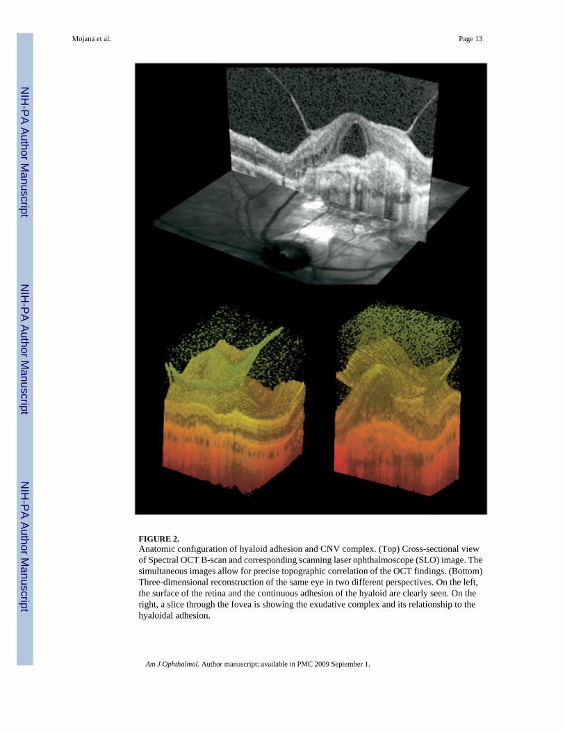
FIGURE 2.Anatomic configuration of hyaloid adhesion and CNV complex. (Top) Cross-sectional viewof Spectral OCT B-scan and corresponding scanning laser ophthalmoscope (SLO) image. Thesimultaneous images allow for precise topographic correlation of the OCT findings. (Bottom)Three-dimensional reconstruction of the same eye in two different perspectives. On the left,the surface of the retina and the continuous adhesion of the hyaloid are clearly seen. On theright, a slice through the fovea is showing the exudative complex and its relationship to thehyaloidal adhesion.
Mojana et al. Page 13
Am J Ophthalmol. Author manuscript; available in PMC 2009 September 1.
NIH
-PA Author Manuscript
NIH
-PA Author Manuscript
NIH
-PA Author Manuscript
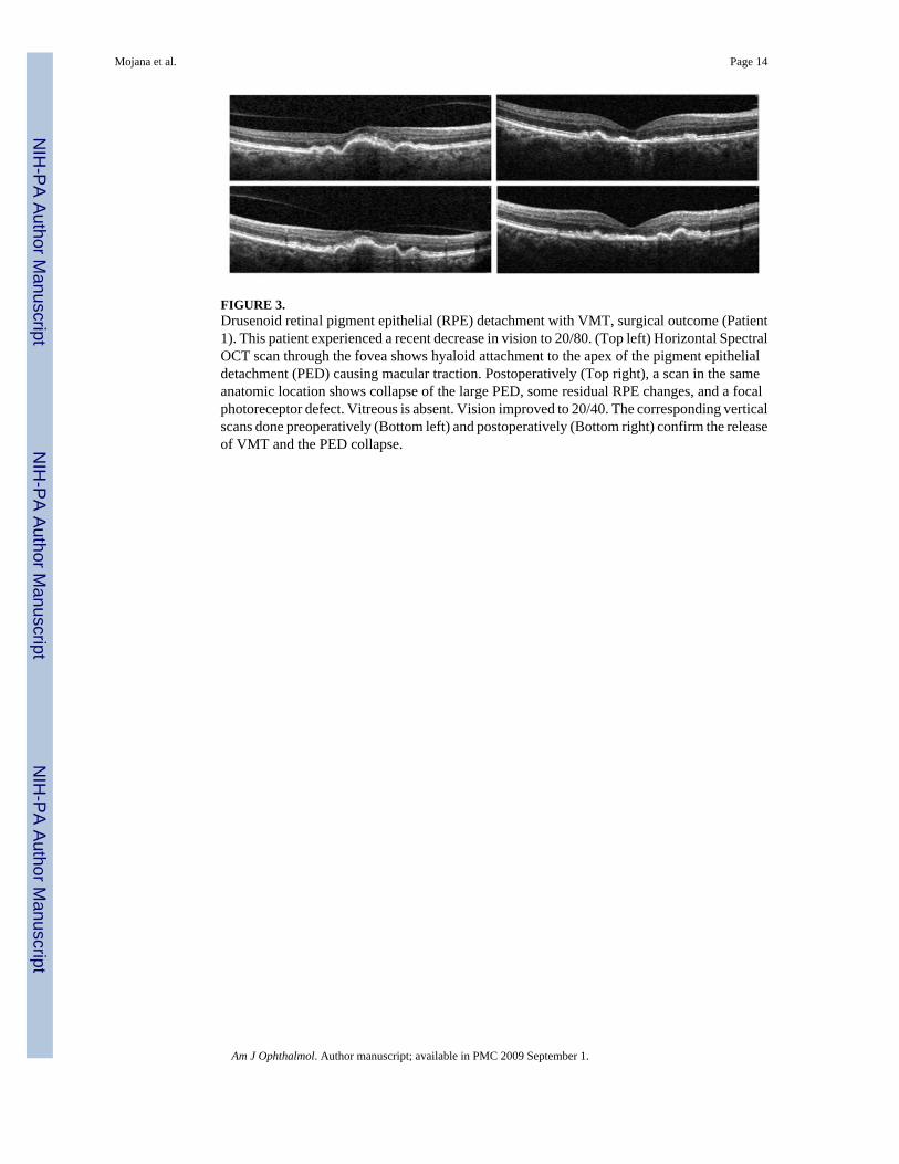
FIGURE 3.Drusenoid retinal pigment epithelial (RPE) detachment with VMT, surgical outcome (Patient1). This patient experienced a recent decrease in vision to 20/80. (Top left) Horizontal SpectralOCT scan through the fovea shows hyaloid attachment to the apex of the pigment epithelialdetachment (PED) causing macular traction. Postoperatively (Top right), a scan in the sameanatomic location shows collapse of the large PED, some residual RPE changes, and a focalphotoreceptor defect. Vitreous is absent. Vision improved to 20/40. The corresponding verticalscans done preoperatively (Bottom left) and postoperatively (Bottom right) confirm the releaseof VMT and the PED collapse.
Mojana et al. Page 14
Am J Ophthalmol. Author manuscript; available in PMC 2009 September 1.
NIH
-PA Author Manuscript
NIH
-PA Author Manuscript
NIH
-PA Author Manuscript
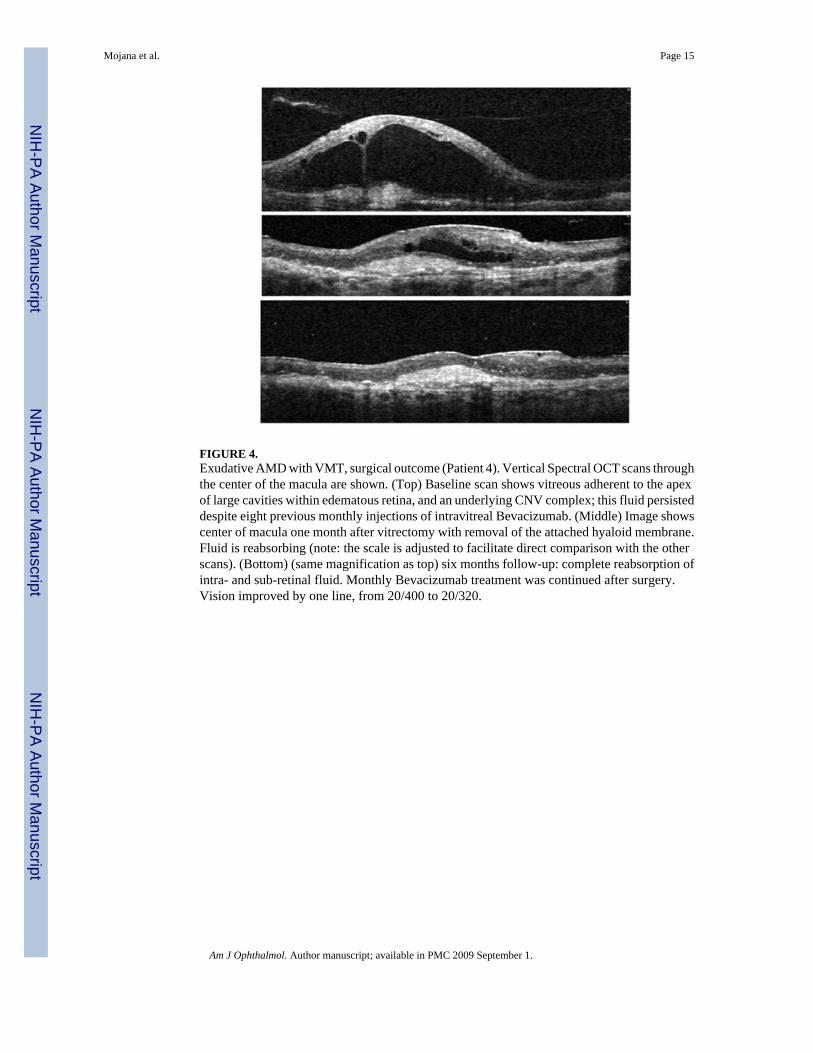
FIGURE 4.Exudative AMD with VMT, surgical outcome (Patient 4). Vertical Spectral OCT scans throughthe center of the macula are shown. (Top) Baseline scan shows vitreous adherent to the apexof large cavities within edematous retina, and an underlying CNV complex; this fluid persisteddespite eight previous monthly injections of intravitreal Bevacizumab. (Middle) Image showscenter of macula one month after vitrectomy with removal of the attached hyaloid membrane.Fluid is reabsorbing (note: the scale is adjusted to facilitate direct comparison with the otherscans). (Bottom) (same magnification as top) six months follow-up: complete reabsorption ofintra- and sub-retinal fluid. Monthly Bevacizumab treatment was continued after surgery.Vision improved by one line, from 20/400 to 20/320.
Mojana et al. Page 15
Am J Ophthalmol. Author manuscript; available in PMC 2009 September 1.
NIH
-PA Author Manuscript
NIH
-PA Author Manuscript
NIH
-PA Author Manuscript

FIGURE 5.Spontaneous resolution of VMT in exudative AMD. Four scans through the fovea of the sameeye. (Top left) Large amount of intraretinal fluid with cystic formation that persisted after threemonthly intravitreal injections of Bevacizumab. (Top right) Same view after five months. Aspontaneous release of the central VMT is detected by Spectral OCT. (Bottom left) Verticalscan through the fovea at the same time as the top left panel. (Bottom right) Vertical view afterthe spontaneous release of traction. There was no improvement in vision despite ongoingtreatment.
Mojana et al. Page 16
Am J Ophthalmol. Author manuscript; available in PMC 2009 September 1.
NIH
-PA Author Manuscript
NIH
-PA Author Manuscript
NIH
-PA Author Manuscript
NIH
-PA Author Manuscript
NIH
-PA Author Manuscript
NIH
-PA Author Manuscript
Mojana et al. Page 17
TABLE 1Ordinal Logistic Regression of Hyaloid Appearance in Exudative Age-Related Macular Degeneration, NonexudativeAge-Related Macular Degeneration and Control Group with Spectral Optical Coherence Tomography
No Evidence ofHyaloid Adhesion
Hyaloid Adhesion toDisk Only
Hyaloid Adhesionto Macula Total
Exudative AMD (%) 40 (65.6) 4 (6.6) 17 (27.8) 61
Nonexudative AMD (%) 38 (64.4) 6 (10.2) 15 (25.4) 59
Control (%) 39 (78) 3 (6) 8 (16) 50
Total 117 13 40 170*P =.002
AMD = age-related macular degeneration.
In parenthesis are reported the percent of eyes.
*Ordinal Logistic Regression (GEE) for decrease severity association with hyaloid adhesion corrected for covariates: gender and age.
Am J Ophthalmol. Author manuscript; available in PMC 2009 September 1.

NIH
-PA Author Manuscript
NIH
-PA Author Manuscript
NIH
-PA Author Manuscript
Mojana et al. Page 18
TABLE 2Binary Logistic Regression of Vitreomacular Adhesion in Exudative AMD, Nonexudative Age-Related MacularDegeneration, and Control Group
GroupHyaloid Adhesion toMacula (%) *P Odds Ratio (Confidence Intervals)
Exudative AMD 17 (27.8).005 5.3 (1.64 to 17.45)
Control 8 (16)
Exudative AMD 17 (27.8).15 1.975 (0.78 to 4.99)
Nonexudative AMD 15 (25.4)
Nonexudative AMD 15 (25.4).06 2.7 (0.96 to 7.65)
Control 8 (16)
AMD = age-related macular degeneration; vs = versus.
In parenthesis are reported the percent of eyes.
*Binary Logistic Regression (GEE).
Am J Ophthalmol. Author manuscript; available in PMC 2009 September 1.
NIH
-PA Author Manuscript
NIH
-PA Author Manuscript
NIH
-PA Author Manuscript
Mojana et al. Page 19
TABLE 3Role of Traction in Eyes with Vitreomacular Adhesion. SubAnalysis of Eyes with Vitreomacular Adhesion (n = 40);Spectral Optical Coherence Tomography Proportion of Vitreomacular Traction vs Absence of Traction in ExudativeAge-Related Macular Degeneration, Nonexudative Age-Related Macular Degeneration, and Control Eyes
Hyaloid Adhesion to Macula withoutVMT
Hyaloid Adhesion to Macula withVMT
Exudative AMD (%) 7 (41) 10 (59)
Nonexudative AMD (%) 13 (87) 2 (13)
Control (%) 7 (88) 1 (12)
Total (%) 27 (68) 13 (32)
P = .0082 (likelihood ratio, Chi-square)
P = .007 (Cochran-Armitage trend test)
AMD = age-related macular degeneration; OCT = optical coherence tomography; VMT = vitreomacular traction.
In parenthesis are reported the percent of eyes.
Am J Ophthalmol. Author manuscript; available in PMC 2009 September 1.
NIH
-PA Author Manuscript
NIH
-PA Author Manuscript
NIH
-PA Author Manuscript
Mojana et al. Page 20TA
BLE
4Su
rgic
al R
esul
ts o
f Hya
loid
Rem
oval
in F
ive
Con
secu
tive
Cas
es w
ith V
itreo
mac
ular
Tra
ctio
n
Patie
ntN
umbe
rG
roup
Len
s Sta
tus
Follo
w-u
p(w
eeks
)Pr
eope
rativ
eT
reat
men
tsSu
rgic
alA
djuv
ant
Post
oper
ativ
eT
reat
men
ts
Bas
elin
eV
A(E
TD
RS
Cha
rt)
Fina
l VA
(ET
DR
SC
hart
)C
hang
e in
VA
(ET
DR
S L
ines
)
Bas
elin
eO
CT
Fov
eal
Thi
ckne
ss(m
icro
n)
Fina
l OC
TFo
veal
Thi
ckne
ss(m
icro
n)
Cha
nge
in O
CT
Fove
al T
hick
ness
(mic
ron)
1N
onex
udat
ive
AM
D(D
ruse
noid
PED
)Ph
akic
24N
one
Non
eN
one
20/8
020
/40
+340
916
0−2
49
2Ex
udat
ive
AM
DPs
eudo
phak
ic32
PDT#
2R
anib
izum
ab#3
Bev
aciz
umab
#3
C3F
8 Bev
aciz
umab
Bev
aciz
umab
#4(2
.5 m
g)20
/40
20/4
00
343
296
−47
3Ex
udat
ive
AM
DPs
eudo
phak
ic36
Bev
aciz
umab
#6SF
6 Bev
aciz
umab
Bev
aciz
umab
#3(2
.5 m
g)2/
200
20/4
00+2
1103
224
−879
4Ex
udat
ive
AM
DPs
eudo
phak
ic24
Bev
aciz
umab
#8B
evac
izum
abB
evac
izum
ab#6
(2.5
mg)
20/4
0020
/320
+111
5650
4−6
52
5Ex
udat
ive
AM
DPs
eudo
phak
ic8
FVT#
2PD
T#1
Ran
ibiz
umab
#9
SF6 B
evac
izum
abB
evac
izum
ab#2
(2.5
mg)
20/4
0020
/320
+185
885
80
Mea
n ±
SD20
/250
20/1
50+1
.4 ±
1.1
477
3.8
408.
4−3
65.4
± 3
85* P
.052
.092
s
AM
D =
age
-rel
ated
mac
ular
deg
ener
atio
n; E
TDR
S =
Early
Tre
atm
ent D
iabe
tic R
etin
opat
hy S
tudy
; FV
T =
feed
er v
esse
l tre
atm
ent;
PED
= p
igm
ent e
pith
elia
l det
achm
ent;
PDT
= ph
otod
ynam
ic th
erap
y; S
D =
stan
dard
dev
iatio
n.
* Two-
taile
d pa
ired
Stud
ent t
test
.
Am J Ophthalmol. Author manuscript; available in PMC 2009 September 1.