Structure and expression profile of the sucrose synthase ...
Upload
independentCategory
view
2download
0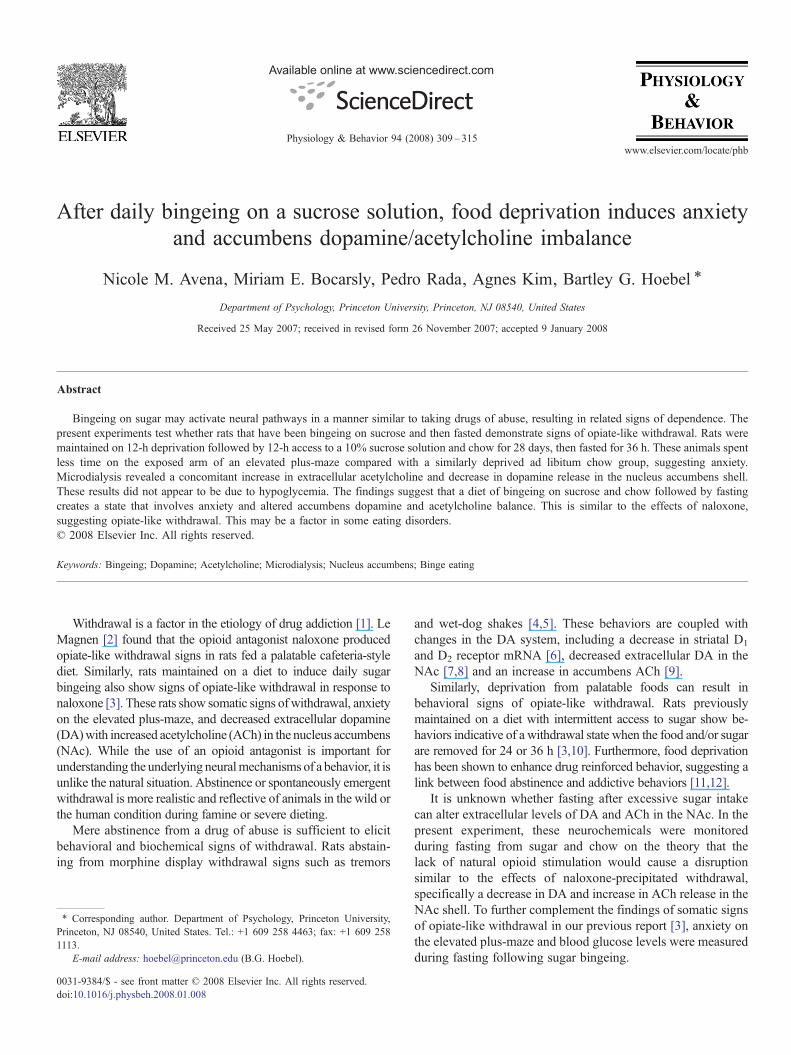
Available online at www.sciencedirect.com
4 (2008) 309–315www.elsevier.com/locate/phb
Physiology & Behavior 9
After daily bingeing on a sucrose solution, food deprivation induces anxietyand accumbens dopamine/acetylcholine imbalance
Nicole M. Avena, Miriam E. Bocarsly, Pedro Rada, Agnes Kim, Bartley G. Hoebel ⁎
Department of Psychology, Princeton University, Princeton, NJ 08540, United States
Received 25 May 2007; received in revised form 26 November 2007; accepted 9 January 2008
Abstract
Bingeing on sugar may activate neural pathways in a manner similar to taking drugs of abuse, resulting in related signs of dependence. Thepresent experiments test whether rats that have been bingeing on sucrose and then fasted demonstrate signs of opiate-like withdrawal. Rats weremaintained on 12-h deprivation followed by 12-h access to a 10% sucrose solution and chow for 28 days, then fasted for 36 h. These animals spentless time on the exposed arm of an elevated plus-maze compared with a similarly deprived ad libitum chow group, suggesting anxiety.Microdialysis revealed a concomitant increase in extracellular acetylcholine and decrease in dopamine release in the nucleus accumbens shell.These results did not appear to be due to hypoglycemia. The findings suggest that a diet of bingeing on sucrose and chow followed by fastingcreates a state that involves anxiety and altered accumbens dopamine and acetylcholine balance. This is similar to the effects of naloxone,suggesting opiate-like withdrawal. This may be a factor in some eating disorders.© 2008 Elsevier Inc. All rights reserved.
Keywords: Bingeing; Dopamine; Acetylcholine; Microdialysis; Nucleus accumbens; Binge eating
Withdrawal is a factor in the etiology of drug addiction [1]. LeMagnen [2] found that the opioid antagonist naloxone producedopiate-like withdrawal signs in rats fed a palatable cafeteria-stylediet. Similarly, rats maintained on a diet to induce daily sugarbingeing also show signs of opiate-like withdrawal in response tonaloxone [3]. These rats show somatic signs of withdrawal, anxietyon the elevated plus-maze, and decreased extracellular dopamine(DA)with increased acetylcholine (ACh) in the nucleus accumbens(NAc). While the use of an opioid antagonist is important forunderstanding the underlying neuralmechanisms of a behavior, it isunlike the natural situation. Abstinence or spontaneously emergentwithdrawal is more realistic and reflective of animals in the wild orthe human condition during famine or severe dieting.
Mere abstinence from a drug of abuse is sufficient to elicitbehavioral and biochemical signs of withdrawal. Rats abstain-ing from morphine display withdrawal signs such as tremors
⁎ Corresponding author. Department of Psychology, Princeton University,Princeton, NJ 08540, United States. Tel.: +1 609 258 4463; fax: +1 609 2581113.
E-mail address: [email protected] (B.G. Hoebel).
0031-9384/$ - see front matter © 2008 Elsevier Inc. All rights reserved.doi:10.1016/j.physbeh.2008.01.008
and wet-dog shakes [4,5]. These behaviors are coupled withchanges in the DA system, including a decrease in striatal D1
and D2 receptor mRNA [6], decreased extracellular DA in theNAc [7,8] and an increase in accumbens ACh [9].
Similarly, deprivation from palatable foods can result inbehavioral signs of opiate-like withdrawal. Rats previouslymaintained on a diet with intermittent access to sugar show be-haviors indicative of a withdrawal state when the food and/or sugarare removed for 24 or 36 h [3,10]. Furthermore, food deprivationhas been shown to enhance drug reinforced behavior, suggesting alink between food abstinence and addictive behaviors [11,12].
It is unknown whether fasting after excessive sugar intakecan alter extracellular levels of DA and ACh in the NAc. In thepresent experiment, these neurochemicals were monitoredduring fasting from sugar and chow on the theory that thelack of natural opioid stimulation would cause a disruptionsimilar to the effects of naloxone-precipitated withdrawal,specifically a decrease in DA and increase in ACh release in theNAc shell. To further complement the findings of somatic signsof opiate-like withdrawal in our previous report [3], anxiety onthe elevated plus-maze and blood glucose levels were measuredduring fasting following sugar bingeing.
310 N.M. Avena et al. / Physiology & Behavior 94 (2008) 309–315
1. Materials and methods
1.1. General methods
Male Sprague-Dawley rats were obtained from TaconicFarms (Germantown, NY) or bred at the Princeton Universityvivarium from a stock originating from Taconic Farms. Ratswere housed individually on a reversed 12-h light: 12-h darkcycle. All procedures were approved by the Princeton Univer-sity Institutional Animal Care and Use Committee.
1.2. Experiment 1: Is anxiety evident while fasting in sugar-bingeing rats?
Rats (300–450 g) in the main experimental group (in-termittent sugar+chow; n=9) were maintained on a diet of 12-hdeprivation followed by 12-h access to a 10% (w/v) sucrosesolution plus standard rodent chow (LabDiet #5001, PMI, St.Louis, MO, 3.02 kcal/g) starting 4 h into the dark phase eachday for 28 days [13]. A control group (ad libitum chow; n=7)was allowed ad libitum access to standard rodent chow. Allanimals had water available ad libitum. Other groups (in-termittent chow and ad libitum sugar) used in Experiments 2and 3 were not tested for anxiety because they failed to showbehavioral signs of withdrawal following naloxone or fasting ina previous report [3].
On Day 28, after the usual 12-h deprivation, the rats in theexperimental group were denied access to sugar and chow for anadditional 24 h. The control group was also deprived for chow for36 h. During this time, the animals continued to have ad libitumaccess to water. Then the animals were individually placed in theelevated plus-maze for 5 min using the technique of File, Lippa,Beer, and Lippa [14]. The apparatus had four arms, each 10 cmwide by 50 cm long, and was elevated 60 cm above the floor. Twoopposite arms were enclosed with high opaque walls. The othertwo arms had no protective walls. The experiment was conductedunder red-light. The rats were placed in the center of the maze andalternated facing an open or closed arm. Each plus-maze trial wasvideo taped and scored for the amount of time spent with head andforepaws on the open arm, closed arm or middle section of themaze by an observer blind to the treatment condition.
1.3. Experiment 2: Do sugar-bingeing rats have altered DAand ACh release in the accumbens while fasting?
A separate group of rats (350–450 g) underwent surgery toimplant guide cannulas for microdialysis. Rats were anesthe-tized with 20 mg/kg xylazine and 100 mg/kg ketamine (i.p.)supplemented with ketamine as needed (100 mg/kg, i.p.).Bilateral 21 gauge stainless-steel guide cannulas were aimed atthe posterior medial accumbens shell (anterior: +1.2 mm,lateral: 0.8 mm and ventral: 4.0 mm, with reference to bregma,midsagittal sinus, and surface of the level skull, respectively)using a stereotaxic instrument.
Rats were allowed to recover from surgery for at least 1 week.Similar to the procedures in Experiment 1, an experimental group(n=6)wasmaintained on daily 12-h deprivation followed by 12-h
access to 10% sucrose and standard rodent chow, starting 4 h intothe dark phase, for 28 days to induce bingeing (i.e., intermittentsugar+chow). One control group was maintained on the sameschedule with no sucrose (intermittent chow, n=7), while anothergroup was maintained on daily ad libitum chow (n=6). On Day28, each rat was moved into the microdialysis chamber and aprobe was inserted and fixed in place with acrylic cement 14–16 hbefore the experiment to allow neurotransmitter recovery tostabilize. Microdialysis probes were constructed of silica glasstubing (37 µm inner diameter, Polymicro Technologies Inc.,Phoenix, AZ) inside a 26 gauge stainless steel tube with amicrodialysis tip of cellulose tubing sealed at the end with epoxy(Spectrum Medical Co., Los Angeles, CA, 6000 MW, 0.2 mmouter diameter×2.0 mm long) [15]. The probes protruded 5 mmfrom the guide cannula to reach the intended site in the accumbensshell. Probes were perfused with buffered Ringer's solution(142 mM NaCl, 3.9 mM KCl, 1.2 mM CaCl2, 1.0 mM MgCl2,1.35mMNa2HPO4, 0.3 mMNaH2PO4, pH 7.35) at a flow rate of0.5 µL/min for the stabilization period and at 1.3 µL/min 2 hbefore and throughout the experiment. Neostigmine (0.3 µM)wasadded to the Ringer's solution to improve basal recovery of AChby hindering its enzymatic degradation.
When the final 12-h sucrose access period was over on Day28, chow, sucrose and water were removed from all of the rats.Water was removed for the 36 h of dialysis experiment becausedrinking water can alter baseline levels of DA and ACh [16],which would confound the results. Microdialysis samples werecollected for 1 h (3×20-min samples) after 12, 24 and 36 h offasting (no food, sugar or water available). Each sample wassplit, half for DA analysis and half for ACh.
1.4. Dopamine and acetylcholine assays
DA and its metabolites 3,4-dihydroxy-phenylacetic acid(DOPAC) and homovanillic acid (HVA) were analyzed by reversephase, high performance liquid chromatography with electroche-mical detection (HPLC-EC). Samples were injected into a 20-µLsample loop leading to a 10-cm column with 3.2 mm-bore and3 µm, C18 packing (Brownlee Co. Model 6213, San Jose, CA).The mobile phase contained 60 mM sodium phosphate, 100 μMEDTA, 1.24 mM heptanosulfonic acid, and 5% vol/vol methanol.DA, DOPAC and HVAwere measured with a coulometric detector(ESA Co. Model 5100A, Chelmsford, MA) with the conditioningpotential set at +500 mV, and working cell potential at −400 mV.
ACh was measured by reverse phase HPLC-EC using a 20-µLsample loop with a 10-cm C18 analytical column (ChrompackInc., Palo Alto, CA). AChwas converted to betaine and hydrogenperoxide by an immobilized enzyme reactor (acetylcholinesteraseand choline oxidase from Sigma, St Louis, MO and column fromChrompack Inc., Palo Alto, CA). The mobile phase was 200 mMpotassium phosphate at pH 8.0. An amperometric detector wasused (EG&G Princeton Applied Research, Lawrenceville, NJ).The hydrogen peroxide was oxidized on a platinum electrode(BAS, West Lafayette, IN) set at 500 mV with respect to an Ag–AgCl reference electrode (EG&G Princeton Applied Research).
Three, 20-min samples were collected at 12, 24 and 36 h offasting. For each hour, data for the three samples were averaged.

Fig. 1. Sugar and chow intake during the 28-day access period. A) Rats withintermittent sugar+chow escalated their total daily sugar intake over time. B)Rats with intermittent sugar+chow ate fewer grams of chow than theintermittent chow and ad libitum chow control groups; however, C) there wasno difference between groups in total daily caloric intake.
311N.M. Avena et al. / Physiology & Behavior 94 (2008) 309–315
Data for DA and ACh were converted to percent of the 12-hdeprivation time point for each group, when the intermittent-fedrats would normally expect food.
1.5. Histology
At the end of the experiment histology was performed toverify microdialysis probe placement. Rats received an over-dose of sodium pentobarbital and when deeply anesthetizedwere intracardially perfused with 0.9% saline followed by 10%formaldehyde. The brains were removed, frozen, and the expe-rimenter inspected the sections as they were cut (40 µm slices,starting anterior to the accumbens) until the sites of probe tipswere located. Once probe tracks were visualized, they wereplotted using the atlas of Paxinos and Watson [17].
1.6. Experiment 3: Are there changes in blood glucose levelsdue to chronic bingeing on sucrose?
Rats (300–350 g) in three groups were maintained for 28 dayson (a) intermittent sugar+chow (12-h deprivation followed by12-h access to a 10% sucrose solution and chow, starting 4 h intothe dark phase; n=10), (b) intermittent chow (12-h deprivationfollowed by 12-h access to standard rodent chow (no sucrose),starting 4 h into the dark phase; n=10), or (c) ad libitum chow(n=9). Chow and sugar were removed and tail-blood sampleswere collected following 12, 24 and 36 h of deprivation. Bloodwas collected from the tip of the tail by one experimenter gentlyholding the animal while another made a small incision about5 mm from the tip of the tail with a sterile scalpel. Blood wascollected in a capillary tube, centrifuged and serum was thenanalyzed for glucose levels with an Analox GM7 Fast EnzymaticMetabolizer (Analox, Lunenburg, MA). During the 28-dayaccess period, sugar and chow intakes were measured daily, andbody weights were measured weekly. Body weights were alsomeasured at each time point during deprivation.
1.7. Statistics
Plus-maze data were analyzed with a one-tailed, unpairedStudent's t-test. Cohen's d, which measures effect size [18], andprep, which provides the probability of replication [19], were alsocalculated. Data for DA and ACh were analyzed as percentdifference from normalized baseline as described above, using atwo-way repeated measures ANOVA followed by post hocTukey tests. Blood glucose levels, body weight and intake datawere analyzed by two-way repeated measures ANOVA.
2. Results
2.1. Body weight, sugar intake and chow intake
Data collected during the 28-day access period in Experiment3 revealed that rats with binge-access to sucrose escalated theirintake of sucrose over the 28-day exposure period (F(27, 279)=4.9, pb0.001; Fig. 1A), a finding similar to what has beenshown in our previous reports with sucrose or glucose [3,20].
Chow intake data showed a significant difference among groups.The rats with intermittent sugar access ate less chow than the adlibitum and intermittent chow groups (F(2,26)=60.8, pb0.001;Fig. 1B). However, there was no difference among groups intotal daily caloric intake (Fig. 1C).
There were no differences in bodyweight among groups duringthe 28-day access period; however, therewas an effect of time,withall three groups gaining weight over the 28 days (F(4,104)=298.9,pb0.001). During the 36 h of deprivation, body weight decreasedover time for all groups (F(2,52)=1957.8, pb0.001), with nodifference among groups at any time point (12, 24 or 36 h).
2.2. Experiment 1: Behavioral indices of anxiety after fasting insugar-bingeing rats
When placed in the elevated plus-maze for 5 min, after 36 hof food deprivation the rats that had previously been maintained
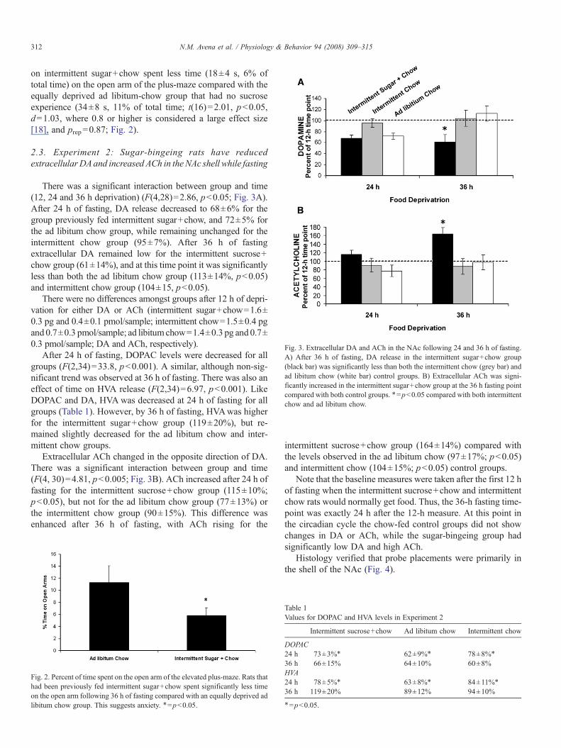
Fig. 3. Extracellular DA and ACh in the NAc following 24 and 36 h of fasting.A) After 36 h of fasting, DA release in the intermittent sugar+chow group(black bar) was significantly less than both the intermittent chow (grey bar) andad libitum chow (white bar) control groups. B) Extracellular ACh was signi-ficantly increased in the intermittent sugar+chow group at the 36 h fasting pointcompared with both control groups. ⁎=pb0.05 compared with both intermittentchow and ad libitum chow.
312 N.M. Avena et al. / Physiology & Behavior 94 (2008) 309–315
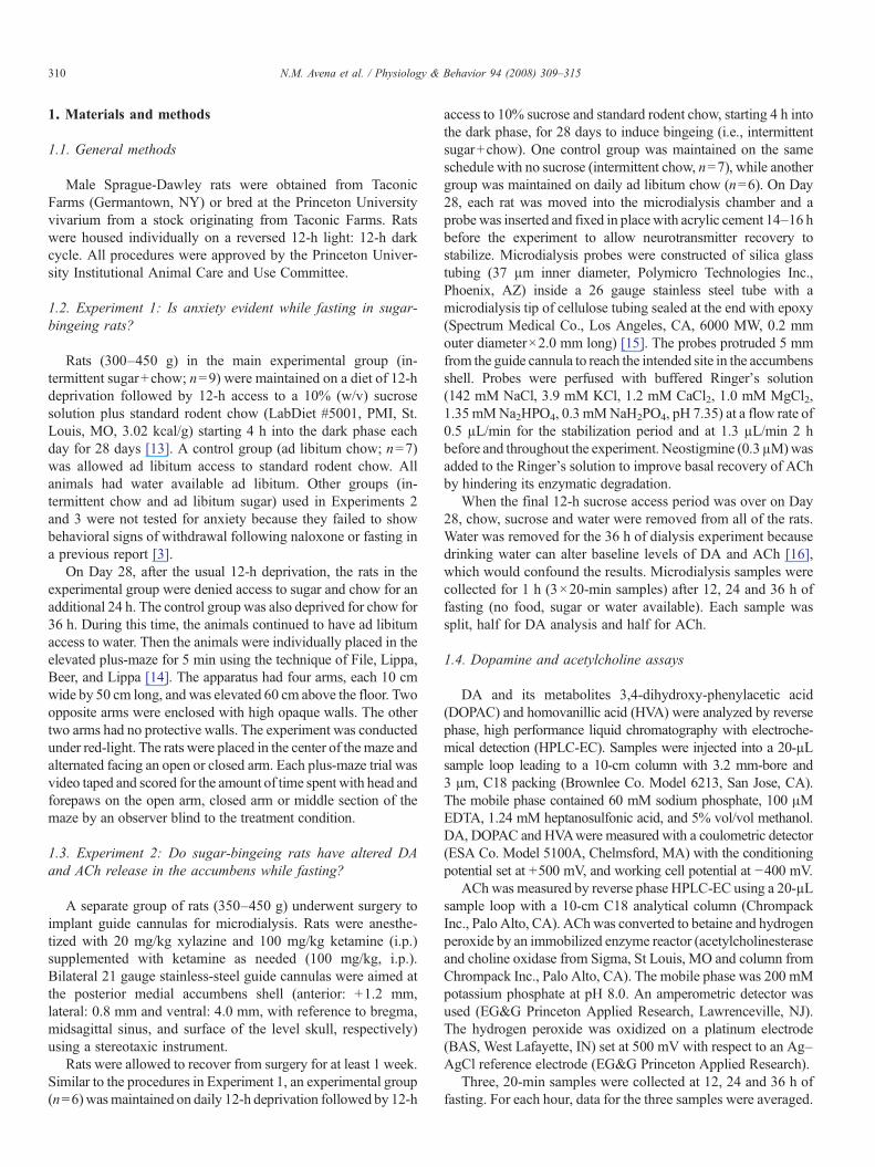
on intermittent sugar+chow spent less time (18±4 s, 6% oftotal time) on the open arm of the plus-maze compared with theequally deprived ad libitum-chow group that had no sucroseexperience (34±8 s, 11% of total time; t(16)=2.01, pb0.05,d=1.03, where 0.8 or higher is considered a large effect size[18], and prep=0.87; Fig. 2).
2.3. Experiment 2: Sugar-bingeing rats have reducedextracellularDAand increased ACh in the NAc shell while fasting
There was a significant interaction between group and time(12, 24 and 36 h deprivation) (F(4,28)=2.86, pb0.05; Fig. 3A).After 24 h of fasting, DA release decreased to 68±6% for thegroup previously fed intermittent sugar+chow, and 72±5% forthe ad libitum chow group, while remaining unchanged for theintermittent chow group (95±7%). After 36 h of fastingextracellular DA remained low for the intermittent sucrose+chow group (61±14%), and at this time point it was significantlyless than both the ad libitum chow group (113±14%, pb0.05)and intermittent chow group (104±15, pb0.05).
There were no differences amongst groups after 12 h of depri-vation for either DA or ACh (intermittent sugar+chow=1.6±0.3 pg and 0.4±0.1 pmol/sample; intermittent chow=1.5±0.4 pgand 0.7±0.3 pmol/sample; ad libitumchow=1.4±0.3 pg and 0.7±0.3 pmol/sample; DA and ACh, respectively).
After 24 h of fasting, DOPAC levels were decreased for allgroups (F(2,34)=33.8, pb0.001). A similar, although non-sig-nificant trend was observed at 36 h of fasting. There was also aneffect of time on HVA release (F(2,34)=6.97, pb0.001). LikeDOPAC and DA, HVAwas decreased at 24 h of fasting for allgroups (Table 1). However, by 36 h of fasting, HVAwas higherfor the intermittent sugar+chow group (119±20%), but re-mained slightly decreased for the ad libitum chow and inter-mittent chow groups.
Extracellular ACh changed in the opposite direction of DA.There was a significant interaction between group and time(F(4, 30)=4.81, pb0.005; Fig. 3B). ACh increased after 24 h offasting for the intermittent sucrose+chow group (115±10%;pb0.05), but not for the ad libitum chow group (77±13%) orthe intermittent chow group (90±15%). This difference wasenhanced after 36 h of fasting, with ACh rising for the
Fig. 2. Percent of time spent on the open arm of the elevated plus-maze. Rats thathad been previously fed intermittent sugar+chow spent significantly less timeon the open arm following 36 h of fasting compared with an equally deprived adlibitum chow group. This suggests anxiety. ⁎=pb0.05.
intermittent sucrose+chow group (164±14%) compared withthe levels observed in the ad libitum chow (97±17%; pb0.05)and intermittent chow (104±15%; pb0.05) control groups.
Note that the baseline measures were taken after the first 12 hof fasting when the intermittent sucrose+chow and intermittentchow rats would normally get food. Thus, the 36-h fasting time-point was exactly 24 h after the 12-h measure. At this point inthe circadian cycle the chow-fed control groups did not showchanges in DA or ACh, while the sugar-bingeing group hadsignificantly low DA and high ACh.
Histology verified that probe placements were primarily inthe shell of the NAc (Fig. 4).
Table 1Values for DOPAC and HVA levels in Experiment 2
Intermittent sucrose+chow Ad libitum chow Intermittent chow
DOPAC24 h 73±3%⁎ 62±9%⁎ 78±8%⁎
36 h 66±15% 64±10% 60±8%HVA24 h 78±5%⁎ 63±8%⁎ 84±11%⁎
36 h 119±20% 89±12% 94±10%
⁎=pb0.05.
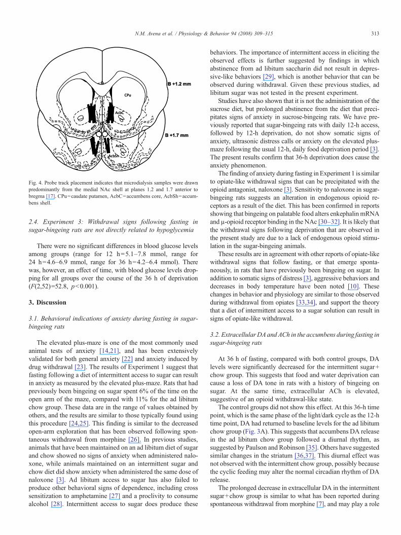
Fig. 4. Probe track placement indicates that microdialysis samples were drawnpredominantly from the medial NAc shell at planes 1.2 and 1.7 anterior tobregma [17]. CPu=caudate putamen, AcbC=accumbens core, AcbSh=accum-bens shell.
313N.M. Avena et al. / Physiology & Behavior 94 (2008) 309–315
2.4. Experiment 3: Withdrawal signs following fasting insugar-bingeing rats are not directly related to hypoglycemia
There were no significant differences in blood glucose levelsamong groups (range for 12 h=5.1–7.8 mmol, range for24 h=4.6–6.9 mmol, range for 36 h=4.2–6.4 mmol). Therewas, however, an effect of time, with blood glucose levels drop-ping for all groups over the course of the 36 h of deprivation(F(2,52)=52.8, pb0.001).
3. Discussion
3.1. Behavioral indications of anxiety during fasting in sugar-bingeing rats
The elevated plus-maze is one of the most commonly usedanimal tests of anxiety [14,21], and has been extensivelyvalidated for both general anxiety [22] and anxiety induced bydrug withdrawal [23]. The results of Experiment 1 suggest thatfasting following a diet of intermittent access to sugar can resultin anxiety as measured by the elevated plus-maze. Rats that hadpreviously been bingeing on sugar spent 6% of the time on theopen arm of the maze, compared with 11% for the ad libitumchow group. These data are in the range of values obtained byothers, and the results are similar to those typically found usingthis procedure [24,25]. This finding is similar to the decreasedopen-arm exploration that has been observed following spon-taneous withdrawal from morphine [26]. In previous studies,animals that have been maintained on an ad libitum diet of sugarand chow showed no signs of anxiety when administered nalo-xone, while animals maintained on an intermittent sugar andchow diet did show anxiety when administered the same dose ofnaloxone [3]. Ad libitum access to sugar has also failed toproduce other behavioral signs of dependence, including crosssensitization to amphetamine [27] and a proclivity to consumealcohol [28]. Intermittent access to sugar does produce these
behaviors. The importance of intermittent access in eliciting theobserved effects is further suggested by findings in whichabstinence from ad libitum saccharin did not result in depres-sive-like behaviors [29], which is another behavior that can beobserved during withdrawal. Given these previous studies, adlibitum sugar was not tested in the present experiment.
Studies have also shown that it is not the administration of thesucrose diet, but prolonged abstinence from the diet that preci-pitates signs of anxiety in sucrose-bingeing rats. We have pre-viously reported that sugar-bingeing rats with daily 12-h access,followed by 12-h deprivation, do not show somatic signs ofanxiety, ultrasonic distress calls or anxiety on the elevated plus-maze following the usual 12-h, daily food deprivation period [3].The present results confirm that 36-h deprivation does cause theanxiety phenomenon.
The finding of anxiety during fasting in Experiment 1 is similarto opiate-like withdrawal signs that can be precipitated with theopioid antagonist, naloxone [3]. Sensitivity to naloxone in sugar-bingeing rats suggests an alteration in endogenous opioid re-ceptors as a result of the diet. This has been confirmed in reportsshowing that bingeing on palatable food alters enkephalin mRNAand µ-opioid receptor binding in the NAc [30–32]. It is likely thatthe withdrawal signs following deprivation that are observed inthe present study are due to a lack of endogenous opioid stimu-lation in the sugar-bingeing animals.
These results are in agreement with other reports of opiate-likewithdrawal signs that follow fasting, or that emerge sponta-neously, in rats that have previously been bingeing on sugar. Inaddition to somatic signs of distress [3], aggressive behaviors anddecreases in body temperature have been noted [10]. Thesechanges in behavior and physiology are similar to those observedduring withdrawal from opiates [33,34], and support the theorythat a diet of intermittent access to a sugar solution can result insigns of opiate-like withdrawal.
3.2. ExtracellularDAandACh in the accumbens during fasting insugar-bingeing rats
At 36 h of fasting, compared with both control groups, DAlevels were significantly decreased for the intermittent sugar+chow group. This suggests that food and water deprivation cancause a loss of DA tone in rats with a history of bingeing onsugar. At the same time, extracellular ACh is elevated,suggestive of an opioid withdrawal-like state.
The control groups did not show this effect. At this 36-h timepoint, which is the same phase of the light/dark cycle as the 12-htime point, DA had returned to baseline levels for the ad libitumchow group (Fig. 3A). This suggests that accumbens DA releasein the ad libitum chow group followed a diurnal rhythm, assuggested by Paulson and Robinson [35]. Others have suggestedsimilar changes in the striatum [36,37]. This diurnal effect wasnot observed with the intermittent chow group, possibly becausethe cyclic feeding may alter the normal circadian rhythm of DArelease.
The prolonged decrease in extracellular DA in the intermittentsugar+chow group is similar to what has been reported duringspontaneous withdrawal from morphine [7], and may play a role

314 N.M. Avena et al. / Physiology & Behavior 94 (2008) 309–315
in fostering reinstatement of sugar intake after abstinence [38].The results obtained with the intermittent chow group, whichshowed relatively little change in DA release at any time point,suggests that the combination of bingeing on sugar and chow, notjust chow, is important in producing the observed effects.
Although DOPAC and HVA usually follow patterns similarto DA, this is not always the case. In the present experiment,DOPAC and HVA did not show a diurnal variation like thatobserved with DA, and instead remained suppressed over time.Although others have reported circadian fluctuations in thesemetabolites in the NAc [35], we are not aware of any papers thathave measured these levels during fasting for 36 h. Thus, in thepresent experiment, fasting may have affected DA metabolismin the chow control group.
ACh levels showed a significant difference between groupsafter 36 h of fasting. ACh in the NAc has been implicated iningestive behavior [39] and satiety in particular [40–44], and,when DA is low, ACh may foster aversion [40,45–48]. Thesignificant increase in ACh observed in the intermittent sugar+chow rats during fasting in the present experiment may corres-pond to the negative aspects of being reward-deprived. Previousstudies lend support to the theory that the findings reported hereare the result of deprivation from the sucrose diet. Rats bingeingon sucrose release DA and show attenuation of ACh release inthe NAc [20,49], which is the opposite of the present resultsseen during prolonged deprivation. The imbalance betweenaccumbens DA and ACh in the intermittent sugar+chow group,but not in the control groups, may contribute to the anxietyobserved in Experiment 1.
3.3. Blood glucose levels during fasting in sugar-bingeing rats
Hypoglycemia can lead to an aversive state from which ananimal may attempt to escape by eating. Behaviors associatedwith this aversive state are similar to those observed duringnaloxone administration or fasting in sucrose-bingeing rats [50].A multitude of factors can influence the brain reward system.However, because of the similarity between behaviors observedduring hypoglycemia and those observed during anxiety, thisstudy measured blood glucose levels to ensure that the observedeffects were not simply due to an aberrant glycemic status.Blood glucose levels were similar in all groups and therefore donot appear to account for the behavioral differences or changesin DA and ACh release. It can be inferred that mean insulinlevels remained consistent across groups, since alterations inblood glucose levels were not observed and body weights didnot differ as a result of the feeding schedules. Thus, the presentfindings, as well as those in our previous report [30], suggest thebehavioral and neurochemical changes are not the result ofdifferences in blood glucose levels. Instead they may be due to acombination of alterations in the endogenous opioid and DAsystems.
4. Conclusion
Long-term deprivation following sugar bingeing can result inbehavioral and neurochemical adaptations similar to those ob-
served when opioid-dependent animals are deprived of an abusedsubstance, such as morphine. These indicators of opiate-like with-drawal are signs of dependence. This finding, combined withprevious studies showing that sugar bingeing can result in othersigns of dependence, including dopaminergic and opioid changes[30,32], naloxone-precipitated and spontaneous withdrawal [3],cross-sensitization with drugs of abuse [27,51], increased intake ofsugar after abstinence [38], a time-dependent increase in respond-ing for cues previously associated with sugar [52], and a proclivityto consume alcohol [28], suggests that dependency is evident onseveral dimensions [53,54]. The present findings may be importantfor understanding the aversive components that could contribute tobinge eating.
Acknowledgements
This research was supported by USPHS grant AA-12882(to BGH) and DA-16458 and DK-79793 (fellowships to NMA).
References
[1] Koob GF, Le Moal M. Drug addiction, dysregulation of reward, andallostasis. Neuropsychopharmacology 2001;24(2):97–129.
[2] Le Magnen J. A role for opiates in food reward and food addiction. In:Capaldi PT, editor. Taste, Experience, and Feeding. Washington, D. C.:American Psychological Association; 1990. p. 241–52.
[3] Colantuoni C, Rada P, McCarthy J, Patten C, AvenaNM,Chadeayne A, et al.Evidence that intermittent, excessive sugar intake causes endogenous opioiddependence. Obes Res 2002;10(6):478–88.
[4] Martin WR, Wikler A, Eades CG, Pescor FT. Tolerance to and physicaldependence on morphine in rats. Psychopharmacologia 1963;4:247–60.
[5] Blasig J, Herz A, Reinhold K, Zieglgansberger S. Development of physicaldependence on morphine in respect to time and dosage and quantificationof the precipitated withdrawal syndrome in rats. Psychopharmacologia1973;33(1):19–38.
[6] Georges F, Stinus L, Bloch B, Le Moine C. Chronic morphine exposureand spontaneous withdrawal are associated with modifications of dopa-mine receptor and neuropeptide gene expression in the rat striatum. Eur JNeurosci 1999;11(2):481–90.
[7] Acquas E, Di Chiara G. Depression of mesolimbic dopamine transmissionand sensitization to morphine during opiate abstinence. J Neurochem1992;58(5):1620–5.
[8] Rossetti ZL, Hmaidan Y, Gessa GL. Marked inhibition of mesolimbicdopamine release: a common feature of ethanol, morphine, cocaine andamphetamine abstinence in rats. Eur J Pharmacol 1992;221(2–3):227–34.
[9] Fiserova M, Consolo S, Krsiak M. Chronic morphine induces long-lastingchanges in acetylcholine release in rat nucleus accumbens core and shell: anin vivomicrodialysis study. Psychopharmacology (Berl) 1999;142(1):85–94.
[10] Wideman CH, Nadzam GR, Murphy HM. Implications of an animal modelof sugar addiction, withdrawal and relapse for human health. NutrNeurosci 2005;8(5–6):269–76.
[11] Carroll ME, Stotz DC, Kliner DJ, Meisch RA. Self-administration oforally-delivered methohexital in rhesus monkeys with phencyclidine orpentobarbital histories: effects of food deprivation and satiation.Pharmacol Biochem Behav 1984;20(1):145–51.
[12] Carr KD. Chronic food restriction: enhancing effects on drug reward andstriatal cell signaling. Physiol Behav 2007;91(5):459–72.
[13] Avena N, Rada P, Hoebel B. Unit 9.23C sugar bingeing in rats. In: CrawleyJ, et al, editor. Current Protocols in Neuroscience. Indianapolis: JohnWiley& Sons, Inc.; 2006. p. 9.23C.1–6.
[14] File SE, Lippa AS, Beer B, Lippa MT. Unit 8.3 animal tests of anxiety. In:Crawley JN, et al, editor. Current Protocols in Neuroscience. Indianapolis:John Wiley & Sons, Inc.; 2004. p. 8.3.1–22.

315N.M. Avena et al. / Physiology & Behavior 94 (2008) 309–315
[15] Hernandez L, Stanley BG, Hoebel BG. A small, removable microdialysisprobe. Life Sci 1986;39(26):2629–37.
[16] Mark GP, Rada P, Pothos E, Hoebel BG. Effects of feeding and drinking onacetylcholine release in the nucleus accumbens, striatum, and hippocam-pus of freely behaving rats. J Neurochem 1992;58(6):2269–74.
[17] Paxinos G, Watson C. The rat brain in stereotaxic coordinates. New York:Academic Press; 2005.
[18] Cohen JD. A power primer. Psychol Bull 1992;112(1):155–9.[19] Killeen PR. An alternative to null-hypothesis significance tests. Psychol
Sci 2005;16(5):345–53.[20] Rada P, Avena NM, Hoebel BG. Daily bingeing on sugar repeatedly
releases dopamine in the accumbens shell. Neuroscience 2005;134(3):737–44.
[21] Kliethermes CL. Anxiety-like behaviors following chronic ethanolexposure. Neurosci Biobehav Rev 2005;28(8):837–50.
[22] Pellow S, Chopin P, File SE, Briley M. Validation of open:closed arm entriesin an elevated plus-maze as a measure of anxiety in the rat. J NeurosciMethods 1985;14(3):149–67.
[23] File SE, Andrews N. Low but not high doses of buspirone reduce theanxiogenic effects of diazepam withdrawal. Psychopharmacology (Berl)1991;105(4):578–82.
[24] Kokare DM, Chopde CT, Subhedar NK. Participation of alpha-melanocytestimulating hormone in ethanol-induced anxiolysis and withdrawal anxietyin rats. Neuropharmacology 2006;51(3):536–45.
[25] Irvine EE, Cheeta S, File SE. Tolerance to nicotine's effects in the elevatedplus-maze and increased anxiety during withdrawal. Pharmacol BiochemBehav 2001;68(2):319–25.
[26] Schulteis G, Yackey M, Risbrough V, Koob GF. Anxiogenic-like effects ofspontaneous and naloxone-precipitated opiate withdrawal in the elevatedplus-maze. Pharmacol Biochem Behav 1998;60(3):727–31.
[27] Avena NM, Hoebel BG. A diet promoting sugar dependency causesbehavioral cross-sensitization to a low dose of amphetamine. Neuroscience2003;122(1):17–20.
[28] Avena NM, Carrillo CA, Needham L, Leibowitz SF, Hoebel BG. Sugar-dependent rats show enhanced intake of unsweetened ethanol. Alcohol2004;34(2–3):203–9.
[29] Sukhotina IA, Malyshkin AA, Markou A, Bespalov AY. Lack of de-pression-like effects of saccharin deprivation in rats: forced swim test,differential reinforcement of low rates and intracranial self-stimulationprocedures. Behav Neurosci 2003;117(5):970–7.
[30] Colantuoni C, Schwenker J, McCarthy J, Rada P, Ladenheim B, Cadet B,et al. Excessive sugar intake alters binding to dopamine and mu-opioidreceptors in the brain. Neuroreport 2001;12(16):3549–52.
[31] Kelley AE, Will MJ, Steininger TL, Zhang M, Haber SN. Restricted dailyconsumption of a highly palatable food (chocolate ensure(R)) alters striatalenkephalin gene expression. Eur J Neurosci 2003;18(9):2592–8.
[32] Spangler R, Wittkowski KM, Goddard NL, Avena NM, Hoebel BG,Leibowitz SF, et al. Opiate-like effects of sugar on gene expression in rewardareas of the rat brain. Brain Res Mol Brain Res 2004;124(2):134–42.
[33] Thor DH, Teel BG. Fighting of rats during post-morphine withdrawal:effect of prewithdrawal dosage. Am J Psychol 1968;81(3):439–42.
[34] Martin WR, Wikler A, Eades CG, Pescor FT. Tolerance to and physicaldependence on morphine in rats. Psychopharmacologia 1963;65:247–60.
[35] Paulson PE, Robinson TE. Regional differences in the effects of amphet-amine withdrawal on dopamine dynamics in the striatum. Analysis ofcircadian patterns using automated on-line microdialysis. Neuropsycho-pharmacology 1996;14(5):325–37.
[36] Smith AD, Olson RJ, Justice Jr JB. Quantitative microdialysis of dopaminein the striatum: effect of circadian variation. J Neurosci Methods 1992;44(1): 33–41.
[37] Dluzen D, Ramirez VD. In vitro dopamine release from the rat striatum:diurnal rhythm and its modification by the estrous cycle. Neuroendocri-nology 1985;41(2):97–100.
[38] Avena NM, Long KA, Hoebel BG. Sugar-dependent rats show enhancedresponding for sugar after abstinence: evidence of a sugar deprivationeffect. Physiol Behav 2005;84(3):359–62.
[39] Kelley AE, Baldo BA, Pratt WE. A proposed hypothalamic-thalamic-striatal axis for the integration of energy balance, arousal, and food reward.J Comp Neurol 2005;493(1):72–85.
[40] Mark GP, Blander DS, Hoebel BG. A conditioned stimulus decreasesextracellular dopamine in the nucleus accumbens after the development ofa learned taste aversion. Brain Res 1991;551(1–2):308–10.
[41] Hoebel BG, Rada P, Mark GP, Pothos E. Neural systems for reinforcementand inhibition of behavior: relevance to eating, addiction, and depression.In: Kahneman D, Diener E, Schwartz N, editors. Well-being: theFoundations of Hedonic Psychology. New York: Russell Sage Foundation;1999. p. 558–72.
[42] Leibowitz SF, Hoebel BG. Behavioral neuroscience and obesity. In: BrayG, Bouchard C, James P, editors. The Handbook of Obesity. New York:Marcel Dekker; 2004. p. 301–71.
[43] Rada PV, Hoebel BG. Supraadditive effect of D-fenfluramine plusphentermine on extracellular acetylcholine in the nucleus accumbens:possible mechanism for inhibition of excessive feeding and drug abuse.Pharmacol Biochem Behav 2000;65(3):369–73.
[44] Rada P, Mark GP, Hoebel BG. Galanin in the hypothalamus raisesdopamine and lowers acetylcholine release in the nucleus accumbens:a possible mechanism for hypothalamic initiation of feeding behavior.Brain Res 1998;798(1–2):1–6.
[45] Rada P, Johnson DF, Lewis MJ, Hoebel BG. In alcohol-treated rats,naloxone decreases extracellular dopamine and increases acetylcholine inthe nucleus accumbens: evidence of opioid withdrawal. PharmacolBiochem Behav 2004;79(4):599–605.
[46] Rada P, Jensen K, Hoebel BG. Effects of nicotine and mecamylamine-induced withdrawal on extracellular dopamine and acetylcholine in the ratnucleus accumbens. Psychopharmacology (Berl) 2001;157(1):105–10.
[47] Mark GP, Weinberg JB, Rada PV, Hoebel BG. Extracellular acetylcholineis increased in the nucleus accumbens following the presentation of anaversively conditioned taste stimulus. Brain Res 1995;688(1–2):184–8.
[48] Hoebel BG, Avena NM, Rada P. Accumbens dopamine-acetylcholinebalance in approach and avoidance. Curr Opin Pharmacol 2007;7(6):617–27.
[49] Avena NM, Rada P, Moise N, Hoebel BG. Sucrose sham feeding ona binge schedule releases accumbens dopamine repeatedly and eliminatesthe acetylcholine satiety response. Neuroscience 2006;139(3):813–20.
[50] Cox DJ, Irvine A, Gonder-Frederick L, Nowacek G, Butterfield J. Fear ofhypoglycemia: quantification, validation, and utilization. Diabetes Care1987;10(5):617–21.
[51] Gosnell BA. Sucrose intake enhances behavioral sensitization produced bycocaine. Brain Res 2005;1031(2):194–201.
[52] Grimm JW, Fyall AM, Osincup DP. Incubation of sucrose craving: effectsof reduced training and sucrose pre-loading. Physiol Behav 2005;84(1):73–9.
[53] Avena NM. Examining the addictive-like properties of binge eating usingan animal model of sugar dependence. Exp Clin Psychopharmacol 2007;15(5):481–91.
[54] Avena NM, Rada P, Hoebel BG. Evidence of sugar addiction: behavioraland neurochemical effects of intermittent, excessive sugar intake. NeurosciBiobehav Rev 2008;32(1):20–39.
Copyright © 2022 FDOKUMEN