Acute Encephalitis, a Poliomyelitis-like Syndrome and ... - NCBI
12
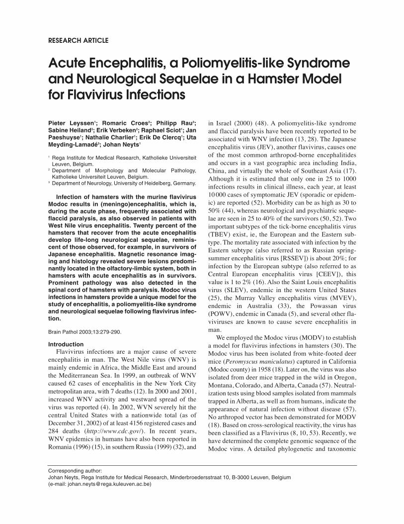
Pieter Leyssen 1 ; Romaric Croes 2 ; Philipp Rau 3 ; Sabine Heiland 3 ; Erik Verbeken 2 ; Raphael Sciot 1 ; Jan Paeshuyse 1 ; Nathalie Charlier 1 ; Erik De Clercq 1 ; Uta Meyding-Lamadé 3 ; Johan Neyts 1 1 Rega Institute for Medical Research, Katholieke Universiteit Leuven, Belgium. 2 Department of Morphology and Molecular Pathology, Katholieke Universiteit Leuven, Belgium. 3 Department of Neurology, University of Heidelberg, Germany. Infection of hamsters with the murine flavivirus Modoc results in (meningo)encephalitis, which is, during the acute phase, frequently associated with flaccid paralysis, as also observed in patients with West Nile virus encephalitis. Twenty percent of the hamsters that recover from the acute encephalitis develop life-long neurological sequelae, reminis- cent of those observed, for example, in survivors of Japanese encephalitis. Magnetic resonance imag- ing and histology revealed severe lesions predomi- nantly located in the olfactory-limbic system, both in hamsters with acute encephalitis as in survivors. Prominent pathology was also detected in the spinal cord of hamsters with paralysis. Modoc virus infections in hamsters provide a unique model for the study of encephalitis, a poliomyelitis-like syndrome and neurological sequelae following flavivirus infec- tion. Brain Pathol 2003;13:279-290. Introduction Flavivirus infections are a major cause of severe encephalitis in man. The West Nile virus (WNV) is mainly endemic in Africa, the Middle East and around the Mediterranean Sea. In 1999, an outbreak of WNV caused 62 cases of encephalitis in the New York City metropolitan area, with 7 deaths (12). In 2000 and 2001, increased WNV activity and westward spread of the virus was reported (4). In 2002, WVN severely hit the central United States with a nationwide total (as of December 31, 2002) of at least 4156 registered cases and 284 deaths (http://www.cdc.gov/). In recent years, WNV epidemics in humans have also been reported in Romania (1996) (15), in southern Russia (1999) (32), and in Israel (2000) (48). A poliomyelitis-like syndrome and flaccid paralysis have been recently reported to be associated with WNV infection (13, 28). The Japanese encephalitis virus (JEV), another flavivirus, causes one of the most common arthropod-borne encephalitides and occurs in a vast geographic area including India, China, and virtually the whole of Southeast Asia (17). Although it is estimated that only one in 25 to 1000 infections results in clinical illness, each year, at least 10 000 cases of symptomatic JEV (sporadic or epidem- ic) are reported (52). Morbidity can be as high as 30 to 50% (44), whereas neurological and psychiatric seque- lae are seen in 25 to 40% of the survivors (50, 52). Two important subtypes of the tick-borne encephalitis virus (TBEV) exist, ie, the European and the Eastern sub- type. The mortality rate associated with infection by the Eastern subtype (also referred to as Russian spring- summer encephalitis virus [RSSEV]) is about 20%; for infection by the European subtype (also referred to as Central European encephalitis virus [CEEV]), this value is 1 to 2% (16). Also the Saint Louis encephalitis virus (SLEV), endemic in the western United States (25), the Murray Valley encephalitis virus (MVEV), endemic in Australia (33), the Powassan virus (POWV), endemic in Canada (5), and several other fla- viviruses are known to cause severe encephalitis in man. We employed the Modoc virus (MODV) to establish a model for flavivirus infections in hamsters (30). The Modoc virus has been isolated from white-footed deer mice (Peromyscus maniculatus) captured in California (Modoc county) in 1958 (18). Later on, the virus was also isolated from deer mice trapped in the wild in Oregon, Montana, Colorado, and Alberta, Canada (57). Neutral- ization tests using blood samples isolated from mammals trapped in Alberta, as well as from humans, indicate the appearance of natural infection without disease (57). No arthropod vector has been demonstrated for MODV (18). Based on cross-serological reactivity, the virus has been classified as a Flavivirus (8, 10, 53). Recently, we have determined the complete genomic sequence of the Modoc virus. A detailed phylogenetic and taxonomic RESEARCH ARTICLE Acute Encephalitis, a Poliomyelitis-like Syndrome and Neurological Sequelae in a Hamster Model for Flavivirus Infections Corresponding author: Johan Neyts, Rega Institute for Medical Research, Minderbroedersstraat 10, B-3000 Leuven, Belgium (e-mail: [email protected])
-
Upload
khangminh22 -
Category
Documents
-
view
3 -
download
0
Transcript of Acute Encephalitis, a Poliomyelitis-like Syndrome and ... - NCBI
Pieter Leyssen1; Romaric Croes2; Philipp Rau3;Sabine Heiland3; Erik Verbeken2; Raphael Sciot1; JanPaeshuyse1; Nathalie Charlier1; Erik De Clercq1; UtaMeyding-Lamadé3; Johan Neyts1
1 Rega Institute for Medical Research, Katholieke UniversiteitLeuven, Belgium.
2 Department of Morphology and Molecular Pathology,Katholieke Universiteit Leuven, Belgium.
3 Department of Neurology, University of Heidelberg, Germany.
Infection of hamsters with the murine flavivirusModoc results in (meningo)encephalitis, which is,during the acute phase, frequently associated withflaccid paralysis, as also observed in patients withWest Nile virus encephalitis. Twenty percent of thehamsters that recover from the acute encephalitisdevelop life-long neurological sequelae, reminis-cent of those observed, for example, in survivors ofJapanese encephalitis. Magnetic resonance imag-ing and histology revealed severe lesions predomi-nantly located in the olfactory-limbic system, both inhamsters with acute encephalitis as in survivors.Prominent pathology was also detected in thespinal cord of hamsters with paralysis. Modoc virusinfections in hamsters provide a unique model for thestudy of encephalitis, a poliomyelitis-like syndromeand neurological sequelae following flavivirus infec-tion.
Brain Pathol 2003;13:279-290.
IntroductionFlavivirus infections are a major cause of severe
encephalitis in man. The West Nile virus (WNV) ismainly endemic in Africa, the Middle East and aroundthe Mediterranean Sea. In 1999, an outbreak of WNVcaused 62 cases of encephalitis in the New York Citymetropolitan area, with 7 deaths (12). In 2000 and 2001,increased WNV activity and westward spread of thevirus was reported (4). In 2002, WVN severely hit thecentral United States with a nationwide total (as ofDecember 31, 2002) of at least 4156 registered cases and284 deaths (http://www.cdc.gov/). In recent years,WNV epidemics in humans have also been reported inRomania (1996) (15), in southern Russia (1999) (32), and
in Israel (2000) (48). A poliomyelitis-like syndromeand flaccid paralysis have been recently reported to beassociated with WNV infection (13, 28). The Japaneseencephalitis virus (JEV), another flavivirus, causes oneof the most common arthropod-borne encephalitidesand occurs in a vast geographic area including India,China, and virtually the whole of Southeast Asia (17).Although it is estimated that only one in 25 to 1000infections results in clinical illness, each year, at least10 000 cases of symptomatic JEV (sporadic or epidem-ic) are reported (52). Morbidity can be as high as 30 to50% (44), whereas neurological and psychiatric seque-lae are seen in 25 to 40% of the survivors (50, 52). Twoimportant subtypes of the tick-borne encephalitis virus(TBEV) exist, ie, the European and the Eastern sub-type. The mortality rate associated with infection by theEastern subtype (also referred to as Russian spring-summer encephalitis virus [RSSEV]) is about 20%; forinfection by the European subtype (also referred to asCentral European encephalitis virus [CEEV]), thisvalue is 1 to 2% (16). Also the Saint Louis encephalitisvirus (SLEV), endemic in the western United States(25), the Murray Valley encephalitis virus (MVEV),endemic in Australia (33), the Powassan virus(POWV), endemic in Canada (5), and several other fla-viviruses are known to cause severe encephalitis inman.
We employed the Modoc virus (MODV) to establisha model for flavivirus infections in hamsters (30). TheModoc virus has been isolated from white-footed deermice (Peromyscus maniculatus) captured in California(Modoc county) in 1958 (18). Later on, the virus was alsoisolated from deer mice trapped in the wild in Oregon,Montana, Colorado, and Alberta, Canada (57). Neutral-ization tests using blood samples isolated from mammalstrapped in Alberta, as well as from humans, indicate theappearance of natural infection without disease (57).No arthropod vector has been demonstrated for MODV(18). Based on cross-serological reactivity, the virus hasbeen classified as a Flavivirus (8, 10, 53). Recently, wehave determined the complete genomic sequence of theModoc virus. A detailed phylogenetic and taxonomic
RESEARCH ARTICLE
Acute Encephalitis, a Poliomyelitis-like Syndromeand Neurological Sequelae in a Hamster Modelfor Flavivirus Infections
Corresponding author:Johan Neyts, Rega Institute for Medical Research, Minderbroedersstraat 10, B-3000 Leuven, Belgium(e-mail: [email protected])

analysis of the entire coding region of the genome con-firmed the classification of MODV in the cluster of fla-viviruses with no known vector (29).
Here, we present a unique model for the study ofacute flavivirus encephalitis, flavivirus-induced flaccidparalysis, a flavivirus-associated poliomyelitis-likesyndrome and long-lasting neurological sequelae fol-lowing flavivirus infection.
Materials and Methods
Cells and viruses. African green monkey kidney(Vero) cells were grown in minimum essential medium(MEM, Gibco, Paisley, Scotland) supplemented with10% inactivated fetal calf serum (FCS, Integro, Zaandam,The Netherlands), 1% L-glutamine, and 0.3% bicar-bonate. MODV was obtained from the American TypeCulture Collection (ATCC VR-415) and cultured onVero cells as previously described (30).
Animals. Eight- to 12-week old Gold hamsters(Mesocricetus auratus) were used throughout the exper-iments. All animals were bred at the Rega Institute. Thehamsters were maintained under artificial diurnal light-ing conditions with free access to food and water. Theprinciples of good laboratory animal care were fol-lowed. All experiments were approved by the ethicalcommittee on vertebrate animal experiments of the K. U.Leuven.
Animal experiments. Hamsters were inoculatedwith 104 PFU (plaque forming units) of Modoc viruseither via the intraperitoneal route (i.p.) (104 PFU in500 �l) or, following brief ether anesthesia, via theintranasal route (i.n.) (104 PFU in 50 �l). All animals wereweighed daily and examined for clinical manifestationsof encephalitis such as tremor, muscle weakness, paral-ysis, closed eye(s), and posture. Every day, the animalsreceived a single dose of 16 mg/kg of oxytetracycline bysubcutaneous (s.c.) injection to suppress bacterialinfection of the gut (a condition seen in most MODV-infected hamsters), thus increasing the animal’s comfort.Hamsters with severe encephalitis, that was judged to bevery likely lethal, were subjected to magnetic reso-nance imaging shortly before fatal outcome of the dis-ease was expected, after which the animals were eutha-nized and the brain and spinal cord dissected (seebelow). Animals with moderate encephalitis and thatwere assumed to survive the acute phase of the infectionwere kept for several months (up to 1.5 years) to allowevaluation of residual neurological brain damage by
MRI and histopathology, long after the symptoms ofthe acute encephalitis had resolved.
Magnetic resonance imaging. The hamsters wereanesthetized by intramuscular injection with a volume (2�l/g) of a 1:2:2 mixture of atropine (0.5 mg/ml, 0.2mg/kg, Sterop, Brussels, Belgium), ketamine (50mg/ml, 40 mg/kg, Ketalar®, Parke-Davis, Zaventem,Belgium) and xylazine (20 mg/ml, 16 mg/kg, XYL-M®
2%, VMD, Arendonk, Belgium). T2-weighted MRIscans were generated with a 2.4 T MRI system (BrukerB – C 24/40, Ettlingen, Germany; magnetic center 588mm) with the following parameters: 10 slices; slicethickness: 2 mm; interslice distance: 2 mm; field ofview (FOV): 4 � 4 cm. A multi-spin-multi-echosequence was used (TR = 3000 ms; TE = 8, 16, 24 up to96 ms [a total of 12 echoes]; matrix 256 �192). The rawdata were processed with the ANALYZE™ softwarepackage (version 7.5, Mayo Biomedical ImagingResource, Mayo Foundation, Rochester, Minn).
Histopathology. Following imaging of the brain, theanimals were sacrificed by ether anesthesia and per-fused transcardially with 20 ml of 4% buffered forma-line. The brain and spinal cord were dissected and post-fixed in 4% buffered formaline, embedded in paraffinblocks, and processed following standard histologicalprocedures. For each animal, six consecutive axial sec-tions of 6 �m were cut at 40 different positions of thebrain at a mutual distance of ±0.3 mm. This meticulousapproach ensured that all major anatomical areas of thebrain were represented in the tissue sections. For routinemicroscopic evaluation, one slide per position wasstained according to the Luxol-Fast blue MBS stain(Klüver-Barrera). Remaining slides were available forsupplementary staining, such as immunohistochem-istry, when necessary.
Results
Modoc virus infection in hamsters. Followingintranasal or intraperitoneal inoculation with the virus,100% of the hamsters (59 animals; 4 independentexperiments) developed acute MODV encephalitis, ofwhich about 50% (30/59) succumbed during the acutephase of the infection. Hamsters with acute MODVencephalitis presented with closed eyes, muscle weak-ness and flaccid paralysis (Figure 4A) or apoliomyelitis-like syndrome (Figure 4B), often accom-panied by tremor. Of the hamsters that survived theacute phase of the infection, about 80% (23/29) did not
280 Flavivirus Encephalitis in Hamsters—Leyssen et al

show obvious neurological deficits, whereas theremaining 20% (6/29) of the survivors presented withclearly visible neurological sequelae, ie, flaccid paraly-sis of one of the front legs or paralysis of one of the hindlegs, and wasting of the muscles thereof, resembling apoliomyelitis-like syndrome (Figure 5A, B).
Cerebral MRI of hamsters with acute Modoc virus-induced encephalitis. MRI was employed to generate T2-weighted images of the brain of 7 hamsters that hadbeen inoculated with Modoc virus via the intranasalroute and that presented with signs of severe acuteencephalitis. These images clearly demonstrate severe andnearly symmetrical tissue damage in both temporallobes of all 7 animals (Figure 1B). Hyperintense signalsof variable intensity were observed in theolfactory/frontobasal lobes (7/7 animals) (Figure 1A)and in the (hypo)thalamic area (6/7) (Figure 1C), sug-gestive of mild to severe brain lesions. In 2 animals, theMRI scans indicated the presence of mild tissue damagein the cingulate gyri (Figure 1D). Mild to severe widen-ing of the lateral ventricles was distinguishable on MRI
scans of 3 animals (Figure 1E). One of the latter hamstersshowed blockage of the foramen of Monroe of a lateralventricle accompanied by widening of the contralateralventricle (Figure 1E).
The brain of 4 hamsters that had been inoculatedwith MODV via the intraperitoneal route and that had alsopresented with signs of severe encephalitis at the time ofcranial MRI, showed a similar distribution pattern oflesions on T2-weighted images. However, the lesionsappeared to be less pronounced and more focused as com-pared to those observed in animals that were inoculatedvia the intranasal route (Figure 1). MRI abnormalitieswere observed in the hindbrain of one animal (Figure 1F).
Histopathology of the brain and spinal cord ofhamsters with acute Modoc virus-induced encephali-tis. All hamsters, that were infected via the intranasalroute, presented with a diffuse and moderate to severe lep-tomeningoencephalitis, which was characterized by aspecific and recurrent topographic distribution pattern(Figure 2A). The lesions showed a remarkable olfacto-ry-limbic predominance, with damage to the bulbus
281Flavivirus Encephalitis in Hamsters—Leyssen et al
Figure 1. Abnormalities in the brain of hamsters during the acute phase of Modoc virus encephalitis as monitored by MRI. Intensi-ty of the lesions (or widening in case of the ventricles): (-) = none; (+) = mild; (++) = moderate; (+++) = severe; * indicates the num-ber of animals displaying none, mild, moderate, or severe MRI abnormalities. = T2-weighted MRI images that are presented inthe panel of figures on the right. Arrows indicate the corresponding abnormality (abnormalities) on the MRI images. Animal identi-fication codes: (a) R3N1; (b) R3N1; (c) R3N5; (d) R3N5; (e) R3N3 and (f) R4N5.

282 Flavivirus Encephalitis in Hamsters—Leyssen et al

olfactorius, cortex piriformis, prelimbic cortices, hip-pocampi, corpora amygdaloidea, corpora mamillaria,hypothalamic nuclei, and preoptic nuclei. Cerebellum,medulla oblongata, medulla spinalis, and basal ganglia,with exception of the nucleus accumbens, were notaffected. The meningitis component was rather limitedto the leptomeninges covering the affected anatomicalregions (Figure 3A, B). The worst affected regions, pri-marily the olfactory bulbs and cortex piriformis, werenecrotic with neuronal cell loss and granular destructionof the white and gray matter architecture (Figure 3A, B).Adjacent to these necrotic areas, less affected regionswere found to be spongiotic with increased cellularitybecause of infiltration by lymphocytes and reactiveastrocytic proliferation (Figure 3A) with the formationof microglial nodules. Furthermore, satellitosis, neu-ronophagia, and singular cell necrosis were observed(Figure 3B). Infiltration of inflammatory cells in theependymal cell layer and plexus choroideus was appar-ent, as was an increased cellularity of the cerebrospinalfluid (data not shown). Polymorphonuclear inflammatorycells were not observed. Intracellular viral inclusions orpathognomonic viral cytotoxic effects were not distin-guishable. The acute phase of MODV encephalitis fol-lowing intranasal inoculation is thus characterized by a
moderate to severe non-specific viral leptomeningoen-cephalitis with a predominant tropism for the olfactoryand limbic system, which is consistent with the route ofinoculation and cerebral MRI findings.
In contrast to hamsters that were inoculated via theintranasal route, no specific or recurrent distributionpattern of CNS damage was noted for hamsters thatwere infected via the intraperitoneal route. Brainlesions were found to be more focal and not diffusely con-tinuous as was observed in intranasally infected hamsters(Figures 2B; 3C, D). On one hand, the olfactory bulbswere completely spared, but on the other hand, the thal-amic and hypothalamic nuclei, as well as the basal gan-glia, were more severely affected in intraperitoneallyinfected hamsters. Occasionally, also the nuclei habenulaeshowed histopathological damage. The inflammatoryresponse was not different from that described for ham-sters infected via the intranasal route (Figure 3C, D), andis consistent with a mild to moderate non-specific viralencephalomyelitis with leptomeningitis. The lesionswere predominantly observed in morphological regionsbelonging to the olfactory and limbic system, as wasobserved for hamsters infected via the intranasal route.
The spinal cord of hamsters that had been inoculat-ed via the intraperitoneal route was characterized by
283Flavivirus Encephalitis in Hamsters—Leyssen et al
Figure 2. (Opposing page) Axial brain sections of hamsters with acute MODV encephalitis. A. Tissue sections of the brain of a moribund hamster (animal identification code R3N3) with acute MODV encephalitis that was inoc-ulated via the intranasal route: (1a) Inflammation and necrosis of the olfactory nuclei with loss of the tissue architecture (right handside of the image); (1b) Inflammation and necrosis below the rhinal fissure (right hand side of the image); (2) Inflammation and necro-sis involving (from left to right below the dotted line) cortex piriformis, dorsal endopiriform nucleus, tuberculum olfactorium, medialforebrain bundle and the septal nuclei; (3) Inflammation and necrosis involving (from left to right below the dotted line) cortex piri-formis, dorsal and ventral endopiriform nuclei, anterior amygdaloid area and nuclei of the preoptic, hypothalamic and thalamic area;(4) Inflammation and necrosis involving (from left to right below the dotted line) cortex piriformis, dorsal and ventral endopiriformnuclei, amygdaloid nuclei, nuclei of the hypothalamic and thalamic area, nuclei of the habenular area, damage to the CA1, CA2 andCA3 area of the hippocampus (encircled by the striped line); (5) Inflammation and necrosis involving the lateral entorhinal and perirhi-nal cortex, dorsal endopiriform nucleus, and nuclei of the amygdaloid area (from upper left to lower right below the dotted line), necrot-ic damage to CA1, CA2 and CA3 area of the hippocampus (encircled by the striped line). B. Tissue sections of the brain of a moribund hamster (animal identification code R4N5) that was inoculated via the intraperitonealroute: Focus of inflammation and necrosis (1) in the dorsal endopiriform nucleus; (2) in the paratenial and paraventricular thalam-ic nucleus; (3) in the paratenial and paraventricular thalamic nucleus, and in the preoptic nuclei; (4) in the hippocampus, mediodor-sal thalamic nucleus, dorsale endopiriform nucleus, medial amygdaloid area and anterior hypothalamic area; (5) in the perirhinaland lateral entorhinal cortex, and CA3 of the hippocampus.C. Tissue sections of the brain of a hamster without neurological sequelae (animal identification code R1/G; dissection of the brainat 652 days postinfection): (1) Focus of cystic fibrosis in the agranular/granular insular cortex and secondary somatosensory cor-tex, and in the central amygdaloid nucleus; (2) Continuity between the lateral ventricle and the subarachnoidal space because oftransmural necrosis of the temporal cortex with rupture of the ependymal cell layer and pia mater; (3-5) decortication and decere-bration involving the agranular insular cortex, perirhinal cortex, primary auditory cortex, secondary and primary visual cortex.D. Tissue sections of the brain of a hamster with apparent neurological sequelae (animal identification code R1/X; dissection of thebrain at 575 days postinfection): (1-3) decortication in the perirhinal/primary auditory cortex with widening of the lateral ventricle,focus of neuronal loss in the CA3 area of the hippocampus; (4) decortication in the perirhinal/primary auditory/secondary visual cor-tex with widening of the lateral ventricle; (5) area of cystic fibrosis involving the ectorhinal/temporal association/secondary visualcortex.E. Tissue sections of the brain of a hamster without apparent neurological sequelae (animal identification code R1/D; dissection ofthe brain at 652 days postinfection): (1-2) Area of cystic fibrosis in the anterior cortical amygdaloid nucleus (3-4) extending to involvethe amygdaloid area; (4-5) widening of the lateral ventricle.

extensive bilateral (Figure 4C) or unilateral (Figure 4D,E) damage to the grey matter. Perivascular cuffing,infiltration of the neuronal tissue by inflammatory cells,destruction of neurons and spongiosis were perceptible(Figure 3E, F). No signs of pathology were observed inthe spinal cord of hamsters that had been infected via theintranasal route.
Cerebral MRI of long-term surviving hamsterswith and without neurological sequelae. T2-weightedMRI images were obtained from the brains of 2 hamsterswith neurological sequelae and of 7 hamsters withoutapparent neurological sequelae (on day 317 postinfection[2 hamsters], day 575 [3 hamsters], and day 652 [4hamsters]). The residual neurological sequelae in 2 of thehamsters that survived acute MODV infection (examinedon day 575 and 652 postinfection, respectively) werecharacterized by paralysis of one of the hind legs andwasting of the muscles thereof, resembling apoliomyelitis-like syndrome (Figure 5A, B). This was fur-
ther visualized by the ink footprint trail of one of thesehamsters (Figure 5C). Another hamster (not included inthe MRI study) showed long-lasting flaccid paralysis ofone of the front legs as visualized by the ink footprint trail(Figure 5D). The footprint trail of a healthy hamster isshown for comparison (Figure 5E). MRI abnormalitieswere moderate in the temporal lobes of all hamsters(Figure 6B), regardless of the presence of (observable)sequelae. Mild lesions were also observed in the orbito-frontal cortex of 2 animals (22%) (Figure 6A), in the(hypo)thalamic area of one animal (11%) (Figure 6C), andin the cingulate gyrus of another animal (11%) (Figure6D). Mild widening of the lateral ventricles was visiblein 4 hamsters (44%) (Figure 6E). No abnormalitieswere detected in the hindbrain of any of the animals(Figure 6F).
Histopathology of the brain and spinal cord oflong-term survivors with and without neurologicalsequelae. Signs of mild residual meningoencephalitis and
284 Flavivirus Encephalitis in Hamsters—Leyssen et al
Figure 3. Histopathology of MODV encephalitis. (A) Immunohistochemical staining for glial fibrillary acidic protein (GFAP) and (B)staining with hematoxylin/eosin (H&E) of the brain of a hamster with acute encephalitis following intranasal inoculation with MODV(higher magnification of a tissue section adjacent to that depicted in Figure 2-A4). The rhinal fissure (arrow) represents an imagi-nary boundary between (i) affected parenchyma under the fissure, of which the leptomeninges show clear signs of meningitis andin which a clearly visible inflammatory infiltrate can be observed and (ii) above the rhinal fissure, less affected parenchyma with reac-tive astrocytic proliferation; (C) GFAP staining and (D) H&E staining of the brain of a hamster with acute encephalitis following intraperi-toneal inoculation with MODV (higher magnification of a tissue section adjacent to that depicted in Figure 2-B1). Focal and well delin-eated lesion with severe tissue destruction and extensive inflammatory infiltrate in the dorsal endopiriform nucleus, surrounded byless affected gray matter with clear signs of reactive astrocytic proliferation at the border of the focal lesion; (E) GFAP staining and(F) H&E staining of the spinal cord of a hamster with acute encephalitis following intraperitoneal inoculation with MODV (higher mag-nification of a tissue section adjacent to that depicted Figure 4C). Perivascular cuffing, infiltration of the neuronal tissue by inflam-matory cells, destruction of neurons and spongiosis are apparent.

irreversible tissue destruction were observed in thebrain of hamsters more than one year after they recov-ered from acute MODV encephalitis. In all hamsters,more or less pronounced dilatation of the ventricularsystem was noted, as was an inconspicuous meningeallymphocytic infiltrate (data not shown). In one ham-ster, very severe irreversible tissue damage wasobserved with continuity between the lateral ventricle andthe subarachnoidal space due to transmural necrosis ofthe temporal cortex (Figure 2C). In the majority of thehamsters, a more or less pronounced mass reduction ofthe temporal and frontobasal cortices was observed.Pseudocystic lesions, particularly located in the tempo-ral and frontobasal cortices, indicated focal tissue loss dueto infarction (Figure 2D, E). Cortical cell layers adjacentto the cortex piriformis and the cell layers of the hip-pocampus were less dense as a result of individual neu-ronal cell loss. The residual neurons presented withatypical, irregular and hyperchromatic nuclei (data notshown). Viral inclusions could not be observed withlight microscopy. In the brain parenchyma, a sparselymphocytic infiltrate was still present and a slightperivascular accumulation of inflammatory cells in theVirchow Robin spaces of small and medium-sized
blood vessels (perivascular cuffing) was observed (datanot shown).
Only minor histopathological changes, such as spon-giosis around the central canal and small, pseudocysticlesions (Figure 5F), were noted in the spinal cord ofseveral hamsters that survived the acute encephalitis for>1 year without neurological sequelae. Severe residualtissue damage, such as loss of grey matter, widening ofthe central canal and the presence of inflammatory cellsin the spinal cord tissue, was, however, detected inhamsters that presented with apparent neurologicalsequelae such as a poliomyelitis-like syndrome (Figure5G).
DiscussionFollowing intranasal or intraperitoneal inoculation
with MODV, hamsters develop acute encephalitis,which is amongst other characterized by muscle weak-ness, flaccid paralysis or a poliomyelitis-like syndrome,and tremor. About 50% of the animals succumb duringthe acute phase of the infection, about 40% survivewithout neurological sequelae, and the remaining 10%recover with obvious long-lasting neurological sequelae.
285Flavivirus Encephalitis in Hamsters—Leyssen et al
Figure 4. Acute MODV encephalitis and MODV-associated spinal cord damage. (A) hamster (animal identification code R4N4) withacute MODV encephalitis presenting with muscle weakness, flaccid paralysis of the 4 limbs and closed eyes; (B) hamster (animalidentification code R4N5) with acute MODV encephalitis presenting with a poliomyelitis-like syndrome characterized by paralysisof the hind limbs; (C) axial section of the spinal cord of a hamster (animal identification code R4N4) with severe MODV encephali-tis showing bilateral damage to the grey matter; (D, E) axial sections of the spinal cord of a hamster (animal identification code R4N5)with severe MODV encephalitis showing unilateral damage to the grey matter.

286 Flavivirus Encephalitis in Hamsters—Leyssen et al

MRI examination of the brain of hamsters during theacute phase of MODV encephalitis (either followingintranasal or intraperitoneal inoculation), revealedmoderate to severe lesions in both temporal lobes of allanimals. Also the thalamic-hypothalamic area appearedto be generally affected and widening of the lateral ven-tricles was observed. Histological study of the brain ofanimals with acute encephalitis not only revealedsevere tissue damage in the above mentioned areas, butalso revealed that the brain pathology was more exten-sive than was expected from the MRI scans. Generally,a 1.5 T MRI scanner is used in the clinical setting.Because of the smaller brain size of hamsters, we used
2.4 T MRI equipment to obtain images with higher res-olution. A good correlation was observed when the MRIdata and the histopathological data were compared.However, the resolution of the images might still havebeen too low to adequately visualize less pronounced, butyet important brain lesions, that were only detectablemicroscopically. Diffuse (midbrain) lesions, that wereobserved with MRI in the brain of hamsters withMODV encephalitis, have also been reported in patientswith Japanese encephalitis (39).
Multiple MRI studies on patients with Japaneseencephalitis (11, 21, 24, 26, 38, 45), tick-borneencephalitis (3, 31), and Saint Louis encephalitis (54) sug-
287Flavivirus Encephalitis in Hamsters—Leyssen et al
Figure 5. (Opposing page) Lifelong neurological sequelae and spinal cord damage following MODV encephalitis. (A, B) Hamster(animal identification code R2/Z), >1 year postinfection, with long-lasting paralysis of the left hind leg and wasting of the musclesthereof; (C) ink footprints of a hamster with paralysis of the left hind leg >1 year postinfection; (D) ink footprints of a hamster withparalysis of the left front leg >1 year postinfection; (E) ink footprints of a healthy uninfected hamster; (F) pseudocystic lesion, sug-gesting tissue loss due to infarction, in the spinal cord of a hamster without neurological sequelae (animal identification code R1/G);and (G) severe, unilateral loss of grey matter, loss of cell bodies in the contralateral half, and widening of the central canal in thespinal cord of a hamster with residual neurological sequelae >1 year postinfection (animal identification code R2/Z).lf = left front leg footprint, lh = left hind leg footprint, rf = right front leg footprint, rh = right hind leg footprint. Underlining marks the para-lysed leg.
Figure 6. Abnormalities in the brain of hamsters that survived the acute phase of MODV encephalitis as monitored by MRI. Inten-sity of the lesions (or widening in case of the ventricles): (-) = none; (+) = mild; (++) = moderate; (+++) = severe; * indicates the num-ber of animals displaying none, mild, moderate, or severe MRI abnormalities. = T2-weighted MRI images that are presented inthe panel of figures on the right. T2-weighted MRI image are shown in panel of figures on the right. Arrows indicate the correspon-ding abnormality (abnormalities) on the MRI images. Animal identification codes: (a) R1/Q; (b) R1/Q; (c) R2/AC; (e) R1/Q; (f) R1/Qand (g) R2/AC.

gested that lesions located in the thalamic nuclei arehighly indicative for flavivirus encephalitis. The MRIabnormalities and the corresponding histopathologicallesions that we observed in the thalamic area of thebrain of MODV-infected hamsters further corroborate theimportance of these lesions as a hallmark of flavivirusencephalitis. However, thalamic lesions have not beenobserved in all patients (or hamsters) with flavivirusencephalitis and may therefore not be regarded as a pre-requisite for the diagnosis thereof. For example, insome patients, lesions were found to be confined to thesubstantia nigra, phenotyped by Japanese encephalitis-associated Parkinsonism (23, 47). Similar findings havealso been reported in patients with Saint Louisencephalitis (11). In another report, the MRI and EEGcharacteristics of Japanese encephalitis were observed ina patient with HSE (43). Similar to what we observed inMODV-infected hamsters, brain lesions in patients withflavivirus encephalitis have also been observed in the cor-tical grey matter, brain stem nuclei (19, 41, 46), whitematter (45), and, less frequently, in the basal ganglia andcerebellum (11, 22, 36).
During the acute phase of MODV encephalitis, alsospinal cord abnormalities were noted. In humans, fla-vivirus-associated spinal cord damage, as detected byMRI, has been observed in patients with acute tick-borne encephalitis (7, 11, 22), Japanese encephalitis(35) and West Nile virus (13, 28).
Long after the obvious symptoms of the acuteMODV encephalitis resolved, all hamsters, even hamstersthat appeared healthy, presented with residual lesions (eg,in the temporal lobes) that were detectable by MRI.Residual histopathological changes such as small cyst-like lesions of presumable encephaloclastic responseand posthemorrhagic lesions with hemosiderin depositswere detectable in the brain of the hamsters. Theseabnormalities were similar to those reported to occur inthe post-acute stage following severe Japaneseencephalitis (38, 46). Decerebration and decortication,as observed in hamsters that survived the acute phase ofMODV disease, are also frequently noted in patientsthat survive severe Japanese encephalitis (37, 41). In con-trast to humans, on which a standard array of neurolog-ical tests can be performed to specifically identify theseneurological deficits, animals need to be trained to per-form certain tasks. The hamsters in this study were nottrained to participate in any of such behavioral or cog-nitive experiments. Therefore, the actual percentage ofanimals with residual neurological deficits followingacute MODV encephalitis may have been higher than the
observed 10%, which only represents the percentage ofanimals with obvious neurological sequelae.
Although there are other hamster models available forthe study of flavivirus encephalitis (51, 56), MODVinfections in hamsters represent, to the best of ourknowledge, the first model in which small laboratory ani-mals develop long-lasting neurological sequelae fol-lowing acute flavivirus encephalitis induced byintraperitoneal inoculation with the virus. Followingintracerebral inoculation with West Nile virus, apoliomyelitis-like syndrome has been described inmonkeys (34) and a similar syndrome has beendescribed in horses following natural infection withWest Nile virus (9). The situation in hamsters that sur-vived the acute phase of MODV encephalitis is remi-niscent of that of patients that survived severe Japaneseencephalitis (1, 36, 38, 40, 46, 50), Saint Louisencephalitis (55), West Nile encephalitis (2) or tick-borne encephalitis (7, 14, 20, 27, 42). Movement disor-ders, or a poliomyelitis-like syndrome, have beenreported regularly following severe acute Japaneseencephalitis (6, 35, 36, 49), and muscle wasting wasreported in up to 25% of these patients (38). Acute flac-cid paralysis of the limbs, as well as a poliomyelitis-likesyndrome have been observed in patients with severeWest Nile virus infections (2, 13, 28). In addition, cog-nitive and emotional deficits have been observed inpatients that survived acute flavivirus encephalitis.
In conclusion, we here presented a unique animalmodel, reminiscent of flavivirus encephalitis and a fla-vivirus-induced poliomyelitis-like syndrome inhumans. Hamsters that survive the acute phase of theinfection may develop long-lasting neurological seque-lae. This model will be instrumental for the study of thepathogenesis and therapy of flavivirus infectionsinvolving the central nervous system.
AcknowledgmentsThis work was supported by a grant from the
“Flemisch Institute for the stimulation of scientific-technological research in industry” to P. Leyssen(IWT/SB/981025/ Leyssen), a grant from “Geconcer-teerde Onderzoeksacties-Vlaamse Gemeenschap”(GOA: Project no 00/12) and the “Fonds voor Weten-schappelijk Onderzoek-Vlaanderen (FWO)” (G. 0122-00). J. Neyts is a fellow of the “Fonds voor Weten-schappelijk Onderzoek-Vlaanderen” (FWO).
288 Flavivirus Encephalitis in Hamsters—Leyssen et al

References
1. Abe T, Kojima K, Shoji H, Tanaka N, Fujimoto K, UchidaM, Nishimura H, Hayabuchi N, Norbash AM (1998)Japanese encephalitis. J Magn Reson Imaging 8:755-761.
2. Ahmed S, Libman R, Wesson K, Ahmed F, Einberg K(2000) Guillain-Barre syndrome: An unusual presenta-tion of West Nile virus infection. Neurology 55:144-146.
3. Alkadhi H, Kollias SS (2000) MRI in tick-borne encephali-tis. Neuroradiology 42:753-755.
4. Anonymous (1999) Update: West Nile-like viralencephalitis-New York, 1999. Morb Mortal Wkly Rep48:890-892.
5. Anonymous (2001) Outbreak of Powassan encephalitis-Maine and Vermont, 1999-2001. Morb Mortal Wkly Rep50:761-764.
6. Arya SC (1998) Japanese encephalitis virus andpoliomyelitis-like illness. Lancet 351:1964.
7. Beer S, Brune N, Kesselring J (1999) Detection of ante-rior horn lesions by MRI in central European tick-borneencephalomyelitis. J Neurol 246:1169-1171.
8. Calisher CH, Karabatsos N, Dalrymple JM, Shope RE,Porterfield JS, Westaway EG, Brandt WE (1989) Anti-genic relationships between flaviviruses as determined bycross-neutralization tests with polyclonal antisera. J GenVirol 70:37-43.
9. Cantile C, Di Guardo G, Eleni C, Arispici M (2000) Clini-cal and neuropathological features of West Nile virusequine encephalomyelitis in Italy. Equine Vet J 32:31-35.
10. Casals J (1960) Antigenic relationships between Powas-san and Russian spring-summer encephalitis viruses.Can Med Ass J 82:355-358.
11. Cerna F, Mehrad B, Luby JP, Burns D, Fleckenstein JL(1999) St. Louis encephalitis and the substantia nigra:MR imaging evaluation. Am J Neuroradiol 20:1281-1283.
12. Deubel V, Gubler DJ, Layton M, Malkinson M (2001)West Nile virus: a newly emergent epidemic disease.Emerg Infect Dis 7:536.
13. Glass JD, Samuels O, Rich MM (2002) Poliomyelitis dueto West Nile virus. N Engl J Med 347:1280-1281.
14. Gunther G, Haglund M, Lindquist L, Forsgren M, Skold-enberg B (1997) Tick-bone encephalitis in Sweden inrelation to aseptic meningo-encephalitis of other etiology:a prospective study of clinical course and outcome. JNeurol 244:230-238.
15. Han LL, Popovici F, Alexander JJ, Laurentia V, TengelsenLA, Cernescu C, Gary JH, Ion NN, Campbell GL, Tsai TF(1999) Risk factors for West Nile virus infection andmeningoencephalitis, Romania, 1996. J Infect Dis179:230-233.
16. Heinz FX, Mandl CW (1993) The molecular biology oftick-borne encephalitis virus. APMIS 101:735-745.
17. Hennessy S, Liu Z, Tsai TF, Strom BL, Wan CM, Liu HL,Wu TX, Yu HJ, Liu QM, Karabatsos N, Bilker WB, HalsteadSB (1996) Effectiveness of live-attenuated Japaneseencephalitis vaccine (SA14-14-2): a case-control study.Lancet 347:1583-1586.
18. Johnson HN (1967) Ecological implications of antigenicallyrelated mammalian viruses for which arthropod vectors areunknown and avian associated soft tick viruses. Jap JMed Sci Biol 20:160-166.
19. Johnson RT, Burke DS, Elwell M, Leake CJ, Nisalak A,Hoke CH, Lorsomrudee W (1985). Japanese encephali-tis: immunocytochemical studies of viral antigen andinflammatory cells in fatal cases. Ann Neurol 18:567-573.
20. Kaiser R (1999) The clinical and epidemiological profile oftick-borne encephalitis in southern Germany 1994-98: aprospective study of 656 patients. Brain 122:78.
21. Kalita J, Das BK, Misra UK (1999) SPECT studies ofregional cerebral blood flow in 8 patients with Japaneseencephalitis in subacute and chronic stage. Acta NeurolScand 99:213-218.
22. Kalita J, Misra UK (2000) Comparison of CT scan and MRIfindings in the diagnosis of Japanese encephalitis. JNeurol Sci 174:3-8.
23. Kalita J, Misra UK (2000) The substantia nigra is alsoinvolved in Japanese encephalitis. Am J Neuroradiol21:1978-1980.
24. Kimura K, Dosaka A, Hashimoto Y, Yasunaga T, Uchino M,Ando M (1997) Single-photon emission CT findings inacute Japanese encephalitis. Am J Neuroradiol 18:465-469.
25. Kramer LD, Presser SB, Hardy JL, Jackson AO (1997)Genotypic and phenotypic variation of selected SaintLouis encephalitis viral strains isolated in California. AmJ Trop Med Hyg 57:222-229.
26. Kumar S, Misra UK, Kalita J, Salwani V, Gupta RK, GujralR (1997) MRI in Japanese encephalitis. Neuroradiology39:180-184.
27. Kuntzer T, de Marval F, Ochsner F, de Torrente A, Kuhn M,Fitting JW (1995) Meningoencephalo-myeloradiculitisdue to Flavivirus: bi-brachial paralysis and respiratoryinsufficiency. Schweiz Med Wochenschr 125:634-638.
28. Leis AA, Stokic DS, Polk JL, Dostrow V, Winkelmann M(2002) A poliomyelitis-like syndrome from West Nile virusinfection. N Engl J Med 347:1279-1280.
29. Leyssen P, Charlier N, Lemey P, Billoir F, Vandamme A-M, De Clercq E, de Lamballerie X, Neyts J (2002) Com-plete genome sequence, taxonomic assignment, andcomparative analysis of the untranslated regions of theModoc virus, a flavivirus with no known vector. Virology239:125-140.
30. Leyssen P, Van Lommel A, Drosten C, Schmitz H, DeClercq E, Neyts J (2001) A novel model for the study of thetherapy of Flavivirus infections using the Modoc virus.Virology 279:27-37.
31. Lorenzl S, Pfister HW, Padovan C, Yousry T (1996) MRIabnormalities in tick-borne encephalitis. Lancet 347:698-699.
32. Lvov DK, Butenko AM, Gromashevsky VL, Larichev VP,Gaidamovich SY, Vyshemirsky OI, Zhukov AN,Lazorenko VV, Salko VN, Kovtunov AI, Galimzyanov KMet al (2000) Isolation of two strains of West Nile virus dur-ing an outbreak in southern Russia, 1999. Emerg Infect Dis6:373-376.
289Flavivirus Encephalitis in Hamsters—Leyssen et al

33. Mackenzie JS, Broom AK (1995) Australian X disease, Mur-ray Valley encephalitis and the French connection. VetMicrobiol 46:79-90.
34. Manuelidis EE (1956) Neuropathology of experimentalWest Nile virus infection in monkeys. J Neuropathol ExpNeurol 15:448-460.
35. Misra UK, Kalita J (1997) Anterior horn cells are alsoinvolved in Japanese encephalitis. Acta Neurol Scand96:114-117.
36. Misra UK, Kalita J (1997) Movement disorders in Japan-ese encephalitis. J Neurology 244:299-303.
37. Misra UK, Kalita J (1998) A comparative study of Japan-ese and herpes simplex encephalitides. ElectromyogrClin Neurophysiol 38:41-46.
38. Misra UK, Kalita J, Jain SK, Mathur A (1994) Radiologicaland neurophysiological changes in Japanese encephali-tis. J Neurol Neurosurg Psychiatry 57:1484-1487.
39. Pradhan S, Pandey N, Shashank S, Gupta RK, Mathur A(1999) Parkinsonism due to predominant involvement ofsubstantia nigra in Japanese encephalitis. Neurology53:1781-1786.
40. Richter RW, Shimojyo S (1961) Neurologic sequelae ofJapanese B encephalitis. Neurology 11:553-559.
41. Roos KL (1999) Encephalitis. Neurol Clin 17:813-833.
42. Schellinger PD, Schmutzhard E, Fiebach JB, Pfausler B,Maier H, Schwab S (2000). Poliomyelitic-like illness incentral European encephalitis. Neurology 55:299-302.
43. Shindo K, Iida R, Tsunoda S, Shiozawa Z (1993) Herpessimplex encephalitis simulating Japanese encephalitison brian MRI and EEG findings: A case report. Neurol Med38:137-142.
44. Shoji H, Azuma K, Nishimura Y, Fujimoto H, Sugita Y,Eizuru Y (2002) Acute viral encephalitis: the recentprogress. Intern Med 41:420-428.
45. Shoji H, Kida H, Hino H, Matsuura S, Kojima K, Abe T,Utsunomiya H, Okada Y, Nakamura Y, Okudera T (1994)Magnetic resonance imaging findings in Japaneseencephalitis. White matter lesions. J Neuroimaging4:206-211.
46. Shoji H, Murakami T, Murai I, Kida H, Sato Y, Kojima K, AbeT, Okudera T (1990). A follow-up study by CT and MRI in3 cases of Japanese encephalitis. Neuroradiology32:215-219.
47. Shoji H, Watanabe M, Itoh S, Kuwahara H, Hattori F(1993) Japanese encephalitis and parkinsonism. J Neu-rol 240:59-60.
48. Siegel-Itzkovich J (2000) Twelve die of West Nile virus inIsrael. BMJ 321:724.
49. Solomon T, Kneen R, Dung NM, Khanh VC, Thuy TT, HaDQ, Day NP, Nisalak A, Vaughn DW, White NJ (1998)Poliomyelitis-like illness due to Japanese encephalitisvirus. Lancet 351:1094-1097.
50. Steinhoff MC (1996) Japanese encephalitis: a Chinesesolution? Lancet 347:1570-1571.
51. Tesh RB, Travassos da Rosa AP, Guzman H, Araujo TP,Xiao SY (2002) Immunization with heterologous fla-viviruses protective against fatal West Nile encephalitis.Emerg Infect Dis 8:245-251.
52. Tsai TF, Yu YX (1994) Japanese encephalitis vaccines. In:Vaccines, Plotkin SA, Mortimer, EA. Philadelphia: WBSaunders pp.672-713.
53. Varelas-Wesley I, Calisher CH (1982) Antigenic relation-ships of flaviviruses with undetermined arthropod-bornestatus. Am J Trop Med Hyg 31:1273-1284.
54. Wasay M, Diaz-Arrastia R, Suss RA, Kojan S, Haq A,Burns D, Van Ness P (2000) St Louis encephalitis: areview of 11 cases in a 1995 Dallas, Tex, epidemic. ArchNeurol 57:114-118.
55. Whitley RJ (1990) Viral encephalitis. N Engl J Med323:242-250.
56. Xiao SY, Guzman H, Zhang H, Travassos da Rosa AP, TeshRB (2001) West Nile virus infection in the golden hamster(Mesocricetus auratus): a model for West Nile encephali-tis. Emerg Infect Dis 7:714-721.
57. Zarnke RL, Yuill TM (1985) Modoc-like virus isolated fromwild deer mice (Peromyscus maniculatus) in Alberta. J WildlDis 21:94-99.
290 Flavivirus Encephalitis in Hamsters—Leyssen et al