Poliomyelitis - Global HELP
293
R.L. Huckstep Poliomyelitis
-
Upload
khangminh22 -
Category
Documents
-
view
1 -
download
0
Transcript of Poliomyelitis - Global HELP

Poliomyelitis

PoliomyelitisA Guide fo r Developing Countries — including Appliances and Rehabilitation fo r the D isabled
R. L. HUCKSTEPC.M.G., M.A., M.D.(Cantab.), F.R.C.S.(Edin.), F.R.C.S.(Eng.), F.R.A.C.S.Professor o f Traum atic and Orthopaedic Surgery, andChairman o f the School o f Surgery, University o f New South Wales,Sydney, Australia.Chairman o f the Department o f Orthopaedic Surgery, and Director o f Accident Services,Prince o f W ales and Prince Henry Hospitals, Sydney, Australia. Lately Professor o f Orthopaedic Surgery, Makerere University, Kam pala, and Honorary Consultant Orthopaedic Surgeon to Mulago and Mengo Hospitals and to the Round Table Polio C lin ic, Uganda, East Africa.
Hunterian Professor, Royal College o f Surgeons o f England.
Corresponding Editor, Journal o f Bone and Jo in t Surgery,Journal o f Western Pacific Orthopaedic Association and Injury.
Illustrations
Joyce F. MarriottN .D .D ., A .T .D .(Lo n do n )Lately Honorary Assistant to the Round Table Polio C lin ic, Kam pala Uganda.
Frank B. Price
t > p t>nnnnnnC H U R C H IL L LIVINGSTONE Edinburgh London and New York 1975

C H U R C H IL L LIVINGSTONEMedical D ivision o f Longman G roup Lim ited
Distributed in the United States o f Am erica by Longman Inc., New Y o rk and by associated companies, branches and representatives throughout the world.
© Longman G roup Lim ited, 1975
A ll rights reserved. N o part o f this publication may be reproduced, stored in a retrieval system, or transmitted in any form or by any means, electronic, mechanical, photocopying, recording or otherwise, without the prior permission o f the publishers (Churchill Livingstone, 23 Ravelston Terrace, Edinburgh).
IS B N 0 443 01312 8
Library of Congress Catalog Card Number 75-13513
Printed in Hong Kong

To Crippled Polio Patients
- Wherever They May Be -
In the hope that this book
will enable many of them to walk,
earn their own living,
and take their rightful place in society

PoliomyelitisDeath before maturity is the usual fate of the untreated crawling
crippled child in developing countries.
Most children with poliomyelitis, however, when upright and walking with supports, or following operation, are accepted by the community, educated by parents and relatives and employable when they reach maturity.
It is more economic to prevent 100 polio casesthan to treat one hopelessly crippled child.
It is often quicker to straighten 100 deformed limbs by simple subcutaneous operations, than to treat a single patient by complicated procedures.
It costs less for 100 crawling paralysed children to walk in simple, locally made calipers and clogs than for one patient to be mobile in expensive imported appliances and boots.
It is essential to educate or rehabilitate patients in addition to making them mobile.
The final aim should be a patient returned to his own village or town, accepted and integrated into his own community, and earning his own living among his friends.

vii


It is hoped that this hook, which has been written in a simple dogmatic style for clarity, will be of value not only to doctors working in developing countries, but also to those who need to visit such countries. It could also be of practical use to physiotherapists, nurses, orthopaedic technicians, orthopaedic assistants, workers in the social and rehabilitation fields and all those interested in treating the paralysed and otherwise disabled patient both in developing and developed countries. It could also be of value to students both in medicine and in the various paramedical disciplines who will need to understand the management of such patients as well as have a simple overall approach to deformity and paralysis in disabled patients.
I should like to acknowledge the assistance given by the many voluntary and other workers over the past lU years in various economically poor countries. It would be impossible in a simple practical book such as this to mention them all individually, but I would like to name a few who have been of particular help.
The National Fund for Research into Crippling Diseases and its Director, Mr. Duncan Guthrie, have given considerable assistance since 1961 to the research and development of the simple appliances described, to the organisation of a huge immunisation scheme and to the setting up of numerous rehabilitation centres in Uganda. Without this assistance little could have been achieved. The members of the Kampala Round Table, past and present, have also given me constant help in the development of the Round Table Polio Clinic, and through their efforts many of the voluntary workers were recruited. The Royal College of Surgeons of England elected two Laming Evans Research Fellows each for a two year period, Mr. A. M. Bain and Mr. C. V. Horn and both proved of great assistance.
Other individuals who have been of particular help have included Mr. George Wilson, Mr. Donald Gibson, and my various registrars, orthopaedic colleagues and house surgeons, physiotherapists, technicians and orthopaedic assistants over many years in Uganda. In particular I should like to mention Professor Sir Ian McAdam, Sir Herbert Seddon,Dr. B. Ganatra Messrs. G. Ealdon, Sharad Hardikar, E. Johnson, David Kisumba,J. Mackenzie, I. Misanvu, B. Mugaya, I. G. Nantamu, B. Nyirinkindi,E. Sendi, W. Serumaga & Leo De Souza. The deep interest shown by His Holiness Pope Paul VI_ has also been of great help to the patientsin the Clinic.
ix

The research workers who have helped to extract and collate the vast amount of statistics in this survey have included Mrs. M. J. Aerni, Miss E. M. Evans, Miss J. Gits, Mrs. D. Leeland, Mrs. C. Lewis, Mrs. G. Nelson and Miss Susan Huckstep.
Mrs. Joyce Marriott, a voluntary worker, was responsible for all the original drawings in this book, which were drawn accurately from actual patients, and these speak for themselves, as does the clear way in which Mr. Frank Price has converted them into the line drawings suitable for this book.
The Managing Director and the publication staff at Churchill Livingstone have been as usual most helpful at all times, and I am very grateful to them.
I am indebted to Mrs. Rosemary Blaxland, Mrs. Carol Bye,Miss Nola Johnson, Miss Hilary Newman, Mrs. Judy Nieuwenhuisen, Mrs. M. C. Polo, Mrs. Jane Rogers and Mrs. Maureen Sara for secretarial assistance, and to Dr. Elizabeth Cooke for checking the manuscript. I am also indebted to Care/Medico for their help in the publication of this work.
Finally, I should like to thank the many voluntary workers who have worked in the Kampala Polio Clinic, and in particular my wife who has spent many hours over a 12 year period helping in the development and documentation of polio and orthopaedic clinics in developing countries, and who has typed and edited the many booklets, papers and films which were the precursors of this book.
Sydney, 1975 R. L. HUCKSTEP
x

ContentsCHAPTER PAGE
1 Introduction to Poliomyelitis 12 Poliomyelitis in Developing Countries 1+3 History of Poliomyelitis 10
GENERAL 1+ Virology 12ASPECTS 5 Epidemiology Ik
6 Pathology 207 Causation of Deformities 268 Organisation of Polio Clinics 3*+9 Training 38
PROPHYLAXIS 10 Prophylactic Immunisation 1+211 General Prophylaxis 1+8
12 Clinical Diagnosis 5213 Muscle Charting 62
DIAGNOSIS lit Measurement of Deformity 6715 X-Ray Changes in Polio 77l6 Laboratory Diagnosis 81+IT Differential Diagnosis 86
18 Nursing 90GENERAL 19 Respiratory and Bulbar Polio 9kTREATMENT 20 Physiotherapy 102
21 Mobilisation of Patients 108
22 Contractures - Conservative Management 117MANAGEMENT 23 Hip and Knee Contractures 125OF 2 It Ankle and Foot Deformities 1U2DEFORMITIES 25 Management of Shortening 152
26 Spine 15k27 Upper Limb Weakness 158
SPECIFIC 28 Extensive Paralysis 161+TREATMENT 29 Complications 170
30 Research in Polio 176
APPLIANCES 31 Appliances for Paralysis 179
REHABILITATION 3233
Rehabilitation Advice for Patients
238267
31+ The Future 276


1. Introduction to PoliomyelitisPoliomyelitis is an infectious disease epidemic and endemic
throughout the world. It is caused hy one of three types of an ultra-microscopic virus. It is transmitted by droplet infection and by oral ingestion, the latter probably being much the more important mode of transmission in developing countries. The incubation period varies from three to thirty days, while seven to fourteen days is the most common interval between infection and the clinical illness.
The disease in the 1970's mainly affects children under five in the developing countries of the tropics and subtropics, but many cases still occur each year in Europe and in other temperate climates. The age of onset in the developed countries, however, is higher, and in Europe unimmunised adults are affected more commonly than children.
In the non-immune the virus can cause a generalised disease which can be divided into three parts - an initial incubation periodj a prodromal non-paralytic stage, and a definitive paralytic illness. Only a sm«l1 proportion of those infected ever become ill, however, and less than half of those who become ill ever become paralysed.
The paralysis is made worse by injections or exercise in the prodromal phase, and any, or all, of the limbs and trunk may be affected. The respiratory or swallowing muscles, or both, may also be affected, and may require urgent treatment to save the patient's life.
The residual effects of polio are due to destruction of the anterior horn cells of the spinal cord and the brain stem. This causes a lower motor neurone type of paralysis with flaccid paralysis and normal sensation.
The muscles affected depend on which level of the spinal cord is involved, but the paralysis tends to affect some muscles more than others, and the lower limb much more often than the upper limb.As a result of this, contractures are liable to occur, and this is mainly due to imbalance of muscles. These deformities are seen particularly in the lower limbs where the flexors of the hip, knee and ankle are often less paralysed than the extensors. Flexion contractures of the hip and knee, and equinus deformity of the ankle are therefore common sequelae.
1

Treatment in the early convalescent stage is mainly directed at treating muscle pain and spasm and to preventing these deformities. This latter is achieved by gently stretching muscles daily, and by fitting splints and supporting calipers at an early stage.
Some degree of muscle recovery usually occurs, but many paralysed patients will need calipers permanently to support unstable limbs, especially where the residual power in the limb is less than that necessary to hold the limb up against gravity. Crutches may also be required, but the upper limbs must be strong enough to use them.
Contractures prevent the fitting of calipers, and either cause patients to walk badly or prevent walking at all. The contractures should always be straightened in children, but only if the patient will thereby be enabled to walk, or, in the case of the upper limb, otherwise benefited. Some adults with severe bilateral lower limb contractures in developing countries, are best left crawling or given a wheelchair, especially if weak arms are associated with severe deformities.
Children with calipers should be followed up at least once every six months to renew outgrown and outworn supports. They will also require education and rehabilitation so that they can be given every opportunity to be independent in the future.
Every adult patient must also be regarded as a human being rather than a pair of paralysed limbs, and treatment must be geared to his individual needs, and his rehabilitation to his entire future.
The prevention of further cases of poliomyelitis is essential if this disease is to be eradicated. This is best achieved by an oral polio vaccine (Sabin) manufactured with all three types of attenuated live virus. At least two, and preferably three (and sometimes more) doses should be given to all children and to all babies from the age of three months onwards. Intensive immunisation campaigns are necessary in the developing countries of the world. This is because, paradoxically, the likelihood of epidemics will increase rather than decrease as the infant mortality falls below 80 and the health of the community improves for reasons which are discussed in the chapter on epidemiology.
Epidemics of paralytic poliomyelitis in the developing countries of the tropics and subtropics have, in fact, shown a threefold increase in the past 10 years and are continuing to increase. Nationwide immunisation campaigns are therefore an urgent necessity for all developing countries and, once started, must continue if future epidemics are to be prevented.2

The
NA
TURA
L CO
URS
E of
U
NTR
EATE
D
PA
RA
LYT
ICP
OLI
OM
YEL
ITIS
3
FULL
RE
COVE
RY
OR
MILD
PA
RALY
SIS
OR
SEVE
RE
PARA
LYSI
S

2. Poliomyelitis in Developing Countries
"That surely the weak shall perish and only the fit survive"
WILLIAM ROBERT SERVICE
The developing countries of the world include many countries in the tropics and subtropics and over half the world's population.This probably amounts to over two thousand million people. Life is often precarious and a single storm or cholera epidemic, such as occurred in Pakistan in 1970 and 1971, or a famine or war can cause hundreds of thousands of deaths, and affect the permanent health of millions. Those already crippled in these circumstances will often be the first to die. Many, in addition, will die of intercurrent disease and malnutrition before maturity.
Economically poor countries in the tropics and subtropics include most of Asia and Africa, the Pacific Islands and Indonesia, and much of Central and South America. The terrain varies from jungle to desert, and from tropical coasts to ice and snow in the mountainous districts. The population varies from some of the most sparsely populated to some of the most densely inhabited areas of the world.
The conditions in which many people exist, although improving, are still often extremely primitive. Houses are usually made of mud, grass and reeds, and only the rich have a corrugated iron roof. Windows may be scanty or absent, and overcrowding is the rule rather than the exception. Sanitation is primitive, and water supplies are not only limited, but often heavily polluted with human excreta.
Numerous intestinal diseases are endemic, and these include typhoid, bacillary dysentery and other gastrointestinal disorders. The enterovirus flora in the tropical and subtropical areas are often extensive as a result, and may considerably decrease the ability of oral polio vaccine to multiply in the gut and produce an immunity. Helminthic infections, schistosomiasis and chronic malaria are also almost universal. Malnutrition is often more common than undernutrition, and few so-called healthy patients are well. Average haemoglobin levels are often less than 8 grams per cent, even in apparently healthy patients.
The infant mortality rate is appallingly high,and the average expectation of life in those who survive infancy is still low, although improving. Infants are likely to be infected at an early
4

THE UNTREATED POLIO PATIENT
THE QUADRIPLEGIC PATIENT
LOWER LIMBS
(ur
ALL FOURS
Fig. 2(a)
5

stage with many gastrointestinal infections, including endemic poliomyelitis. These will usually occur in the first few months of life, however, when the haby still has maternal antibodies from the placenta or breast milk. Subclinical infection with the polio virus resulting in immunity without paralysis will therefore occur. It is only when standards of health improve and the child first becomes infected with the polio virus at the age of two or three years, when maternal antibodies are negligible, that paralysis in appreciable numbers of patients may occur.
Epidemics are likely to become much more frequent unless overall immunisation schemes are put rapidly into effect. In many tropical and subtropical countries the cost of vaccine, the difficulty of distribution in many inaccessible parts of the world, and the high enterovirus content of intestinal flora in warmer climates, will considerably diminish the chance of success.
The economic results of poliomyelitis can be devastating. Each epidemic will leave its residue of paralysed patients, and unlike most other diseases, these patients will usually remain disabled. Lack of hospital facilities will mean that many will be condemned to crawling on the ground, and deformities and contractures will result. (Fig. 2(a)) The difficulties of getting these unfortunate children upright and walking in suitable supports, and with operations if necessary, is more difficult the longer they remain untreated.
The number of severely paralysed patients with poliomyelitis in Uganda alone is about 30,000, but the total number with some degree of residual paralysis following polio is probably nearer 90,000 out of a total population of only 10 million people. Most other countries in Africa have very similar problems, and the number of untreated paralysed polio patients in Nigeria has been estimated at between 200 - 300 thousand. The total number of untreated polio patients in Africa is therefore likely to be well over one million and, in the developing countries of the world, several million.
Little has been done for any of these patients, and most, through ignorance and poverty, will die before maturity. (Fig.2(b)) The majority are children, who are often left uneducated and neglected and allowed to drag themselves through the mud and dust, their wasted bodies covered in callosities, sores and insect bites, and their apathetic expressions only one indication of their hopeless plight. Their physical state, however, usually belies a normal though uneducated mentality.
6

POLIOMYELITIS IN AFRICA
THE UNTREATED POLIO PATIENTFig. 2(b)

In view of their normal mentality and the fact that most of these patients can he got walking with simple calipers and other supports preceded hy simple operations if necessary, the need for this urgent treatment before they are allowed to die of neglect and intercurrent infection is obvious.
This is more so in view of the fact that having been got upright and walking, most of them due to their normal mentality can be educated or rehabilitated at a small fraction of the cost of supporting an otherwise helpless, uneducated, crawling cripple.
In the following pages after the introductory chapters and chapters on the assessment of the polio patient the remainder of this book will deal with the polio patient from the acute stage through to the management of paralysis with and without contractures, the splinting of the paralysed limb and trunk, through to the final rehabilitation of the patient and his return to his own community whenever possible.
8

KAMPALA POLIO CLINIC
U P C O U N T R Y CLINICS
MISSION HOSPITALS KISUBI + NAM ILYANGO HOSTEL
L W E Z A AGRICULTURAL
REHABIL ITATION UNIT
OTHER REHABIL ITATION CENTRES
6000 + PATIENTS
1000+ PATIENTS
300 + PATIENTS45 CHILDREN AT SPECIAL SCHOOL
+400 IN N ORM AL SCHOOLS
20 POLIO ADULTS TRAIN ING
12 CENTRES-EACH WITH
20-130 ADULTS IN TRAIN ING
Fig. 2(c)
9

3. History of PoliomyelitisPoliomyelitis is said to have first occurred nearly 6,000
years ago in the time of the Ancient Egyptians. The evidence for this, which is not yet fully proved, is in the withered and deformed limbs of certain Egyptian mummies.
The following are the more important dates in the history of polio:ANCIENT EGYPT3,700 B.C. - An Egyptian mummy with ? polio. If this was polio, cases almost certainly occurred before then.1,580 - 1,350 B.C. - The Priest Ruma with a withered leg and equinus foot - shown on a plaque and probably poliomyelitis.1,209 B.C. - Mummy Giptah with an equinus foot.
MIDDLE AGES1559 - Painting by Pieter Bruegel showing a crippled beggar.Not necessarily polio although it probably did occur during this period in England.
EIGHTEENTH CENTURY1789 - First known description of poliomyelitis by Underwood.
NINETEENTH CENTURY1834 - First epidemic of poliomyelitis in the island of St. Helena.1855 - First description by Duchenne of the pathological process in poliomyelitis with the involvement of the anterior horn cells of the spinal cord.
TWENTIETH CENTURY1908 - Transmission of poliomyelitis to a monkey by Landsteiner.1909 - Passage of the virus through a monkey by Flexner.1949 - Growth of the virus on tissue culture.1951 - Three types of polio virus isolated and identified.1954 - First large scale trial of Salk (dead vaccine) by injection.1958 - First general use of Sabin (live attenuated vaccine) by mouth.
10

POLIO THROUGH THE AGES
PROBABLY POLIO
OIL PAINTING BY PIETER BRUEGEL OF BEGGAR IN I5S9
1500 A.D. EUROPE
POLIO AS A CHILD
2000 A. D. THE WORLD
I RESIDUAL PARALYSIS AS AN ADULT
WORLDWIDE IMMUNIZATION BY 2000 A.D.
N O MORE N E W CASES IN C H ILD REN
Fig. 3
EGYPTIAN MUMMY WITH EQUINUS FOOT
POSSIBLYPRIEST RUMA
OF EGYPT STONE
CARVING
11
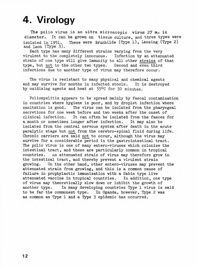
4. VirologyThe polio virus is an ultra microscopic virus 27 in
diameter. It can be grown on tissue culture, and three types were isolated in 1951. These were Brunhilde (Type l), Lansing (Type 2) and Leon (Type 3).
Each type has many different strains varying from the very virulent to the completely innocuous. Infection by an attenuated strain of one type will give immunity to all other strains of that type, but not to the other two types. Second and even third infections due to another type of virus may therefore occur.
The virus is resistant to many physical and chemical agents and may survive for months in infected stools. It is destroyed by oxidising agents and heat at 55°C for 30 minutes.
Poliomyelitis appears to be spread mainly by faecal contamination in countries where hygiene is poor, and by droplet infection where sanitation is good. The virus can be isolated from the pharyngeal secretions for one week before and two weeks after the onset of clinical infection. It can often be isolated from the faeces for a month or sometimes longer after infection. It may also be isolated from the central nervous system after death in the acute paralytic stage but not from the cerebro-spinal fluid during life. Chronic carriers are said not to occur, although the virus may survive for a considerable period in the gastrointestinal tract.The polio virus is one of many entero-viruses which colonise the intestinal tract, and these are particularly common in tropical countries. An attenuated strain of virus may therefore grow in the intestinal tract, and thereby prevent a virulent strain growing. On the other hand, other entero-viruses may prevent the attenuated strain from growing, and this is a common cause of failure in prophylactic immunisation with a Sabin type live attenuated vaccine in tropical countries. In addition, one type of virus may theoretically slow down or inhibit the growth of another type. In many developing countries Type 1 virus is said to be far the commonest type. In Uganda, however, Type 2 was as common as Type 1 and a Type 3 epidemic has occurred.
12

POLIO VIRUSENTRY INTO BODY
N O IN FECTIO N SUB CLIN ICA L IN FECTIO N
ft OR IMMUNITY PREVENT'; U GROWTH OF VIRUS
a
IMMUNITY OCCURS W ITH O UT SYMPTOMS OR IMMUNITY PREVENTS INFECTION
CLINICAL INFECTION W IT H O U T PARALYSIS
ENTRY VIA INTESTINE AND OR PHARYNX
IMMUNITY OCCURS WITH nvSYMPTOMS
CLIN ICA L INFECTION W ITH PARALYSIS
BLOOD BRAIN BARRIER
PROTECTS SPINAL CORD
VIRUS SPREAD UT
BODY
IMMUNITY W ITH PARALYSIS
ANTERIORHORNCELLS
INVOLVED
Fig. 4
13

5. EpidemiologyEpidemics of poliomyelitis have become more frequent in the
last fifty years, and particularly since 19^0 a number of large epidemics have been notified throughout the world.
It also appears that as soon as the hygiene of a community improves an epidemic is much more likely, and this usually occurs when the infant mortality of a country falls below 80 per 1,000. Conversely, where hygiene is poor most children are immunised by faecal contamination in infancy. Those infected in infancy while still carrying maternal antibodies are, however, only seldom seriously paralysed. In Cairo it has been found that nearly 100% of all children are immune by the age of four.
This paradoxical phenomenon of increased paralysis with improved hygiene means that it is essential to carry out mass polio immunisation campaigns at the same time as other standards of hygiene and health are improved lest a trail of polio epidemics and paralysed patients are the results of otherwise welcome aid. Unfortunately, vaccine is expensive, and methods of distribution are often difficult in the very countries where the need is most acute.
NEW EPIDEMICS IN AN ENDEMIC COMMUNITY
These may be caused by -
1) An unusual strain of virus.
2) Increased virulence of a strain already present.
3) A different type of virus, i.e., Type 1 or Type 3in an endemic community with Type 2.
H) Lack of immunity due to improved hygiene, and lackof subclinical infection in infancy.
EPIDEMICS IN A NON-ENDEMIC AND NON-IMMUNE COMMUNITY
I) These may be caused by any virulent strain ortype of virus.
2) These may lead to widespread severe infection among the non-immune.
14

WO
RLD
MA
PFU
TURE
EP
IDEM
ICS
OF P
OLI
O
• •• • • i ■ |• 1 L
OZUJO
<zLL.ooo2- ± ad
— D
uzLUQCLOQC<Ooo2- ±: CL— D
JOQ ^£ .2> < Li. iDQ_oQ-
uOcdZLO
15

3) Epidemics have been seen in the past when anoutsider has introduced infection in isolated communities such as the islanders of St. Helena and among the Eskimos. They have also been seen when non-immune people visit endemic countries without prophylactic prior immunization.
The economically rich countries must therefore continue their campaigns. This is essential with the increasing number of visitors from the developing countries to developed countries, as well as visitors returning from these countries after visits, who would act as carriers. The rapidity of air travel makes the dissemination of infection all the more likely. Although this sounds obvious, it is estimated that no less than 25$ of children in Britain and many other developed countries in 1969 were either incompletely protected against polio, or had no vaccine. The public have shown a lethargic interest in vaccination schemes, with possible risks of polio epidemics in the future in Europe, North America and elsewhere.
FUTURE EPIDEMICS
Epidemics of polio in the world to-day have never been so large, and many further epidemics will occur in the countries of the tropics and subtropics before adequate immunization of the populations at risk can be carried out. Even then, further epidemics will occur as complacency and inadequate continuation of immunisation campaigns allow a loophole in protection, especially in babies and young children.
Epidemics in non-endemic and non-immune communities will periodically continue to be caused by a virulent strain or type of polio virus either introduced by visitors from endemic countries, or given to visitors to endemic countries who have not been sufficiently immunised. New epidemics in endemic communities will also occur if the standard of hygiene is allowed to rise, while the immunisation of newborn children at risk is neglected.
Endemic communities may also, in addition, be infected by a foreign strain of virus newly introduced, by increased virulence in a strain already present, or by a different type of virus, i.e., Type 1 or 3 in an endemic community normally infected with Type 2.
16

LIBYA594/1964
U A R 417/1961
MALI233/1963SENEGAL
3 09>71965SUDAN
243/1962NIGERIA465/1964
I ETHIOPIA974/1963
I KENYA,456/1965CONGO
KINSHASA1039/1969
TANZANIA,407/1965
ANGOLA149/1963 MALAWI,121/1963
(ZAMBIA112/1962
RHODE!I93/I96V
SOUTH J AFRICA423/1968
AFRICAMOROCCO 173/1961
<=> MALTA 426/ 1942-3
TOGO301/1963
UGANDA148/1965
ST HELENA 217/1945
NOTIFIEDRECENTCASES
oMAURITIUS1000/1945
CD
REUN IO N160/1949
MALAGASY REPUBLIC 383/1947
ACTUAL PARALYTIC CASES MANY TIMES NUMBER OF NOTIFIED CASES
Fig. 5(b)
17

IMPORTANT EPIDEMICS IN THE 20th CENTURY
The mean annual incidence of paralytic poliomyelitis in the non-immune countries of the world is said to be 50 - 100 per million of population. The vast majority of cases in the underdeveloped countries of the world, however, are never notified, and official figures in these countries bear little relation to the true epidemiological picture. The following are only a few of the many epidemics which have occurred in thepast and are continuing to occur
EUROPE AND THE U.S.A.
Early 20th Century -
1931 - 1941
1941
1947
1948
1950
1952
DEVELOPING COUNTRIES
1939 - 1949
1952 - 1959
1945 - 1975
1954 - 1975
1975 __
U.S.A., Norway and Sweden
Many European countries
Malta
Great Britain and Germany
Mauritius and the Eskimos
U.S.S.R. and France
U.S.A. - Severe epidemic with 20,000 residual paralysed cases
South Africa, Northern Rhodesia, Belgian Congo and Egypt
North Viet-Nam, Singapore, China and Ceylon
Belgian Congo, Kenya, Uganda, Tanzania, and most countries in Africa and Asia
Various South American countries
Epidemics continuing to occur and getting larger and more frequent in many unimmunized countries of the world in the tropics and subtropics and in over half the world's population
18

CASES OF POLIOMYELITIS IN UGANDA AND IMMUNISATION SCHEMES
Fig. 5(c)
19

6. Pathology
ACUTE DISEASE
Poliomyelitis is a generalised infection which may involve the whole body, including the muscles direct, and liver, spleen and gut in the acute stage. The central nervous manifestations (Fig.6(a)), however, with involvement of the brain stem and anterior horn cells of the spinal cord are the only permanent manifestations of a systemic disease.
SPINAL CORD
In the acute stage, the whole cord may be hyperaemic and congested, and there may be slight meningeal inflammation. Inflammatory cells in the acute stages are polymorphs, which are later replaced by lymphocytes.
The anterior horn cells are those chiefly affected and every degree of degeneration may be seen from loss of Nissl's granules and eccentricity of the nucleus to complete disappearance of the cell. Cell death and disintegration may be rapid, and numerous phagocytes may be seen on section.
Section of the cord in the chronic stage may reveal atrophy of the anterior horn cells on one or both sides. The cells are replaced by astrocytes. The motor fibres arising from the destroyed cell disappear.
Occasionally posterior horn cells, posterior columns and other spinal cells may be affected in the acute stages, but residual effects are rare.
Poliomyelitis is essentially a disease affecting anterior horn cells with resulting paralysis of a lower motor neurone type with asymmetrical flaccid paralysis and normal sensation.
BRAIN AND BRAIN STEM
Examination of the brain on postmortem examination is sometimes made difficult by the secondary effects of respiratory paralysis and anoxia.
The true cerebral lesions are confined to the brain stem, medulla, pons and mid-brain and are rare in the basal ganglia and cerebral cortex. The residual lesions are usually confined to the brain stem with involvement of the lower cranial nuclei.
20

PATHOLOGY OF POLIOM YELITIS
BRAIN STEM "' INVOLVEMENT
MOTOR CORTEX BRAIN (RARE)
ANTERIOR HORN CELLS OF SPINAL CORD
SECTION OF SPINAL CORD SHOWING ONE HALF AFFECTED
NORMAL : DESTROYEDANTERIOR HORN ANTERIOR
CELLS HORN CELLS
Fig. 6(a)
21

CHRONIC DISEASE
LOCOMOTOR SYSTEMMUSCLES
The effect of destruction of the horn cells is to cause every degree of paralysis from a minimal degree which may recover completely, to complete quadriplegia with severe trunk involvement It is more usual, however, for one or two limbs to be affected, and the lower limbs are much more often involved than the upper limbs. Certain muscle groups tend to be much more often affected than others, and these are shown in Fig. 6(b).
The paralysed muscles affected show atrophy, fatty infiltration and replacement by connective tissue.
The resulting imbalance of muscles leads to deformities. This is mainly due to the excessive pull of the stronger group of muscles. The effects of gravity + position are often of secondary importance. Contractures may also be due to fibrosis in partly or completely paralysed muscles. Secondary contractures of ligaments and joint capsules may often result from longstanding shortening of muscles and tendons.
BONE AND JOINTS
The effect of paralysis on a growing limb results in shortening, and a decrease in diameter of the bone. This is due to lack of muscle pull with added vascular and neurological causes.Fractures may also occur fairly commonly, but unite well.
The effect of longstanding contractures on the joint surfaces may be to cause them to become flattened, deformed and occasionally subluxed or dislocated. Unsupported walking on weak joints may also lead to some of these changes.
The joints, however, are completely painless and usually fully mobile within the range allowed by the contracted soft tissues surrounding them. The articular surfaces are glistening, and the cartilage is thin but appears normal. Intra-articularadhesions only form if attempts are made to straighten the joints by operations or manipulation, and osteoarthritis without previous trauma or operation is almost unknown.
22

MUSCLES COMMONLY AFFECTED IN POLIOMYELITIS
SPINAL MUSCLES ..
EXTENSORS OFHIP
.. -~ ..
EXTENSORS OF KNEE
DORSIFLEXORS OF ANKLE ··· · ······ J
THENAR MUSCLES
Fig. 6(b)
23

SKIN AND SUBCUTANEOUS TISSUESDependent limbs may become oedematous in cold climates due to
stasis and gravity. In cold climates chilblains may be troublesome and may become infected.
RESPIRATORY SYSTEMLung infections are common due to paralysis of intercostals or
diaphragm.
EFFECTS OF BULBAR PARALYSIS
Bulbar paralysis usually recovers with no residual effects.A palsy in the acute stage may, however, result in a lung abscess in the chronic stage due to inhaled secretions.
24

THE NEGLECTED POLIO PATIENT.
25

7. Causation of DeformitiesThe initial cause of deformities in poliomyelitis appears to be
muscle spasm followed by interstitial fibrosis and collagen deposition in paretic muscle groups. This may lead to severe deformities as early as a month after the onset of paralysis.The exact cause of the muscle spasm is unknown, but basically it appears to be due to inco-ordinated involuntary contractions of surviving fibres in partly paralysed muscle.Although spasm of muscles is important in acute polio, the
growth of the limb is the most important factor in the progress of all deformities in chronic polio. This is why deformities are always much worse in children than in the adult or in adolescence. (Fig. 7(a))
HIP DEFORMITIESHip deformities in polio consist usually of a flexion abduction
deformity, due to relative weakness of the adductors and extensors of the hip compared to the abductors and flexors. Occasionally the converse may occur, especially with a flail hip. In this case an adduction deformity may lead to subsequent subluxation or dislocation of the hip.
KNEE DEFORMITIESThe commonest deformity of the knee is a flexion deformity due
to inbalance between the flexors and extensors in the initial phase of paralysis. This is followed by growth of the bone without equivalent growth of paretic muscles which have areas of fibrosis.An equally common deformity of the knee is a mild valgus
deformity which is almost always less than 30%. This deformity is so common in the paretic or paralysed lower limb in poliomyelitis that the absence of a slight valgus deformity should make one seriously reconsider the diagnosis of poliomyelitis. Lateral rotation of the tibia on the femur, and lateral subluxation of the knee may also occur as may genu recurvatum.This latter deformity is more often due to early weight-bearing on a weak knee, rather than inbalance between the extensors and flexors of the knee joint. (Fig. 7(b))ANKLE DEFORMITIESThe commonest deformity of the ankle is an equinus deformity due
to weak dorsiflexors and a relatively stronger calf muscle.
26

CAU
SATI
ON
OF
D
EFO
RM
ITIE
SIM
BALA
NCE
OF
M
USC
LES
2
d)iZ
27
SPAS
M OF
M
US
CLE
S
IMPO
RTA
NT
IN AC
UTE
PO
LIO

Other common deformities are a valgus deformity often associated with some degree of equinus, a varus deformity due to imbalance between the invertors and evertors, and a cavus foot due to weak intrinsic muscles and strong flexors of the toes. Occasionally a calcaneus deformity of the foot may occur due to weak calf muscles, but this is not common. (Fig. 7(b))
EFFECT OF POSTURE (Fig. 7(c))Prolonged bed rest, particularly in the severely paralysed
patient with inadequate physiotherapy, may cause a flexed hip, knee and ankle. This is the commonest cause of deformities in poliomyelitis after imbalance of muscles. In developing countries squatting and crawling can also help to potentiate these deformities.EFFECT OF GRAVITY (Fig. 7(c))Gravity acting on the paralysed limb may be responsible for an
equinus ankle and an adducted shoulder, as well as other deformities.SHORT LEG AND OTHER ASSOCIATED DEFORMITIES (Fig. 7(d))A short leg or a flexed hip, knee or ankle may cause the pelvis
to tilt, and a compensatory scoliosis may develop in the spine (Fig.7(d)). This deformity is usually mild and may take some time to develop. If the shortening or deformities are left uncorrected, however, they may become permanent.WEIGHT BEARING ON WEAK JOINTS (Fig> y(e))Weight-bearing on weak joints, as already discussed, may lead
to a genu recurvatum or a valgus knee, or to a valgus ankle.This deformity may become progressively worse with stretching of the ligaments of the joints without external support to the deforming joint.PROGRESS OF CONTRACTURES (pig. 7(f))All the above deformities may progress if left uncorrected
in children, where bone growth is not equalled by growth in fibrotic muscle. Secondary bone deformities are common in children.In the spine untreated severe scoliosis may be associated with
apparent kyphosis. This unsightly deformity of the spine may progress to wedging and rotation of vertebrae, and to crowding of the ribs on the concave side of the deformity.
28

CAUS
ATIO
N
OF
DEF
OR
MIT
IES
(/)UJ— Iu«/)Ds
uz<— I<00£
d)il
29
OF
TOES

CAUSES OF DEFORMITIES
GRAVITY
Fig. 7(c)
30

CAUSES OF DEFORMITIESFLEXION OF
SHORT LEG OTHER JOINTS
FLEXED HIP
FLEXE KNEE
QUINU ANKLE
TILTED PELVIS DEFORMITY IN ONEABDUCTION OF HIP JOINT MAY CAUSECOMPENSATORY SCOLIOSIS DEFORMITY IN THE OF SPINE OTHER TWO JOINTS
Fig. 7(d)
31

WEIGHT BEARING ON WEAK JOINTS
VALGUS VALGUS ANKLE KNEE
GENU RECURVATUMFig. 7(e)
32

MA
INTE
NA
NCE
AN
D PR
OG
RESS
ION
OF
C
ON
TR
AC
TU
RES
uz z>- £ Q
o _jz 0£ X0 a u
U Q
33

8. Polio ClinicsThe optimum organisation in a developing country is one large
central clinic and orthopaedic workshop with a peripheral ring of sub-clinics and smaller workshops. In a fairly large country this not only helps deal with a vast clinical load, but is also more acceptable to patients who are often unwilling to leave their own regions, as well as being faced with transport difficulties.
CENTRAL CLINIC (Fig. 8(a))
The central clinic is run in close conjunction with the main hospital of the country. All difficult polio cases are admitted to the wards and operated on in the theatre of this hospital. This main clinic is also used for all grades of training, which includes up-country surgeons and doctors, medical students, orthopaedic assistants, physiotherapists, social workers and ancillary helpers. Monday is the best major clinic day, as this enables patients to travel over the weekend and attend this clinic early on Monday morning. An ambulant hostel is used to help with accommodation.
A team of workers, which includes all the staff except doctors, then documents the patients throughout the morning. Clinical histories, both medical and social, muscle charting, simple physiotherapy and the changing of calipers and other supports are carried out. The patients are then given lunch and at 1.30 p.m. the team of surgeons and doctors arrives, complete with voluntary workers, who write down the detailed examination on special proforma as dictated. Supports are confirmed or changed, further treatment is ordered, and with this organised team all treatment, including the fitting of new calipers, can be completed by U.00 p.m. or 5>00 p.m.The only exceptions to this are operations which are given a future appointment, and patients requiring specialised physiotherapy, who are admitted, if possible, with a relative to the ambulant hostel for further treatment.
34

MAJOR POLIO CLINIC, HOSPITAL, HOSTEL.
# MEDICAL AND SOCIAL HISTORY# MUSCLE CHART AND PHYSIOTHERAPY# EXAMINATION BY SURGEON
i

UP-COUNTRY CLINICS (Fig. 8(b))
Clinics with a Trained Surgeon or Doctor - In these the same routine as in the major clinic is carried out, but on a much smaller scale. All difficult cases are referred to the Central Clinic, and all straightforward patients treated and operated on, where necessary, in the local clinic.
Clinics without a Trained Surgeon and New Clinics - These clinics usually require an initial two or three visits by a polio team in order both to train the doctor in charge and organise suitable facilities. The doctor in charge is asked to collect all polio patients from his district, and to arrange for them to stay in a makeshift hostel for 2 - 3 days. A team consisting of a surgeon, physiotherapist, two orthopaedic assistants, a workshop worker, a social worker (if possible) and a secretary, complete with a good supply of calipers, crutches and stencilled proforma, then arrives on the appointed day.Documentation of patients is carried out by the team on the
first day while the surgeon does in-patient rounds and organises future administration. On the second day the surgeon, assisted by the doctor, examines and documents all polio patients. On the third day simple operations are carried out by the local doctor, assisted and taught by the surgeon. In the meantime, calipers are fitted and physiotherapy given by the remainder of the team. Difficult operations are booked for the central clinic and not done up-country. Simple operations not performed on the third day are booked by the doctor in charge, and are carried out over a subsequent period of weeks or months. A supply of calipers, clogs and crutches must be left in order to start a small caliper bank.
A large number of patients can be properly examined if the organisation is good. The record number of patients fully examined in one day in Uganda was lU3 in 1965. One hundred and ninety-four calipers were also fitted in the same day.Visits should continue at least once every six months until the organisation is fully operational and the doctor in charge competent to keep his clinic running. As soon as practicable an orthopaedic assistant, a workshop worker and a small workshop is started. The doctor should visit the central clinic. It should then be possible for the doctor concerned to be almost independent of further visits, but close co-operation must continue with the central major clinic.
36

POLIO CLINIC & PHYSIOTHERAPY DEPT.♦--------------------------- —42’------------------------------ -»
FRONT ELEVATION
Fig. 8(b)
37
9. TrainingThe training of staff to look after the paralysed polio
patient is important in dealing with the vast polio problems in developing countries. There are many millions of untreated paralysed polio patients in these countries, and it would obviously be impossible for one surgeon or even several surgeons in each country to deal with the vast problems unaided.
It is essential, therefore, to train not only doctors, but also ancillary staff such as physiotherapists, orthopaedic assistants, workshop personnel and social and rehabilitation workers, and the patients' relatives themselves,so that a large team is dealing with the great majority of the problems.
In Uganda a vast training scheme was developed to cope with the problem, and this has now extended to other developing countries. Training is not only by teaching at the central hospital and up-country, but also by the means of thousands of booklets. Papers, films, slides and symposia are also used. Training schemes recommended are as follows:-
SURGEONS AND DOCTORS
Up-country surgeons and doctors, and surgeons from other countries, attend the main centre for short two or three day refresher courses. They are also visited in their own hospitals and clinics and operation sessions are run with training in view. They, in turn, are expected to train their own junior staff on returning up-country.
MEDICAL STUDENTS
Medical students are taught by means of booklets, films, slides, clinical demonstrations and lectures.
PHYSIOTHERAPISTS
Physiotherapists should be taught by apprenticeship.
38

TRAINING FOR ORTHOPAEDIC ASSISTANTS
3 m o n a
rfic/Fracture
\ ' I .. >(jOrthopaeq^
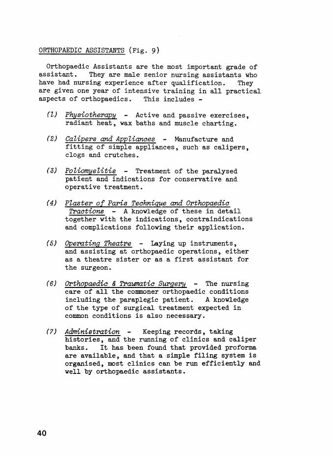
ORTHOPAEDIC ASSISTANTS (Fig. 9)
Orthopaedic Assistants are the most important grade of assistant. They are male senior nursing assistants who have had nursing experience after qualification. They are given one year of intensive training in all practical aspects of orthopaedics. This includes -
40
(l) Physiotherapy Active and passive exercises, radiant heat, wax baths and muscle charting.
(2) Calipers and Appliances Manufacture and fitting of simple appliances, such as calipers, clogs and crutches.
(3) Poliomyelitis Treatment of the paralysed patient and indications for conservative and operative treatment.
(4) Plaster of Paris Technique and Orthopaedic Tractions A knowledge of these in detail
together with the indications, contraindications and complications following their application.
(5) Operating Theatre Laying up instruments, and assisting at orthopaedic operations, either as a theatre sister or as a first assistant for the surgeon.
(6) 01'thopaedic & Traumatic Surgery The nursing care of all the commoner orthopaedic conditions including the paraplegic patient. A knowledge of the type of surgical treatment expected in common conditions is also necessary.
(7) Administration Keeping records, taking histories, and the running of clinics and caliper banks. It has been found that provided proforma are available, and that a simple filing system is organised, most clinics can be run efficiently and well by orthopaedic assistants.

Ninety-five orthopaedic assistants were trained in Uganda, and many of these have been posted to up-country hospitals.This has led to a considerable improvement in the standards of orthopaedic and traumatic treatment throughout the country and particularly the care of the polio patient, and there is no reason why this type of assistant cannot be used in all countries, both developing and developed.
WORKSHOP WORKERS
Disabled patients should be used where possible. Several up-country orthopaedic workshops were started and workshop workers attended for a three-month course in the main clinic before starting on their own. These workers need help and visits initially to organise their workshops and facilities.
VOLUNTARY WORKERS
These can be of great help in taking notes and helping with the organisation of clinics. Rehabilitation and social workers should also be trained.
PATIENTS' RELATIVES
These can be taught passive stretching of contractures and help with the rehabilitation of paralysed patients, both in the polio hostel and at home. The use of relatives to help in the management of paralysed patients is essential in developing countries, where adequate physiotherapy and nursing help will be very short for many years to come.
Thus it may be seen that much can be achieved by efficient team work and over 7,000 polio patients were treated over a 12 year period by this method. The efficient organisation of the large clinic is essential at the very start if a large number of patients are to be properly treated. This is especially so in poliomyelitis where many patients are children, and where most will require constant supervision for the rest of their lives.
41

10. Prophylactic Immunisation"Governments with poliomyelitis problems (in developing
countries) hesitate to venture on an enterprise soon to collapse because of lack of funds, personnel, communications and transport. Live polio vaccine is unstable without refrigeration, while the more stable Salk vaccvne is so expensive that few can afford even a single dose."
(PERABO, 1970)
The main defence against poliomyelitis is prophylactic immunisation (Fig. 10), as public health measures only play a very small and subsidiary role in prevention. This situation is unlikely to improve, as the exact mode of infection is still incompletely understood. World-wide immunisation campaigns are, therefore, essential both to diminish and eventually eradicate polio. At present, with the exception of some islands, only the developed countries of the world have been adequately immunised, but even these countries may not continue to be protected due to increasing public apathy to vaccination. It is also likely that over a million new cases of poliomyelitis now occur every year, mainly in the populations of the developing tropical and subtropical countries of the world and the risk of spread to Europe and North America in the future is considerable, if vaccination is not continued energetically. The problem is greatly underestimated by economically rich countries, as probably less than 1 in 100 patients with poliomyelitis in developing countries are even notified. The figures provided to the World Health Organisation can only show a tiny fraction of the total number of cases. Despite this lack of notification, the World Health Organisation have shown an average threefold increase in polio epidemics over the past 10 years in most tropical and subtropical countries of the world.
TYPES OF POLIO VACCINE
There are two main types of vaccine, a killed vaccine given by injection (Salk) and a live attenuated vaccine given by mouth (Sabin). It has now been shown that the live attenuated vaccine is not only much cheaper, but has many other advantages over the dead vaccine. In addition, by modern methods of manufacture, it is extremely safe, and the chances of causing paralysis are less than one in a million.
42

PROPHYLACTIC IMMUNIZATION
v a c c in eby injection
SABIN
v a c c in eby mouth
^ DROPSof vaccine on sugar
cubeFig. 10
43

It is also becoming apparent in developing countries that as the infant mortality falls below 80 per thousand, due to a general improvement in health and hygiene, paradoxically the likelihood of epidemics of polio recurring increases. This is because the polio virus is normally endemic in most of these countries, and therefore most children are infected with the virus during infancy when they are still protected with a high circulating level of maternal antibodies. They therefore acquire immunity to the disease without becoming paralysed as the maternal antibodies give them adequate protection against paralysis. As the health and hygiene of a community improves, however, the children are les.s likely to get subclinical infections as infants, and may become infected instead for the first time at the age of 1 or 2 years or even much later when they have lost their maternal antibodies and protection. They will therefore be susceptible to paralytic poliomyelitis, and epidemics will become more frequent.
In communities with an even higher degree of hygiene the age of acquiring the disease and immunity become later and later until at the present date in Europe and North America only one-third of all new polio cases are under the age of five. No less than one-third are over 15 and one-third between the ages of 5 and 15. This lack of immunity makes it essential that people from these countries be immunized before travelling to parts of the world where poliomyelitis is endemic or epidemic. It must also be remembered that, while early immunity appears to occur in urban communities in countries with a poor hygienic standard, the same is not necessarily true for isolated rural communities. It has been shown, for instance, that while over 90I of the cases of polio occurring in urban populations in Uganda occur in those under five years of age, in some rural communities in Uganda only one-third of those under 5 have any immunity at all.
Thus it may be seen that it is essential for immunization campaigns to be carried out as a matter of urgency in all developing countries.
44

SALK VACCINE
This is composed of killed strains of virus given by- injection. It has the advantage of being safe and usually effective, and of not being suppressed by the intestinal enteroviruses which may prevent immunity with the Sabin vaccine. It has, however, the following disadvantages
1) Repeated Injections
These appear to be necessary, both to achieve and maintain immunity. This may be dangerous in the prodromal phase of a wild polio virus infection and the injection may precipitate paralysis in the injected limb.
2) Delay in Immunity
This takes three weeks to achieve, and there is limited or no intestinal protection. Vaccine is therefore of limited value during an epidemic.
3) Incomplete Immunity
Immunity is less than that achieved by the Sabin vaccine in countries with a low enterovirus infection rate.
SABIN VACCINE
This consists of attenuated strains of live poliovirus.The trivalent vaccine made up of all three types of poliovirus should normally be used. The vaccine made by reputable authorities is now safe for use. The only disadvantage to its use at present in tropical countries is the fact that a high intestinal enterovirus content may inhibit colonization of the poliovirus in the intestinal tract. It should normally be used in preference to the Salk vaccine.
Advantages
l) Intestinal protection after the first dose is obtained within three days in over 60% of people. Intestinal colonization of the vaccine prevents growth of a virulent strain of poliovirus. Blood stream protection also rapidly occurs.
45

2) Immunity is much longer lasting and may be lifelong.
3) Immunity may also be conferred on contacts and othermembers of the family and community.
It is much cheaper to make and distribute.
Composition
The three attenuated viruses make up the trivalent Sabin vaccine. They differ in their prevalence and their abilities to colonize the intestinal tract and cause immunity. Vaccines for Africa should contain an increased amount of Type 1 virus (the least able to colonize the intestinal tract) and decreased amounts of Type 2 and Type 3 vaccine.
The following vaccine was recommended in 1975, hut composition may change and vary from country to country
Type 1 - L Sc 2ab - 105'T3 hType 2 - P712 Ch 2ab - 10' ’k 7Type 3 - Leon 12a^b - 10
TCID50 per dose
The vaccine can be stored for several weeks in a refrigerator between +2°C and +10°C (i.e. ordinary part of refrigerator - not the deeg freeze or cold compartment). A genuine deep freeze (-^0 C or below) is necessary for prolonged storage, and optimum storage temperatures are indicated with each batch. A temperature of -10 to +2 (i.e. the temperature of the cold compartment of a refrigerator) should be AVOIDED. The vaccine can be transported in ordinary "picnic"cold bags with cold packs or tins, to keep the vaccine cool.
Dosage
Three drops of trivalent vaccine (or the dosage stipulated on the container) should be given either by drops into the back of the mouth or on a lump of sugar. Plastic dropper bottles from which a dose can be squeezed are vastly superior to solid or glass bottles and save contamination.
46

Dosage should start at the age of two or three months and three doses are given at approximately k - 6 weekly intervals.A booster dose at the age of 18 months and another at school entry is advisable.
In communities with a low enterovirus content, the first dose protects about 65% of the population, the second dose about 95% and the third dose 100$. In many developing countries in the tropics the protection rate is probably much lower due to enterovirus interference. Vaccine should not be given in the presence of throat or intestinal infections.
Cost and Distribution
Each dose of vaccine has been reduced in cost to about 15 cents (6p) by simplifying its composition. Developing countries of the world still cannot get help from UNICEF or WHO for its routine purchase, although help can be obtained with distribution costs and in an epidemic. In Uganda, therefore, the cost of vaccine had to be raised by voluntary contributions. As a result of the President's Polio Appeal in 1966, thirty thousand pounds was raised by this means.The Polio Research Fund of Great Britain provided another thirty thousand pounds for vaccine and research into immunization. Other Governments also helped, and UNICEF provided vehicles.
The vaccine campaign was initially part of a National Immunization Campaign. A more rapid distribution, however, can be effected by having a separate scheme for polio.Villages and towns are notified by radio, television and newspapers, and local chiefs as to the next proposed visit of polio immunization vans. Distribution takes place in hospitals, schools, village centres or even under a tree.
Over 5 million doses have been distributed to date, and over 80% of the children under five in Uganda immunized at least once. Booster doses and continued immunization in infancy may cause difficulties, as it is essential that these be continued. The age group immunized will also have to be increased, as the endemic state (and therefore the likelihood of early natural immunity) decreases in all developing countries.
47

11. General ProphylaxisActive immunisation with trivalent Sabin vaccine is the
only effective prophylactic measure against polio. Improvements in hygiene and public health education can be of only relatively limited value due to the large numbers of asymptomatic carriers in an epidemic, who may continue to excrete the virus. It has been estimated that approximately 100% will continue to excrete the Virus in the stools for two weeks, 50% for three weeks and 25% for six weeks. A pathogenic strain of polio virus can nearly always be cultured from the sewage of towns, even when no_ cases of poliomyelitis are occurring. In most tropical countries there is a high incidence of endemic poliomyelitis among the populations. In addition, the virus can also often be cultured for a few days from the oral and nasal secretions of asymptomatic carriers.
The overall results of improvement in hygiene, and the lowering of infant mortality rates to below about 75 per 1 ,000, paradoxically, is to change a non-paralytic endemic state in a country into a paralytic epidemic state if polio vaccine is not given at the same time, (W.H.O. 1968), and the reasons for this are discussed in Chapter 5.
In addition to public measures of limited value in preventing epidemics, the only other important prophylactic measures are the avoidance of exercise, operations and injections where possible during epidemics and in suspected early cases.
Passive immunisation with antibodies in early suspected cases is of doubtful value, and contacts of polio patients and those at risk are best given oral vaccine which will prevent the multiplication of the wild virus in the intestinal tract.
48

ISOLATION OF PATIENTS
This is probably unrealistic in the acute phases of illness in developing countries. Careful hygiene on the part of the patient and all those looking after the patient , the washing of hands after defaecation with an antiseptic oxidising agent, the careful washing of soiled linen, and the adequate disposal of excreta, are the most that can be expected. Adults are best treated in hospital if possible and children without respiratory complications at home.
SCHOOLS
Schools should not be closed during epidemics, as this would lead to children disseminating the infection.School children with upper respiratory or gastro-intestinal infections, however, must be kept at home, and enforced rest ensured. It is also wise to stop organized games and physical education during epidemics, as exercise is known to make paralysis worse in the exercised limbs during the prodromal phase.
SWIMMING POOLS, MEETINGS, THEATRES,CINEMAS AND DANCE HALLS
It is probably wise to close swimming pools during an epidemic, and make sure that they are heavily chlorinated when they are opened again. This will also prevent unwarranted exercise during the possible prodromal phase.
During the height of an epidemic it is best to avoid large gatherings of people indoors. This should only apply to those at risk. In developing countries, where over 90% of cases occur in the under 5 age group, little would be gained by preventing adults attending meetings.
49

OPERATIONS AND INJURY
There is considerable evidence to show that previous tonsillectomy could precipitate bulbar paralysis, and that injury or operation to limbs during the prodromal or early phases of the major illness can precipitate severe paralysis in the limbs subjected to trauma.All non-urgent operations should therefore be stopped during the height of a polio epidemic, and in particular tonsillectomy. This ideal may, however, be difficult to implement in developing countries.
INJECTIONS
These should be avoided where possible during epidemics as it has been shown that injections during the prodromal phases of polio may precipitate or increase paralysis in the limb or limbs injected. Lumbar puncture should be avoided unless it is urgently required for other reasons.
CONCLUSIONS
General prophylaxis is of limited practical value due to the large number of asymptomatic carriers present in a community. Certain measures, however, may help to minimise the spread of the virus, while the avoidance of strenuous exercises, trauma and operations will diminish the severity of paralysis.
50

FACT
ORS
W
HIC
H
MAY
PO
TEN
TIA
TE
PARA
LYSI
S IN
THE
PR
OD
RO
MA
L PH
ASE
-
OPE
RA
TIO
NS
AND
TON
SILE
CT
OM
Y
51
Fig.
11

12. Clinical DiagnosisPRODROMAL OR PREPARALYTIC STAGE OF POLIO
The illness is usually of vague and variable duration. It maylast from a few hours to a few days, and one to three days is theusual duration. It may be very severe or so mild as to passunnoticed. It is usually, but not always, followed by an asymptomatic stage before the onset of the paralysis. Many patients never progress beyond this stage, and are only diagnosed by the laboratory finding of the poliovirus in the throat or stools. (Fig. 12(a))
The importance of this stage is that exercise, injection or operations may precipitate severe paralysis in the limbs exercised or traumatised.
SIGNS AND SYMPTOMS (Fig. 12(b))
These are variable and vague, and mimic other virus infections such as influenza. The more common symptoms and signs are -
(1) Headache and malaise
(2) Sore throat and upper respiratory infection
(3) Slight cough
(4) Diarrhoea or constipation
(5) Backache and joint pains
(6) Pyrexia of variable duration and severity
(7) Mild neck stiffness
Many other symptoms may occur, and the only safe way to deal with the problem is to regard all children with the above symptoms as suspects during an epidemic.
TREATMENT
Where facilities allow, all suspects should be put to bed and rested if possible. This is, of course, completely impracticable in many developing countries.
52

ANALYSIS of PATIENTS PARALYSED in POLIOMYELITIS
THE M A JO R ITY OF THOSE W H O ARE ILL W IT H
POLIOMYELITIS
ARE NEVER PARALYSED
OF THOSE PARALYSED
30%ARE PARALYSED BUT RECOVER COMPLETELY
30%HAVE MILD PARALYSIS
30%HAVE MODERATE OR SEVERE PARALYSIS
HAVE SEVERE RESPIRATORY OR HAVE BU LBA R IN VO LVEM EN T PO L IO M Y E L IT IS
Fig. 12(a)
O R DIE
53

The only practical measures which can he taken in suspects in economically poor countries is to stop the playing of games and manual work if possible, avoid injections for any hut the most severe general infections, and not perform tonsillectomies or non-emergency operations at the height of an epidemic.
It is also important to realise that the naso-pharyngeal secretions and faeces are highly infective at this stage of the illness, and children with suspected infection should he isolated if possible, particularly from other children and babies.
The signs and symptoms in paralytic polio are very variable in both duration and severity, and it is the onset of a flaccid paralysis of lower motor neurone type in a patient who is mentally alert, and who has had a prodromal illness, which makes the diagnosis probable. The presence of an epidemic will also be of help. The following are the common signs and symptoms
PERIPHERAL PARALYSIS
This may take from a few hours to three days to reach its maximum. It may appear to improve and then become worse again. It may affect one muscle group or the entire body, as• follows
In addition there may be transient involvement of the bladder with urinary retention which always improves.
The degree of initial paralysis bears little relation to the final degree of recovery.
BULBAR PARALYSIS
Pharyngeal Paralysis (Swallowing)
The most important sign of bulbar paralysis is the inability to swallow due to pharyngeal paralysis. The patient chokes on both solid and liquid food and also cannot swallow his own saliva. As a result, the patient may drown in his own secretions.
DEFINITIVE OR PARALYTIC STAGE (Fig. 12(c))
(1) Arms )(2) Legs )(3) Trunk )
Lower Motor Neurone Type of Flaccid Paralysis
54

THE PRODROMAL AND PREPARALYTIC STAGES OF POLIOMYELITIS
55

In addition, the patient cannot cough properly due to paralysis of the larynx. He also has difficulty in speaking due to paralysis of the palate.
Clinically, there may he bubbling at the back of the throat due to bubbling of air through a pool of mucus. Examination of the soft palate may show a patulous or loose lingula and accumulated secretions in the back of the throat.
If the patient survives the acute stage the prognosis for recovery is good.
Cranial Palsies
Any other cranial nerves may be paralysed, particularly the facial and the nerves to the ocular muscles.
Respiratory and Cardiovascular Centre Involvement
Signs of involvement of the respiratory centre are cessation of respiration for a few seconds at a time and irregularity of breathing. Involvement of the cardiovascular centre may cause the pulse to become irregular in both rate and volume.This may result in severe cardiovascular collapse.
RESPIRATORY PARALYSIS
This may be caused by involvement of -
Respiratory Centre - This is rare and mentioned above.
Inter postal Muscles - This may be asymmetrical and may be complete or incomplete. The anterior horn cells in thethoracic region are involved. Clinically, the chest eitherdoes not move, or moves poorly or asymmetrically.
Diaphragm - The anterior horn cells of the mid-cervical region are involved. Paradoxical respiration can be seen by observing the abdominal movements.
The early signs of respiratory involvement include breathlessness, a feeling of suffocation, slight cyanosis and the use of the alae nasae, sternomastoids and other accessory muscles of respiration. The chest and abdomen must be inspected, and the inability to take a deep breath and hold it, or to count to more than twenty in one breath are suspicious signs.
56

THE PARALYTIC STAGE OF POLIOMYELITIS
MENTAL CLARITYSORE THROATSLIGHT NECK RETRACTION
PYREXIA
PAINFUL JOINTS, MUSCLES & BACK
SEVERE HEADACHE M ALAISE
FLACCID PARALYSISOF THE LIMBS-ASYM M ETRICALWITH NORMAL SENSATION
BULBAR AND/OR RESPIRATORY PARALYSIS IN SEVERE CASES
Fig. 12(c)
57

A rising respiratory and pulse rate, and increasing restlessness and irritability are other signs of value.
POLIO-ENCEPHALITIS
This is rare and is usually associated with bulbar paralysis. Mental disturbances and even coma may occur, and there is nearly always paralysis of the facial muscles.
PAINFUL MUSCLES
This is a very constant finding and the muscles may be excruciatingly tender. The back and limb muscles which are not paralysed are both tender and in spasm. The patient is therefore very reluctant to sit up or bend forward, and slight pressure of even the bedclothes on the limbs may cause severe pain.
Pain in the neck with a mild degree of neck retraction may also occur.
FIBRILLATION OF MUSCLES
The muscles paralysed may show fine ripples of contractures. This is known as fibrillation.
PAINFUL JOINTS
The joints may be painful and this is mainly due to pain in the surrounding muscles. There is never any swelling or redness of the joints themselves; an important differentiating point from an acute arthritis.
PYREXIA
This may be variable in both duration and severity.
UPPER RESPIRATORY SYMPTOMS
Sore throat and other upper respiratory symptoms are common and variable.
58

OTHER SYMPTOMS
General malaise, headache and bowel upsets, either constipation or diarrhoea, may also occur.
ADEQUATE DOCUMENTATION OF PATIENTS
It is essential that an adequate history be taken and examination made of patients in order that the degree of paralysis, the progress of patients, and the value of treatment should be assessed. This is especially so in developing countries, where treatment may not always be carried out.
The assessment forms illustrated (Figs. 12(d) and (e)) have been designed with a view to realistic ease of filling out by voluntary workers and others with limited knowledge of poliomyelitis and with the examination assessment being dictated by the doctor at the clinic.
59

POLIO CLINIC FIRST ATTENDANCE
Research Case: Yes/No Today's Date:...... Polio No.:...NAME:....................AGE:. . . DATE OF BIRTH:....... SEX:ADDRESS:.........................MAP REF. :...............LENGTH OF HISTORY ............... DATE OF PARALYSIS ......
PARALYSIS: Legs| * Arms|^" Trunk Respiration
MOBILITY: Walk before paralysis?....Walk Now?....Only Crawl?. What supports?............... How far walk/crawl?.........
Any injections within How long before1 month of paralysis? ........... paralysis? ........How many injections? ........... Which limbs injected?___
POLIO VACCINE: Yes/No Injection or Mouth?...... Dates
LOWER LIMB DEFORMITIES
RT.LEG
LT.LEG
LOWER LIMB POWER
RT.LEG
LT.LEG
HIP l.Flex/Abd.2.
KNEE 1.FIexion2.Valgus3.Genu Recurv.
ANKLE 1.Equinus2.Valgus3 .Varusk.
HIP 1 .2.
KNEE 1.Extensors 2 .3.
ANKLE l.Dorsiflex 2.3.k.
REAL SHORTENING 1i: APPARENT SHORTENING
RIGHT ARM LEFT ARM
TRUNK Scoliosis?
TREATMENT: Calipers/Footwear Crutches Passive StretchingOPERATION RECOMMENDED OTHER TREATMENTNEXT ATTENDANCE DATE OTHER DATA 60
OPERATION DATE SURGEON
Fig. 12(d)

POLIO CLINIC
(Subsequent Attendances)NAME:.........................................................Attendance No: ......... Polio No:........... Date:.........HISTORY: Walking? Using Crutches?Previous Operations & Dates? What Kind of Calipers/Footwear?
LOWER LIMB DEFORMITIES
RT.LEG
■----LT.LEG
--------------------LOWER LIMB POWER
RT.LEG
LT.LEG
HIP l.Flex/Abd.2.
KNEE 1.Flexion2.Valgus3.Genu Recurv.
ANKLE 1.Equinus2 .Valgus3.Varusu.
HIP 1.2.
KNEE 1.Extensors 2.3.
ANKLE l.Dorsiflex 2 .3.
REAL SHORTENING APPARENT SHORTENING
RIGHT ARM LEFT ARM
TRUNK Scoliosis?
TREATMENT: Calipers/Footwear Crutches Passive StretchingOTHER TREATMENT
NEXT ATTENDANCE DATE OPERATION DATE SURGEON
OTHER DATA:Fig. 12(e)
61

13. Muscle ChartingMuscle charting is an assessment of the power of individual
muscles, in order that the degree of paralysis can be assessed.
It is important to assess all the muscle groups as soon as the tenderness in the muscles will allow, i.e., three weeks after the onset of paralysis (approx.). The degree of recovery should then be assessed at succeeding attendances, and an approximate idea as to the final degree of recovery and the necessity for calipers can be known at three months.
The muscle power is graded from 0 - 5 and the important figure is 3_. A power of 3 indicates the ability of a muscle just to do its work against gravity. This will naturally vary according to the individual muscle, i.e., the quadriceps has to lift a heavy leg against gravity while the extensor digitorum to the little finger has minimal weight to lift in order to do its work. If each respectively can lift the leg or the little finger each must have a power of at least 3 .
MAIN GRADES OF POWER
Muscle charting can be summarised as follows: (Fig. 13(a))
0 - No power
1 - Flicker of movement only
2 - Movement with gravity eliminated
3 - Movement gust against gravity
4 - Movement against gravity plus resistance
5 - Normal power
The illustrations show this diagramatically.
Occasionally power 6 is considered to be full power, but this extra grade of power, although an improvement, has not yet gained universal acceptance.
A muscle must not be given a power of 5 or 6 unless it has full power. A power of 0 indicates that there is not even a flicker of movement, while 1 signifies that a flicker is present but no more.
62

QUADRICEPS 3
JUST AGAINST GRAVITY
MUSCLE CHARTING(MAIN GRADES OF POW ER)
QUADRICEPS 0 QUADRICEPS 1 QUADRICEPS 2
NO MOVEMENT AT ALL | COMPLETELY FLAIL
FLICKER OF
CONTRACTION 'k ONLY
MOVEMENT ONLY WITH GRAVITY ELIMINATED
QUADRICEPS 4
X
QUADRICEPS 5
FULL NORMAL POWER
Fig. 13(a)
63

Muscle charting may be difficult to do accurately, especially in young children. Another difficulty may be trick movements which may deceive the examiner and give a false reading. A simplified muscle chart (Fig. 13(c)), is therefore indicated for those with limited experience.
FINER DEGREES OF ASSESSMENT
This is shown diagramatically on the opposite page. It is useful to have fractional muscle powers between the main assessments, especially if the power has improved from one figure and yet has not quite reached the next.
The powers around three, for instance, can be assessed as follows:
2+ - A power between 2 and 3, i.e., a littlemore than 2
3- - A power just
3 - The limb can
3+ - The limb can very slight
4- - A power more
The gradations naturally vary according to the individual muscle and joint. It is unrealistic to apply the above criteria to the extensor of the 2nd toe, for instance, but they may be quite helpful in assessing a knee extensor.
APPROXIMATE ASSESSMENT OF FINAL RECOVERY
The following should only be taken as a very rough but useful guide -
(1) Add 2 to the assessment of power at three weeksi.e. a muscle power of 2 could finally have a power of h
(2) Add I to the power at three monthsi.e. a muscle of power 2 at three months could have a final power of 3 (except when the power is 0 and is then likely to remain at 0).
(3) After six monthsAll recovery is now due to hypertrophy of residual muscles
0 4 or to trick movements.

MUSCLE CHARTING(Finer Grades of Power)
QUADRICEPS 2 +
QUADRICEPS 3
QUADRICEPS 3
JU ST A G A IN S T G R A V IT Y
QUADRICEPS 3 +
A G A IN S T G R A V IT Y A N D SL IG H T R E S IS T A N C E
Fig. 13(b)

SIMPLE MUSCLE CHART
DArRT.
rE
LT.DArRT.
rELT.
DART.
TELT.
DART.
TELT.
uppER
LIMBS
SHOULDER (Deltoid(Trapezius
ELBOW (Extensors (Flexors
WRIST (Dorsiflexors(Palmar Flexors
HAND (Grip(Finger Extensors (Finger Flexors (Opponens
T
R
U
N
K
NECK (Extensors (Flexors
SPINE (Extensors (Abdominals
RESPIRATION(Intercostals (Diaphragm
L0WER
LIMBS
HIP (Extensors (Flexors (Abductors (Adductors (internal Rotators (External Rotators
KNEE (Extensors (Flexors
ANKLE (Dorsiflexors(Plantarflexors(Evertors(invertors
Fig. 13(c)
66

14. Measurement of DeformityAn accurate assessment of deformities is essential at the time
the patient is first seen, not only in order that the optimum treatment should be prescribed, but also because the subsequent management may depend on prognosis.A number of contractures in poliomyelitis are missed because
the patient may disguise those with a lordotic spine or tilted pelvis. (Figs. lMa) and (d)).Deformities which are not apparent when the patient lies on a
couch, or even sits, may become very apparent when he stands.It is, therefore, essential to examine every joint thoroughly, and to check deformities not only with the patient lying, but also sitting, and if possible standing and walking. In those patients who cannot either stand or walk, the patient should sit up, with support if necessary, after the assessment has been completed in a supine position.
HIP DEFORMITIES (Fig. lU(a))
The flexed abducted hip is the usual common deformity in polio. This may be missed because the lumbar spine may disguise as much as 60$ of fixed flexion. Abduction of the hip may be responsible for most of the deformity, and an associated tilted pelvis may allow considerable abduction in flexion to be missed. The much rarer deformity of flexion without abduction is due to a tight iliopsoas tendon.Examination of the hip must therefore start by eliminating
lumbar lordosis by flexing the opposite hip fully and then extending the affected hip in slight adduction, instead of abduction.
KNEE DEFORMITIES (Fig. lU(b))
Flexion deformity of the knee should be measured with the hip extended as much as possible. This is because the knee deformity may be increased when the hip is flexed due to tight hamstrings. Movement of the knee is usually assessed from as much extensionallowed by the flexion deformity to full flexion, and the kneewith no deformity is graded as 0 .A valgus deformity of the knee is assessed from the line of the
femoral shaft. Backward and lateral subluxation of the tibia on the femur, however, is assessed as mild, moderate or severe.
67

External rotation of the tibia on the femur can be assessed in degrees, but it is important that the knee is extended as much as possible before the rotation is assessed, as the rotation is often correctable when the knee is fully flexed.
ANKLE AND FOOT DEFORMITIES (Fig. lU(c ))
These are assessed as illustrated the ankle being assessed at its neutral position at 901 to the tibia. The amount of equinus or calcaneus is measured from this plane.Foot deformities are also assessed from neutral with varus and
valgus and adduction of the forefoot being assessed in degrees. Clawing or cavus of the foot or toes can only be assessed as mild, moderate or severe.
SPINE (Fig. lU(d))
The usual deformity is a scoliosis of the spine, and this can only be accurately measured by x-ray. This should initially be assessed as mild, moderate or severe and a note made as to whether it is correctable and the position of the apex of the curve. When the curve is severe there may be a compensatory secondary curve which should also be noted. In addition there may be rotation of the vertebra in severe deformities leading to prominence of one part of the chest or lateral spinal muscles, which is assessed as a kypho-scoliosis. There may in addition be a smooth kyphosis or lordosis due to severe weakness of the posterior spinal muscles, and the patient may have to arch his back to maintain his balance.
UPPER LIMB DEFORMITIES (Fig. lU(e))
Deformities of the upper limb are measured from the neutral position. The commonest deformities are an adduction deformity of the shoulder, a flexion deformity of the elbow, a rotation deformity of the forearm, and flexion deformities of the Joints of the hand and wrist. Each of these deformities should be measured with the joints above and below the deformity as straight as possible.
UNEQUAL GROWTH IN POLIOMYELITIS (Fig. lU(f))
Unequal growth is common when polio occurs in the growing child, and is not seen after the epiphyses have closed. A short arm as such is not an appreciable disability and its symptoms arise from its weakness or deformity. A short leg,on the other hand,
68

MEASUREMENT OF HIP DEFORMITIES
Fig. 14(a)
69

MEASUREMENT OF KNEE DEFORMITIES
95° FLEXION DEFORMITY
MOVEMENTS
OTHER DEFORMITIESVALGUS DEFORMITY MODERATE BACKW ARD SUBLUXATION
M ODERATE LATERAL SUBLUXATION
M ARKED EXTERNAL ROTATION OF
Fig. 14(b)
70

MEASUREMENT OF ANKLE AND FOOT DEFORMITIES
40° CALCANEUS ANKLE _ 25° or MODERATE VALGUS50 VARUS FOOT
35° ADDUCTION FOREFOOT
71

M EASUREMENT OF DEFORMITIES OF THE SPINE
COMPENSATORY SECONDARY
" CURVE IN TH O RAC IC
SPINE
M ARKED SCOLIOSIS OF SPINECONCAVITY TO LEFT IN UPPER LUMBAR REGION
Fig. 14(d)
72

MEASUREMENT OF DEFORMITIES OF THE UPPER
73
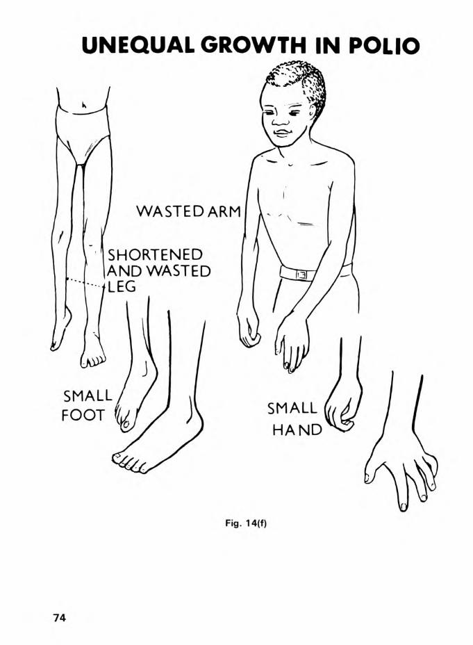
UNEQUAL GROWTH IN POLIO
Fig. 14(f)
74

SHORTENINGAPPARENT
P ELV IS H O R IZ O N T A L

may be a very severe disability, and lead not only to a severe limp, but also to poor posture with secondary deformities in other joints as well as the opposite leg and in the spine.
ASSESSMENT OF SHORTENING (Fig. lU(a))
Shortening must be assessed accurately. Apparent shortening is much more important than true shortening, and is usually caused by a tilted pelvis. Tilting of the pelvis may also lead to an increase or decrease of the apparent shortening of the leg, and it will increase with adduction, which can sometimes be corrected.
Apparent shortening is assessed with the legs held in the position in which the patient would stand or walk. True shortening, on the other hand, is measured with the leg in as similar a position as possible, and if one leg is in fixed abduction or adduction the other leg must be put in an identical position. It is important always to measure length to the sole of the foot, rather from the anterior superior iliac spine to the medial malleolus. This is because there is often a cavus (clawing) or a flattening of the foot in polio,which may lead to as much as 2 centimetres of increased or decreased shortening respectively of the actual foot below the medial malleolus. Other factors that have to be taken into consideration in the assessment of shortening are a dislocated hip, a flexed knee and a deformed foot and ankle.
76

15. X-Ray Changes in PolioThe x-ray appearances of bone and joints in the patient with
severe long-standing poliomyelitis may be bizarre, but are often quite typical.
INITIAL X-RAY CHANGES
The initial changes seen in paralysed or paretic limbs or trunk in the growing child are due to the lack of the normal muscular pull, and the diminished vascularity of the paralysed and paretic overlying muscles with collagen infiltration and contracture.This usually results in osteoporotic short bones with a thin cortex, and a relatively wide medullary cavity.
SUBSEQUENT CHANGES
Subsequent to the initial changes, the imbalance of muscle power, plus the differential growth in contracted and shortened muscles compared to the underlying bones, the normal shape of the epiphyses is affected. The effects of weight-bearing on osteoporotic weak joints without the benefit of supporting muscles to protect their ligaments may also cause marked changes in the ends of growing bones, while deformities may also result from fractures in the shafts of the osteoporotic bone.
LONG-STANDING DEFORMITIES
Long-standing -untreated contractures of joints may cause flattening of the epiphyses due to the direct pressure effects on the contracted joints in abnormal contact. Subluxations and dislocations of joints, such as the hip and knee due to unbalanced muscle action may also occur. Osteoarthritis, however, is very rare in joints affected by poliomyelitis, except where the paralysed joint has been immobilised in plaster, operated upon, manipulated or damaged in other ways. In these cases severe degenerative changes may occur much earlier than in the normal joint.
The following is a summary of the common deformities seen in long-standing untreated poliomyelitis. These appearances are mainly restricted to the growing child, and once growth has ceased these changes, apart from osteoporosis and thinning of cortices, usually do not occur.
77

SPINE AND RIBS (FIG. 15(a))
The vertebrae may become rotated and wedge-shaped with diminution of disc spaces and lipping on the concave side of the disc under compression.
The ribs are usually crowded together on the concave side of the scoliotic curve and broader and thinner on the convex weak side of the scoliosis where the' are more prominent and usually flattened with widened intercostal spaces.
PELVIS, HIP, FEMUR (FIGS. 15(a) and (b))
The paralysed pelvis is often tilted, deformed and smaller on the more paralysed side. The acetabulum may be more oblique and the hip may be subluxed or dislocated, if the neck is in varus. The head of the femur is often more globular than normal, and the neck and shaft of the upper femur is usually decreased in diameter and associated with a varus or a valgus deformity of the neck of the femur.
KNEE JOINT AND TIBIA FIG. 15(b))
The lower end of the femur may be more globular than normal, and the posterior articular surface of the femoral condyles may be flattened in long-standing flexion deformities of the knee. The patella is often much smaller than normal, and the tibial plateau may be deformed and sloped either backwards or forwards. There may be lateral or posterior subluxation of the femur on the tibia in severe flexion deformities, and both tibia and femur may show a thin cortex and widened medullary cavity.
ANKLE AND FOOT FIG. 15(c))
The ankle joint often shows the lateral malleolus higher than the medial and this results in the ankle mortice being tilted laterally in the valgus ankle. X-ray of the tarsal bones often shows marked changes in both shape and position, and there may be evidence of either flattening of the foot or cavus. The Uth metatarsal is sometimes quite short compared to the other metatarsal bones in a severe deformity.
78

X-RAY CHANGES IN POLIO. (TRUNK)

UPPER LIMB (FIG. 15(d))
The shoulder in severe weakness is commonly subluxed downwards due to the lack of muscular support. The head of the humerus tends to be more globular than normal and the whole scapula and humerus may be decreased in size.
The elbow may be misshapen with a poorly developed medial epicondyle and the head of the radius may be dislocated.
A rotation deformity of the forearm may be present, and the wrist may be deformed and often in palmarflexion. It may be subluxed or even dislocated.
Deformities of the metacarpals and phalanges, if present, may show narrow shafts and globular heads in relation to the shaft.
80

X-RAY CHANGES IN HIP AND KNEE
EXTERNAL ROTATION OF TIBIA
FIBULA POSTERIOR A.R X-RAY
PATELLA LATERAL
Fig. 15(b)
LATERAL AND POSTERIOR SUBLUXATION OF KNEE LATERAL AND POSTERIOR
SUBLUXATION OF KNEE AP X-RAY
81

X-RAY CHANGES IN POLIO, ANKLE AND FOOT
LOWER MEDIAL MALLEOLUS
FLAT FOOT
CAVUS FOOT

X-RAY CHANGES IN UPPER LIMBS
ADDUCTION DEFORMITY SHOULDER
Fig. 15(d)
83

16. Laboratory DiagnosisThe laboratory can give comparatively little help in
poliomyelitis, except to confirm a diagnosis, or to help in the differential diagnosis. The following are the more important laboratory investigations.
E.S.R.
There is only a slight or moderate increase of the E.S.R. in polio.WHITE BLOOD COUNTThis may be normal or there may be a slight or moderate
leucocytosis. A count of over 20,000 or a polymorphonuclearcy- tosis of over 90% is against a diagnosis of polio.CEREBRO-SPINAL FLUIDThis is normal in the pre-paralytic phase. In the paralytic
phase there is often a raised pressure with an increase of neutrophils and protein early. After a few days the lymphocytes increase.The fluid is clear and free of pus cells and bacteria both on
microscopy and culture. This may be useful in differentiating a pyogenic or tuberculous meningitis.CULTURE OF THE P0LI0VIRUSTHROAT - It may be cultured readily from the throat for one week before and one week after the onset of paralysis.FAECES - The poliovirus may be cultured from the faeces for several weeks and sometimes longer after an attack of poliomyelitis.C.S.F. - It is rare to culture the poliovirus from the C.S.F. except at post-mortem in cases dying in the acute stage.
TESTS FOR ANTIBODY AGAINST POLIOMYELITISThe two main tests are a tissue culture neutralisation test
and a complement fixation test. Both are of very limited value unless the immune status of the patient is known just before the attack of polio.The antibody picture in a population at risk is, however, a
valuable method of assessing the immunological status of the community and the necessity for an immunization campaign. It isalso useful for assessing the results of mass administration of vaccine.
84
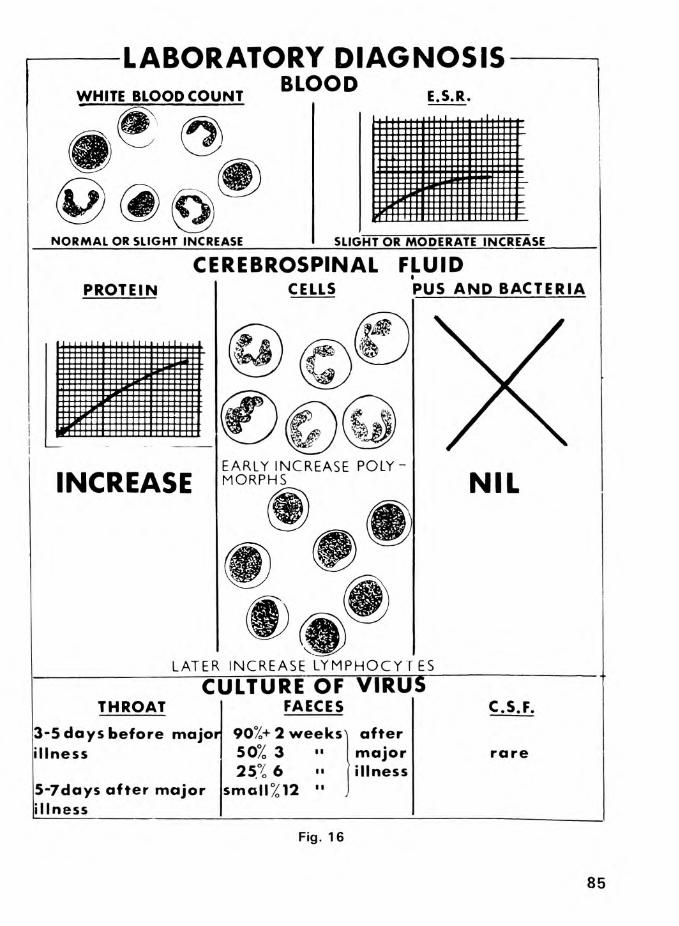
LABORATORY DIAGNOSISBLOODWHITE BLOOD COUNT
N O R M A L O R S L IG H T IN C R EA S E
E.S.R.
S L IG H T O R M O D E R A T E IN C R E A S E
CEREBROSPINAL FLUIDPROTEIN CELLS PUS AND BACTERIA
INCREASEEARLY IN C REASE POLY M O RPH S NIL
LATER IN C R E A SE L Y M P H O C Y T E SCULTURE OF VIRUS
THROAT FAECES C.S.F.
3-5 days before major 90%+ 2 weeks] afterillness 50% 3 major rare
25% 6 illness5-7days after major small%12 ••illness
Fig. 16
85

17. Differential Diagnosis ACUTE POLIOMYELITIS
In the pre-paralytic stage the diagnosis can only be confirmed by culture of the polio virus.
In the paralytic and pseudoparalytic stages, the following are the more important differential diagnoses:
MENINGITIS AND ENCEPHALITIS Polioencephalitis is rare. Mild meningitis or encephalitic symptoms are not uncommon, however, and a lumbar puncture will differentiate a meningitis. A virus encephalitis may be more difficult to diagnose except with the help of a laboratory. Neck pain is comruon in polio, but severe neck retraction and photophobia are rare. Flaccid paralysis also occurs in poliomyelitis, but not in meningitis or encephalitis. ~-
ACUTE INFECTIVE POLYNEURITIS This may be confused with poliomyelitis. The paralysis, however, is symmetrical and affects all four limbs and trunk with associated sensory loss. The cerebro-spinal fluid has a high protein content, but no cells. There is usually complete recovery.
PSEUDOPARALYSIS Osteomyelitis, arthritis and trauma in children may cause apparent paralysis of a limb, due to the child's unwillingness to move it. A thorough clinical examination will reveal a swollen, tender limb or joint, and the regional glands may be enlarged and tender in infections.
CJPHER VIRUS INFECTIONS The Coxsackie Groups A & B and the E.C.H.O. virus are the most important viruses in a large group which may cause a paralysis very similar to polio. The diagnosis is sometimes only made by the laboratory, or by a knowledge of an epidemic of the particular virus in question. The paralysis tends to be very much less severe than in polio and recovery is usually complete. Bladder involvement and sensory loss may also be present.
OTHER CAUSES OF ACUTE PARALYSIS Any other causes of paralysis include traumatic palsies of peripheral nerves, acute neuritis due to vitamin deficiency or poisoning by heavy metals, and epidemic pleurodynia (Bornholm's Disease). A thorough history and examination will usually establish the diagnosis.
86

DIFFERENTIAL DIAGNOSIS OF ACUTE POLIOMYELITIS
MENINGITIS AND ENCEPHALITIS
MENTAL IRRITATION NECK RETR A C T IO N OR C O N FU S IO N
POLYNEURITIS
PERSISTENT BLADDER INVOLVEMENT
ALL FOUR LIMBS PARALYSED
PSEUDOPARALYSIS IN CHILDREN(Osteomyelitis, Arthritis, Injury,etc.)
HEAT SW ELL IN G OR SEVERE LOCALISED TENDERNESS IN BONE OR JOINT INVOLVED
Fig. 17(a)
87

CHRONIC POLIOMYELITIS
SPINAL CONDITIONS - The back should always be examined in all cases of paralysis and the following differentiated
Tuberculosis, and Infection of the Spine: These may causeeither a spastic or flaccid paralysis of a limb or limbs, and is common in developing countries. There is usually, but not always, an obvious kyphos, and sensory and bladder disturbances may be present.
Spinal Fracture: This may be overlooked in a baby or youngchild.
Transverse Myelitis: This usually causes a spastic type ofparalysis with sensory loss.
Tumours of the Spinal Cord or Vertebrae: These are uncommon in children. In Africa the most common tumour in children causing paralysis with sensory, motor and bladder involvement, and a normal X-Ray, is a Burkitt tumour. In an adult a secondary deposit in a vertebral body is common.
SPASTIC HEMIPLEGIA AND DIPLEGIA - Spastic hemiplegia and diplegia in children are very common in Africa. There is usually a history dating from birth, but in some cases the condition dates from early childhood. In most cases the arm and leg on the same side are involved, but all or only one limb may be paralysed.The child is usually,but not always, mentally affected to some extent. The limbs are always spastic and there may be athetoid movement. The muscles look normal, quite different from the wasted limbs of the polio patient. Contractures are usually much less than in the untreated polio patient. Sensation is normal and shortening absent or much less than in poliomyelitis.
CONGENITAL TALIPES EQUINO-VARUS (C.T.E.V.) - This is a commondifferential, diagnostic problem, as polio may also cause an equino- varus deformity. The deformity dates from birth, power is initially normal, and there is no other evidence of poliomyelitis.
The back should always be examined, as spina bifida is another diagnostic pitfall which is usually associated with sensory loss in the legs.
88

DIFFERENTIAL DIAGNOSIS OF CHRONICPOLIOMYELITIS
SPINAL CONDITIONS'FRACTURES. INFECTIONS,SECONDARY DEPOSITS ETC.)
RETENTION OF URINE
KYPHOS OR DEFORM (EXCEPT IN TRANSVERSEMYELITIS AND BURKITT'S TU M O U R)
SPASTIC HEMIPLEGIAMOTOR PLUS SENSORY LOSS (.SPASTICITY OR FLACCIDITYJ
FOOT OR FEET ONLY AFFECTED NORMAL MUSCLE P O W ER INITIALLY HISTORY SINCE BIRTH
SPASTICLEG
USUALLY ARM OR LEG ON SAME SIDE (MAY BE O N E LIMB OR ALL FOUR LIMBS)
SPASTIC ARMCONGENITAL TALIPES EQUINO VARUS
Fig. 17(b)
89

18. NursingIn view of the likelihood of painful muscles and spasm of
hack muscles, particularly in the older child or adult patient, good nursing is essential in acute poliomyelitis. The patient should be treated on a sorbo mattress on fracture boards (Fig. 18 ) with adequate support of paralysed limbs. A good supply of small soft pillows and pads should be available for limbs, and the back should be adequately supported.
Rigid splinting should be avoided where possible. The feet tend to develop equinus deformities, and they should be supported by foot boards or by soft expanded polyethylene (Plastazote) back splints. A well padded below-knee caliper with backstop and clogs may serve the same purpose.
Knees and hips should be slightly flexed for comfort, rather than held completely straight. Paralysed arms are best supported, as illustrated, on pillows with a slight degree of abduction.
Paralysis of the spine, with spasm and pain, may be relieved by the support of a small pillow in the upper lumbar region, but this may not always be so when the patient is lying flat. Adequate lumbar support, however, is essential when sitting.
The care of the skin, particularly of the lower back and heels, is important, and the severely paralysed patient must be turned every two to four hours, day and night, to prevent bed sores and to keep the skin dry. The semi-prone, prone and lateral positions are usually more comfortable than the supine. The supine position in any case should not be permitted, if there is any danger of bulbar paralysis. The nurse must change the position of paralysed limbs frequently, day and night, as there is seldom enough physiotherapy help.
In the acute stages retention of urine may occur, and catheterisation may be necessary. Adequate fluid intake is essential, especially in the tropics, and in the case of repeated catheterisation, antibiotic cover to prevent urinary infection is important. Constipation may necessitate the use of suppositories and careful adjustment of the diet.
90

NURSING IN ACUTE POLIOMYELITISG O O D - m e n t a l a n d p h y s i c a l r e s t
SM ALL P IL L O W O R PAD TO SU PPO RT LU M BA R SP INE
KN EES SL IG H TLY FL E X E D
H EA D O N P I L L O W ARM SU PPO R T ED
F O O T B O A R D TO KEEP FEET AT R IG H T A N G LE
BAD -UN COM FORTABLE AND LIKELY TO CAUSE CONTRACTURES
Fig. 18
91

In severe paralysis of the upper limbs and trunk, respiratory infection may occur, and daily breathing exercises and chemotherapy are essential in order to prevent a respiratory tract infection, even when the respiratory muscles are not severely impaired. The patient should also be turned from one side to the other to minimise respiratory complications.The hygiene of the nose and oral cavity are also important.
Feeding, when there is no associated bulbar or respiratory palsy, is usually straightforward, but it is essential in the early stages not to miss the onset of paralysis, and especially regurgitation of food or fluids. (Chapter 19).
PAINFUL MUSCLES M D SPASMS
Paralysed muscles, especially in adults in the acute stage, may be very tender. Those that are not fully paralysed may also be in spasm, and contracted. Hot, moist towels wrung out and placed over the painful muscles will often relieve some of the spasm. A hot water bottle is a less effective, but more practical method where there is a shortage of nursing staff, and this is especially useful for spasm of the back muscles. It is important, however, that towels should not be too hot, or the paralysed patient may be burned.
The Sister Kenny method of regular application of hot packs has been over-emphasized in the past, and the claims for its efficiency in preventing or curing paralysis exaggerated.There is no doubt, however, that moderate heat, especially if moist, does relieve spasm and pain, and is soothing to the patient, but there is little place for a great deal of time-wasting ritual, especially in developing countries.
Analgesics, and some of the more modern drugs with a specific effect on muscle spasm, are sometimes also indicated in the acute stages, but their side effects should be noted, and watched for. It is essential that, in the first three or four days, the patient should receive the minimum of drugs which could depress or mask the diagnosis of the onset of respiratory or bulbar palsy.
In moving the patient, painful muscles should not be squeezed, and limbs should be lifted gently near the joint, and not at a painful muscle belly. Joints in paralysed limbs should be put through their full range of movement at least once a day, provided that this is not too painful, and provided this does not cause too much spasm. The position of paralysed limbs92

should also be gently changed several times a day by the nurse caring for the patient.
PSYCHOLOGICAL REST
The paralysed patient may be very apprehensive, and it is essential that he should be sedated and reassured if necessary. Sedatives must, however, be used with great care in the first three days of paralysis, due to the danger of unrecognised respiratory or bulbar palsy.
OBSERVATION OF THE PATIENT
Careful observation of the patient is essential, in case of an increase of paralysis, especially respiratory or bulbar palsy. These are discussed in Chapter 19. In severe cases, half-hourly checks on pulse and respiration should be carried out in the first three or four days. In addition to respiratory rate, the volume and rhythm of the respiration should be carefully noted at this stage of the illness, and also the use of any of the accessory muscles of respiration.
The colour of the patient, and his mental alertness, may also be valuable guides to early complications, as may nasal regurgitation, difficulty with swallowing, a "nasal voice" and weakness of voice power.
Paralysis affecting new muscle groups, and new areas of tenderness and spasm must also be noted. A rise in temperature may herald the onset of infection; alternatively, it may in the early severe case indicate involvement of the bulbar centres.
The fluid intake and urinary output chart is essential in tropical countries where dehydration is particularly likely to occur. A bell within easy access of a limb that the patient can use, or, in the case of very severe paralysis, a laryngeal microphone, is essential for the peace of mind of both patient and attendants.
OTHER POINTS OF MANAGEMENT
Complete barrier nursing of patients in the acute stages of polio is of limited use, owing to the presence of asymptomatic carriers of the virus during epidemics. Simple precautions, such as the wearing of masks, the isolation of eating utensils, scrupulous cleanliness of attendants, and the correct disposal of linen and excreta are, however, indicated. Encephalitic or meningeal signs may require special nursing, sedatives and a darkened room.

19. Respiratory and Bulbar PolioRespiratory paralysis may be of sudden onset, and in Africa
the patient may be hundreds of miles from the nearest hospital equipped with a respirator.
In severe respiratory paralysis the only method of saving a patient's life may be to transport him to the hospital, and assisted respiration may be essential during the journey.
EMERGENCY TREATMENT - RESPIRATORY PARALYSIS ALONE (Fig.19(a))
A hand operated bellows, as shown, is simple. No oxygen cylinder is necessary although provision is made for connection to an oxygen cylinder, and this would be indicated for severe respiratory insufficiency. In the place of a bellows a self-filling bag such as an 'Ambu' could be used. An ordinary anaesthetic face mask with expiratory valve should be used with either of these methods.
The patient can be given continuous or intermittent assisted respiration, (depending on the degree of respiratory paralysis). Guides to respiratory distress are increase of pulse and respiratory rate, use of accessory muscles of respiration, cyanosis and restlessness.
EMERGENCY TREATMENT - BULBAR PALSY WITH OR WITHOUT RESPIRATORY PALSY (Fig. 19(aD
In combined respiratory plus bulbar palsy the patient cannot swallow, and will drown in his own secretions if these are not drained away as they form. The easiest method is by tilting the head of the bed well down, and by having the patient's head to one side. The patient can then be transported on his side, again with the head of the bed down. This will drain secretions more efficiently, but is less effective for assisted respiration.
Associated respiratory insufficiency can then be treated by a bellows type of respirator as above. A foot-operated sucker to aspirate the secretions should always be available, and should be used as necessary to keep the airway clear.
94

2</)
h J * < O ai o . <
z2* < DC qq
DC 3 O flfl
Z z
*2<si- <>- £5u c l 212 o «DC Z
k li
zU<
o CO—1 Z< o
( / )LU>
J75 O t( J—1 <
>■ >- j
< O tCt t 3 *< £ 3Q. o z> -O f
2 * .
^ if/10
□cLU IV '
H - Q_ V
<O \
tc QZ
CLU )
<X
U iDC
«n
<cc2DC<GO- JDCO«/>3—iOl>-ocOt-<t t
Ewn
(0CD
05
z
oh-uDl/i
95
TREATMENT OF BULBAR PALSY ALONE (Fig. 19(b))
In bulbar palsy without respiratory paralysis, the patient cannot swallow and must be prevented from aspirating his own secretions. The best method of doing this is by tilting the bed head downwards with the patient either lying on his face or on his side, with his head to one side.Emergency Management
A useful method of treating children is by a bed broken and angulated in the middle as shown. Pillows can be used to bolster up the centre of the mattress.Secretions will dribble out of the patient's mouth.
They should in addition be aspirated.Facial Palsy
Facial palsy is usually associated with difficulty in swallowing. Care must be exercised in keeping eye, nose and mouth clean, especially when the patient cannot shut one or both eyes properly.Physiotherapy
Physiotherapy is essential to keep the lungs well aerated, as the patient cannot cough properly due to a paralysed larynx.Alarm System
Par^Lysis of the larynx will mean a poor voice and the patient should have a buzzer or bell within reach of a working group of muscles so that he can summon aid if necessary.Feeding
The patient should be fed by gastric tube or parenterally, and initially nothing should be given by mouth.Associated Respiratory Paralysis
A careful watch must be kept in the first three days for the onset of associated respiratory paralysis, or central palsy affecting the cardiovascular or respiratory centres.Prognosis
The prognosis is good in swallowing paralysis alone, provided that the secretions can be kept drained. Recovery is usually c omplet e.
96

TREATMENT OF PATIENT WITH BULBAR PALSY ALONE
POSITION OF PATIENT
FACE DOWN HEAD TO ONE SIDE
FOOT SUCTION PUMP
Fig. 19(b)
USE SPECIAL BED OR PILLOWS AS SHOWN
97

TREATMENT OF RESPIRATORY PARALYSIS ALONE (Fig. 19(c))Diagnosis
Respiratory paralysis is usually due to paralysis of the intercostal muscles or of the diaphragm or of both.Paralysis due to involvement of the respiratory centre itself is very rare.A careful watch for decreasing respiratory muscle activity
is essential, and the patient should be given assisted respiration as soon as early signs of respiratory insufficiency appear. These include a rising respiration and pulse rate, the use of accessory muscles or respiration (a late sign), slight cyanosis, restlessness and irritability, and an inability to count to more than twenty in one breath.Tank Respirator
The tank type of respirator is best, and there are many models on the market. It is essential to make sure that there is no bulbar palsy and that the airway is clear before putting the patient into a respirator on his back.The use of the respirator must be explained to the patient.
Oral fluids, apart from sips of water, should not be given in the first twelve hours. Gastric or intestinal distension may require gastric aspiration or a flatus tube, and urinary retention may require catheterisation.The respiration rate in a tank respirator should vary from
30 to Uo/min. in infants to l6 to 20/min. in adults.The negative pressure should be set at 12 cm/water in an
infant and lh to 18 cm/water in an adult.Other Respirators
Other types of respirator, which include a rocking bed and a cuirass which fits chest or abdomen or both, are much less efficient than the tank respirator and should be avoided except for short periods.Weaning from Respirators
The patient should be weaned from the respirator gradually. When this happens he should be kept in the same room as the tank respirator until his confidence in breathing on his own has been regained.
98


RESPIRATORY PLUS BULBAR PALSY (Fig. 19(d))Many patients have drowned in their own secretions in the
past due to the doctors not realising the dangers of a tank respirator in bulbar palsy. The accumulation of secretions in the back of the throat will be sucked into the lungs at the first inspiratory phase of the machine, and the patient will continue to aspirate his own secretions until he drowns.Tilting of the head downwards with the head on one side will
allow the secretions to dribble out of the mouth as they form, and the patient should in addition be lying on his face.Alternatively, the secretions can be prevented from entering
the lungs at all by the use of a cuffed tracheotomy tube, or by a cuffed endotracheal tube - the lung being inflated by a positive pressure pump. Each of these methods will be described.Patient on Face, Head Tilted Down and Tank Respirator
This is the best method of treating a patient with respiratory plus bulbar palsy in a developing country.The patient can be fed by a gastric tube, and the secretions dribble out of the mouth as they form, and are not sucked into the lung.Tracheotomy and Cuffed Tracheotomy Tube and Intermittent Positive Pressure Respiration
This is the standard method of treating patients in Europe and the U.S.A. A cuffed endotracheal tube can be used as a temporary measure if necessary.This is theoretically a much better method than a tank
respirator plus head tilted down position. In actual practice in developing countries, however, intermittent positive pressure respiration machines are expensive, are in very short supply, and tend to give trouble and break down.The cuffed tube, if used for long periods, causes pressure necrosis on the trachea and may get blocked. Biochemical upsets, and over or under ventilation of the lungs are much more common than in a tank respirator. In addition, the patient cannot talk, and psychologically does not tolerate the cuffed tube well.A head-down position in a tank respirator should therefore
always be the treatment of choice in developing countries, except occasionally in specialised units.
100

TREATMENT OF COMBINED RESPIRATORY AND BULBAR PALSY
CABINET RESPIRATOR IN HEAD DOWN POSITION
ALARM
USE ONLY WITH TRAINED NURSING & MEDICAL STAFF
CUFFED TRACHEOSTOMY TUBE
Fig. 19(d)
101

20. PhysiotherapySimple physiotherapy has a very definite place in the treatment
of poliomyelitis in developing countries. Few hospitals, however, are able to employ physiotherapists solely for the treatment of patients with poliomyelitis. In addition, only a small proportion of a physiotherapist's overall comprehensive training can be applied to the treatment of patients with poliomyelitis.
The "assistant" physiotherapist, and in particular the "orthopaedic assistant" (Chapter 10), on the other hand, with limited practical experience in stretching of contractures, fitting of calipers and getting patients walking, will have a very definite and valuable place in the treatment of patients in polio clinics in developing countries.
The fully trained physiotherapist plays her most important role in the major hospital of a country or in the main provincial hospitals. Her value as a member of the orthopaedic team is dependent on her preparedness to adapt to local conditions.She must spend a considerable part of her time teaching junior staff simple physiotherapy techniques, and in supervising these. She must also be prepared to involve herself in a considerable amount of administrative work, and to devote most of her individual therapy time to patients who require the specialised types of physiotherapy which only she can safely carry out, such as short wave diathermy and ultrasound.
The duties of a physiotherapist can be summarised in the following order of priority
Teaching of Relatives and Friends of Patients: Relatives orfriends of patients can carry out many simple procedures, and should be taught passive stretching of muscles and contracted joints, supervision of muscle re-education, correct fitting of calipers, and assisting the patient to walk.
The duty of the physiotherapist is to make sure that calipers do, in fact, support the necessary joints, and that they have the correct raise, backstop or other modification for optimum comfort and mobility.
102

PREVENTION OF DEFORMITIESPOSTURE,SPLINTING,& SUPPORT
H IP S
Fig. 20(a)
103

Teaching Patients to Walk: Patients who have never walked,or those who have been crawling for a long time, or who walk badly, must he taught to walk in calipers and crutches in a realistic time, relative to bed shortages and waiting lists.
Teaching of Orthpaedic Assistants: Ancillary staff need tobe taught the simpler and more important aspects of physiotherapy, and especially those methods which they themselves will be expected to carry out in up-country hospitals and polio clinics. These include simple active and passive exercises, fitting of calipers and supports, getting patients walking and very simple muscle charting.
Simple Muscle Charting: Physiotherapists should be able tocarry out muscle charting accurately, as this will assist the doctor in the assessment of the patient's disability and she should also complete simple proforma (Fig. 20(d)).
Post-operative Treatment: Post-operative treatment inpoliomyelitis should be directed particularly to the active and passive movement of joints, and to getting the patient up and walking. Breathing exercises may also be necessary post- operatively for those patients with respiratory insufficiency.
Posture Splinting and Support: It is more important toprevent deformities than to treat them, and also much easier.The correct posture of the patient in bed will often achieve much. This is frequently difficult to maintain, and detachable supports, particularly in the form of above-knee calipers, will not only support knees and ankles and prevent deformities, but by their weight and the fact that they keep the knee straight, will help to prevent or correct any hip flexion deformities as well.
Spinal supports for the convalescent patient with a flail spine will help support a back. It can not prevent a deformity, however, unless it is of the Milwaukee type from chin to pelvis, which is seldom tolerated or kept adequately adjusted.
Active and Passive Movements of Joints: Joints should not beforced in the first two or three weeks of illness while the muscles are still in spasm and very tender. Within the limits of pain, however, much can be achieved in keeping hips, ankles and other joints mobile and moving through a full range each day. Where possible this should be done by active movements, but as many of the joints are paralysed this may have to be done by passive movements instead.
104

PREVENTION OF DEFORMITIESMOVEMENT-ACTIVE OR PASSIVE-LOW ER LIMB
K N EES
Fig. 20(b)
105

PREVENTION OF DEFORMITIESUPPER LIMB - ACTIVE AND PASSIVE MOVEMENTS
IN T E R N A L A N D E X T E R N A L R O T A T IO N O F S H O U L D E R
F L E X IO N A N D E X T E N S IO N P R O N A T IO N A N DO F E L B O W SU P IN A T IO N O F F O R E A R M
F L E X IO N A N D E X T E N S IO N O F W R IS T
F L E X IO N A N D E X T E N S IO N O F F IN G E R S
Fig. 20(c)
106

POLIO NO:.......NAME AGE SEX......Date Paralysis Date First Seen Date Admitted Hospital
POLIO PHYSIOTHERAPY PROFORMA
Date Physio Started Date Caliper Fitted Date Next Attendance
LIMBS AND TRUNK PARALYSEDJOINTS CONTRACTED ......OPERATION DATE & SUMMARY MOBILITY BEFORE TREATMENT MOBILITY AFTER TREATMENT
DATE & INITIALS PHYSIO
PHYSIOTHERAPYI
Hip
DEFORMITIES & POWER!gAnkle
REMARKSIt. Leg Lt. Le Knee Ankle Hip Knee
(Walking Support etc.)
1stAttendance
COMMENTS
Fig. 20(d)
107

21. Mobilisation of PatientsThe patient should be out of bed and in a chair at an early
stage unless paralysis is very severe. In the case of children this can usually be achieved within a few days. In adults, however, the back muscles may remain very tender, and full mobilisation may take one or two months. Patients should always be propped up gradually in bed and then in a chair before being stood up.
Methods of gradually mobilising the convalescent patients are illustrated in Figs. 2l(a) and (b). This progresses from supports in bed, to support in a chair through to exercises in bed or a couch and then to sitting and walking. Mobilisation in a swimming pool, if available, is also helpful. It should be stressed that swimming pools, unless warm and properly maintained, are greatly over-rated in the treatment of poliomyelitis. Polio clinics in small countries without adequate finance should forego a swimming pool in favour of building extra facilities for the main polio clinic or having extra Balkan beams on beds so as to exercise paralysed limbs on supporting slings, as shown.
The progressive mobilisation of the paralysed patient out of bed is shown in Fig. 21(b), and this varies from a wheel-chair in the severely paralysed to a walking machine, parallel bars and crutches and calipers in the less severely disabled.
Some patients with weak quadriceps are capable of walking without calipers by forcing back their knee or by supporting their knee with a hand (Fig. 21(c)). This may be satisfactory in the patient with strong hip extensors and a moderate plantar flexion power of the ankle. In the child, however, this method of walking may cause either flexion or hyper-extension deformities of the knee.
All children with weak limbs and with the possibilities of deformity should be encouraged to wear calipers until they have at least completed their growth period, even if they can walk without support.
INDICATIONS FOR CALIPERS IN CHILDREN
Above-Knee Caliper
I.Where the quadriceps has power less than 3(i.e., the leg cannot be held up against gravity)
108

MOBILISATION OF THE CONVALESCENT PATIENT
PADDED CH A IRSU PPO RTED S ITT IN G IN BED
EXERCISES W ITH LIMBS SUPPORTED IN FRAM E
PO LIO S W IM M IN G PO O L
Fig. 21(a)
109
2. To prevent a possible contracture developingin a growing child, due to imbalance of muscles
2. As a night or day splint to correct a slight degree of contracture
Below-Knee Caliper
When the quadriceps has a power greater than 3 and yet the foot is flail and dropping or tending to go into valgus or varus, and therefore needs support
Above or Below-Knee Caliper with Backstop
When the tendo Achillis is tight, with a resulting tendency for an equinus deformity of the ankle to develop
CONTRA-INDICATIONS TO A CALIPER
1 In a patient with an uncorrected deformity3unless it is of slight degree, and the caliper designed to correct it. However, where operation must be delayed or is refused, a caliper should be ordered, even though it would otherwise be contraindicated.
2. Where there is severe weakness of both legsand at the same time appreciable weakness of one or both arms and the trunk. (i.e., the patient would not be able to propel himself forward with crutches)
INDICATIONS FOR CRUTCHES
Bilateral calipers or a caliper on one leg associated with weakness of the opposite leg or spine. Also in some patients with weakness of the hip on the side with a severely affected lower leg.
110

PROGRESSIVE MOBILISATION OF THE PARALYSED PATIENT
1 1 1

INDICATIONS FOR CALIPERS AND CRUTCHES IN ADULTSSimilar to those in children except that new deformities tend
not to occur in long-standing cases. In some cases, the patient can manage without any support at all hy bracing back a weak knee with a slightly equinus ankle or strong hip extensor.DIFFERENCES IN ABOVE AND BELOW-KNEE CALIPERSApart from the length of the caliper, the diameter of the
ring, and the fact that the above-knee caliper has a knee piece, both are identical in every way.Inside or outside irons and T-straps for varus or valgus
deformities are not justified in under-developed countries.All irons should be double, as they hold a tendency to inversion or eversion as well as, if not better than, a maladjusted single iron.CALIPERS AND CRUTCHESIt is important that the caliper fits properly and that the
crutches are correct both in length and in the position of the hand grip. The more important points are illustrated in the appropriate diagram. An above-knee caliper is required if the knee extensors are less than three in power and a below-knee caliper if they are more than three and the ankle weak. An ankle with a tendency to equinus will need a back-stop in addition.BELOW-KNEE CALIPERThis should allow full flexion of the knee, and the strap at
the top of the caliper should be fairly tight. The sockets in clog or shoe should fit firmly and not allow too much free movement, and there must always be a supporting ankle strap.
In the case of a foot which tends to go into equinus, a backstop must also be used.ABOVE-KNEE CALIPERThis should extend no higher than l" below the groin on the
inner side. Straps should be adjusted fairly tightly and the same other criteria apply as with fitting a below-knee caliper.The knee piece must be adjusted to give the knee adequate
support anteriorly, and except in the case of genu recurvatum, the posterior knee strap should be slightly loose.
A valgus knee is common and it is essential that the metal of the caliper does not rub on the inside of the knee in these patients, especially when weight-bearing.112

METHODS OF WALKING WITH WEAK KNEE EXTENSORS
BRACING BACK LEG W ITH HAND ON HYPEREXTENSION OF W EAK KNEE THIGH W ITH HIP EXTENSORS OR W IT H
PLANTARGRADE FOOTFig. 21(c)
113

CRUTCHES
Their length should be l" less than the distance of the anterior axillary fold to the sole of the foot. The hand grip is approximately one third down the crutch from the top but may be less. In the case of weakness of the trunk or arms the top of the crutch should be well padded to avoid pressure in the axilla and a radial nerve palsy.
PLASTIC KNEE SPLINT
A plastic knee splint is sometimes indicated in patients who have knee extensors with power less than three and a fairly stable and serviceable foot and ankle which does not require the support of a caliper. The place of a plastic knee splint, however, is very limited as it takes longer to adjust and fit, and they are often not as comfortable as a knee-bending caliper. They have a place in a few adult patients, but are seldom worth fitting in children.
114

FITTIN G OF CA LIPERS AND CRUTCHES
I hI" BELOW A X ILLA AND [ HPADDED TOP
HAND GRIP I/3 rd WAY D O W N THE CRUTCH
BELO WKNEE CALIPER
A LLO W S FLEX ION OF THE KNEE
CLO G FITS W EL L AN D STRAPS SUPPORT FO O T AN D ANKLE
ABO VE KNEE CALIPER I"BELOW 'G RO IN
KNEE PIECE A ['-FIRM AND
COMFORTABLE FIT
STRAP BEHIND KNEE SLIGHTLY LOOSE EXCEPT IN GENU RECURVATUM
Fig. 21(d)
115

PLASTIC KNEE SPLINT
EDGES PADDED WITH SORBO OR PLASTIC
116
Fig. 21(e)

22. Contractures — Conservative Management
Contractures will usually occur if there is imbalance of opposing groups of muscles which are not held in check. Although this is the most important cause, there are other primary causes as well, and these include the effects of gravity, the effects of flexed joints in bed, and the results of bearing weight on a weak leg.
Secondary to the primary causes are the effects on joint, bone and connective tissue of the initial contracture. Joint capsules become contracted on the flexed side, the epiphysis may become flattened or deformed, and intermuscular septa, nerves and vessels become shortened in time. The skin over the flexed surface may also become tight.
Long-standing contractures, especially of a major joint such as hip or knee, may cause multiple problems in their correction. Standard open operations are often successful in correcting the contracture, but these may be major operations requiring blood transfusion, plus a qualified surgeon. Complications which may follow these operations include skin loss, infection, and damage to important structures.
Subcutaneous methods of dividing tight structures and straightening contracted hips, knees and ankles are not new. They were in fact used in the 19th century, but have been superseded now by open methods. These simple methods, however, have their virtues in developing countries. They can be done by doctors inup-country hospitals, require no blood, and complications seldomoccur. In less severe contractures these procedures have many advantages over major open operations. Modifications to the more conventional methods of subcutaneous tenotomies will therefore be described in detail.
Fig. 22(a) shows the commoner deformities and contractures of the hip, knee and ankle in poliomyelitis.
Knee and ankle contractures may prevent the fitting of calipers, if severe. Operative correction is required, with certainexceptions which will be discussed.
117

PREVENTION OF CONTRACTURES
The prevention of contractures in the acute and subacute stages is very important.
In many cases the patients are children who will be treated at home, and the parents must be shown how to stretch the paralysed limbs daily to prevent contractures. In children the muscles in acute polio are tender for a much shorter duration than in adults, and stretching can be commenced almost immediately with very little pain. This is essential, ascontractures can occur in a matter of days.
Splints
Splints, except for back slabs for a drop foot, are now seldom used. Reliance is placed much more on daily stretching plus correct support of paralysed limbs in bed, as already illustrated.
Stretching of Muscles and Joints (Fig. 22(b))
Joints must be stretched in the direction opposite to that of the contracture, i.e. an equinus ankle dorsally. This must becarried out at least once a day by the physiotherapist or nurse,and at least three times a day by relatives.
The important contractures are those of hip, knee and ankle, but other contractures such as a varus contracture of the foot, or an adduction contracture of the shoulder may occur.
The illustrations show the correct method of stretching contracted joints. The same method of manipulation is used by the surgeon. In this case a general anaesthetic is used and this allows a greater degree of correction to be obtained.
Flexion Contracture of the Hip Fig. 22(b)
Pressure backwards should be in the upper third of the thigh, lest excessive leverage should cause a fracture. The opposite hip must be fully flexed to eliminate lumbar lordosis, and the leg should be brought down in slight adduction to stretch the abductors which are usually tight as well.
118

THE TYPICAL CONTRACTURES OF POLIO
Fig. 22(a)
119

MANIPULATION OF.‘-FLEXION ABDUCTION DEFORMITIES OF THE HIP
C O R R E C T
MANIPULATION OF>FLEXION DEFORMITY OF THE KNEE


Lying the patient on his face in ted, as shown, with a pillow under the lower thigh is useful, provided the patient will tolerate the position. The hips can also he extended while the patient is in this position.
Flexion Contracture of Knee Fig. 22(b)
It is essential that the knee is manipulated as shown in the illustration and that pressure is exerted near the joint. If this is not done fractures of the tibia or femur, slipping of the epiphyses and backward subluxation of the tibia on the femur are liable to occur.
Preventing a Recurrence of Contracture
Apart from stretching imbalanced muscles, the only way of preventing a recurrence of a contracture is to hold a joint in an overcorrected position so that the deforming muscles are acting at a mechanical disadvantage. This is most easily achieved by fitting calipers as soon as the tender muscles will allow, (in small children within a few days or even immediately) and leaving the calipers on for most of the day and night in the acute and subacute stages.
Calipers for Deformed Knees
The special supports for deformed knees are illustrated in Fig. 23 (c). This caliper for a slight flexion deformity of the knee is merely an ordinary caliper with a loose posterior strap and a tight knee piece which may need to be padded. This type of support, which helps correct the knee as a patient walks, can only be used to correct a deformity of 30 degrees or less due to the difficulty of fitting.
A caliper for genu recurvatum has the opposite effect with a tight broad posterior strap and the knee piece needs to be fairly loose anteriorly. In this type of support only slight tension on the posterior strap is required as the deformity is easily correctable when the patient ceases to stand.
The caliper in genu valgum requires to be bent to compensate for this, and in addition a second knee-piece will need to be fitted so that it presses on the medial side of the knee-joint and correct the valgus as the patient walks. This knee-piece is in addition to the ordinary anterior knee-piece.
122

MANIPULATION OF ANKLE AND FOOT DEFORMITIESSUPPORT FOREFOOT
EQUINUS OF ANKLE c o r r e c t
ADDUCTION OF FOREFOOT
IN C O R R E C TSUPPORT ANKLE
FORCED EVERSION AND DORSIFLEX FOR AT LEAST 5 MINUTES ^
VARUS OF FOOT
SUPPORT ANKLE
FORCIBLE CORRECTION OF FOOT FOR AT LEAST 5 MINUTES
SMALL PADDED WEDGE
Fig. 22(d)
123

Manipulation of Ankle and Foot Deformities Fig. 22(d)
This is shown, and the most important deformity to correct is equinus. The correct method is demonstrated with the ankle firmly supported as the foot is dorsi-flexed. In the case of varus of the foot, or adduction of the forefoot it is important to he firm yet gentle and to avoid too rapid or forceful a manipulation. Much more is achieved hy firm pressure for at least five minutes in the opposite direction to the deformity, and this will usually need to he repeated and followed hy surgical correction to prevent recurrence.
124

23. Hip and Knee ContracturesIt is essential that the patient is assessed in detail before
any operative procedures are considered. The main criteria with any operation on the lower limb are whether the patient is likely to be able to walk with or without a caliper following an operative procedure, and whether the patient will be socially benefited by being able to walk.
As a general guide line all children who have a reasonable chance of being able to walk post-operatively, even with difficulty and even with calipers on both legs, should have their limbs straightened, and every attempt made to get the child up and mobile.
In the case of adults, operative procedures are only indicated where there is a good chance of reasonable mobility. The future employment and wishes of the patient should be taken into consideration, as well as age. As a general rule old patients with very severe deformities requiring extensive surgery should have a low priority on operative correction, while young patients and those with minimal deformities should be considered first.
CONTRAINDICATIONS TO OPERATION IN CHILDREN
1. Both legs severely involved with one or both arms, particularly the triceps, weak. i.e. the child will not be able to use crutches or walk in calipers. Good arms on both sides are important if both legs and the trunk are severely affected. There are notable exceptions to this rule and each child should be assessed as to the possibility of future walking if the contractures are straightened.
2. Minimal contracture of the hip alone of less than 30°.
In all other circumstances the child should be operated on if necessary and got upright and walking.
The psychological effects of mobility on a child are tremendous, and parents will often educate and look after an upright child when they tend to neglect a crawling one.
125

CONTRAINDICATIONS TO OPERATION IN ADULTS
1. Where one or both arms are weak in addition to both legs being severely paralysed, i.e. the use of crutches will be difficult or impossible. Fairly strong arms, particularly the triceps, are important if the patient is to progress upright. Again, as in the case of children, a patient with determination may manage surprisingly well with limited weakness in one or both arms provided his trunk is strong.
2. When there is only a minimal degree of contracture and the patient is managing to walk well.
3. When there is severe contracture of both knees and the patient earns his livelihood on the ground and is happy to continue doing so.
1+. Where operative facilities are poor and contractures severe.
In an adult with polio contractures it is essential to consider the patient as a whole and not only the contracture. An adult patient who can crawl fast on the ground and earn his own living is often better off than one who can only progress very slowly upright with stiff knees and two crutches. The latter may look better, and almost certainly feels better. He may have difficulty in planting crops in an agricultural community, which may be his only means of livelihood. He may therefore die of starvation and neglect following well-meaning operative intervention.
Serious consideration must be given to the future occupation and mobility before bilateral severe contractures are straightened.A simple wheelchair may be a far better method of progression for long distances, while pads on knees and hands may allow fast local progression, and mobility indoors.
Isolated Hip Contractures of Less than 30°
No treatment is required for these when there are no_ other contractures. The stability of the hip is often improved, and shortening compensated for, if there is a small degree of abduction/flexion contracture.
126

Isolated Knee Contractures of Less than 30°
In a child these are best treated by fortnightly manipulations under anaesthesia until at least 2 of genu recurvatum is obtained. An above knee caliper is then fitted. If there is associated hip contracture preliminary soft tissue correction will also be necessary, (see below).
Russell traction will also correct this deformity but will take time and will necessitate hospitalisation. In adults this contracture may be much more difficult to correct. Manipulation alone usually fails. Russell traction again takes time but may be effective.
In some adult patients with a mild degree of flexion deformity no treatment, however, is necessary. In others a supracondylar osteotomy may be indicated, but only if the foot is stable, and a caliper may thereby be discarded.
INDICATIONS FOR OPERATION
Hip and Knee Contractures of Over 30°
These will all require operation, with the exceptions already mentioned. In a young child with fairly recent contractures the most important single factor responsible for the deformity is a tight tensor fascia lata and ilio-tibial band. In the older child or adult, however, other ligamentous and tendinous structures play an important part and must be divided as well. (See below).
The subcutaneous method of division is very satisfactory for the less severe contractures, provided it is done correctly and as extensively as necessary. Care must be taken to avoid damaging the femoral and popliteal arteries and the lateral popliteal nerve. The biceps, however, should always be divided under direct vision because of the risk of damaging the adjacent lateral popliteal nerve.
The operative techniques employed are illustrated, and will also be described in detail.
127

Sterility and Position on Operation Table
OPERATIVE DETAILS (Fig. 23(a))
The operation must be done under full sterile precautions with adequate skin preparation and sterile towels. It is better to sterilise both legs even if only one is to be operated on, so that the opposite hip can be kept flexed to compensate for a lumbar lordosis.
The affected hip and knee should be kept as straight as possible when the tight structures are being divided so that these are kept under tension.
First Incision
This is situated on the lateral side of the thigh about 1 inch above the knee joint. The tensor fascia lata is kept under tension, and can usually be felt as a tight band.
The knife is inserted horizontally from a lateral to a medial direction just behind the tight band, and directed towards the femoral shaft until the tip touches the lateral cortex of the bone. The blade is then twisted through 90° so that its sharp edge is pointing vertically anteriorly. All the subcutaneous structures anterior to the blade and lateral to the lateral surface of the femoral shaft are then cut.
It is important that the tight structures be cut anteriorly enough, and that the relative positions of the popliteal artery and lateral popliteal nerve be borne constantly in mind.
If the flexion contracture of the knee is more than 30° it is also essential that the biceps tendon is divided by open division under direct vision (see below). This will also enable the lateral intemruscular septum and the posterior part of the tensor fascia lata to be divided much more safely and efficiently than by the subcutaneous method alone.
Second and Third Incisions
These are situated one-third up the thigh and two-thirds up the thigh on the lateral aspect. One incision instead of these two, and situated in the middle of the thigh, would be adequate, however.
128

SUBCUTANEOUS FASCIOTOMY FOR HIP& KNEE CONTRACTURES
SKIN INCISIONSFLEX O PPOS ITE HIP W H E N
PERFORM ING O PER A T IO N
DIVISION OF TENSOR ANTERIOR TO HIP
FASCIA LATA AND TIGHT STRUCTURESKEEP LATERAL TO LA TERA L BO RD ER OF FEMUR IN L O W E R
THREE IN C IS IO N S
CARE W IT H PO PL IT EA L VESSELS AND CARE W IT H FEMORAL LATERAL PO PL IT EA L NERVE
ARTERY
129

The tensor fascia lata should he palpated and the knife should be inserted in the same way as in the first incision along its posterior border down to bone. It should then be twisted through 90 and the cut made anteriorly and lateral to the lateral border of the femoral shaft.
If the tensor fascia lata is not palpated the knife should still be inserted into the lateral aspect of the thigh, as there will be other tight structures to be cut including the vastus lateralis.
Fourth Incision
This is situated about one finger's breadth below the anterior superior iliac spine. The position of the femoral artery should be palpated and borne constantly in mind during the operation. The position of the inguinal ligament should also be noted and care taken not to divide it. The femoral nerves just lateral to the femoral artery are usually tight, but should not be divided. They usually protect the artery just medial to them.
The blade of the knife is inserted subcutaneously below the anterior superior iliac spine and in a lateral to medial direction, and slightly downwards, for about three-quarters of an inch. It is then twisted through 90° so that the blade is facing backwards and all the subcutaneous tight structures are cut. If the hip contracture is severe all the tight structures lateral to the femoral nerves and right down to the front of the femoral neck and trochanter should be cut. It is important that the tip of the blade, when inserted deeply, should also be angled downwards so that the blade of the knife is parallel with the inguinal ligament. This will avoid the risk of cutting the ligament.
The anterior tight structures having been cut, the blade is then twisted so that it cuts laterally, and all the tight structures on the anterio-lateral side of the hip are cut. The blade should not go further back than the coronal plane of the anterior part of the hip joint, and the abductors posterior to this are left intact to give stability to the hip.
The hip must be kept in as much adduction and extension as possible while the tight structures are being divided. The tight structures should also be palpated through the skin at intervals during the operation to ensure that no tight deforming bands have been left undivided.
130

POST OPERATIVE TREATMENT (Fig. 23(b))
It is important that all blood clot is squeezed out periodically during the operation and at the end. This applies particularly to the hip incision which may bleed considerably. It is also essential that after the plaster has been applied, the hip incision be checked again, and any blood which might have reformed squeezed out with full sterile precautions. This incision should then have a small pad of wool with tight elastoplast strapping across it. The other incisions only require a plastic spray and a small gauze dressing.
Subcutaneous elongation of the tendo Achillis, if necessary, should be carried out after the hip and knee contractures have been divided.
The leg or legs are then put in well padded above-knee plasters, or in Russell traction. Plaster is best used when the knee contractures after the first operation are less than U5 degrees or when shortage of hospital beds make outpatient treatment essential. Russell traction, however, is better for severe knee contractures, especially in older children and adults. It may entail a stay of 2 or 3 months or longer in hospital, but is less likely to lead to painful stiff knees and late osteoarthritis.
If plaster is used the ankle should be firmly dorsiflexed. The knee, however, should not be put under any tension at all, otherwise pain and pressure on articular cartilage will result.
With plaster correction the hip and knee should be manipulated every two weeks until the knee is in at least 2 degrees of genu recurvatum and the hip has 10 degrees or less of flexion deformity. It is essential that the manipulation is done by the methods already discussed, and that at least five to ten minutes be spent on each individual joint. Backward subluxation of the knee should be corrected or avoided, and lateral rotation of the tibia on the femur may also require correction.
It is also essential for the knee to be fully flexed with each manipulation and for the tibia to be rotated both medially and laterally to maintain this very essential mobility.
The leg should be put into a well padded above-knee plaster after each manipulation, but again the knee should not be under any tension.
131

A walking piece is attached to the bottom of the plaster as soon as the flexion deformity is less than above bo degrees, and the patient should be got up walking with the help of crutches.
The final plaster is left on for two weeks, and then replaced by an above-knee caliper. It is essential that the posterior knee strap of this caliper is loose, and that the patient wears his caliper day and night for the first two or three weeks until the liability for the flexion contracture of the knee to recur has diminished. It should then be worn during the day.
If possible the patient should have physiotherapy or assisted exercises after the plaster has been removed.
The use of turnbuckles to straighten a severely contracted knee after operation, or wedged plasters to straighten slight contractures, is seldom indicated, although theoretically desirable. Knees become very painful and may sublux backwards, and stiffness is much more common than after correction by manipulation.
Painful stiff knees often occur after prolonged correction of severe contractures. Intensive physiotherapy and Russell traction may be required for severe intractable cases, and occasionally patellectomy or arthrodesis.
It must be remembered that many knees which are painful and stiff after prolonged immobilisation in plaster regain some and often all their movement within a period of weeks or months after operative correction. Pain usually also disappears even in those patients who do not regain a full range of movement.
OTHER PROCEDURES FOR KNEE CONTRACTURES
Russell Traction (Fig. 25(c))
Russell traction, if used alone post-operatively, for severe knee contractures, should be supplemented by daily passive stretching. Manipulation under anaesthesia in addition will not be necessary, except occasionally for the last 10 or 20 of a flexion contracture which may be difficult to correct, and may require plaster in addition. Alternatively this can be treated by continued traction with a weight of 2 or 3 lbs. attached to a sling over the lower half of the thigh. This will then pull the
132

POSTOPERATIVE TREATMENT OF LOWER LIMB CONTRACTURES
SEVERE KNEE DEFORMITIES
MILD AND MODERATE KNEE AND HIP DEFORMITIES
ABOVE KNEEPADDED PLASTERS
>VOID PRESSURE ON KNEEM A N IPULA T IO N EVERY T W O W EEKS UN T IL 1° G EN U RECURVATUM
W A LK IN G HEELS W H E N FLEX IO N OF KNEE LESS THAN 30*
Fig. 23(b)133

femur downwards while a sling under the calf will exert countertraction upwards.
Russell traction will also in many cases correct some of the severe flexion contractures of the hip.
An equinus deformity of the ankle, however, may recur during prolonged traction unless it is supported with a back slab. Inaddition the ankle should be passively stretched each day tomaintain correction if it is contracted, or if an equinus deformity has been corrected.
Open Biceps Tenotomy (Fig. 22(d))
The biceps tendon should be divided in all knee contractures of over 30 degrees. It must always be done by open operation because of the close proximity of the lateral popliteal nerve.
The incision for this is shown in Fig. 23(d). The biceps tendon is easily palpated and found and can be quickly mobilised out of the wound by a pair of scissors inserted behind it to hook it out. It is important to make sure that it isindeed the biceps tendon, and only the biceps tendon, which isbeing divided, as the lateral popliteal nerve may resemble it closely. Muscle fibres should be identified actually being inserted into the tendon. In addition the division should be done with great care as the lateral popliteal nerve may sometimes be adherent to the back of the tendon.
After division a finger should be placed in the wound to palpate any other tight structures. The lateral intermuscular septum and the posterior part of the iliotibial band will often be found in need of division, and sometimes the most anterior part of the deep fascia lata as well.
MANAGEMENT OF OTHER DEFORMITIES
Genu Recurvation In children this should be treated by an aboveknee caliper with a tight posterior strap if it is more than 10°. In adults it should be left untreated if it is not getting worse or causing complications. A corrective osteotomy is occasionally required.
134

ADDITIONAL CORRECTION OF DEFORMITIESCORRECTION OF KNEE FLEXION
TERMINAL 30°
CORRECTION OF ROTATION
USUALLY COMBINED WITH
SKIN TRACTION
TRANSVERSE SECTION
REMOVABLE SPLINT FOR EQUINUS
WIRE OR PLASTER BACK SPLINTS
Fig. 23(c)
CANVAS PADDING THIG LACE
1 KANbVtRSE SECTION
LACE
135

Valgus Deformity of the Knee This is common and seldom requires specific treatment in polio except for adjustment of a caliper to prevent it rubbing. In severe cases an additional valgus knee strap can be used.
Tibial Rotation and Backward and Lateral Subluxation of theTibial Plateau These are usually but not always associated with
a flexion deformity of the knee. They should be corrected if possible at the same time as the knee deformity. Often these deformities are asymptomatic and alone do not require specific treatment.
Fractures These are common in polio, and should be treated in the same way as fractures in patients without polio. The opportunity should be taken, however, to use the fracture to correct any existing deformity in the fractured limb.
Dislocation or Subluxation of the Hips This is occasionally seen with severe paralysis of the hip muscles. It is more usual, however, after an extensive open division of the hip muscles and ligaments for a flexion deformity. Dislocations require reduction and treatment by an abduction spica. Some cases of recurrent dislocation are best left untreated. Others may require an osteotomy, an arthrodesis, or occasionally a psoas transfer.
SEVERE FLEXION DEFORMITY OF THE KNEE
This may be associated with backward and lateral subluxation of the tibia, and also with lateral rotation of the tibial plateau.
In children the subcutaneous methods of correction already described should be tried.
In adults a painful stiff knee may often result, quite apart from the difficulty and time involved in straightening a severe longstanding contracture.
In adults with one deformed knee a compression arthrodesis (Fig. 23(g)) may be indicated in the patient with a stable ankle and minimal shortening. A supracondylar osteotomy (Fig. 23(e) & (f)), however, is better for the patient who will require a caliper for either a weak ankle or severe shortening.
136

OPEN BICEPS TENOTOMY
SEPARATION OF BICEPS TENDON
LATERAL POPLITEAL NERVE INTACT
BICEPS TENOTOMYALSO DIVIDE LATERAL INTER-
MUSCULAR SEPTUM AND REMAINS OF ILIO-TIBIAL
TRACT
Fig. 23(d)
137

SUPRACONDYLAR OSTEOTOMY OF FEMUR FOR A SEVERE VALGUS DEFORMITY
IN CO M PLETE OSTEOTOMY PLUS BO N E CHIPS
O STEO CLASIS AN D OVER CORRECTIO N 3 W EEKS AFTER OPERATION
PO SIT IO N AFTER CO RRECTIO N FINAL RESULT
Fig. 23(e)
138

SUPRACONDYLAR OSTEOTOMY FOR DIFFICULT FLEXION CONTRACTURES OF THE KNEE
2nd STAGE SUPRACONDYLAR OSTEOTOMYF L E X IO N LESS TH A N 4 0 FLEXIO N
3 W EEKS LATER
RESECT SHADED AREA OF BONE
ROTATE LO W ER FEMUR INTO SLIGHT GENU RECURVATUM
2 S T A G E O STEO TO M Y / O S T E O C L A S IS
1 STAGE O S T E O T O M YR E S E C T IO N O F B O N E
Fig. 23(f)
139

In adults with both knees severely contracted, there is a definite indication for leaving the patient crawling if his only means of livelihood is from the cultivation of crops. It may be much more difficult, and occasionally impossible, for a patient with two stiff knees and weak hips to cultivate the land, especially if there is associated weakness of the spine.
In some cases where a permanent job for the patient can be found in a factory or office, this may be a good indication for getting him walking, provided his arms are powerful enough to use crutches. In these cases bilateral supracondylar osteotomies, or an arthrodesis on one side and an osteotomy on the other side, is indicated.
140

COMPRESSION ARTHRODESIS OF THE KNEEDEFORMITY INCISION
BONE EXCISED
COMPRESSION OF BONE ENDS FOR
141

24. Ankle and Foot DeformitiesAn equinus deformity of the foot in children with polio is the
commonest deformity seen and nearly always requires operation.A tight tendo Achillis is the major cause when there is no_ associated varus, and this is easily corrected hy subcutaneous elongation of the tendon.
CONTRAINDICATIONS TO OPERATION ON EQUINUS FEET
Contraindications to Operation in Children.
The only contraindications to operation in children are:-
(1) A child who will never walk because of weak arms.
(2) Where there is a minimal degree of deformity and the child is managing well with a shoe or boot with or without a caliper.
(3) Infection of toes or feet, or lack of permission for operation. These are indications fob delay only.
Contraindications to Operation in Adults.
In an adult the same criteria apply, but in addition in the following cases operation is not usually required
(1) Where a slightly equinus foot is stabilising an unstable knee.
(2) Where an equinus foot is compensating for shortening in a patient who does not wear shoes.
OPERATION
Type of Knife
The knife is preferably a small tenotomy knife. If this is not available an old cataract knife can be used. Care must be taken not to break the blade. A gentle sawing motion should be used.
142

SUBCUTANEOUS ELONGATION OF TENDO ACHILLIS
LO W ER SKIN INC IS ION
SKIN INCISION(Small Tenotomy Puncture Only)
UPPER SKIN INCISION
INCISIONS IN TENDO ACHILLIS
PO STER IO R 2/3 OF UPPER PART OF TEN D O
..AC H ILL IS
MEDIAL 2/3rd'O F LO W ER PART OF •T E N D O ACHILLIS
POSITION OF FOOT AFTER MANIPULATION
AT LEAST30° D O RS IFLEX IO N OF FOO T
POSITION OF TENOTOMY KNIFE FOOT IN PLASTER
USE AN O LD ACATARACT K N IFE IF '
m NO TENO TO M Y 11 KN IFE AVAILABLE
PADDED W ALK IN G PLASTER W IT H FO O T W E L L DORSl- FLEXED
Fig. 24(a)
143

Sterility
All subcutaneous operations should be carried out under full sterile precautions, with the use of adquate sterile towels.
OPERATIVE TECHNIQUE (Fig. 2^(a))
The rotation present in most tendons must be taken into account and this may be up to 90 .
First Incision
This should be in the upper third of the tendo Achillis (tendo calcaneus) about 3" from its insertion. The posterior two- thirds of the upper part of the tendon should be divided.
The tenotomy knife should be inserted transversely from the side at the junction of the anterior third and posterior two- thirds of the tendon. It should then be twisted through 90° and a cut made backwards until the blade can be felt under the skin.
Second. Inaision
This should be about V above the insertion of the tendo Achillis. The medial two-thirds of the tendon should be divided at this site, and also the plantaris tendon which is inserted slightly anterior and medial to the tendo Achillis. Care must be taken not to injure the posterior tibial vessels and nerves.
The knife is inserted in a posterior-anterior direction at the junction of the lateral third and the medial two-thirds of the tendon with the blade pointing downwards. It is then twisted through 90° and a cut made medially until the blade can be felt under the skin. The blade should try to cut the plantaris tendon, and it must be deep enough to do this without injuring the vessels and nerves. The tendon should be under moderate tension while the cuts are being made.
Manipulation
If the equinus cannot be corrected after these two incisions it usually means that not enough of the upper third of the tendon has been divided and a deeper incision should be made.
The foot should then be forcibly dorsi-flexed, and in young children with polio more than 30 of dorsi-flexion can be easily
144

obtained. It is important that more tendon is divided if a good correction is not obtained, otherwise a recurrence of contracture is likely. In adults and older children with long-standing polio a lesser degree of correction may be obtained, and in these patients a second manipulation after two weeks may be necessary.
There is usually only slight bleeding, but it is important that all subcutaneous blood clot is squeezed out. No sutures are necessary and a plastic spray and a small dressing are all that are required.
Post-Operative Care
In children with a stable knee, a well padded below-knee walking plaster should be applied with the foot in at least 30 dorsi- flexion. Post-operatively the leg is well elevated, and the child can usually be sent home after a day or two. Young children return to the clinic after three weeks, and older children and adults after six weeks. The plaster is then removed as an outpatient, and a below-knee caliper with backstop fitted.
In the case of an unstable knee with no_ contracture an above-knee plaster should be given instead of a below-knee one. The subsequent treatment is identical with that in a patient with a stable knee, except that an above-knee caliper is fitted instead of a below-knee one.
Complete Division of Tendo Achillis
Occasionally the whole tendo Achillis may be divided accidentally. The vast majority of these patients achieve a good repair within the sheath of the tendon. With rare exceptions there is no_ indication for operative repair.
Recurrence of Deformity
It is essential that a child wears a caliper with a backstop after elongation of the tendo Achillis, otherwise some deformity will nearly always recur.
145

ASSOCIATED FLEXION CONTRACTURES OF HIP AND KNEE
There is often a flexion contracture of the hip and knee associated with an equinus of the ankle. These contractures should be dealt with at the same time as the ankle, and the post-operative treatment is that of the hip and knee contractures.
It is always important to assess the equinus deformity of the ankle with the knee as straight as possible, otherwise the equinus will appear less than it really is, as the gastrocnemei will be relaxed when the knee is flexed. It is important also to foresee the effects on the ankle of straightening a flexed knee. For instance, an ankle with only 10 equinus with a 60 flexion contracture of knee may theoretically go into TO equinus if the knee is fully straightened, if the gastrocnemius is entirely responsible for the equinus.
OTHER DEFORMITIES OF THE FOOT AND ANKLE
Although an equinus deformity is by far the commonest and most important of the ankle and foot contractures, other contractures do frequently occur. These are:-
Calcaneus Deformity
In this deformity the calf muscles are weak and the dorsiflexors of the foot strong. The only treatment which is usually necessary for mild cases is a lace-up boot on the affected side. In more severe cases a below-knee caliper may be required as well with a "reversed" backstop to prevent dorsiflexion of the foot.
Varus deformity (Fig. 24(b) <$ (c))
In this deformity the foot is inverted and there is often associated adduction of the forefoot. It is caused by overaction of the invertors of the foot with weakness of the evertors.
Mild degrees of varus are best treated by a below-knee caliper with double irons. A single inside iron and outside T-strap, as classically advocated is not usually kept adjusted well enough to be of use in developing countries.
Severe degrees of deformity may warrant manipulation, a soft tissue correction, or a subtaloid triple arthrodesis. It is essential that the deforming tendons be transferred as well. If this is not done, deformities will tend to recur.
146

EQUINOVARUS & ADDUCTION FOREFOOT CORRECTION OF DEFORMITIES
MILD CASES
BOOTS WORN WRONG WAY ROUND
BELOW KNEE CALIPER & BACKSTOP
MODERATE & SEVERE CASESSUBCUTANEOUS
CORRECTIONELONGATION \OF TENDO U \ACHILLES & \POSTERIOR -v.CAPSULOTOMY L O
OVERCORRECTION IN PADDED PLASTER
OPEN SOFT TISSUE CORRECTIONINCISION
I DIVISION OR ELONGATION OF J ALL TIGHT STRUCTURES
T==4 EXCEPT VESSELS & NERVES
Fig. 24(b)
147

Valgus deformity (Fig. 24(d))
This is a very common deformity in polio and is often associated with a tight tendo Achillis. Elongation of the tendo Achillis, followed by a below-knee caliper with backstop, will usually correct most deformities. A more permanent correction may necessitate a transfer of one or both peroneal tendons medially and also a small bone graft into the subtaloid joint. (Grice operation).
Cavus Deformity (Fig. 24(e))
This will sometimes require tenotomy or tendon transfer or even arthrodesis of the toes as illustrated.
Indications for Tendon Transfers and Mag or Operations
In countries where beds and operating time are in short supply the indications must be very good before major operations on feet and ankle are embarked upon. It is usually not worth doing a major operation on an ankle if a caliper will still be needed to support an unstable knee, unless a boot or clog cannot be worn due to deformity. Tendon transfers require prolonged physiotherapy post-operatively and are seldom indicated as isolated procedures.
The best operation on the foot is a triple arthrodesis (not done before the age of twelve) when and only when this will enable a caliper to be completely discarded, or where it will enable the patient to wear a boot or clog. It can be done for either a varus or valgus foot and is particularly indicated in the weak equinus foot when the knee is stable. Conversely, it is also indicated where it will enable a weak knee to be stabilised and braced back by the resulting stable foot. Transfer of the tibialis anterior tendon laterally may also be indicated if of sufficient power (i.e. over power U).
148

EQUINOVARUS& ADDUCTION OF FOREFOOT
TENDON TRANSFERS
SUBTALOID ARTHRODESES (After 12 years of age.)
Fig. 24(c)149

TREATMENT OF VALGUSMILD DEFORMITY
BOOT WITH KAiSR ON INNER SIDE SOLE & HEEL
DEFORMITY OF THE ANKLE
BELOW KNEE CALIPER & BOOT & BACKSTOP
MODERATE OR SEVERE DEFORMITY
SUBCUTANEOUS ELONGATION OF TENDO
ACHILLIS
Fig. 24(d)
150

CLAW ING OF THE FOOT AND TOESTRANSFER
OF PERONEUS LONGUS & SOFT TISSUE CORRECTION
Fig. 24(e)
151

25. Management of ShorteningLimb shortening is common in poliomyelitis. Occasionally limb
lengthening can initially occur after paralysis, but after a few months gradually increasing shortening is the rule in the growing child.
Shortening of an upper limb is of little importance. Shortening of the lower limb is very important, and may cause a patient to walk with a more severe limp than would otherwise be the case if he only had a weak leg. It is also of much greater importance in people who walk barefoot and who could neither tolerate nor afford a special boot or shoe with a compensatory raise.
A small degree of flexion/abduction deformity (30°) of the hip or pelvic tilt is often useful in these patients as it not only stabilises the hip, but also causes a degree of apparent lengthening which may compensate for true shortening.
If the apparent lengthening is too great, however, the leg may be longer than the opposite one, especially if the opposite leg is also paralysed.
Treatment in leg shortening should be conservative in most cases. Apparent, and not true shortening, should always be measured. The more important methods for barefooted people and others can be summarised as follows:
Apparent Shortening of less than 3/4": No treatment.
Apparent Shortening of 3/4" - with the opposite leg not requiring a caliper: A clog on one leg only. No_clog on the opposite side. Alternatively, a pair of boots with a raise on the heel V' less than the shortening.
Apparent Shortening of one leg of more than lk" with the opposite leg not requiring a caliper: A clog on one leg only with a raise so that the total height of the clog or boot is V' - 3A " less than the apparent shortening.
Shortening where calipers are required on both legs or where the patient wears boots: No raise for differences of lessthan ^ . For differences of more than h" a raise on boot or clog h" less than the actual degree of apparent shortening.
152

ozzLUKccoXLO00§3
IAJ
*O- Ju _
oI -zUls5Ult t
Z 'o oQ- UJ Q_£ o
!«ts§<LU _
uoLU LU
O Q Q
O y d l uO t m O w
00<00
Q-—1/>LU
z £ o <LU OC
5 KX X
oot O O £ - 1 - 10 u Ul
00<oo
zo>jzoZ _J
a . <
< oU Z
-J ozzD zLU uj
< CL So
o--f i;> xO LUo aLyoU Z
1
2 (/ ) 00 U lOOz zo"§2
S <
153

26. SpineThe main differential diagnosis of the paralytic scoliotic
spine is from idiopathic scoliosis and congenital abnormalities of the vertebrae. In both these cases there is seldom other evidence of weakness as there practically always is in polio. Tuberculosis and fractures of the spine and most other common spinal conditions produce a kyphosis or kyphos, and not a scoliosis, and scoliosis if present, is only secondary to the pr imary kypho sis.
In severe deformities due to paralysis, rotation of the vertebrae may occur and lead to scoliosis and apparent kyphosis, a condition known as kyphoscoliosis (Fig. 26(a))
CAUSATION
Scoliosis of the spine in polio is usually due to an imbalance of the lateral flexors of the trunk. The spine flexes with a concavity to the stronger side, and there are usually compensatory curves above and below the main curve in the opposite direction except when the spine is completely flail. Deformities may also be due to a much lesser extent to a tilt of the pelvis resulting from unequal leg lengths, or to an abduction contracture of a paralysed leg.
Kyphosis of the spine is usually due to general spinal weakness as well as being associated with the rotation of the vertebrae in a severe scoliosis.
Lordosis or hyperextension of the lumbar spine is the opposite deformity to kyphosis and is usually an attempt by the patient to balance a weak spine on a weak pelvis.
TREATMENT
The indications for a supporting corset are mainly in the case of severe deformity. A corset, usually made out of expanded polyethylene and Vitrathene, will only give incomplete support, and will do little to prevent the deformity becoming worse. The only type of support which will really correct a paralytic scoliosis is one in which distracting pressure is exerted between the sknl1 and chin above, and the pelvis below. They also have lateral pressure pads. These supports, of which the "Milwaukee" brace is one of the best, are seldom practical as outpatient treatment in developing countries as they require accurate
154

SEVERE KYPHOSCOLIOSIS DUE TO WEAK SPINAL MUSCLES
155

adjustment and regular supervision.
The problem of the scoliotic spine in polio is an extremely difficult one which has yet to be solved in Europe and the U.S.A. Complicated apparatus to prevent the scoliosis becoming worse, and spinal grafting which requires bone bank bone and long hospitalisation are seldom practical in underdeveloped countries except in specialised centres, and even then only when the indications are good and adequate facilities are available.
A plastic corset made out of Vitrathene and Plastazote is probably the best compromise in most moderate and severe cases (Fig. 26(b)).
A child with scoliosis should be kept under observation if possible without a support. No treatment will be required unless the deformity is progressive.
Most cases of kyphosis or lordosis do not require treatment other than that prescribed for the often associated scoliosis except where backache requires support when the patient is sitting.
In most cases of spinal deformity, however, it is the patient rather than the deformity that requires management. Cumbersome supports are not only uncomfortable but often inhibit walking in a paralysed patient, and relief of back pain or improvement of stability when sitting are the main indications for a support or rarely for operative fusion.
156

SPINAL SUPPORTS IN POLIO
157

27. Upper Limb WeaknessIn developing countries the indications must be good before
operations for upper limb weakness and deformity are embarked upon. It is also important that there is a possibility that the patient will be considerably improved by the operation. For instance, it is obviously of little use to operate on a shoulder if the hand is useless.
Except in the large specialist hospitals, only the well proved operations should be embarked upon. The conditions for which operations and other procedures on the upper limb are warranted in polio, are as follows
Shoulder Girdle Paralysis
Deltoid paralysis is common in poliomyelitis, and abduction splints designed to compensate for shoulder girdle paralysis are too cumbersome to be tolerated by the ambulant patient.
In the severely paralysed patient, however, slings suspended from a beam on the bed, wheelchair or workbench have a very definite place in supporting a shoulder when there is function in the wrist and hand, and they will also support a weak arm and allow a disabled patient to feed, paint, use a typewriter or sewing machine, or carry out many other activities.
In the case of a patient who has a functional hand and wrist and a functional elbow, or one which can be supported in a splint, arthrodesis of the shoulder is the procedure of choice (Fig. 27(a)). It is important, however, that there is good evidence that the patient will benefit considerably from this operation. The shoulder should be arthrodesed in forty to sixty degrees of abduction and about thirty degrees of forward flexion and internal rotation in order that the hand can be brought to the mouth. It is important to make sure that the trapezius is powerful and of over power k before arthrodesis is considered as this muscle will be responsible for abducting the shoulder girdle and the shoulder.
Elbow Weakness
If the biceps can flex the arm against gravity (power 3) a weak triceps is of limited disability, as gravity will extend the elbow. The patient will be disabled, however, if he is dependent upon a wheelchair or crutches, and has a weak trunk and legs. There will be difficulty in getting up from the bed or chair because the
158

ARTHRODESIS OF THE SHOULDER
INDICATIONS
GOOD TRAPEZ IUS
W EA K DELTOID
GOOD FOREARM AND HAND
TECHNIQUE
ACHROMION INTO TUBEROSITY HUMERUS
TRIFIN NAIL
FORWARD FLEXION \UMMCD1 INTERNAL ROTATION J H u n tK U ;)
40°-60° ABDUCTION
ALL CARTILAGE EXCISED FROM HEAD OF HUMERUS AND GLENOID CAVITY
30°30°
IMMOBILISATION IN PLASTER SHOULDER SPICA UNTIL CONSOLIDATED
Fig. 27(a)
159

triceps is an important muscle with which to push down, and a patient with severe weakness of the lower limbs may be dependent to a considerable extent on the strength of the triceps.Although operations have been described for transposing the biceps into the triceps, these leave the flexors of the elbow weak, and are not usually indicated. The best treatment in developing countries for elbow weakness is a simple plastic support from wrist to upper arm with an angle of about 100° depending on the use of the other arm. A trial should always be made with a plaster of Paris support before a final vitrathene or Plastazote support is made.
Arthrodesis of the elbow is seldom indicated, as patients usually prefer a mobile though weak elbow.
Forearm
In poliomyelitis there may be a fixed supination or pronation deformity of the forearm, but fixed supination is by far the more common, due to the unbalanced action of the biceps.
A fixed supination deformity may make use of the hand difficult as the palm faces upwards. The best operation for this deformity is division of the lower radius one inch proximal to its articular margin and impaction of the cut end of the radius onto the smaller cut end of the ulna with pronation to correct the deformity.
Wrist
Weakness of the extensors of the wrist is not uncommon and results in wrist drop. Associated clawing of the fingers with paralysis of the intrinsic muscles of the hand, especially of the opponens and the short abductor of the thumb, may also be present.
An attempt should be made to support the dropped wrist in a plaster splint, and if this is comfortable either a plastic splint or a simple wire type of cock-up splint may be all that the patient will require.
If the patient is considerably improved by this splint, however, and if the fingers are functioning better with the wrist in the improved position, arthrodesis of the wrist should be considered, but this should not be embarked upon before growth has ceased.
160

TREATMENT OF O PPO N EN S PALSY
PARALYSED HAND TEMPORARY SPLINT TENDON TRANSFER
FLEXOR D IG ITORUM SUBLIMIS DETACHED
PULLEY FROM FLEXO R CARPI U LN A R IS
Fig. 27(b)
O PPO S IT IO N & ROTATION OF TH U M B RESTORED
161

Paralysis of the Thumb
The thenar muscles are commonly paralysed in polio, and as a result lack of opposition of the thumb is common. The patient can he considerably improved, provided the hand is otherwise functional, by transfer of the flexor digitorum sublimis from the third or fourth fingers to allow the thumb to oppose (Fig. 27(b)).
It is important, however, that a trial should be made with a temporary splint if there is any doubt in the patient's mind about the operation. It is also essential that the power of the flexor sublimis should be over four before transfer is embarked upon, and that the flexor profundus of that finger is also over the power of four. As with all operations on the upper limb it is essential to make sure that the rest of the hand is functional and that it will be improved to a significant extent by such an operation.
Management of the Flail Arm
In patients with a flail arm there is seldom any indication to embark on any operative procedures. A plastic splint to support the wrist or elbow, or a simple wire support, may be all that is indicated to improve the cosmetic appearance, and occasionally the function of an arm which can be used as a prop if for nothing else.
Various upper limb contractures can occur in poliomyelitis due to imbalance of muscles, and these are illustrated in Fig. 27(c). The treatment for these is prevention by physiotherapy and splinting, as once they have occurred they may be very difficult to correct.
162

COMMON UPPER LIMB CONTRACTURES
ADDUCTEDSHOULDER
FOREARM
STIFF FINGERS AND LIMITATION OF OPPOSITION OF THUMB
Fig. 27(c)
WRISTDROP
163

28. Extensive ParalysisThe severely paralysed child in a developing country usually
dies long before maturity (Fig. 28(a)). Initial respiratory paralysis, or respiratory infection during the first few months after paralysis, is often lethal in the early stages of the disease. In addition, many children tend to be neglected in favour of the able-bodied, especially in countries where food is in short supply and families are large. Relatively few children, therefore, ever reach hospital or see a doctor, and even fewer survive to adult life. Some survive because of the care of relatives and a strong will to live on the part of the patient.In some poor communities the severely disabled are taken care of as well as the able-bodied, but they are the exception rather than the rule.
The correct decision regarding management of these patients is a difficult one for the doctor responsible for treatment. He knows that, if he sends the patient away as "hopeless" and "untreatable", there will be little chance of the child surviving, or ever returning for more definitive treatment. On the other hand, prolonged treatment of one severely paralysed patient may mean neglecting twenty or thirty children with easily correctible deformities, and some of these latter patients will also die if left untreated.
The compromise is often to perform the simplest procedure initially which will keep the patient alive and which will allow mobility to be achieved if possible. Extensive operations, prolonged physiotherapy and expensive apparatus should be left for the future, and should not even be considered except in very special cases. Simple apparatus can be made very easily in developing countries (Chapter 31 & 32), or locally adapted from the practical designs available through the National Fund for Research into Crippling Diseases.
PRINCIPLES OF TREATMENT (Fig. 28(b))
Some type of independent mobility, however slow and however bizarre, is often a great advance over no mobility at all. On the other hand, this does not mean that months, or even years, should be spent in straightening legs, fitting calipers or giving extensive physiotherapy to enable the patient to take two or three slow steps on a level floor. It is far better that he should be able to crawl fast or swing his body between his arms
164

THE EXTENSIVELY PARALYSED PATIENT.
Fig. 28(a)
165

or use a wheelchair well. The exception to this concept of limited, attempted walking is a child who should be got walking if at all possible, as it is often surprising how well a child will progress while he or she is upright.
Four categories of patients will now be considered under extensive paralysis.
MODERATELY SEVERE PARALYSIS ONLY
Some patients will be found who are able to walk with supports despite some weakness in all four limbs, and these patients axe usually the ones with adequate power in at least part of one leg plus the trunk. Encouragement of this type of patient is essential and mobility should be the aim, particularly in children.
TYPE I SEVERE DISABILITY
This is the type of patient who, with treatment, might be able to walk. An important aspect of treatment after careful assessment, is the character and the determination of the patient and the likely co-operation of relatives and friends. Such a patient should have a trial with supports and crutches in order that a practical assessment can be carried out before any major surgery is embarked upon, except for the simpler subcutaneous corrections. However, major surgery is not indicated unless there is a considerable likelihood that the patient will be benefited by such procedures. Major operations, especially those involving bone division, should not be considered, the possible exception being in the case of a child.
TYPE II SEVERE DISABILITY
The second category of patient is the one who will obviously need a considerable amount of effort to enable him just to progress, whether this is by operation or physiotherapy, or both. This is the patient who should be given a wheelchair, if possible, and if the patient is crawling, supports for the arms and feet or knees. Prolonged, painful operations which may leave stiff and sometimes painful joints, are not indicated except in very special cases, as the end result may be a patient who can neither crawl nor walk and who has exchanged a painless disability for a painful one and is worse off than before.
166

M O B I L E IN W H E E L C H A I R
MOBILISATION OF THE QUADRIPLEGIC PATIENT
KNEE PADS
C R A W L I N G SUPPORTS
H A N DHOLDS
FOREARMSUPPORT
S E V E R E L Y D IS A B L E D
P U S H E D IN W H E E L C H A I R
SUPPORTING CORSET IF NECESSARY
Fig. 28(b)
167

TYPE III SEVERE DISABILITY
Finally, the third category of patient is the one in which the paralysis of the limbs and trunk is so extensive that there is no possibility of getting the patient walking. This is the type of patient for whom a wheelchair is indicated without delay. Rehabilitation for such a patient is important and every effort should be made to find him a job and also to teach him to feed, dress and work. In this latter connection, a support for an unstable spine to let a patient sit up properly in a wheelchair, or operations on the upper limb to improve function may be indicated.
APPLIANCES FOR THE SEVERELY DISABLED
Wheelchairs, elbow crutches, walking frames, and modifications to bed, chair and wheelchair are discussed in Chapter 31 for all these severely disabled patients. In Chapter 32, further designs are discussed, and in Chapter 31 a design for a wheelchair and an overhead beam for the very severely disabled patient is described.
OTHER TYPES OF SUPPORT FOR THE SEVERELY DISABLED PATIENTS
Many patients in developing countries, even if they are lucky enough to be given a wheelchair, will need supports for the arms and legs in order that they should be able to progress outside of a wheelchair.
Supports for the hands and the knees are illustrated and although these may look crude, they are in fact, comfortable and practical for a patient, particularly one living outside of major cities.
Each patient should be assessed on his or her merits and it is sometimes better to give a severely paralysed patient hand holds so that he can swing his legs between his arms rather than try to get him to crawl with very severe contractures.
Finally, there is a whole series of patients who will only just be able to progress, often in a bizarre type of manner. It
168

is seldom that such patients cannot he helped to some extent by supports on arms or legs or both, and these patients should be considered individually and made an appropriate support, as illustrated.

29. ComplicationsA variety of complications may occur in poliomyelitis,
(Fig. 29(a)), and it is often difficult to differentiate between a complication and the natural history of the disease in an untreated, severely paralysed patient, so varied and numerous are the manifestations of poliomyelitis. Complications in growing patients, with untreated paralysis of several years' duration, may be bizarre. They can include callosities over severely flexed knees in crawling patients, and dislocations, subluxations and changes in joint anatomy due to longstanding deformities.Secondary complications also occur after operations on
severely contracted joints. These include painful knees and dislocated hips following operative correction of severe flexion contractures, and fractures due to forced post-operative manipulation in osteoporotic contracted limbs.HIP JOINT
Subluxation or dislocation of the hip is probably best treated in a developing country by a subtrochanteric osteotomy which will have the advantage of giving some stability to the hip joint, without the dangers and expense associated with a total hip replacement which might be considered in some economically rich countries.Transfer of the iliopsoas muscle is not usually indicated, but
arthrodesis, despite its disadvantages in a community that squats, has a very real place if an osteotomy is not considered advisable.KNEE COMPLICATIONS
Roughly these have been dealt with already to some extent and these should not be embarked on lightly especially the more major osteotomies unless there is a good likelihood of the patient walking post-operatively. The same applies to a valgus deformity of the knee.ANKLE AND FOOT DEFORMITIES
These may be numerous and have already been discussed. The same principles apply as for the knee in the very severely paralysed patient.Most foot deformities, however severe, are usually painless
unless they have had plaster or operation or both. Indications therefore must be good before a major operation is embarked upon.
170

GENERAL COMPLICATIONS
M
RENAL CALCULI
COMPLICATIONS DUE TO APPLIANCES
RADIAL NERVE PALSYFig. 29(a)
171

UPPER LIMB
Although upper limb paralysis is much less common than lower limb weakness in developing countries, the severely paralysed patient may depend on his upper limbs for his livelihood. It is therefore important to consider the management of these deformities and a trial should be made where possible with a splint before an arthrodesis is embarked upon. An arthrodesis of the shoulder and opponens transfer to restore opposition of the thumbs are probably the best two procedures in the severely paralysed patient. These operations may, if the rest of the arm is fairly reasonable, enable the patient to write or work at a factory, where before he might be bed-ridden or confined to a wheel chair.PELVIS AND SPINE
The weakness in poliomyelitis is usually a scoliosis or kyphoscoliosis. Although arthrodesis of the spine is indicated, this is seldom so in severely paralysed patients and some immobility of the spine is often essential for locomotion or progression due to the variety of movements used in the severely paralysed patient. A Milwaukee Brace is seldom indicated and a simple thoraco-lumbar supporting corset to enable the patient to sit up in bed or in a wheel chair is all that is necessary.GENERAL COMPLICATIONS (Fig. 29(a))These include bronchial pneumonia due to respiratory paralysis
and renal calculi due to prolonged immobilisation in bed. Both these complications can be minimised by the early mobilisation of the patient.Many complications may occur due to ill-fitting appliances, and
it is therefore important that the physiotherapist, if available, should supervise the fitting of supports and make sure that pressure areas do not occur.Complications due to the poor circulation in the skin such as
chilblains and oedema are more common in cold weather (Fig. 29(b)) Callosities and bursae due to crawling on deformed joints on the other hand occur mainly in the tropics and subtropics.Many avoidable complications occur through excessive zeal in
trying to straighten the contractures. The main one of these is fractures due to excess manipulation and care must be taken due to the osteoporotic bone. (Fig. 29(c)). Joints during manipulation should be put where possible through a full range of movement. No joint should also be immobilised under pressure in plaster, and prolonged immobilisation should be avoided.
172

CUTANEOUS COMPLICATIONS OF POLIOMYELITIS
CALLOSITIES BURSAE
OEDEMA CHILBLAINSFig. 29(b)
173

FRACTURES DUE TO MANIPULATIONSM A N IP U L A T IO N S FO R H IP C O N T R A C T U R E S
M A N IP U LA T IO N O F A D D U C T IO N O F
SH O U LD ER S
M A N IP U L A T IO N FOR A N K LE
D E F O R M IT IE S
LOWER T IB IAFig. 29(c)
174

POST-OPERATIVE COMPLICATIONS OF POLIO
SEPSIS OR KELOID AFTER OPEN ETA
H AEMOTOM A OR INFECTION OF HIPWOUND
NECROSIS AFTER TRIPLE ARTHRODESIS
DAMAGE TO FEMORAL OR POPLITEAL ARTERY WITH GANGRENE
KNEE STIFFNESS AND PAIN AFTER MANIPULATION
OSTEOARTHRITIS OF KNEE AFTER FORCED MANIPULATION
Fig. 29(d)
175

30. Research in PolioOver 7,000 polio patients have been personally examined, and
over 6,8U6 were seen between April 1st i960 and April 1st 1971. 1,583 patients were summarised on the 7 5*+ punch cards illustrated (Fig.30 a,b) and all the other patients in the series summarised in variable amounts of detail. In addition over 1,000 other polio patients were examined in countries outside Uganda, including 608 in the British Solomon Islands in 1970.
A considerable amount of information was accumulated, not only on the natural history of untreated patients with poliomyelitis but also on the effects on muscle, joint and bone of severe deformities in the growing child.
The causative virus was also isolated in a large number of early cases and the role of various muscle groups in the causation of deformities was also worked out.
The value of various methods of non-operative and operative procedures in the treatment of various deformities, particularly in the lower limb were evaluated and the whole new series of simple methods of operative correction of deformities of particular application to developing countries were evolved.
The long-term follow up of these patients for up to 11 years post-operatively allowed an evaluation to be made as to the relative merits of different types of treatment.
A number of simple appliances including calipers, clogs, crutches and wheelchairs were evolved made out of simple material which was available in developing countries at little cost and designed so as to be manufactured by unskilled or semi-skilled technicians. These supports were tested to a considerable extent on patients and evaluated as to durability, comfort, strength and function.
Much of this research has, or will be published and the information obtained is available on request for those who require this.
176

• •
^ nvnji^ livn
• •4 41 H 11 +• •J H JH• •44
• •4 4• 4J H J | *
• •‘4 4
• s*g*s*s* =
•s*;4s«s»?- - N Ml o
Si.
S]H 3inM 3
•=»-«o#v*Z
•*•=•!•?• S
•s*s»:»s»s» • » 0JX3JM3 SJ10N
• • •
• •4 4 4 4
• • • 44 4 4 4 4 4
• 4I t * SR
• •31 ♦ ER• •4 4
• •44MIL 21*• •♦- 31*• •4 4
• •4 4 4 431* ER • •4 4
44 • 44 4 4 4
•LarisH3MV3S3N
J»a h io t
• « 4 4 t ?• • •
• • •MIR JNI 4*ft 4• • •
OF EOU OTM
J So Wbv ■t“ * ® s » ~
M10 034 4 TF
M10• HiO
t “10f4 4 44 4 4 .4• • • 4 4t,4> 4if f f t• • • • • • •
d O - i S O d
03
4.14. t * *
,»S«T •.ft?*;*;**
r#7*T*s*t#
• • •• • • •• • • •
•eie|#ieS N O l l y H 3 d O
o n / . i

• • •
DIAGNOSIS RESEARCH CASE
RESEARCH INTEREST
POLIO No.DATE 1st. ATT.
ASSESS & DATE
CHECKED: RLH/OTHER DATE
FULL NAME AGE DATE OF BIRTH
DISTRICT MILES FROM KAMPALA SEX M/F
LENGTH HISTORY DATE PARALYSED
AGE PARALYSIS TOTAL FOLLOW UP
LIMBS AFFECTED R LEG L LEG R ARM L ARM TRUNK RESP.
FIRST ATT: WALK BEFORE PARALYSIS YES NO WALK NOW YES NO
WHAT SUPPORTS CRAWL YES NO / FAR LITTLE
LAST ATT: WALK W'O SUPPORTS YES NO WALK YES NO
CRUTCHES YES NO CALIPERS R AK BK L AK L BK
PLASTIC SPLINTS REMARKSINJECTIONS BEFORE PARALYSIS YES NO HOW LONG BEFORE
No. OF INJECTIONS LIMBS AFFECTED
POLIO VACCINE YES NO INJECTION/MOUTH
OPERATIONS R LEG L LEGTYPE OP.DATE & SURGEON
P.O.P./RUSS. TRACT
TIME IN P.O.P./RUSS TRACTNo. MANIP.REMARKS & OTHER OPERATIONS
OTHER SURGICAL INTEREST EARLY CONTR. YES/NODISLOCATED/SUBLUX. HIP R LOTHER REMARKSPHYSIOTHERAPY PASS STRETCH YES NO NO MUSCLE CHARTSPROFORMA YES NOARMS R ARM L ARMDATE DELT. FLEX. DELT FLEXDR. F & H EXT. F & H EXT.
TRUNKDATEDR.
SPECIAL REHABILITATION INTEREST: SCHOOL YES/NO WORKING YES/NOREHAB. YES NO REMARKS
DATE LAST ATTENDANCE No. OF ATTENDANCESSURGEONS ASSESSING RLH AMB OTHERS HS/REG. SMHTOTAL TIME HOSPITAL & HOSTELOTHER REMARKS
f « * MOUNT RE GO. TRADE MARK OO'C.C. 02404 P
• # • • • • • • • • • • • • •
• • • • • • • •
• •• •
• • •
• • •
4 —tr%

31. Appliances for ParalysisEQUIPMENT AND STAFF FOR SIMPLE WORKSHOPS
An attempt should always he made in all developing countries to start a simple orthopaedic workshop even if, initially, this is with a single worker. In too many developing countries, it is erroneously felt that appliances can only he made by a highly trained sophisticated orthopaedic technician. This type of person, in the early stages of starting an orthopaedic workshop, is often a drain on the economy of the country, and with notable exceptions may be the worst type of person to employ.
In Uganda up to i9 6 0, the only calipers being made were copies of European and North American types of support with welded tops, adjustable side arms and specially fitted heel sockets in opentoed boots. These were exorbitantly expensive, production was appallingly low, and hundreds of patients, as is the case in most developing countries, were crawling because of lack of supports. (Fig. 31(a)). The totally impractical and expensive open-toed boots made by a local shoemaker only lasted a short time before they broke where the sole joined the open toe. These boots also allowed toes to get dirty and wet without affording the usual protection of a boot. They did not even support the foot properly, as the weak forefoot in polio was incompletely splinted.
Metal crutches made in a loop, while ingenious in concept, were made of steel which required heating before bending. Rubber tubing on the bottom of the metal wore through to the metal within a few weeks, and the wooden handles broke where they had been bored to slip over the steel.
Simple appliances were designed in Uganda which could be simply manufactured, and 50 - 100 of them could be made in the time taken to weld, mill and drill a single complicated and completely unnecessarily complex caliper. In most developing countries, however, millions of patients still crawl through lack of simple supports.
In I960, the first workshop to make simple calipers was started in Uganda, at a cost of a few shillings. A derelict shed, a vice, hammer, hacksaw, tape measure, an old workbench, a pair of scissors and some simple leather hand-sewing materials were the only tools initially used.
179
The materials were 3/l6" (k.Q mm) galvanised wire, bought by the hundred-weight roll, and l/V (6.U mm) and 5/l6" (&.mm) iron used for reinforcing concrete. Locally produced lining and brown leather, buckles, and thread were used for the calipers, and old blanket, subsequently replaced by cotton waste or cheap cotton wool, was used for padding the top of the calipers.
The only two workers in this first workshop were a paraplegic patient with transversemyelitis, who was shown how to use the leather sewing materials, and a completely uneducated and otherwise unemployable labourer to do all the metal work. The metal of the first caliper took less than five minutes to cut and bend, and the first 300 calipers were made without outside assistance in two months by these two almost untrained workers.
Subsequently, a secondhand leather sewing machine was bought to mass-produce the knee pieces. A simple metal bending machine, in conjunction with an adjustable jig and metal cutter, followed and allowed the metal of a complete caliper to be cut and bent in under one minute.
A mass-production line for wooden clogs and crutches, with handsaws, planers and drills at present enables over ten thousand of each of these to be made each year. Initially, however, these were cut, carved and drilled by hand, and there is no reason why, in a small workshop in developing countries, this should not continue until increased financial assistance permits expansion and mass-production.
It cannot be stressed often enough that, unless a start is made with a simple workshop, and unless some supports are produced, however simply and however crudely, with voluntary money if necessary, governments are unlikely to take more than a passing interest in such projects. "Others help those who help themselves", is a well-worn but true saying, and Ministries of Health in economically poor countries like to support projects which they car actually see are viable.
There is no reason why calipers, crutches and clogs should not be made in any developing country, with only the basic equipment mentioned above.
180

SUPPORTS IN DEVELOPING COUNTRIES
BEFORE ATTENDING POLIO CL I NIC
AFTER ATTENDING POLIO CLINIC
* i , . . i i
TROLLEYFig. 31(a)
181
The equipment needed for a wooden crutch is a wood saw, drill, screwdriver, wooden dowels or hroom handles, screws, nails, rubber or motor car tyre for the bottoms of crutches, and some l" and l V square lengths of hard wood. In countries where bamboo is readily available, this can be used instead of the wood and dowels.
The basic equipment required for a clog, in addition to the above, is hard wood, floor linoleum, a chisel and mallet, some glue and some broad-headed carpet tacks. Wood preservative or linseed oil will prolong the life of clogs in tropical countries.
An electric drill on a stand for drilling clogs and crutches, and a smaller leather sewing machine for the knee-pieces of calipers are useful, but not essential.
It can be seen that more than enough equipment to start a workshop to make calipers, clogs and crutches can be bought for a few dollars or pounds, and any small shed will do as the first workshop. One hard-working technician without any formal education, but with the most elementary knowledge of wood and leatherwork is the only person needed initially for making these simple appliances.
It is recommended that, where there is already a workshop making expensive and complicated appliances, with a technician who is more interested in his own narrow standards than in the fate of thousands of disabled patients, both workshop and technician should be completely by-passed, unless he be an exceptional and dedicated person prepared to make simple cheap supports. He can continue to make his sophisticated and expensive appliances for the few who can pay for them, but should not in any way be involved in the simple mass-production workshop, unless he can completely adapt to the needs of a poor country.
A voluntary organisation can usually be persuaded to start a practical workshop, and once this is working and supplying simple supports free to disabled patients, the government will soon support this. This has happened in several developing countries, when a selfish orthopaedic technician, tied to union rules, has refused to consider crippled patients, unable to pay anything for supports.
182

ORTHOPAEDIC WORKSHOPMASS PRODUCTION
(Diagramatic Only)
Wood MetalWorkshop Workshop
Plastics AssemblySpecial and
Appliances Leatherand Fitting Workshop
Training LivingWorkshop Accommodation
Store StoreCompleted RawProducts Materials
Offices
Car Park
Fig. 31(b)
183

LARGER WORKSHOPS
Once mass-production starts, and many thousands of supports are made each year, separate workshops for each type of material can be built. (Fig. 31(b))
Wood Workshop
Basic woodworking equipment will help speed production. Many thousands of crutches, the wooden parts of clogs, the wooden seats of wheelchairs, fracture boards, foot-pieces for plasters, wooden spreaders for tractions, and the wooden part of pulleys can then be mass-produced.
Metal Workshop
This should be provided with the equipment for bending the metal of calipers, and for making wheelchairs, Balkan beams, weights, and the metal of simple pulleys. Knee-bending pieces for calipers may also be manufactured here.
Leather and Assembly Workshop (Fig. 31(c))
This requires the equipment for putting the leather tops on calipers and for making knee-pieces and straps for calipers.Clogs will be assembled here, as well as boots and special leather supports for the knees and hands of very severely paralysed patients.
Special Appliances Workshop
This workshop should be responsible for all plastics, such as expanded polyethelene and Vitrathene corsets, as well as knee, foot and arm supports manufactured from these materials and from polyester resin.
It should be combined with the artificial limb workshop, as much of the equipment required is similar to that required for prostheses.
Training Workshop
In a fairly small workshop technicians are best trained by apprenticeship, and should go from one workshop to another actually learning to make all the parts necessary for the various supports.
184

ASSEMBLY WORKSHOP^ ________________________________
n P n 11 n n
Fig. 31(c)
185

In countries where several clinics have been started, caliper banks are indispensable, and clinics spread throughout the country are invaluable, especially if there are technicians available to assemble and repair calipers as well as to make clogs and crutches. Ideally, this simple training should take not more than three to six months, provided the technician has a basic knowledge of woodwork and leatherwork. More specialised prosthetic manufacture will take longer to learn.
It has been found that it is better in large countries to provide one large well organised mass-production central workshop, turning out thousands of prefabricated parts, which can be assembled throughout the small centres, than several smaller workshops all trying to do the same thing.
A ring of small workshops in upcountry hospitals is necessary because minor repairs, raises on clogs and adjustments to calipers are sometimes necessary to the mass-produced article. The technician who is required to carry out these modifications on clinic days can also be employed making or assembling appliances from prefabricated parts, and stock-taking at other times.
The second grade of technician who requires training is a much more skilled person. He may have come for training from another country and may have to be solely responsible for organising a major workshop. Such a technician must know not only how to make all the equipment himself, but must also have received training in the manufacture of artificial limbs and corsets.
The minimum training necessary for such a senior technician is one year, and he must, in addition, have a good knowledge of metal, wood and leatherwork before he starts the course. In Uganda such a course was run with 31 technicians from lU different countries, and although this particular course was compressed into six months, this period would normally be found to be too short.A second course for lU technicians of one year's duration followed the first course. Large courses will need large workshops, but in most countries the training of three or four technicians by apprenticeship will not necessitate special accommodation, as they will rotate through the workshops as working and contributing members of the staff.
186

Stoves for Raw Materials and Finished Articles
Stores for raw materials and finished articles are essential and in large workshops a storekeeper must be employed, who uses recognised book-keeping methods for issuing raw materials and accounting for wastage.
All finished articles, which may number several thousands, must be properly issued, both to the major centres and to upcountry hospitals.
Offices
Ideally, at least two offices are needed. One should be a small one for the technician in charge of the workshop. The other is a larger one for the keeping of records, the issue of equipment and for general administration.
UPCOUNTRY WORKSHOP
An upcountry workshop is similar to the first small workshop in any country, i.e., a single workroom with one technician provided with simple wood, metal and leatherworking tools.There must be a lockable store for raw materials, with a smaller store of finished articles ready for fitting. Ideally, this workshop should be attached to the room used as a polio clinic and as a physiotherapy room.
CONCLUSIONS
An orthopaedic workshop can vary in size from a large complex, as in Uganda, employing TO workers, and training another 20, and mass-producing tens of thousands of supports each year, to a small one-man workshop consisting of a few dollars or pounds worth of equipment with which a few calipers, clogs and crutches can be made.
It is important that all developing countries should start initially with the manufacture of simple supports. Crippled unskilled labour should be used where possible, and some attempt should be made to get at least a few of the thousands of crawling cripples upright and walking, who cannot pay anything at all for supports. If necessary, sophisticated expensive workshops employing expatriate technicians should be by-passed unless they are really prepared to make simple cheap appliances out of local materials.
187

INDIVIDUAL APPLIANCES FOR POLIO PATIENTS
Any appliance, provided that it is comfortable and supports the joint or joints it is intended to support, is better than no appliances at all, and a patient without a support is often a crawling patient. A patient with a support is usually upright and walking.
Orthopaedic technicians in over 90% of the developing countries however, feel that nothing but the best for their patients is good enough. They feel that it is better to get one patient up in a well-fitting, expensive caliper made out of duralumin, sometimes after many weeks of personal attention and fitting, rather than 50 or 100 patients walking in sturdy, unsophisticated supports.
They forget that this short-sighted policy may mean that thousands of cripples, many of them children, will remain crawling, while a few lucky patients, often those with money, are upright.
They forget that it may take months, and often years, before the patient or his relatives summon up enough courage to come for treatment for the first time. These patients usuallycannot afford to wait weeks for a support, and often cannotafford to pay anything at all for an appliance.
The fate of sending such patients home without a support isoften to condemn them to an early death. Parents will oftenregard upright children as worth educating. They seldom feel the same about crawling children, especially in countries where poverty is common and families are large. Even when the child does not die, continued crawling will usually make his contractures worse and the subsequent management more difficult.
It will be many years before the developing countries of the world will be able to afford anything but a limited number of cheap supports for their crawling millions. Every extra screw and every extra sophisticated part of a support will prolong the time for which all crippled patients will have to wait for the supports they urgently need.
188

MANUFACTURE OF A SIMPLE CALIPERLEATHER TOP OF CALIPER
LEATHER BEHIND KNEE
W OOL PADDING' UNDER LEATHER
TOP
KNEE PIECES ( 2 TYPES >
METAL(GALVANISED WIRE OR MILD STEEL)
COMPLETED SIMPLE ABOVE AND BELOW KNEE CALIPERS
ABOVE KNEE CALIPER W ITH BACKSTOP
BELOW KNEE.,' CALIPER WITHOUT BACKSTOP
CLOGS OR BOOTS WITH OR WITHOUT BACKSTOPS CAN BE USED WITH EITHER ABOVE OR BELOW
KNEE CALIPERS
NO HEATING OR WELDING REQUIRED
Fig. 31(d)
189

COMPONENTS OF A SIMPLE CALIPER
METAL FRAME 0
Calipers of less than 12" (30.5cm)
Calipers 12" - 20" (50.8cm)Calipers over 20" (50.8cm)Calipers with a simple knee-piece Calipers for very heavy patients
SIZE OF RING OF
Calipers of less than 12" (30.5cm) Calipers of 12" - 20" (50.8cm) Calipers over 20" (50.8cm)Very large calipers
CALIPER
.. 3/16" (0.1+8cm) GalvanisedWire.
.. V (0.6Ucm) ) Mild Steel or
. . 5/16" (0.8cm)j Iron Used for
.. 5/16" (0.8cm)) Reinforcing
. . 3/8" (0.96cm)| Concrete.
CALIPER
.. .. 3" (7.6cm)
.. .. V (10.2cm)
. . .. 1+ 5/1 6" (11cm)b 3 /b " (ll.Hem)
LEATHER ON CALIPER
Lining Leather(a) Centre of Knee-pieces (h) Inside caliper ring
Brown Leather(a) Outside of knee-pieces(b) Outside caliper ring(c) Straps
Cheap Cotton Wool or Cotton WastePadding for top of caliper under leather
Buckles(a) l" (2.5^cm) for small sizes of caliper(b) l V (3.2cm) for large sizes of caliper
190

A simple orthopaedic workshop must, therefore, he set up in every developing country to mass produce the cheapest and most durable calipers, clogs and crutches that can be made.(Figs. 31(d), (g), (j)). These are needed by the thousands in almost every tropical country, and they must be paid for by the state, and made where possible by disabled labour.
Sophisticated supports should only be made for those who can pay for them once the mass-production is under way. One exception to the rule for simple supports is the patient, who, because of the special nature of his disability, such as a severely deformed foot, requires a special boot or support made to measure or an appliance to help him or her to become more mobile or self-sufficient in the home or in employment.
In future, when all crippled patients have been fitted with calipers or other supports, a gradual process of improvement should be instituted in a phased programme. A simple knee bending caliper (Fig. 31(e)), should, however, always be made before a complicated one, improvements to wheelchairs should not affect the quantity produced, and long delays should not supersede immediate fitting, except for specially made appliances.
CALIPERS (Fig. 31(d))
Millions of calipers will be needed each year in the developing countries of the world, and the need will increase in the future. The calipers are used mainly for the flail lower limbs of polio patients, and to give support to otherwise weak and unstable knees and ankles. Other supports are also needed to a lesser extent for weak hips and spines.
Calipers of the type made in Europe and the U.S.A. have little application in countries where the expense, complicated design and length of time necessary for manufacture are totally impractical, except for the wealthy few. A cost of £10 to £30 ($US25-75) or even more, is quite an impossible sum when a single country may require many thousands each year.
The screws of the complicated caliper tend to work loose and get lost. The knee mechanisms may clog up with sand and mud in a country where people wade through muddy streams and cross ploughed fields or sandy deserts. The duralumin of the "gleaming" calipers bends and breaks, and uncared for leather deteriorates quickly in such conditions. In addition, a delay
191

of four to six weeks or more, while calipers are made to measure, is impossibly long, in countries where patients travel long distances and sometimes find it difficult to stay for even one night. These polio patients are usually children, and the calipers are outgrown or outworn in three to six months, so from every point of view a simple, cheap, foolproof caliper is required, which is immediately available in all sizes.
Attempts have been made in many countries to make simple calipers, and some of these are considerably cheaper than those manufactured in Europe and the U.S.A. Most, however, suffer from the fault that they are merely cheap copies of European ones still requiring welded parts and screws. This all adds to labour costs, and necessitates the employment of skilled and semi-skilled workers.
A very simple design of caliper will be described, which is easy to make and costs less than one-fiftieth of the price of an imported duralumin caliper with knee pieces. Several thousand have been made each year in the past 10 years in Uganda, and they do not require heating or welding. They appear to be satisfactory and strong, and are often lighter than their more expensive counterparts from Europe and North America. They cost less than a dollar, including the use of virtually unskilled labour.
TYPES OF CALIPER
Below-Knee Caliper
The components of the caliper without a knee-bending piece are shown in Fig. 31(d). The main components are 3/l6" (U.8 mm) galvanised wire or V (6.U mm) or 5/l6" (8 mm) iron or mild steel. The latter is more difficult to bend, but is stronger. The top of the ring is padded with wool or cotton waste, and covered with leather as shown, soft lining leather being next to the skin.
Above-Knee Caliper
This is identical with a below-knee caliper except for its length, size of the ring, and the fact that it has a simple knee-piece to support the knee (Fig. 31(d)).
192

The knee-piece can be adjusted to compensate for a mild flexion deformity (Fig. 22(c)), or for varus or valgus (Fig. 22(c)). In the case of genu recurvatum, a tight posterior strap a little wider than normal prevents the knee from displacing backwards.
Knee-Bending Calipers
These are shown in the illustration, and are only indicated for some adults. In developing countries it is not often worth going to the trouble of making them for children, as children outgrow their supports and require frequent changes.
The caliper is identical with an ordinary above-knee caliper, except for a joint opposite the knee joint, which has a rivet on each side and a piece of piping which slides up to unlock the joint, and down to lock it.
This is shown in Fig. 3l(e). It can be seen that it is very easily made from an ordinary type of caliper, the only difficult part of this caliper being the exact diameter of piping. It is found in practice, however, that too loose a fit over the piping is not important, as the combination of leg, knee-piece and two side supports usually give all the stability necessary. The Type I caliper is easier to make but the Type II is indicated for all but the simplest of workshops.
It must be stressed that a knee-bending caliper in poor countries is not essential even for adult patients, and patients should initially be fitted with ordinary standard, non-bending supports.
As the economy of the country improves the better quality supports with a knee-piece can be gradually made, adults and older children taking initial priority.
Hip Flexion Pieces
In patients with very unstable hips, especially those with an internal or external rotation deformity, a simple hinge on a pelvic band will improve gait.
In those with a very unstable spine this is attached to the corset (Fig. 31(f)), instead of to a simple pelvic band.
193


It should be stressed that, where there are inadequate facilities, nothing but the simplest caliper should be made initially, and the patient can at least be got upright and walking, however badly. The next stage can be the fitting of pelvic bands or corset, if necessary.
It should also be stressed that the immobilisation afforded by the extra attachment to the spine or pelvis may mean that the patient may not walk as fast, or as well, as without a corset. This is especially so when the patient can only just manage to walk with a support.
Sophisticated Calipers
This is shown in Fig. 31(f), complete with knee-bending mechanism. This is but one common design of caliper, and has the advantage of being adjustable for a growing child.
In practice, however, in the tropics, it is found that the leather of the caliper usually wears, or requires repair by the time the caliper is outgrown, and it is much better to replace the entire caliper. In the simple mass-produced caliper the metal can be used several times, and the caliper re-covered as necessary.
COST OF CALIPERS AND CLOGS
The more usual sizes of caliper cost between 15 - 30 pence (U.S. Uo - 80 cents) excluding the cost of relatively unskilled labour. The metal frame for a 10" caliper (25 cm.) of 3/l6"(1+.8 mm) galvanised wire is less than 1 penny (U.S. 3 cents), and a 19" (U8 cm.) one of V' (6.U mm) iron only Uo cents - 2 pence (U.S. 5 cents).
The expensive part of the caliper is the leather work. The use of plastic instead of leather could reduce the cost of this.
Clogs cost 10 - 25 pence each (U.S. 25 - ^0 cents), compared with £ 1.50 (U.S. $5) for the cheapest type of locally madeleather boot, and £10 - £30 (U.S. $25 - 75) for speciallymade imported boots or shoes.
KNEE-BENDING CALIPER
The caliper can be made specifically for a particular patient, but the simplest method is to take a caliper 2" (5 cm.) longer than that required, cut it about lh" (1* cm.) above the knee, and flatten the congruous surfaces. It should be riveted at 195

SOPHISTICATED SUPPORTS FOR POLIO PATIENTS
BILATERAL ABOVE KNEE CALIPERS,SUPPORTING CORSET WITH HIP
HINGE
S IM P L E H IN G E TO C O N T R O L R O T A T IO N
USE PELVIC BAND INSTEAD OF CORSET IF SPINE HAS ADEQUATE PO W ER
KNEE BENDING CALIPERP A W L L O C K H IN G E
TOE RAISING SPRING
the knee joint after the piping has been threaded on the upper limb of the caliper (Fig. 3l(e) - Type I).
It is best to use not less than 5/l6" (8 mm) mild steel (forlarger calipers) as V' (6.1+ mm) may break. The diameter ofthe piping for this is approximately 7/l6" (ll.l mm) with alength of lh" (U cm.).
The leather front knee-piece should be a little longer than normal. The leather strap behind the knee is only placed below the hinge, as shown.
The piping should fit fairly firmly, and the rivet needs to be flush. This may sometimes be difficult to make properly. A disadvantage of this type of knee-piece is that the sharp ends may tear the trousers when the knee is bent. Shorts should therefore be worn, or strengthening material sewn on the inside of the trousers to prevent this. Apart from this disadvantage, the knee-piece is simple, cheap and foolproof, and is probably worth making in small workshops, for special categories of adult patient.
A more sophisticated version of this bending caliper (Fig. 31(e) Type II) consists of a 'hinge' with a rivet merely at the joint itself without the protruding sharp upper end.This hinge is more difficult to make, as the two ends must be flush and interlocking, and the rivet should be congruous with this surface. Half the piping can then slide over the actual hinge rather than only down to it, as in the simple model. A simple "stop" 3/b" (1.9 cm) below the rivet prevents the piping slipping any further down the caliper.
FOOTWEAR
Boots have many disadvantages in a barefooted community.Apart from expense, they often deteriorate rapidly in wet and muddy conditions. The manufacture of a raise or backstop is a time-consuming and expensive procedure. Patients, whether adults or children, often refuse to wear a boot on one leg and no boot on the other to compensate for moderate shortening, which is one way of dealing with a limited degree of disparity in length.
Clogs have the advantage of cheapness and ease of manufacture, and their cost is a mere fraction of that of boots. They give more support to an equinus foot than a boot, and most barefooted patients prefer them to boots. They will also wear a clog on
197

MANUFACTURE OF A CLOG(CARVED WOOD)
O N E PIECE OF W O O D
SOLE AND HEEL M UST BE BOTH FIRMLY N A IL E D A N D G LUED
Fig. 31(g)
RUBBER
,4 VITRATHENE OR S IM ILAR
MATERIAL
LEATHER STRAPS LEATHER STRAPS

one foot and not on the other to compensate for moderate shortening. Clogs also seem to last considerably better in muddy conditions than boots, especially if waterproofed. They do not, however, support a severely calcaneus, valgus or varus foot adequately and a boot must be used for these categories of patients. Boots should also be used for those patients who normally wear shoes, except in the case of young children.
CLOGS
Details regarding the manufacture of a clog are shown in Figs. 31(g) and 31(h), and it may be seen that this simple design can be mass-produced by the thousand, and has proved satisfactory over a period of several years.
It is essential that the heel of the clog in the large sizes is made either of thick floor linoleum, vitrathene or a similar strong material, and that it is properly glued and also attached with large tacks.
Some patients may need padding in the heel in addition, and where the heel is thin, a specially made clog may be necessary.
It is stressed that a varus heel, or one with much equinus, will either need operative correction before a clog is fitted, or a boot.
Clog with a Backstop
A foot with a tendency to a footdrop is most easily corrected by a simple clog with a backstop (Fig. 3l(i)).
This backstop, if reversed, can also be used to prevent a calcaneus foot occurring. The latter, however, is probably best held with a lace-up boot which will prevent the foot dorsi-flexing more than it should.
A toe-raising spring attached to a caliper (Fig. 31(f)) will give a more dynamic foot, but it is more difficult and expensive to make, and, more important, it is more difficult to repair and adjust.
BOOTS
Boots are necessary for the deformed foot, especially one in varus or severe equinus where a clog cannot be fitted easily. They are also indicated where special raises have to be made or a special shape required. 199

Patients who also normally wear shoes will require hoots or shoes, and adjustments and raises can he made in the same way as on clogs, although they will naturally be more expensive.
The exorbitant profit made by the appliance makers of Europe and North America has no place in the economically poor countries. An ordinary pair of boots allowing an adequate profit margin should cost no more than £1.5, and raises should not add more than another £1 to the cost, if local leather is used, and if local boot makers are employed.
It should be stressed that there is little place for the opentoed boot which, theoretically, will allow for growth. It breaks much too easily and will not support an equinus or deformed foot adequately.
BANK SYSTEM FOR APPLIANCES
A "bank" system for storing supports enables the fitting of any size of caliper in two or three minutes. In addition, because of its simple design, a special size of caliper can be made to order in a few minutes.
Sizes of caliper from 5" (13 cm.) to 3^V (8 7 cm.) in 3g"(13 mm.) sizes, with different diameters of the back of the thigh ring as the length increases, enable a complete range of sizes to be available. In a large workshop, at least two pairs of the common sizes and one pair each of the uncommon sizes are kept constantly in the bank, and this will cover the usual daily requirements of a busy clinic. The sizes of the ring vary from 6" (15 cm.) to l6" (Uo cm.) (inside measurement of half ring).
Six sizes of knee-piece are mass-produced from 3" x 3"(7.6 x 7*6 cm.) square to 6" x 6" (15 x 15 cm.) square. A large stock is kept and the appropriate size fitted as required.
Crutches in all sizes from 21" (53 cm.) to 56" (lUO cm.) should be kept so that the patient can be fitted immediately.
All the different sizes of clog and boot, with and without backstops, should also be kept in stock so that the patient can be fitted without delay. The sizes of boots are from 1 to 13 (children's size) and 1 to 12 (adult size). The sizes of the clog are 3" (7.6 cm.) to 10V' (27 cm.) in h" (13 mm.) sizes.It has been found that boots with toe caps are considerably stronger than those without toes and that there are few advantages for an open-toe boot for a growing child, as boots are usually200

MANUFACTURE OF A CLOG(SEPARATE COMPONENTS)
LEATHER STRAPS
I o r* P L Y W O O D O R H A R D W O O D
C
,6 V ITRATH ENE OR SIMILAR MATERIAL MVULE OR O THER
H A R D W O O D
^RUBBER
S O L E A N D H EEL M UST BE B O T H N A IL E D A N D G L U E DFig. 31(h)
201

USE E ITHER SEPARATE C O M P O N E N T S O R C A R V E D W O O D C L O G SFig. 31 (i)
MANUFACTURE OF A BACKSTOP FOR A CLOG
TOPREVENT
EQUINUS
TO PREVENTCALCANEUS
202

outworn much faster than they are outgrown in developing countries. Clogs too have many advantages over boots in the majority of cases.
A renovating side of the workshop should also be kept in operation so that boots can be repaired, and calipers given new leather tops and used again. In economically poor countries little need be wasted, and even clogs and boots can often be repaired and re-issued.
There should, preferably, be one central mass-production workshop, with small workshops scattered throughout the country, rather than several medium-sized workshops.
Mass-production of caliper parts and the manufacture of difficult appliances, such as wheelchairs and corsets, are best done at the major centre alone. Assembly, repairs and the manufacture of simple appliances such as calipers, clogs and crutches, can be carried out at the small centres as well as at the central workshop.
CRUTCHES AND WALKING AIDS
The Indications for Crutches
Crutches are essential for patients with above-knee calipers on both knees. These patients often have weak hips and lower trunk muscles as well, and the only method of propulsion forward is by the use of crutches.
Except for special cases the patient must fulfill the following criteria before crutches are prescribed:-
(1) A grip strong enough to hold the crutch
(2) Triceps strong enough to propel himself forward
(3) A spine strong enough to allow him to sit up unaidedunless the arms are very powerful.
It must be stressed that there are exceptions to this rule. More commonly, however, the converse is true and patients in whom a gross weakness of the triceps and other arm muscles has been missed are prescribed crutches.
203

MANUFACTURE WOODEN CRUTCH
Material fl" X 1" -: Hard Wood ^ _ size 21" size 1*0"
- 39"- 60"
Length of Crutch
Length of Hand-Hold from Top
Length of Unsplit Wood from Bottom
DowelLength(Top)
DowelLengt](Hand
21 - 23V 7 - 8" 1*" 3V 3"2k - 26V 9 - 9 V 5" 3V 3"27 - 29V io - ioV 5" 3V 3"30 - 3-5V n - n V 5" - 6" 1*" 3V36 - k l" 12 - ll*V 6" 1*" 3h"
k2 - 1*6" 15 - 16V 6" 1*" 3V1*6 - 50" 17 - 17V 7" 1*V i*"
51 - 60" 18 - 21" 8" 5" i*V
NOTE: The hand rest is usually slightly less than
l/3rd the way down the shaft of the crutch.
The distance will vary with individual
patients and adjustments may he necessary.
204

Simple Wooden Crutch (Fig. 3l(,j))
There axe many types of crutch which can be made.Simplicity, lightness, strength, and cheapness of manufacture, however, are essential in developing countries, and the wooden crutch is probably one of the simplest that could be designed with these criteria in view. It has the disadvantage, however, of having a fairly narrow base for patients who are very unstable, and it is not as strong as a well made metal crutch.
It can be made adjustable by providing several holes for the hand-hold and a through-bolt and a wing nut.
Length can be adjusted by making a sliding base which can be elongated and held by two bolts with wing nuts.
Experience has shown, however, that it is far cheaper and easier to make the crutch in one piece as shown. It is also stronger, and there is far less to break or get lost. The hand-hold can be easily adjusted by putting two screws at a new level, and an exchange system for a longer crutch when required will allow a new rubber bottom to be added. A well used crutch has usually been found to become outworn much more quickly than it is outgrown.
Metal Crutch (Fig. 31(D)
This has the advantage over the wooden crutch of durability and stability. It has the disadvantages of being heavier, more expensive and much more difficult to make. It may be useful in certain categories of patient, especially those who require a stronger, more stable crutch.
The crutch should usually be made of steel, and this has the advantage over iron of lightness and strength. It has the disadvantage of requiring heating before bending, but this should not present problems in the type of workshop in which this more complicated design would be made.
It must be stressed that it is essential to weld a metal strip around the bottom of the crutch, as shown, rather than rely on rubber tubing, as this latter wears out much too quickly. The old motor tyre used on this strip must be both wired firmly and glued, and even then it is likely to be the weakest part of the crutch. It is,however, easy to repair.
205

MA
NU
FAC
TUR
E OF
A
WO
OD
EN
CR
UTC
HocLCL.CL.<-in
xocCQ_Q_<<NIO
X
Q uo
3CO6)
Z<
CLO
OQOoo
QO
- ~ - §* 0 * Q_i„ CL
- < X
206
RUBB
ER
GLUE
D AN
D N
AIL
ED

SIMPLE W ALKING STICKSCREW Y
WOODDOWEL-
I "l-'i
LEATHER
NAILSFig. 31 (k)
207

The other weak point of this crutch is the wear which takes place in the broom handle holes after prolonged use.Here again, renewal is comparatively easy, and can be done by using a branch of a tree, if necessary.
Walking Stick (Fig. 31 (k))
Many patients with a single paralysed leg who are given crutches could, in fact, manage well with a single stick.It is essential that the stick is held in the hand of the side opposite to that of the weak or weaker leg, and not on the same side.
SIMPLE PARALLEL BARS
Parallel bars made simply by friends or relatives out of branches of trees or bamboo can be made almost anywhere (Fig. 33 (d)). These will allow a patient who cannot quite manage to walk with crutches to learn how to walk, or who can walk slowly to walk better.
A design for hospital use is shown in Fig. 31 (n). This is made out of furniture tubing, but can be made out of conduit or almost any other type of tubing.
WALKING MACHINE
The design of a simple walking machine is shown in Fig. 3l(m)• It is stressed that this design can be modified in many ways for ease of manufacture, that angle iron or wood can be used instead of tubing, and that the principle, rather than the design, is shown as an example of what can be made.
PLASTIC LIMB AND SPINAL SUPPORTS
Two thermo-plastics have proved useful for body and limb supports. One of these is Plastazote, which is an expanded polyethylene. It has the advantage of being light, soft and comfortable, and can be moulded directly on to the patient.It has the disadvantage that it is less strong than Vitrathene. It therefore often requires strengthening struts of this latter material when it is made into body jackets, or into any other supports which are either large or are subjected to strain.
The other material is high density polyethylene or vitrathene. 1/8" and 3/l6" sheets are the usual sizes used, but different diameters are available for use as a strengthening material for other splints, or it can be used alone as described below.
208

METAL CRUTCHESBROOM HAN DLE OR W O O D D O W EL( D CIZD
; h ig h t e n s il e STEEL
(HEAVIER GAUGE IRON COULD BE USED'
METAL
WASHERSFig. 31(1)
Fig. 31 (m)
PADDED TOP
W ELD ED WASHERS & METAL STRIP
METAL STRIP
MOTOR TYRE W IRED & GLUED
OLD MOTOR TYRE
A SIMPLE WALKING MACHINEPADDED ARM RESTS
4 -5" OFFSET
ADJUSTABLE HANDLES
ADJUSTABLEFRAME
5 OFFSET
6 W H EEL CASTOR
209

210
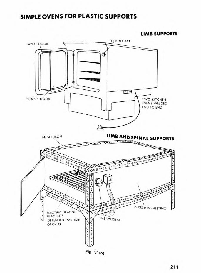
SIMPLE OVENS FOR PLASTIC SUPPORTS
LIMB SUPPORTS
f '9- 31(o)
211

Details of manufacture of appliances are usually given t>y the manufacturer.
The brief summary below demonstrates the basic principles of the manufacture of a simple oven (Fig. 31(o)), and the manufacture of a Vitrathene knee support (Fig. 31(p)).
"Plastazote" or expanded polyethylene requires heating like "Vitrathene", and differs in that it can be moulded directly on to the patient. Polyester resin or Lightcast light-weight fibreglass frame supports, "Skelecasts", can also be moulded on to the patient direct, but are not detachable. They do not require heating, and because of their frame nature allow the patient to have a daily bath or swim without removing the support.
”Plastazote" (Expanded Polyethylene) Supports
The material which comes in V , h", 3 A" and l" sheets is cut approximately to size, and then heated to 130 C in an oven. It is then moulded directly on to the patient and this is possible despite the temperature because of its very low conductivity. Similarly, the patient can step directly on to a heated sheet to mould a special sole.
"Plastazote" supports often require strengthening, and for this a Vitrathene sheet or strip should be laid on the surface of a Plastazote strip or sandwiched between two layers of Plastazote before heating. As soon as the splint has cooled it should be trimmed, and buckles orr'Velcro"fasteners added as necessary.
Most sheets of "Plastazote" come perforated for coolness.Added holes, however, can easily be added if necessary. The material can also be reheated to alter its shape once it has been shaped.
"Vitrathene" (High Density Polyethylene - Surgical Grade)Supports
"Vitrathene" cut slightly larger than the size required, is heated on a stockinette sheet in a special oven to about 130 C until it becomes clear. This material, however, unlike "Plastazote" is a good conductor and therefore must be moulded on to a plaster cast of the limb or trunk rather than the limb itself, and held in place by crepe bandages while cooling (Fig. 3l(p)). It is then trimmed, and buckles or fasteners attached as in the case of a "Plastazote" splint. Sheets often212

VITRATHENE KNEE SUPPORTMETAL ROD
POSITIVECAST
NEGATIVECAST
SPLIT
PLASTER CAST OF LEG NEGATIVE CAST FILLEDWITH PLASTER
C— , aOR ,? VITRATHENE
NEGATIVE CAST REMOVED
POSITIVECAST
VITRATHENE-ON
S t o c k in e t t e
STOCKINETTE
VITRATHENEMOULDEDVITRATHENE HEATED
POSITIVE CAST FIXED
HOLES FOR VENTILL ATION
PADDINGVITRATHENE SPLINT CUT OFF COMPLETION OF SPLINT WITHBANDAGED ON AND TRIMMED STRAPS AND PADDINGTO CAST
Fig. 31 (p)
213

come perforated, but extra holes can easily be added for coolness.
In the case of "Vitrathene" corsets, these can be made in front and back halves, and joined together after moulding if the oven is found not to be large enough to take a complete sheet of plastic.
"Skeleaast" Supports
These are simple supporting frames made out of polyester resin on bandage, or fibreglass tape, with expanded polyethylene (Plastazote) strips under the padding. Their main function is the support of the limbs and trunk in various orthopaedic conditions where the support has to remain in place for several weeks. In weakness due to polio the support may have to remain for years but, on the other hand, be detachable during the night. "Plastazote" and "Vitrathene" detachable supports are therefore of much more use than "Skelecasts".
"Skelecasts" can be applied directly on to the patient, but cannot be removed for bathing and sleeping. They do, however, allow the patient to swim or bathe, as mentioned, as they are waterproof, and they are much cooler than complete supports.
Their main use would be in the acute or subacute stage of poliomyelitis as a temporary support for a weak spine, and post- operatively instead of plaster of Paris where they would need to remain for several weeks undisturbed.
SIMPLE WHEELCHAIRS & OTHER VEHICLES
Wheelchairs which are cheap, strong and can be locally manufactured, are required by the thousands by many economically poor countries of the world to-day. In these countries there are millions of crawling patients, not only with severe poliomyelitis but also with paraplegia and quadriplegia due to spinal tuberculosis, injuries and transverse myelitis. There are also numerous patients with severely deformed or amputated limbs due to trauma, untreated congenital deformities, severe osteomyelitis, pyogenic and tuberculous arthritis and leprosy. The necessity for a wheelchair may be urgent, as the penalty for lack of mobility in developing countries is usually early death through neglect and starvation.
214

SIMPLE WHEEL CHAIRSTYPE
TYPE n

It will be many years before adequate surgical treatment will be available for most of these patients, and even with simple calipers and simple operations to correct deformities, there will still be many thousands of patients who, through the severity of their deformities or paralysis, or the weakness of their arms, will require wheelchairs for the rest of their lives.
Wheelchairs as manufactured in Europe and North America usually cost over i. 50 (U.S. $120) and sometimes much more by the time they reach most developing countries. They have the advantage of lightness and of being collapsible. They have the very marked disadvantage, apart from cost, of being unable to stand up to rough roads and of being difficult to repair locally. A very simple wheelchair is, therefore, necessary and has been designed in an effort to find a solution to this problem.
In an effort to design a suitable chair, the first prototypes were made out of old broken metal frame chairs which were rewelded, with a supporting frame for the wheels added. Canvas seats and backs were made out of old canvas mail bags, bogie wheels were obtained from old hospital trolleys and wooden chocks were used for brakes. More sophisticated versions of this chair were developed from this initial prototype. The design considerations involved will first be discussed before the technical details of construction are described, as these may have a bearing on modifications of the chair for different countries, and in different conditions.
Design Considerations for Wheelahairs
The design of a suitable wheelchair for developing countries presents several problems. It must be simple enough to be manufactured locally out of easily available materials with semi-skilled labour, cheap enough to be a practical proposition and strong enough to stand up to rough roads, mud, dust and water.
The wheelchairs which will be described fulfill these criteria. They can be manufactured in any simple metal workshop, and all the materials required for their construction are usually readily available in economically poor countries. The retail cost of materials is relatively cheap £9 - £12 (U.S. $23 - 30), and this could be reduced considerably if materials were bought wholesale, or if old frame chairs were available at minimal cost.
216
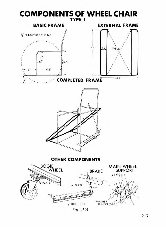
COMPONENTS OF WHEEL CHAIRTYPE I
BASIC FRAME EXTERNAL FRAME
\ FURNITURE TUBING
16 5"
COMPLETED
OTHER COMPONENTS
217

Eight prototypes of different types were extensively tested over several months before the present designs were settled upon (Fig. 3l(q.) - (v)). Over 1,000 have been made to date, and hard usage for over seven years has not produced any wear except in that easily replaceable spindles and ball bearings of the main wheels and the cheaper types of bogie wheel. This is in contradistinction to the imported wheelchairs which all needed extensive repairs after much less use.
Patient-propelled wheelchairs, in addition, should be a compromise of being low enough for patients with weak arms to climb in and out of easily, and yet high enough to clear potholes and small drains without outside help. They should not be too wide, as this decreases their usefulness on paths, increases the difficulties of propulsion, and makes it difficult to get through the usual narrow doors of houses. The width of the wheelchairs described depends on the frame chair used, but it is a little wider than a conventional wheelchair due to the necessity of supporting both ends of the axles of the main wheels. The advantages of using cheap and universally available bicycle wheels in manufacture, however, far outweigh disadvantages of width. If necessary, the chair can be narrowed by removing segments of the transverse struts and re-welding them.
The ability to be collapsible is of secondary importance to strength and cheapness of construction. This is especially so in countries where private cars are the exception rather than the rule, and where buses and taxis, when available, have roof racks which can take wheelchairs and bicycles without difficulty.
The size of the main propulsion wheels is 28", the usual size available in developing countries, and therefore cheap and easily repairable. In countries where the standard bicycle wheels are of a different size, this size, of course, should be used. The large wheels afford good tractive power for a patient with weak arms, and are far better over rough ground than small wheels in an unsprung chassis. The centre of gravity of the patient should be just behind the axis of the wheels to obtain maximum tractive effect on the main wheels. This will also enable a patient to tilt the chair forward by sliding forward in the chair to get the bogie wheel out of a drain or pothole, as well as facilitating getting in and out of the chair. A single trailing bogie wheel will allow both main driving wheels to be always on the ground, especially in uneven country. This is not so with two wheels.
218

TYPE I WHEELCHAIR
MATERIALS & COSTU.S. $
Basic Chair 3/V' (1.9cm) Outside diameter 22ftfurniture tubing (693cm) l.UU
External Frame 3A " furniture tubing AftA 27cm) 0.92
Brake V (0.6cm) Steel Rod 3hft(1 0 7cm) 0 . 1 7
Handle l" x 1/8" (2 .5cm x 0 .3cm) steel flat
1ft(30.5cm) 0 .0L
Gate 3A " x 1 /8" (1 . 9 x 0.3cm) steel flat
1ft0.03
Wheel Supports I V x V' (3.8 x 0 .6cm) steel flat
1ft0.08
Castor SupportsAngle l" x 1 /8" steel angle
(2 .5 x 0 .3cm)3ft
(91 Acm) 0.36Plate V x V steel flat
(1 0 .2 x 0.6cm)6"(1 5 .2cm) 0 .1 5
Main Wheels 28" (122cm) complete with tyre, tube & valve 2 10 .8 0
Bogie Wheel 5" castor (1 2 .7cm) or k" or 6" 1 6 .1 2
Seat and Back 3/8" plywood sq.ft.0 .8L
Paint White Undercoat
White finishing coat Wood stain
\ pint ( A 8 ml) \ pint \ pint
0.230.250.30
Miscellaneous Bolts and nuts Woodscrews No. 10. l V
k ) 8 ) 0 .1 8
TOTAL U.S.$ 21.91
N.B. Due to inflation, prices shown above can only be regarded as a comparative guide.
219

The bicycle wheel tyres were originally used instead of propulsion rails for propelling the chair forward, as they were cheaper, and many patients prefer to use the tyres themselves when clean. A propulsion rail, however, can also be attached without the necessity of widening the external frame. The extra cost of materials is approximately 12/- (0.60 Pounds, U.S.$1.50),and can be easily attached. (Fig. 3l(v)).
Bogie wheels were experimented with, taking into consideration both size and position; 4", 5", 6", and 8" sizes were tried with single or double wheels either in the front or the back. Front bogie wheels, whether single or double, were discarded in patient-propelled wheelchairs for rough road use, as they were easily obstructed by potholes and gullies. A single trailing wheel not only becomes obstructed less, but can also be tilted out of a pothole. It also acts as a prop to prevent the wheelchair tilting backwards when going up hills.
The size of the bogie wheel depends on availability and cost.The larger wheels are heavier, more expensive and more difficult to obtain. They are, however, better on rough ground; 5" or 6" wheels are probably the best compromise, but V wheels are cheaper and have proved fairly satisfactory where patients do not have to go over rough surfaces. Bogie wheels have been found on the whole to be hard-wearing, but poor quality ones may wear at the spindle. It has been found advisable, therefore, to bolt rather than to weld the bogie wheel on to the back plate in order to facilitate renewal of worn wheels.
The use of old chairs as the main frame has the advantage that the basic chair is available without the necessity of a jig.Once mass-production starts, however, the manufacture of the entire chair should be embarked upon from scratch, and modifications introduced if necessary. The use of three-quarter inch conduit tubing can make the chair easier to manufacture, but heavier; three-quarter inch furniture tubing is lighter and stronger, but will require a tube-bending machine.
Various other chairs were experimented with and three types of wheelchair will be described. Attempts were also made to use wooden frame chairs. These proved easier to fit with bicycle wheels, but using a supporting frame of lh" x V (3.8 c.m. x 6.b c.m.) flat steel instead of tubing screwed to the chair. Although this proved fairly satisfactory for gentle indoor use, these chairs did not stand up to hard usage or outdoor conditions, and were heavier and more cumbersome.
220

SIMPLE WHEEL CHAIRSTYPE in

WHEEL CHAIRTYPE m
BACK VIEW
222
Fig. 31 (s-a)
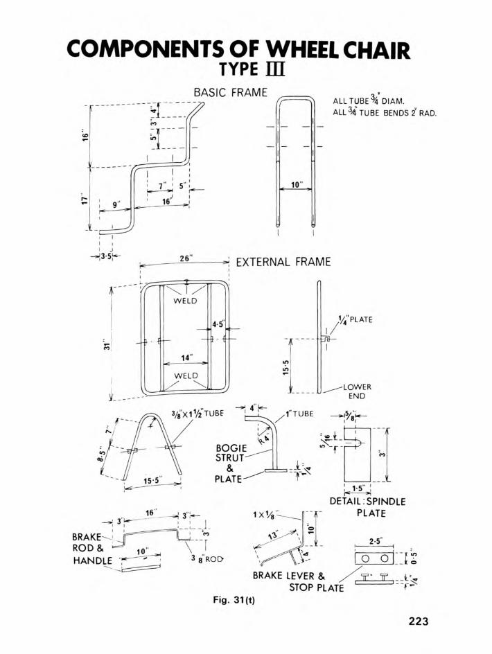
COMPONENTS OF WHEEL CHAIRTYPE m
BASIC FRAME
10"
ALL TUBE \ DIAM.ALL34 TUBE BENDS 2" RAD.
*j EXTERNAL FRAME
1 - .....................
W ELD
14"
W ELD
4-5 v4" PLATE
-■-Era—
3/8Xl1/2TUBE ~̂ |. 4 'T /T'TUBE
-LOWEREND
BOGIESTRUT"
&PLATE"
VS-t-d r y i
-1CO
BRAKE'RO D & y Q
HANDLE
1 co\ '" f
3 8 ROD
1 X1/8 -
\1
!c 1-5"al
DETAIL ?SPINDLE PLATE
2-5"
o o n
BRAKE LEVER &
Fig. 31 (t)
223

WHEEL CHAIR TYPE mDETAILED SPECIFICATIONS
©
_ „ BRAKE iT . X -, lock $o o 3/ i 6X % X 3 " HJ FO O T
PLATE
3/4 ^
f
15 ©8V2XI V2 " I/2THICK J SEAT BACK
Vi TH IC K 13V2' A
in
ROUND14’
SEAT m
151/ 2 ©
2 2 4
Fig. 31 (u)

TYPE III WHEELCHAIRMATERIALS & COST
Basic Chair 3/U" (l.9cm) Outside Diameter Furniture tubing
Aft A 27 cm)
U.S.$
0.92Seat Frame 3A " furniture tubing 1 0ft
(305 cm) 0.53Support Frame 3A " furniture tubing
l" (2 .5 +̂cm) furniture tubing
3ft 2" (96.5cm) 1ft 9" (53.3cm)
0 . 1 7
0.06
Castor Supports V (0.6U cm) steel plate 25 sq.in. (l6l sq.cm,. )0.13
Main Wheels 28" (7 1cm) complete with tyre, tube & valve
2 10 .8 0
Bogie Wheel 5" (1 2 . 7 cm) castor or 4" or 6"
1 6.12
Hand Rims V' (1 - 2 7 cm) furniture tubing 13 ft. (396 cm) 0 .6 3
Brake 3A " furniture tubing
3/1 6" x 3A " (0A 8 x 1 .9cm)
V' (1 .27cm) furniture tubing
3/8" (0 .96cm) steel rod
1 0"(25 A cm) 2ft.(6l cm) v(10 cm)2ft A"( ( 7 1 cm)
0 .0U
0 .1 0
0 .0 1
0.08
Brake Lock 3/1 6" (0 A 8 x 1.9 cm) flat steel
V 1 x V' (0.6U x 1.27 cm)
3"(7 . 6 cm)2
0 . 0 10 .0 1
rivets
Continued:
225

u.s.$
Paint Grey undercoat
White finishing coat Thinner
^ pint (296 ml) \ pint 1 pint
0.21+
0.1+80.21+
Seat Back 5/l6" (0.80 cm) Plywood 1.7 sq.ft. (1+8 sq.cm)
C.19
Seat 5/l6" Plywood 1.5 sq.ft. (1+0 sq.cm) 0.17
Foot Support 5/16" Plywood 0.75 sq.ft (20 sq.cm) 0.08
Padding 3" (7.6cm) foam for seat ) 2" (5.1cm) foam for back ) Artificial leather covering
2.7 sq.ft. (71.5 sq.cm 7.5 sq.ft. (201 sq.cm)
jl.12
3.2U
Miscellaneous Bolts and nuts5/16" x 3/1+" (0.80 x 1.9 cm) 1+ 0.01+Wood screws (No.10) lV' 10 0.05Electrodes (No.12) 2 0.06
Welding rods 10 0.06
Glue 0.08
TOTAL U.S. $ 25-66
N.B. Due to inflation, prices shown above can only be regarded as a comparative guide.
226

The important point in the design and mass-production of wheelchairs for economically poor countries, as with other appliances, is the necessity of keeping the design simple and cheap. Over- enthusiastic workshop supervisors should not be allowed to make too many complicated additions at the expense of quantity and strength. The production line should concentrate on the simplest satisfactory design, and modifications should be added as required. These include foot-boards, specially built up foot rests, arm rests and extra narrow or high seats with a head rest, if necessary. A scaled down wheelchair can be made for children.In the case of paraplegic patients 3" foam plastic or other padding is essential for the seat, and this should be covered by waterproof material to keep it in place. If cost allows, however, all wheelchairs should be padded with 3" foam plastic for the seats and 2" foam plastic for the backs.
In the case of patients who cannot propel themselves, two bogie wheels at the front should be put in place of the back wheel (Fig. 3l(v)), and a strut added to prevent the chair tilting backwards. The external frame should be welded so that the axis of the wheels is now level with the back of the seat and the centre of gravity of the chair, therefore, in front of this. The top of the type I chair, however, is ideal for pushing and does not require modification in any way. A piece of three-quarter inch (19mm.) tubing for pushing should be welded on to the type II chair, and is already present on the type III chair.
Another modification of the standard wheelchair is a double tubing extension forward in the place of bogie wheels in order to turn the wheelchair into a vehicle suitable for being towed by a bicycle. Numerous modifications and types of this "pedicab" are naturally possible on the various types of chair available.
A frame of V (6.Umm) or 5/l6" (8mm) mild steel rod can also be easily attached to a chair, and to this is added a detachable canvas awning with a front and back transparent panel. This totally encloses the chair and makes it weatherproof in the rainy season.
It should be possible to detach the whole frame completely as well, and the canvas panels on the front, sides and back should be made so that they can be rolled up to the roof and fastened there when not temporarily required.
The technical details of manufacture of simple type I, II and III wheelchairs are illustrated in Figs. 31(q) - (y).
227

SIMPLE WHEEL CHAIRADDITIONAL MODIFICATIONS
PROPULSION RIM
PADDING ARM AND FOOT RESTS
Fig. 31 (v)
228

THREE-WHEELED CHAIRS (Hand-Propelled) - Figs. 3l(w), (x), (y).
There are a variety of hand-propelled chairs on the market in economically rich countries, tut these are not made to stand up to rough roads. Alternatively, a simple, rugged, cheap chair can he built from a simple locally produced wheelchair by welding a front bicycle wheel fork with a single 20" wheel in front, and two 28" bicycle rear wheels at the back. Propulsion is by bicycle pedals and chain direct on to the front wheel, and two ordinary bicycle brakes are used on the back wheels. The addition of a three-speed gear,front and back lights, a simple hood, a horn and a luggage carrier under the seat can all improve this. The three-wheel chair illustrated (Figs. 3l(w), (x) and (y)) is one in which simple bicycle components have been adapted to an ordinary type III wheelchair. This three-wheel chair has proved itself on rough roads, and can be propelled for long distances much more easily, and much faster, than the ordinary wheelchairs with two main wheels.
Numerous prototypes were tried out with a variety of propulsion mechanisms before the chair illustrated went into production. Despite this, further modifications in the future will probably help to stabilise the chair further and improve on its propulsion. A motorised version is also being designed.
MECHANICALLY PROPELLED CHAIRS
Most manufacturers of wheelchairs sell a variety of chairs, from electrically propelled wheelchairs to sophisticated three-wheel chairs with small two-stroke and even four-stroke engines. An ingenious handyman in a workshop in a developing country could easily adapt a locally produced three-wheel chair to propulsion by means of an old lawn mower or motorscooter engine.
CARS ADAPTED FOR DISABLED DRIVERS
Standard cars, adapted with controls for invalid drivers, are usually a much more practical proposition for the disabled who can afford them in developing countries than invalid cars themselves. They may initially cost more, but they stand up to rough roads much better than invalid cars designed for well surfaced roads, and they are much safer.
229

THREE-WHEELED CHAIRTYPE IV
Fig. 31 (w)
230

141A
FRAMES OF THREE-WHEELED CHAIRTYPE JSL
SEAT FRAME 3/4 tu b e
57 " 4 6 " 3314" , 183/ 4 " 10 " ,2 V i r t * h *-= i— :
i114
C U TT IN G & BEND IN G LENGTHS
BASIC FRAME I ' t u b eALL BENDS 3" RAD
65" _ 57" _ 50" 42'
l_i:15
C=li------2»t
130C U T T IN G & BEN D IN G LENGTHS
Fig. 31 (x)
231

COMPONENTS OF 3 WHEELED CHAIRTYPE E?
MAIN W H EEL SUPPORTS
n41/2/ '
I]
-.i."V
ml/i'THICK a
3/8W H EEL
SUPPORT
16
7 3/4' 32 Vs" 453/4' 521/4“ +2''
CUTTING & BEND ING LENGTHS
HEADTUBEd Z Zi 6 " _
11/4TUBE
BICYCLE CHAIN DRIVE W ITH 3" CHAIN WHEEL FASTENED TO HAN D LEBAR
1 TUBEFRONT W H EEL SUPPORT
1 mm GROOVEC 3/gGROOVE
I '1V? 1/ 2"
TUBE 3/4"
5''12 y2"
Vs'
5"' 13"
: 9 v i :273/4- ~ ^
CUTTING & BENDING LENGTHS
BRAKE
% WASHER 3/g"STEEL Vi' TUBE RO°
REAR CYCLE BRAKE
Ms'x 3/4"X3''Fig. 31 (y)
232

SUPPORTS FOR THE MODERATELY DISABLED PATIENT
CROUCHINGCRAWLING
KNEE PADS HAND SUPPORTS HAND HOLDSC O N D U IT TU B IN G TREE RO O TS
FOAM RUBBERLEATHER
LEATHERRUBBER
RUBBER
RUBBER
RUBBER
Fig. 31(z-a)
233

SUPPORTS FOR THE SEVERELY DISABLED
These are discussed in Chapter 28, and illustrated in Figs. 3l(za) and (zb). Such supports may have a very real place for the severely disabled in economically poor countries.
BALKAN BEAMS (Figs. 3l(zb) and (zc))
Simple beams which can be used to support limbs both post- operatively and during physiotherapy are illustrated. They are much cheaper and lighter than imported types of beam, and are easily made locally out of angle iron or furniture tubing as shown.
234

SUPPORTS FOR THE SEVERELY DISABLED CRAWLING PATIENTS
SIMPLE KNEE, ELBOW AND HAND PADS FOR CRAWLING PATIENTSF O R E A R M
K N E E P A D E L B O W P A D S U P P O R T
FOAM RUBBER
LEATHER
RUBBER
Fig. 31 (z-b)
235

SIMPLE BALKAN BEAM
CDW OODEN BROOM
CD METAL PIPES
GALVANISED ,1 WIRE
236
Fig. 31 (z-c)

4 4
i
SIMPLE BALKAN BEAMS(CONDUIT TU B IN G )
237

32. RehabilitationINTRODUCTION
The patient must always be regarded as a human being, rather than a paralysed limb or a disabled body. Every aspect of rehabilitation as a wage-earning member of the community should be planned as early as possible in the treatment. (Fig. 32(a)). There will, of course, be occasional patients who are so severely disabled physically, and so psychologically affected by their paralysis that they will be perpetually confined to a hospital respirator, a rehabilitation centre, or a sheltered workshop if such facilities exist. A patient can be very severely disabled and yet can still be fully rehabilitated into the community,(Fig. 32(h)), especially if relatives and friends are prepared to help. In economically poor countries this is more difficult.Even in Western communities the difficulty of re-educating local people to accept the disabled person as a contributing member, and not just a useless cripple, may be considerable.
The severely disabled patient, with severe upper limb and trunk weakness, may have difficulties in attending to the needs of daily living. It is important that he or she should be able to manage these without assistance, if possible, before intensive education or rehabilitation is begun.
He should be taught to wash, dress, feed and attend to his toilet needs. This may entail modifications to clothes, cooking and bathroom appliances. (Figs. 32(c) & (d)). The ability to be mobile, at least on a fairly level surface, is an additional attribute which will help to make him independent.
In the following pages the rehabilitation of the patient will be discussed from overall social, educational and rehabilitation aspects. It is presumed that all that can be done to get him or her upright and mobile medically or surgically has already been done, including operations on upper and lower limbs, supports and provision of a wheelchair, if necessary.
238

REHABILITATION OF THE CRIPPLED PATIENT
PLANTING CROPS
SCHOOL
BOOTMAKING
OFFICE WORK
Fig. 32(a)
239

THE PSYCHOLOGICAL EFFECTS OF GETTING A CRAWLING PATIENT UPRIGHT
240

AIDS FOR DISABLED
W R IT IN G |
TYPEW RITER(e l e c t r ic if p o s s ib l e )
PAD CLIPPED TO BOARD
Fig. 32(c)
241

SPONGE MOP WITH LONG HANDLE
LONG HANDLE WITH INTERCHANGEABLE COMB OR TOOTH BRUSH ATTACHMENTS
RAZOR WITH HANDLE
TOWEL WITH LOOPS TO AID DRYING
242

AIDS TO DAILY LIVINGPARALYSED PATIENT IN
THE VILLAGE
LOW STOVE
SLIDING DOORS WITH FOR WHEEL CHAIRS LOW HANDLESPATIENT CONFINED TO BED IN MAJOR HOSPITAL
Fig. 32(e)
243

SOCIAL REHABILITATION
The rehabilitation of the severely paralysed patient must start as soon as possible after paralysis, and preferably, in hospital. This is especially so if paralysis is so severe that additional equipment to allow movement will be required. Appliances and supports which may be necessary include the following
IN BED AND IN HOSPITAL
Lifting Handles and Hoists (Fig. 32(e))
The patient with a weak trunk and weak legs will find difficulty in moving in bed, and a lifting handle can prove invaluable.Various hoists are also available for the patient with chronic paralysis, and ancillary aids such as a simple backrest, arm supports, book rests and pillows properly positioned can help to ease the patient's life.
Extension appliances (Fig. 32(e)) are easily made and can be used by the severely paralysed for reaching articles, while simple adjustments to light and radio switches, for those few fortunate enough to have these luxuries in developing countries, will allow much more independence.
Extensively paralysed patients, and patients in respirators, may require extra aids. Alarm bells worked by the patient's mouth or head, or by minor movements of the patient's shoulder, are an example of these. The patient should be rehabilitated, if possible, while still in hospital and this will help considerably in the long-term management and rehabilitation of the future.
Eating
Patients with weak arms may find eating very difficult, and there are various simple aids which can support weak arms and help to guide food to the mouth (Fig. 32(c)).
These vary from special knives and forks with larger handles, so that a patient with a weak grip can hold them, to slings on springs which can support a flail shoulder or weak elbows. These slings can be attached to Balkan beams, to special attachments on wheelchairs, or even to the ceiling of the room.
244

Washing and Toilet
It is essential to make the patient as self-reliant as possible (Fig. 32(d)), and aids which can help with the washing and toilet needs are important and often forgotten. These include supporting rails, bath or shower seats, and adjustments to the height of washing bowl, basin or bath. A rubber mat in the bath and on the floor can often prove a valuable adjunct.
Dressing
A severely paralysed patient who can undress without assistance can become self-reliant and much less of a burden on nurses, relatives or friends. This may mean experimenting with special clothes, zip fasteners, Velcro and similar attachments to clothes and appliances for undoing or doing up buttons. A variety of the simplest types of aids may make all the difference to the patient's life. The items of clothing which often give difficulty are shoes, and those with elastic tops which can slip on and off may prove of considerable assistance. Alternatively, in warm climates, simple sandals or clogs may prove acceptable and easy to manage.
Housing, Domestic Aids and Furniture for the Severely Disabled
Patients should be taught how to cook and do housework from a wheelchair or, in the case of walking patients, when both knees are in above-knee calipers, how to work with the arms occupied by crutches. Stoves, sinks, refrigerators (if available) and other kitchen utensils and appliances may have to be lowered or raised, and a trial with those appliances in actual use is essential before the correct adjustments can be decided upon.
Patients of different nationalities, religions and economic backgrounds may have different customs. It is a waste of time, for instance, to train a patient to use a sophisticated electric or gas stove when an outdoor coal, charcoal, wood or paraffin stove is all that is available. The social worker or almoner, if one is available, can be of great assistance.
In most developing countries there are unlikely to be enough trained social workers, and a nurse, nursing assistant or orthopaedic assistant may be the best person available to help.
Switches for lights, if they exist, may have to be lowered, and furniture in houses may require alteration. A severely disabled
245

person with a two-storey house or flat would be well advised to change this for a bungalow or ground floor room.
Steps should be changed to ramps for patients in wheelchairs or in calipers, and doors may have to be widened for wheelchairs. Patients may have to move their home so as to be closer to their work or to suitable transport.
Transport
Where possible, a patient should be enabled to leave home or hospital and thus become as independent as possible. Wheelchairs must be strong enough for rough roads and be patient-propelled whenever possible. The streamlined and expensive lightweight folding chairs, as produced in Europe and North America, have little place outdoors on the rough tracks and roads of most developing countries. A simple rugged type of wheelchair designed for such uneven terrain has already been described in Chapter 31.
Motorised wheelchairs, and cars with special controls,are available for the more wealthy patients, but in economically poor countries they have a limited place, and it is far better to adapt an ordinary rugged small car, such as a Volkswagen, Ford Escort, or DAF, with known good servicing facilities than to use threewheeled motorised invalid carriages which will disintegrate on rough roads.
Electric wheelchairs have little place in countries where few houses have electricity. On the other hand, a small two-stroke engine on the back of a strong wheelchair, or a rugged threewheeled wheelchair (Chapter 31) may be a practical proposition, and will often allow the patient considerable mobility.
A motorised wheelchair, or a car with special controls, will cost money to run and maintain, and a poor patient given such transport without also being given adequate means to maintain and run it, may be worse off than a patient who has a very simple type of maintenance-free wheelchair.
The use of unskilled manpower to help a trained patient with transport and the problems of daily living may enable the patient to work at a skilled job which he could not otherwise manage.Only a small fraction of his salary need be used in paying for the unskilled assistance necessary to push his wheelchair, help him wash and dress, and to fetch and carry in his job. This compromise is often far more satisfactory than supplying a patient with sophisticated apparatus which is often expensive to run and impossible to maintain.
246
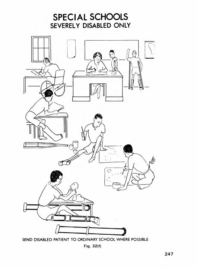
SPECIAL SCHOOLSSEVERELY DISABLED ONLY
SEND DISABLED PATIENT TO ORDINARY SCHOOL WHERE POSSIBLEFig. 32(f)
247

It is important that the people in the village or town where the rehabilitated patient will work and live should be educated.It should be explained that the disabled patient is not infectious and that, apart from lack of mobility, he is as normal as anyone else. This co-operation and support from the local population may make all the difference between the disabled patient being able to manage the physical and psychological problems he will face, and failing completely.
EDUCATION OF CHILDREN (Figs.32(f) & (g))
Over 90 per cent of polio cases in tropical countries occur in children under the age of five. Some of these children can walk with the aid of a stick or with a relative helping them. In some only one arm may be paralysed, but in the majority of cases both legs are paralysed and the patients can only crawl.
In economically poor countries where families are large, schools are in short supply and money is scanty, the able-bodied children are often educated at the expense of the disabled. In countries where there is no money to send many of the physically normal children to school, it is not surprising that the disabled children are left uneducated. Another reason for the lack of education of the physically disabled is the problem of transport.
The popular belief is that the cripple in body is a cripple in mind, and the lack of education in these mentally normal children helps to foster this idea.
Most of the children seen in developing countries are, therefore, uneducated or have had a scanty primary education. In the case of a physically disabled child, who may have to earn a wage more by brain than brawn, education may be even more important than in the physically normal. The physically handicapped, too, have more reason to work and less scope for play, and therefore may, in fact, do better than the physically normal in many jobs.
Both the parents and the child in question must be told this, and every effort made to get the child into a school for normal children if possible. The very severely disabled may need a special school, and those with moderate physical disability may need a special hostel or dormitory attached to a normal school.The vast majority of physically handicapped, however, should be able to attend normal classes in a normal school, whether this is a day school or a boarding school.
248

REHABILITATION OF THE CHILD
Fig. 32(g)
249

The best way to demonstrate to parents that it is possible to educate physically disabled children is to provide a small school attached to a polio clinic and polio hostel. Children attending for inpatient treatment can thus start their education in hospital, and accompanying relatives and even relatives of outpatient children attending for treatment at the clinic, can see the school in action. They are then often stimulated to continue education once the child has returned home.
Once the child is upright and walking, or even mobile in a wheelchair, relatives in developing countries regard the cripple in quite a different way, and as a normal human being. They will then often go to even more trouble to educate the disabled child than in the case of the physically normal brothers and sisters.
POLIO CLINIC SCHOOL
This should be attached to each polio clinic and need only be a small room with a blackboard and chalk, and a few simple materials such as crayons, paper, old illustrated magazines, flour and water paste, scissors and perhaps modelling clay and plasticine. Old toys, building bricks, and other equipment are gradually acquired. Desks and chairs are unnecessary, as many children prefer to sit on the floor, and even a few cushions are a luxury.
As in the case of the workshop a start, however small, should be made somehow. A voluntary worker can usually be persuaded to act as a teacher, even if it is only for two hours a aay, three days a week. Once a start has been made, there are few communities which will not ensure that this simple class continues to flourish.
EDUCATION AT NORMAL SCHOOLS
Headmasters of schools, if properly approached, seldom refuse to take two or three physically disabled children. Adjustments may have to be made to desks and benches, and the other able-bodied children may have to help the disabled ones up steps and in and out of wheelchairs. The danger is not that the disabled children will be neglected, but that they will be spoilt by the normal children, such is the competition to push a wheelchair to school, or to help a disabled friend. Teachers also soon realise that the physically normal children usually benefit from learning to help those less fortunate than themselves.
250

It is far better too, that disabled children should be sent to normal schools to mix with the rough and tumble of normal life, even though there might be obstacles to be overcome, than to put them in the artificial surroundings of a school for only the disabled, with every facility to make their life easier.
In the world outside they will have to learn to deal with obstacles and normal people, and this is best started at school.
HOSTELS AND SPECIAL DORMITORIES ATTACHED TO SCHOOLS
Certain schools, especially those run by the missions, will often accept more than two or three disabled children. In Uganda there is a hostel with 10 boys crippled with poliomyelitis attached to one mission school, and a hostel with 20 disabled girls attached to another.
The children in these schools attend ordinary classes with physically normal children, but receive extra attention out of school hours in their dormitories, with a resident matron specifically to look after them. This has worked well for 10 years in Uganda, and is a happy compromise between complete dependence and complete independence.
SCHOOL FOR SEVERELY DISABLES CHILDREN (Fig. 32(f))
The very severely disabled require a special school with extra staff to look after them, and extra care with both their education and their daily needs.
Such a school has been started in Uganda, and has boarding as well as day accommodation for those who live out with relatives or friends.
It is natural that such a school, with its extra staff and extra facilities will cost more than an ordinary school, and it would therefore be uneconomic to educate partly disabled children in the same school as those who are more severely disabled.
In some countries special schools cater for all the disabled children, but these are expensive and have many psychological disadvantages for those who are only slightly paralysed.
251

EDUCATION OF THE OLDER DISABLED CHILD AND YOUNG ADULT
There are many disabled older children who are bright and intelligent. They are usually considered only fit for repetitive clerical jobs, or for work in a factory, whereas their actual intelligence might suit them for jobs requiring much more skill.
The otherwise bright older child, or young adult, may have received only scanty education because of his disability. In a developing country, provided the child or young adult is intelligent, and the finance and facilities exist, there is no_ reason at all why he should not continue at school until the age of 20 or 25 or even older. The system whereby a child finishes school at 17, 18 or 19 has little place in a developing country. The shortage of good, able candidates for universities should favour a person of 30 who wishes to enter university almost as much as one of 20, provided his qualifications are suitable. The same will apply to adults who wish to take teacher training courses, and technical or other qualifications which will suit them for available posts.
REHABILITATION OF THE ADULT (Fig. 32(h))
In most developing countries little has been done to rehabilitate the disabled adult. In countries where this has been attempted, the usual result has been a sheltered workshop, often run at a loss, and supported by voluntary agencies with sporadic government support.
Some of these so-called rehabilitation centres consist of magnificent buildings with the name of the donor prominently displayed, and some even have highly skilled and dedicated staff. In many of these centres the permanently disabled patients are well looked after, and in some employment is provided for outpatients. It is usually found, however, that few disabled patients are ever discharged. However large the centre, all the beds soon become full and, apart from death, or the occasional patient being found a job elsewhere, no further patients can be catered for. The chronic residents prefer to become permanent fixtures in a sheltered workshop, whereas many of them would be suitable for employment on the open labour market.
At the opposite extreme is the hostel catering for the destitute and beggars, often run by an overworked and dedicated missionary, and supplying shelter and food for the poor who would otherwise die. These centres are usually so short of money and so crowded
252

by the chronic beggars and physically normal destitute people, that little or nothing is done to rehabilitate or train the crippled occupants.
There must always be sheltered workshops for those too crippled to live and work in a normal community. There must be hostel accommodation for the destitute. On the other hand, neither of the above methods is the real method of dealing with the vast majority of the disabled adults and older children in an economically poor community. In Uganda, for instance, the number of permanently crippled patients is estimated at 250,000, and even if only 100,000 of these required sheltered employment, this would place an intolerable burden on an economically poor country.
The other mistake made in many countries is to establish the rehabilitation centres in the main cities. This not only has the disadvantage of cost, but also means that many disabled people from country areas either stay hundreds of miles from their homes, or, more likely, can never be trained.
The optimum method of management is to provide a ring of rehabilitation centres throughout the country, and to train patients, where possible, in skills that will enable them to earn their own living in their own community, away from the centre that trained them.
They may, of course, need help in acquiring a piece of land, or obtaining a job. They may need supervision for the first year or two in their new work, and their conditions of employment may require modification. Financial help may be necessary in starting their own farm or shoe-making and repairing business.
Once independence is achieved, however, the cripple who was a burden on the taxpayer can become a taxpayer himself, and more important, can regain self-respect for both himself and his family.
His place can then be taken in the rehabilitation centre by another deserving disabled patient. Equally important, in many developing countries, the example of the disabled earning their own living will do much to dispel the attitude that the cripple is incapable of earning a livelihood. This will ensure that future disabled patients are viewed in quite a different way by the population.
In Uganda, in 1963, the first attempt to start an agricultural rehabilitation scheme for the disabled was made on 30 acres of
253

land outside Kampala. Many mistakes were made in this pilot project, but much was learnt. It was found, for instance, that polio patients with two above-knee calipers could actually hoe a field, and that severely crippled patients with no education at all could be successfully rehabilitated on the land.
As a direct result of this the Government and voluntary agencies in Uganda decided to support the Ministry of Culture and Community Development in starting a ring of rehabilitation schemes in Uganda. Thirteen rehabilitation centres were opened around the country between 1968 and 1 9 7 2, each with between 20 and 130 inmates. Nearly 2,000 patients were trained during this time, and disabled patients were found jobs in industry and in agriculture. Most important, a large number were rehabilitated back into their own communities, and much of the population of the country as a whole soon appreciated that the disabled could earn their own living.
The various rehabilitation centres have proved a real success, and several of them are now self-supporting, and the same type of centres would be of similar value in other developing countries.
SELECTION OF PATIENTS FOR TRAINING
Not all disabled patients are suitable for employment. An interview is essential in order to assess whether a patient is worth training in the first place, whether a job exists for him in the trade or profession for which he is most suited, whether he will be able to work full-time or part-time, and whether any medical or surgical treatment is required before he is accepted for training.
Patients placed in an industrial job must be able to do as well as the able-bodied, provided adjustments are made to machines or workbenches.
Patients, however, are not machines, to be used only during the hours of work and turned off afterwards. Suitable housing, adjustment to doorways, ramps, tables and beds for those in wheelchairs, suitable transport, and, if possible, someone to look after the patient, at least in the critical first few months are essential.
It must also be realised that many patients in developing countries, especially if severely crippled, have never worked, and have never been to school. They have always been regarded
254

REHABILITATION OF ADULTSAGRICULTURE
TELEPHONE OPERATOR
LEATHERWORK
iuCLERICAL WORK
Fig. 32(h)
SEWING WOODWORKOTHER JOBS
HOEING PLANTINGINDUSTRY
255

as parasites on the community, and their own attitude is often one of rejection. It is essential, therefore, that whatever their training, they should receive a course of re-orientation before definitive training even starts, and that this continues until a new job has been found, and even afterwards.
INDUSTRIAL REHABILITATION CENTRE (KIREKA, NEAR KAMPALA)(Fig 32(i))
Many agricultural communities have little place for industrial rehabilitation centres. On the other hand, many developing countries are now starting light industries. Rehabilitation geared to the needs of such a country, and to possible new industries developing in the future, in which disabled patients can be employed, should always be considered.
It is important that the disabled should be able to hold their jobs by their ability rather than by sympathy. Many disabled people are capable of working more conscientiously than the able- bodied, and can sometimes do even better work, provided simple adjustments can be made to machinery or workbenches. Obviously, it is necessary to make use of the limbs which are not disabled in order to obtain the optimum work from a paralysed worker.
At the Kireka Industrial Rehabilitation Centre in Uganda, these factors are carefully taken into consideration when deciding which skills to teach the patients during their four to eight month period of training.
In addition, certain severely crippled patients stay in the sheltered workshop attached to this centre, but it is essential that the vast majority stay merely for training and are then discharged for employment, or to their homes, and replaced by others awaiting their turn for training.
DETAILS OF AN INDUSTRIAL REHABILITATION CENTRE (Fig. 32(i))
Kireka is a residential centre for 130 disabled persons, eight miles from Kampala. The majority of the trainees will stay an average of six months and are then fitted into industrial employment, living quite separate from the centre. UO out of the 130 stay permanently in a sheltered workshop atmosphere, but it is important, as mentioned above, that this number remains small in relation to the total number in Uganda, or else the whole object of a training centre for industry, rather than a permanent centre for a few lucky inmates, is lost.
256

INDUSTRIAL REHABILITATION CENTRE(Diagramatic Only)
ConcreteTubularFurniture Engineering
Wood Woodwork CentralProducts & Paint
SprayingMachinery Assembly Store
Umbrel la TailoringCompleted
AssemblyLeatherwork Handicrafts Printing Products
Store
Staff
Housing
Men'sDormitory
(40)10
Rooms each with
4 Beds
LoungeKitchenDining
RoomWomans
Dormitory(10)
Accommodation for 30
Sheltered Workshop
Employees
ModelThatched
Hut
ShowOffices Room Mam Hall
Shop
Seed and Plant
Nurseries
Car Park and Entrance
Gardens, Playing Fields and Crop Growing around BuildingsFig. 32(i)
257

The training in the workshop is divided into three parts:-
1) Re-orientation of the Patient to Work
This includes self-discipline, good time-keeping, and the ability to be hardworking, conscientious and loyal.
2) Ability of the Patient to Look after Himself
This includes hygiene, the adjustment of clothing, transport, eating and toilet facilities to meet the needs of a disabled patient. These have already been discussed.
J) Training
This will be variable, and each patient will need this adjusted according to his or her physical and mental ability and the needs of the country. Some of the more obvious forms of training will be discussed individually.
Patients with Previous Education - These may be suited to further education to fit them for teaching or other professional jobs . Adjustments to their methods of transport, and ways of thinking, may enable them to be rehabilitated back into their former jobs. Training in typing, shorthand or other clerical work may suit them to vacancies which might exist or be created for them. Their st~ndards of work must be competitive in the open market if they are to keep a job, and their tYaining may have to be extended to make sure that they are adequately qualified.
Training can be given for office jobs such as filing clerks, office machine operators, receptionists, office messengers and telephonists.
Patients with Previous Training in Special Fields - If a patient has had previous training it is essential that he should be able to make use of this, if this is at all possible.
Uneducated - There are a variety of trades which are taught, and the exact one will depend on the patient's physical and mental ability and the possible available jobs in the future.
258

Heavy Jobs - These are available to patients with a single caliper, and include metal work using heavy and light machinery, and carpentry to include all types of woodwork, such as wood machining, furniture-making, building carpentry and the making of crutches and clogs, and painting.
Light Jobs - These include tailoring, leather work, handicrafts, silk screen printing and umbrella assembly.
It should be pointed out that there are all gradations between light and heavy jobs, and that many other types of training may be indicated in specific countries and for specific patients.The management of the severely disabled and the question of sheltered workshops is discussed below.
AGRICULTURAL REHABILITATION (Fig. 32(j))
The emphasis in the rural rehabilitation centres in Uganda is different from that in the industrial centre, and each patient spends, on an average, eight months, or one and a half times as long as in the industrial centre.
The emphasis in these centres is not only on teaching the patient to cultivate his own small holding, but also on a number of skills which may be useful to him and his family. The training therefore includes the following
PLANNING OF TIME
The disabled worker is encouraged to plan the week in such a way that the heavier part of his work is done in the cooler partof the day, and the sedentary part while the sun is hot. Duringthe first one or two months he is also rehabilitated both physically and mentally.
AGRICULTURAL TRAINING
The patient is taught shortcuts to working efficiently, and also how to grow the vegetables which will not only feed him and his family, but also provide a cash crop.
He is supplied with the tools which will be of most use to him,and these are modified, if necessary, to cater for his disability.
259

AGRICULTURAL REHABILITATION CENTRE(30 Acres)(Diagramatic Only)
40 M E N +10 W O M EN - IN TRAINING 10 FAM ILIES - SHELTERED V ILLA G E
TRAININGA REA
Kitchen Workshops and Stores& Store for Training
51Plots
in10 Acres( 1 Plot for eachTrainee
+Extra Land for Centre
itse lf)
Women's Dining RoomDormitory and
(10) Lounge
WashRooms
1 |Dormitories - Men Sick Bay
1 &5 X 8 Men
. . 1 iSurgery
RecreationHall
Car Park & Lawns
SHELTERED V ILLA G E FO R 10 PERMANENTLY DISABLED + F A M IL IE S
10 Houses, each m 1 Plot of 2 Acres each
Fig. 320)
260

OTHER TRAINING
Other skills which are encouraged are small animal farming, home tailoring, shoemaking, home repairing, rural crafts with local material, literacy, hygiene, citizenship and current affairs.
The programme is designed to create someone who has a variety of skills, and it is often found that he can use one or more, even if he cannot use them all.
FOLLOW-UP
Disabled patients are resettled on land provided by the Government and are supervised for at least two years after returning to their own communities. Their difficulties are resolved where possible, and both the patient and the community in which he works are encouraged to recognise that the disabled worker is capable of earning his own living in his own community.
It should be stressed that only fertile land should be given to the disabled, and that some of this should be broken up and tilled initially. This is essential, in order that, at least in the early stages, the new disabled farmer is given every encouragement to make a success of his future.
SHELTERED WORKSHOP
In every society there are disabled patients who can contribute to their own upkeep to a variable extent, but who cannot do a full day's work.
Some of these can live at home and work, sometimes assisted by relatives or friends. Others will have to live and work in a sheltered workshop where they can be assisted with the daily processes of living, and where the problem of transport is relatively easily overcome. Modifications to utensils are also easier in a sheltered community, and these are discussed above.
At present, in Uganda, a number of severely disabled patients live in hostels attached to rehabilitation centres and work in a sheltered workshop. They earn at least part of their own living by the following trades, and these are trades which would be of value in most other developing countries
261

1. Cotton cloth printing and dressmaking.
2. Handicrafts made out of local material, such as barkcloth and desertwood, of interest to tourists.
3. The assembly of umbrellas and the manufacture of articles from bamboo and local woods.
Naturally the extent and type of work will vary according to the disability of the patient, and there are many other trades, such as watch and clock repairing, that can be taught. It is essential that patients should be assessed very carefully so that they can be given the job for which they are most suited.
It is also essential that every help be employed to make these disabled workers as efficient as possible. This can be done in various ways such as providing arm supports (Fig. 32(e)) to support a paralysed arm with a functional hand, elbow and wrist splints, and spinal supports to help the patient to walk or sit better. Operations can be performed on the upper limbs to improve functions, such as an arthrodesis of the shoulder to restore abduction, and an opponens transfer for a weak thumb.
A careful individual assessment of each patient may do much to improve the functional result, and also the capacity for work.
It is also important that the sheltered workshops should employ the disabled at standard rates of pay, under normal industrial conditions. Wage earning disabled patients should also always contribute something to their own keep, however small the amount, as this does much to help them to regain their self-respect.
At present there are ho beds for patients in sheltered employment at the main industrial rehabilitation centre, as well as a further limited number in the other 13 rehabilitation centres throughout the country.
MOBILE REHABILITATION CENTRES FOR RURAL DISABLED WOMEN
Disabled women, especially in rural areas, often tend to be overlooked where rehabilitation is considered. A number of these women have children, and therefore must live at home. Few, because of family commitments, can travel many miles to spend several months in a major rehabilitation centre learning a trade.
262

It is therefore essential to rehabilitate women such as these at village level. A scheme was started which enabled mobile instructors to travel around the country, to teach these patients how to overcome their disabilities and to operate their small holdings to the best advantage in their own homes. Much can be done by good organisation and the adaptation of homes, furniture, small holdings and tools to meet the needs of the disabled housewife or farmer.
Training of these disabled women also includes child care, home management, hygiene, nutrition, sewing, literacy and citizenship. Much can be achieved by relatively simple, but essential, training. Much can also be achieved by discussing with village chiefs, or leading citizens the necessity of accepting and helping the disabled, so that they can become fully integrated into their own communities.
REHABILITATION OF DISABLED BEGGARS
The relatively small number of severely disabled polio patients who survive their disability into adult life usually become beggars, or are completely dependent on relatives or friends in many developing countries. They are often completely uneducated and rely on their deformed limbs and bodies to arouse the sympathy of the passers-by. These patients will resist, with understandable justification, any attempt to straighten their deformed legs if, at the same time, no attempt is made to rehabilitate them and give them employment. If they cease to receive sympathy, they cease to be effective beggars. If they are no longer given money they may starve and die, and often their families with them.
It is therefore essential that jobs be found for them, or that they should at least be promised a place in a rehabilitation centre before their disabilities are treated.
In Uganda 2k beggars, together with their families, have been rehabilitated on a 25-acre site at Mbale, 150 miles to the west of Kampala. They are given training similar to that provided in a rural rehabilitation centre, and, where possible, they are then moved to a small holding near their own homes. Once established with their own land and homes, the patients are often, but not always, willing to undergo surgery to correct contractures and enable them either to walk or at least become more mobile or self- sufficient .
263

It is essential to assess a patient completely, both mentally and physically, before any complicated procedure or operation is advocated, to see if he will, in fact, be better off afterwards.
If a patient has a single deformed leg which makes it necessary for him to walk with a stick, he will almost always benefit from having this straightened.
Children, however deformed, will almost always benefit from being helped to walk, provided that there is enough power in arms and trunk to make this a possibility.
Older adults, on the other hand, with two severely deformed legs, necessitating prolonged surgery if mobility is to be achieved, are often best given a wheelchair, plus pads for hands and knees to enable them to crawl fast. They can always have their limbs straightened in the future, if necessary, when operative time, post-operative physiotherapy, and the bed state allows. Many adults, however, prefer to crawl fast, with painless joints, than walk slowly with painful or partially stiff ones, and the patient should not be forced to have an operation against his will.
ORTHOPAEDIC WORKSHOP
The ideal workshop in which to employ crippled polio patients is obviously an orthopaedic workshop. Disabled patients manufacturing orthopaedic appliances for other disabled individuals, will use a large variety of skills. These range from heavy labour to the lighte~t jobs, and this will therefore employ patients with nearly every grade of disability. All the workers in the assembly and leather workshop in Kampala are disabled. A smaller percentage are employed in the wood, metal and special appliances workshop. On the whole they have proved, with a few exceptions, to be both hardworking and reliable.
It is also of help to patients requiring appliances to see these being made by disabled workers with disabilities similar to their own. It shows that the disabled are capable, not only of working but also of earning a good living. This information is soon disseminated throughout any country where the disabled are normally considered useless parasites incapable of working at all, let alone earning any kind of living.
264

OTHER REHABILITATION PROJECTS
There are many other rehabilitation projects for disabled patients. These include vocational training courses, conducted by the Ministry of Education at technical schools and colleges, while crippled patients also attend apprenticeship courses and employers' training schemes.
In addition, courses in telephone switchboard operating, record keeping and filing are included in the rehabilitation centres.In the case of women and girls, training is also available as home helps and nursery attendants.
Schooling for all children and for suitable young adults is important, and this has already been discussed above.
RECREATION FOR THE DISABLED
Few people realise that the disabled require any recreation or sport. In view of their disability it is considered that a radio or television set will cater for all their recreational needs outside their work, and that they will be content just to sit and look.
This attitude of mind adopted by the general public gradually transfers itself to the disabled, who will often gradually appear to be content to be the spectators of life rather than the participants.
The Stoke Mandeville Games in Britain have demonstrated that paraplegic patients including those with paralysis due to poliomyelitis can participate in competitive sport. These annual games have shown that many patients even more severely disabled than those with poliomyelitis, can do surprisingly well in a wide range of sports, played with other disabled competitors.
Archery, netball, swimming, water polo, table tennis and precision javelin throwing are a few of the more popular of these games for these patients, and a polio patient in a wheelchair from Uganda won a gold medal in the 1970 Commonwealth Paraplegic Games. In addition it has been shown that painting and photography can be made into lucrative hobbies for those who are even more severely disabled and have the necessary skill.
265

The provision of a wheelchair, and a little interest and encouragement in their various enterprises, may completely change the life of crippled patients. Many severely paralysed patients in their cheap, but sturdy, wheelchairs were found many miles from their homes, watching football matches and making friends everywhere. Their attitude of mind changes, and their self-respect increases. They realise that, apart from their lack of speed, they may be just as good as, and sometimes considerably better than their physically normal fellows.
CONCLUSIONS
It is essential to regard the patient as a human being rather than a crippled limb, and to rehabilitate the whole patient following operative correction. Straightening a beggar's twisted limbs to enable him to walk without rehabilitating him and finding him employment, may lose him his livelihood of begging and condemn him to an early death from starvation.
The aims of rehabilitation should be not only to enable a patient to become mobile, but also to dress, feed, look after his toilet needs, and, if possible, live a fully independent life. He should be encouraged to partake in social activities, and in sport for the disabled, and if possible take up a recreational activity or hobby.
Children should go to schools for normal children, unless they are very severely paralysed, and should take part in as many school activities as possible.
The final aim should be an individual rehabilitated, and socially integrated into his own community, earning his own living in his own village or town.
266

33. Advice for PatientsRelatives and friends of polio patients can do much to help a
paralysed patient, both in the early stages of paralysis and later when the patient is moving more and may need to wear calipers and other supports. This is especially so in countries where there are shortages of hospital beds and doctors and other staff.
The following are some brief notes for the guidance of patients and for those who care for them. These notes, together with the four illustrations 33(a) to 33(d), can be reproduced together with the text from this chapter without further permission from either the author or the publisher if they are needed for the education of patients or staff.
Nature of Poliomyelitis:
Poliomyelitis is a disease caused by one of three types of virus which is spread mainly through the gut but to a lesser extent by the nose and throat of patients during the first week of the disease. Great care in cleanliness and hygiene is therefore essential.
Poliomyelitis causes paralysis of the legs much more than the arms and can affect breathing. It does not, however, affect the brain, and the intelligence of the patients is quite normal even though they may suffer from effects of deprivation of education.
Prevention of Poliomyelitis:
Poliomyelitis is prevented by a vaccine given by mouth containing all three types of a virus in safe form. This is usually given on a lump of sugar at intervals of from 6 to 8 weeks. Three initial doses at the age of 3 months, Mg months and 6 months, with booster doses at school entry and in times of epidemic are those usually recommended.
Treatment of the Acute Stage of Polio:
Rest and the avoidance of injections is essential in the early stages of this condition. Warm packs are soothing for tender muscles, and a patient should be put in a comfortable position and paralysed joints moved through their full range each day to prevent deformity.
267

Exercises for Deformities:
1. Upper Limb Exercises (Fig. 33(c)) These are illustrated, and the paralysed shoulder should he lifted upwards as well as rotated inwards and outwards as shown. The elbow should be bent and straightened and the forearm should be twisted through its full extent both inwards and outwards.The paralysed wrist and hand should be closed and opened and deformities should be prevented by these exercises.
2. Lower Limb Exercises (Fig. 33(b)) The child should be placed on the face in order to stretch the hip muscles fully, the knees should be straightened daily and gently moved through their full range of movement. The ankle and foot should be moved both upwards, inwards and outwards to prevent deformity.
Supports for Lower Limbs (Fig. 33(a) and 33(d))
These consist mainly of below-knee and above-knee supports for paralysed lower limbs and the type of support will depend on the extent of the paralysis. Clogs may be used instead of boots for coolness and cheapness, especially in children. A backstop in the clog may be used to prevent the foot going down, and in older children and adults a bending piece may be fitted for the knee. Sometimes supports are given for patients who are still in bed and cannot walk or for babies before they start walking. The reason for this is that these supports, which are removable, are used to prevent deformity as the child lies in bed.
Fitting of Calipers: (Fig. 33(d))
It is important that the caliper should fit well and not rub the skin or any protruding bony prominence under them. The top of the caliper should be at least l" below the groin, as illustrated, and occasionally extra padding is necessary to prevent the caliper rubbing. The caliper can also be bent if uncomfortable and the leather can easily be repaired by gluing or sewing. It is important, however, that the caliper is not discarded even though the patient may be able to walk without the support, as deformities may occur or recur should a support not be present.
Fitting Crutches and Walking Sticks:
Crutches should extend to l" below the axilla and the top should be padded in those patients who are severely paralysed and need to rest on them.268

INSTRUCTIONS FOR POLIOMYELITIS PATIENTS AND THEIR RELATIVES
i/1'
_ i,
FRONTISPIECE OF PAMPHLET ISSUED TO ALLPATIENTS
Fig. 33(a)
269

Walking sticks are usually held in the hand opposite the side of maximum paralysis.
Other Supports:
Supports may be given for weakness of the spine and it is important to make sure they are not causing irritation of the skin.
Getting the Patient Walking:
Parallel bars for home use are illustrated in Fig. 33(d) and every encouragement should be given to the patient to walk with these supports if necessary.
Wheelchairs:
Patients with weakness in the arms as well as the legs may require wheelchairs. Some patients who can walk a few steps may also be given a wheelchair for longer distances. It is important that wheelchairs should be maintained properly with the tyres pumped up and the hubs oiled and rust prevented. The wheels are mainly bicycle wheels which can usually be replaced locally, and minor repairs can be carried out without much trouble.
Operations in Poliomyelitis:
Operations are only performed by the doctor if they are felt to be absolutely necessary, and these are divided into those operations where a twisted joint is straightened in order to allow the leg to fit into a caliper or other support, and those operations which may enable the patient to dispense with a support by switching a muscle from one side of the leg to another, or performing an operation to stiffen a joint.
All operations are done with the patient asleep -under an anaesthetic so that they do not feel pain, and many operations are now done which require few or no stitches. A patient may need to be treated in plaster for a few weeks after an operation before being given a caliper.
Although most severe deformities in poliomyelitis can be corrected, muscles that have been paralysed for a long time usually remain permanently weak, and therefore operations are designed to improve a patient and not usually to cure a paralysis.
270

INSTRUCTIONS FOR PATIENTS AND RELATIVES
STRETCHING OF TIGHT MUSCLES
SUPPORT BACK W IT H LEFT HAN D
HOLD LEG ABOVE
Fig. 33(b)
271
Aids to Daily Living: (Figs. 32(c), (d)3 and (e))
There are certain adjustments at home which might make life for the paralysed patient easier, and these will he carried out where possible by those looking after the patient. These improvements include simple fasteners to help the patient to dress himself, larger handles on knives, forks and spoons for patients with weak hands, and lowered washing utensils and a simple toilet seat so that the patient can attend to his needs without assistance.
Beds can be adjusted for height and a lifting pole over the end may help the patient lift himself from a wheelchair. In addition, many simple methods of helping the patient include lower chairs and tables, together with lower stoves to allow easier cooking and preparation of food, ramps instead of stairs for the patient who is in calipers or in a wheelchair, and many other types of aids to make a patient as independent as possible.
Education & Rehabilitation:
Polio patients are mentally quite normal, and every effort should be made to return children to a normal school and continue their education. In the case of adults they should be trained for a suitable job wherever possible.
Sport for the Disabled:
There are various sports that the disabled in wheelchairs and calipers can enjoy, and these include basketball, netball, tennis, table tennis, archery, throwing the javelin and swimming. They should be encouraged to participate with other disabled patients or even with the able bodied. They should also join in other social activities, as these will do much to rehabilitate them both mentally and physically. These activities are very popular once the initial barriers of shyness and social acceptance are overcome.Conclusions:
After the first two months of illness the polio patient is non- inf ectious and will never be a source of infection, despite his disabilities. A polio patient is also mentally quite normal, and backwardness is largely due to shyness, and lack of education rather than to any mental defect.
272

INSTRUCTIONS FOR PATIENTS AND RELATIVESSTRETCHING OF TIGHT MUSCLES OF ARM
ELBOW
FLEX & EXTEN D FINGERS & WRIST AS FULLY AS POSSIBLE
WRIST & HAND
FLEX &EXTEND ELBOW
STRETCH EACH JO INT AT LEAST 20TIMES EACH DAY
FITTING OF CORSET
SLIGHTLY PADDED UPPER & LOWER ENDS & OVER PROMINENCES
NO PRESSURE UNDER ARMS
ALLOWS FLEXION OF HIPS
BUCKLES FIRMLY TIGHTENED
Fig. 33(c)
273

Relatives and friends could do much to encourage the paralysed patient to take an active role in the community, and in most cases lead a normal life within the confines of their physical weakness. Many are fully capable of earning their own living in suitable employment, and often do better than the able bodied as conscientious employees.
274
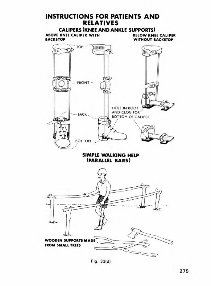
INSTRUCTIONS FOR PATIENTS AND RELATIVES
CALIPERS (KNEE AND ANKLE SUPPORTS)A B O V E K N E E C A L IP E R W IT H B A C K S T O P
B E L O W K N E E C A L IP E R W IT H O U T B A CK STO P
SIMPLE WALKING HELP (PARALLEL BARS)
Fig. 33(d)
275

34. The Future Poliomyelitis will remain a major epidemiological problem in the
tropics and subtropics until at least 1980 and probably much later. The three-fold increase in poliomyelitis during the past 10 years in over half the world's population, who live in economically poor countries without any appreciable national immunisation scheme will mean the incidence of paralytic disease will continue to rise for some time. Up to 1975 virtually nothing had been done on a massive scale to control this increase despite the fact that polio vaccine had been manufactured for nearly two decades. Little also is likely to happen on a national scale for some time due to economic factors in purchase, and particularly in distribution of the vaccine. An added important factor is that as public health measures in economically poor countries improve and infant mortality rate falls below 75 per thousand universal endemic non-paralytic poliomyelitis will become both epidemic and paralytic as already discussed.
It is therefore recommended in the future that the following be instituted as a matter of urgency.
1. IMMUNISATION
A massive well organised immunisation programme is required which will have to be financed and co-ordinated by economically viable countries, for their economically poor counterparts. These campaigns once started must continue lest epidemics be made worse by insufficient continuation of immunisation as occurred in Nigeria.
2. TREATMENT
276
Millions of crippled patients are at present still untreated, and further millions of children will become paralysed between now and the time when these immunisation campaigns become internationally successful. These will require treatment and many will need surgery. Most, however, will need simple supports and nearly all will require education and rehabilitation.
The priorities in treatment should be firstly to get as many children as possible upright and walking in simple appliances with simple operations if necessary.

Complicated procedures involving many months of hospitalisation should not be initially performed except in special cases.
The next priority would be those adults who would be able to walk by having simple operations or with calipers alone but who would not require long hospitalisation.
The third priority would be a performance of more difficult operations which would prevent deformities from progressing, or which would enable supports to be dispensed with altogether.
The last priority would be the management of patients, mainly adult, with very severe deformities.
3. TEACHING
It is quite unrealistic to expect a few skilled surgeons to perform all the operations or to run all the clinics. It is therefore absolutely essential that doctors working in smaller hospitals be taught all the simpler procedures, and that clinics should be started in small up-country hospitals.
In addition to doctors it is essential that paramedical personnel should be trained to relieve doctor time where possible. This includes orthopaedic assistants to help run clinics, to do simple physiotherapy, and to get patients upright and walking. Orthopaedic technicians will be required to make simple calipers. There is little place for sophisticated long training except in large centres, and the accent should be the treatment of the maximum number of patients by the simplest possible methods as soon as possible.
U. REHABILITATION AND EDUCATION
This is essential to the community and rehabilitation and education of patients should be geared realistically to the needs of the community and the possible jobs available for the disabled.
277

In addition the education of the local community is essential in order that the paralysed patient should gain social acceptance in the lesser developed countries of the world, and without this much of the benefits of rehabilitation are lost.
5. CONCLUSIONS FOR THE FUTURE
Although prevention, treatment and rehabilitation are the three mainstays in the management of polio in the future, all this would fail without the common ingredient, education.
Education should not only include the patient and the community hut should extend to paramedical personnel such as orthopaedic technicians, physiotherapists, orthopaedic assistants and social workers. It should extend to medical students, doctors and surgeons. In countries where all medical and paramedical staff are in desperately short supply the use of semi-skilled labour where possible will go far towards making the most efficient use of the skilled services that are available. Massive training schemes and the use of audio-visual aids geared to the needs of developing countries rather than those of sophisticated centres, are needed urgently in order that the most efficient use be made of the facilities available.
278

OTHER PUBLICATIONS ON PARALYSIS, APPLIANCES & REHABILITATION BYTHE AUTHOR
APPLIANCESOrthopaedic Appliances for Developing Countries - (1971)- Tropical Doctor 1_ 6h & 108Simple Appliances for Developing Countries -International Society for Rehabilitation of the Disabled- Papers & Reports P.UWheelchairs for Developing Countries of the World - (With S. C. M. Hunt) - (1 9 6 7)Journal of Bone & Joint Surgery U9 B 595
FILMSPolio in Uganda (1966 & 1971) l6 mm Colour with Sound - Royal Society of Medicine - London
"Skelecasts" (197*0 16 mm Colour - Merck Sharp & Dohme,Sydney
GENERAL ASPECTS & REHABILITATIONOrthopaedic Problems in the Newer World (1970 & 1972)Commonwealth Foundation Occasional Paper Number X - London
Orthopaedic Problems in East Africa (1970)Journal Western Pacific Orthopaedic Association VII 65
The Cripple in Africa (1970)Makerere Medical Journal - Kampala l*j_ 9
Rehabilitation of the Cripple in Africa (196U)Rehabilitation in Africa - Polio Research Fund - London
A Simple Guide to Polio (19 68) - 2nd EditionMinistry of Community Development - Kampala
Un Guide Simple de la Poliomyelite (1 9 6 7)Centre de Reeducation pour Handicapes Physiques - Kinshasa
SKELECASTSA Simple Guide to Trauma (1970, 1972, 197*+) - Churchill Livingstone - Edinburgh and London
Skelecasts - A Lightweight Concept of Immobilisation - (19 6 9) - East African Medical Journal U6 60I
279

Printed in Hong Kong by Shock VVah Tong Printing Press

Copyright © 2016 Global-HELP OrganizationOriginally published by Churchill Livingstone (1975)Original ISBN-10: 0-44301-312-8
![Tachdjian's Pediatric Orthopaedics [Chapter 24] - Global HELP](https://static.fdokumen.com/doc/165x107/6326e077e491bcb36c0b02be/tachdjians-pediatric-orthopaedics-chapter-24-global-help.jpg)