Age-dependent single-species population dynamics with delayed argument
Upload
independentCategory
view
2download
0
RESEARCH PAPERbph_977 1645..1660
Activation of PDE2 andPDE5 by specific GAFligands: delayed activationof PDE5Ronald Jäger1, Frank Schwede2, Hans-Gottfried Genieser2,
Doris Koesling1 and Michael Russwurm1
1Institut für Pharmakologie und Toxikologie, Medizinische Fakultät, Ruhr-Universität-Bochum,
Bochum, Germany, and 2BIOLOG Life Science Institute, Bremen, Germany
CorrespondenceMichael Russwurm, Institut fürPharmakologie und Toxikologie,Medizinische Fakultät,Ruhr-Universität-Bochum, 44780Bochum, Germany. E-mail:michael.russwurm@ruhr-uni-bochum.de----------------------------------------------------------------
Keywordsphosphodiesterases; cyclicguanosine monophosphate;cyclic nucleotide analogues; GAFdomains; phosphodiesterase 2;phosphodiesterase 5----------------------------------------------------------------
Received8 January 2010Revised5 July 2010Accepted22 July 2010
BACKGROUND AND PURPOSEBy controlling intracellular cyclic nucleotide levels, phosphodiesterases (PDE) serve important functions within varioussignalling pathways. The PDE2 and PDE5 families are allosterically activated by their substrate cGMP via regulatory so-calledGAF domains. Here, we set out to identify synthetic ligands for the GAF domains of PDE2 and PDE5.
EXPERIMENTAL APPROACHUsing fluorophore-tagged, isolated GAF domains of PDE2 and PDE5, promising cGMP analogues were selected. Subsequently,the effects of these analogues on the enzymatic activity of PDE2 and PDE5 were analysed.
KEY RESULTSThe PDE2 ligands identified, 5,6-DM-cBIMP and 5,6-DCl-cBIMP, caused pronounced, up to 40-fold increases of the cAMP-and cGMP-hydrolysing activities of PDE2. The ligand for the GAF domains of PDE5, 8-Br-cGMP, elicited a 20-foldGAF-dependent activation and moreover revealed a time-dependent increase in PDE5 activity that occurred independently ofa GAF ligand. Although GAF-dependent PDE5 activation was fast at high ligand concentrations, it was slow at physiologicallyrelevant cGMP concentrations; PDE5 reached its final catalytic rates at 1 mM cGMP after approximately 10 min.
CONCLUSIONS AND IMPLICATIONSWe conclude that the delayed activation of PDE5 is required to shape biphasic, spike-like cGMP signals. Phosphorylation ofPDE5 further enhances activity and conserves PDE5 activation, thereby enabling PDE5 to act as a molecular memorybalancing cGMP responses to nitric oxide or natriuretic peptide signals.
AbbreviationsFRET, fluorescence resonance energy transfer; GAF, protein domain first identified in cGMP-specific andcGMP-stimulated phosphodiesterases, Anabaena adenylyl cyclases, Escherichia coli FhlA; PDE, phosphodiesterase
Introduction
The second messenger molecules cAMP and cGMPregulate a variety of physiological processes, particu-larly in the cardiovascular and nervous systems. Theintracellular levels of cAMP and cGMP in a tissuedepend on the activity of the cyclic nucleotide-forming adenylyl and guanylyl cyclases but are alsoprofoundly determined by the action of the cyclicnucleotide-degrading phosphodiesterases present.
In mammals, 11 families of PDEs exist that eitherdegrade cGMP, cAMP or both and are characterizedby different regulatory properties (Bender and Beavo,2006). With the exception of the photoreceptorPDE6, all the phosphodiesterases are homodimerswith C-terminal conserved catalytic domains andN-terminal regions containing different regulatorymodules. Five of the 11 PDE families (PDE2, 5, 10,11 and the photoreceptor PDE6) contain a tan-dem of GAF domains in their N-terminal region
BJP British Journal ofPharmacology
DOI:10.1111/j.1476-5381.2010.00977.xwww.brjpharmacol.org
British Journal of Pharmacology (2010) 161 1645–1660 1645© 2010 The AuthorsBritish Journal of Pharmacology © 2010 The British Pharmacological Society

designated GAF-A and GAF-B respectively. GAFdomains are small molecule-binding motifs iden-tified by sequence homology in more than 7400proteins mostly from lower organisms (Aravind andPonting, 1997). In most cases, their ligands areunknown. The acronym GAF originates from theproteins in which these domains were first identified(cGMP-specific and cGMP-stimulated phosphodi-esterases, Anabaena adenylyl cyclases, Escherichia coliFhlA). In mammals, PDEs are the only proteinscontaining GAF domains. PDE2 and PDE5 arestimulated by cGMP binding to the GAF domains(Beavo et al., 1971; Russell et al., 1973; Moss et al.,1977; Corbin et al., 2003; Mullershausen et al.,2003; Rybalkin et al., 2003). The GAF domainsof PDEs 10 and 11 have been reported to mediatestimulation of the catalytic domain of Anabaenaadenylyl cyclases by cAMP and cGMP, respectively,in chimeric proteins (Gross-Langenhoff et al.,2006).
Apparently, the cyclic nucleotide only binds toone of the tandem GAF domains, for example, toGAF-B in PDE2A and in GAF-A in PDE5A (Martinezet al., 2002; Rybalkin et al., 2003; Zoraghi et al.,2005). Whereas stimulation of the dual substratePDE2 by cGMP was reported as long ago as the 1970s(Beavo et al., 1971; Russell et al., 1973; Moss et al.,1977; for review see Manganiello et al., 1990), cGMPactivation of the cGMP-specific PDE5 was des-cribed only a few years ago (Corbin et al., 2003;Mullershausen et al., 2003; Rybalkin et al., 2003).Unexpectedly, the kinetics of GAF domain-mediatedactivation differ considerably: activation of PDE2was fast while PDE5 activation was reported to occurafter a lag phase of several minutes (Rybalkin et al.,2003). In the cGMP-bound state, PDE5 is pho-sphorylated at serine-102 by the cGMP- and cAMP-dependent protein kinases in vivo and vitro, and thisstabilizes cGMP binding (Wyatt et al., 1998; Corbinet al., 2000; Francis et al., 2002; Rybalkin et al., 2002).As PDE5 degrades cGMP and is activated by cGMP,the GAF-mediated activation is difficult to monitorprecisely. Therefore, specific ligands for the GAFdomains are highly desirable and would be valuabletools for a better understanding of GAF-mediatedPDE regulation.
Here, we report on a screen for GAF domainligands using fluorophore-tagged GAF domainsderived from PDE2 and PDE5. Whereas agonistsspecific for PDE2 or PDE5 GAF domains were iden-tified, of the nucleotide analogues tested no GAFdomain antagonists were discovered. By use of theligand specific for PDE5, a GAF domain-dependentactivation was differentiated from an additionaltime-dependent increase in activity togethercausing unusual enzyme kinetics.
Methods
Expression of PDE2 and 5 and of fluorescenceresonance energy transfer constructs of theirGAF domainsThe 3′,5′-cyclic nucleotide phosphodiesterases(E.C.3.1.4.17; Alexander et al., 2009) human PDE5A(NM_001083.2) and murine PDE2A1 (NR_026574)were amplified from human or mouse cDNA librar-ies and subcloned into pcDNA3 (Invitrogen). DNAencoding the catalytic domain of PDE5 (S531..N880)was PCR-amplified and subcloned into pcDNA3.
PCR-amplified DNA encoding the GAF domainsof human PDE5 (V93-A515) was subcloned intopcDNA3 containing the coding regions of CFP andYFP as described previously (Russwurm et al., 2007).PCR-amplified DNA encoding the GAF domains ofmurine PDE2A1 (T199F200..S540H541) was subclonedinto the same vector using an EcoRI site locatedbetween the BsmBI sites. In the resulting protein,the C-terminal end of CFP (. . . T226A227A228) isconsecutively followed by a linker (GDGIH), thetandem GAF domains (see above), a second linker(RIRL) and YFP (MVS . . .).
HEK 293 cells were grown in 75 cm2 flasks asdescribed previously (Russwurm et al., 2009) andtransfected with 8 mg of the respective plasmid and24 mL FuGene6 (Roche) according to the instruc-tions of the manufacturer. Cells were harvested48–72 h post transfection, lysed in 500 mL of 50 mMNaCl, 50 mM triethanolamine/HCl, pH 7.4 con-taining 2 mM D,L-dithiothreitol, 1 mM EDTA andprotease inhibitor cocktail (mammalian, Sigma-Aldrich) and a cytosolic fraction was obtained bycentrifugation (100 000¥ g, 40 min, 4°C). Proteinconcentrations were determined using the BradfordProtein Assay (Bio-Rad).
Analysis of isolated GAF domains byfluorescence resonance energy transfermeasurementsSamples, 5 mL, of the cytosolic fractions containingthe isolated GAF domains as fluorescence resonanceenergy transfer (FRET) constructs, obtained as des-cribed above, were analysed in a total volume of100 mL of buffer (25 mM triethanolamine/HCl, pH7.4 containing 2 mM D,L-dithiothreitol and 10 mMMgCl2) using a Cary Eclipse spectrofluorometerwith a microplate accessory (Varian) and white halfarea 96 well microplates (Greiner). Nucleotides wereadded at the concentrations indicated and emis-sions were recorded for 30 min [excitation 436 nm,emissions 475 nm (CFP) and 525 nm (YFP), 5 nmexcitation and emission slits]. To identify anta-gonists, a half-maximally effective concentration
BJP R Jäger et al.
1646 British Journal of Pharmacology (2010) 161 1645–1660
of cGMP (0.1 mM) was added subsequentlyand emission changes were again recorded for30 min. Background values were obtained from awater-containing well and subtracted.
PDE activity measurementsThe cGMP- and cAMP-hydrolysing activities weredetermined essentially as described by Friebe et al.(1998). In short, PDE-containing cytosolic fractionswere incubated for 5 min at 37°C in a total volumeof 100 mL of 50 mM triethanolamine/HCl, pH 7.4containing 0.5 g·L-1 bovine serum albumin, 3 mMD,L-dithiothreitol, 3 mM MgCl2, 32P-cGMP (~3 kBq)at the concentration indicated, and 1 U calf intes-tinal alkaline phosphatase (Sigma-Aldrich). Reac-tions were stopped by addition of 900 mL 30% (v/v)charcoal in 50 mM KH2PO4, pH 2.3 and after cen-trifugation, formed 32P in the supernatant wasdetermined as Cerenkov radiation in a liquid scin-tillation counter.
For concentration–response curves, between4 ng and 0.15 mg of cytosolic protein of PDE2- orPDE5-expressing HEK cells were used; the actualamount used was adjusted to yield a product for-mation between 2¥ the value determined in theabsence of enzyme and 20% of total substrateused.
To determine the time courses of the activity,PDE-containing cytosolic fractions were pre-warmed for 5 min in a total volume of 500 mL of50 mM triethanolamine/HCl, pH 7.4, containing0.5 g·L-1 bovine serum albumin and 3 mM D,L-dithiothreitol and subsequently mixed with 500 mLof substrate [32P-cGMP (~3 kBq) at the indicatedconcentration in 50 mM triethanolamine/HCl, pH7.4, containing 3 mM MgCl2 and 10 U calf intesti-nal alkaline phosphatase]. At the time points indi-cated, aliquots were withdrawn, stopped and anyproduct formed was determined as describedabove. The amounts of enzyme, adjusted asdescribed above, were between 0.2 and 1.2 mg ofcytosolic protein. All data are presented as means� SEM of at least three independent experimentsperformed in duplicate. To obtain average enzy-matic activities (Figures 4 and S2), the GMP formedwas determined at the time points indicated by thevertical lines (10, 20, 30, 45, 60, 90, 120 s . . .).Average PDE activity during the intervals isdepicted by the bars and was calculated by divid-ing the amount of GMP formed during the intervalby the length of the interval; for example, toobtain the average PDE activity between 30 and45 s, GMP already present at the beginning of theinterval (30 s) was subtracted from GMP at the endof the interval (45 s) to obtain the additional GMP
formed in the interval and subsequently dividedby the length of the interval (15 s= 45 s - 30 s).
For statistical analysis of product accumulationtime courses, the integral
accumulated GMP t final velocity tt
final velocit
_ _
_
( ) = × +( )
×1 2
2ln
yy initial velocity e tt
−( ) × −⎛
⎝⎜⎞
⎠⎟−
( )×
_ln 2
1 2 1
of an exponential ‘decay’ (which is actually anincrease) of enzymatic velocity from initial_velocityto final_velocity
actual velocity final velocity
final velocity initial velo
_ _
_ _
= −
− ccity e tt
( ) ×−
( )×ln 2
1 2
was fitted using non-linear regression analysis tothe measured GMP accumulation curves to obtainvalues for half-life and 95% confidence intervals(Frieden, 1979).
Phosphorylation of PDE5In vitro phosphorylation of 25 mL of PDE5-containingcytosolic fractions (approx. 60 mg of protein) wasperformed for 10 min at 37°C in a total volume of100 mL containing 50 mM triethanolamine/HCl, pH7.4, 0.5 g·L-1 bovine serum albumin, 3 mM D,L-dithiothreitol, 3 mM MgCl2, 500 mM ATP, 300 mM8-Br-cGMP and catalytic subunit of cAMP-dependentprotein kinase (total activity 1.6 nmol·min-1, JenaBioscience). For control incubations, ATP wasomitted and the kinase was replaced by 2 U of calfintestinal alkaline phosphatase to degrade residualATP. After the phosphorylation experiment, aliquotswere saved for Western blots to check successfulphosphorylation (Mullershausen et al., 2003) andeither cGMP-degrading activity was determineddirectly or deactivation of the enzyme was moni-tored after dilution of the sample to reduce the8-Br-cGMP concentration.
Direct determination of cGMP-degrading activitywas performed in 10 s incubations using aliquotscorresponding to 0.04–6 mg of cytosolic protein(depending on the substrate concentration, adjustedas described above) without alkaline phosphatase.Subsequently, reactions were stopped by incubatingthe sample for 5 min at 95°C, and [32P]-GMP formedwas degraded to 32P and guanosine by incuba-ting the sample with 2 U alkaline phosphatase for20 min at 37°C.
Deactivation was monitored after a 1000-folddilution to reduce the 8-Br-cGMP concentration to0.3 mM, and PDE activity was determined at the
BJPGAF ligands of PDE2 and PDE5
British Journal of Pharmacology (2010) 161 1645–1660 1647
indicated time points in 10 s incubations usinga substrate concentration of 0.03 mM [32P]-cGMP,subsequent heating to 95°C and [32P]-GMP degra-dation as described above. All data presentedare means � SEM of at least three independentexperiments performed in duplicate.
Results
Analysis of PDE2 and PDE5 activation induced byGAF domain ligands is hampered by the concomi-tant degradation of cGMP at the catalytic domains.Therefore, we set out to identify GAF ligands that donot serve as substrate or at least display a higherselectivity for the GAF versus the catalytic domains.To identify these ligands, we used FRET constructscomposed of tandem GAF domains of PDE2 andPDE5 sandwiched between two fluorescent proteins,CFP and YFP. The FRET construct derived from theGAF domains of PDE2 was generated according tothe crystal structure of the PDE2 GAF domains avail-able (Martinez et al., 2002) and displayed cGMP-dependent FRET changes. The first attempts togenerate PDE5 constructs based on the respectiveregions of PDE5 were unsuccessful, that is, the con-structs did not display cGMP-dependent FRETchanges. Therefore, the constructs were sequentiallyelongated, that is, amino acids preceding GAF-Awere added at ~10 amino acid intervals. Interest-ingly, 51 amino acids had to be added to yieldcGMP-dependent FRET changes (Russwurm et al.,2007). Using these constructs, we screened forcGMP analogues eliciting conformational changessimilar to cGMP, thereby qualifying as GAF agonistsand potential PDE activators. In addition, wesearched for analogues that block cGMP activationthereby acting as GAF antagonists and inhibitors ofPDE activation.
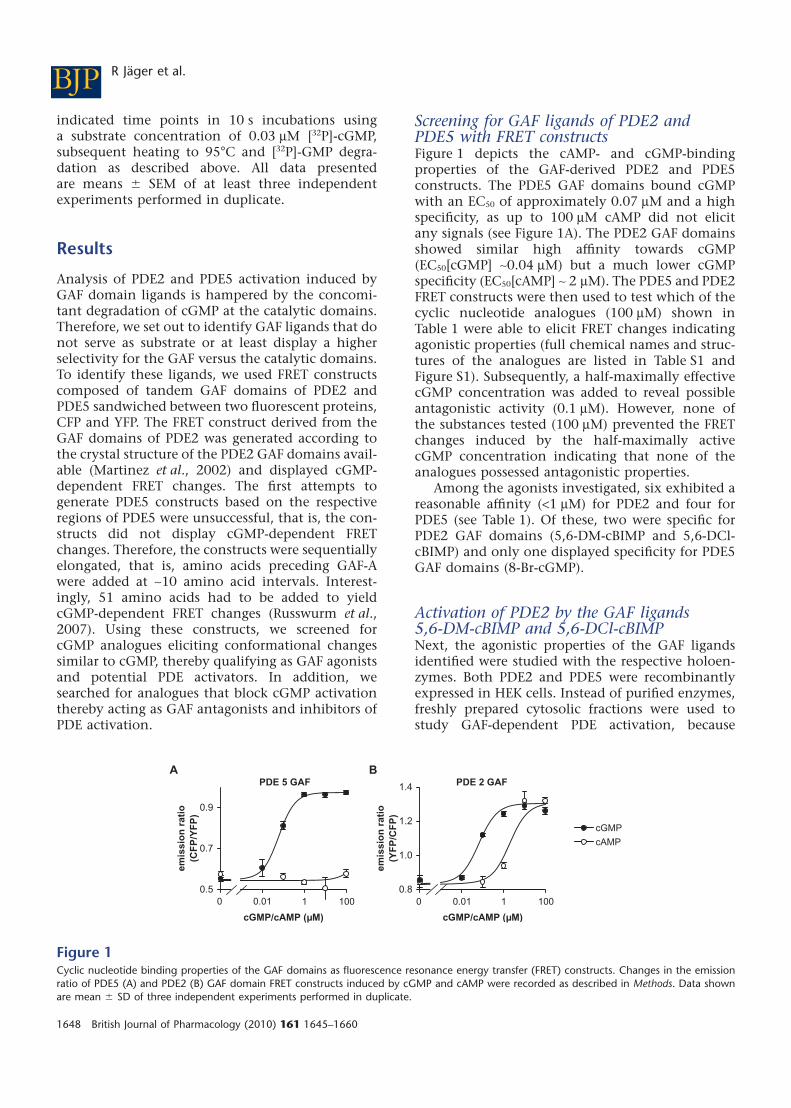
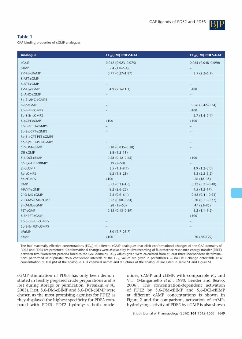
Screening for GAF ligands of PDE2 andPDE5 with FRET constructsFigure 1 depicts the cAMP- and cGMP-bindingproperties of the GAF-derived PDE2 and PDE5constructs. The PDE5 GAF domains bound cGMPwith an EC50 of approximately 0.07 mM and a highspecificity, as up to 100 mM cAMP did not elicitany signals (see Figure 1A). The PDE2 GAF domainsshowed similar high affinity towards cGMP(EC50[cGMP] ~0.04 mM) but a much lower cGMPspecificity (EC50[cAMP] ~ 2 mM). The PDE5 and PDE2FRET constructs were then used to test which of thecyclic nucleotide analogues (100 mM) shown inTable 1 were able to elicit FRET changes indicatingagonistic properties (full chemical names and struc-tures of the analogues are listed in Table S1 andFigure S1). Subsequently, a half-maximally effectivecGMP concentration was added to reveal possibleantagonistic activity (0.1 mM). However, none ofthe substances tested (100 mM) prevented the FRETchanges induced by the half-maximally activecGMP concentration indicating that none of theanalogues possessed antagonistic properties.
Among the agonists investigated, six exhibited areasonable affinity (<1 mM) for PDE2 and four forPDE5 (see Table 1). Of these, two were specific forPDE2 GAF domains (5,6-DM-cBIMP and 5,6-DCl-cBIMP) and only one displayed specificity for PDE5GAF domains (8-Br-cGMP).
Activation of PDE2 by the GAF ligands5,6-DM-cBIMP and 5,6-DCl-cBIMPNext, the agonistic properties of the GAF ligandsidentified were studied with the respective holoen-zymes. Both PDE2 and PDE5 were recombinantlyexpressed in HEK cells. Instead of purified enzymes,freshly prepared cytosolic fractions were used tostudy GAF-dependent PDE activation, because
0.5
0.7
0.9
cGMP/cAMP (µM)
emis
sio
n r
atio
(C
FP
/YF
P)
0 0.01 1 1000.8
1.0
1.2
1.4
cGMP/cAMP (µM)
emis
sio
n r
atio
(Y
FP
/CF
P)
0 0.01 1 100
BA
cGMP
cAMP
FAG 2 EDPFAG 5 EDP
Figure 1Cyclic nucleotide binding properties of the GAF domains as fluorescence resonance energy transfer (FRET) constructs. Changes in the emissionratio of PDE5 (A) and PDE2 (B) GAF domain FRET constructs induced by cGMP and cAMP were recorded as described in Methods. Data shownare mean � SD of three independent experiments performed in duplicate.
BJP R Jäger et al.
1648 British Journal of Pharmacology (2010) 161 1645–1660
cGMP stimulation of PDE5 has only been demon-strated in freshly prepared crude preparations and islost during storage or purification (Rybalkin et al.,2003). First, 5,6-DM-cBIMP and 5,6-DCl-cBIMP werechosen as the most promising agonists for PDE2 asthey displayed the highest specificity for PDE2 com-pared with PDE5. PDE2 hydrolyses both nucle-
otides, cAMP and cGMP, with comparable Km andVmax (Manganiello et al., 1990; Bender and Beavo,2006). The concentration-dependent activationof PDE2 by 5,6-DM-cBIMP and 5,6-DCl-cBIMPat different cAMP concentrations is shown inFigure 2 and for comparison, activation of cAMP-hydrolysing activity of PDE2 by cGMP is also shown
Table 1GAF binding properties of cGMP analogues
Analogue EC50(mM) PDE2-GAF EC50(mM) PDE5-GAF
cGMP 0.042 (0.023–0.075) 0.065 (0.048–0.090)
cAMP 2.4 (1.0–5.4) –
2-NH2-cPuMP 0.71 (0.27–1.87) 3.5 (2.2–5.7)
8-AET-cGMP – –
8-APT-cGMP – –
1-NH2-cGMP 4.9 (2.1–11.1) >100
2′-AHC-cGMP – –
Sp-2′-AHC-cGMPS – –
8-Br-cGMP – 0.56 (0.42–0.74)
Rp-8-Br-cGMPS – >100
Sp-8-Br-cGMPS – 2.7 (1.4–5.4)
8-pCPT-cGMP >100 >100
Rp-8-pCPT-cGMPS – –
Sp-8-pCPT-cGMPS – –
Rp-8-pCPT-PET-cGMPS – –
Sp-8-pCPT-PET-cGMPS – –
5,6-DM-cBIMP 0.10 (0.035–0.28) –
DB-cGMP 3.8 (1.2–11) –
5,6-DCl-cBIMP 0.28 (0.12–0.65) >100
Sp-5,6-DCl-cBIMPS 19 (7–50) –
2′-dcGMP 3.5 (1.3–9.4) 1.9 (1.2–3.0)
Rp-cGMPS 6.2 (1.8–21) 3.3 (2.2–5.2)
Sp-cGMPS >100 26 (18–35)
cIMP 0.72 (0.33–1.6) 0.32 (0.21–0.48)
MANT-cGMP 8.2 (2.6–26) 4.5 (1.2–17)
2′-O-MS-cGMP 2.5 (0.9–6.4) 0.62 (0.41–0.93)
2′-O-MS-TME-cGMP 0.22 (0.08–0.64) 0.20 (0.11–0.37)
2′-O-ME-cGMP 28 (15–53) 47 (23–95)
PET-cGMP 0.35 (0.13–0.89) 3.2 (1.1–9.2)
8-Br-PET-cGMP – >100
Rp-8-Br-PET-cGMPS – –
Sp-8-Br-PET-cGMPS – –
cPuMP 8.0 (2.7–23.7) –
cXMP >100 70 (38–129)
The half-maximally effective concentrations (EC50) of different cGMP analogues that elicit conformational changes of the GAF domains ofPDE2 and PDE5 are presented. Conformational changes were assessed by in vitro recording of fluorescence resonance energy transfer (FRET)between two fluorescent proteins fused to the GAF domains. EC50 values given were calculated from at least three independent determina-tions performed in duplicate; 95% confidence intervals of the EC50 values are given in parentheses. –, no FRET change detectable at aconcentration of 100 mM of the analogue. Full chemical names and structures of the analogues are listed in Table S1 and Figure S1.
BJPGAF ligands of PDE2 and PDE5
British Journal of Pharmacology (2010) 161 1645–1660 1649
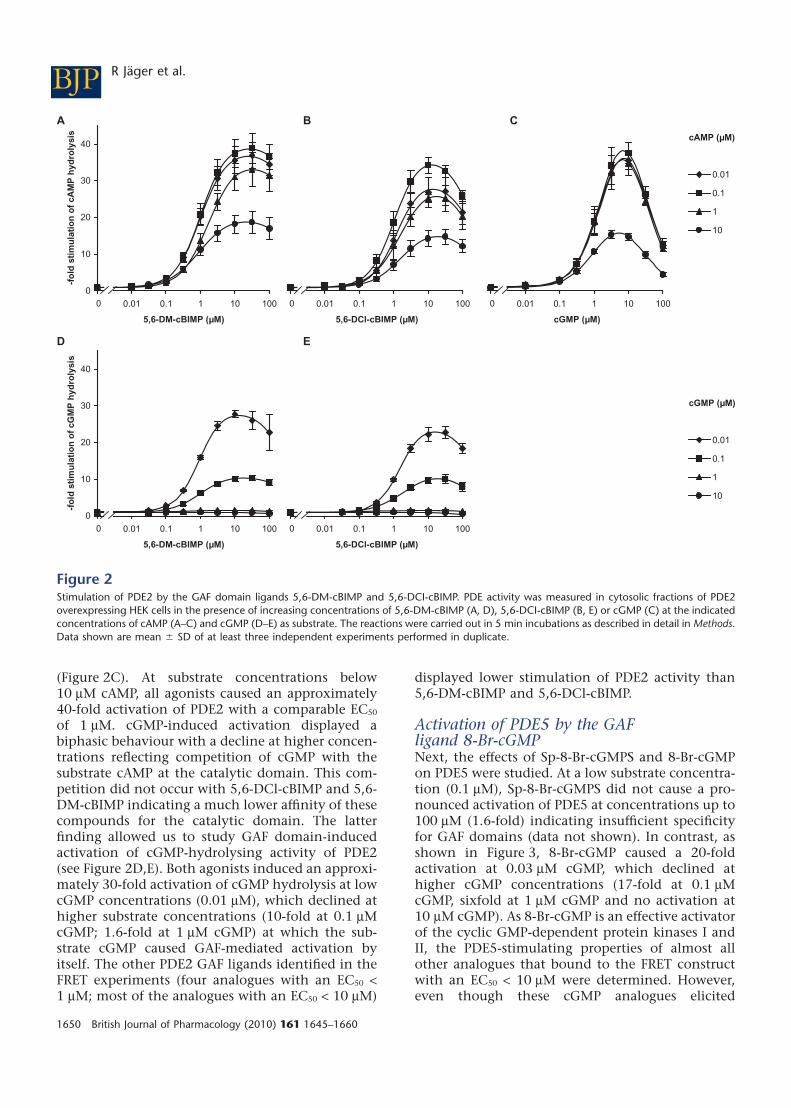
(Figure 2C). At substrate concentrations below10 mM cAMP, all agonists caused an approximately40-fold activation of PDE2 with a comparable EC50
of 1 mM. cGMP-induced activation displayed abiphasic behaviour with a decline at higher concen-trations reflecting competition of cGMP with thesubstrate cAMP at the catalytic domain. This com-petition did not occur with 5,6-DCl-cBIMP and 5,6-DM-cBIMP indicating a much lower affinity of thesecompounds for the catalytic domain. The latterfinding allowed us to study GAF domain-inducedactivation of cGMP-hydrolysing activity of PDE2(see Figure 2D,E). Both agonists induced an approxi-mately 30-fold activation of cGMP hydrolysis at lowcGMP concentrations (0.01 mM), which declined athigher substrate concentrations (10-fold at 0.1 mMcGMP; 1.6-fold at 1 mM cGMP) at which the sub-strate cGMP caused GAF-mediated activation byitself. The other PDE2 GAF ligands identified in theFRET experiments (four analogues with an EC50 <1 mM; most of the analogues with an EC50 < 10 mM)
displayed lower stimulation of PDE2 activity than5,6-DM-cBIMP and 5,6-DCl-cBIMP.
Activation of PDE5 by the GAFligand 8-Br-cGMPNext, the effects of Sp-8-Br-cGMPS and 8-Br-cGMPon PDE5 were studied. At a low substrate concentra-tion (0.1 mM), Sp-8-Br-cGMPS did not cause a pro-nounced activation of PDE5 at concentrations up to100 mM (1.6-fold) indicating insufficient specificityfor GAF domains (data not shown). In contrast, asshown in Figure 3, 8-Br-cGMP caused a 20-foldactivation at 0.03 mM cGMP, which declined athigher cGMP concentrations (17-fold at 0.1 mMcGMP, sixfold at 1 mM cGMP and no activation at10 mM cGMP). As 8-Br-cGMP is an effective activatorof the cyclic GMP-dependent protein kinases I andII, the PDE5-stimulating properties of almost allother analogues that bound to the FRET constructwith an EC50 < 10 mM were determined. However,even though these cGMP analogues elicited
0.01
0.1
1
10
cAMP (µM)
0
10
20
30
40
0.001 0.01 0.1 1 10 100
cGMP (µM)
-fo
ld s
tim
ula
tio
n
0
0
10
20
30
40
0.001 0.01 0.1 1 10 100
5,6-DCl-cBIMP (µM)
-fo
ld s
tim
ula
tio
n
0
0
10
20
30
40
0.001 0.01 0.1 1 10 100
5,6-DCl-cBIMP (µM)
-fo
ld s
tim
ula
tio
n
0
0
10
20
30
40
0.001 0.01 0.1 1 10 100
5,6-DM-cBIMP (µM)
-fo
ld s
tim
ula
tio
n o
f cG
MP
hyd
roly
sis
0
C
E
0
10
20
30
40
0.001 0.01 0.1 1 10 100
5,6-DM-cBIMP (µM)
-fo
ld s
tim
ula
tio
n o
f cA
MP
hyd
roly
sis
0
0.01
0.1
1
10
cGMP (µM)
A
D
B
Figure 2Stimulation of PDE2 by the GAF domain ligands 5,6-DM-cBIMP and 5,6-DCI-cBIMP. PDE activity was measured in cytosolic fractions of PDE2overexpressing HEK cells in the presence of increasing concentrations of 5,6-DM-cBIMP (A, D), 5,6-DCI-cBIMP (B, E) or cGMP (C) at the indicatedconcentrations of cAMP (A–C) and cGMP (D–E) as substrate. The reactions were carried out in 5 min incubations as described in detail in Methods.Data shown are mean � SD of at least three independent experiments performed in duplicate.
BJP R Jäger et al.
1650 British Journal of Pharmacology (2010) 161 1645–1660

conformational changes comparable to cGMP in theFRET constructs, none of the tested compoundscaused any relevant PDE5 activation. The lack ofGAF domain-induced activation can be attributedto competition of the analogues with the sub-strate turnover at the catalytic domain, indicating asimilar affinity ratio of the analogues and cGMP tothe GAF and catalytic domains respectively.
A construct containing only the catalytic domainfor PDE5 was used to confirm that inhibition by8-Br-cGMP observed at the highest concentration(1000 mM) was caused by competition at the cata-lytic site (Figure 3B, upper curve). As in the holoen-zyme, the highest concentration of 8-Br-cGMP(1000 mM) inhibited the catalytic domain construct.For comparison, inhibition of the catalytic domainby 8-Br-cAMP was also determined and was foundto be more marked than that by 8-Br-cGMP asdescribed previously (Poppe et al., 2008).
Time course of PDE5 activationWhile recording the concentration–response curvesfor 8-Br-cGMP, we got the impression that cGMPdegradation was not linear over time. Therefore, wemonitored the time course of PDE5 activation byassessing GMP accumulation at 10 s intervals withlow, medium and high cGMP concentrations (0.03,1, 100 mM; Figure 4A–C; results with additionalconcentrations shown in Figure S2). At all substrateconcentrations, a time-dependent increase in PDEactivity was observed. Even at a substrate concen-
tration of 0.03 mM cGMP, at which the GAF domainsof PDE5 can be considered ligand-free, maximalactivity was not observed until 120 s. The values forthe half-life of the activity increase were derivedfrom the product accumulation curves (Figures 5and S3). Because the scatter was predominantly dueto different PDE5 preparations, the curves had to benormalized to the 240 s values for the purpose ofstatistical analysis. As a control for our assay condi-tions, PDE2 was measured for comparison. Asexpected, PDE2 was fully active at the first timepoint measured (10 s), which shows that the slowtime-dependent increase in PDE5 activity was notcaused by the experimental conditions (Figure 6A).Moreover, PDE activity in platelet cytosol, whichcan be attributed almost exclusively to PDE5 underthe conditions applied (data not shown), showed acomparable time-dependent increase in activitywhen measured under identical assay conditions(Figure 6B,C). The activity increase observed istherefore a general feature of PDE5 and not limitedto the recombinant enzyme.
Next, we analysed activation of PDE5 at thesame substrate concentration (0.03 mM) in the pres-ence of a maximally activating concentration of theGAF ligand 8-Br-cGMP (Figure 4E). Under these con-ditions, the enzyme showed a 20-fold activationalready at the first point measured (10 s) indi-cating that at a high ligand concentration, GAF-induced activation is fast. However, also in thesemeasurements, an activity increase (threefold) overtime also occurred (120 s). At an intermediate ligand
8-Br-cGMP
8-Br-cAMP
0
5
10
15
20
0 1 10 100 1 000
8-Br-cGMP (µM)
-fo
ld s
tim
ula
tio
n o
f cG
MP
hyd
roly
sis
0
0.03
0.1
1
10
cGMP (µM)
A
cGMP (µM) 1
0
0.25
0.5
0.75
1
0 1 10 100 1 000
8-Br-cGMP/8-Br-cAMP (µM)
-fo
ld c
GM
P h
ydro
lysi
s
0
B
Figure 3Stimulation of PDE5 by the GAF domain ligand 8-Br-cGMP. (A) PDE5 activity was measured in cytosolic fractions of PDE5 overexpressing HEK cellsin the presence of increasing concentrations of 8-Br-cGMP at the indicated concentrations of cGMP as substrate. (B) Activity of a PDE5 constructcontaining only the catalytic domain was measured at 1 mM cGMP in the presence of increasing concentrations of 8-Br-cGMP or 8-Br-cAMP. Thereactions were carried out for 5 min as described in detail in Methods. Data shown are mean � SD of at least three independent experimentsperformed in duplicate.
BJPGAF ligands of PDE2 and PDE5
British Journal of Pharmacology (2010) 161 1645–1660 1651

concentration (30 mM 8-Br-cGMP, Figure 4D),GAF-induced activation at 10 s was negligible, but6 min were required to reach final velocity (10-foldactivation).
Subsequently, we studied cGMP as a GAF ligand.At a very high cGMP concentration (100 mM cGMP),the GAF domains should be occupied immediately.
Accordingly, PDE5 activity at the first time pointwas 200-fold higher than with 0.03 mM cGMP. Thefactor 200 is explained by a 10-fold km effect due tothe higher substrate concentration and a 20-foldGAF-dependent activation of PDE5. The GAF-dependent activation of PDE5 by cGMP occurs asfast as the one observed with the high 8-Br-cGMP
100
0
50
100
150
200
0 60 120 180 240
0
10
20
30
0 240 480 720 9600
0.1
0.2
0.3
0 60 120 180 240
time (s)
aver
age
PD
E a
ctiv
ity
0
1
2
3
0 200 400 600
time (s)
aver
age
PD
E a
ctiv
ity
0
20
40
60
0 60 120 180 240
time (s)
0.03
8-B
r-cG
MP
(µM
)
030
CBA
D
F
cGMP (µM)
130.0
0
2
4
6
8
0 60 120 180 240
time (s)
aver
age
PD
E a
ctiv
ity
(nm
ol·m
in-1
·mg-1
)(n
mo
l·min
-1·m
g-1
)(n
mo
l·min
-1·m
g-1
)
300
E
Figure 4Kinetics of PDE5 activation. Time courses of PDE activation were obtained by measuring GMP accumulation in cytosolic fractions of PDE5overexpressing HEK cells at 0.03 mM (A, D, E), 1 mM (B, F) or 100 mM (C) cGMP as substrate either in the absence (A, B, C) or in the presenceof 30 mM (D) or 300 mM (E, F) 8-Br-cGMP. The reactions were carried out as described in detail in Methods. Average PDE activity in the indicatedtime intervals was calculated as described in Methods. The width of a bar depicts the length of the respective interval, the height represents theaverage enzymatic velocity during the interval and the area of the bar is therefore equivalent to GMP formed during the interval. Data shown aremean � SD of at least three independent experiments performed in duplicate.
BJP R Jäger et al.
1652 British Journal of Pharmacology (2010) 161 1645–1660
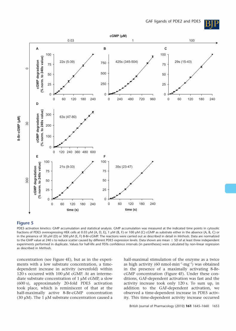
concentration (see Figure 4E), but as in the experi-ments with a low substrate concentration, a time-dependent increase in activity (sevenfold) within120 s occurred with 100 mM cGMP. At an interme-diate substrate concentration of 1 mM cGMP, a slow(600 s), approximately 20-fold PDE5 activationtook place, which is reminiscent of that at thehalf-maximally active 8-Br-cGMP concentration(30 mM). The 1 mM substrate concentration caused a
half-maximal stimulation of the enzyme as a twiceas high activity (60 nmol·min-1·mg-1) was obtainedin the presence of a maximally activating 8-Br-cGMP concentration (Figure 4F). Under these con-ditions, GAF-dependent activation was fast and theactivity increase took only 120 s. To sum up, inaddition to the GAF-dependent activation, weobserved a time-dependent increase in PDE5 activ-ity. This time-dependent activity increase occurred
0
25
50
75
100
0 60 120 180 240
0
250
500
750
0 240 480 720 960
0
25
50
75
100
0 60 120 180 240
time (s)
0
25
50
75
100
0 60 120 180 240
cGM
P d
egra
dat
ion
(%
no
rm. t
o 2
40s
valu
e)
0
100
200
300
0 120 240 360 480 600
cGM
P d
egra
dat
ion
(%
no
rm. t
o 2
40s
valu
e)
0
25
50
75
100
0 60 120 180 240
time (s)
cGM
P d
egra
dat
ion
(%
no
rm. t
o 2
40s
valu
e)
)34-51( s92)405-543( s524)93-5( s22
63s (47-80)
)74-32( s53)33-9( s12
CBA
D
FE
cGMP (µM)001130.0
8-B
r-cG
MP
(µM
)
030
300
Figure 5PDE5 activation kinetics: GMP accumulation and statistical analysis. GMP accumulation was measured at the indicated time points in cytosolicfractions of PDE5 overexpressing HEK cells at 0.03 mM (A, D, E), 1 mM (B, F) or 100 mM (C) cGMP as substrate either in the absence (A, B, C) orin the presence of 30 mM (D) or 300 mM (E, F) 8-Br-cGMP. The reactions were carried out as described in detail in Methods. Data are normalizedto the GMP value at 240 s to reduce scatter caused by different PDE5 expression levels. Data shown are mean � SD of at least three independentexperiments performed in duplicate. Values for half-life and 95% confidence intervals (in parentheses) were calculated by non-linear regressionas described in Methods.
BJPGAF ligands of PDE2 and PDE5
British Journal of Pharmacology (2010) 161 1645–1660 1653

within approximately 120 s and was independentof the GAF ligand, whereas the GAF-dependentactivation was fast at high ligand concentrationsand took minutes at cGMP concentrations aroundthe EC50.
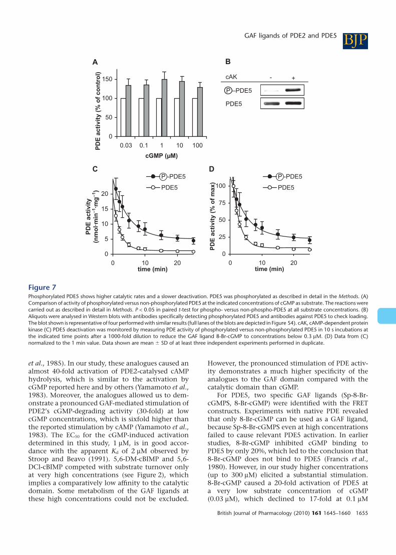
Phosphorylation of PDE5Cyclic GMP binding to the GAF domain of PDE5has been reported to be stabilized by phosphoryla-tion at serine-102. To analyse the impact of PDE5phosphorylation, we performed experiments withphosphorylated PDE5 and used antibodies thatspecifically recognize the phosphorylated enzyme tocheck phosphorylation. Our attempt to phosphory-late ligand-free PDE5 was unsuccessful; we wereunable to detect any phosphorylation without thesubstrate or a GAF domain ligand (data not shown)confirming that cGMP bound to the GAF domainis a prerequisite for phosphorylation, as found pre-viously (Turko et al., 1998). Hence, we incubatedrecombinantly expressed PDE5 in the presence of8-Br-cGMP with and without the cAMP-dependentkinase to compare activity of the phosphorylatedversus the non-phosphorylated PDE5. After incuba-tion, measurement of PDE activity at various sub-strate concentrations (0.03, 0.1, 1, 10 and 100 mM)showed an approximately 35% higher activity ofthe phosphorylated species indicative of an increasein Vmax (Figure 7). Phosphorylation has beenreported to have an important impact on the disso-ciation of the GAF ligand. Therefore, subsequent to
phosphorylation induced by incubation with kinaseand 8-Br-cGMP, samples were diluted to decrease theconcentration of the free GAF ligand. Dissociationof the GAF ligand was then monitored by measu-ring the activity at a low concentration of cGMP(0.03 mM) in 10 s incubations (Figure 7C). Witha half-life of 3.4 versus 1.5 min, deactivation ofphospho-PDE5 was clearly slower than that of thenon-phosphorylated enzyme. We concluded thatphosphorylation enhances cGMP-induced PDE5activation by increasing the activity and slowingdown the dissociation of the GAF ligand.
Discussion
GAF domain ligands for PDE2 and PDE5To enable a better characterization of the allostericGAF domain-dependent activation of PDE2 andPDE5, we performed a systematic search for GAFdomain ligands among 30 nucleotide analoguesusing GAF domain-containing FRET constructs.Although our screen with PDE2 and PDE5 GAFdomain FRET constructs disappointingly did notyield any antagonists, several agonists for the PDE2and PDE5 GAF domains were identified.
5,6-DM-cBIMP and 5,6-DCl-cBIMP were rela-tively specific for PDE2 compared with PDE5. Bothanalogues have been suggested before to act as PDE2activators (Genieser et al., 1992), cBIMP has beenshown to activate PDE from rat liver or bovineadrenal gland twofold (Erneux et al., 1981; Miot
0
25
50
75
100
0 60 120 180 240
time (s)
cGM
P d
egra
dat
ion
(%
no
rm. t
o 2
40s
valu
e)
PDE2A
0
0.02
0.04
0.06
0 60 120 180 240
time (s)
aver
age
PD
E a
ctiv
ity
(nm
ol·m
in-1
·mg-1
)0
25
50
75
100
0 60 120 180 240
time (s)
cGM
P d
egra
dat
ion
(%
no
rm. t
o 2
40s
valu
e)
C PlateletsPlateletsB
Figure 6(A) cGMP degradation by PDE2 was measured at the indicated time points in cytosolic fractions of PDE2 overexpressing HEK cells at 1 mM cGMPas substrate. Data were normalized to the GMP value at 240 s. (B) Time course of PDE5 activation was determined in human platelet cytosolicfractions at a substrate concentration of 0.03 mM cGMP as described in Methods and the legend to Figure 4. (C) Statistical analysis of the GMPaccumulation determined in platelet cytosolic fractions. Data were normalized to the GMP value at 240 s. Values for half-life and 95% confidenceinterval (in parentheses) were calculated by non-linear regression as described in Methods. Data shown are mean � SD of at least threeindependent experiments performed in duplicate.
BJP R Jäger et al.
1654 British Journal of Pharmacology (2010) 161 1645–1660
et al., 1985). In our study, these analogues caused analmost 40-fold activation of PDE2-catalysed cAMPhydrolysis, which is similar to the activation bycGMP reported here and by others (Yamamoto et al.,1983). Moreover, the analogues allowed us to dem-onstrate a pronounced GAF-mediated stimulation ofPDE2’s cGMP-degrading activity (30-fold) at lowcGMP concentrations, which is sixfold higher thanthe reported stimulation by cAMP (Yamamoto et al.,1983). The EC50 for the cGMP-induced activationdetermined in this study, 1 mM, is in good accor-dance with the apparent Kd of 2 mM observed byStroop and Beavo (1991). 5,6-DM-cBIMP and 5,6-DCl-cBIMP competed with substrate turnover onlyat very high concentrations (see Figure 2), whichimplies a comparatively low affinity to the catalyticdomain. Some metabolism of the GAF ligands atthese high concentrations could not be excluded.
However, the pronounced stimulation of PDE activ-ity demonstrates a much higher specificity of theanalogues to the GAF domain compared with thecatalytic domain than cGMP.
For PDE5, two specific GAF ligands (Sp-8-Br-cGMPS, 8-Br-cGMP) were identified with the FRETconstructs. Experiments with native PDE revealedthat only 8-Br-cGMP can be used as a GAF ligand,because Sp-8-Br-cGMPS even at high concentrationsfailed to cause relevant PDE5 activation. In earlierstudies, 8-Br-cGMP inhibited cGMP binding toPDE5 by only 20%, which led to the conclusion that8-Br-cGMP does not bind to PDE5 (Francis et al.,1980). However, in our study higher concentrations(up to 300 mM) elicited a substantial stimulation.8-Br-cGMP caused a 20-fold activation of PDE5 ata very low substrate concentration of cGMP(0.03 mM), which declined to 17-fold at 0.1 mM
0
5
10
15
20
0 10 20time (min)
PD
E a
ctiv
ity
(nm
ol·m
in-1
·mg-1
)
P -PDE5
PDE5
C
P –PDE5
PDE5
cAK - +
B
0
50
100
150
0.03 0.1 1 10 100
cGMP (µM)
PD
E a
ctiv
ity
(% o
f co
ntr
ol)
A
0
25
50
75
100
0 10 20time (min)
PD
E a
ctiv
ity
(% o
f m
ax)
P -PDE5
PDE5
D
Figure 7Phosphorylated PDE5 shows higher catalytic rates and a slower deactivation. PDE5 was phosphorylated as described in detail in the Methods. (A)Comparison of activity of phosphorylated versus non-phosphorylated PDE5 at the indicated concentrations of cGMP as substrate. The reactions werecarried out as described in detail in Methods. P < 0.05 in paired t-test for phospho- versus non-phospho-PDE5 at all substrate concentrations. (B)Aliquots were analysed in Western blots with antibodies specifically detecting phosphorylated PDE5 and antibodies against PDE5 to check loading.The blot shown is representative of four performed with similar results (full lanes of the blots are depicted in Figure S4). cAK, cAMP-dependent proteinkinase (C) PDE5 deactivation was monitored by measuring PDE activity of phosphorylated versus non-phosphorylated PDE5 in 10 s incubations atthe indicated time points after a 1000-fold dilution to reduce the GAF ligand 8-Br-cGMP to concentrations below 0.3 mM. (D) Data from (C)normalized to the 1 min value. Data shown are mean � SD of at least three independent experiments performed in duplicate.
BJPGAF ligands of PDE2 and PDE5
British Journal of Pharmacology (2010) 161 1645–1660 1655
cGMP and sixfold at 1 mM cGMP. At 10 mM cGMP,no 8-Br-cGMP-dependent activation was observed.These results show that even at a substrate concen-tration of 0.1 mM cGMP, some cGMP is bound to theGAF domains already, at 1 mM cGMP approximatelyhalf of GAF domains are saturated, and at 10 mMcGMP the GAF domain is fully saturated. Due totechnical reasons, we were unable to measure PDEactivity at a substrate concentration below 0.03 mMcGMP, but as 8-Br-cGMP activation of PDE5 at 0.03is only slightly higher than at 0.1 mM cGMP (20- vs.17-fold), the GAF domains can be considered to belargely unoccupied at 0.03 mM cGMP. An EC50 ofabout 1 mM cGMP at PDE5’s GAF domains can alsobe estimated from the kinetic analysis of PDE5 acti-vation (see below), which is in reasonable agree-ment with the Kd of 0.2 mM observed by Zoraghiet al. (2005).
In a millimolar concentration, 8-Br-cGMPcompeted with substrate turnover (see Figure 3),remarkably, 8-Br-cGMP inhibited turnover at theseconcentrations less effectively than 8-Br-cAMP.In an earlier study that used microcalorimetry todirectly assess turnover of cyclic nucleotide ana-logues, degradation of 8-Br-cAMP but not of 8-Br-cGMP by PDE5 was described (Poppe et al., 2008),an unexpected finding that nevertheless is in linewith our observations.
It should be noted that the cGMP affinity wasa lot higher in the FRET constructs than in theholoenzymes (as judged by activation of PDE activ-ity) in both PDE5 and PDE2, which is in accordancewith results from earlier binding studies (Wu et al.,2004; Zoraghi et al., 2005). The discrepancy impliesthat the catalytic domain alters the properties of theGAF domain-containing regulatory domain, anobservation that is also supported by the findingthat sildenafil binding to PDE5’s catalytic domainhas an impact on cGMP binding to the GAF domain(Turko et al., 1999).
Similarly, the half-maximal effective 8-Br-cGMPconcentration for PDE5 stimulation was in therange of 30 mM and far higher than the EC50
observed with the FRET construct (0.6 mM). Yet,because this study used isolated FRET-tagged GAFdomains only as tools to identify promising cGMPanalogues, a thorough investigation of the molecu-lar basis of this discrepancy was not performed.Furthermore, the principle of reciprocity impliesthat the catalytic domain in the holoenzymeshould affect ligand binding to the GAF domains,if the GAF domains can regulate the catalyticturnover.
Direct assessment of the stimulating effect ofcGMP binding on PDE5 cGMP hydrolysis inevitablyrepresents a challenge, because cGMP acts as an
activator and substrate. Different approaches havebeen made to demonstrate the consequences ofcGMP binding to the GAF domain on the catalyticdomain. In one of these studies, the effect of thephysiological GAF ligand, cGMP, on binding of thesynthetic PDE5 inhibitor sildenafil was examined(Corbin et al., 2003). An increase in sildenafil affin-ity in the presence of low cGMP concentrationssuggested that cGMP binding to the GAF domainsinduced a conformational change leading to highersildenafil affinity and presumably activation of theenzyme. Another study (Rybalkin et al., 2003) tookadvantage of the relatively slow cGMP dissociationand investigated activation of the enzyme aftercGMP pre-incubation. In that study, a 10-fold stimu-lation of PDE5 and a higher potency of sildenafil toinhibit the stimulated enzyme were found. Ourapproach to employ a reasonably specific, syntheticGAF domain ligand (8-Br-cGMP) to investigatestimulated turnover of the physiological substrate,cGMP, represents the second possible strategy tosolve the problem that cGMP acts as activator andsubstrate and revealed a 20-fold dynamic range ofPDE5 activation.
The almost unchanged 8-Br-cGMP EC50 valuesfor stimulation of the PDE5 holoenzyme at dif-ferent cGMP concentrations raise the questionwhy the EC50 values were not shifted by increa-sing cGMP concentrations. The finding can beexplained in the case that the Kd values at the GAFdomains for 8-Br-cGMP and cGMP are in the rangeof the observed EC50s. Under the assumption of Kd
of 30 and 1 mM for 8-Br-cGMP and cGMP, respec-tively, the Cheng and Prusoff equation (Cheng andPrusoff, 1973) yields 8-Br-cGMP EC50 values of31, 33 and 60 mM at increasing cGMP con-centrations (0.03, 0.1 and 1 mM). The small dif-ference expected would be barely visible on alogarithmic scale, especially as the stimulationfactors by 8-Br-cGMP also vary with the cGMPconcentration.
PDE5 activation kineticsIn contrast to the fast GAF domain-dependent acti-vation of PDE2, the increase in PDE5 activity wasslow. A non-linear PDE5 activity has been describedbefore and attributed to slow binding of cGMP (0.1and 1 mM) to the GAF domains because blockingcGMP binding to the GAF domains or saturatingthe GAF domains with cGMP linearized the kinetics(Rybalkin et al., 2002). In contrast, our study per-formed on a more detailed time scale revealeda time-dependent but GAF ligand-independentincrease in PDE5 activity in addition to the GAFligand-dependent activation.
BJP R Jäger et al.
1656 British Journal of Pharmacology (2010) 161 1645–1660

GAF ligand-dependent PDE5 activationThe GAF ligand-dependent PDE5 activation wasfast at high GAF ligand concentrations (300 mM8-Br-cGMP or 100 mM cGMP) as stimulated PDE5activity (20-fold) was already observed after 10 s.However, at intermediate GAF ligand concentra-tions (30 mM 8-Br-cGMP or 1 mM cGMP), the GAFdomain-dependent PDE5 activation was muchslower and dominated the time course of PDE5activity described below (600 s). The ligandconcentration-dependence of the activation kinet-ics indicates cGMP binding to the GAF domains asthe rate-limiting step.
cGMP binding experiments with the PDE5holoenzyme and the isolated GAF domains havebeen published in the past (McAllister-Lucas et al.,1993; Turko et al., 1996; Francis et al., 2002;Zoraghi et al., 2005). In summary, these studiesdescribed a slow and a fast cGMP binding site/component on PDE5 that did not correspond tothe GAF-A and GAF-B domains, respectively, butwere components of cGMP binding to the GAF-Adomain. The slow site displayed a dissociationhalf-life between 72 and 465 min. Together with acGMP Kd between 0.1 and 1 mM, an associationhalf-life between 10 and 232 min can be derived ata 1 mM cGMP concentration. For the fast site, theobserved dissociation half-life between 29 and42 min would result in an association half-lifebetween 2.6 and 20 min at 1 mM cGMP. The acti-vation half-life of 7 min (see Figure 5B) determinedin our study fits fairly well within this rangeand suggests that the fast cGMP binding sitedescribed previously mediates activation of theenzyme.
Time-dependent PDE5 activity increaseSurprisingly, in addition to the GAF ligand-dependent PDE5 activation, we found a time-dependent increase in activity (threefold activityincrease within 120 s), which was independent of aGAF domain ligand as it was observed under condi-tions at which the GAF domains can be consideredligand-free (0.03 mM cGMP) or fully saturated (addi-tion of 300 mM 8-Br-cGMP or 100 mM cGMP). Themolecular basis for the time-dependent increase inPDE5 activity is unknown. As the increase occurredat very low and very high substrate concentrations,cGMP binding to the catalytic domains does notappear to be the limiting step. One can only specu-late about the existence of at least two conforma-tions of PDE5’s catalytic domain with the transitionbetween the lower and higher activity states occur-ring within 2 min. Untypically slowly responding,so-called hysteretic behaviour has been described
for regulatory as well as metabolic enzymes previ-ously (Frieden, 1979; 2008).
A recent study described a new ‘super-high’ sen-sitivity state for sildenafil inhibition (Rybalkinaet al., 2010) raising the question whether the differ-ent sensitivity states are related to the two confor-mations described above. As the two sensitivitystates were observed only in the non-stimulatedenzyme whereas the two conformations describedin our study were observed at non-stimulating(0.03 mM cGMP) and stimulating cGMP concen-trations (100 mM), the sildenafil sensitivity statesappear to be distinct from the two activity states.
Recently, a general model for GAF-mediated PDEactivation derived from the structure of full lengthPDE2A was proposed (Pandit et al., 2009). Accordingto this model, a dimer interface is formed betweenthe catalytic domains if the H-loop occludes thecatalytic pocket (closed conformation). PDE2 cyclesbetween the closed and open conformations andcGMP binding to GAF-B stabilizes the open confor-mation by moving the two catalytic domains apart.This allows the H-loop to swing out therebyenabling substrate binding. It is tempting to spe-culate that cGMP binding in PDE5 induces an evenlarger conformational reorganization, becausecGMP binds to the more distant GAF-A in thisenzyme. These changes could therefore requiremore time, although the time scale of minutes atcGMP concentrations near the EC50 of 1 mM remainsnevertheless unexpected. With regard to the pro-posed model, it can further be speculated that thetwo conformations of PDE5’s catalytic domain withlower and higher activity correspond to differentorientations of the H-loop. Structural analyses of theH-loop of unligated PDE5 and inhibitor PDE5 com-plexes demonstrated the existence of four differentconformations of this loop (Wang et al., 2006), allof which differed from the H-loop conformationsin other PDEs. Furthermore, in contrast to PDE2,the H-loop in the unligated PDE5 did not occludethe catalytic pocket. Whether the lower andhigher activity states correspond to different confor-mations of the H-loop therefore awaits furtherstructural analyses.
Consequences of PDE5 activation kinetics forintracellular cGMP signalsPDE5-rich cells respond to NO stimulation witha cGMP spike (Mullershausen et al., 2001). A slowresponse of cGMP-degrading activity to variationsin cGMP is a prerequisite for development of sucha spike. Extensive mathematical models havebeen developed to describe the activity profiles ofcGMP-forming and cGMP-degrading activities that
BJPGAF ligands of PDE2 and PDE5
British Journal of Pharmacology (2010) 161 1645–1660 1657
underlie these cGMP spikes (Mo et al., 2004). Inthese models, besides the impact of cGMP concen-tration on PDE5 activity, rate constants for increasesin PDE activity were required to explain theobserved cGMP peaks. Thus, without a slowly releas-ing ‘break’ on PDE5 activity, the observed intracel-lular cGMP signals in cells containing high PDE5levels would remain inexplicable.
PDE5 phosphorylationPhosphorylation analysis of PDE5 confirmed theresults by others that only the enzyme with a boundGAF ligand can be phosphorylated efficiently bythe cAMP- and cGMP-dependent protein kinases(Thomas et al., 1990). In this context, it is interest-ing to note that only those FRET constructs, whichincluded PDE5’s phosphorylation site, were respon-sive to cGMP. Phosphorylated PDE5 exhibitedapproximately 35% higher catalytic rates than thenon-phosphorylated species at various substrateconcentrations indicating a general Vmax effect. Inaddition to an increased catalytic rate, dissociationof the GAF ligand was significantly slower in thephosphorylated PDE5 compared with the non-phosphorylated enzyme indicating that phosphory-lation conserves PDE5 activation (half-life of 3.4 vs.1.5 min). In earlier studies, a longer half-life hasbeen suggested (Mullershausen et al., 2004). Inthose studies, dissociation of the GAF ligand wasdetermined by monitoring PDE5 activity at a sub-strate concentration of 100 nM cGMP, a concentra-tion at which – as shown in the present study –some PDE5 activation already occurs.
The impact of PDE5 phosphorylation on cGMPbinding to the isolated regulatory domain of PDE5has been described before (Francis et al., 2002). Inthat study, phosphorylation shifted the 50:50 ratiobetween the high and a low affinity cGMP bindingsites (corresponding to the slow and fast sitesdiscussed above, half-life 39 and 265 min respec-tively) completely to the high affinity component(slow site, half-life 339 min). Albeit deactivation ofboth, the non-phosphorylated and phosphorylatedholoenzymes, was considerably faster in our study,the data are in accordance with PDE5 phosphoryla-tion slowing down deactivation of the enzyme.
In summary, our data show that PDE5 activationunder physiological conditions occurs unexpectedlyslow, is long-lasting – in the range of minutes – andgreatly depends on the cGMP levels under restingconditions in a specific tissue. Although the cGMP-induced activation of PDE2 and PDE5 are bothmediated by GAF domains, the activation kineticsare quite diverse and reflect their different biologicalfunctions; PDE2, responsible for the cross-talk
between cGMP and cAMP has to respond fast tovariations in cGMP levels, whereas PDE5 as amolecular memory mediating feedback inhibitionwithin the cGMP cascade has to act in a delayed butsustained manner.
Acknowledgements
This work was supported by the DeutscheForschungsgemeinschaft (Ko1157) and the KOFFER(Kommission für Finanzautonomie und Ergän-zungsmittel of the Medical Faculty). The authorsgratefully acknowledge the technical assistance ofUlla Krabbe, Arkadius Pacha, Erika Mannheim andCaroline Vollmers.
Conflicts of interest
H.G.G. is CEO and F.S. is head of Research andDesign of BIOLOG Life Science Institute, which sellscyclic nucleotide analogues for research purposes.
References
Alexander SP, Mathie A, Peters JA (2009). Guideto Receptors and Channels (GRAC). 4th edn.Br J Pharmacol 158: S1–S254.
Aravind L, Ponting CP (1997). The GAF domain:an evolutionary link between diverse phototransducingproteins. Trends Biochem Sci 22: 458–459.
Beavo JA, Hardman JG, Sutherland EW (1971).Stimulation of adenosine 3′,5′-monophosphatehydrolysis by guanosine 3′,5′-monophosphate.J Biol Chem 246: 3841–3846.
Bender AT, Beavo JA (2006). Cyclic nucleotidephosphodiesterases: molecular regulation to clinicaluse. Pharmacol Rev 58: 488–520.
Cheng Y, Prusoff WH (1973). Relationship betweenthe inhibition constant (K1) and the concentration ofinhibitor which causes 50 per cent inhibition (I50) ofan enzymatic reaction. Biochem Pharmacol 22:3099–3108.
Corbin JD, Turko IV, Beasley A, Francis SH (2000).Phosphorylation of phosphodiesterase-5 by cyclicnucleotide-dependent protein kinase alters its catalyticand allosteric cGMP-binding activities. Eur J Biochem267: 2760–2767.
Corbin JD, Blount MA, Weeks JL 2nd, Beasley A,Kuhn KP, Ho YS et al. (2003). [3H]sildenafil bindingto phosphodiesterase-5 is specific, kineticallyheterogeneous, and stimulated by cGMP. MolPharmacol 63: 1364–1372.
BJP R Jäger et al.
1658 British Journal of Pharmacology (2010) 161 1645–1660
Erneux C, Couchie D, Dumont JE, Baraniak J, Stec WJ,Abbad EG et al. (1981). Specificity of cyclic GMPactivation of a multi-substrate cyclic nucleotidephosphodiesterase from rat liver. Eur J Biochem 115:503–510.
Francis SH, Lincoln TM, Corbin JD (1980).Characterization of a novel cGMP binding protein fromrat lung. J Biol Chem 255: 620–626.
Francis SH, Bessay EP, Kotera J, Grimes KA, Liu L,Thompson WJ et al. (2002). Phosphorylation of isolatedhuman phosphodiesterase-5 regulatory domain inducesan apparent conformational change and increases cGMPbinding affinity. J Biol Chem 277: 47581–47587.
Friebe A, Mullershausen F, Smolenski A, Walter U,Schultz G, Koesling D (1998). YC-1 potentiates nitricoxide- and carbon monoxide-induced cyclic GMP effectsin human platelets. Mol Pharmacol 54: 962–967.
Frieden C (1979). Slow transitions and hystereticbehavior in enzymes. Annu Rev Biochem 48: 471–489.
Frieden C (2008). A lifetime of kinetics. J Biol Chem283: 19873–19878.
Genieser HG, Winkler E, Butt E, Zorn M, Schulz S,Iwitzki F et al. (1992). Derivatives of1-beta-D-ribofuranosylbenzimidazole 3′,5′-phosphatethat mimic the actions of adenosine 3′,5′-phosphate(cAMP) and guanosine 3′,5′-phosphate (cGMP).Carbohydr Res 234: 217–235.
Gross-Langenhoff M, Hofbauer K, Weber J, Schultz A,Schultz JE (2006). cAMP is a ligand for the tandem GAFdomain of human phosphodiesterase 10 and cGMPfor the tandem GAF domain of phosphodiesterase 11.J Biol Chem 281: 2841–2846.
McAllister-Lucas LM, Sonnenburg WK, Kadlecek A,Seger D, Trong HL, Colbran JL et al. (1993). Thestructure of a bovine lung cGMP-binding, cGMP-specificphosphodiesterase deduced from a cDNA clone. J BiolChem 268: 22863–22873.
Manganiello VC, Tanaka T, Murashima S (1990). CyclicGMP-stimulated cyclic nucleotide phosphodiesterases.In: Beavo J, Houslay MD (eds). Cyclic NucleotidePhosphodiesterases: Structure, Regulation and DrugAction. John Wiley and Sons Ltd: New York, pp. 61–85.
Martinez SE, Wu AY, Glavas NA, Tang XB, Turley S,Hol WG et al. (2002). The two GAF domains inphosphodiesterase 2A have distinct roles in dimerizationand in cGMP binding. Proc Natl Acad Sci USA 99:13260–13265.
Miot F, Van Haastert PJ, Erneux C (1985). Specificityof cGMP binding to a purified cGMP-stimulatedphosphodiesterase from bovine adrenal tissue.Eur J Biochem 149: 59–65.
Mo E, Amin H, Bianco IH, Garthwaite J (2004). Kineticsof a cellular nitric oxide/cGMP/phosphodiesterase-5pathway. J Biol Chem 279: 26149–26158.
Moss J, Manganiello VC, Vaughan M (1977).Substrate and effector specificity of a guanosine3′:5′-monophosphate phosphodiesterase from ratliver. J Biol Chem 252: 5211–5215.
Mullershausen F, Russwurm M, Thompson WJ, Liu L,Koesling D, Friebe A (2001). Rapid nitric oxide-induceddesensitization of the cGMP response is caused byincreased activity of phosphodiesterase type 5 paralleledby phosphorylation of the enzyme. J Cell Biol 155:271–278.
Mullershausen F, Friebe A, Feil R, Thompson WJ,Hofmann F, Koesling D (2003). Direct activation ofPDE5 by cGMP: long-term effects within NO/cGMPsignaling. J Cell Biol 160: 719–727.
Mullershausen F, Russwurm M, Koesling D, Friebe A(2004). In vivo reconstitution of the negative feedbackin nitric oxide/cGMP signaling: role ofphosphodiesterase type 5 phosphorylation. Mol BiolCell 15: 4023–4030.
Pandit J, Forman MD, Fennell KF, Dillman KS,Menniti FS (2009). Mechanism for the allostericregulation of phosphodiesterase 2A deduced fromthe X-ray structure of a near full-length construct.Proc Natl Acad Sci USA 106: 18225–18230.
Poppe H, Rybalkin SD, Rehmann H, Hinds TR, Tang XB,Christensen AE et al. (2008). Cyclic nucleotide analogsas probes of signaling pathways. Nat Methods 5:277–278.
Russell TR, Terasaki WL, Appleman MM (1973).Separate phosphodiesterases for the hydrolysis of cyclicadenosine 3′,5′-monophosphate and cyclic guanosine3′,5′-monophosphate in rat liver. J Biol Chem 248:1334–1340.
Russwurm M, Mullershausen F, Friebe A, Jager R,Russwurm C, Koesling D (2007). Design of fluorescenceresonance energy transfer (FRET)-based cGMP indicators:a systematic approach. Biochem J 407: 69–77.
Russwurm C, Zoidl G, Koesling D, Russwurm M (2009).Dual acylation of PDE2A splice variant 3: targeting tosynaptic membranes. J Biol Chem 284: 25782–25790.
Rybalkin SD, Rybalkina IG, Feil R, Hofmann F, Beavo JA(2002). Regulation of cGMP-specific phosphodiesterase(PDE5) phosphorylation in smooth muscle cells. J BiolChem 277: 3310–3317.
Rybalkin SD, Rybalkina IG, Shimizu-Albergine M,Tang XB, Beavo JA (2003). PDE5 is converted to anactivated state upon cGMP binding to the GAF Adomain. EMBO J 22: 469–478.
Rybalkina IG, Tang XB, Rybalkin SD (2010). Multipleaffinity states of cGMP-specific phosphodiesterase forsildenafil inhibition defined by cGMP-dependent andcGMP-independent mechanisms. Mol Pharmacol 77:670–677.
Stroop SD, Beavo JA (1991). Structure and functionstudies of the cGMP-stimulated phosphodiesterase. JBiol Chem 266: 23802–23809.
Thomas MK, Francis SH, Corbin JD (1990). Substrate-and kinase-directed regulation of phosphorylation ofa cGMP-binding phosphodiesterase by cGMP. J BiolChem 265: 14971–14978.
BJPGAF ligands of PDE2 and PDE5
British Journal of Pharmacology (2010) 161 1645–1660 1659
Turko IV, Haik TL, McAllister-Lucas LM, Burns F,Francis SH, Corbin JD (1996). Identification of keyamino acids in a conserved cGMP-binding site ofcGMP-binding phosphodiesterases. A putativeNKXnD motif for cGMP binding. J Biol Chem 271:22240–22244.
Turko IV, Francis SH, Corbin JD (1998). Bindingof cGMP to both allosteric sites of cGMP-bindingcGMP-specific phosphodiesterase (PDE5) is requiredfor its phosphorylation. Biochem J 329: 505–510.
Turko IV, Ballard SA, Francis SH, Corbin JD (1999).Inhibition of cyclic GMP-binding cyclic GMP-specificphosphodiesterase (Type 5) by sildenafil and relatedcompounds. Mol Pharmacol 56: 124–130.
Wang H, Liu Y, Huai Q, Cai J, Zoraghi R, Francis SHet al. (2006). Multiple conformations ofphosphodiesterase-5: implications for enzyme functionand drug development. J Biol Chem 281: 21469–21479.
Wu AY, Tang XB, Martinez SE, Ikeda K, Beavo JA (2004).Molecular determinants for cyclic nucleotide binding tothe regulatory domains of phosphodiesterase 2A. J BiolChem 279: 37928–37938.
Wyatt TA, Naftilan AJ, Francis SH, Corbin JD (1998).ANF elicits phosphorylation of the cGMPphosphodiesterase in vascular smooth muscle cells.Am J Physiol 274: H448–H455.
Yamamoto T, Manganiello VC, Vaughan M (1983).Purification and characterization of cyclicGMP-stimulated cyclic nucleotide phosphodiesterase
from calf liver. Effects of divalent cations on activity.J Biol Chem 258: 12526–12533.
Zoraghi R, Bessay EP, Corbin JD, Francis SH (2005).Structural and functional features in human PDE5A1regulatory domain that provide for allosteric cGMPbinding, dimerization, and regulation. J Biol Chem280: 12051–12063.
Supporting information
Additional Supporting Information may be foundin the online version of this article:
Figure S1 Structures of the tested analogues.Figure S2 Kinetics of PDE5 activation.Figure S3 PDE5 activation kinetics: GMP accumu-lation and statistical analysis.Figure S4 Complete lanes of the Western blotshown in Figure 7B.Table S1 Full chemical names of the testedanalogues.
Please note: Wiley-Blackwell are not responsiblefor the content or functionality of any supportingmaterials supplied by the authors. Any queries(other than missing material) should be directed tothe corresponding author for the article.
BJP R Jäger et al.
1660 British Journal of Pharmacology (2010) 161 1645–1660
Copyright © 2022 FDOKUMEN