Acquisition of mitochondrial dysregulation and resistance to mitochondrial-mediated apoptosis after...
9
Acquisition of mitochondrial dysregulation and resistance to mitochondrial-mediated apoptosis after genotoxic insult in normal human fibroblasts: A possible model for early stage carcinogenesis Kristen P. Nickens a, 1 , Ying Han d , Harini Shandilya d , Ashley Larrimore c , Gary F. Gerard d , Eric Kaldjian e , Steven R. Patierno a, b, c , Susan Ceryak a, b, c, ⁎ a Department of Pharmacology and Physiology, The George Washington University Medical Center, 2300 I Street NW, Washington, DC 20037 b GW Cancer Institute, The George Washington University Medical Center, 2300 I Street NW, Washington, DC 20037 c Department of Medicine, The George Washington University Medical Center, 2300 I Street NW, Washington, DC 20037 d Transgenomic, Incorporated, 12325 Emmet Street Omaha, NE 68164 e Hearing Health Science, 1902 Austin Ave, Ann Arbor, MI 48104 abstract article info Article history: Received 25 July 2011 Received in revised form 14 October 2011 Accepted 17 October 2011 Available online 25 October 2011 Keywords: Death-resistance Genotoxin Chromium Mitochondria Early-stage carcinogenesis Acquisition of death-resistance is critical in the evolution of neoplasia. Our aim was to model the early stages of carcinogenesis by examining intracellular alterations in cells that have acquired apoptosis-resistance after exposure to a complex genotoxin. We previously generated sub-populations of BJ-hTERT human diploid fi- broblasts, which have acquired death-resistance following exposure to hexavalent chromium [Cr(VI)], a broad-spectrum genotoxicant. Long-term exposure to certain forms of Cr(VI) is associated with respiratory carcinogenesis. Here, we report on the death-sensitivity of subclonal populations derived from clonogenic survivors of BJ-hTERT cells treated with 5 μM Cr(VI) (DR1, DR2), or selected by dilution-based cloning with- out treatment (CC1). Following Cr(VI) treatment, CC1 cells downregulated expression of the anti-apoptotic protein Bcl-2 and exhibited extensive expression of cleaved caspase 3. In contrast, the DR cells exhibited no cleaved caspase 3 expression and maintained expression of Bcl-2 following recovery from 24 h Cr(VI) ex- posure. The DR cells also exhibited attenuated mitochondrial-membrane depolarization and mitochondrial retention of cytochrome c and SMAC/DIABLO following Cr(VI) exposure. The DR cells exhibited less basal mtDNA damage, as compared to CC1 cells, which correlates with intrinsic (non-induced) death-resistance. Notably, there was no difference in p53 protein expression before or after treatment among all cell lines. Taken together, our data suggest the presence of more resilient mitochondria in death-resistant cells, and that death-resistance can be acquired in normal human cells early after genotoxin exposure. We postulate that resistance to mitochondrial-mediated cell death and mitochondrial dysregulation may be an initial phe- notypic alteration observed in early stage carcinogenesis. © 2011 Elsevier B.V. All rights reserved. 1. Introduction Despite making huge strides in understanding the molecular basis for pre-malignant progression and conversion to neoplasia little is known about the earliest stages of carcinogenesis. It has been sug- gested that genotoxic exposure is sufficient to trigger a mutagenic “initiation step” [1]. However, the causal and temporal relationships between genotypic and phenotypic alterations, as well as the poten- tial role of the cellular stress response in mediating or modulating early stage carcinogenesis, are not clear. Following extensive genetic damage by a genotoxicant, most normal cells will be eliminated from the proliferating population by either apoptosis or terminal growth ar- rest (TGA). This might constitute a selective pressure for expansion of cells with the ability to survive after exposure to apoptogenic levels of a DNA damaging agent; and may yield a precursor pool of cells from which death-resistant variants, that are more predisposed towards neo- plastic evolution, may emerge. Escape from or resistance to apoptosis or TGA seems to be a requirement for cells to achieve limitless replicative potential, making acquisition of death-resistance one of the most criti- cal characteristics attained by cancer cells [1]. Biochimica et Biophysica Acta 1823 (2012) 264–272 Abbreviations: Cr(VI), hexavalent chromium; TGA, terminal growth arrest; ROS, reactive oxygen species; SMAC/DIABLO, second mitochondria-derived activator of caspases/direct inhibitor of apoptosis binding protein; hTERT, human telomerase; mtDNA, mitochondrial DNA; Δψm, mitochondrial membrane potential; ND1, NADH dehydrogenase 1 ⁎ Corresponding author at: Department of Pharmacology and Physiology, The George Washington University Medical Center, 2300 I Street N.W., Room 650, Ross Hall, Washington, D.C. 20037. Tel: +1 202 994 3896; fax: +1 202 994 2870. E-mail address: [email protected] (S. Ceryak). 1 This work was conducted in partial fulfillment of the requirements of the Ph.D. degree in Molecular Medicine, Columbian College of Arts and Sciences, The George Washington University. 0167-4889/$ – see front matter © 2011 Elsevier B.V. All rights reserved. doi:10.1016/j.bbamcr.2011.10.005 Contents lists available at SciVerse ScienceDirect Biochimica et Biophysica Acta journal homepage: www.elsevier.com/locate/bbamcr
Transcript of Acquisition of mitochondrial dysregulation and resistance to mitochondrial-mediated apoptosis after...

Biochimica et Biophysica Acta 1823 (2012) 264–272
Contents lists available at SciVerse ScienceDirect
Biochimica et Biophysica Acta
j ourna l homepage: www.e lsev ie r .com/ locate /bbamcr
Acquisition of mitochondrial dysregulation and resistance tomitochondrial-mediated apoptosis after genotoxic insult in normal humanfibroblasts: A possible model for early stage carcinogenesis
Kristen P. Nickens a,1, Ying Han d, Harini Shandilya d, Ashley Larrimore c, Gary F. Gerard d, Eric Kaldjian e,Steven R. Patierno a,b,c, Susan Ceryak a,b,c,⁎a Department of Pharmacology and Physiology, The George Washington University Medical Center, 2300 I Street NW, Washington, DC 20037b GW Cancer Institute, The George Washington University Medical Center, 2300 I Street NW, Washington, DC 20037c Department of Medicine, The George Washington University Medical Center, 2300 I Street NW, Washington, DC 20037d Transgenomic, Incorporated, 12325 Emmet Street Omaha, NE 68164e Hearing Health Science, 1902 Austin Ave, Ann Arbor, MI 48104
Abbreviations: Cr(VI), hexavalent chromium; TGA,reactive oxygen species; SMAC/DIABLO, second mitocaspases/direct inhibitor of apoptosis binding proteinmtDNA, mitochondrial DNA; Δψm, mitochondrial memdehydrogenase 1⁎ Corresponding author at: Department of Pharm
George Washington University Medical Center, 2300 IHall, Washington, D.C. 20037. Tel: +1 202 994 3896; fa
E-mail address: [email protected] (S. Ceryak).1 This work was conducted in partial fulfillment of
degree in Molecular Medicine, Columbian College of AWashington University.
0167-4889/$ – see front matter © 2011 Elsevier B.V. Aldoi:10.1016/j.bbamcr.2011.10.005
a b s t r a c t
a r t i c l e i n f oArticle history:Received 25 July 2011Received in revised form 14 October 2011Accepted 17 October 2011Available online 25 October 2011
Keywords:Death-resistanceGenotoxinChromiumMitochondriaEarly-stage carcinogenesis
Acquisition of death-resistance is critical in the evolution of neoplasia. Our aim was to model the early stagesof carcinogenesis by examining intracellular alterations in cells that have acquired apoptosis-resistance afterexposure to a complex genotoxin. We previously generated sub-populations of BJ-hTERT human diploid fi-broblasts, which have acquired death-resistance following exposure to hexavalent chromium [Cr(VI)], abroad-spectrum genotoxicant. Long-term exposure to certain forms of Cr(VI) is associated with respiratorycarcinogenesis. Here, we report on the death-sensitivity of subclonal populations derived from clonogenicsurvivors of BJ-hTERT cells treated with 5 μM Cr(VI) (DR1, DR2), or selected by dilution-based cloning with-out treatment (CC1). Following Cr(VI) treatment, CC1 cells downregulated expression of the anti-apoptoticprotein Bcl-2 and exhibited extensive expression of cleaved caspase 3. In contrast, the DR cells exhibitedno cleaved caspase 3 expression and maintained expression of Bcl-2 following recovery from 24 h Cr(VI) ex-posure. The DR cells also exhibited attenuated mitochondrial-membrane depolarization and mitochondrialretention of cytochrome c and SMAC/DIABLO following Cr(VI) exposure. The DR cells exhibited less basalmtDNA damage, as compared to CC1 cells, which correlates with intrinsic (non-induced) death-resistance.Notably, there was no difference in p53 protein expression before or after treatment among all cell lines.Taken together, our data suggest the presence of more resilient mitochondria in death-resistant cells, andthat death-resistance can be acquired in normal human cells early after genotoxin exposure. We postulatethat resistance to mitochondrial-mediated cell death and mitochondrial dysregulation may be an initial phe-notypic alteration observed in early stage carcinogenesis.
© 2011 Elsevier B.V. All rights reserved.
1. Introduction
Despite making huge strides in understanding the molecular basisfor pre-malignant progression and conversion to neoplasia little is
terminal growth arrest; ROS,chondria-derived activator of; hTERT, human telomerase;brane potential; ND1, NADH
acology and Physiology, TheStreet N.W., Room 650, Rossx: +1 202 994 2870.
the requirements of the Ph.D.rts and Sciences, The George
l rights reserved.
known about the earliest stages of carcinogenesis. It has been sug-gested that genotoxic exposure is sufficient to trigger a mutagenic“initiation step” [1]. However, the causal and temporal relationshipsbetween genotypic and phenotypic alterations, as well as the poten-tial role of the cellular stress response in mediating or modulatingearly stage carcinogenesis, are not clear. Following extensive geneticdamage by a genotoxicant, most normal cells will be eliminated fromthe proliferating population by either apoptosis or terminal growth ar-rest (TGA). This might constitute a selective pressure for expansion ofcells with the ability to survive after exposure to apoptogenic levels ofa DNA damaging agent; and may yield a precursor pool of cells fromwhich death-resistant variants, that aremore predisposed towards neo-plastic evolution,may emerge. Escape from or resistance to apoptosis orTGA seems to be a requirement for cells to achieve limitless replicativepotential, making acquisition of death-resistance one of the most criti-cal characteristics attained by cancer cells [1].

265K.P. Nickens et al. / Biochimica et Biophysica Acta 1823 (2012) 264–272
Certain forms of hexavalent chromium [Cr(VI)] compounds arewell known occupational human respiratory carcinogens [2]. Thesecompounds are classified by the International Agency for Researchon Cancer as group 1 human carcinogens [3,4], as they were encoun-tered by workers in the chromium mining and smelting industry.The intracellular metabolic reduction of Cr(VI) yields an array ofstructural genetic lesions, including direct DNA damage in the formof Cr(VI)-DNA adducts, DNA strand breaks and DNA crosslinks, aswell as potential indirect damage through generation of reactive ox-ygen species (ROS) [5]. The damage disrupts cellular homeostasisand triggers both apoptosis and TGA, which have been previouslyestablished as the primary mode of Cr(VI)-induced cell death[6–8]. These characteristics of Cr(VI)-induced damage, make it a use-ful model genotoxin for investigating the balance between cell deathand survival.
Apoptosis is the best characterized mechanism of controlled celldeath. It occurs via two main pathways, the extrinsic/deathreceptor-mediated pathway and the intrinsic/mitochondrial-mediatedpathway, the latter of which is found to be compromised in mostcancer cells [9–11]. Once triggered, signals from both pathways canlead to the activation of a group of cysteine proteases known as cas-pases, and the initiation of apoptotic cell death via cleavage of effec-tor caspases (i.e. caspase 3) [9]. The mitochondria are criticalregulators of apoptosis, as they harbor apoptogenic factors such ascytochrome c and the second mitochondria-derived activator of cas-pases/direct IAP binding protein with low pI (SMAC/DIABLO) thatinitiate cell death upon release [9]. Moreover, the mitochondria arealso the prime functional location of the apoptotic regulatory pro-teins of the Bcl-2 family, some of which have been shown to preventapoptosis by inhibiting mitochondrial-membrane permeabilization[12], cytochrome c release and caspase activation [13], as well as oxida-tive stress-induced damage [14,15].
The mitochondria contain and maintain their own genetic materi-al, mitochondrial DNA (mtDNA). Dysregulation of mitochondria-controlled functions, such as apoptosis and energy metabolism, maybe related to mtDNA defects that affect the synthesis and functionof encoded electron transport chain protein subunits [16]. mtDNAdamage can occur at high frequencies, due to the elevated residentlevels of ROS produced by mitochondrial respiration, absence of his-tones, and limited DNA repair mechanisms functioning within the mi-tochondria [16,17]. Notably, excessive oxidative damage to themtDNA may be a trigger for apoptosis [16,18].
The goal of this study was to examine the phenotypic alterationsin cells that have acquired apoptosis-resistance as a result of an “ini-tiating” genotoxin exposure. Previously, our lab has reported on apopulation of Cr(VI)-resistant human fibroblasts, B-5Cr cells, derivedfrom survivors of BJ-hTERT cells (stably transfected with human telo-merase (hTERT)) exposed to 5 μM Cr(VI) [19]. To ensure that the ob-served resistance to Cr(VI)-induced cell death was due to acquiredapoptosis-resistance and not a result of clonogenic selection pressure,the B-5Cr cells, as well as untreated B-0Cr cells, were further sub-cloned. The subclonal populations include the clonogenic controlcell line, CC1 (derived from B-0Cr), and the DR1 and DR2 cell lines de-rived from the B-5Cr cells.
We tested the hypothesis that survival after an initiating geno-toxic insult may involve the selection of cells with mitochondrial-associated dysregulated apoptotic control, leading to death-resistance. In this study, we investigated cleaved caspase 3 expressionfollowing exposure to both Cr(VI) and cisplatin, Bcl-2 protein expres-sion, mitochondrial-membrane depolarization, and the release of cy-tochrome c and SMAC/DIABLO following exposure of normal anddeath-resistant cells to Cr(VI). In addition, we evaluated mtDNA dam-age and copy number before and after Cr(VI) exposure. Ourresults show that resistance to apoptosis can be acquired in normaldiploid human cells following an apoptogenic exposure to a chemicalgenotoxicant. These observations suggest that the acquisition of
resistance to mitochondrial-mediated cell death, and/or selection formitochondrial dysregulation, may contribute to the earliest stages ofcarcinogenesis.
2. Materials and Methods
2.1. Cell lines and culture
Cr(VI) resistant fibroblasts (B-5Cr) were derived from humanforeskin fibroblasts transfected with the hTERT gene (BJ-hTERT;Geron Corp.), and further subcloned into both clonogenic control(death-sensitive) cell lines (CC1 from B-0Cr cells) and death-resistant cell lines (DR1 and DR2 from B-5Cr cells), as previously de-scribed [19]. All cell lines were maintained in Dulbecco's ModifiedEagle Medium (DMEM) (Invitrogen Corporation), containing Medi-um 199 (Invitrogen Corporation), 10% fetal bovine serum (HycloneLaboratories, Inc.), 5 μg/ml gentamicin (Life Technologies), and0.75 μg/ml fungizone antimycotic (Invitrogen Corporation). ThehTERT transgene was selected for by the addition of 10 μg/ml hygro-mycin B (Life Science Technologies) to the medium. All cell lines wereincubated in a 95% air and 5% CO2 humidified atmosphere at 37 °C,and the medium was replaced every 48 h.
2.2. Experimental treatment of cells
Sodium chromate [Cr(VI)] (Na2CrO4.4H2O, J.T. Baker ChemicalCo.) was dissolved and diluted in DMEM and sterilized by passagethrough a 0.2 μm filter. Where indicated, some experiments in-cluded a recovery period, in which Cr(VI) treated medium was re-moved from cells, cells were rinsed with phosphate bufferedsaline (PBS), and were incubated for 24 h in untreated DMEM.For all experiments, cells were incubated at 37 °C for 24 h priorto treatment.
2.3. Immunoblotting
Cells were seeded at a density of 4 X 10⁵ cells/60 mm dish. Follow-ing treatment, cells were scraped from the dish in CHAPS cell lysisbuffer (Cell Signaling Technology) supplemented with 50 mM NaF,1 mM Na3V04, and 1 mM PMSF. The cells were lysed by freezing andthawing. The lysates were centrifuged and immunoblotting was per-formed as previously described [20]. Antibodies used were as follows:cleaved caspase 3, Bcl-2 (Cell Signaling Technology), hTERT (Rock-land), p53 (EMD), and Bax (Calbiochem). An antibody to β-actin(Sigma-Aldrich) was used to confirm equal protein loading.
2.4. Cr(VI)-DNA adducts
Cells were seeded at 8×10⁵ cells/100 mm dish and treated withCr(VI) containing 5 μCi Na251CrO4/dish for 2 h, 24 h, or 24 h followedby a 24 h recovery period in fresh DMEM. The cells were suspendedin lysis buffer (10 mM Tris–HCl, 0.5% SDS, 0.5% Triton X-100) contain-ing proteinase K at 37 °C overnight. DNA was phenol:chloroformextracted, precipitated, and quantified using a multi-purpose scintil-lation counter LS 6500 (Beckman Coulter) and expressed as pmole51Cr(VI)/μg DNA.
2.5. Immunofluorescence
Cells were seeded at a density of 1.5×10⁴/well in 8-well chamberslides (Nunc International). Following treatment the cells werestained with 500 nM MitoTracker Red CMXRos (Mitotracker; Molec-ular Probes) for 10 min at 37 °C, and fixed with 3.7% paraformalde-hyde in DMEM for 20 min at room temperature (RT). The cells werepermeabilized with 0.1% Triton-X, and blocked with 1% BSA/1%heat-inactivated FBS. The cells were incubated with cytochrome c

Fig. 1. DR cells are resistant to Cr(VI)-induced caspase 3-mediated apoptosis. CC1, DR1and DR2 cells (a) were treatedwith 0 or 6 μMCr(VI) for 24 h, followed by a 24 h recoveryperiod in fresh DMEM. Total cell protein was extracted after the recovery period. Proteinswere separated by SDS-PAGE and cleaved caspase 3 expressionwas detected by immuno-blotting at 17 and 19 kDa. The same amount of protein loading was confirmed by immu-noblotting for β-actin. The data are expressed as percent of respective control, in theabsence of Cr(VI) and are the mean +/− SE of four experiments. * indicates statisticallysignificant difference from respective untreated control (pb0.05).
266 K.P. Nickens et al. / Biochimica et Biophysica Acta 1823 (2012) 264–272
(1.5 μg/ml) (Calbiochem), or SMAC/DIABLO (1:250) (Cell SignalingTechnology) for 1 h at RT. The slides were rinsed, blocked, and incu-bated with goat anti-mouse IgG secondary antibody conjugated toAlexa 488 (1:500) (Molecular Probes). Hoechst dye was used to indi-cate the nucleus (1:1000) (Molecular Probes). The cells were visual-ized at 63X using a Zeiss laser scanning microscope, at excitationwavelengths of 543, 488, and 405 nm for MitoTracker, Alexa 488,and Hoechst, respectively. Cells were individually analyzed usingImage-Pro Plus software (Media Cybernetics, Inc.). For each experi-ment twenty cells per condition were analyzed. Segmentation analy-sis was done to threshold the images with dynamic ranges ofapproximately 30–255 (Red channel) and 40–255 (Green channel).Pixel area was counted and measured for the selected cell in the spe-cific dynamic ranges to analyze Red only, Green only, Green withinred, and Green outside of Red. Values for Green outside of Red werenormalized to Red only.
2.6. Mitochondrial DNA damage analysis
Cells were seeded at a density of 1×10⁶/100 mm dish and incu-bated at 37 °C for 24 h prior to treatment. Cells were treated with0 or 6 μM Cr(VI) for 24 h, washed with PBS, trypsinized, and centri-fuged at 1000 rpm for 5 min. DNA was extracted using a QiagenQIAamp DNA Mini Kit, according to the manufacturer's instructions(Qiagen Incorporated). The DNA concentration and integrity were de-termined, and mtDNA and nuclear DNA (nuDNA) copy number wereassessed by real-time polymerase chain reaction (RT-PCR) using SYBRGreen I Dye, AmpliTaq gold DNA polymerase (Applied Biosystems In-corporated), and primers for the amplification of mitochondrialNADH dehydrogenase 1 (ND1) and the β2microglobulin (β2m) geneto assess mtDNA and nuDNA, respectively. Cell copy number wasmeasured assuming diploid cells and calculated by dividing nuDNAcopy number by two. mtDNA damage was measured by the amplifi-cation of an 8.9-kb mtDNA target by quantitative PCR (QPCR) accord-ing to a modification of the method of Santos et al. [21]. mtDNAdamage was expressed as amount of 8.9-kb DNA/106 ND1 copies am-plified, with amplification being inversely proportional to mtDNAdamage.
2.7. Statistical analysis
To determine significant differences among experimental groups,statistical analyses were performed using GraphPad Prism version4.00 as previously described [22].
3. Results
3.1. Resistance to genotoxin-induced caspase 3-mediated cell death ac-quired after Cr(VI) exposure
To investigate relative death-sensitivity in our model system,we assessed susceptibility to apoptosis. Cleavage of caspase 3was measured after 24 h exposure to the apoptogenic concentra-tion of 6 μM Cr(VI) followed by a 24 h recovery period (Fig. 1).In the absence of Cr(VI) treatment, cleaved caspase 3 protein ex-pression was undetectable in all of the cell lines. The CC1 cellline exhibited a 73-fold increase in cleaved caspase 3 expressionafter Cr(VI) treatment, as compared to the untreated control. Insharp contrast, the DR1 and DR2 cell lines showed significant re-sistance to Cr(VI)-induced caspase 3 cleavage, which was only9.5 and 11-fold of the untreated control, respectively. Theapoptosis-resistance observed in the DR cells was not Cr(VI)-spe-cific: the BJ-hTERT parental cells exhibited a marked increase incleaved caspase 3 expression, which was 99 and 857-fold of con-trol, respectively, after 3 h treatment with apoptogenic doses ofcisplatin (20 μM and 40 μM) followed by a 48 h cisplatin-free
recovery period. In contrast, the respective cisplatin-induced incre-mental caspase 3 cleavage was sharply attenuated in the DR1 cells,at just 56 and 169-fold of control (Supplemental Fig. 1). Notably,our data show similar sensitivity to Cr(VI) in the BJ-hTERT andCC1 cell lines (data not shown).
To determine whether the observed death-resistance was onlya consequence of decreased Cr(VI) uptake during the treatment,we measured Cr(VI)-DNA adduct formation, which serves as a rig-orous, dose-sensitive biomarker for Cr(VI) exposure and uptake.There was no significant difference among the cell lines inCr(VI)-DNA adduct formation after exposure to 6 μM Cr(VI),which was approximately 0.6 and 2.9 pmol/μg DNA after 2 h(Fig. 2a) and 24 h (Fig. 2b) exposure, respectively. The slight de-crease in Cr(VI)-DNA adduct formation observed in both DR2and CC1 cells following a 24 h recovery period is not statisticallysignificant, and does not correlate with death sensitivity (Fig. 2b,striped bars).
Apoptosis resistance in cancer cells has also been directly associat-ed with the increased expression of Bcl-2 and/or the loss of Bax ex-pression [23]. Therefore, we determined the protein expressionlevels of Bcl-2 after 24 h treatment with 0, 6, or 9 μM Cr(VI) followedby a 24 h recovery period, which temporally correlates with the ob-served cleaved caspase 3-mediated apoptosis resistance to Cr(VI).There was no significant difference in the basal expression levelsamong all cell lines. However, we observed a significant, 2-fold de-crease in Bcl-2 protein following recovery from Cr(VI) treatment inthe CC1 cells, which was not observed in the DR cells (Fig. 3). Wealso determined the protein expression of Bax basally and after geno-toxin exposure, but found no difference among DR and CC cell lines(data not shown).
3.2. DR cells are uniquely resistant to mitochondrial-mediated apoptosisafter Cr(VI) exposure
To investigate the potential involvement of the death-receptorpathway of apoptosis in our death-resistant cell model system, eachcell line was treated with or without 100 nM [24] of anti-Fas for

Fig. 2. DR cell resistance to Cr(VI)-induced cleaved caspase 3-mediated apoptosis is notrelated to decreased Cr(VI)-DNA adduct formation. CC1, DR1 and DR2 cells were trea-ted with 0 or 6 μM Cr(VI) radiolabeled with Na251CrO4 for (a) 2 h, or (b) 24 h (solidbars) followed by a 24 h recovery period in fresh DMEM (hatched bars). DNA was iso-lated by phenol:chloroform extraction. The samples were lysed and analyzed for intra-cellular 51Cr using a multi-purpose scintillation counter. Data are mean +/− SE of 2experiments done in triplicate and are expressed as picomole of 51Cr normalized toμg of DNA.
Fig. 3. DR cells maintain Bcl-2 protein expression levels following post-Cr(VI) treat-ment recovery. CC1, DR1 and DR2 cells were treated with 0, 6, or 9 μMCr(VI) for 24 h, fol-lowed by a 24 h recovery period in fresh DMEM. Total cell protein was extracted afterrecovery period. Proteinswere separated by SDS-PAGE and Bcl-2 expressionwas detectedby immunoblotting. The same amount of protein loading was confirmed by immunoblot-ting for β-actin. Data for 0 and 6 μM Cr(VI) exposure are mean +/− SE of three experi-ments, while 9 μM Cr(VI) exposure data are mean +/− SE of two experiments. All dataare normalized to β-actin. * indicates a statistically significant difference from respectiveuntreated control (pb0.05).
267K.P. Nickens et al. / Biochimica et Biophysica Acta 1823 (2012) 264–272
24 h, followed by a 24 h recovery period and analysis of cleaved cas-pase 3 protein expression by immunobloting (Supplemental Fig. 2a).Our data show that the Fas ligand-mediated death-receptor pathwayof apoptosis is not activated, as cleaved caspase 3 expression was notobserved in the cells studied. Additionally, we investigated the ex-pression levels of caspase 8 in our model system. Caspase 8 is criticalfor the activation of the death-receptor pathway of apoptosis and isactivated upon cleavage [25]. We show that there is no decrease incaspase 8 expression after 24 h Cr(VI) treatment followed by a 24 hrecovery period among all cell lines (Supplemental Fig. 2b). Thesedata are in keeping with previous reports from other laboratories,showing that fibroblasts are resistant to death-receptor-mediated ap-optosis [26,27].
The involvement of the mitochondrial-mediated pathway of ap-optosis was assessed in the death-resistant cell model system, byinvestigating the dissipation of the mitochondrial-membrane poten-tial (Δψm), as well as the release of the apoptogenic factors, cyto-chrome c and SMAC/DIABLO, from the mitochondrial innermembrane space (IMS) into the cytoplasm. Previous data from ourlab using flow cytometric analysis of the JC-9 Δψm dependent dyeshow that BJ-hTERT parental cells exhibit dissipation of Δψm,resulting in a significant 1.7 and 4.9-fold increase in mitochondrialmembrane depolarization following 24 h treatment with 9 μMCr(VI) and 0.5 h treatment of 45 μM hydrogen peroxide (H2O2), re-spectively. In sharp contrast, the DR1 cells were unaffected by eithertreatment (Supplemental Fig. 3). In light of these data, we deter-mined the effect of Cr(VI) on the release of cytochrome c andSMAC/DIABLO from the mitochondrial IMS by immunofluorescentconfocal microscopy.
As shown in Fig. 4a and b, cytochrome c in untreated CC1, DR1 andDR2 cells was localized to the mitochondria, as evidenced by the ex-clusive co-localization of the cytochrome c antibody with Mitotracker(far right panel, merge). Exposure of the CC1 cells to 6 μM Cr(VI) for24 h induced a respective 3.3-fold increase in the mitochondrial re-lease of cytochrome c into the cytosol, as compared to control. In con-trast, cytochrome c remained predominantly localized to themitochondria in the DR1 and DR2 cells following 24 h treatmentwith 6 μM Cr(VI), with just a 1.8 and 1.4-fold increase in cytosolic re-lease as compared to untreated control, respectively. Similar resultswere observed when we measured the localization of SMAC/DIABLOfollowing genotoxin exposure (Fig. 5a and b). When untreated, theCC1, DR1 and DR2 cells exhibited distinct mitochondrial localizationof SMAC/DIABLO, as indicated by its exclusive co-localization withMitotracker (far right panel, merge). However, following 6 μMCr(VI) treatment, the CC1 cells showed a significant 11-fold increasein the release of SMAC/DIABLO into the cytosol, as compared to re-spective control and treated DR1 cells. Taken together, our datashow that after Cr(VI) treatment, the release of both cytochrome cand SMAC/DIABLO was attenuated in the DR cells when comparedto the CC1 cells.
3.3. DR cells display less basal mtDNA damage
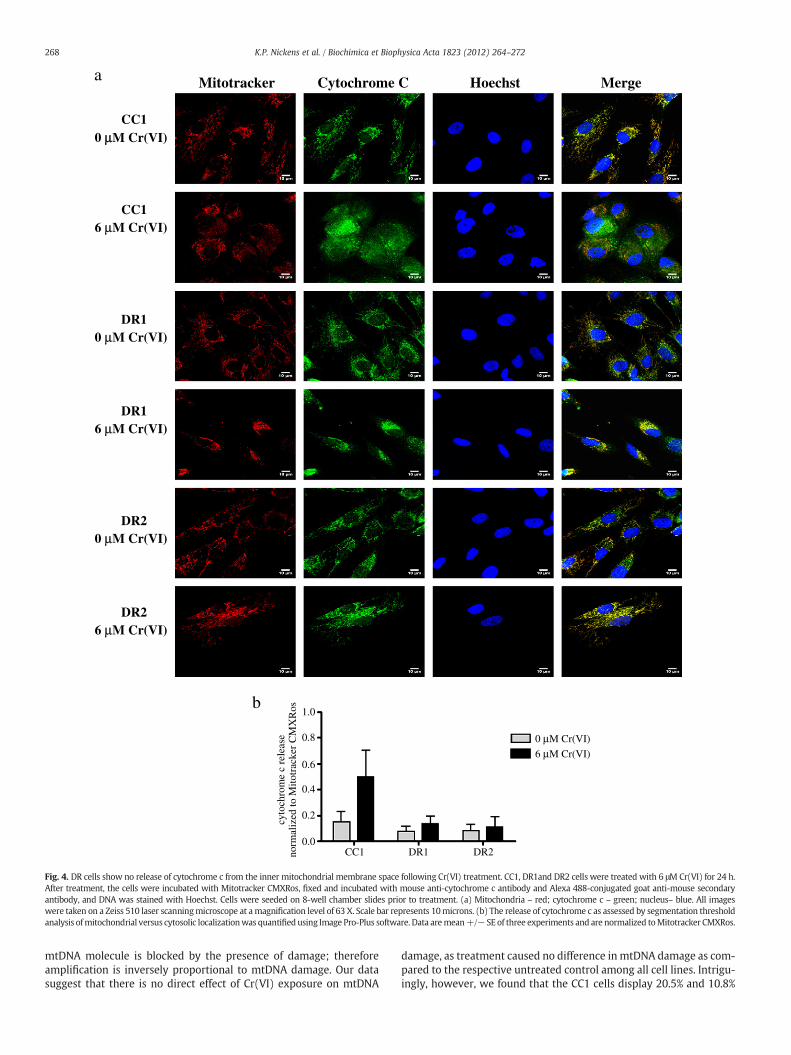
Several studies have shown that in response to certain DNA dam-aging agents, mitochondrial biogenesis can be induced as a survivalmechanism in an effort to repopulate damaged cells with functionalmitochondria [28–32]. mtDNA content correlates with mitochondrialquantity, therefore, we measured mtDNA content before and after24 h exposure to 6 μM Cr(VI), and found no significant differenceamong the cell lines (Fig. 6a).
Loss of mitochondrial function, growth arrest, and apoptosis havebeen correlated with mtDNA damage [33]. Therefore, to further in-vestigate the stability and potential involvement of mtDNA indeath-resistance in our model system, we measured mtDNA damagebefore and after Cr(VI) treatment (Fig. 6b). QPCR amplification of aprimer designed to detect an 8.9-kb product along the circular
Cytochrome C Hoechst Merge
CC10 µµM Cr(VI)
CC16 µM Cr(VI)
DR10 µM Cr(VI)
DR16 µM Cr(VI)
DR20 µM Cr(VI)
DR26 µM Cr(VI)
Mitotracker
1.0
0.8
0.6
0.4
0.2
0.0
0 µM Cr(VI)
6 µM Cr(VI)
CC1 DR1 DR2
cyto
chro
me
c re
leas
eno
rmal
ized
to M
itotr
acke
r C
MX
Ros
a
b
Fig. 4. DR cells show no release of cytochrome c from the inner mitochondrial membrane space following Cr(VI) treatment. CC1, DR1and DR2 cells were treated with 6 μM Cr(VI) for 24 h.After treatment, the cells were incubated with Mitotracker CMXRos, fixed and incubated with mouse anti-cytochrome c antibody and Alexa 488-conjugated goat anti-mouse secondaryantibody, and DNA was stained with Hoechst. Cells were seeded on 8-well chamber slides prior to treatment. (a) Mitochondria – red; cytochrome c – green; nucleus– blue. All imageswere taken on a Zeiss 510 laser scanningmicroscope at amagnification level of 63 X. Scale bar represents 10microns. (b) The release of cytochrome c as assessed by segmentation thresholdanalysis ofmitochondrial versus cytosolic localizationwas quantifiedusing Image Pro-Plus software. Data aremean+/− SE of three experiments and are normalized toMitotracker CMXRos.
268 K.P. Nickens et al. / Biochimica et Biophysica Acta 1823 (2012) 264–272
mtDNA molecule is blocked by the presence of damage; thereforeamplification is inversely proportional to mtDNA damage. Our datasuggest that there is no direct effect of Cr(VI) exposure on mtDNA
damage, as treatment caused no difference in mtDNA damage as com-pared to the respective untreated control among all cell lines. Intrigu-ingly, however, we found that the CC1 cells display 20.5% and 10.8%

Mitotracker Hoechst Merge
CC10 µµM Cr(VI)
CC16 µM Cr(VI)
DR10 µM Cr(VI)
DR16 µM Cr(VI)
DR20 µM Cr(VI)
DR26 µM Cr(VI)
2.0
1.5
1.0
0.5
0.0
SMA
C/D
IAB
LO
rel
ease
norm
aliz
ed to
Mito
trac
ker
CM
XR
os
CC1 DR1 DR2
0 µM Cr(VI)
6 µM Cr(VI)*^
b
a SMAC/DIABLO
Fig. 5. DR cells showno release of SMAC/DIABLO following Cr(VI) treatment. CC1, DR1andDR2 cells were treatedwith 6 μMCr(VI) for 24 h. After treatment, the cells were incubatedwithMitotracker CMXRos, fixed and incubated with mouse anti-SMAC/DIABLO antibody and Alexa 488-conjugated goat anti-mouse secondary antibody, and DNA was stained with Hoechst.Cells were seeded on 8-well chamber slides prior to treatment. (a) Mitochondria – red; SMAC/DIABLO – green; nucleus – blue. All images were taken on a Zeiss 510 laser scanningmicroscope at a magnification level of 63 X. Scale bar represents 10 microns. (b) The release of SMAC/DIABLO as assessed by segmentation threshold analysis of mitochondrial versuscytosolic localization was quantified using Image Pro-Plus software. Data are mean +/− SE of three experiments and are normalized to Mitotracker CMXRos. * indicates statisticallysignificant difference from respective untreated control, ^ indicates statistically significant difference from DR1 cells treated with 6 μM Cr(VI) (pb0.05).
269K.P. Nickens et al. / Biochimica et Biophysica Acta 1823 (2012) 264–272
Fig. 6. DR cells display no change in mtDNA copy number, but intrinsically less mtDNAdamage. CC1, DR1, and DR2 cells were treated with 0 or 6 μM Cr(VI) for 24 h. Total DNAwas extracted and quantified, (a) mtDNA and nuclear (nu) DNA copy number were de-termined by RT-PCR. Primers were generated for the 108-bp mitochondrial-encodedND1 gene and an 89-bp β2 microglobulin (β2m) gene to assess mtDNA and nuDNA, re-spectively. Values are averages of three determinations obtained in one RT-PCR run.The copy number/cell was calculated by dividing nuDNA copy number by two (assum-ing the cells were diploid). mtDNA copy number was expressed as mtDNA copies/cell.(b) mtDNA damage was measured by the amplification of an 8.9-kb mtDNA target byQPCR. mtDNA damage was inversely proportional to amount of amplification , andwas expressed as amount of 8.9-kb DNA/106 ND1 copies amplified. * indicates a statis-tically significant difference from CC1 cells, regardless of Cr(VI) treatment. Data aremean +/− SE of three experiments (0 μM Cr(VI), p=0.0516; 6 μM Cr(VI) p=0.0430).
270 K.P. Nickens et al. / Biochimica et Biophysica Acta 1823 (2012) 264–272
less basal amplification of the 8.9-kb mtDNA product per 1 millioncopies of the ND1 gene, as compared to the DR1 and DR2 cells, respec-tively. These data indicate that the DR1 cells have significantly less in-trinsic/steady-state mtDNA damage as compared to the CC1 cells,while the DR2 cells exhibit a moderate difference in damage as com-pared to the CC1 cells (Fig. 6a).
4. Discussion
The selection of cells with the ability to survive after exposure toapoptogenic levels of a DNA damaging agent may yield a precursorpool of cells fromwhich neoplastic variants can emerge [19]. The dys-regulation of mitochondrial-mediated cellular mechanisms such asapoptosis, are frequently observed in malignant tumors, thushighlighting a potential connection between mitochondria, death-resistance and carcinogenesis [28,30]. We postulate that survivalafter genotoxic insult may involve the selection of cells with mito-chondrial dysregulation, leading to death-resistance. Our data dem-onstrate that the DR cells, as compared to the death-susceptiblecells, are resistant to mitochondrial-mediated apoptosis and displaydifferences in intrinsic mtDNA damage that correlate with death-resistance. The results of the present study provide insight into thepossible role of mitochondrial dysregulation and apoptosis-resistance in the mechanisms of early stage carcinogenesis.
The use of human diploid fibroblasts in this study enables us to in-vestigate the earliest genotoxin-induced events that may occur in
normal cells, yielding a pro-survival phenotype. As part of the intri-cate cellular microenvironmental milieu, fibroblasts are associatedwith pathological conditions such as fibrosis and carcinogenesis, asthey affect the behavior of neighboring cells, and contribute to themaintenance of the extracellular matrix [34,35].
Our model system was derived from survivors of Cr(VI)-treated,immortalized human diploid fibroblasts (BJ-hTERT). BJ-hTERT cellswere exposed to a single dose of Cr(VI), yielding 1% cell survival,from which a population of surviving (B-5Cr) cells was expanded[19]. The B-5Cr cells exhibited enhanced long term survival andgrowth potential (also observed in the subclonal populations exam-ined in the current study; Supplemental Fig. 4), resistance to apopto-sis induction from a variety of stimuli (indicated by phosphtidlyserinetranslocation), and a stable p53 response [19]. Of note, the resistantphenotype of the B-5Cr cells diminished with time in cell culture.This phenomenon was attributed to the effect of cumulative cell divi-sions in culture and extensive expansion. Similar progressive pheno-typic changes have been observed in human lung fibroblasts [36].Therefore, to ensure that the observed resistance to Cr(VI)-inducedcell death in the B-5Cr cells was due to acquired apoptosis-resistance and not a result of clonogenic selection pressure, early pas-sages of the B-5Cr cells, that still exhibited the pro-survival pheno-type, were subcloned to establish the DR1 and DR2 cell lines used inthe present study. Untreated BJ-hTERT (B-0Cr) cells were also usedto derive the clonogenic control cell line, CC1.
It is critical to emphasize that the observed death-resistant pheno-type in our model system is a consequence of a single toxic and gen-otoxic exposure. In vitro models of cellular death-resistancegenerated via long-term, chronic, and slowly escalating exposuredoses are advantageous when studying the role of altered transportand intracellular pharmacodynamics in response to cellular genotoxicstress [37–40]. However, our system is unique in that it models initialmolecular events that occur in a normal cell that survived a single,acute, initiating genotoxic challenge. While the selection model inthis study was generated by Cr(VI) treatment, it also exhibits across-resistance to the well-known chemotherapeutic agent, cisplat-in, as well as to H2O2 (Supplemental Figs. 1 and 3). This is in contrastto published reports where Cr(VI)-resistant cell models were gener-ated and exhibit resistance only to Cr(VI) (reviewed in [41]). This un-derlines the utility of our cell model to assess loss of therapeuticefficacy and toxicity of chemotherapeutic agents in cells exhibitingthe earliest hallmarks of cancer.
Disruption of mitochondrial-mediated apoptosis signaling in ma-lignant tumors has been directly associated with the enhanced ex-pression of Bcl-2 [23]. The requirement of Bcl-2 for resistance toCr(VI)-induced death has been highlighted in studies in SV40-transformed lung epithelial cells (BEAS-2B) by Azad et al. [42]. Ourdata are in keeping with these studies in that a maintenance of Bcl-2 protein expression correlated with Cr(VI)-induced cleaved caspase3-mediated death-resistance as compared to a significant decreasein protein expression in the death-sensitive cells (Fig. 3). Studieshave also shown a relationship between Bcl-2 degradation andCr(VI)-induced ROS production [14,43], however; this correlationwas not observed in our system. Moreover, it is generally acceptedthat ROS are mediators of cellular damage, including mtDNA damageand consequently mitochondrial function [44,45]. Along these lines, arecent study by Indran et al. has demonstrated that the basal and en-dogenous production of ROS in cancer cells can be reduced as a con-sequence of hTERT overexpression and lead to the improvement ofmitochondrial function [45]. As the cells in our system are derivedfrom hTERT-immortalized fibroblasts, and exhibit phenotypic alter-ations suggestive of dysregulated mitochondrial-mediated cell deathsignaling, we measured the levels of hTERT protein expression. How-ever, we found no difference basally or after genotoxin exposureamong all cell lines, suggesting lack of TERT involvement in the ob-served DR phenotype (Supplemental Fig. 5a).
271K.P. Nickens et al. / Biochimica et Biophysica Acta 1823 (2012) 264–272
In an effort to further investigate the possible link betweenmitochondrial-mediated apoptosis resistance, the observed pheno-typic alterations, and a potential redox effect in our model system,we assessed mtDNA damage in the cell lines before and after Cr(VI)treatment. Our data were obtained by long-range QPCR amplificationand analysis of an 8.9-kb mtDNA product that is inversely proportion-al to mtDNA damage (Fig. 6b). Interestingly, we found no direct effectof Cr(VI) on mtDNA damage among all cell lines studied. However, inthe absence of any Cr(VI) treatment, both DR1 and DR2 cells exhib-ited less inherent mtDNA damage as compared to the CC1 cells. Thereduced levels of mtDNA damage observed in the DR cells uncover apotentially striking intrinsic mechanism for resistance. These dataimply that the ability of a population of cells to maintain a more sta-ble mitochondrial genome (or potentially enhanced ROS buffering ca-pacity), may increase their ability to survive in the face of cellularoxidative stress. Therefore, we investigated the possibility that theDR cells may exhibit restricted generation of ROS, which would resultin lower reduced intrinsic mtDNA damage. However, we found nodifference in the basal levels of total cellular ROS, total cellular O2
-,or mitochondria-specific O2
- among all cell lines (data not shown).Similar results were reported in a study by Kulawiec et al., wherebreast cancer cell lines containing mutated mtDNA, as compared towild-type cells containing undamaged mtDNA exhibited resistanceto etopside-induced apoptosis, but demonstrated no associationwith increased ROS production [46]. Furthermore, the authors foundthat mutated mtDNA leads to the constitutive activation of theP13K/Akt pathway [46]. Further data from our laboratory show a sig-nificant induction of phosphorylated-Akt serine-473 protein expres-sion following Cr(VI) genotoxic exposure in the DR1 cells, whichwas downregulated in the BJ-hTERT cells (Supplemental Fig. 6). Ofnote, constitutive activation of Akt1, by myristolated Akt transfection,did not induce death-resistance in the BJ-hTERT cells (data notshown), potentially pointing to a further link between mtDNA dysre-gulation and death resistance in the DR1 cells, which exhibit decre-mental basal levels of mtDNA damage, as compared to the controlcells.
5. Conclusion
Here we focused on mitochondrial involvement in the “black box”of early stage carcinogenesis and cellular death-resistance. Mitochon-dria are heterogeneous in nature, varying in content from cell to cell,and varying in genetic material frommitochondria to mitochondria. Itis known that changes in critical cellular functions (ie. oxidative phos-phorylation) can result from damaged or mutated mtDNA, and it hasbeen hypothesized that these alterations correlate with tumorigene-sis [47,48]. Therefore, our hypothesis is supported by the idea that aselected pool of cells with an innately enhanced survival capacity(ie. lower ROS production or enhanced ROS buffering capacity), maysupport or lead to the generation of more robust mitochondria; andin the face of subsequent oxidative stress, these cells and their dysre-gulated mitochondria, are able to survive.
The present study provides evidence that resistance tomitochondrial-mediated apoptosis, dysregulation of mitochondrialassociated proteins, and the stability of mtDNA, may be among thefirst phenotypic alterations observed in early stage carcinogenesis.We demonstrate that heritable resistance to apoptosis, one of thehallmarks of cancer, can be acquired in normal diploid human cells,following a single toxic and genotoxic exposure [1]. If death-resistant cells also harbor mutations, the inappropriate survival andreplication of such genomically unstable cells may predispose themto neoplastic progression. Investigations into the potential role of mi-tochondria in early stage carcinogenesis are critical to our understand-ing of origins of tumor emergence and may implicate mitochondrialdysregulation as a carcinogenic biomarker.
Supplementary materials related to this article can be found on-line at doi:10.1016/j.bbamcr.2011.10.005.
Role of Funding Source
This work was supported by the National Institutes of Health[RO1CA107972 to S.C., RO1CA107972 supplement to K.N.,R01ES05304 and R01ES09961 to S.P.]. The funding source had no in-volvement in the conduct of this research or the preparation of thisarticle.
Acknowledgments
The authors acknowledge Dr. Popratiloff for his help with the con-focal microscopy studies and analysis, Dr. O'Brien for helpful discus-sions, and Dr. Lee for the generous use of his cell culture facility. Wealso thank Dr. Pritchard, Dr. Beaver and Dr. Lizardo-Escano for theirearlier contributions to this study, as well as Dr. Lal-Nag and Dr.Camilli for their helpful insight.
References
[1] D. Hanahan, R.A. Weinberg, The hallmarks of cancer, Cell 100 (2000) 57–70.[2] I.M. Gavin, B. Gillis, Z. Arbieva, B.S. Prabhakar, Identification of human cell re-
sponses to hexavalent chromium, Environ. Mol. Mutagen. 48 (2007) 650–657.[3] Chromium, nickel and welding, IARC Monogr Eval Carcinog Risks Hum 49 (1990)
1–648.[4] NTP 11th Report on Carcinogens, Rep Carcinog, , 2005, pp. 1–A32.[5] K.W. Jennette, Microsomal reduction of the carcinogen chromate produces chro-
mium(V), J. Am. Chem. Soc. 104 (1982) 874–875.[6] L.J. Blankenship, D.L. Carlisle, J.P. Wise, J.M. Orenstein, L.E. Dye III, S.R. Patierno, In-
duction of apoptotic cell death by particulate lead chromate: differential effects ofvitamins C and E on genotoxicity and survival, Toxicol. Appl. Pharmacol. 146(1997) 270–280.
[7] L.J. Blankenship, F.C. Manning, J.M. Orenstein, S.R. Patierno, Apoptosis is the modeof cell death caused by carcinogenic chromium, Toxicol. Appl. Pharmacol. 126(1994) 75–83.
[8] L. Ha, S. Ceryak, S.R. Patierno, Chromium (VI) activates ataxia telangiectasia mu-tated (ATM) protein. Requirement of ATM for both apoptosis and recovery fromterminal growth arrest, J. Biol. Chem. 278 (2003) 17885–17894.
[9] Y. Shi, A structural view of mitochondria-mediated apoptosis, Nat. Struct. Biol.8 (2001) 394–401.
[10] R.C. Taylor, S.P. Cullen, S.J. Martin, Apoptosis: controlled demolition at the cellularlevel, Nat. Rev. Mol. Cell Biol. 9 (2008) 231–241.
[11] P. Gimenez-Bonafe, A. Tortosa, R. Perez-Tomas, Overcoming drug resistance byenhancing apoptosis of tumor cells, Curr. Cancer Drug Targets 9 (2009) 320–340.
[12] M.G. Vander Heiden, N.S. Chandel, E.K.Williamson, P.T. Schumacker, C.B. Thompson,Bcl-xL regulates the membrane potential and volume homeostasis of mitochondria,Cell 91 (1997) 627–637.
[13] A. Srinivasan, L.M. Foster, M.P. Testa, T. Ord, R.W. Keane, D.E. Bredesen, C. Kayalar,Bcl-2 expression in neural cells blocks activation of ICE/CED-3 family proteasesduring apoptosis, J. Neurosci. 16 (1996) 5654–5660.
[14] N. Azad, A.K. Iyer, A. Manosroi, L. Wang, Y. Rojanasakul, Superoxide-mediatedproteasomal degradation of Bcl-2 determines cell susceptibility to Cr(VI)-inducedapoptosis, Carcinogenesis 29 (2008) 1538–1545.
[15] D.M. Hockenbery, Z.N. Oltvai, X.M. Yin, C.L. Milliman, S.J. Korsmeyer, Bcl-2 func-tions in an antioxidant pathway to prevent apoptosis, Cell 75 (1993) 241–251.
[16] V.A. Bohr, T. Stevnsner, N.C. de Souza-Pinto, Mitochondrial DNA repair of oxida-tive damage in mammalian cells, Gene 286 (2002) 127–134.
[17] S.P. LeDoux, G.L. Wilson, E.J. Beecham, T. Stevnsner, K. Wassermann, V.A. Bohr,Repair of mitochondrial DNA after various types of DNA damage in Chinese ham-ster ovary cells, Carcinogenesis 13 (1992) 1967–1973.
[18] B. Van Houten, V. Woshner, J.H. Santos, Role of mitochondrial DNA in toxic re-sponses to oxidative stress, DNA Repair (Amst) 5 (2006) 145–152.
[19] D.E. Pritchard, S. Ceryak, K.E. Ramsey, T.J. O'Brien, L. Ha, J.L. Fornsaglio, D.A. Stephan,S.R. Patierno, Resistance to apoptosis, increased growth potential, and altered geneexpression in cells that survived genotoxic hexavalent chromium [Cr(VI)] exposure,Mol. Cell. Biochem. 279 (2005) 169–181.
[20] M.A. Lal, D. Bae, T.C. Camilli, S.R. Patierno, S. Ceryak, AKT1 mediates bypass of theG1/S checkpoint after genotoxic stress in normal human cells, Cell Cycle 8 (2009)1589–1602.
[21] J.H. Santos, J.N. Meyer, B.S. Mandavilli, B. Van Houten, Quantitative PCR-basedmeasurement of nuclear and mitochondrial DNA damage and repair in mammaliancells, Methods Mol. Biol. 314 (2006) 183–199.
[22] G. Chun, D. Bae, K. Nickens, T.J. O'Brien, S.R. Patierno, S. Ceryak, Polo-like kinase 1enhances survival and mutagenesis after genotoxic stress in normal cells throughcell cycle checkpoint bypass, Carcinogenesis 31 (2010) 785–793.
[23] A. Hasenjager, B. Gillissen, A. Muller, G. Normand, P.G. Hemmati, M. Schuler, B.Dorken, P.T. Daniel, Smac induces cytochrome c release and apoptosis

272 K.P. Nickens et al. / Biochimica et Biophysica Acta 1823 (2012) 264–272
independently from Bax/Bcl-x(L) in a strictly caspase-3-dependent manner inhuman carcinoma cells, Oncogene 23 (2004) 4523–4535.
[24] D. Mahalingam, A. Natoni, M. Keane, A. Samali, E. Szegezdi, Early growthresponse-1 is a regulator of DR5-induced apoptosis in colon cancer cells, Br. J.Cancer 102 (2010) 754–764.
[25] C. Kantari, H. Walczak, Caspase-8 and bid: caught in the act between death recep-tors and mitochondria, Biochim. Biophys. Acta 1813 (2011) 558–563.
[26] A. Nesterov, M. Nikrad, T. Johnson, A.S. Kraft, Oncogenic Ras sensitizes normalhuman cells to tumor necrosis factor-alpha-related apoptosis-inducing ligand-induced apoptosis, Cancer Res. 64 (2004) 3922–3927.
[27] T. Tanaka, M. Yoshimi, T. Maeyama, N. Hagimoto, K. Kuwano, N. Hara, Resistanceto Fas-mediated apoptosis in human lung fibroblast, Eur. Respir. J. 20 (2002)359–368.
[28] X. Fu, S. Wan, Y.L. Lyu, L.F. Liu, H. Qi, Etoposide induces ATM-dependent mito-chondrial biogenesis through AMPK activation, PLoS One 3 (2008) e2009.
[29] E. Nisoli, E. Clementi, S. Moncada, M.O. Carruba, Mitochondrial biogenesis as acellular signaling framework, Biochem. Pharmacol. 67 (2004) 1–15.
[30] E. Nisoli, E. Clementi, C. Paolucci, V. Cozzi, C. Tonello, C. Sciorati, R. Bracale, A.Valerio, M. Francolini, S. Moncada, M.O. Carruba, Mitochondrial biogenesis inmammals: the role of endogenous nitric oxide, Science 299 (2003) 896–899.
[31] E. Nisoli, S. Falcone, C. Tonello, V. Cozzi, L. Palomba, M. Fiorani, A. Pisconti, S. Brunelli,A. Cardile, M. Francolini, O. Cantoni, M.O. Carruba, S. Moncada, E. Clementi, Mito-chondrial biogenesis by NO yields functionally active mitochondria in mammals,Proc. Natl. Acad. Sci. U. S. A. 101 (2004) 16507–16512.
[32] J. Xu, C. Shi, Q. Li, J. Wu, E.L. Forster, D.T. Yew, Mitochondrial dysfunction in plate-lets and hippocampi of senescence-accelerated mice, J. Bioenerg. Biomembr. 39(2007) 195–202.
[33] F.M. Yakes, B. Van Houten, Mitochondrial DNA damage is more extensive andpersists longer than nuclear DNA damage in human cells following oxidativestress, Proc. Natl. Acad. Sci. U. S. A. 94 (1997) 514–519.
[34] R.J. McAnulty, Fibroblasts and myofibroblasts: their source, function and role indisease, Int. J. Biochem. Cell Biol. 39 (2007) 666–671.
[35] A. Vaheri, A. Enzerink, K. Rasanen, P. Salmenpera, Nemosis, a novel way offibroblast activation, in inflammation and cancer, Exp. Cell Res. 315 (2009)1633–1638.
[36] D.E. Pritchard, S. Ceryak, L. Ha, J.L. Fornsaglio, S.K. Hartman, T.J. O'Brien, S.R.Patierno, Mechanism of apoptosis and determination of cellular fate in
chromium(VI)-exposed populations of telomerase-immortalized human fibro-blasts, Cell Growth Differ. 12 (2001) 487–496.
[37] V.I. Grishko, L.I. Rachek, D.R. Spitz, G.L. Wilson, S.P. LeDoux, Contribution of mito-chondrial DNA repair to cell resistance from oxidative stress, J. Biol. Chem. 280(2005) 8901–8905.
[38] Y.Y. Lu, J.L. Yang, Long-term exposure to chromium(VI) oxide leads to defects insulfate transport system in Chinese hamster ovary cells, J. Cell. Biochem. 57(1995) 655–665.
[39] C.F. Rodrigues, A.M. Urbano, E. Matoso, I. Carreira, A. Almeida, P. Santos, F.Botelho, L. Carvalho, M. Alves, C. Monteiro, A.N. Costa, V. Moreno, M.C. Alpoim,Human bronchial epithelial cells malignantly transformed by hexavalent chromi-um exhibit an aneuploid phenotype but no microsatellite instability, Mutat. Res.670 (2009) 42–52.
[40] K.H. Son, M. Zhang, E. Rucobo, D. Nwaigwe, F. Montgomery, H. Leffert, Derivationand study of human epithelial cell lines resistant to killing by chromium trioxide,J. Toxicol. Environ. Health A 67 (2004) 1027–1049.
[41] K.P. Nickens, S.R. Patierno, S. Ceryak, Chromium genotoxicity: A double-edgedsword, Chem. Biol. Interact. 188 (2010) 276–288.
[42] N. Azad, A.K. Iyer, L. Wang, Y. Lu, D. Medan, V. Castranova, Y. Rojanasakul, Nitricoxide-mediated bcl-2 stabilization potentiates malignant transformation ofhuman lung epithelial cells, Am. J. Respir. Cell Mol. Biol. 42 (2010) 578–585.
[43] Y.O. Son, J.A. Hitron, X. Wang, Q. Chang, J. Pan, Z. Zhang, J. Liu, S. Wang, J.C. Lee, X.Shi, Cr(VI) induces mitochondrial-mediated and caspase-dependent apoptosisthrough reactive oxygen species-mediated p53 activation in JB6 Cl41 cells, Toxicol.Appl. Pharmacol. 245 (2010) 226–235.
[44] Z.X. Chen, R. Velaithan, S. Pervaiz, mitoEnergetics and cancer cell fate, Biochim.Biophys. Acta 1787 (2009) 462–467.
[45] I.R. Indran, M.P. Hande, S. Pervaiz, hTERT overexpression alleviates intracellularROS production, improves mitochondrial function, and inhibits ROS-mediatedapoptosis in cancer cells, Cancer Res. 71 (2011) 266–276.
[46] M. Kulawiec, K.M. Owens, K.K. Singh, Cancer cell mitochondria confer apoptosisresistance and promote metastasis, Cancer Biol. Ther. 8 (2009) 1378–1385.
[47] H. Fang, L. Shen, T. Chen, J. He, Z. Ding, J. Wei, J. Qu, G. Chen, J. Lu, Y. Bai, Cancertype-specific modulation of mitochondrial haplogroups in breast, colorectal andthyroid cancer, BMC Cancer 10 (2010) 421.
[48] O. Warburg, On respiratory impairment in cancer cells, Science 124 (1956)269–270.