Acoustic Events and “Optophonic” Cochlear Responses Induced by Pulsed Near-Infrared LASER
21
Acoustic Events and “Optophonic” Cochlear Responses Induced by Pulsed Near-Infrared LASER Ingo Ulrik Teudt, Institute of Audioneurotechnology and the Department of Experimental Otology ENT-Clinics, Hannover School of Medicine, 30625 Hannover, Germany ([email protected]) Hannes Maier, Institute of Audioneurotechnology and the Department of Experimental Otology ENT-Clinics, Hannover School of Medicine, 30625 Hannover, Germany ([email protected]) Claus-Peter Richter, and Department of Otolaryngology, Feinberg School of Medicine, Northwestern University, Chicago, IL 60611 USA ([email protected]) Andrej Kral Institute of Audioneurotechnology and the Department of Experimental Otology ENT-Clinics, Hannover School of Medicine, 30625 Hannover, Germany ([email protected]) Abstract Optical stimulation of neural tissue within the cochlea was described as a possible alternative to electrical stimulation. Most optical stimulation was performed with pulsed lasers operating with near-infrared (NIR) light and in thermal confinement. Under these conditions, the coexistence of laser-induced optoacoustic stimulation of the cochlea (“optophony”) has not been analyzed yet. This study demonstrates that pulsed 1850-nm laser light used for neural stimulation also results in sound pressure levels up to 62 dB peak-to-peak equivalent sound pressure level (SPL) in air. The sound field was confined to a small volume along the laser beam. In dry nitrogen, laser-induced acoustic events disappeared. Hydrophone measurements demonstrated pressure waves for laser fibers immersed in water. In hearing rats, laser-evoked signals were recorded from the cochlea without targeting neural tissue. The signals showed a two-domain response differing in amplitude and latency functions, as well as sensitivity to white-noise masking. The first component had characteristics of a cochlear microphonic potential, and the second component was characteristic for a compound action potential. The present data demonstrate that laser-evoked acoustic events can stimulate a hearing cochlea. Whenever optical stimulation is used, care must be taken to distinguish between such “optophony” and the true optoneural response. Index Terms Auditory system; hearing aids; infrared-radiation effects; optical-radiation effects; photoacoustic effects © 2011 IEEE Correspondence to: Ingo Ulrik Teudt. Color versions of one or more of the figures in this paper are available online at http://ieeexplore.ieee.org. NIH Public Access Author Manuscript IEEE Trans Biomed Eng. Author manuscript; available in PMC 2012 September 24. Published in final edited form as: IEEE Trans Biomed Eng. 2011 June ; 58(6): 1648–1655. doi:10.1109/TBME.2011.2108297. NIH-PA Author Manuscript NIH-PA Author Manuscript NIH-PA Author Manuscript
-
Upload
independent -
Category
Documents
-
view
3 -
download
0
Transcript of Acoustic Events and “Optophonic” Cochlear Responses Induced by Pulsed Near-Infrared LASER
Acoustic Events and “Optophonic” Cochlear ResponsesInduced by Pulsed Near-Infrared LASER
Ingo Ulrik Teudt,Institute of Audioneurotechnology and the Department of Experimental Otology ENT-Clinics,Hannover School of Medicine, 30625 Hannover, Germany ([email protected])
Hannes Maier,Institute of Audioneurotechnology and the Department of Experimental Otology ENT-Clinics,Hannover School of Medicine, 30625 Hannover, Germany ([email protected])
Claus-Peter Richter, andDepartment of Otolaryngology, Feinberg School of Medicine, Northwestern University, Chicago,IL 60611 USA ([email protected])
Andrej KralInstitute of Audioneurotechnology and the Department of Experimental Otology ENT-Clinics,Hannover School of Medicine, 30625 Hannover, Germany ([email protected])
AbstractOptical stimulation of neural tissue within the cochlea was described as a possible alternative toelectrical stimulation. Most optical stimulation was performed with pulsed lasers operating withnear-infrared (NIR) light and in thermal confinement. Under these conditions, the coexistence oflaser-induced optoacoustic stimulation of the cochlea (“optophony”) has not been analyzed yet.This study demonstrates that pulsed 1850-nm laser light used for neural stimulation also results insound pressure levels up to 62 dB peak-to-peak equivalent sound pressure level (SPL) in air. Thesound field was confined to a small volume along the laser beam. In dry nitrogen, laser-inducedacoustic events disappeared. Hydrophone measurements demonstrated pressure waves for laserfibers immersed in water. In hearing rats, laser-evoked signals were recorded from the cochleawithout targeting neural tissue. The signals showed a two-domain response differing in amplitudeand latency functions, as well as sensitivity to white-noise masking. The first component hadcharacteristics of a cochlear microphonic potential, and the second component was characteristicfor a compound action potential. The present data demonstrate that laser-evoked acoustic eventscan stimulate a hearing cochlea. Whenever optical stimulation is used, care must be taken todistinguish between such “optophony” and the true optoneural response.
Index TermsAuditory system; hearing aids; infrared-radiation effects; optical-radiation effects; photoacousticeffects
© 2011 IEEE
Correspondence to: Ingo Ulrik Teudt.
Color versions of one or more of the figures in this paper are available online at http://ieeexplore.ieee.org.
NIH Public AccessAuthor ManuscriptIEEE Trans Biomed Eng. Author manuscript; available in PMC 2012 September 24.
Published in final edited form as:IEEE Trans Biomed Eng. 2011 June ; 58(6): 1648–1655. doi:10.1109/TBME.2011.2108297.
NIH
-PA Author Manuscript
NIH
-PA Author Manuscript
NIH
-PA Author Manuscript
I. IntroductionNEURAL stimulation with near-infrared (NIR) optical radiation has been suggested as analternative to neural stimulation with electrical current. Such optical stimulation is possiblein peripheral nerves [1]–[6] and in the cochlea [7]–[10]. The potential advantages of opticalstimulation include the absence of physical contact between neural tissues and thestimulation source and the high spatial selectivity of optical stimulation compared toelectrical stimulation. However, the method also has its limitations. Tissue between theradiation source and the neurons can be a confounding factor for stimulation. The tissue mayscatter or absorb the radiation. Both will result in a need for increase of the radiationexposure to stimulate the target tissue. Scatter of the radiation will spread the stimulation,while absorption will result in heating and subsequent damage of the overlying structures.
The laser parameters recently used for optical stimulation include radiation wavelengthsbetween 1850 and 2120 nm, pulse lengths between 5 µs and 10 ms, and radiant exposures upto 2 J/cm2. The laser–tissue interaction thus takes place in so-called “thermal confinement.”In thermal confinement, even at low energies used to stimulate the neurons, the fast heatingof the small volume in front of the optical fiber results in expansion of this volume and in astress-relaxation wave. For neural stimulation in the cochlea, it is important to determinewhether this relaxation wave is audible and constitutes a confounding factor for neural-stimulation techniques. Although it has been well established that audible acoustic eventsappear at high stimulus energies [11], [12] or in the condition of stress confinement [13],acoustic events in the thermal confinement at low energies have not been investigated invivo and their spatiotemporal characteristics have not been provided yet. For the potentialuse of neuronal optical stimulation in the cochlea, the awareness of generated pressurewaves is highly important. Laser-induced pressure waves might interfere with auditory-nerve stimulation by stimulating remaining hair cells. On the other hand, laser-inducedpressure waves could also serve as a new mechanism for sound stimulation in the hearing-impaired cochlea [14].
II. Material and MethodsA. Laser Stimulation
Optical stimulation was performed with a diode laser (Capella, Model R-1850, Aculight)with 1850-nm wavelength and variable pulse energies. The laser was operated at 33 or 100Hz repetition rate. The radiation was coupled to an optical fiber (5 m length, NA 0.22 ±0.02) with 200 µm core diameter (Ocean Optics, VIS/NIR, FL). The overall stability of thelaser output was maintained by internal closed feedback loops that regulated the diodetemperature (and thus, the emission wavelength) and output energy. Different outputenergies per pulse, 1 mm from tip of the optical fibers, were determined in air with anenergy meter (Coherent J50LP-1A with 3Σ Coherent, FL). For comparability with previousreports all energies were related to the 200 µm core diameter of the used glass fiber.Dependent on the selected laser-setting radiant exposure ranged from 0.004 to 0.35 J/cm2.
During the experiments, the optical fiber was mounted on a three-axis micromanipulator(MM33, Narishige, Japan). The optical fiber was oriented onto the capsule of the middlecochlear turn (1 mm distance), next to the cochlea toward the inner wall of the bulla andperpendicular to the bulla opening (in air). Timing of the laser was controlled by customwritten software (Audiology Laboratory, Otoconsult, Germany) from an external PC using aNational Instruments 6259 M-Series MIO card.
Teudt et al. Page 2
IEEE Trans Biomed Eng. Author manuscript; available in PMC 2012 September 24.
NIH
-PA Author Manuscript
NIH
-PA Author Manuscript
NIH
-PA Author Manuscript
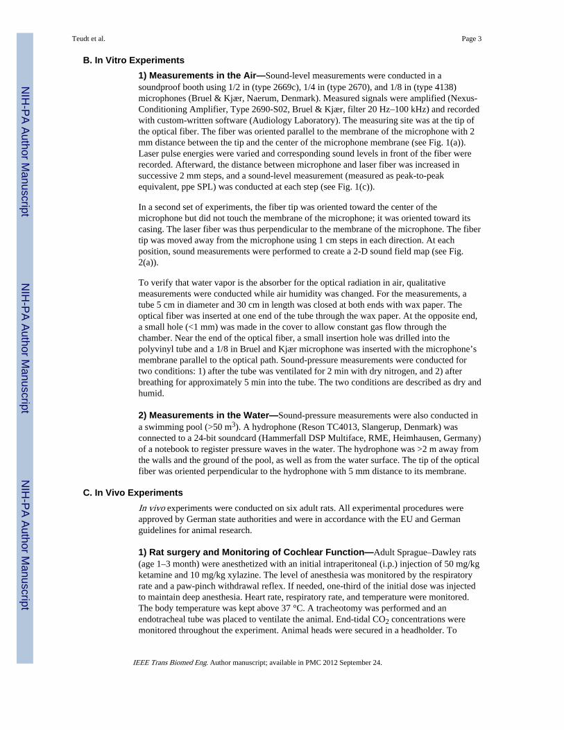
B. In Vitro Experiments1) Measurements in the Air—Sound-level measurements were conducted in asoundproof booth using 1/2 in (type 2669c), 1/4 in (type 2670), and 1/8 in (type 4138)microphones (Bruel & Kjær, Naerum, Denmark). Measured signals were amplified (Nexus-Conditioning Amplifier, Type 2690-S02, Bruel & Kjær, filter 20 Hz–100 kHz) and recordedwith custom-written software (Audiology Laboratory). The measuring site was at the tip ofthe optical fiber. The fiber was oriented parallel to the membrane of the microphone with 2mm distance between the tip and the center of the microphone membrane (see Fig. 1(a)).Laser pulse energies were varied and corresponding sound levels in front of the fiber wererecorded. Afterward, the distance between microphone and laser fiber was increased insuccessive 2 mm steps, and a sound-level measurement (measured as peak-to-peakequivalent, ppe SPL) was conducted at each step (see Fig. 1(c)).
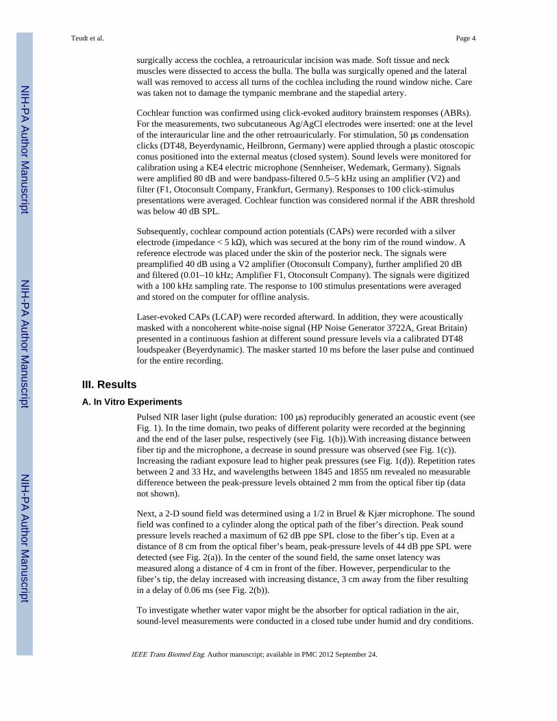
In a second set of experiments, the fiber tip was oriented toward the center of themicrophone but did not touch the membrane of the microphone; it was oriented toward itscasing. The laser fiber was thus perpendicular to the membrane of the microphone. The fibertip was moved away from the microphone using 1 cm steps in each direction. At eachposition, sound measurements were performed to create a 2-D sound field map (see Fig.2(a)).
To verify that water vapor is the absorber for the optical radiation in air, qualitativemeasurements were conducted while air humidity was changed. For the measurements, atube 5 cm in diameter and 30 cm in length was closed at both ends with wax paper. Theoptical fiber was inserted at one end of the tube through the wax paper. At the opposite end,a small hole (<1 mm) was made in the cover to allow constant gas flow through thechamber. Near the end of the optical fiber, a small insertion hole was drilled into thepolyvinyl tube and a 1/8 in Bruel and Kjær microphone was inserted with the microphone’smembrane parallel to the optical path. Sound-pressure measurements were conducted fortwo conditions: 1) after the tube was ventilated for 2 min with dry nitrogen, and 2) afterbreathing for approximately 5 min into the tube. The two conditions are described as dry andhumid.
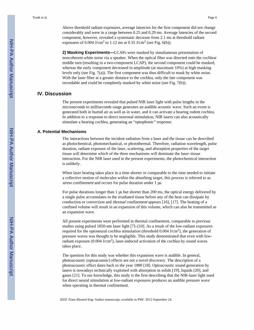
2) Measurements in the Water—Sound-pressure measurements were also conducted ina swimming pool (>50 m3). A hydrophone (Reson TC4013, Slangerup, Denmark) wasconnected to a 24-bit soundcard (Hammerfall DSP Multiface, RME, Heimhausen, Germany)of a notebook to register pressure waves in the water. The hydrophone was >2 m away fromthe walls and the ground of the pool, as well as from the water surface. The tip of the opticalfiber was oriented perpendicular to the hydrophone with 5 mm distance to its membrane.
C. In Vivo ExperimentsIn vivo experiments were conducted on six adult rats. All experimental procedures wereapproved by German state authorities and were in accordance with the EU and Germanguidelines for animal research.
1) Rat surgery and Monitoring of Cochlear Function—Adult Sprague–Dawley rats(age 1–3 month) were anesthetized with an initial intraperitoneal (i.p.) injection of 50 mg/kgketamine and 10 mg/kg xylazine. The level of anesthesia was monitored by the respiratoryrate and a paw-pinch withdrawal reflex. If needed, one-third of the initial dose was injectedto maintain deep anesthesia. Heart rate, respiratory rate, and temperature were monitored.The body temperature was kept above 37 °C. A tracheotomy was performed and anendotracheal tube was placed to ventilate the animal. End-tidal CO2 concentrations weremonitored throughout the experiment. Animal heads were secured in a headholder. To
Teudt et al. Page 3
IEEE Trans Biomed Eng. Author manuscript; available in PMC 2012 September 24.
NIH
-PA Author Manuscript
NIH
-PA Author Manuscript
NIH
-PA Author Manuscript
surgically access the cochlea, a retroauricular incision was made. Soft tissue and neckmuscles were dissected to access the bulla. The bulla was surgically opened and the lateralwall was removed to access all turns of the cochlea including the round window niche. Carewas taken not to damage the tympanic membrane and the stapedial artery.
Cochlear function was confirmed using click-evoked auditory brainstem responses (ABRs).For the measurements, two subcutaneous Ag/AgCl electrodes were inserted: one at the levelof the interauricular line and the other retroauricularly. For stimulation, 50 µs condensationclicks (DT48, Beyerdynamic, Heilbronn, Germany) were applied through a plastic otoscopicconus positioned into the external meatus (closed system). Sound levels were monitored forcalibration using a KE4 electric microphone (Sennheiser, Wedemark, Germany). Signalswere amplified 80 dB and were bandpass-filtered 0.5–5 kHz using an amplifier (V2) andfilter (F1, Otoconsult Company, Frankfurt, Germany). Responses to 100 click-stimuluspresentations were averaged. Cochlear function was considered normal if the ABR thresholdwas below 40 dB SPL.
Subsequently, cochlear compound action potentials (CAPs) were recorded with a silverelectrode (impedance < 5 kΩ), which was secured at the bony rim of the round window. Areference electrode was placed under the skin of the posterior neck. The signals werepreamplified 40 dB using a V2 amplifier (Otoconsult Company), further amplified 20 dBand filtered (0.01–10 kHz; Amplifier F1, Otoconsult Company). The signals were digitizedwith a 100 kHz sampling rate. The response to 100 stimulus presentations were averagedand stored on the computer for offline analysis.
Laser-evoked CAPs (LCAP) were recorded afterward. In addition, they were acousticallymasked with a noncoherent white-noise signal (HP Noise Generator 3722A, Great Britain)presented in a continuous fashion at different sound pressure levels via a calibrated DT48loudspeaker (Beyerdynamic). The masker started 10 ms before the laser pulse and continuedfor the entire recording.
III. ResultsA. In Vitro Experiments
Pulsed NIR laser light (pulse duration: 100 µs) reproducibly generated an acoustic event (seeFig. 1). In the time domain, two peaks of different polarity were recorded at the beginningand the end of the laser pulse, respectively (see Fig. 1(b)).With increasing distance betweenfiber tip and the microphone, a decrease in sound pressure was observed (see Fig. 1(c)).Increasing the radiant exposure lead to higher peak pressures (see Fig. 1(d)). Repetition ratesbetween 2 and 33 Hz, and wavelengths between 1845 and 1855 nm revealed no measurabledifference between the peak-pressure levels obtained 2 mm from the optical fiber tip (datanot shown).
Next, a 2-D sound field was determined using a 1/2 in Bruel & Kjær microphone. The soundfield was confined to a cylinder along the optical path of the fiber’s direction. Peak soundpressure levels reached a maximum of 62 dB ppe SPL close to the fiber’s tip. Even at adistance of 8 cm from the optical fiber’s beam, peak-pressure levels of 44 dB ppe SPL weredetected (see Fig. 2(a)). In the center of the sound field, the same onset latency wasmeasured along a distance of 4 cm in front of the fiber. However, perpendicular to thefiber’s tip, the delay increased with increasing distance, 3 cm away from the fiber resultingin a delay of 0.06 ms (see Fig. 2(b)).
To investigate whether water vapor might be the absorber for optical radiation in the air,sound-level measurements were conducted in a closed tube under humid and dry conditions.
Teudt et al. Page 4
IEEE Trans Biomed Eng. Author manuscript; available in PMC 2012 September 24.
NIH
-PA Author Manuscript
NIH
-PA Author Manuscript
NIH
-PA Author Manuscript
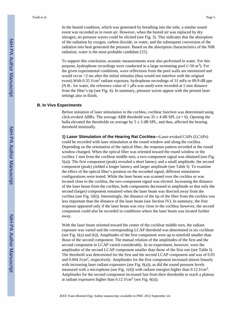
In the humid condition, which was generated by breathing into the tube, a similar soundevent was recorded as in room air. However, when the humid air was replaced by drynitrogen, no pressure waves could be elicited (see Fig. 3). This indicates that the absorptionof the radiation by oxygen, carbon dioxide, or water, and the subsequent conversion of theradiation into heat generated the pressure. Based on the absorption characteristics of the NIRradiation, water is the most probable candidate [15].
To support this conclusion, acoustic measurements were also performed in water. For thispurpose, hydrophone recordings were conducted in a large swimming pool (>50 m3). Forthe given experimental conditions, wave reflections from the pool walls are minimized andwould occur >2 ms after the initial stimulus (thus would not interfere with the originalevent).With 0.35 J/cm2 radiant exposure, hydrophone recordings of 31 mPa or 89.9 dB ppe(N.B.: for water, the reference value of 1 µPa was used) were recorded at 5 mm distancefrom the fiber’s tip (see Fig. 4). In summary, pressure waves appear with the present lasersettings also in fluids.
B. In Vivo ExperimentsBefore initiation of laser stimulation in the cochlea, cochlear function was determined usingclick-evoked ABRs. The average ABR threshold was 35 ± 4 dB SPL (n = 6). Opening thebulla elevated the thresholds on average by 5 ± 3 dB SPL, and thus, affected the hearingthreshold minimally.
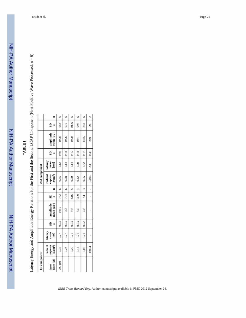
1) Laser Stimulation of the Hearing Rat Cochlea—Laser-evoked CAPs (LCAPs)could be recorded with laser stimulation at the round window and along the cochlea.Depending on the orientation of the optical fiber, the response pattern recorded at the roundwindow changed. When the optical fiber was oriented toward the round window or thecochlea 1 mm from the cochlear middle turn, a two-component signal was obtained (see Fig.5(a)). The first component (peak) revealed a short latency and a small amplitude; the secondcomponent (peak) yielded a longer latency and larger amplitude (see Table I). To examinethe effect of the optical fiber’s position on the recorded signal, different stimulationconfigurations were tested. While the laser beam was scanned over the cochlea or waslocated close to the cochlea, the two-component signal was elicited. Increasing the distanceof the laser beam from the cochlea, both components decreased in amplitude so that only thesecond (larger) component remained when the laser beam was directed away from thecochlea (see Fig. 5(b)). Interestingly, the distance of the tip of the fiber from the cochlea wasless important than the distance of the laser beam (see Section IV). In summary, the firstresponse appeared only if the laser beam was very close to the cochlea; however, the secondcomponent could also be recorded in conditions where the laser beam was located furtheraway.
With the laser beam oriented toward the center of the cochlear middle turn, the radiantexposure was varied and the corresponding LCAP threshold was determined in six cochleae(see Fig. 6(a) and (b)). Amplitudes of the first component were up to ninefold smaller thanthose of the second component. The mutual relation of the amplitudes of the first and thesecond component in LCAP varied considerably. In no experiment, however, were theamplitudes of the second LCAP component smaller than those of the first one (see Table I).The threshold was determined for the first and the second LCAP component and was of 0.05and 0.004 J/cm2, respectively. Amplitudes for the first component increased almost linearlywith increasing laser radiant exposures (see Fig. 6(a)), as did the sound pressure levelsmeasured with a microphone (see Fig. 1(d)) with radiant energies higher than 0.12 J/cm2.Amplitudes for the second component increased fast from their thresholds to reach a plateauat radiant exposures higher than 0.12 J/cm2 (see Fig. 6(a)).
Teudt et al. Page 5
IEEE Trans Biomed Eng. Author manuscript; available in PMC 2012 September 24.
NIH
-PA Author Manuscript
NIH
-PA Author Manuscript
NIH
-PA Author Manuscript
Above threshold radiant exposures, average latencies for the first component did not changeconsiderably and were in a range between 0.25 and 0.29 ms. Average latencies of the secondcomponent, however, revealed a systematic decrease from 2.1 ms at threshold radiantexposures of 0.004 J/cm2 to 1.12 ms at 0.35 J/cm2 (see Fig. 6(b)).
2) Masking Experiments—LCAPs were masked by simultaneous presentation ofnoncoherent white noise via a speaker. When the optical fiber was directed onto the cochlearmiddle turn (resulting in a two-component LCAP), the second component could be masked,whereas the early component decreased in amplitude (at maximum 10%) at high maskinglevels only (see Fig. 7(a)). The first component was thus difficult to mask by white noise.With the laser fiber at a greater distance to the cochlea, only the late component wasrecordable and could be completely masked by white noise (see Fig. 7(b)).
IV. DiscussionThe present experiments revealed that pulsed NIR laser light with pulse lengths in themicroseconds to milliseconds range generates an audible acoustic wave. Such an event isgenerated both in humid air as well as in water, and it can activate a hearing rodent cochlea.In addition to a response to direct neuronal stimulation, NIR lasers can also acousticallystimulate a hearing cochlea, generating an “optophonic” response.
A. Potential MechanismsThe interactions between the incident radiation from a laser and the tissue can be describedas photochemical, photomechanical, or photothermal. Therefore, radiation wavelength, pulseduration, radiant exposure of the laser, scattering, and absorption properties of the targettissue will determine which of the three mechanisms will dominate the laser–tissueinteraction. For the NIR laser used in the present experiments, the photochemical interactionis unlikely.
When laser heating takes place in a time shorter or comparable to the time needed to initiatea collective motion of molecules within the absorbing target, this process is referred to asstress confinement and occurs for pulse duration under 1 µs.
For pulse durations longer than 1 µs but shorter than 200 ms, the optical energy delivered bya single pulse accumulates in the irradiated tissue before any of the heat can dissipate byconduction or convection and thermal confinement appears [16], [17]. The heating of aconfined volume will result in an expansion of this volume, which can also be transmitted asan expansion wave.
All present experiments were performed in thermal confinement, comparable to previousstudies using pulsed 1850-nm laser light [7]–[10]. As a result of the low-radiant exposuresrequired for the optoneural cochlea stimulation (threshold 0.004 J/cm2), the generation ofpressure waves was thought to be negligible. This study demonstrated that even with low-radiant exposure (0.004 J/cm2), laser-induced activation of the cochlea by sound wavestakes place.
The question for this study was whether this expansion wave is audible. In general,photoacoustic (optoacoustic) effects are not a novel discovery. The description of aphotoacoustic effect dates back to the year 1880 [18]. Optoacoustic sound generation bylasers is nowadays technically exploited with absorption in solids [19], liquids [20], andgases [21]. To our knowledge, this study is the first describing that the NIR-laser light usedfor direct neural stimulation at low-radiant exposures produces an audible pressure wavewhen operating in thermal confinement.
Teudt et al. Page 6
IEEE Trans Biomed Eng. Author manuscript; available in PMC 2012 September 24.
NIH
-PA Author Manuscript
NIH
-PA Author Manuscript
NIH
-PA Author Manuscript
B. In Vitro Experiments—Pressure Waves in Air and WaterTo test the pulsed 1850-nm laser for recordable sound phenomena, microphone recordingswere performed in room air and water. In water, the absorption of 1850-nm laser light isreported with lλ=1850 nm ≈ 0.84 mm [15]. As the experiments presented here show, thisabsorption is sufficient to generate an acoustic pressure wave.
In air, the recorded pressure waves were most likely generated by the absorption of laserradiation in water vapor. With ambient conditions during the experiment being 21 °C and35% relative humidity an absolute humidity of 6.46 g/m3 or 0.358 mmol/cm3 (molar mass ofwater 18.015 g/mol) can be calculated. Thus, with the absorption of water using theLambert–Beer law, a length constant of 1/(ε′c) = lλ=1850 nm ≈ 13.0 cm results for theseconditions. Hence, the laser absorption is not restricted to a small volume at the outlet but toan extended narrow cone in front of the fiber. The power density along the direction of thelaser beam at a distance d from the fiber exit is given by
where r is the radius of the laser beam at a distance d from the optical fiber tip, ε′ the molarabsorption coefficient of water and c the concentration. Assuming a linear relationshipbetween sound pressure and power density, the data correspond qualitatively well, showinga “stripe-like” sound source with decreasing intensity along the laser beam (see Fig. 2(a)).However, in biological environments, the parameters might differ substantially from theabove considerations (e.g., 44.2 g/m3 H2O at 100% relative humidity and 37 °C), leading toa less extended sound generation site and more spherical wavefront.
Whereas sound pressure waves in air reached up to 62 dB ppe SPL, well in the audiblerange, the latencies of the sound signals in front of the laser fiber were consistent with anaxially extended sound source along the center beam path (see Fig. 2(b)). Peak pressuresappeared simultaneously along the laser beam axis but were delayed for off-axis positions.For the sound propagation velocity in air (t = 20 °C) of 343 m/s, a delay of 0.089 ms wouldhave been expected for a distance of 3 cm. In our experiment, the latency isocontour plot(see Fig. 2(b)) revealed a time delay of 0.06 ms. The difference can be explained byshortening the effective distance by the radius of the microphone (~0.63 cm) and the radialextension of the generation site. This result demonstrates that the pressure wave is generatedalong the laser beam and propagates sideways as a sound wave.
The cochlea is filled by endo- and perilymph, thus it is of interest to what extent laser-induced sound waves exist when the absorbing volume is significantly decreased byimmersion in water. Measurements in a swimming pool showed that radiant exposures of0.35 J/cm2 generated a sound pressure of 31 mPa in water. Within the cochlea, this 89.9 dBppe (referred to water with reference value 1 µPa) is enough pressure to cause either a directhair cell deflection or to set the basilar membrane into motion. Laser-induced pressurewaves in water are well documented for experiments with high local absorption [22], [23].
Recently, 10 ns pulses of green laser light (532 nm) were used to vibrate circumscribed partsof the Guinea pig basilar membrane [14], [24] with the goal of restoring outer hair cellfunction. The green laser is not absorbed in water but in cellular structures and operates instress confinement and not in thermal confinement as the laser used in this study. Regardlessof the underlying optomechanical process, highly focused laser-induced pressure waves
Teudt et al. Page 7
IEEE Trans Biomed Eng. Author manuscript; available in PMC 2012 September 24.
NIH
-PA Author Manuscript
NIH
-PA Author Manuscript
NIH
-PA Author Manuscript
yield an interesting approach for stimulating the hearing impaired cochlea, but may have thedisadvantage of acoustically generated artifacts in pure neural applications.
C. In Vivo Experiments—The “Optophonic” ResponseWith the present experiments, we have demonstrated that the pressure wave generated bylow-level pulsed NIR-laser light is sufficient to evoke a cochlear response throughacoustical stimulation of a hearing cochlea. This happened with laser pulses in themicrosecond range, far away from the region of stress confinement. Measurements inhearing rats revealed a two-component LCAP when the optical fiber was close to thecochlea and a single LCAP when the fiber was further away from the cochlea. The single-component LCAP and the late component of the two-component LCAP revealed a meanlatency of 1.34 ms and could be masked by acoustic white noise (see Fig. 7(a) and (b)), thusshowing all characteristics of an acoustic CAP response (see Table I). In adult cats, CAPlatencies are reported between 1.2 and 1.8 ms, depending on stimulus intensity [25]. Thiscorresponds to the here presented LCAP mean latency of 1.34 ms. In rats, acousticbrainstem audiometry (ABR) wave I latencies (generated in the auditory nerve) werepublished that are close to the here determined CAP latency: wave I latencies in rats were1.52–1.6 ms for sinusoidal stimulation at 8 and 6 kHz, respectively [26], and about 1.55 ms[27] for 60 dB SPL clicks.
This study analyzed laser-induced sound waves generated by low laser pulse energies. In theexperiments, optophonic responses could be recorded with radiant exposure down to 0.004J/cm2. Based on the present results, the second response of LCAP represents a cochlearresponse to the sound elicited by the laser. In analogy to the “electrophonic response” [28],it thus represents an “optophonic response”—a response of the hearing cochlea (hair cells)to the laser. In contrast to electric stimulation, however, this response is not due to directhair cell stimulation, but due to an indirect acoustic event generated by the laser beam whenit hits water molecules.
D. First LCAP ComponentIn addition to the second component, another response with a shorter latency could berecorded when the laser fiber targeted the cochlea or was positioned very close to thecochlea. This response was more difficult to mask and had a short latency (0.27 ms).Latencies of afferent axons responses have been above 0.8 ms [29], much longer than thepresent first component. One possible source of the signal would be a direct stimulation ofprimary afferent neurons through the cochlear wall (optoneural effect). The aforementionedlatency of 0.27 ms, however, is much shorter than latencies obtained with direct electricalstimulation of the auditory nerve. For example, latencies of electrically evoked CAP withmonophasic current pulses delivered to a monopolar intracochlear electrode (cathodicstimuli) have P2 latencies from 0.67 to 0.82 ms for cats and from 0.65 to 0.75 ms for Guineapigs [30]. The short latency of the first LCAP component and its linear relation to radiantexposure makes a transcochlear optoneural stimulation in the present setup (stimulationfrom outside of the cochlea) improbable. In addition, the long pathway of the laser to theneurons, passing through water like perilymph (a good absorber) and bone, makes a directstimulation unlikely. Another candidate for the early signal would be bone conductionversus air conduction. However, bone conduction has been reported with longer, not shorter,latencies than air conduction, and thus, yields no explanation for the short latency of the firstcomponent [31], [32]. Artifacts caused by laser light interactions with the electrode could beruled out since the signal disappeared ~4 min postmortem in the euthanized animal.
A possible explanation for the early component is a cochlear microphonic potential (CM).CMs generated in hair cells have latencies of 0.2–0.5 ms [33], and are known to have a
Teudt et al. Page 8
IEEE Trans Biomed Eng. Author manuscript; available in PMC 2012 September 24.
NIH
-PA Author Manuscript
NIH
-PA Author Manuscript
NIH
-PA Author Manuscript

similar relation of amplitudes to sound pressure level. Dependent on sound frequency, alinearity remains up to a “limit of linearity,” above which the input–output function flattens[34]. In Guinea pigs, a linear increase of CM could be recorded with acoustic stimuli up to80 dB SPL, and even at 100–110 dB SPL, a further increase in CM amplitude is reported[35].As the sound pressure level of the acoustic event increases in an almost linear fashionwith radiant exposure above 0.08 J/cm2 (see Fig. 1(d)), the first component could representCMs. When stimulating with laser pulses of longer duration (up to 2 ms) in vivo, signals ofdifferent polarity were revealed at the on- and offset of the laser pulse when masking theCAP with noise (data not shown). This result closely corresponds to the signal picked up bythe microphone, again corresponding to the microphonic potentials. The difficulty to maskCM by white noise is easy to explain: masking CAPs acts by desynchronizing the actionpotentials of the auditory nerve. In microphonics, such desynchronization unlikely takesplace; microphonics reflect the salient properties of the basilar membrane vibrations and notthe properties of the auditory nerve fibers and the laser response can still be found in theaverage. Similarly, click-evoked CMs were difficult to mask with white noise (data notshown).
In conclusion, the laser beam not only generates a pressure wave in thermal confinementcondition, the cochlea of an animal also responds to this pressure wave by generating a CMresponse and a CAP. Optophonic responses must be considered one component of theresponse of a hearing cochlea to pulsatile laser stimulation in microsecond range. Underconditions used for neural stimulation, optoacoustic effects are serious by-products. It ishighly probable that previous reports represented a mixture of a true neural stimulation andoptoacoustic effects. More effort in controlling the animals hearing conditions is required ifthe stimulated cochlea is only partially deaf.
E. Future DirectionsThe laser-induced neural stimulation of cochlear structures is an interesting alternative toelectrical stimulation with cochlear implants. Since laser fibers are small and flexible, aninsertion into the cochlea as with traditional cochlea implant electrodes could be possible. Aprevious study on the insertion force of laser fibers revealed similar forces as those producedwith traditional CI electrodes [36]. Thus, research facilities in the auditory field shouldexplore the potential of optoneural cochlea stimulation to restore hearing loss as analternative to electrical stimulation. However, the acoustic component during neural laserstimulation could also yield a disturbing artifact that has to be overcome in a partially deafcochlea. Considering patients with residual hearing only as candidates for optical cochlearimplants, the acoustic by-product might be negligible. More studies in deafened cochleae areaim of a next study.
Combining the optoacoustic stimulation of cochlear structures (optophonic hearing) withdirect laser stimulation of neural tissues within the cochlea (optoneural hearing) has apotential for a new generation of optically driven cochlear implants. Further studies need toanalyze different laser wavelengths, pulsewidths, and repetition rates on multiple locations,in hearing and deafened cochleae, to reveal the optimal parameters for both optophonic andoptoneural stimulation in the mammalian cochlea.
AcknowledgmentsThe authors thank D. Bystron and D. Kühne for excellent technical support and collaboration.
The work of C.-P. Richter was funded by the Federal funds from the National Institute of Deafness and OtherCommunication Disorders, Department of Health and Human Services, National Institutes of Health (NIH) underContract HHSN260-2006-00006-C/Contract NIH N01-DC-6-0006. The work of I. Teudt and A. Kral was supportedby Deutsche Forschungsgemeinschaft (SFB Transregio 37 Project A5). Pilot experiments for this study were
Teudt et al. Page 9
IEEE Trans Biomed Eng. Author manuscript; available in PMC 2012 September 24.
NIH
-PA Author Manuscript
NIH
-PA Author Manuscript
NIH
-PA Author Manuscript
performed in A. Kral’s laboratory at the Institute of Neurophysiology, University Clinics Hamburg-Eppendorfbefore moving the laboratory to Hannover.
References1. Wells J, Kao C, Jansen ED, Konrad P, Mahadevan-Jansen A. Application of infrared light for in
vivo neural. J. Biomed. Opt. 2005; vol. 10:064003-1–064003-6. [PubMed: 16409069]
2. Wells J, Kao C, Konrad P, Milner T, Kim J, Mahadevan-Jansen A, Jansen ED. Biophysicalmechanisms of transient optical stimulation of peripheral nerve. J. Biophys. 2007; vol. 93:2567–2580.
3. Wells J, Konrad P, Kao C, Jansen ED, Mahadevan-Jansen A. Pulsed laser versus electrical energyfor peripheral nerve stimulation. J. Neurosci. Methods. 2007; vol. 163:326–337. [PubMed:17537515]
4. Teudt IU, Nevel AE, Izzo AD, Walsh JT Jr, Richter CP. Optical stimulation of the facial nerve: Anew monitoring technique? Laryngoscope. 2007; vol. 117:1641–1647. [PubMed: 17607145]
5. Fried NM, Lagoda GA, Scott NJ, Su LM, Burnett AL. Laser stimulation of the cavernous nerves inthe rat prostate in vivo: Optimization of wavelength, pulse energy, and pulse repetition rate. Conf.Proc. IEEE Eng. Med. Biol. Soc. 2008; vol. 2008:2777–2780. [PubMed: 19163281]
6. Fork RL. Laser stimulation of nerve cells in Aplysia. Science. 1971 Mar.vol. 171:907–908.[PubMed: 5541653]
7. Izzo AD, Suh E, Pathria J, Walsh JT, Whitlon DS, Richter CP. Selectivity of neural stimulation inthe auditory system: A comparison of optic and electric stimuli. J. Biomed. Opt. 2007; vol.12:021008-1–021008-7. [PubMed: 17477715]
8. Izzo AD, Walsh JT Jr, Jansen ED, Bendett M, Webb J, Ralph H, Richter CP. Optical parametervariability in laser nerve stimulation: A study of pulse duration, repetition rate, and wavelength.IEEE Trans. Biomed. Eng. 2007 Jun; vol. 54(no. 6):1108–1114. [PubMed: 17554829]
9. Richter CP, Bayon R, Izzo AD, Otting M, Suh E, Goyal S, Hotaling J, Walsh JT Jr. Opticalstimulation of auditory neurons: Effects of acute and chronic deafening. Hear Res. 2008; vol.242:42–51. [PubMed: 18321670]
10. Rajguru SM, Matic AI, Robinson AM, Fishman AJ, Moreno LE, Bradley A, Vujanovic I, Breen J,Wells JD, Bendett M, Richter C-P. Optical cochlear implants: Evaluation of surgical approach andlaser parameters in cats. Hear Res. 2010 Jul.vol. 269:102–111. [PubMed: 20603207]
11. Attenborough, K.; Qin, Q. Aspects of Laser-Generated Acoustic Shock Waves in Air. Hull, U.K:Univ. Hull, Acoustics Res. Centre; 2003.
12. Qin Q, Attenborough K. Characteristics and application of laser-generated acoustic shock waves inair. Appl. Acoust. 2004; vol. 65:325–340.
13. van Leeuwen, TG. Pulsed laser tissue interaction. In: Welch, AJ.; Van Gemert, MJ., editors.Optical-Response of Laser-irradiated Tissue. New York, Berlin: Springer; 1995. p. 712-714.
14. Wenzel GI, Balster S, Zhang K, Lim HH, Reich U, Massow O, Lubatschowski H, Ertmer W,Lenarz T, Reuter G. Green laser light activates the inner ear. J. Biomed. Opt. 2009; vol.14:044007-1–044007-6. [PubMed: 19725719]
15. Hale GM, Guerry MR. Optical constants of water in the 200 nm to 200 mm wavelength region.Appl. Opt. 1973; vol. 12:555–563. [PubMed: 20125343]
16. Jacques SL. Laser–tissue interactions. Photochemical, photothermal, and photomechanical. Surg.Clin. North Amer. 1992 Jun.vol. 72:531–558. [PubMed: 1589829]
17. Zhigilei LV, Garrison BJ. Microscopic mechanisms of laser ablation of organic solids in thethermal and stress confinement irradiation regimes. J. Appl. Phys. 2000; vol. 88:1281–1298.
18. Bell AG. On the production and reproduction of sound by light: The photophone. Proc. Amer.Assoc. Adv. Sci. 1880; vol. 29:115–136.
19. Pan Y, Perton M, Audoin B, Rossignol C. Acoustic waves generated by a laser point pulse in atransversely isotropic cylinder. J. Acoust. Soc. Amer. 2006; vol. 119:243–250. [PubMed:16454280]
20. Antonelli L, Blackmon F. Experimental demonstration of remote, passive acousto-optic sensing. J.Acoust. Soc. Amer. 2004; vol. 116:3393–3403. [PubMed: 15658691]
Teudt et al. Page 10
IEEE Trans Biomed Eng. Author manuscript; available in PMC 2012 September 24.
NIH
-PA Author Manuscript
NIH
-PA Author Manuscript
NIH
-PA Author Manuscript
21. Yonak SH, Dowling DR. Gas-phase generation of photoacoustic sound in an open environment. J.Acoust. Soc. Amer. 2003; vol. 114:3167–3178. [PubMed: 14714799]
22. Blackmon F, Antonelli L. Remote, aerial, trans-layer, linear and nonlinear downlink underwateracoustic communication. Proc. Oceans. 2006 Sep.:1–7.
23. Sodha MS, Rai V, Verma MP, Konar S, Maheshwari KP. Underwater optical generation of sound:Oblique incidence. Pramana. 1993 Jul.vol. 41:1–7.
24. Zhang K, Wenzel G, Balster S, Lim H, Lubatschowski H, Lenarz T, Ertmer W, Reuter G.Optoacoustic induced vibrations within the inner ear. Opt. Express. 2009; vol. 17:23037–23043.[PubMed: 20052230]
25. Moore DR. Development of the cat peripheral auditory system: Input–output functions of cochlearpotentials. Brain Res. 1981; vol. 219:29–44. [PubMed: 6266603]
26. Ito T, Tokuriki M, Shibamori Y, Saito T, Nojyo Y. Cochlear nerve demyelination causesprolongation of wave I latency in ABR of the myelin deficient (md) rat. Hear Res. 2004; vol.191:119–124. [PubMed: 15109711]
27. Popelar J, Groh D, Pelanova J, Canlon B, Syka J. Age-related changes in cochlear and brainstemauditory functions in Fischer 344 rats. Neurobiol. Aging. 2006; vol. 27:490–500. [PubMed:16464658]
28. Moxon, E. Ph.D. dissertation. Cambridge, MIT: MIT; 1971. Neural and mechanical responses toelectric stimulation of the cat’s inner ear.
29. Ruggero MA, Rich NC. Timing of spike initiation in cochlear afferents: Dependence on site ofinnervation. J. Neurophysiol. 1987 Aug.vol. 58:379–403. [PubMed: 3655874]
30. Miller CA, Abbas PJ, Rubinstein JT, Robinson BK, Matsuoka AJ, Woodworth G. Electricallyevoked compound action potentials of guinea pig and cat: Responses to monopolar, monophasicstimulation. Hear Res. 1998; vol. 119:142–154. [PubMed: 9641327]
31. Sohmer H, Freeman S. The latency of auditory nerve brainstem evoked responses to air- and bone-conducted stimuli. Hear Res. 2001; vol. 160:111–113. [PubMed: 11591496]
32. Mauldin L, Jerger J. Auditory brain stem evoked responses to bone-conducted signals. ArchOtolaryngol. 1979; vol. 105:656–661. [PubMed: 496714]
33. Møller AR. Effect of click spectrum and polarity on round window N1N2 response in the rat.Audiology. 1986 Jan.vol. 25:29–43.
34. Tasaki I, Davis H, Legouix J-P. The space-time patterns of the cochlear microphonic in GuineaPig. J. Acoust. Soc. Amer. 1952 Jul.vol. 24:502–519.
35. Zidanic M, Brownell WE. Fine structure of the intracochlear potential field. II. Tone-evokedwaveforms and cochlear microphonics. J. Neurophysiol. 1992 Jan.vol. 67:108–124. [PubMed:1552313]
36. Balster, S.; Wenzel, GI.; Zhang, K.; Lim, HH.; Ertmer, W.; Lenarz, T.; Reuter, G. Insertion-Forceand -Depth of Laser Fibers Into a Cochlea Model. Baltimore, MD: ARO; p. 7371-7371.
Biographies
Ingo Ulrik Teudt was born in Hamburg, Germany, in 1976. He received the M.D. degreefrom the University Clinic Hamburg-Eppendorf, Eppendorf, Germany, in 2004.
Since 2010, he has been with the Department of Otolaryngology and the Institute ofAudioneurotechnology (VIANNA),Medical School Hannover, Hannover, Germany. From2004 to 2009, he was with the Department of Otolaryngology, University Clinic Hamburg-Eppendorf, where he received his doctoral thesis from the Institute of Molecular Biology in
Teudt et al. Page 11
IEEE Trans Biomed Eng. Author manuscript; available in PMC 2012 September 24.
NIH
-PA Author Manuscript
NIH
-PA Author Manuscript
NIH
-PA Author Manuscript
Prof. Beisiegels Laboratory in 2006. He was a Postdoctoral Researcher in Prof. RichtersLaboratory, Department of Otolaryngology, Northwestern University, Chicago, IL, in 2006.His current research interests include the development and improvement of cochlear implantdevices, and new strategies for cochlear stimulation using optoacoustic stimulation.
Hannes Maier was born in Frankfurt, Germany, May 2, 1958. He received the P.D. degreein experimental audiology/neurootology from the Hamburg University Medical School,Hamburg, Germany, and the Ph.D. degree in physics from the Max-Planck Institute forBiophysics, Frankfurt, Germany, in 1993.
Since 2011, he has been a Full-Time Research Scientist with the ENT Department and theInstitute of Audioneurotechnology (VIANNA), Medical University Hannover, Hannover,Germany. From 2008 to 2010, he was Full-Time Consultant for research at Phonak AcousticImplants SA, Lonay, Switzerland. He was a Scientist and the Head of Audiology in theDepartment of Otolaryngology, Hamburg University Medical School, from 1998 to 2008.He was a Project Manager/Postdoctoral Researcher in the Hearing Research Laboratory,Department of Otolaryngology, Tübingen University, Germany, until 1997. The authorstudied physics and mathematics at Gießen University, Germany, and the Université deParis Sud, Paris, France, from 1981 to 1984. His current research interests includeimplantable hearing aids for direct, round window, and third window mechanical stimulationof the human cochlea.
Claus-Peter Richter was born in Erlangen, Germany. He received the Medical degree fromthe Johann Wolfgang Goethe-University, Frankfurt, Germany, the Masters degree in physicsfrom the Max-Planck Institute for Biophysics, Frankfurt, and the Habilitation degree(“Privatdozent”) from the Department of Physiology, Johann Wolfgang Goethe-University.
Since 2008, he has been an Associate Professor of otolaryngology and is currently theDirector of the Resident Research in the Department of Otolaryngology, NorthwesternUniversity, Chicago, IL. He has joint appointments in the Departments of BiomedicalEngineering and Communication Sciences and Disorders, and is a Fellow of the HughKnowles Foundation. He joined the Laboratory of Dr. Dallos at the NorthwesternUniversity, in 1996, and became a Full-Time Faculty at the Northwestern UniversityMedical School in 2002. His current research interests include the development andimprovement of cochlear implant electrodes, the micromechanics of the mammaliancochlea, and the maturation of the mammalian inner ear. Recently, the research efforts of hislaboratory are focused to develop cochlear implants that use optical radiation from pulsedlasers rather than electrical currents to stimulate auditory neurons.
Teudt et al. Page 12
IEEE Trans Biomed Eng. Author manuscript; available in PMC 2012 September 24.
NIH
-PA Author Manuscript
NIH
-PA Author Manuscript
NIH
-PA Author Manuscript
Andrej Kral was born in Bratislava, Slovak Republic. In 1994 he received the Medicaldegree from the Comenius University, Bratislava, in 1994, and the Ph.D. degree in 1998from the same university.
Since 2009, he has been a Full Professor of auditory neuroscience and the Director of theInstitute of Audioneurotechnology and the Department of Experimental Otology, MedicalUniversity Hannover, Hannover, Germany. Since 2004, he has been an Adjunct Professor ofcognition and neuroscience with the School of Behavioral and Brain Sciences, University ofTexas at Dallas. He was appointed Associate Professor (“Privatdozent”) in the Departmentof Physiology, Johann Wolfgang Goethe-University, in 2002. In 2005, he was appointedProfessor of neurophysiology with the University Clinics Hamburg-Eppendorf, heading theLaboratory of Auditory Neuroscience. His current research interests include auditoryprostheses, cochlear implants, and maturation and plasticity of the central auditory system,as well as binaural cochlear implants and effects of deafness on the auditory system.
Teudt et al. Page 13
IEEE Trans Biomed Eng. Author manuscript; available in PMC 2012 September 24.
NIH
-PA Author Manuscript
NIH
-PA Author Manuscript
NIH
-PA Author Manuscript
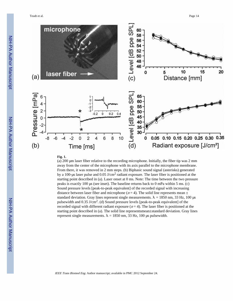
Fig. 1.(a) 200 µm laser fiber relative to the recording microphone. Initially, the fiber tip was 2 mmaway from the center of the microphone with its axis parallel to the microphone membrane.From there, it was removed in 2 mm steps. (b) Biphasic sound signal (asterisks) generatedby a 100-µs laser pulse and 0.05 J/cm2 radiant exposure. The laser fiber is positioned at thestarting point described in (a). Laser onset at 0 ms. Note: The time between the two pressurepeaks is exactly 100 µs (see inset). The baseline returns back to 0 mPa within 5 ms. (c)Sound pressure levels [peak-to-peak equivalent] of the recorded signal with increasingdistance between laser fiber and microphone (n = 4). The solid line represents mean ±standard deviation. Gray lines represent single measurements. λ = 1850 nm, 33 Hz, 100 µspulsewidth and 0.35 J/cm2. (d) Sound pressure levels [peak-to-peak equivalent] of therecorded signal with different radiant exposure (n = 4). The laser fiber is positioned at thestarting point described in (a). The solid line representsmean±standard deviation. Gray linesrepresent single measurements. λ = 1850 nm, 33 Hz, 100 µs pulsewidth.
Teudt et al. Page 14
IEEE Trans Biomed Eng. Author manuscript; available in PMC 2012 September 24.
NIH
-PA Author Manuscript
NIH
-PA Author Manuscript
NIH
-PA Author Manuscript
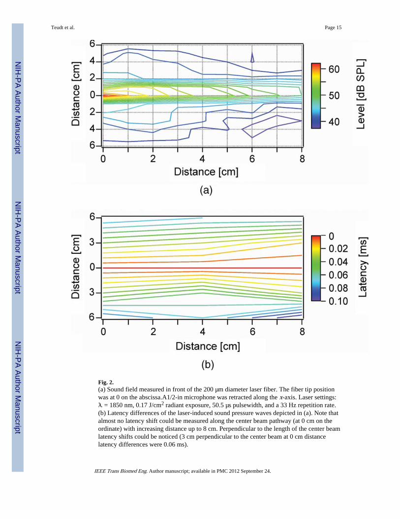
Fig. 2.(a) Sound field measured in front of the 200 µm diameter laser fiber. The fiber tip positionwas at 0 on the abscissa.A1/2-in microphone was retracted along the x-axis. Laser settings:λ = 1850 nm, 0.17 J/cm2 radiant exposure, 50.5 µs pulsewidth, and a 33 Hz repetition rate.(b) Latency differences of the laser-induced sound pressure waves depicted in (a). Note thatalmost no latency shift could be measured along the center beam pathway (at 0 cm on theordinate) with increasing distance up to 8 cm. Perpendicular to the length of the center beamlatency shifts could be noticed (3 cm perpendicular to the center beam at 0 cm distancelatency differences were 0.06 ms).
Teudt et al. Page 15
IEEE Trans Biomed Eng. Author manuscript; available in PMC 2012 September 24.
NIH
-PA Author Manuscript
NIH
-PA Author Manuscript
NIH
-PA Author Manuscript
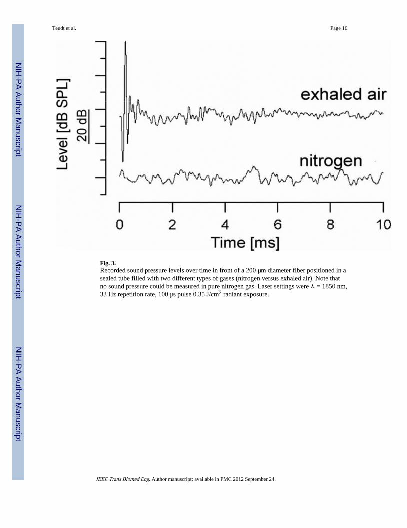
Fig. 3.Recorded sound pressure levels over time in front of a 200 µm diameter fiber positioned in asealed tube filled with two different types of gases (nitrogen versus exhaled air). Note thatno sound pressure could be measured in pure nitrogen gas. Laser settings were λ = 1850 nm,33 Hz repetition rate, 100 µs pulse 0.35 J/cm2 radiant exposure.
Teudt et al. Page 16
IEEE Trans Biomed Eng. Author manuscript; available in PMC 2012 September 24.
NIH
-PA Author Manuscript
NIH
-PA Author Manuscript
NIH
-PA Author Manuscript
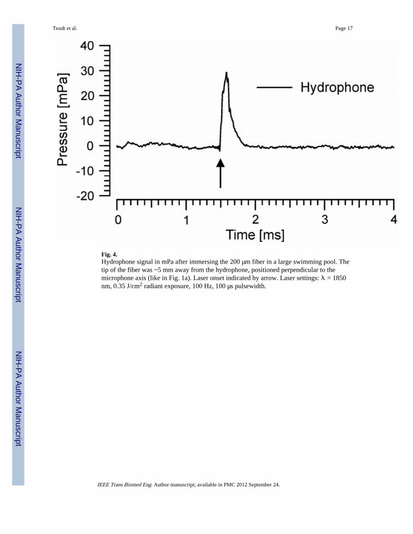
Fig. 4.Hydrophone signal in mPa after immersing the 200 µm fiber in a large swimming pool. Thetip of the fiber was ~5 mm away from the hydrophone, positioned perpendicular to themicrophone axis (like in Fig. 1a). Laser onset indicated by arrow. Laser settings: λ = 1850nm, 0.35 J/cm2 radiant exposure, 100 Hz, 100 µs pulsewidth.
Teudt et al. Page 17
IEEE Trans Biomed Eng. Author manuscript; available in PMC 2012 September 24.
NIH
-PA Author Manuscript
NIH
-PA Author Manuscript
NIH
-PA Author Manuscript
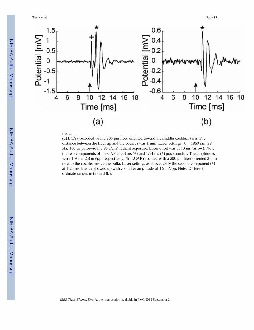
Fig. 5.(a) LCAP recorded with a 200 µm fiber oriented toward the middle cochlear turn. Thedistance between the fiber tip and the cochlea was 1 mm. Laser settings: λ = 1850 nm, 33Hz, 100 µs pulsewidth 0.35 J/cm2 radiant exposure. Laser onset was at 10 ms (arrow). Notethe two components of the CAP at 0.3 ms (+) and 1.14 ms (*) poststimulus. The amplitudeswere 1.9 and 2.8 mVpp, respectively. (b) LCAP recorded with a 200 µm fiber oriented 2 mmnext to the cochlea inside the bulla. Laser settings as above. Only the second component (*)at 1.26 ms latency showed up with a smaller amplitude of 1.9 mVpp. Note: Differentordinate ranges in (a) and (b).
Teudt et al. Page 18
IEEE Trans Biomed Eng. Author manuscript; available in PMC 2012 September 24.
NIH
-PA Author Manuscript
NIH
-PA Author Manuscript
NIH
-PA Author Manuscript
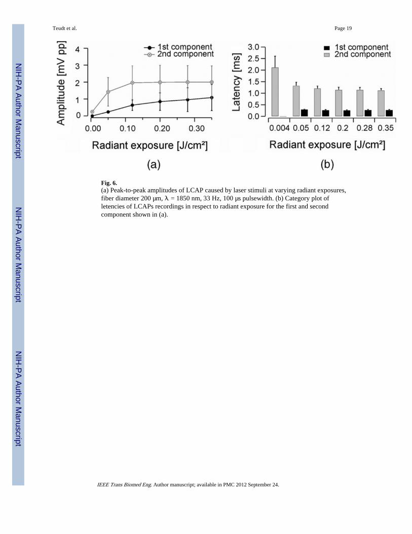
Fig. 6.(a) Peak-to-peak amplitudes of LCAP caused by laser stimuli at varying radiant exposures,fiber diameter 200 µm, λ = 1850 nm, 33 Hz, 100 µs pulsewidth. (b) Category plot ofletencies of LCAPs recordings in respect to radiant exposure for the first and secondcomponent shown in (a).
Teudt et al. Page 19
IEEE Trans Biomed Eng. Author manuscript; available in PMC 2012 September 24.
NIH
-PA Author Manuscript
NIH
-PA Author Manuscript
NIH
-PA Author Manuscript
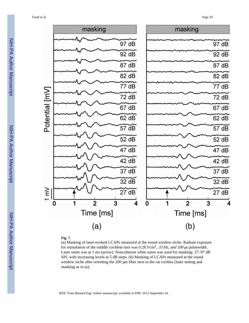
Fig. 7.(a) Masking of laser-evoked LCAPs measured at the round window niche. Radiant exposurefor stimulation of the middle cochlear turn was 0.28 J/cm2, 33 Hz, and 100 µs pulsewidth.Laser onset was at 1 ms (arrow). Noncoherent white noise was used for masking: 27–97 dBSPL with increasing levels at 5 dB steps. (b) Masking of LCAPs measured at the roundwindow niche after orienting the 200 µm fiber next to the rat cochlea (laser setting andmasking as in (a).
Teudt et al. Page 20
IEEE Trans Biomed Eng. Author manuscript; available in PMC 2012 September 24.
NIH
-PA Author Manuscript
NIH
-PA Author Manuscript
NIH
-PA Author Manuscript
NIH
-PA Author Manuscript
NIH
-PA Author Manuscript
NIH
-PA Author Manuscript
Teudt et al. Page 21
TAB
LE I
Lat
ency
Ene
rgy
and
Am
plitu
de E
nerg
y R
elat
ions
for
the
Firs
t and
the
Seco
nd L
CA
P C
ompo
nent
(Fi
rst P
ositi
ve W
ave
Proc
esse
d, n
= 6
)
1st
com
pone
nt2n
d co
mpo
nent
lase
rfi
ber
[Ø]
radi
ant
expo
ser
[J/c
m2 ]
late
ncy
mea
n[m
s]SD ±
ampl
itud
em
ean
[µV
]SD ±
n
radi
ant
expo
ser
[J/c
m2 ]
late
ncy
mea
n[m
s]SD ±
ampl
itud
em
ean
[µV
]SD ±
n
200
µm0,
350,
270,
0310
8577
26
0,35
1,12
0,08
1998
958
6
0,28
0,27
0,03
958
704
60,
281,
140,
1119
9697
96
0,20
0,25
0,03
845
516
50,
201,
140,
1219
9010
066
0,12
0,26
0,03
637
309
40,
121,
200,
1119
6199
66
0,05
0,29
0,02
238
543
0,05
1,32
0,15
1425
842
6
0,00
4-
--
--
0,00
42,
110,
4924
924
2
IEEE Trans Biomed Eng. Author manuscript; available in PMC 2012 September 24.