ciguidelinescigifinal.pdf - Cochlear Implant Group of India
46
THE COCHLEAR IMPLANT GROUP OF INDIA Cochlear Implant Guidelines Third Edition Executive body President: Dr. Rajesh Vishwakarma Secretary: Ms. Nishita Mohandas Immediate Past President: Dr. Shankar Medikeri Vice President: Dr. Madhuri Gore Jt. Secretary: Dr. Mohnish Grover Treasurer: Dr. Ganji Srinivas Executive Council Members: Ms. Rashmi Deshpande Mr. M A Srikanth Dr. Ruchika Mittal Gp.Capt. Himanshu Swami Dr. Milind Navalakhe Dr. Subbarayadu Yarlagadda November 2018 First edition: 2004 Second edition: 2011
-
Upload
khangminh22 -
Category
Documents
-
view
0 -
download
0
Transcript of ciguidelinescigifinal.pdf - Cochlear Implant Group of India

THE COCHLEAR IMPLANT GROUP OF INDIA
Cochlear Implant Guidelines
Third Edition
Executive body
President: Dr. Rajesh Vishwakarma Secretary: Ms. Nishita Mohandas
Immediate Past President: Dr. Shankar Medikeri
Vice President: Dr. Madhuri Gore Jt. Secretary: Dr. Mohnish Grover Treasurer: Dr. Ganji Srinivas
Executive Council Members: Ms. Rashmi Deshpande Mr. M A Srikanth Dr. Ruchika Mittal Gp.Capt. Himanshu Swami Dr. Milind Navalakhe Dr. Subbarayadu Yarlagadda
November 2018
First edition: 2004 Second edition: 2011

2
Expert committee members: 1. Dr Alok Thakar
2. Dr Ameet Kishore
3. Dr Amit Keshri
4. Dr C Preetam
5. Dr Ganji Srinivas
6. Dr Gaurav Gupta
7. Dr Himanshu Swami
8. Ms Jaspal Choudhary
9. Dr Kapil Sikka
10. Dr Kranti Bhavna
11. Dr Madhuri Gore
12. Dr Manoj Manikoth
13. Dr Milind Navlakhe
14. Dr Mohnish Grover
15. Dr Naresh Panda
16. Dr Neelam Vaid
17. Ms Nishita Mohandas
18. Dr Rajesh Vishwakarma
19. Ms Rashmi Deshpande
20. Ms Ritu Nakra
21. Dr Ruchika Mittal
22. Dr Satya Prakash Dubey
23. Dr Shankar B Medikeri
24. Ms Shefali Shah
25. Dr Shitanshu Sharma
26. Mr Srikant
27. Dr Sunil Dutt
28. Dr Sumit Mrig
29. Dr Y Subba Rayudu

3
The following guidelines were made after inputs from all the above members in
the expert committee. The Executive Committee had repeated discussions on
the same and then a draft of guidelines was put up on the CIGI website. A mail
was sent to all CIGI members for suggestions and queries. Various mails were
received which were discussed among EC members. An edited draft was put up
on the website and a mail was again sent to all members. The guidelines were
then discussed in General Body Meeting at Daman on 30th November 2018 and
approved by all. The Executive Committee is thankful to all those who gave
their voluntary contribution to the formation of these guidelines.
IMPORTANT NOTE: THESE ARE GUIDELINES FROM INDIAN POINT
OF VIEW MADE BY MEMBERS OF COCHLEAR IMPLANT GROUP OF
INDIA. THESE ARE RECCOMENDATIONS OF THE EXPERT
COMMITTEE FOR BEST POSSIBLE RESULTS AND NOT BINDING ON
ANYONE. THIS IS NOT A MEDICOLEGAL DOCUMENT

4
Contents
1. Infrastructure and equipment required……………..………5 2. Investigations
a) Audiological and Speech and Language …………...…...7 b) Radiological……………………………………..………8 c) Genetic…………………………………………..………10
3. Effect of age/duration of auditory deprivation………..……11 4. Psychological evaluation…………………..………….……12 5. Preoperative therapy
a) Pediatrics…………………………………………….......13 b) Adults……………………………………………..……..15
6. Vaccination schedule…………………………………..……18 7. Consent……………………………………………………...19 8. Surgical steps……………………………………………..…23 9. Post surgery medications……………………………………25 10. Postoperative therapy and (re)habilitation
a) Pediatrics…………………………………………......26 b) Adults……………………………………………...…27
11. Management of complications: a) Facial palsy………………………………………………29 b) Misplaced electrodes…………………………….………31 c) Incomplete insertion………………………………….….35 d) Acute otitis media ………………………………………37 e) Tympanic membrane perforation……...………………...38 f) Cholesteatoma post cochlear implant…………………....39
12. Surgery in special conditions: a) Ossified cochlea …………………………………………40 b) Malformed inner ear …………………………………….41
13. Bilateral cochlear implantation……………………………...45

5
1. Infrastructure and Equipment required
Mandatory
1. Operating microscope with bright illumination, up to 20 X magnification and focus adjustment
2. Instrument set a. Blade handle for 15 and 11 number blades b. Toothed and non-toothed thumb and Adson forceps c. Dissecting Mayo scissors d. Broad periosteal elevator e. Freer perichondrial/ periosteal elevator f. Micro forceps – fine tip g. Micro forceps – cup h. Micro forceps- upturned cup i. Micro scissors j. Micro pick straight k. Micro pick –curved l. Micro pick- right angled m. Micro pick- angled Shuckenecht type n. Suction tips large 2 numbers o. Suction tip micro nos 12-26 p. Suction tip adapters
3. Needle holder 8cm 4. Cautery – good quality with cutting and bipolar forceps, patient plate and
standard safety settings 5. Micro drill system with operating speed at least 35000 RPM with
brushless motor 6. Drill set
a. Cutting burrs sizes 7mm, 6mm, 5mm, 4.5 mm, 4 mm, 3.5 mm, 3 mm, 2.5 mm, 2mm, 1 mm, .8mm, .6mm
b. Diamond burrs same as above c. Taper diamond long burrs for cochleostomy .8mm and .6mm
7. Anesthesia equipment with gas control, pressure monitoring, ventilation and gas scavenging facilities
8. Multipara meter monitoring with SpO2, ETCO2, blood pressure, pulse rate
9. Efficient suction apparatus with variable suction control 10. Overhead lighting system of at least 60000 lux 11. Operation theatre table with lift, tilt and elevation functions- , manual ,
and with provision for intra operative X-ray 12. Cardiac defibrillator and emergency cart

6
Recommended All of the above, plus
1. Operating microscope with tilt able eyepiece and variable focus, LED or Xenon illumination
2. Facility for recording of surgical video in a reproducible format and viewing of surgery in large screen monitor
3. Micro drill system equivalent to Skeeter with .8mm. .7mm and .5mm tips 4. Facial nerve monitor with facility of live monitoring of cranial nerves and
recording of events 5. Drill system for micro ear surgery with speeds up to 60000 RPM, self-
irrigation system and torque control 6. Cautery systems with bipolar cutting and coagulation systems 7. Thermal blanket specified for Operation theatre use in children 8. Operation theatre tables with lift, tilt and elevation functions- hydro
electronic 9. Hydraulic surgeon chair 10. Laminar airflow operation theatre with hepa filters and positive pressure
systems 11. Intra operative C arm
Optional
1. Intra operative CT 2. Titanium instruments for surgery 3. Operating microscope with facility for neuro navigation

7
2 Investigations
2(a) Audiological
A) Mandatory
Test Indication for CI Audiometry Behavioural Observation Audiometry/Visual Reinforcement Audiometry / Free Field or Sound Field/ Pure Tone Audiometry Immittance Test Battery (Tympanometry and Reflex testing)
Severe to Profound hearing loss Moderately Severe to profound, Bilateral Moderately severe to profound hearing loss Tympanogram : ‘A’ or ‘As’ Type with reflexes absent
Brainstem Evoked Response Audiometry Click stimuli with both rarefaction and condensation polarities and Tone burst stimuli @500Hz and @1000Hz
Bilateral V wave absent or obtained till 90 dBnHL
Oto Acoustic Emissions Screening OAE Diagnostic OAE
Bilateral DPOAEs Absent – Repeated after 15 days for both ears in infants
Response with Hearing Aids - Functional Gain Test after at least 3 months of auditory based intervention
Limited or no benefit as evaluated on the aided audiogram, speech perception tests and therapy progress report
B) Recommended Test Indication for CI 1.Auditory Steady State Response (ASSR)
To assess auditory nerve function when no ABR is present and severity of hearing loss on various frequencies

8
C) Optional Test Indication for CI
TT-EABR (Trans Tympanic-Electrically Evoked Brainstem Response) promontory stimulation test Cortical testing LLR (Late Latency Response) MLR (Middle Latency Response)
To be done in cases of suspected cochlear nerve hypoplasia/aplasia (please see section on cochlear malformations) and ANSD
Speech and Language Evaluation
Assessment of mode of communication, prelinguistic skills, Language age /skills (Receptive and Expressive), associated oral motor issues, ASD. Schedules of speech and language development and standardized assessment can be utilized. Speech Language assessment is indicated for adults developing hearing loss after head trauma.
2(b) Radiological Investigations
A) Mandatory
1. High Resolution Computed Tomography (HRCT) scan of temporal bone (axial, coronal) minimum 64 slice.
2. Magnetic Resonance Imaging (MRI) temporal bone and brain (FIESTA/CISS sequence) with oblique sagittal sections of internal acoustic canals of minimum 1.5 Tesla.
B) Recommended
MRI temporal bone with 3D reconstruction of cochlea
C) Optional
HRCT temporal bone to calculate
a) Cochlear duct length b) Cochlear orientation

9
Checklist for Imaging Mastoid process
Size: Normal Hypoplastic Forward lying Sigmoid
Pneumatization: limited Throughout Beyond Effusion: No
Yes Middle ear Normal Exposed jugular bulb Aberrant carotid artery Aberrant facial nerve Other: specify Cochlear morphology Normal Incomplete partition1
Hypoplasia Common cavity
Aplasia Cochlear ossification2
None Round window Length in basal turn, mm Middle turn Apical turn Vestibular aqueduct Normal Large Internal auditory canal diameter
Normal Stenosed enlarged
Cochlear aperture normal narrow enlarged

10
Other Otosclerosis Fracture
Intra and postoperative plain radiography was done in all patients to determine optimum positioning of the electrode array. An oblique anti Stenver's view was found most useful to show the electrode in the cochlea. References
1. Sennaroğlu L, Demir Bajin M. Classification and Current Management of Inner Ear Malformations. Balkan Medical Journal. 2017;34(5):397-411. doi:10.4274/balkanmedj.2017.0367.
2. Balkany T, Gantz B, Nadol JB Jr. Multichannel cochlear implants in partially ossified cochleas. Ann Otol Rhinol Laryngol 97 (suppl 135): 3-7.
2(c) Genetic Testing
Genetic tests are not mandatory Recommended Genetic testing can be done for mutations common in that particular area and population in cases of:
• Positive family history of congenital/progressive hearing loss • Consanguinity • Syndromic child
Optional All patients should be evaluated for mutations common in that particular area and population.

11
3) Effect of age/duration of auditory deprivation
Pre-lingual: Following US FDA criteria, we recommend that cochlear implant can be done between age 1-17 years. Patients below 1 year and more than 17 years can be operated depending on case to case basis. However, the outcomes of cochlear implant tend to be more variable beyond 5 years of age. Therefore, implantation after 5 years of age should be done after meticulous evaluation with multidisciplinary approach and a well-informed consent regarding outcomes. When the implant is done beyond the age of 5 years more benefits maybe seen with reference to quality of life as compared to speech and language outcomes. Post-lingual: There is no age limitation for cochlear implantation in post lingual recipients. The important considerations are the aetiology of hearing loss and the associated co-morbidities in the geriatric age group. References:
1. Hilly O, Hwang E, Smith L, Shipp D, Nedzelski JM, Chen JM, Lin VW. Cochlear implantation in elderly patients: stability of outcome over time.J Laryngol Otol. 2016 Aug; 130(8):706-11.
2. Maura Cosetti, J. Thomas Roland, Jr. Cochlear Implantation in the Very Young Child: Issues Unique to the Under-1 Population. Trends Amplif. 2010 Mar; 14(1): 46–57.
3. Kalejaiye A et al. Low surgical complication rates in cochlear implantation for young children less than 1 year of age. Laryngoscope. 2017 Mar;127(3):720-724.
4. David M, Valencia Frank L.Rimell Barbara J.Friedman Melisa R.Oblander Josephine Helmbrecht. Cochlear implantation in infants less than 12 months of age. Int J Pediatr Otorhinolaryngol. 2008 Jun;72(6):767-73.
5. Sanchez-Cuadrado I, Lassaletta L, Perez-Mora RM, Zernotti M, Di Gregorio MF, Boccio C, Gavilán J. Is there an age limit for cochlear implantation?. Ann Otol Rhinol Laryngol. 2013 Apr;122(4):222-8.
6. Su Wooi Teoh, MD, David B. Pisoni, PhD, and Richard T. Miyamoto, MD. Cochlear Implantation in Adults with Prelingual Deafness. Part I. Clinical Results. Laryngoscope. 2004 Sep; 114(9): 1536–1540.
12
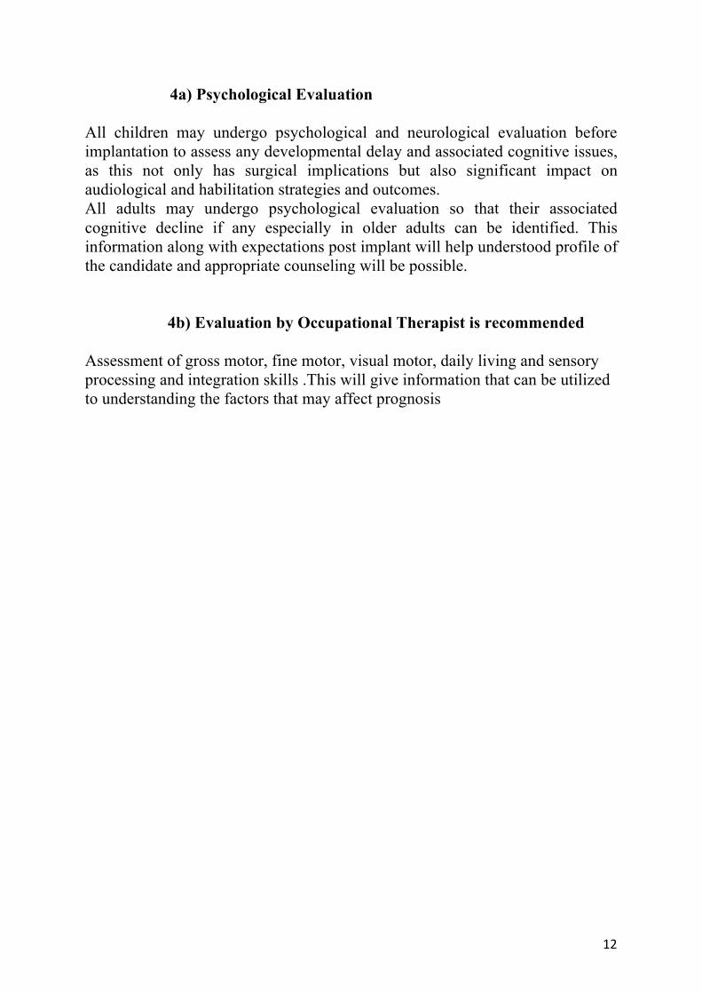
4a) Psychological Evaluation All children may undergo psychological and neurological evaluation before implantation to assess any developmental delay and associated cognitive issues, as this not only has surgical implications but also significant impact on audiological and habilitation strategies and outcomes. All adults may undergo psychological evaluation so that their associated cognitive decline if any especially in older adults can be identified. This information along with expectations post implant will help understood profile of the candidate and appropriate counseling will be possible. 4b) Evaluation by Occupational Therapist is recommended
Assessment of gross motor, fine motor, visual motor, daily living and sensory processing and integration skills .This will give information that can be utilized to understanding the factors that may affect prognosis

13
5(a) Preoperative auditory-based therapy (Pediatrics)
The aim of Auditory based therapy is to establish age-appropriate spoken language skills in babies and young children with hearing loss via listening alone with appropriate hearing devices. This means that Auditory-based Therapy, relates to early intervention (i.e. for babies and very young children).(Please refer Appendix I: Auditory Skill Hierarchy ) Children with hearing loss for whom intervention begins when they are 5 years and above can avail of services in Auditory Based Intervention, using an approach in which listening is supported by visual communication cues.
Mandatory:
INFRA-STRUCTURE: 1. Centers that offer audiological services must have a modern, sound-treated
audiology booth, with modern equipment and an area for fitting hearing aids and MAPping children with cochlear implants.
2. Therapy rooms must be sufficiently sound treated. 3. Furniture appropriate for babies and young children. All furniture may be
fitted with rubber stoppers, for sound dampening. 4. Storage space to store all files and educational and test materials in Audiology
and Auditory-Verbal Therapy/ Auditory Based Intervention sessions. EQUIPMENT: Audiology:
1. A calibrated Clinical Audiometer. 2. A Visual-Reinforcement Audiometry (VRA) system. 3. A calibrated Immittance Analyser. 4. A hearing aid loaner service (to be offered to recipients at the discretion of
the centre). 5. All current programming software related to all the hearing devices
(hearing aids and cochlear implants) that the centre offers its recipients. 6. A calibrated auditory evoked potential system, Otoaacoustic analyser. 7. Ear impression material and equipment for taking ear impressions for
making ear moulds for hearing aided recipients. Those centers which do not offer this service in-house must liase with a facility that is able to do so
Speech and Language
Standardised assessment. (for measuring and monitoring progress in children) should be available (Appendix 2).

14
Skills of Professionals:
1. All staff must be sufficiently trained to manage the age-group of the population they serve. Audiologists: a. Audiologists (preferably with a Masters degree in Audiology/
M.ASLP/A&ST) must have the knowledge and skills to measure and manage hearing loss hearing impaired individuals
b. They must be trained in all the hearing device technologies (w.r.t. hearing aids and cochlear implants) offered by the center to its recipients.
c. They must have undergone specialized training in the management of children with cochlear implants.
Speech language Pathologists (preferably with a Masters degree in SLP/M.ASLP/A&ST)
Should have knowledge to administer standardized speech and language tests, with special reference to children with hearing loss and associated disorders due to expanding criteria .
Speech Language Pathologists/Audiologists/Educators -Therapists
a. All ASLPs/AST D.Eds.(HI), B.Eds.(HI), TODs, M.Eds.(HI), working with children with cochlear implants must have undergone training in Auditory Based Intervention to address the needs of the children they serve. a. Professionals need to be aware of speech and language, communication and
cognitive development of typical hearing children b. Therapists must have the knowledge on how to assess changes during course of therapy.
Guidance and Counselling before surgery:
1. The center must offer an audiological service within its premises or liase closely with an audiological center for the management of all babies and young children in its care.
2. For those children for whom spoken language may not be an option, their families must be sufficiently, realistically and appropriately counselled about outcomes, so that they can make an informed decision regarding communication options for their child.
3. Families of children whose history requires immediate cochlear implant surgery must be guided to surgery without delay.
4. Centers that offer Auditory-Verbal Therapy/ Auditory Based Intervention must offer minimum once a week therapy sessions lasting an hour in duration. Parent participation in these sessions is mandatory.
5. The center must assess the family’s motivation and ability to attend and participate in post-surgery habilitation on an on-going basis (direct or distance)

15
for 2- 3 years. 6. The focus of therapy done prior to surgery must be on ascertaining whether or
not the baby or child demonstrates i) awareness of speech and perceives suprasegmentals, vowels and consonants, and determining his pattern of vocal behavior and progress after stimulation ii) determining candidacy for cochlear implant surgery
7. The complete battery of prescribed audiological tests (as administered under Pre-Op Work up Guidelines) along with progress in therapy must be detailed in a letter to the ENT surgeon, along with a recommendation for surgery.
5(b) Preoperative auditory based training (adults) Mandatory: INFRA-STRUCTURE:
1. The lay-out of the center must be welcoming to those adults and their families in their care. Centers that offer services to the elderly must be accessible: elevators, ramps for wheelchair bound clients. They need to have compliance for barrier free environment
2. Centers that offer audiological services (please also refer 1. under Pre-surgery) must have a sound-treated audiology booth, (please refer 1.under Equipment) and an area for fitting hearing aids and Mapping cochlear implanted adults.
3. Therapy rooms must be relatively quiet 4. A meeting room for discussion. 5. Furniture must be appropriate for adults and the elderly.
All furniture must be fitted with rubber stoppers, for sound dampening. 6. Storage space to store all files in Audiology and Communication
Therapy sessions.
EQUIPMENT: Audiology:
1. A calibrated Clinical Audiometer with facility for speech perception assessment in Free Field.
2. A calibrated Immittance Audiometer 3. Hearing aids on trial or a hearing aid loaner service (to be offered to
recipients at the discretion of the centre). 4. All current programming software related to all the hearing devices
(hearing aids and cochlear implants) that the centre offers its recipients.
5. A calibrated auditory evoked potential system with facility to use clicks and tone bursts/ASSR, Otoacoustic analyzer.

16
6. Ear impression material and equipment for taking ear impressions for making ear moulds for hearing aided recipients. Those centres which do not offer this service in-house must liase with a facility that is able to take well-made ear impressions from which to make ear moulds.
7. Materials for speech perception testing, in accordance with international protocol for English using sentence and word perception tests in auditory only and auditory visual modes.)in regional languages.
8. Adults with congenital hearing loss, must be assessed on psychophysical measures when speech perception testing is not possible. These will include gap detection, loudness, pitch perception and tone adaptation/decay.
Resource Material: 1. Adult questionnaires (e.g. Speech, Spatial and Qualities of Hearing
Scale (SSQ)) 2. The center must be sufficiently and appropriately equipped with
appropriate reading materials (Newspapers, books, magazines). 3. Audiobooks (Refer “Resources for Adults”) 4. Self-practice resource material for adults.(Refer “Resources for Adults”)
Skills of Professionals:
1. All staff must be sufficiently trained to manage the age-group and adult learning styles of the population they serve. Specifically: a. Audiologists (preferably with a Masters degree) must have the
knowledge and skills to measure and manage hearing loss in adults including SSD and asymmetric hearing loss), following the pre- and post implant protocol, as outlined in the Pre-Op Work-up Guidelines.
b. They must be trained in all the hearing device technologies (hearing aids and cochlear implants) offered by the center to its recipients. They must have attended 2-3 workshops or undergone individualized training by the manufacturers in programming of hearing aids and Mapping. They need to have basic skills in how to counsel adults and families in preparation for cochlear implant surgery.
c. Audiologists must have a basic knowledge in Adult Learning Styles.
Services and guidance before surgery: 1. The center must offer an audiological service within its premises or
liase closely with an audiological center for the management of all

17
adults in its care. 2. The complete audiological test battery (Please refer Pre-Op Work up
Guidelines) administered must also include an assessment of cognitive skills.
3. Adults whose history requires immediate cochlear implantation must be guided to surgery without delay.
4. Centers that offer Communication Therapy must offer weekly therapy sessions lasting 30-40 minutes in duration. Family participation in these sessions is mandatory.
5. The results of a complete battery of prescribed audiological testing, with particular focus on Speech Perception testing along with results of questionnaires filled up by the adult must be presented in a letter to the ENT surgeon, along with a recommendation for surgery.

18
6) Vaccination Schedule Meningitis following cochlear implantation is a known complication. Streptococcal pneumoniae and H influenzae are the common pathogens causing the infections both in middle ear and the meninges. In first few days after implantation, the surgical site infections are likely contributor to meninigitis. After 30 days of implantation, Acute ottis media becomes the commoner etiologic factor. Center for disease control and prevention (CDC) and Advisory committee on prevention has from time to time brought about guidelines for cochlear implant recipients. The guidelines are summarized as follows11,12
Children who have cochlear implants or are candidates for cochlear implants should receive PCV13 (Pneumococcal Conjugate vaccine 13).
Children with cochlear implants, from age 2 years through age 5, should receive two doses of PCV13 if they have not received any doses of PCV7 or PCV13 previously. If they have already completed the four-dose PCV7 series, they should receive one dose of PCV13 through age 71 months.
Children 6 through 18 years of age with cochlear implants may receive a single dose of PCV13 regardless of whether they have previously received PCV7 or the pneumococcal polysaccharide vaccine (PPSV23)
In addition to receiving PCV13, children with cochlear implants should receive one dose of PPSV23 at age 2 years or older and after completing all recommended doses of PCV13.
CDC adds guidelines for adults:
Adult patients (≥19 yrs of age) who are candidates for a cochlear implant and those who have received a cochlear implant should be given a single dose of PCV13 followed by a PPSV23 at least 8 weeks later. A second dose of PPSV23 is recommended for those 65 years of age and older.
Universal immunization program in India recommends pentavalent vaccine for all children after 6 weeks of age and hence receive Hib vaccine universally. History should be carefully elicited and Hib vaccine administered in consultation with pediatrician if required. References:
1. Kahue CN, Sweeney AD, Carlson ML, Haynes DS. Vaccination recommendations and risk of meningitis following cochlear implantation. Curr Opin Otolaryngol Head Neck Surg 2014; 22: 359-366
2. http://www.entnet.org/sites/default/files/Cochlear-Implants-and-Meningitis-VaccinationFact-Sheet-update-November-2012.pdf)

19
7) Consent Name: Age / Sex: years Date of Birth: Proposed Date of Surgery: Hospital Registration No: ___________________ Type of Implant: Address: PROPOSED TREATMENT The doctor has explained that I/my _______________________ have /has (condition /diagnosis) _____________________ and that a cochlear implant on the ______ side/s is proposed. OPTIONAL: I have chosen to get ______ model of ____________ Company implanted on myself / my_____________ COCHLEAR IMPLANTATION The cochlear implant is an electronic device that is implanted inside the cochlea (inner ear), which bypasses damaged or absent hair cell and provides electrical stimulation directly to the hearing nerve fibres. Under a general anaesthetic, an opening is made behind the ear and a small area of the mastoid bone hollowed out to lodge the receiver / stimulator part of the device. An array of electrodes is inserted into the cochlea (inner ear). Any bleeding is stopped and the skin wound close over the device. The external part of the device will be programme and fitted when the wound has healed. The programming is usually done after two or three weeks. BENEFITS OF COCHLEAR IMPLANTATION Cochlear implantation is designed for people with severe to profound hearing loss who derive limited benefits from hearing aids. The speech / language / hearing skill will improve following cochlear implantation. The improvement depends upon many factors which have been explained to me by the cochlear implant team, audiologist and therapist. RISK These are the more common risks. There may be other unusual risks that have not listed here. I understand that there are risks associated with any anaesthesia. I / my ________ may have side effects from any of the drugs used. The common side effects include light – headedness, nausea, skin rash and constipation. I understand the procedure has the following specific risks and limitations:

20
• The device will not cure my / my _______deafness nor will it completely restore by hearing. I am likely to need some ‘listening’ training to be able to benefit as much as possible. • There may be circumstances where it may not be possible to insert the implant completely and this may sometimes affect eventual outcomes. • The ability of the implant to improve speech perception will depend on the ability of the auditory nerve to conduct the electrical stimulus and the functional integrity of the auditory cortex. This may not be possible to detect with current investigative methods accurately and so if the auditory cortex and the nerve are defective the outcomes may be poor. • Static electricity may damage the electronic components of the device or the program. • I / my __________ will need to have regular follow- up for ENT consultation; programming and auditory verbal therapy and the device will need regular maintenance. • I / my__________ may have some dizziness, dryness in my mouth and / or ringing in my ear(s) after the operation as a result of the surgery to the ear. • I / my __________ wound may become infected and I may need antibiotics for this. Rarely the skin wound may fail to heal and the device may have to be removed. In post operative period, there may be leakage of fluid from the cochlea perhaps requiring a further operation. • Very rarely I may have bleeding after the operation, which may require local treatment or occasionally a return to the operating theatre. • I may notice some numbness or stiffness around the ear, which in most cases will improve gradually with time. • I may have some loss of taste on the side of the operation, which may be temporary or permanent. • Rarely, I may have some swelling near the facial nerve, which runs close to the operation site. This usually revolves over the course of some weeks, but permanent paralysis may rarely occur. • I may have some pain in the area of the coil, which should improve over time. • Very rarely, my body would ‘reject’ the implant, which may be extruded. • Placement of the implant may simulate new bone growth, which may damage surviving nerves and make replacement of my device difficult. • The implants have been used for over thirty years without any reports of consequences from electrical stimulation. If problems should develop in future, the implant can be easily removed. • The external equipment may fail and require re-mapping. Should there be a need for the patient to undergo Magnetic Resonance Imaging, the magnet in the cochlear implant would need to be surgically removed. • The internal implant may fail and need a second surgery to replace the damaged device. I / my relative will need to avoid sports where there is a potential to damage the device, and must warn medical staff and I / my relative

21
have one, as some procedures or investigations can be damage the device. Hence we should contact the implant surgeon for advice. • I understand some of the above risks are more likely if I / my relative smoke/ s become overweight, diabetic, high blood pressure or other medical conditions. INDIVIDUAL RISKS I understand the following are possible significant risks and complications specific to my ___________ individual circumstances, that I have considered in deciding to have this operation. _______________________________________________________________________________________________________________________________________________________________________________________________________________________________________________________________________________________________________________________________________________________________________________________ DECLARATION BY PATIENT / RELATIVE • I acknowledge that the ENT surgeon & Audiologist have informed me about the procedure, alternative treatments & answered my specific queries & concerns about this matter. • I understand the need for continuous follow up care & therapy for adequate benefit. • I acknowledge that I have discussed with the ENT surgeon regarding any significant risks & complications specific to my / our individual circumstances that I have considered in deciding to have this operation. • I agree to any other additional procedures considered necessary in the judgement of my / our ENT surgeon during this operation. • I understand that a doctor other than the specialist ENT surgeon may perform the procedure, when necessary. • I have received a copy of this form to take home with me. • If a needle stick / sharp injury occur to staff during any procedure I give my permission for blood to be taken & tested for HIV & other blood borne disorders. I understand that I will be advised & counselled as soon as practicable after the operation if this has been necessary. • I understand that the recordings from my surgery / programming may be used at presentations / promotions if necessary without revealing my identity. OPTIONAL: We, _________________________ of ___________________have agreed to use ______________________________ cochlear implant system. We have been informed of the specifications / functioning.

22
We have been informed of the various warranty associated with the cochlear implant system. We have been informed the costs associated with post operative maintenance of the device. Signature of patient / relative: ________________ Relationship to patient: _____________________ Signature of Witness: ______________________ Name of Witness: _________________________ Date: DECLARATION BY DOCTOR • I declare that I have explained the nature & consequences of the operation to be performed, & discussed the risks that particularly concern the patient. • I have given the patient an opportunity to ask questions & I have answered these. Surgeon’s signature:______________ Anaesthetist’s signature: _____________ Surgeon’s Name: ________________ Anaesthetist’s Name: _______________ Date:

23
8) Surgical steps Mandatory:
1. Operative site to be prepared. 2. Perioperative antibiotics and steroids to be administered intravenously 3. Infiltration with local anesthetic and adrenaline is recommended 4. Incision and flaps can be made depending on surgeons preference,
however it should be such that we get 2 flaps (subcutaneous plane and musculoperiosteal) to decrease the chances of implant extrusion.
5. Depending on type and model of cochlear implant being used the distance between pinna and proposed site of receiver stimulator package is to be ensured so that overlapping of internal and external component does not happen and the package should not lie beneath the incision. Even if “off the ear” processor is to be used the implant should be sufficiently posterior so that the patient has an option of using a behind the ear processor.
6. The technique of surgery can be as per surgeons choice and experience. 7. The bed for implant should be drilled as per the thickness of the implant
being used. 8. Cochleostomy/ extended round window/ round window opening should
be done only after everything else is ready. 9. If a separate cochleostomy is being done, then it should be done
anteroinferior or inferior to round window. 10. Direct suctioning over perilymph should not be done. 11. Hemostasis should be ensured before opening the implant. 12. Gloves should be changed before touching the implant. 13. Monopolar cautery should be disconnected before putting the implant on
its bed. 14. Electrode array should be inserted in a slow smooth manner. If resistance
is felt, insertion should not be done forcefully. 15. Minimal handling of active electrodes should be done and electrode
insertion should be done in minimum trials. 16. Any perielectrode space in cochleostomy should be sealed well so that
there is no perilymph leakage. 17. Intraoperative checking of impedances of electrodes and nerve response
should be done as per the type of implant model.
Recommended:
1. Powder-less gloves.
24
2. If bone at site of bed is thin, the bed should be drilled such that a bony island is preserved in center, and dura on periphery can be compressed by pressing the bony island.
3. For opening the cochlea through round window, it should not be opened with a burr, but with a pick or any other sharp instrument. For a separate cochleostomy, diamond burr at low speed should be used until endosteum of scala tympani is revealed. Endosteum is entered with a sharp microinstrument.
4. Steroid solution/ sodium hyaluronate should be instilled inside cochlea after opening it.
5. Intraoperative/postoperative radiology (xray) to see positioning of array is recommended.
Optional:
1. Depth of insertion should be calculated as per the cochlear duct length of every patient.
2. Round window insertion or separate cochleostomy should be done as per radiological evaluation to find out axis of cochlea in relation to obliquity of canal wall (cochlear rotation)
References: Lenhardt E. Intracochlear placement of cochlear implant electrodes in soft surgery technique. HNO 1993;41:356-359. Cohen NL. Cochlear implant soft surgery: fact or fantasy? Otolaryngol – Head Neck Surg 1997;117:214-216. Eshraghi AA. Prevention of cochlear implant electrode damage. Curr Opinion Otoalaryngol Head Neck Surg 2006;14:323-328.

25
9) Post surgery medications Antibiotics: Rates of complications after Cochlear Implantation are low, ranging from 1-11 percent.3,4,5,6 Infection, both at surgical site and in middle ear can be serious. Device exposure and infection can be devastating and at times require device removal. This can have a major cost implication, not only for patient but for society as a whole. Even if the device is salvaged, the implications of infection treatment, hospitalization and delay in activation can be severe. Infections also have strong propensity for intracranial spread through cochlea. There has been previously published literature on the use of prophylactic antibiotics in Otologic surgery.7
Vershchhur et al, in their Cochrane review on antibiotic prophylaxis in clean and clean contaminated ear surgery have concluded no benefit of use of prophylactic antibiotics in clean and clean contaminated surgery. The authors have classified otologic surgeries into Clean, Clean contaminated, and neuro-otologic procedures where dura is exposed, or breached. The review does not address cochlear implants in particular. Systematic review of perioperative versus prophylactic antibiotics for cochlear implantation 8 could identify only 3 low quality studies addressing the concept of antibiotics in cochlear implantation. The authors have generally considered cochlear implantation to be clean surgery unless middle ear has active infection or glue. The study could not generate any conclusion on role of perioperative antibiotics in cochlea implant surgery. The data in literature on type of antibiotics, duration and mode of usage is also highly variable and insufficient to draw conclusions. Ceftriaxone, clarithromycin, amoxicillin Clavulanic acid etc have all been used by different authors. The guidelines for use of antibiotics are hence being drawn on basis of limited existing literature and personal experience. Recommendation: Cochlear implantation is a high stake high cost surgery and there are serious limitations for funding devices, maintenance and treatment of device related complications. We, hence recommend the routine intravenous antibiotic prophylaxis for cochlear implant surgery. The spectrum for coverage should be broad and protective against common pathogens that cause device and middle ear infections. The duration and choice of antibiotics can be individualised as per institutional policies. Optional: Labyrinthine sedatives The impact of cochlear implantation on vestibule has been studied in various studies and provide some conflicting reports.9,10 Our experience and most data excerpts reveal that cochlear implantation surgery causes vertigo and vomiting post-operatively and recommend use of anti-emetic vestibular sedatives in post operative course for 24-48 hours.

26
10) Postoperative therapy and (re)habilitation
10(a) Pediatrics Mandatory: Audiology:
1. Families must receive a thorough orientation to their child’s device by the audiologist, in a language and format that they understand.
2. The child’s cochlear implant must be activated (or switched-on) with some member of the family, preferably both parents, present.
3. The patient identification Card must be filled in by the audiologist, in consultation with the parents, immediately following device activation i.e. on the day of Switch-on.
4. The child’s cochlear implant must be registered with the manufacturer on the day of Switch-on.
5. Mapping sessions must be scheduled in accordance with the child’s progress in therapy.
6. A CI assisted audiogram must be obtained and results correlated with sensitivity levels as reflected in the MAP.
7. Session notes and all records must be filed in the individual child’s file. Auditory-Verbal Therapy/ Auditory Based Intervention:
1.Therapists must prepare weekly plans for Auditory Based Intervention sessions, In preparation for the upcoming session. Lesson plans for Auditory-Verbal Therapy/ Auditory Based Intervention must be individualized and session targets identified and recorded, based on the child’s stage of auditory skills development.
2.Diagnostic notes must be maintained after each session. 3.Family members must be guided to understand session targets so that follow- up during the week is appropriate and sufficient.
4.In addition to informal and on-going assessments and questionnaires, each child’s progress must be assessed using at least 1 standardised assessment. These assessments must be administered annually or six-monthly, as specified in the Instruction Manual.
5.Children in whom little or no progress is seen must have their progress reviewed and referred to those professionals whose help needs to be sought, in a timely manner.
Recommended:

27
1. Centres offering tele-practice services must have the associated web-based technology for high-speed connectivity. 2. The centre must be equipped with the complete battery of prescribed standardised assessments 3. Speech Perception testing, in consultation with the therapist must track the child’s progress and families must understand this collated information.
Optional:
1. Centres may aspire to offer a multi-disciplinary service to families of children with hearing loss with additional needs, at a single location.
10(b) Adults: Mandatory: Audiology:
1. The adult recipient and family must receive a thorough orientation to their device by the audiologist, in a language and format that they understand, prior to device activation (or Switch-on) or immediately following it.
2. The adult’s cochlear implant must be activated (or switched-on), preferably with some member of the family present.
3. The patient identification Card must be filled in by the audiologist, in consultation with the adult, immediately following device activation, on the day of Switch-on.
4. The adult’s cochlear implant must be registered with the manufacturer, as per their prescribed format, on the day of Switch-on or device activation.
5. MAPping sessions must be scheduled in accordance with the adult’s progress in therapy.
6. The adult must understand the details of the changes being made to his hearing levels and must be very conversant with all accessories and their management. .
7. A CI assisted audiogram and CI assisted speech perception testing must be obtained, in accordance with standard procedure and results correlated with sensitivity levels as reflected in the MAP.
8. Session notes and all records must be filed in the individual adult’s file.

28
Auditory traning and Communication strategies: 1. Therapists must prepare weekly plans for therapy sessions, in
preparation for the upcoming session. 2. Sufficient and appropriate resource material including self-practice
resources must be assigned following each therapy session. 3. Weekly plans and all records must be filed in the individual adult’s
file. Recommended:
1. Standardised Speech Perception tests for adults must be developed in all regional languages by national and Government and Educational Institutes such AYJNISHD (D), AIISH, Dr.SRCISH and NISH.
Optional:
1. Adults with hearing loss across India have access to remote re-habilitation services, from the center of their choice by telepractice.

29
11) Management of Complications
11(a) Facial Palsy
Primary facial palsy (>HB Grade IV Palsy)
Ø Review of surgical video/ Surgical notes to look for evidence of exposed facial nerve or nerve injury or compression of nerve by electrode array
Ø Electrophysiological testing for facial nerve function is essential for documentation, but not mandatory
Ø Surgical exploration with nerve decompression or in case of complete nerve transection, end to end anastomosis or cable nerve grafting has to be performed
Primary facial palsy (<HB Grade IV Palsy) Ø Medical management with steroids (Prednisolone 1mg/Kg/day) for 2
weeks in tapering dose should be initiated if no medical contraindication to steroids is evident. Physiotherapy should also be initiated simultaneously.
Ø Review of surgical video/ Surgical notes to look for evidence of exposed facial nerve or compression of nerve by electrode array
Ø Electrophysiological testing of facial nerve function and monitoring of grade of facial palsy should be monitored. In case of deterioration a surgical intervention of facial nerve decompression should be planned.
Secondary facial palsy:
Ø Medical management with steroids (Prednisolone 1mg/Kg/day) for 2 weeks in tapering dose should be initiated if no medical contraindication
Facial palsy
Primary/Immediate
Secondary/Delayed (> 1hour after Surgery)
<HB Grade IV >HB Grade IV

30
to steroids is evident. Antibiotics like Amoxycillin Clavulanate/ Ceftriaxone should be initiated and physiotherapy should also be initiated simultaneously.
Ø Immunological assay and PCR of saliva or serum for HSV 1 / 2 and VZV can be performed to rule out viral reactivation.
Ø Computed tomography should be performed to rule out electrode array migration and if found, it should be managed surgically by repositioning and fixation
Ø Electrophysiological testing of facial nerve function and monitoring of grade of facial palsy should be done. In case of deterioration of grade of palsy in spite of treatment even after 7 days of treatment a surgical intervention of facial nerve decompression can be planned after radiological investigations.
Ø If a definitive evidence of bacterial infection is identified, either clinically or radiologically, the administration of steroids should be withheld.
Ø Surgical intervention should not be planned routinely
Key points: ü Facial nerve palsy is seen more commonly in adult population. ü Secondary facial nerve palsy has a better prognosis than primary cases. ü Prevention of facial nerve palsy by use of intra-operative facial nerve
monitoring is recommended both clinically and medico-legally. ü Avoiding chorda tympani nerve sacrifice and elevation of tympano-
meatal flap, prevents viral reactivation. Meticulous control of bleeding is essential to prevent blood product related nerve injury.
ü MRI scan to evaluate viral reactivation is optional and not mandatory References:
1. Alzhrani F, Lenarz T, Teschner M. Facial palsy following cochlear implantation. Eur Arch Otorhinolaryngol. 2016 Dec; 273(12):4199-4207
2. Thom JJ, Carlson ML, Olson MD, Neff BA, Beatty CW, Facer GW, Driscoll CL. The prevalence and clinical course of facial nerve paresis following cochlear implant surgery. Laryngoscope. 2013 Apr; 123(4):1000-4.
3. Li S, Qin Z, Zhang F, Li L, Qi S, Liu L. Early complications following cochlear implantation in children and their management. Int J PediatrOtorhinolaryngol. 2014 Jul; 78(7):1040-4.
4. Joseph ST, Vishwakarma R, Ramani MK, Aurora R. Cochlear implant and delayed facial palsy.Cochlear Implants Int. 2009 Dec; 10(4):229-36.

31
11(b) Misplaced electrodes
Extra-cochlear (Major) Suspicion of extra-cochlear misplaced electrode array:
o Intra-operative or post-operative imaging o Poor auditory skill development o Absent behavioural responses o Injury to adjacent neurovascular structures o Vestibular signs and symptoms o Non-auditory stimulation like facial nerve twitching or repeated
swallowing
Electrode array in Eustachian tube: Ø After radiological confirmation (computed tomography), surgical re-
exploration is performed and implant is removed. Re-implantation with a preferably new implant should be performed
Ø Intra-operative radiology, X-ray/fluoroscopic guidance/CT scan should be done for confirmation, in addition to electrophysiological tests.
Misplaced Electrode array
Intra-‐cochlear (Minor) Extra-‐cochlear (Major)
• Tip-‐roll over • Kinking • Looping • Scalar transition
• Eustachian tube • ICA/Carotid canal • Internal auditory canal • Vestibule/Superior
semi-‐circular canal/Lateral semi-‐circular canal
• Hypotympanic air cell • Cochlear aqueduct • Hyrtl’s fissure

32
Electrode array in ICA/Carotid canal: Ø After radiological confirmation (computed tomography), angiogram is
performed to rule out any injury Ø High risk consent is taken and endovascular interventionist, skull base
surgeon and adequate blood products are kept stand by. Ø Re-exploration is performed and careful removal of electrode is done and the
tract is obliterated with muscle tissue. Depending on the condition of electrode array and the model of implant, same implant can be used however issues with insertion can be there which may require new implant.
Ø If the patient or the guardians do not provide consent, the electrodes can be left in place after amputation and implantation performed in the opposite ear. This carries a high risk of injury to carotid artery in due course due to foreign body reaction and should ideally be avoided.
Ø Intra-operative radiology, X-ray/fluoroscopic guidance/CT scan should be done for confirmation, in addition to electrophysiological tests.
Electrode array in Internal Auditory canal: Ø After radiological confirmation (computed tomography), surgical re-
exploration is performed and implant is removed. If the implant is not modiolar hugging and minimally entering IAC and is not causing any neural stimulation, careful traction and removal can be done under fluoroscopic guidance. If the array is involving a major portion of IAC and is modiolar hugging then a complete visualisation of IAC through modified trans-labyrinthine approach is warranted. Careful removal of electrodes and if required middle ear obliteration is performed to control the CSF leak. Depending on the condition of electrode array and the model of implant, same implant can be used however issues with insertion can be there which may require new implant.
Ø If the patient or the guardians do not provide consent, the electrodes can be left in place after amputation and implantation performed in the opposite ear. This carries a high risk of injury to neural structures in due course due to foreign body reaction and should ideally be avoided.
Ø Intra-operative radiology, X-ray/fluoroscopic guidance/CT scan should be done for confirmation, in addition to electrophysiological tests.
Electrode array in Vestibule/SSCC/LSCC: Ø After radiological confirmation (computed tomography), surgical re-
exploration is performed and implant is removed by slow traction. The facial recess exposure is widened and cochleostomy is also widened antero-inferiorly. Depending on the condition of electrode array and the model of

33
implant, same implant can be used however issues with insertion can be there which may require new implant.
Ø Intra-operative radiology, X-ray/fluoroscopic guidance should be done for confirmation, in addition to electrophysiological tests.
Electrode array in Hypotympanic air cell system: Ø After radiological confirmation (computed tomography), surgical re-
exploration is performed and implant is removed. The facial recess exposure is widened and cochleostomy is also widened antero-inferiorly. Depending on the condition of electrode array and the model of implant, same implant can be used however issues with insertion can be there which may require new implant.
Ø Intra-operative radiology, X-ray/fluoroscopic guidance should be done for confirmation, in addition to electrophysiological tests.
Electrode array in Cochlear aqueduct: Ø After radiological confirmation (computed tomography), surgical re-
exploration is performed and implant is removed. The tract is plugged with muscle and fascia. CSF leak if any has to be controlled with plugging, medication and strict bed rest. The facial recess exposure is widened and cochleostomy is also widened antero-inferiorly. Depending on the condition of electrode array and the model of implant, same implant can be used however issues with insertion can be there which may require new implant.
Ø Intra-operative radiology, X-ray/fluoroscopic guidance should be done for confirmation, in addition to electrophysiological tests.
Electrode array in Hyrtl’s fissure: Ø After radiological confirmation (computed tomography), surgical re-
exploration is performed and implant is removed. The tract is plugged with muscle and fascia. CSF leak if any has to be controlled with plugging, medication and strict bed rest. The facial recess exposure is widened and cochleostomy is also widened antero-inferiorly. Depending on the condition of electrode array and the model of implant, same implant can be used however issues with insertion can be there which may require new implant.
Ø Intra-operative radiology, X-ray/fluoroscopic guidance should be done for confirmation, in addition to electrophysiological tests.

34
Intra-cochlear (Minor)
Electrode Tip-roll over/Kinking/ Looping/ Scalar transition: Ø If detected on intraoperative radiology then the implant needs to be
withdrawn and reinsertion done immediately on table. The implant integrity test should be performed.
Ø Intra-operative radiology, X-ray/fluoroscopic guidance/CT scan should be done for confirmation, in addition to electrophysiological tests.
Ø If detected in post-operative period and the involvement is limited to few electrodes they can be switched off and the rehabilitation monitored. In case of significant poor scores then a re-exploration. Depending on the condition of electrode array and the model of implant, same implant can be used however issues with insertion can be there which may require new implant. Re-implantation with the same type of new implant or a thinner implant is performed (Newer type or thicker implant runs a risk of partial insertion due to new bone formation or foreign body reaction). The radiology and electrophysiological tests should be done for monitoring intra-operatively.
Key points:
ü Major electrode array misplacement requires surgical intervention ü Minor electrode misplacement does not warrant any surgical intervention
in majority of cases. Proper mapping and close monitoring of rehabilitation scores is warranted.
ü Radiology is to be viewed for any inner ear deformity or anatomical variations.
ü Widening of facial recess, if required by sacrificing chorda tympani nerve is warranted. The cochleostomy or round window opening is widened antero-inferiorly and in a vertical line to prevent anterior displacement.
References:
1. Ying YL, Lin JW, Oghalai JS, Williamson RA. Cochlear implant electrode misplacement: incidence, evaluation, and management.Laryngoscope. 2013 Mar; 123(3):757-66.
2. Dietz A, Wennström M, Lehtimäki A, Löppönen H, Valtonen H. Electrode migration after cochlear implant surgery: more common than expected? Eur Arch Otorhinolaryngol. 2016 Jun; 273(6):1411-8.

35
3. Coombs A, Clamp PJ, Armstrong S, Robinson PJ, Hajioff D. The role of post-operative imaging in cochlear implant surgery: a review of 220 adult cases. Cochlear Implants Int. 2014 Sep; 15(5):264-71.
4. Dirr F, Hempel JM, Krause E, Müller J, Berghaus A, Ertl-Wagner B, Braun T. Value of routine plain x-ray position checks after cochlear implantation. OtolNeurotol. 2013 Dec; 34(9):1666-9.
11(c) Incomplete insertion Incomplete insertion of electrodes in cochlear implantation surgery
Incomplete electrode insertion intra-operatively in normal inner ear anatomy:
Ø No pressure should be applied for insertion Ø Implant has to be withdrawn and cochleostomy or round window site
needs to be widened to avoid any bony obstruction Ø Care should be given to correction of abnormal angle of insertion by
widening of facial recess & proper patient head positioning. Ø Lubricant (hyaluronic acid/Steroid) based re-insertion can be tried with
slight manipulation of the angle of insertion
Incomplete electrode insertion detected post-operatively in normal inner ear anatomy:
Ø The most common cause is migration of implant due to scarring or fibrous tissue.
Ø The extra-cochlear electrodes need to be switched off and rehabilitation scores noted.
Incomplete electrode insertion
Normal inner ear anatomy Abnormal inner ear anatomy
Intra-‐operatively
Post-‐operatively

36
Ø Re-implantation in such cases carries a high risk of partial insertion again and thus poorer scores.
Ø If the speech in noise responses are poor, the patient should be considered for re-exploration and re-insertion of electrodes or implantation with a new implant of similar type or thinner size needs to be done with fixation of array.
Incomplete electrode insertion in abnormal inner ear anatomy: Common conditions associated with incomplete insertion:
o Post meningitis/Labyrinthine ossificans o Cochlear Otosclerosis o Osteogenesis imperfecta o Fracture of temporal bone o Inner ear deformities
• Please see section on cochlear malformations and cochlear ossification
for details regarding this problem. References:
1. Shin SH, Park S, Lee WS, Kim HN, Choi JY. Revision cochlear implantation with different electrodes can cause incomplete electrodeinsertion and poor performance. OtolNeurotol. 2013 Apr; 34(3):549-53.
2. Lee J, Nadol JB Jr, Eddington DK. Factors associated with incomplete insertion of electrodes in cochlear implant surgery: a histopathologic study.AudiolNeurootol. 2011; 16(2):69-81.
3. Kameswaran M, Kumar RS, Murali S, Natarajan K, Krishnan V.Cochlear implantation in ossified cochlea-Merf experience.Indian J Otolaryngol Head Neck Surg. 2005 Oct; 57(4):327-9.
4. Kemink JL, Zimmerman-Phillips S, Kileny PR, Firszt JB, Novak MA. Auditory performance of children with cochlear ossification and partial implant insertion. Laryngoscope. 1992 Sep; 102(9):1001-5.

37
11(d) Acute otitis media (AOM)
1. AOM occurring within 8 weeks of implant • Complicated :
(a) Mandatory: IV antibiotics (Ceftriaxone/Cefotaxim/Amoxy clav) for 10 days followed by 3 weeks* of oral antibiotics (amoxicillin clavulanate/cephalexin) (b) Recommended: Myringotomy and grommet insertion should be considered in unresolved middle ear effusion.
• Uncomplicated: IV antibiotics (Ceftriaxone/ Cefotaxim/Amoxy clav)
for 5 days followed by 2 weeks of oral antibiotics (amoxicillin clavulanate/cephalexin)
* Oral antibiotics can be continued until middle ear effusion resolves.
2. AOM occurring after 8 weeks of implant • Complicated :
(a) Mandatory: IV antibiotics (Ceftriaxone/Cefotaxim/Amoxy clav) for 10 days followed by 3 weeks* of oral antibiotics (amoxicillin clavulanate/cephalexin) (b) Recommended: Myringotomy and grommet insertion should be considered in unresolved middle ear effusion. * Oral antibiotics can be continued until middle ear effusion resolves.
• Uncomplicated : Initial empiric treatment with an oral antimicrobial agent (eg, amoxicillin or amoxicillin/clavulanate, at a dose of 80–90 mg/kg per day) is reasonable if all of the following criteria are fulfilled: (1) the episode occurs 2 or more months after cochlear implantation;(2) the patient does not have an uncorrected Mondini or similar inner-ear malformation or CSF/ middle-ear fistula; (3) the patient does not appear severely ill and there is no clinical evidence of mastoiditis or meningitis; and (4) the cochlear implant does not have a spacer/positioned.

38
References: (1) Acute otitis media and mastoiditis following cochlear implantation.Migirov L, Yakirevitch A, Henkin Y, Kaplan-Neeman R, Kronenberg J.Int J Pediatr Otorhinolaryngol.2006 May;70(5):899-903. Epub 2005 Nov 23. (2) Acute mastoiditis in children with cochlear implants: is explantation required?Zawawi F, Cardona I, Akinpelu OV, Daniel SJ.Otolaryngol Head Neck Surg. 2014 Sep; 151(3):394-8. Epub 2014 Jun 4. (3) The management of acute mastoiditis in children with cochlear implants: saving the device.Osborn HA, Cushing SL, Gordon KA, James AL, Papsin BC.Cochlear Implants Int. 2013 Nov;14(5):252-6. (4) Cochlear implants in children: surgical site infections and prevention and treatment of acute otitis media and meningitis.Rubin LG, Papsin B, Committee on Infectious Diseases and Section on Otolaryngology-Head and Neck Surgery.Pediatrics. 2010 Aug; 126(2):381-91. (5) Management of acute otitis media in cochlear implant recipients: to tube or not to tube? Preciado D et al. Laryngoscope 122: April 2012:709-10
11(e) Tympanic membrane perforation
• Active/ Perforation with discharge: IV/ Oral antibiotics to reduce the active inflammation followed by myringoplasty.
• Dry Perforation: Wait for 3 months for spontaneous healing with regular ENT follow up. Myringoplasty if the perforation persists even after 3 months. Reference: (1) Surgical complications and their management in a series of 300 consecutive pediatric cochlear
implantations.Bhatia K, Gibbin KP, Nikolopoulos TP, O'Donoghue GM.Otol Neurotol. 2004 Sep; 25(5):730-9.
11(f) Cholesteatoma post cochlear implant
1. Disease in which Implant can be saved (Inactive /Limited Disease): Conservative surgery with regular ENT follow up for 2 yrs
2. Disease requiring removal of Implant (Active/ Extensive Disease): Surgery requires cutting the electrode array at the site of intracochlear insertion and leaving it in situ, clearing the disease and Subtotal Petrosectomy along with cul -de -sac closure for 6 month. The patient is advised to go in for reimplantation after 6 months. Prior to this it is recommended that patient undergoes imaging to rule out any residual/ recurrence of the pathology. The patient can be given the option of implantation in the other ear to avoid any hearing deprivation if the other ear is fit for surgery.
References:

39
(1) The role of subtotal petrosectomy in cochlear implant surgery--a report of 32 cases and review on indications.Free RH, Falcioni M, Di Trapani G, Giannuzzi AL, Russo A, Sanna M.Otol Neurotol. 2013 Aug; 34(6):1033-40. (2) The role of subtotal petrosectomy in cochlear implantation.Casserly P, Friedland PL, Atlas MD.J Laryngol Otol. 2016 Jul; 130 Suppl 4:S35-40.

40
12.Surgery in special conditions:
12(a) Ossified cochlea
1. Cochlear implantation in severe to profound hearing loss post meningitis with impending fibrosis and ossification of the cochlea should be considered an emergency (1). Where funding is an issue, emergency “stenting” of the cochlea with a custom made or dummy sterile electrode may be considered to keep the lumen patent for future implantation (2). This is an off label use only, performed by a few surgeons (2).
2. Serial audiological evaluations may be performed in patients postmeningitis, starting as soon after the acute attack as possible. If there is hearing loss of more than 30 dBHL four tone average, MRI of the cochlea, auditory pathways and brain is recommended. If there is evidence of early fibrosis in the cochlea, a HRCT scan of the inner ear is ordered (3).
3. Impending ossification may be graded with the Balkany grading system and also in relation to the segments involved: a) round window membrane b) lower segment of the cochlea c) upper segment of the cochlea (4).
4. Realistic expectations regarding the outcome of cochlear implantation in postmeningitic ossified cochlea need to be explained to patient/parents.
5. The standard procedure for ossified cochlea is a “drill out” of the basal turn with a view to find the lumen of the scala tympani. It is possible an adequate lumen may be obtained after up to 9 mm drill out along the basal turn towards the ascending limb. This may sometimes be into the scala vestibuli of the basal turn (5). A depth gauge or an insertion test device is used to measure the actual length of the electrode that can be inserted before opening the actual implant and the electrode.
6. Should a drill out of the basal turn be unsuccessful, or should the lumen available be inadequate, second turn drill out is performed. The second turn of the cochlea would be drilled open after removing the incus, and working in front of the oval window below the processus cochleariformis (5).
7. Complete insertion of a standard or compressed electrode with optimal number of active electrodes inside the cochlear lumen, either basal, or through the second turn (anterograde or retrograde) is desirable (6).
8. Where a single optimal channel active electrode is unlikely to be inserted, as gauged by the depth measure (dummy electrode), there exists the option of a split electrode array insertion into the basal and the second turns with image guidance (5).

41
9. Should there be complete ossification with no visible lumen of the cochlea, an auditory brainstem implant may be considered with realistic expectations and guarded prognosis (7).
REFERENCES:
1. Dodge PR, Davis H, Feigin RD, et al. Prospective evaluation of hearing impairment as a sequela of acute bacterial meningitis. N Engl J Med. 1984, 311(14):869-74.
2. Kirtane MV, More YI, Mankekar G. Cochlear stenting: how I do it. Eur Arch Otorhinolaryngol. 2010, 267(6): 985-7.
3. Mercus P, Free RH, Mylanus EAM et al. Dutch Cochlear Implant Group (CI-ON) Consensus Protocol on Postmeningitis Hearing Evaluation and Treatment. Otol Neurotol 2010, 31: 1281-86.
4. Balkany T, Gantz B, Nadol JB Jr. Multichannel cochlear implants in partially ossified cochleas. Ann Otol Rhinol Laryngol 97 (suppl 135): 3-7.
5. Wanna GB, Carlson ML, Blachon GS, Noble JH, Dawant BM, Labadie RF, et al. Implantation of the completely ossified cochlea: An image-guided approach. Otol Neurotol 2013 34(3):522–5.
6. Senn P, Rostetter C, Arnold A et al. Retrograde cochlear implantation in postmeningitic basal turn ossification. Laryngoscope 2012 122: 2043-50.
7. Grayeli AB, Boucarra D, Kalamaridis M et al. Auditory brainstem implant in bilateral and completely ossified cochleae. Otol Neurotol 2003 24(1): 79-82.
12(b) Malformed inner ear Preoperative considerations Detailed audiological evaluation Mandatory: Apart from the audiological tests routinely done (refer to audiological tests section), ear specific aided assessments must be carried out, as this can be helpful in determining the presence and function of an auditory nerve, and deciding the side for cochlear implantation Recommended: If possible, and appropriate, middle-latency, and cortical auditory evoked potentials should also be considered. Optional: Tests required: MLR, LLR, cortical testing, TTEABR The results of this electrical stimulation can be useful as a prognostic indicator of outcomes in these cases. Detailed Imaging
42
Mandatory: Radiologic assessment of inner ear structures with high-resolution computed tomography and magnetic resonance imaging is mandatory for accurate identification of the anomalies. Refer to the classification of inner ear anomalies (reference) and all findings must be documented as such. The evaluation of the cochleovestibular nerve and especially its cochlear branch is of extreme importance prior to cochlear implantation. Differentiation between aplasia– hypoplasia and hypoplasia and a normal size of the cochlear branch can be very difficult and requires the highest possible resolution. The presence of a small internal auditory canal can make the diagnosis even more difficult. Visualization of the VIIIth nerve and its branches is performed on MR. MR – protocol The patient is examined at the highest possible resolution. The resolution of the heavily T2 weighted sequence in the axial plane is increased. The spatial resolution becomes: 0.29 Å~ 0.29 mm in plane with a thickness of 0.25 mm When an abnormality is seen or suspected on these axial images then direct parasagittal images with the same heavily T2 weighted sequence are made perpendicular on the nerves in the internal auditory canal and CPA. This is done on both sides so that bilateral pathology can be recognized or comparison with a normal side is possible. These direct images have a better resolution and are sharper than reconstructed images that are acquired using the axial images. CT – protocol High resolution is performed using Multidetector conventional CT to study the anatomy of the cochlea and vestibule, and to confirm the presence of a canal for the cochlear nerve between the fundus of the internal auditory canal and the modiolus at the base of the cochlea. If available, a high resolution Cone Beam CT may be used as it boosts the resolution from 0.625/0.5 mm for MDCT to 0.15 mm for CBCT. Intraoperative considerations

43
This surgery should be performed by surgeons or centers experienced in CI. In such cases, patients are at risk for incomplete insertion of the electrode array, misplaced electrode arrays, CSF leak, facial nerve injury and sub-optimal performance of the cochlear implant device. The surgeon should not hesitate to seek the opinion or help from colleagues experienced in dealing with cochlear malformations. The routine surgical protocols must be followed as for any CI surgery (Refer to surgical protocols) The surgical approach used for electrode insertion will depend on the nature of the malformation, the relationship of the facial nerve to the labyrinth and the degree of pneumatisation of the temporal bone. The technique may thus need to be adapted to ensure safe, complete and atraumatic insertion of the electrode array. A decision may have to be made to select the best type of electrode array for that particular malformation. The ideal electrode is one that lies in close proximity to the neuroepithelium. Length of electrode array will need to be decided on basis of length of cochlea. If modiolus is absent or partly deficient then half band electrodes and precurved electrode arrays should be avoided. The surgeon should be aware of the high possibility of a CSF leak and the techniques to seal such a leak. Bony defects of the modiolous and lamina cribrosa at the fundus of the IAC can result in communication between the perilymphatic space and the subarachnoid space within the IAC. This leads to CSF egress through the cochleostomy. This must be sealed tightly using fascia or periosteum. Occasionally a lumbar drain may have to be used. The facial nerve may have an aberrant course in upto 15 % of cochleo-vestibular malformations. The use of Facial nerve monitor is desirable to reduce the chance of facial nerve injury during the procedure. Malformations with defects of the lamina cribrosa are at risk of IAC implantation. If unrecognized, IAC implantation can lead to CSF leak, sub optimal hearing outcome and non auditory stimulation of the facial and vestibular nerves. The surgeon may consider Fluoroscopy assisted cochlear implantation using the technique referred to below to ensure correct placement of the electrode array and avoid inadvertent penetration of the IAC. (see reference)

44
References Sennaroglu, Levent. (2010). Cochlear Implantation in Inner Ear Malformations — A Review Article. Cochlear implants international. 11. 4-41. Paul. R. K., Teresa. A. Z., (2004), Pre- Operative Trans-Tympanic Electrically Evoked Auditory Brainstem Response, International Journal of Audiology, 43; S16-S21. Fishman A J, Roland Jr J T et al (2003) . Fluroscopically assisted cochlear implantation. Otology Neurotology, 24. 882-886. Coelho D H, Waltzman J T (2008). Implanting common cavity malformations using intraoperative fluoroscopy. Otology Neurotology (29) 914-919.

45
13. Bilateral cochlear Implantation
The advantages of bilateral cochlear implantation have now been established beyond doubt. Various studies across the world have pointed out the beneficial effects of stimulating both cochlea simultaneously (ref 1,2). The benefits that are mentioned include, but are not limited to, improved hearing in noisy situations and improved sound localization. It is also mentioned in well conducted studies across the world that simultaneous bilateral cochlear implantation is better than sequential implantation because of the benefits of avoiding a second surgery, and its attendant costs and patient discomfort (ref 3). Also, after 3 to 4 years of bilateral CI use, normal-like patterns of bilateral cortical activity are promoted in children receiving bilateral CI with minimal inter-implant delays and young ages but are not present in older children who had longer inter-implant delays. (Ref 4) Bilateral cochlear implantation however increases cost to the patient, and to the funding agency and careful weightage must be given to these factors before a blanket recommendation of the same. It is therefore proposed that bilateral cochlear implantation should be offered immediately to children with profound deafness following meningitis (as the second ear may undergo ossification and may preclude the use of a cochlear implant) and some congenital abnormalities where the added benefit of the second implant may improve outcomes (eg., Mondini dysplasia) . In the rest of the patients, it should be clearly mentioned that the above mentioned benefits of hearing in noise and localisation are small, even if it does prove beneficial over time, data do not clearly show absolute benefits of unilateral vs bilateral cochlear implantation. Similarly bilateral cochlear implantation is not usually offered to adults with age onset hearing loss due to significant residual hearing in the other ear. While performing bilateral cochlear implantation, care should also be exercised with the anesthetic risks of longer surgery and the issues of sterility, which may compromise both ears at the same time. Therefore it is strongly recommended that the OT standards be of the highest standard and particular care be taken to position the receiver stimulator package in exactly the same position in both ears. The posterior skin flap should also be of adequate thickness and should be atraumatically elevated to prevent implant movement and malposition. It is also recommended that the surgery, if bilateral be completed in less than 4-5 hours of time and any extension beyond this time unnecessarily increases the anesthetic risk and recovery morbidity, especially in a young child. In late 2016, the Food and Drug Administration (FDA) released a warning that “repeated or lengthy use” of general anesthetic or sedation drugs during surgeries or

46
procedures in children younger than 3 years of age may affect development of children’s brains. The FDA defined “lengthy” as greater than three hours of exposure (ref 5). References 1. Das S, Buchman CA. Bilateral cochlear implantation: current concepts. Curr Opin Otolaryngol Head Neck Surg 2005;13(5):290-3. 2. Brown KD, Balkany TJ. Benefits of bilateral cochlear implantation: a review. Curr Opin Otolaryngol Head Neck Surg 2007;15(5):315-8. 3. Self-reported benefits from successive bilateral cochlear implantation in post-lingually deafened adults: randomised controlled trial. Quentin Summerfield A, Barton GR, Toner J, McAnallen C, Proops D, Harries C, Cooper H, Court I, Gray R, Osborne J, Doran M, Ramsden R, Mawman D, O’Driscoll M, Graham J, Aleksy W, Meerton L, Verschure C, Ashcroft P, Pringle M Int J Audiol.2006; 45 Suppl 1:S99-107. 4. Cortical function in children receiving bilateral cochlear implants simultaneously or after a period of interimplant delay. Gordon KA, Wong DD, Papsin BC , Otol Neurotol. 2010 Oct; 31(8):1293-9. 5. Anesthesia and Developing Brains — Implications of the FDA Warning Dean B. Andropoulos and Michael F. Greene. New Engl J Med 376;10 nejm.org March 9, 2017.