A YAC Mouse Model for Huntington’s Disease with Full-Length Mutant Huntingtin, Cytoplasmic...
12
Neuron, Vol. 23, 181–192, May, 1999, Copyright 1999 by Cell Press A YAC Mouse Model for Huntington’s Disease with Full-Length Mutant Huntingtin, Cytoplasmic Toxicity, and Selective Striatal Neurodegeneration cally beginning in midadulthood and progressing toward death z18 years from onset (Hayden, 1981; Harper, 1991). The hallmark neuropathological feature of HD is neuronal loss in the caudate and putamen (Vonsattel et al., 1985), primarily affecting the medium spiny projec- J. Graeme Hodgson, 1 Nadia Agopyan, 3 Claire-Anne Gutekunst, 5 Blair R. Leavitt, 1 Fred LePiane, 2 Roshni Singaraja, 1 Desmond J. Smith, 6 Nagat Bissada, 1 Krista McCutcheon, 1 Jamal Nasir, 1 Laure Jamot, 4 Xiao-Jiang Li, 5 Mary E. Stevens, 6 tion neurons. The underlying genetic defect is expansion Erica Rosemond, 3 John C. Roder, 4 to .35 of a CAG trinucleotide repeat in the HD gene Anthony G. Phillips, 2 Edward M. Rubin, 6 (Huntington’s Disease Collaborative Research Group, Steven M. Hersch, 5 and Michael R. Hayden 1,7 1993). 1 Centre for Molecular Medicine and Therapeutics Recent studies have implicated the formation of ag- University of British Columbia gregates containing truncated polyglutamine-contain- Vancouver, British Columbia V5Z 4H4 ing fragments in the pathogenesis of CAG trinucleotide 2 Department of Psychology diseases (Davies et al., 1997). Immunohistochemical University of British Columbia analyses have demonstrated the presence of neuronal, Vancouver, British Columbia V6T 1Z4 intranuclear inclusions in postmortem material from HD 3 Faculty of Pharmacy (DiFiglia et al., 1997), spinocerebellar ataxia (SCA) 1 University of Toronto (Skinner et al., 1997), SCA3 (Paulson et al., 1997), and Toronto, Ontario L4V 1V7 dentatorubral pallidoluysian atrophy (DRPLA) (Becher 4 Department of Molecular et al., 1997) patients and in mice expressing exon 1 Immunology and Neurobiology of huntingtin (htt). Despite the observed association of University of Toronto aggregates with neurodegeneration, the relationship be- Toronto, Ontario M5G 1X5 tween aggregation and cell death remains unresolved Canada (Klement et al., 1998; Saudou et al., 1998). 5 Emory University School of Medicine Our goal was to generate transgenic mice that repli- Atlanta, Georgia 30322 cate, within the context of the full-length HD gene, the 6 Human Genome Center disease-causing genetic mutation, expressed in the Lawrence Berkeley Laboratories same developmental and tissue- and cell-specific man- Berkeley, California 94720 ner seen in patients with the disease. To this end, yeast artificial chromosomes (YACs) containing human geno- mic DNA spanning the full-length gene, including all Summary regulatory elements, were used. The HD clones were engineered to contain CAG sizes similar to those seen We have produced yeast artificial chromosome (YAC) in either adult onset (YAC46 containing 46 CAG repeats) transgenic mice expressing normal (YAC18) and mu- or juvenile onset (YAC72 containing 72 CAG repeats) HD. tant (YAC46 and YAC72) huntingtin (htt) in a develop- We found that YAC46 and YAC72 mice develop pro- mental and tissue-specific manner identical to that gressive electrophysiological abnormalities that pre- observed in Huntington’s disease (HD). YAC46 and cede nuclear translocation and aggregation of htt. YAC72 mice show early electrophysiological abnor- YAC72 mice have behavioral abnormalities, with onset malities, indicating cytoplasmic dysfunction prior to influenced by the level of mutant protein. A mouse ex- observed nuclear inclusions or neurodegeneration. By pressing mutant htt with 72 glutamines at higher levels 12 months of age, YAC72 mice have a selective degen- presented with an early onset behavioral phenotype (z6 eration of medium spiny neurons in the lateral striatum weeks) and had intranuclear aggregates and neurode- associated with the translocation of N-terminal htt generation specifically in the striatum. Other YAC72 fragments to the nucleus. Neurodegeneration can be mice expressing lower levels of mutant protein had pro- present in the absence of macro- or microaggregates, gressive electrophysiologic abnormalities at 6 and 10 clearly showing that aggregates are not essential to months, followed by selective striatal neurodegenera- initiation of neuronal death. These mice demonstrate tion first seen at 12 months of age. No aggregates were that initial neuronal cytoplasmic toxicity is followed by visible by light or electron microscopy, clearly indicating cleavage of htt, nuclear translocation of htt N-terminal that aggregates are not necessary for initiation of selec- fragments, and selective neurodegeneration. tive neuronal loss. On the other hand, translocation of N-terminal fragments of htt into the nucleus is seen, providing in vivo evidence of cleavage of htt and the Introduction toxic gain of function of these fragments, as first pro- Huntington’s disease (HD) is characterized by personal- posed by Goldberg et al. (1996). These mice represent ity change, involuntary movements, and dementia typi- the first animal model expressing full-length mutant hu- man htt under the control of its own promoter, providing insights into the sequential molecular and cellular events 7 To whom correspondence should be addressed (e-mail: mrh@ cmmt.ubc.ca). underlying HD.
-
Upload
independent -
Category
Documents
-
view
2 -
download
0
Transcript of A YAC Mouse Model for Huntington’s Disease with Full-Length Mutant Huntingtin, Cytoplasmic...
Neuron, Vol. 23, 181–192, May, 1999, Copyright 1999 by Cell Press
A YAC Mouse Model for Huntington’s Diseasewith Full-Length Mutant Huntingtin, CytoplasmicToxicity, and Selective Striatal Neurodegeneration
cally beginning in midadulthood and progressing towarddeath z18 years from onset (Hayden, 1981; Harper,1991). The hallmark neuropathological feature of HD isneuronal loss in the caudate and putamen (Vonsattel etal., 1985), primarily affecting the medium spiny projec-
J. Graeme Hodgson,1 Nadia Agopyan,3
Claire-Anne Gutekunst,5 Blair R. Leavitt,1
Fred LePiane,2 Roshni Singaraja,1
Desmond J. Smith,6 Nagat Bissada,1
Krista McCutcheon,1 Jamal Nasir,1
Laure Jamot,4 Xiao-Jiang Li,5 Mary E. Stevens,6 tion neurons. The underlying genetic defect is expansionErica Rosemond,3 John C. Roder,4 to .35 of a CAG trinucleotide repeat in the HD geneAnthony G. Phillips,2 Edward M. Rubin,6 (Huntington’s Disease Collaborative Research Group,Steven M. Hersch,5 and Michael R. Hayden1,7 1993).1 Centre for Molecular Medicine and Therapeutics Recent studies have implicated the formation of ag-University of British Columbia gregates containing truncated polyglutamine-contain-Vancouver, British Columbia V5Z 4H4 ing fragments in the pathogenesis of CAG trinucleotide2 Department of Psychology diseases (Davies et al., 1997). ImmunohistochemicalUniversity of British Columbia analyses have demonstrated the presence of neuronal,Vancouver, British Columbia V6T 1Z4 intranuclear inclusions in postmortem material from HD3 Faculty of Pharmacy (DiFiglia et al., 1997), spinocerebellar ataxia (SCA) 1University of Toronto (Skinner et al., 1997), SCA3 (Paulson et al., 1997), andToronto, Ontario L4V 1V7 dentatorubral pallidoluysian atrophy (DRPLA) (Becher4 Department of Molecular et al., 1997) patients and in mice expressing exon 1
Immunology and Neurobiology of huntingtin (htt). Despite the observed association ofUniversity of Toronto aggregates with neurodegeneration, the relationship be-Toronto, Ontario M5G 1X5 tween aggregation and cell death remains unresolvedCanada (Klement et al., 1998; Saudou et al., 1998).5 Emory University School of Medicine Our goal was to generate transgenic mice that repli-Atlanta, Georgia 30322 cate, within the context of the full-length HD gene, the6 Human Genome Center
disease-causing genetic mutation, expressed in theLawrence Berkeley Laboratories
same developmental and tissue- and cell-specific man-Berkeley, California 94720
ner seen in patients with the disease. To this end, yeastartificial chromosomes (YACs) containing human geno-mic DNA spanning the full-length gene, including all
Summary regulatory elements, were used. The HD clones wereengineered to contain CAG sizes similar to those seen
We have produced yeast artificial chromosome (YAC) in either adult onset (YAC46 containing 46 CAG repeats)transgenic mice expressing normal (YAC18) and mu- or juvenile onset (YAC72 containing 72 CAG repeats) HD.tant (YAC46 and YAC72) huntingtin (htt) in a develop- We found that YAC46 and YAC72 mice develop pro-mental and tissue-specific manner identical to that gressive electrophysiological abnormalities that pre-observed in Huntington’s disease (HD). YAC46 and cede nuclear translocation and aggregation of htt.YAC72 mice show early electrophysiological abnor- YAC72 mice have behavioral abnormalities, with onsetmalities, indicating cytoplasmic dysfunction prior to influenced by the level of mutant protein. A mouse ex-observed nuclear inclusions or neurodegeneration. By pressing mutant htt with 72 glutamines at higher levels12 months of age, YAC72 mice have a selective degen-
presented with an early onset behavioral phenotype (z6eration of medium spiny neurons in the lateral striatum
weeks) and had intranuclear aggregates and neurode-associated with the translocation of N-terminal htt
generation specifically in the striatum. Other YAC72fragments to the nucleus. Neurodegeneration can be
mice expressing lower levels of mutant protein had pro-present in the absence of macro- or microaggregates,gressive electrophysiologic abnormalities at 6 and 10clearly showing that aggregates are not essential tomonths, followed by selective striatal neurodegenera-initiation of neuronal death. These mice demonstratetion first seen at 12 months of age. No aggregates werethat initial neuronal cytoplasmic toxicity is followed byvisible by light or electron microscopy, clearly indicatingcleavage of htt, nuclear translocation of htt N-terminalthat aggregates are not necessary for initiation of selec-fragments, and selective neurodegeneration.tive neuronal loss. On the other hand, translocation ofN-terminal fragments of htt into the nucleus is seen,providing in vivo evidence of cleavage of htt and theIntroductiontoxic gain of function of these fragments, as first pro-
Huntington’s disease (HD) is characterized by personal- posed by Goldberg et al. (1996). These mice representity change, involuntary movements, and dementia typi- the first animal model expressing full-length mutant hu-
man htt under the control of its own promoter, providinginsights into the sequential molecular and cellular events7 To whom correspondence should be addressed (e-mail: mrh@
cmmt.ubc.ca). underlying HD.
Neuron182
Figure 1. Generation of 46 and 72 CAGs in YACs
(A) Homologous recombination was used to introduce 46 and 72 CAG repeats into YACs containing 18 CAG repeats.(B) Southern blot of YAC72 transgenic founder mice using human HD–specific probe cD70–2. The copy number in founder F2498 is higherthan that of two other full-length founders (2510 and 2511) and human DNA. Founders 2494, 2712, and 2787 are missing this portion of theYAC. Equal amounts (10 mg) of DNA were loaded per lane.
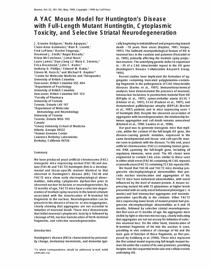
Results Additionally, in light of the observed electrophysiolog-ical changes in the hippocampi of YAC46 and YAC72mice (see below), we confirmed that human htt wasYAC Transgenic Miceexpressed in the hippocampi of YAC transgenic mice byTwo well-characterized YACs (YGA2 and 353G6) con-Western blot (Figure 2B) and by immunohistochemistrytaining 18 CAG repeats and spanning the entire HD gene(data not shown). Expression was low in peripheral tis-(Hodgson et al., 1996) were used.sues, similar to humans.We adopted a previously described strategy using
Mutant htt expression in fibroblast cell lines revealedhomologous recombination in yeast to introduce ex-htt expression in YAC72 founder 2498 to be approxi-panded CAG repeats (46 and 72) into YACs (Duff et al.,mately three times that seen in founder 2511 (Figure1994) (Figure 1A). Since YACs are inherently unstable,2C) and approximately twice that of endogenous levelsand this strategy relies on homologous recombination,(data not shown). Expression levels in YAC18, YAC46,all clones were extensively screened by Southern analy-and YAC72 (2511) lines were equivalent, representingsis, PCR, and pulse-field gel electrophoresis.one-third to one-half of endogenous level expressionSouthern analysis comparing signal intensity with hu-(Figure 2D).man genomic DNA demonstrated that all YAC46 trans-
genic lines had integrated one to two copies of the YACRescue of Embryonic Lethality(data not shown). Two of the seven lines injected withWe next tested for appropriate developmental regula-YAC72 integrated the complete HD gene (2498 andtion by assessing whether mice expressing mutant hu-2511). Densitometric scanning showed that founderman htt (YAC46 or YAC72) could compensate for the2498 had integrated approximately four copies of theloss of endogenous htt. Targeted disruption of htt resultstransgene (two wild-type), whereas line 2511 integratedin embryonic lethality at 7.5 days gestation (Duyao etone to two copies (Figure 1B). We were able to establishal., 1995; Nasir et al., 1995; Zeitlin et al., 1995). Welines from all founder mice with the exceptions of YAC46crossed the YAC46 and YAC72 lines onto the null back-664 and YAC72 2498, which developed an early neuro-ground and demonstrated efficient rescue of the em-logical phenotype at 6 weeks of age (see below) andbryonic lethality. The genotype of the YAC-positivedid not breed.offspring at the mouse HD locus had the expected Men-delian 1:2:1 ratio for homozygous, heterozygous, and
Transgene-Derived htt Is Expressed in a Pattern wild-type offspring (35:68:26, respectively). In contrast,Similar to Endogenous htt cDNA transgenic mice expressing full-length htt withBoth normal and mutant human htt were expressed in 18, 44, and 128 glutamines under the control of thethe same tissues as the endogenous mouse protein, cytomegalovirus promoter failed to rescue the embryowith highest levels seen in brain and testes (Figure 2A). lethality.Subcellular localization experiments on cortical tissue These data provide conclusive evidence of appro-from wild-type and transgenic mice showed that mutant priate developmental expression of mutant htt underYAC-derived htt primarily localized to the cytosolic and the control of the endogenous promoter and show thatmembrane-associated fractions (data not shown), simi- expansion of the polyglutamine tract does not disrupt
normal function of htt in mammalian development.lar to human htt (DiFiglia et al., 1995).
YAC Transgenic Mouse Model for HD183
Figure 2. Human htt Expression in YAC Transgenic Mice Parallels that Seen with Endogenous Mouse htt Expression
(A) The tissue expression patterns of wild-type (wt) and human htt in YAC18 (tg 18), YAC46 (tg 46), and YAC72 (tg 72) mice. Normal and YACtransgenic proteins (100 mg of total protein from each tissue) were probed with BKP1 (detects murine and human htt) and GHM1 (detectshuman htt only). Htt expression levels are highest in cortex, cerebellum, and testes. A longer exposure of the blots indicated murine andhuman htt are also expressed at low levels in other tissues tested.(B) Western blot using human-specific monoclonal antibody GHM1 showing hippocampal expression of human htt in two YAC46 (1749 and1746) and YAC18 (line 29) mice.(C) YAC transgenic mice expressing human htt with 72 glutamines have varying levels of expression, depending on the copy number of thegene. YAC72 founder 2498 expresses human htt at higher levels than founder 2511 using lysates from fibroblast lines assessed using GHMI.Equal loading was confirmed by equivalent actin levels in each lane.(D) YAC18, YAC46, and YAC72 transgenic mice express human htt at equivalent levels that are approximately one-third to one-half ofendogenous htt expression.
Motor and Behavioral Analysis YAC72The weight of the YAC72 mouse (2498) was approxi-Tests were designed to detect behavioral and motor
deficits in wild-type (n 5 7), YAC18 (line 30, n 5 7), and mately half that of her female siblings (16 g versus 29.9g; n 5 3) when sacrificed at 1 year. YAC72 mice fromYAC46 (line 668, n 5 7; line 1747, n 5 3) mice. Tests
were repeated at 2 month intervals between 3 and 20 line 2511 had body weight similar to controls at all timesassessed.months of age. In addition, observations were made on
the YAC72 mice lines and age-matched controls at 3, Observations of spontaneous behavior were carriedout on the 2498 mouse (Figure 3A) and age-matched5, 7, and 9 months of age. All mice used in this study
were pure FVB/N, eliminating the confounding effects controls (Figure 3B) at the ages of 3, 5, 7, and 9 months.The 2498 mouse showed obvious circling that was per-of strain differences. YAC18 mice behaved identically
to wild-type mice at all ages tested. sistent and by 9 months was associated with obviouschoreoathetoid movements of the head and neck asYAC46
Weekly observation of mouse behavior and handling of well as gait ataxia. The circling was tight and rapid, witha full turn being completed in approximately one-thirdthe mice revealed no obvious differences in behavior
compared with wild-type or YAC18 mice up to 20 months of a second (Figure 3C). When held by the tail, 2498was disoriented and displayed a foot-clasping postureof age. Responsiveness to sensory stimuli, including
auditory cues, olfactory cues, and touch; spontaneous reminiscent of HD exon 1 transgenic mice (Mangiariniet al., 1996). Additionally, as it was lowered to the benchactivity measurements; motor control; and coordination
were normal. top, unlike wild-type FVB/N mice, it failed to orient itself
Neuron184
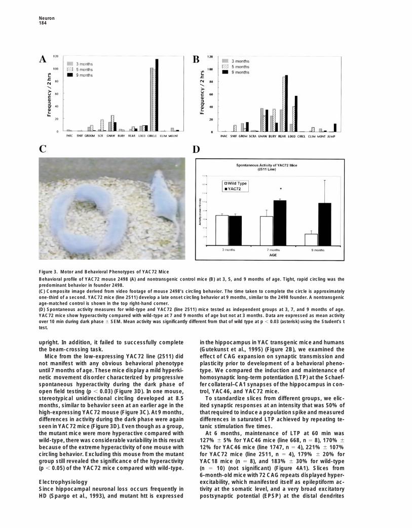
Figure 3. Motor and Behavioral Phenotypes of YAC72 Mice
Behavioral profile of YAC72 mouse 2498 (A) and nontransgenic control mice (B) at 3, 5, and 9 months of age. Tight, rapid circling was thepredominant behavior in founder 2498.(C) Composite image derived from video footage of mouse 2498’s circling behavior. The time taken to complete the circle is approximatelyone-third of a second. YAC72 mice (line 2511) develop a late onset circling behavior at 9 months, similar to the 2498 founder. A nontransgenicage-matched control is shown in the top right-hand corner.(D) Spontaneous activity measures for wild-type and YAC72 (line 2511) mice tested as independent groups at 3, 7, and 9 months of age.YAC72 mice show hyperactivity compared with wild-type at 7 and 9 months of age but not at 3 months. Data are expressed as mean activityover 10 min during dark phase 6 SEM. Mean activity was significantly different from that of wild type at p , 0.03 (asterisk) using the Student’s ttest.
upright. In addition, it failed to successfully complete in the hippocampus in YAC transgenic mice and humans(Gutekunst et al., 1995) (Figure 2B), we examined thethe beam-crossing task.
Mice from the low-expressing YAC72 line (2511) did effect of CAG expansion on synaptic transmission andplasticity prior to development of a behavioral pheno-not manifest with any obvious behavioral phenotype
until 7 months of age. These mice display a mild hyperki- type. We compared the induction and maintenance ofhomosynaptic long-term potentiation (LTP) at the Schaef-netic movement disorder characterized by progressive
spontaneous hyperactivity during the dark phase of fer collateral–CA1 synapses of the hippocampus in con-trol, YAC46, and YAC72 mice.open field testing (p , 0.03) (Figure 3D). In one mouse,
stereotypical unidirectional circling developed at 8.5 To standardize slices from different groups, we elic-ited synaptic responses at an intensity that was 50% ofmonths, similar to behavior seen at an earlier age in the
high-expressing YAC72 mouse (Figure 3C). At 9 months, that required to induce a population spike and measureddifferences in saturated LTP achieved by repeating te-differences in activity during the dark phase were again
seen in YAC72 mice (Figure 3D). Even though as a group, tanic stimulation five times.At 6 months, maintenance of LTP at 60 min wasthe mutant mice were more hyperactive compared with
wild-type, there was considerable variability in this result 127% 6 5% for YAC46 mice (line 668, n 5 8), 170% 612% for YAC46 mice (line 1747, n 5 4), 221% 6 107%because of the extreme hyperactivity of one mouse with
circling behavior. Excluding this mouse from the mutant for YAC72 mice (line 2511, n 5 4), 179% 6 20% forgroup still revealed the significance of the hyperactivity YAC18 mice (n 5 8), and 183% 6 30% for wild-type(p , 0.05) of the YAC72 mice compared with wild-type. (n 5 10) (not significant) (Figure 4A1). Slices from
6-month-old mice with 72 CAG repeats displayed hyper-excitability, which manifested itself as epileptiform ac-Electrophysiology
Since hippocampal neuronal loss occurs frequently in tivity at the somatic level, and a very broad excitatorypostsynaptic potential (EPSP) at the distal dendritesHD (Spargo et al., 1993), and mutant htt is expressed
YAC Transgenic Mouse Model for HD185
Figure 4. Effect of CAG Expansion on Synaptic Transmission
Graphs in (A1) and (A3) summarize LTP obtained from 6- and 10-month wild-type (wt), YAC18, YAC46, and YAC72 (line 2511) mice, respectively.Data are plotted as the mean fEPSP slope 6 SEM (normalized with respect to the 10 min immediately preceding the tetanus) versus time.Each point is an average of ten traces within 5 min bins. The baseline stimulation intensity was 30%–50% of the maximal response deliveredevery 30 s and was constant. Arrows indicate the time at which individual HFS was delivered. The traces are representative field recordings(averages of ten sweeps) showing the responses 10 min before (baseline) and 60 min after HFS. In (A2), D-AP5 blocks the hyperactivity seenin the dendritic region of 6-month-old YAC72 (2511) mice. The bar histograms show the area under the fEPSP in the absence and presenceof D-AP5 (25 mM).(B) Elevated resting calcium levels and lack of calcium influx in response to glutamate in 10-month-old YAC46 mice. Intracellular restingcalcium concentrations are increased in CA1 pyramidal neurons from YAC46 mice (B) (335 6 70 [n 5 6]) compared with wild-type mice (A)(165 6 12 [n 5 6] [p , 0.05]). Neurons from 10 month YAC46 (line 668) mice did not respond to either 10 mM or 100 mM glutamate (arrows)in contrast to wild-type mice.(C) PPF is not altered in YAC46 mice compared with wild-type mice at 6 and 10 months of age.

Neuron186
Figure 5. N-Terminal htt Aggregates in the YAC72 2498 Mouse
(A) and (B) are light micrographs showing EM48 immunoreactivity in the striatum of a wild-type control and mouse 2498, respectively. EM48immunoreactivity is obviously more intense in the nuclei of the striatal mouse 2498 than in wild type. EM48 immunoreactivity was very intensein neuronal nuclei (B), whereas HD549 immmunoreactivity was restricted to the cytoplasm (C). EM48 nuclear immunoreactivity (arrow) did notoccur in VAT- (D), NOSI- (E), or PARV- (F) immunoreactive neurons (arrowheads). Nuclear staining was diffuse and also included small (G)and large ([H], arrowheads) puncta. By electron microscopy (I), immunogold particles were seen both in the cytoplasm (c) and nucleus (n).Microaggregates containing more than five particles were found in the nuclei ([I], arrows). At higher magnification, microaggregates appearto be associated with amorphous, electron-dense material (J). Particles were also found in nuclear pores ([K], arrows). Nuclear pores notcontaining immunogold particles are indicated by the arrowheads (K). Scale bar, 30 mm (A–C); 10 mm (D–F); 8 mm (G and H); 3 mm (I); 50 nm(J); and 100 nm (K).
(Figure 4A1). 2-amino-5-phosphonovalerate (D-AP5) (25 is a crucial factor in the induction of LTP (Malenka etal., 1988, 1992), was intact in these mice. Calcium tran-uM), which blocks NMDA receptors (Morris et al., 1986)
and under normal conditions has no effect on the fast sients in Fura-2-loaded, acutely dissociated hippocam-pal CA1 neurons from 10-month-old YAC46 mice consis-synaptic response, blocked the somatic spiking and re-
duced the area under the field EPSP (fEPSP) curve (Fig- tently failed to produce a calcium signal in response toglutamate application (Figure 4B) in contrast to controls.ure 4A2; 211.75 6 0.189 mV.ms in control slices (n 5
4) and 23.82 6 0.24 mV.ms in D-AP5-treated slices (n 5 Lack of calcium influx can result from downregulationof NMDA receptors and/or reduced driving force for4) in 6-month-old YAC72 mice. These findings suggest
that in 6-month-old YAC72 mice, the NMDA component calcium. In keeping with these possibilities, resting lev-els of calcium in neurons from these mutant mice wereis prominent during fast synaptic transmission. Follow-
ing tetanization, most of the slices from YAC72 mice higher (335 6 70 nM [n 5 6]) than those of controls(165 6 12 nM [n 5 6] [p , 0.05]) (Figure 4B), suggestingdisplayed a greater short-term potentiation (372% 6
124%, p 5 0.20 when compared with wild-type and that the calcium-buffering capacity of mutant neuronswas reduced.YAC18), which progressively declined over time.
By 10 months of age, obvious and significant differ- Elevated intracellular calcium concentration can alsoaffect presynaptic release mechanisms. To assess pre-ences were seen in both YAC46 and YAC72 mice. In
contrast to wild-type, LTP was not induced in CA1 neu- synaptic efficacy, we investigated the extent of thepaired-pulse facilitation (PPF) in wild-type, YAC18, androns of either line of mice expressing mutant htt (Figure
4A3). The potentiation at 60 min after the fifth tetanus YAC46 mice, which showed no differences from controlsat 6 and 10 months of age (Figure 4C).in slices from 10 month YAC46 mice was 101% 6 25%
(line 668, n 5 6) and 108% 6 12% (line 1746, n 5 6) However, posttetanic potentiation, which is the poten-tiation seen immediately after tetanic stimulation and iscompared with 199% 6 31% (n 5 6) in wild-type mice
(p , 0.01). High-frequency stimulation (HFS) induced also indicative of presynaptic release mechanisms, wasreduced in 10-month-old YAC46 mice. The normalizeddepression instead of potentiation in 10 month YAC72
mice (Figure 4A3). Lack of LTP induction in mutant slices fEPSP slope immediately after the first HFS was 224% 6
15% in wild-type mice (n 5 6), 167% 6 25% in Trans-was not due to the deterioration of slices, as no changein the responses within the same slice from the nonteta- genic line 668 (n 5 5), and 102% 6 9% in transgenic
line 1746 (n 5 6) (p , 0.05). These data suggest thatnized control pathway was recorded.To determine the mechanism underlying the progres- presynaptic terminals in mutant mice, while responding
normally to single stimuli, are not able to sustain neuro-sive loss of LTP in 10 month YAC46 mice, we next inves-tigated whether glutamate-mediated calcium influx, which transmitter release during high-frequency stimulation.

YAC Transgenic Mouse Model for HD187
Figure 6. Neurodegeneration in Mouse 2498
(A) through (C) are micrographs of 1.5 mm striatal sections from YAC72 2498 (A and B) and YAC18 (C) immunoreacted with EM48 andcounterstained with toluidine blue. In YAC72 2498, many degenerating neurons (stars) are present in the most lateral regions of the striatum([A], stars) as compared with the more medial regions (B). In YAC18, neurons had a normal appearance, with a regular and well-roundednuclear envelope (C). Intense EM48 staining (brown) is seen in a neuronal nucleus (A) in the lateral striatum and in the cytoplasm of neuronsin the medial portion of the striatum (B). Many degenerated neurons contained immunogold particles in their nuclei (n) and cytoplasm (c). In(D), evidence of degeneration included condensed cytoplasm, swelling of mitochondria (m), vacuolation of Golgi (g), and condensation andmarginization of the heterochromatin ([E], arrows). By electron microscopy, many neurons that did not show frank degenerative signs hadabnormal scalloped nuclear membranes (F). Scale bar, 20 mm (A–C); 3 mm (D); 200 nm (E); and 3 mm (F).
Neuropathology: Nuclear Translocation We and others have proposed that mutant htt is cleavedin the cytoplasm and that only the N-terminal portion ofof N-Terminal htt
Brains from wild-type (n 5 7), YAC18 (n 5 4), and YAC46 the protein is translocated into the nuclei, where it mayform aggregates (Goldberg et al., 1996; Cooper et al.,(n 5 5) mice were examined at 5, 9, 12, and 20 months
of age. The brains of YAC72 mice were examined only 1998; Hackam et al., 1998; Martindale et al., 1998; Well-ington et al., 1998). We also performed immunocyto-at 5 (n 5 2), 8 (n 5 2), 10 (n 5 3), and 12 ([n 5 3] [2511],
[n 5 1] [2498]) months. The gross morphology of each chemistry in adjacent sections using EM48 and HD549,a rat monoclonal antibody that reacts with an internalbrain appeared normal, and a reduction in brain weight
was observed only in YAC72 mouse 2498 (0.41 versus region of htt (amino acids 549–679). Sections stained withHD549 alone exhibited intense staining of neuronal peri-0.58 g in controls [n 5 3]).
In all mice examined, faint EM48 staining was present karya and dendrites as described previously (Gutekunstet al., 1995). HD549 immunoreactivity was not detectedin neurons throughout the brain. In contrast, in the 12-
month-old YAC72 mouse (2498), EM48 intensely stained in neuronal nuclei (Figure 5C), suggesting that onlyN-terminal fragments of mutant htt (,549 amino acids)many nuclei within neurons in the striatum (Figure 5B),
olfactory tubercle, nucleus accumbens, and fundus stri- translocate to the nuclei.ati. Small numbers of neurons in the septum and thegranule cell layer of the cerebellar cortex also had nu- Nuclear Staining Is Specific to Medium
Spiny Neuronsclear EM48 immunoreactivity. This intense nuclear stain-ing was not seen in any other brain regions, including We performed double immunolabeling using EM48 and
antibodies specific for each interneuron population, in-hippocampus, thalamus, and brain stem, nor was it pres-ent in YAC18 or YAC46 or in wild-type controls (Figure cluding the vesicular acetylcholine transporter (VAT),
which labels cholinergic interneurons; nitric oxide synth-5A). EM48 nuclear staining was more intense in the lat-eral compared with the medial striatum. ese (NOSI), which labels neurons containing somato-
statin; neuropeptide Y and reduced nicotinamide adenineIn the 12-month-old YAC72 line 2511 (n 5 3), there wasan increased number of EM48-immunoreactive neuronal dinucleotide phosphate– (NADPH-) diaphorase; and parv-
albumin (PARV), which labels a distinct population ofnuclei in the striatum (Figure 7A) and cortex (Figure 7B).
Neuron188
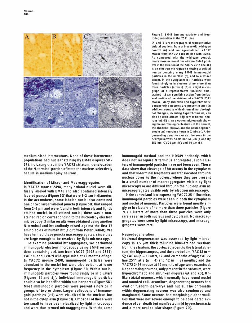
Figure 7. EM48 Immunoreactivity and Neu-rodegeneration in the 2511 Line
(A) and (B) are micrographs of representativestriatal sections from a 1-year-old wild-typecontrol (A) and an age-matched YAC72mouse from line 2511 (B) stained with EM48.As compared with the wild-type control,many more neuronal nuclei were EM48 posi-tive in the striatum of the YAC72 2511 line. (C)is an electron micrograph showing a striatalneuron containg many EM48 immunogoldparticles in the nucleus (n), and to a lesserextent, in the cytoplasm (c). Particles werefound singly or in clusters of no more thanthree particles (arrows). (D) is a light micro-graph of a representative toluidine blue–stained 1.5 mm semithin section from the lat-eral portion of the striatum of a YAC72 2511mouse. Many shrunken and hyperchromaticdegenerating neurons are present (stars). Inaddition, neurons with abnormal morphologi-cal changes, including hyperchromasia, canalso be seen (arrows) adjacent to normal neu-rons (n). (E) is an electron micrograph show-ing the morphological features of the normal,the abnormal (arrow), and the neurodegener-ated (star) neurons shown in (D) (inset). A de-generating dendrite can also be seen in theneuropil (arrow). Scale bar, 60 mm (A and B);350 nm (C); 20 mm (D); and 10 mm (E).
medium-sized interneurons. None of these interneuron immunogold method and the HD549 antibody, whichdoes not recognize N terminus aggregates, such clus-populations had nuclear staining by EM48 (Figures 5D–
5F), indicating that in the YAC72 striatum, translocation ters of immunogold particles have not been seen. Thesedata show that cleavage of htt occurs in the cytoplasmof the N-terminal portion of htt to the nucleus selectively
occurs in medium spiny neurons. and that N-terminal fragments are translocated throughnuclear pores to the nucleus, where they are presentin a small number of macroaggregates visible by lightIdentification of Micro- and Macroaggregatesmicroscopy or are diffused through the nucleoplasm asIn YAC72 mouse 2498, many striatal nuclei were dif-microaggregates visible only by electron microscopy.fusely labeled with EM48 and also contained intensely
In the control and low-expressing YAC72 2511 line mice,labeled puncta (Figure 5G) that were 1–2 mm in diameter.immunogold particles were seen in both the cytoplasmIn the accumbens, some labeled nuclei also containedand nuclei of neurons. Particles were found mostly sin-one or two larger labeled puncta (Figure 5H) that rangedgly or in clusters of no more than three particles (Figurefrom 2–5 mm and were found in both intensely and lightly7C). Clusters of more than three particles were onlystained nuclei. In all stained nuclei, there was a non-rarely seen in both nucleus and cytoplasm. No macroag-stained region corresponding to the nucleoli by electrongregates were seen by light microscopy, and microag-microscopy. Similar results were obtained using anothergregates were rare.N-terminal anti-htt antibody raised against the first 17
amino acids of human htt (a gift from Peter Detloff). Wehave termed these puncta macroaggregates, since they Neurodegeneration
Neuronal degeneration was assessed by light micros-are large enough to be resolved by light microscopy.To examine potential htt aggregates, we performed copy in 1.5 mm thick toluidine blue–stained sections
from the striatum, the cortex adjacent to the lateral stria-immunogold electron microscopy using EM48 on sec-tions containing striatum from YAC72 (2498 and 2511), tum, the hippocampus, and the cerebellum. YAC18 (n 5
5); YAC46 (n 5 10) at 9, 12, and 20 months of age; YAC72YAC18, and FVB/N wild-type mice at 12 months of age.In YAC72 mouse 2498, immunogold particles were line 2511 at 8 (n 5 4) and 12 (n 5 3) months; and the
YAC72 2498 mouse at 12 months of age were examined.abundant in the nuclei but were also evident at lowerfrequency in the cytoplasm (Figure 5I). Within nuclei, Degenerating neurons, only present in the striatum, were
hyperchromatic and shrunken (Figures 6A and 7D). Un-immunogold particles were found singly or in clusters(Figures 5I and 5J). Individual immunogold particles like striatal neurons, which normally have round nuclei
and rounded cellular outlines, degenerating neurons hadcould also be identified within nuclear pores (Figure 5K).Most immunogold particles were present singly or in oval or fusiform perikarya and nuclei. The chromatin
within degenerating neurons was also condensed andgroups of two or three. Larger collections of immuno-gold particles (.5) were visualized in the nucleus but marginated. Some neurons had morphologic abnormali-
ties that were not severe enough to be considered evi-not in the cytoplasm (Figure 5I). Almost all of these weretoo small to have been visualized by light microscopy dence of cell death but manifested mild hyperchromasia
and a more oval cellular shape (Figure 7D).and were thus termed microaggregates. With the same

YAC Transgenic Mouse Model for HD189
Electron microscopy using the same tissue blocks con- months, YAC72 mice had an initial increase in the NMDAcomponent of synaptic response, which was antago-firmed the degenerative and abnormal nature of thesenized by D-AP5, an NMDA receptor antagonist. This ismorphologic changes. By electron microscopy, degen-consistent with the NMDA receptor hyperactivity associ-erative changes varied in severity and included reducedated with the presence of mutant htt. In animals, over-neuronal size, the presence of nuclear and plasma mem-activation of NMDA receptors in the striatum has beenbrane irregularities, increased electron density of theshown to reproduce biochemical and neuropathologicalcytoplasm and the nucleus, swelling of some mitochon-changes seen in HD (Ferrante et al., 1985; Beal et al.,dria, dilation of Golgi cisternae, nuclear shrinkage and1989). Recent in vitro data also reveal that mutant httcondensation, and margination of heterochromatin (Fig-increases the size of currents through the NMDA recep-ures 6D, 6E, and 7E). These degenerative features aretor subtypes, which are preferentially expressed in theconsistent with apoptosis. In addition to frank degenera-medium spiny neostriatal neurons (Chen et al., 1999).tion, electron microscopy also revealed frequent irregu-These data represent in vivo evidence in favor of NMDAlar nuclear envelopes in neurons (Figure 6F).receptor hyperactivity as an early abnormality in theSignificant degeneration was present in the striatumpathogenesis of HD, consistent with the hypothesis ofof the YAC72 2498 mouse and in the YAC72 2511 lineexcitotoxicity in HD (Albin and Greenamyre, 1992; Coyleat 12 months. No degeneration was found in the YAC46and Puttfarchen, 1993).or YAC18 (Figure 6C) or in wild-type controls. In the
An increase in NMDA receptor function would give risestriatum of mouse 2498, there was extensive neuronalto an elevated influx of calcium during normal synapticdegeneration, most severe laterally, decreasing in atransmission. Our studies indicated higher resting levelsgraded fashion more medially (Figures 6A and 6B). Theof calcium in neurons from mutant mice at 10 monthsproportion of degenerating neurons ranged from 4% ofof age. Increases in intracellular calcium levels are asso-observable neurons medially to 80% of neurons laterally.ciated with calcium-dependent desensitization of bothIn the striatum of 12-month-old low-expressing YAC72voltage- and ligand-gated channels, including NMDAmice (2511), numerous degenerating neurons (Figurereceptors. It is therefore possible that during high-fre-7D) were present. As in 2498, degenerating neuronsquency stimulation, NMDA receptors are inactivatedwere most frequent in the lateral striatum. The medial-due to high-calcium influx, which could underlie the lackto-lateral gradient of neurodegeneration was less dra-of LTP in those mice with mutant htt. In both YAC46matic than in the 2498 mouse, ranging from 4% mediallyand YAC72 mice, the electrophysiological abnormalitiesto z40% laterally. We found no sign of degenerationprecede any obvious behavioral abnormalities as wellin the YAC72 2511 line at 8 months of age. There wasas any evidence of neurodegeneration or aggregate for-no evidence of reactive gliosis in these mice by glialmation. Taken together, these results suggest that ex-fibrillary acidic protein immunocytochemistry (data notpression of mutant htt in these mice causes cytoplasmicshown).dysfunction before the observed behavioral and neuro-The hippocampus was examined, since YAC transgenicpathologic changes.mice had abnormal hippocampal LTP, and neuronal loss
Examination of N terminus htt fragment aggregationin the CA1 region of the hippocampus is seen in mostin the YAC72 2498 and YAC72 2511 lines does not sup-patients with HD (Spargo et al., 1993). Examination ofport the hypothesis that large nuclear aggregates under-semithin and Nissl-stained sections (50 mm) from wild-lie neurodegeneration. In the YAC72 2498 mouse, whichtype, YAC18, and YAC46 mice at ages 5, 9, 12, and 20expressed high levels of mutant htt and had an obviousmonths showed no obvious differences in the grossbehavioral phenotype by 6 weeks of age, 80% of theorganization and neuronal density of the CA1 pyramidalneurons still observable in the lateral striatum at 1 yearcell layer and no increase in the presence of degenerat-of age were undergoing neurodegeneration. In both de-ing neurons.generating and normal-appearing neurons, N-terminalnuclear htt fragments were labeled with solitary and
Discussion small clusters of immunogold particles, and only rarelywere macroaggregates observed. Furthermore, the com-
We have produced YAC transgenic mice that express plete absence of macroaggregates and the rarity of mi-normal and mutant htt in a developmentally regulated croaggregates in the normal and degenerating striataland cell-specific pattern identical to that of endogenous neurons in the YAC72 2511 line at 12 months of agehtt and develop functional, behavioral, and pathological suggest that even microaggregates may not be neces-changes similar to those of patients with HD. This is sary for cell death. The immunogold method, however,manifest with selective neurodegeneration affecting does not permit distinguishing solitary N terminus frag-specifically the medium spiny neurons of the striatum. ments from the smallest possible multimers.Furthermore, we show that neurodegeneration can oc- In vitro evidence has suggested that proteolytic cleav-cur in the absence of aggregates, providing evidence age of htt plays an important role in the pathogenesisthat aggregates are not essential for the initiation of the of htt (Hackam et al., 1998, 1999; Martindale et al., 1998).illness. The studies in the YAC transgenic mice now provide direct
YAC transgenic mice expressing mutant htt with 46 evidence of cleavage of htt by demonstration of N-termi-CAG repeats do not have a clinical phenotype by de- nal fragments in the nucleus and C-terminal fragmentstailed behavioral analysis for up to 20 months of age. in the cytoplasm. Furthermore, immunogold-labeled elec-However, electrophysiological abnormalities are evident tron microscopy has revealed gold-labeled htt frag-much earlier. The severity of electrophysiological abnor- ments traversing the nuclear pore to the nucleus frommalities is more obvious in mice with 72 compared with the cytosol. Importantly, the N-terminal translocation of
htt appears to be only evident in those neurons that are46 CAG repeats at the same age. Interestingly, at 6
Neuron190
repeats, was linearized with BstEII and transformed into yeast sphe-susceptible to subsequent neurodegeneration, namely,roplasts. The region of DNA containing the expanded CAG repeatthe medium spiny neurons. The striatal interneurons,plus sufficient flanking homologous DNA (EE640) was cloned intowhich are largely spared in HD, did not show presencethe pRS406 vector. The EE640 fragment, containing 48 CAG repeats,
of htt in the nucleus. This suggests that one reason was cloned from the L191F1 cosmid containing an expanded CAGfor selective neuronal degeneration in HD might be the repeat. This same fragment, containing 72 repeats, was shotgun
cloned from patient DNA. These clones were transformed into yeast,generation of proteolytic cleavage of htt in specific cells,and pop-in and pop-out clones were selected as described pre-with resultant N-terminal translocation of htt into theviously (Duff et al., 1994).nucleus specifically in those cells.
The increased intracellular calcium concentrationGeneration and Screening of YAC Transgenic Miceseen in the YAC46 mice at 10 months of age, prior to anyYAC DNA was prepared for microinjection into FVB/N pronuclei,evidence of nuclear translocation of htt or apoptosis,and founder pups were screened (Hodgson et al., 1996). Furthersupports a model in which the initial trigger for the illnessanalysis of the 59 region of YAC YGA2 was done by Southern analysis
is cytoplasmic toxicity, which is associated with in- using the probes OS14, D4S95, and OT17, a gift from Dr. S. Hadano.creased cytoplasmic calcium concentration. These find-ings are highly reminiscent of the recent reports in early
Protein Expression Analysisonset familial Alzheimer’s disease, whereby activation Bone marrow fibroblast cultures were established from YAC72of caspases by mutant presenilin 1 is preceded by an founders 2498 and 2511 and an age-matched FVB/N mouse in Dul-elevation in cytoplasmic calcium (Buxbaum et al., 1998). becco’s modified Eagle’s medium, 10% fetal calf serum, penicillin/
streptomycin, and 20 mM L-glutamine (Canadian Life Technologies).Altered concentrations of cytoplasmic calcium may beCells were maintained at a humidified atmosphere of 378C, 6% CO2.an important trigger for activation of the proteolyticFor the isolation of proteins, cells were lysed in buffer (1.0% NP40,pathways, leading to the generation of truncated frag-0.15 M NaCl, 50 mM Tris [pH 8.0], and 0.01% azide). Protein (100
ments for both Alzheimer’s disease and HD (Kim andmg) was resolved in 7.5% SDS–PAGE minigels. Membranes were
Tanzi, 1998). reacted with the anti-htt monoclonal antibody GHM1 (Kalchman etRecently, there have been reports of other transgenic al., 1997) or an anti-actin monoclonal antibody (Sigma) by standard
conditions.mice expressing different portions of htt (Mangiarini etal., 1996; White et al., 1997; Reddy et al., 1998; Schillinget al., 1999) under the control of varying promoters. Behavioral Analysis
Mice were handled and checked on a weekly basis. For a moreThese mice display a significant neurological phenotypequalitative assessment, YAC46, YAC18, and control mice were vid-associated with the development of intranuclear aggre-eotaped for 2 hr in a fresh cage identical to the one in which theygates and in some cases, neurodegeneration, but as yet,were housed every 2 months from 8 to 20 months of age. For a 10no evidence of the selective striatal neurodegenerations interval every minute, the behavior of the subjects was recorded;
seen in HD. This suggests that expression of full-length the frequency of such behaviors as inactivity, grooming, scratching,htt and/or expression in a cell-specific and develop- sniffing, gnawing, burying, rearing, locomotion, climbing, circling,
jumping, and mounting was noted. YAC72 mice (line 2511) weremental manner, as seen with its own promoter, mayassessed at 3, 7, and 9 months, while YAC72 mouse 2498 wasbe important for accurately recapitulating the diseaseassessed every 2 months from 3 to 12 months of age.phenotype.
Motor control was tested by the ability to climb a wire screenA major question still left unanswered is, what is thesecured against the wall at a 608 angle and was observed until they
nature of the initial toxic stimulus in the cytoplasm that reached the top or bottom. For coordination, mice were placedinitiates the cell death pathways, leading to neurodegen- midway on a suspended beam and observed for latency of initiative
movement, number of falls, and time to reach safety. Mice wereeration? One possibility is that there may be disturbedassessed for abnormal righting or posturing by suspension by theinteraction with an interacting protein such as HAP1 (Litail. Spontaneous motor activity was measured with photo beamset al., 1995) or HIP1 (Kalchman et al., 1997; Wankerin a novel test chamber during 2 hr of light and 2 hr of dark aset al., 1997), which then induces cellular toxicity anddescribed previously (Nasir et al., 1995).
promotes the proteolytic processing of htt. ElectrophysiologyThese YAC transgenic mice represent the first animal Hippocampal slices (400 mm thick) from 6- and 10-month-old sex-
matched mice were obtained as described previously (Dingledine,model with mutant full-length htt expressed under the1984) and perfused in an interface chamber at 328C with oxygenatedcontrol of its endogenous promoter, with all intronicartificial cerebrospinal fluid (ACSF) containing (in mM): NaCl, 124;regulatory sequences included. This developmentallyKCl, 3.0; KH2PO4, 1.25; CaCl2, 4.0; NAHCO3, 26.0; and D-glucose,appropriate expression of mutant htt in the appropriate10 and 10 mm bicucculline methiodide (GMI; Sigma Chemical). In
cellular compartments has produced animals with be- selected slices, 25 mM D-AP5 (Tocris) was added to the perfusatehavioral, electrophysiological, and pathological pheno- to block NMDA receptors. To invoke tetanus, LTP consisted of five
trains of 100 Hz stimulations lasting 200 ms at an intertrain intervaltypes occurring at different stages of development, giv-of 10 s. A control pathway, which did not receive pattern stimulation,ing insights into the sequence of cellular and molecularwas also monitored to determine the general state of the slice. Slicesevents underlying the pathogenesis of HD. In particular,that demonstrated either an increase or decrease in the fEPSP slopethe YAC72 line, with selective striatal neurodegenerationevoked by the stimulation of the control pathway following HFS of
by 1 year of age, allows therapeutic strategies to be the test pathway were omitted from data analysis.assessed that delay or prevent HD. Data are expressed as mean 6 SEM, n 5 number of animals.
Significance was tested by the Student’s t test. Experiments weredone double blind.Calcium MeasurementsExperimental ProceduresPunched cultures of CA1 neurons were dissociated as describedpreviously (Cohen and Wilkin, 1995). Cells were loaded with 5 mMYAC Mutagenesis
YAC mutagenesis was performed as described previously (Duff et Fura2-AM (Molecular Probes) by incubating them for 45 min at roomtemperature in ACSF containing (in mM): NaCl, 140; KCl, 5; MgCl2,al., 1994). The pop-in construct (see text), containing 46 or 72 CAG
YAC Transgenic Mouse Model for HD191
1; CaCl2, 2; and HEPES, 10 (buffered to pH 7.3 with NaOH). Fluores- X. J. L.). Dr. Michael Hayden is an established investigator of theBritish Columbia Children’s Hospital.cence measurements were conducted by the use of a PTI D104F
series microscope (Nikon Diaphot 200) and a 403 oil immersionobjective at room temperature. Cell densities were one to five cells Received March 8, 1999; revised April 12, 1999.per field. Baseline (30 s) measurements were obtained prior to gluta-mate (10–100 mM) application. Digitonin (20 mM) was added at the
Referencesend of the experiment to record background fluorescence. Ratiomeasurements were converted into estimates of [Ca21]i according
Albin, R.L., and Greenamyre, J.T. (1992). Alternative excitotoxic hy-to the Grynkiewicz equation (Grynkiewicz et al., 1985). Kd, Rmin, Rmax,pothesis. Neurology 42, 733–738.Fb380, and Ff380 values were determined with a calcium calibration
kit containing Mg21 (Molecular Probes) and were rechecked every Beal, M.F., Lowall, N.W., Swartz, K.J., Ferrante, R.J., and Martin,week. Estimated values were Kd, 227 6 5 nM; Rmin, 0.44; and Rmax, 5.7. J.B. (1989). Differential sparing of somatostain-neuropeptide Y and
cholinergic neurons following striatal excitotoxin lesions. Synapse3, 38–47.
AntibodiesBecher, M.W., Rubinsztein, D.C., Leggo, J., Wagster, M.V., Stine,Four antibodies against various regions of htt were used: a rabbitO.C., Ranen, N.G., Franz, M.L., Abbott, M.H., Sherr, M., MacMillan, etpolyclonal antibody (EM48) to amino acids 1–256 (Gutekunst et al.,al. (1997). Dentatorubral and pallidoluysian atrophy (DRPLA). Clinical1999), a rabbit polyclonal antibody (HD-N) to the first 17 amino acidsand neuropathological findings in genetically confirmed North Amer-of human htt (provided by Peter Detloff), a rat monoclonal antibodyican and European pedigrees. Mov. Disord. 12, 519–530.(HD549) to amino acids 549–679 (Gutekunst et al., 1995), and aBuxbaum, J.D., Choi, E.K., Luo, Y., Lilliehook, C., Crowley, A.C.,monoclonal antibody (GHMI) directed to the internal region of httMerriam, D.E., and Wasco, W. (1998). Calsenilin: a calcium-binding(Hodgson et al., 1996). Other antibodies were directed against theprotein that interacts with the presenilins and regulates the levelsVAT, which labels cholinergic interneurons; NOSI (Chemicon Inter-of a presenilin fragment. Nat. Med. 4, 1177–1181.national, Temecula, CA), which labels neurons containing somato-
statin, neuropeptide Y, and NADPH-diaphorase activity; and PARV Chen, N., Luo, T., Wellington, C., Metzler, M., McCutcheon, K., Hay-(Sigma, St. Louis, MO), which labels another distinct population of den, M.R., and Raymond, L.A. (1999). Subtype-specific enhance-medium-sized interneurons. ment of NMDA receptor currents by mutant huntingtin. J. Neuro-
chem. 72, 1820–1898.
Cohen, J., and Wilkin, G.P. (1995). Neural Cell Culture (New York:Light and Electron Microscopic ImmunocytochemistryOxford University Press).Each mouse was anesthetized with sodium pentobarbitol, injectedCooper, J.K., Schilling, G., Peters, M.F., Herring, W.J., Sharp, A.H.,intraperitoneally with 300 IU of heparin, and perfused intracardiallyKaminsky, Z., Masone, J., Khan, F.A., Delanoy, M., Borchelt, D.R.,with 200 ml of 3% paraformaldehyde and 0.15% glutaraldehyde inet al. (1998). Truncated N-terminal fragments of huntingtin with ex-0.1 M phosphate buffer (PB) at pH 7.2. Brains were removed andpanded glutamine repeats form nuclear and cytoplasmic aggregatespostfixed overnight in 2% paraformaldehyde. Brains were sectionedin cell culture. Hum. Mol. Genet. 7, 783–790.in ten series of 40 mm coronal sections with a vibratome (Pelco,
Redding, CA), collected in PB, and rinsed in 0.05 M phosphate- Coyle, J.T., and Puttfarchen, P. (1993). Oxidative stress, glutamate,buffered saline (PBS [pH 7.2]). Light and electron microscopy immu- and neurodegenerative diseases. Ann. Neurol. 38, 357–366.nocytochemistry were performed as described previously (Gute- Davies, S.W., Turmaine, M., Cozens, B.A., DiFiglia, M., Sharp, A.H.,kunst et al., 1995, 1999). Ross, C.A., Scherzinger, E., Wanker, E.E., Mangiarini, L., and Bates,
For double immunolabeling experiments, EM48 antibody was G.P. (1997). Formation of neuronal intranuclear inclusions (NII) un-combined with either VAT, NOSI, or PARV antibody in the primary derlies the neurological dysfunction in mice transgenic for the HDincubation. Controls included the omission of primary antisera and mutation. Cell 90, 537–548.single labeling for each antibody.
DiFiglia, M., Sapp, E., Chase, K., Schwarz, C., Meloni, A., Young,C., Martin, E., Vonsattel, J.P., Carraway, R., Reeves, S.A., et al.(1995). Huntingtin is a cytoplasmic protein associated with vesiclesAssessment of Neurodegenerationin human and rat brain neurons. Neuron 14, 1075–1081.Neurodegeneration was assessed in semithin sections (1.5 mm) from
striatum, cortex, hippocampus, and cerebellum in all lines. Sections DiFiglia, M., Sapp, E., Chase, K.O., Davies, S.W., Bates, G.P., Vonsat-were cut with a Leica Ultracut S ultramicrotome, counterstained tel, J.P., and Aronin, N. (1997). Aggregation of huntingtin in neuronalwith toluidine blue, differentiated in 95% alcohol, and coverslipped. intranuclear inclusions and dystrophic neurites in brain. ScienceNeuronal degeneration was quantified as follows: sections were 277, 1990–1993.visualized on a Nikon Labophot-2 microscope with a 603 lens Dingledine, R. (1984). Brain Slices (New York: Plenum Press).equipped with a 1 mm2 ocular grid. A starting frame was systemati-
Duff, K., McGuigan, A., Huxley, C., Schulz, F., and Hardy, J. (1994).cally selected so that one side of the ocular grid was touching theInsertion of a pathogenic mutation into a yeast artificial chromosomemost lateral portion of the corpus callosum. From this starting point,containing the human amyloid precursor protein gene. Gene Ther.counts were made with three 240 mm2 frames obtained by moving1, 70–75.the grid toward the medial portion of the striatum, one grid lengthDuyao, M.P., Auerbach, A.B., Ryan, A., Persichetti, F., Barnes, G.T.,at a time. This was repeated in each section. Within each frame, allMcNeil, S.M., Ge, P., Vonsattel, J.-P., Gusella, J.F., Joyner, A.L., andneurons with visible nuclei were counted and categorized as normalMacDonald, M.E. (1995). Inactivation of the mouse Huntington’sor degenerating. In each counting frame, the top and right framedisease gene homolog Hdh. Science 269, 407–410.lines were excluded from analysis.Ferrante, R.J., Kowall, N.W., Beal, M.F., Richardson, E.P., Jr., Bird,E.D., and Martin, J.B. (1985). Selective sparing of a class of striatal
Acknowledgments neurons in Huntington’s disease. Science 230, 561–563.
Goldberg, Y.P., Nicholson, D.W., Rasper, D.M., Kalchman, M.A.,We thank our colleagues in our laboratories for useful commentsKoide, H.B., Graham, R.K., Bromm, M., Kazemi-Esfarjani, P.,and discussion, in particular, Dr. Cheryl Wellington, Dr. AbigailThornberry, N.A., Vaillancourt, J.P., and Hayden, M.R. (1996). Cleav-Hackam, and Ryan Brinkman. We would also like to thank Georgeage of huntingtin by apopain, a proapoptotic cysteine protease, isSakellaropoulos, Hong Yi, Courtney Dorn, and James S. Mulroy formodulated by the polyglutamine tract. Nat. Genet. 13, 442–449.their technical assistance. This work was supported by grants fromGrynkiewicz, G., Poenie, M., and Tsien, R.Y. (1985). A new generationthe Medical Research Council of Canada (M. R. H. and B. R. L.),of Ca21 indicators with greatly improved fluorescence properties.the Canadian Genetic Diseases Network, the Huntington DiseaseJ. Biol. Chem. 260, 3440–3450.Society of America (M. R. H., C. A. G., and X. J. L.), and the National
Institutes of Health (NS35255 to S. H. and C. A. G. and NS36232 to Gutekunst, C.-A., Levey, A.I., Heilman, C.J., Whaley, W.L., Yi, H.,
Neuron192
Nash, N.R., Rees, H.D., Madden, J.J., and Hersch, S.M. (1995). Iden- Pittman, R.N. (1997). Intranuclear inclusions of expanded polyglu-tification and localization of huntingtin in brain and human lympho- tamine protein in spinocerebellar ataxia type 3. Neuron 19, 333–344.blastoid cell lines with anti-fusion protein antibodies. Proc. Natl. Reddy, P.H., Williams, M., Charles, V., Garrett, L., Pike-Buchanan,Acad. Sci. USA 92, 8710–8714. L., Whetsell, W.O., Jr., Miller, G., and Tagle, D.A. (1998). BehavioralGutekunst, C.-A., Li, S.-H., Yi, H., Mulroy, J.S., Kuemmerle, S., Jones, abnormalities and selective neuronal loss in HD transgenic miceR., Rye, D., Ferrante, R.J., Hersch, S.M., and Li, X.-J. (1999). Nuclear expressing mutated full-length HD cDNA. Nat. Genet. 20, 198–202.and neuropil aggregates in Huntington’s disease: relationship to Saudou, F., Finkbeiner, S., Devys, D., and Greenberg, M.E. (1998).neuropathology. J. Neurosci. 19, 2522–2534. Huntingtin acts in the nucleus to induce apoptosis but death doesHackam, A.S., Singaraja, R., Wellington, C.L., Metzler, M., McCut- not correlate with the formation of intranuclear inclusions. Cell 95,cheon, K., Zhang, T., Kalchman, M., and Hayden, M.R. (1998). The 55–66.influence of huntingtin protein size on nuclear localization and cellu- Schilling, G., Becher, M.W., Sharp, A.H., Jinnah, H.A., Duan, K.,lar toxicity. J. Cell Biol. 141, 1097–1105. Kotzuk, J.A., Slunt, H.H., Ratovitski, T., Cooper, J.K., Jenkins, N.A.,Hackam, A.S., Singaraja, R., Zhang, T., Gan, L., and Hayden, M.R. et al. (1999). Intranuclear inclusions and neuritic aggregates in(1999). In vitro evidence for both the nucleus and cytoplasm as transgenic mice expressing a mutant N-terminal fragment ofsubcellular sites of pathogenesis in Huntington disease. Hum. Mol. huntingtin. Hum. Mol. Genet. 8, 397–407.Genet. 8, 25–33. Skinner, P.J., Koshy, B.T., Cummings, C.J., Klement, I.A., Helin, K.,Harper, P.S. (1991). Huntington’s Disease (Philadelphia: W.B. Servadio, A., Zoghbi, H.Y., and Orr, H.T. (1997). Ataxin-1 with anSaunders). expanded glutamine tract alters nuclear matrix–associated struc-
tures. Nature 389, 971–974.Hayden, M.R. (1981). Huntington’s chorea (Berlin: Springer-Verlag).
Spargo, E., Everall, I.P., and Lantos, P.L. (1993). Neuronal loss inHodgson, J.G., Smith, D.J., McCutcheon, K., Koide, H.B., Nishiyama,K., Dinulos, M.B., Stevens, M.E., Bissada, N., Nasir, J., Kanazawa, the hippocampus in Huntington’s disease: a comparison with HIVK., et al. (1996). Human huntingtin derived from YAC transgenes infection. J. Neurol. Neurosurg. Psychiatry 56, 487–491.compensates for loss of murine huntingtin by rescue of the embry- Vonsattel, J.P., Myers, R.H., Stevens, T.J., Ferrante, R.J., Bird, E.D.,onic lethal phenotype. Hum. Mol. Genet. 5, 1875–1885. and Richardson, E.P., Jr. (1985). Neuropathological classification ofHuntington’s Disease Collaborative Research Group (1993). A novel Huntington’s disease. J. Neuropathol. Exp. Neurol. 44, 559–577.gene containing a trinucleotide repeat that is expanded and unstable Wanker, E.E., Rovira, C., Scherzinger, E., Hasenbank, R., Walter, S.,on Huntington’s disease chromosomes. Cell 72, 971–983. Tait, D., Colicelli, J., and Lehrach, H. (1997). HIP1: A huntingtinKalchman, M.A., Koide, H.B., McCutcheon, K., Graham, R.K., Nichol, interacting protein isolated by the yeast two-hybrid system. Hum.K., Nishiyama, K., Lynn, F.C., Kazemi-Esfarjani, P., Wellington, C.L., Mol. Genet. 6, 487–495.Metzler, M., et al. (1997). HIP1, a human homolog of S. cerevisiae Wellington, C.L., Ellerby, L.M., Hackam, A.S., Margolis, R.L., Trifiro,Sla2p, interacts with membrane-associated huntingtin in the brain. M.A., Singaraja, R., McCutcheon, K., Salvesen, G.S., Propp, S.S.,Nat. Genet. 16, 44–53.
Bromm, M., et al. (1998). Caspase cleavage of gene products associ-Kim, T.-W., and Tanzi, R.E. (1998). Neuronal intranuclear inclusions ated with triplet expansion disorders generates truncated fragmentsin polyglutamine diseases: nuclear weapons or nuclear fallout? Neu- containing the polyglutamine tract. J. Biol. Chem. 273, 9159–9167.ron 21, 657–659.
White, J.K., Auerbach, W., Duyao, M.D., Vonsattel, J.P., Gusella,Klement, I.A., Skinner, P.J., Kaytor, M.D., Yi, H., Hersch, S.M., Clark, J.F., Joyner, A.L., and MacDonald, M.E. (1997). Huntingtin is requiredH.B., Zoghbi, H.Y., and Orr, H.T. (1998). Ataxin-1 nuclear localization for neurogenesis and is not impaired by the Huntington’s diseaseand aggregation: role in polyglutamine-induced disease in SCA1 CAG expansion. Nat. Genet. 17, 404–410.transgenic mice. Cell 95, 41–53.
Zeitlin, S., Liu, J.-P., Chapman, D.L., Papaioannou, V.E., and Efstrati-Li, X.J., Li, S.H., Sharp, A.H., Nucifora, F.C., Jr., Schilling, G., Lana- adis, A. (1995). Increased apoptosis and early embryonic lethalityhan, A., Worley, P., Snyder, S.H., and Ross, C.A. (1995). A huntingtin- in mice nullizygous for the Huntington’s disease gene homologue.associated protein enriched in brain with implications for pathology.
Nat. Genet. 11, 155–162.Nature 378, 398–402.
Malenka, R.C., Kauer, J.A., Zucker, R.S., and Nicoll, R.A. (1988).Postsynaptic calcium is sufficient for potentiation of hippocampalsynaptic transmission. Science 242, 81–84.
Malenka, R.C., Lancaster, B., and Zucker, R.S. (1992). Temporallimits on the rise in postsynpatic calcium required for the inductionof long-term potentiation. Neuron 9, 121–128.
Mangiarini, L., Sathasivam, K., Seller, M., Cozens, B., Harper, A.,Hetherington, C., Lawton, M., Trottier, Y., Lehrach, H., Davies, S.W.,and Bates, G.P. (1996). Exon 1 of the HD gene with an expanded CAGrepeat is sufficient to cause a progressive neurological phenotype intransgenic mice. Cell 87, 493–506.
Martindale, D., Hackam, A.S., Wieczorek, A., Ellerby, L., Wellington,C.L., McCutcheon, K., Singaraja, R., Kazemi-Esfarjani, P., Devon,R., Bredesen, D.E., Tufaro, F., and Hayden, M.R. (1998). Lengthof the protein and polyglutamine tract influence localization andfrequency of intracellular aggregates of huntingtin. Nat. Genet. 18,150–154.
Morris, R.G., Anderson, E., Lynch, G.S., and Baudry, M. (1986). Se-lective impairment of learning and blockade of long-term potentia-tion by an N-methyl-D-aspartate receptor antagonist, AP5. Nature19, 774–776.
Nasir, J., Floresco, S.B., O’Kusky, J.R., Diewert, V.M., Richman, J.M.,Zeisler, J., Borowski, A., Marth, J.D., Phillips, A.G., and Hayden,M.R. (1995). Targeted disruption of the Huntington’s disease generesults in embryonic lethality and behavioral and morphologicalchanges in heterozygotes. Cell 81, 811–823.
Paulson, H.L., Perez, M.K., Trottier, Y., Trojanowski, J.Q., Subra-mony, S.H., Das, S.S., Vig, P., Mandel, J.-L., Fischbeck, K.H., and