
A Case of Acute Heart Failure Following Immunotherapy for ...

Aβ-Immunotherapy for Alzheimer's Disease Using Mannan–Amyloid-Beta Peptide Immunoconjugates
ANAHIT GHOCHIKYAN1,*, IRINA PETRUSHINA2,*, ANDREW LEES3, VITALY VASILEVKO2,NINA MOVSESYAN1, ADRINE KARAPETYAN2, MICHAEL G. AGADJANYAN1,2, and DAVIDH. CRIBBS2,4
1The Institute for Molecular Medicine, Department of Immunology, Huntington Beach, California. 2TheInstitute for Brain Aging and Dementia, University of California—Irvine, Irvine, California. 3BiosynexusIncorporated, Gaithersburg, Maryland. 4Department of Neurology, University of California—Irvine, Irvine,California.
AbstractIn Alzheimer's disease (AD) the accumulation of pathological forms of the beta-amyloid (Aβ) peptideare believed to be causal factors in the neurodegeneration that results in the loss of cognitive functionin patients. Anti-Aβ antibodies have been shown to reduce Aβ levels in transgenic mouse models ofAD and in AN-1792 clinical trial on AD patients; however, the clinical trial was halted when somepatients developed meningoencephalitis. Theories on the cause of the adverse events includeproinflammatory “primed patients,” a Th1-inducing adjuvant, and Aβ autoreactive T cells. Newimmunotherapy approaches are being developed to eliminate these putative risk factors. Mannan,which is recognized by pattern recognition receptors of the innate immune system, can be utilizedas a molecular adjuvant to promote a Th2-mediated immune response to conjugated B cell epitopes.The N-terminus of Aβ was conjugated to mannan, and used to immunize mice with lowconcentrations of immunoconjugate, without a conventional adjuvant. Mannan induced a significantand highly polarized toward Th2 phenotype anti-Aβ antibody response not only in BALB/c, but alsoin B6SJL F1 mice. New preclinical trials in AD mouse models may help to develop novelimmunogen–adjuvant configurations with the potential to avoid the adverse immune response thatoccurred in the first clinical trial.
INTRODUCTIONAlzheimer's disease (AD) is a most common form of dementia in the elderly and ischaracterized by a progressive loss of memory and general cognitive decline. Theneuropathological features of the disease include neurofibrillary tangles (NFT), deposition ofamyloid-beta (Aβ) in senile plaques, and neuronal loss in affected brain regions (Price andSisodia, 1994). The amyloid cascade hypothesis that proposed a central role of Aβ depositionin the brain in the onset and progression of AD (Hardy and Higgins, 1992;Hardy and Selkoe,2002), remains to be a rationale for therapeutic strategies (Golde, 2005). Thus, reduction ofthe level of Aβ in the brain may diminish learning and memory deficits observed in AD patients.Recently, several groups have demonstrated that active immunization of amyloid precursorprotein (APP) transgenic (Tg) mice with fibrillar Aβ, as well as passive transfer of anti-Aβantibodies, significantly reduced Aβ plaque deposition, neuritic dystrophy, and astrogliosis inthe brains of these mice (Schenk et al., 1999;Bard et al., 2000;Morgan et al., 2000;Wilcock et
Address reprint requests to: David H. Cribbs, Ph.D. The Institute for Brain Aging and Dementia University of California—Irvine 1207Gillespie NRF Irvine, California 92697-4540 E-mail: [email protected]*These authors contributed equally to this work.
NIH Public AccessAuthor ManuscriptDNA Cell Biol. Author manuscript; available in PMC 2007 October 31.
Published in final edited form as:DNA Cell Biol. 2006 October ; 25(10): 571–580.
NIH
-PA Author Manuscript
NIH
-PA Author Manuscript
NIH
-PA Author Manuscript

al., 2004a). Improvements in learning and memory were also observed after either active orpassive immunization of APP/Tg mice (Janus et al., 2000;Morgan et al., 2000;Dodart et al.,2002;Sigurdsson et al., 2004;Wilcock et al., 2004a;2004b).
Based on these results, the AN-1792 vaccine clinical trial was initiated with AD patients, butwas halted because a subset of participants developed meningoencephalitis. Although theresults of the first vaccination of elderly AD patients with Aβ42 peptide raised concerns aboutthe safety of AN-1792 vaccine, follow-up studies suggest that anti-Aβ antibodies were capableof reducing AD pathology and, at least in some patients, diminishing the progressive cognitivedecline associated with the disease (Hock et al., 2003;Nicoll et al., 2003;Ferrer et al.,2004;Bayer et al., 2005;Fox et al., 2005;Gilman et al., 2005;Masliah et al., 2005). Second-generation vaccines, which induce a Th2-polarized immune response or utilize nonself T-cellepitopes in the immunogen to amplify the antibody response to the B-cell epitope of Aβ, mayprovide safer alternatives for active immunization (Cribbs et al., 2003b;Agadjanyan et al.,2005;Cribbs and Agadjanyan, 2005). Previously, it was demonstrated that a Th2-type ofhumoral immune response in APP/Tg mice was therapeutically effective (Weiner et al.,2000). Thus, an adjuvant that can direct the immune response towards a Th2 phenotype maybe critical for the design of a safe and effective immunotherapy for AD patients.
Mannan has been investigated extensively as a molecular adjuvant due to its ability to enhanceboth B- and T-cell immune responses (Okawa et al., 1992;Apostolopoulos et al., 1995,2000;Karanikas et al., 1997;Vaughan et al., 2000;Stambas et al., 2002a,2002b,2005). Theadjuvant function is dependent on the ability to target the immunogen to antigen-presentingcells expressing receptors specific to mannosylated sugars. Mannose-binding receptors(MBRs) are expressed on dendritic cells, some endothelial cells, and macrophages (Engeringet al., 1997a;Gröger et al., 2000;Linehan et al., 2000). In addition, Mannose-binding lectin(MBL) (Turner, 1996;Tenner, 1999;Vasta et al., 1999), which also has an opsonic functionsimilar to complement C1q (homologue of MBL), binds to complement receptor type 1 (CD35)(Ghiran et al., 2000), and therefore stimulates phagocytosis of antigen conjugated to mannan.Dendritic cells are able to present very low concentrations of mannosylated antigen 100–1000fold more efficiently than non-mannosylated antigen (Engering et al., 1997a). Besidespromoting phagocytosis and antigen presentation, antigens conjugated to mannan may alsoprovide a stronger signal to antigen-specific B cells by simultaneous triggering of BCR andCD21 and/or BCR and CD35 C' receptors (Molina et al., 1996;Kozono et al., 1998).Enhancement of immune responses against several mannosylated antigens, including peptideantigens, has been demonstrated (Okawa et al., 1992;Apostolopoulos et al., 1995,2000). Undercertain conditions, mannosylated antigens induced strong Th2 type anti-inflammatoryresponses with high levels of IgG1 antibodies, IL4 and IL10 production (Okawa et al.,1992;Apostolopoulos et al., 1995,1996;Vaughan et al., 1999;Apostolopoulos and McKenzie,2001).
In this paper we report the development of a novel AD vaccine consisting of the N-terminusof Aβ (Aβ28) conjugated to mannan. We demonstrated that low doses of Aβ28 conjugated withmannan, in the absence of conventional adjuvant, are capable of eliciting a robust Th2-typeanti-Aβ immunity in mice. The antibodies were specific for the N-terminus and were judgedfunctional based on strong binding to Aβ-plaques in brain tissue from an AD case.
MATERIALS AND METHODSPreparation of peptides and conjugation with mannan
Aβ peptides spanning aa 1–42 (Aβ42), 1–28 (Aβ28), 1–15 (Aβ1–15), 6–20 (Aβ6–20), 11–25(Aβ11–25), and 16–30 (Aβ16–30) of Aβ42 were synthesized at the UCI core facility (Cribbs etal., 2003b;Petrushina et al., 2003). The Aβ28 peptide with an N-terminal linker (n-CAGA)
GHOCHIKYAN et al. Page 2
DNA Cell Biol. Author manuscript; available in PMC 2007 October 31.
NIH
-PA Author Manuscript
NIH
-PA Author Manuscript
NIH
-PA Author Manuscript
sequence was synthesized, and mannan–Aβ28 conjugate was prepared as previously described(Inman, 1993;Lees et al., 1996). More specifically, mannan from Saccharomyces cerevisiae(cat #M-3640; Sigma, St. Louis, MO) was further purified by passage over a Q Sepharose FFcolumn. Purified mannan (10 mg/ml) was activated by addition of the organic cyanylatingreagent 1-cyano-4-dimethylaminopyridinium tetrafluoroborate (CDAP) (25 μl/ml, 100 mg/mlin acetonitrile). After 30 sec 25 μl of aqueous 0.2 M triethylamine (TEA) was added. Afteranother 2 min an equal volume of 0.5 M hexanediamine, pH 9, was added and the reactionallowed to continue overnight. The solution was then dialyzed against water, and the mannanconcentration and the free amine content were determined as previously described (Lees et al.,1996). The amino-mannan was bromoacetylated using NHS bromoacetate and desalted bydialysis. The n-CAGA–Aβ28 peptide (5.0 mg/ml) was dissolved in 0.15 M HEPES, 2 mMEDTA, pH 7.3, and the free thiol content determined using Ellman's reagent. The peptide wascombined with the bromoacetylated mannan at a molar ratio of 30 thiols/100 kDa ofcarbohydrate under a stream of nitrogen. After an overnight reaction, the solution was quenchedby the addition of mercaptoethanol, concentrated with an Ultra 4 (10-kDa cutoff) device(Millipore, Bedford, MA), and free peptide removed by gel filtration on a Superose 12 columnequilibrated with 0.1 M HEPES, pH 8. The void volume peak was pooled. The peptide contentwas determined from the UV spectrum and its calculated extinction coefficient. The finalproduct contained 50 μM peptide and five molecules of peptides per 100 kDa of mannanformulated in PBS.
Immunization of miceSix- to 8-week old mice of different immune haplotypes BALB/c) and APP/Tg 2576 animalswere immunized four times (biweekly) subcutaneously (s.c.) with 100 μg of fibrillar Aβ28 inalum as previously described (Cribbs et al., 2003b). To evaluate the efficacy of mannan as anadjuvant we immunized BALB/c mice s.c. four times (biweekly) with 2.5, 5, or 10 μg ofmannan–Aβ28 peptide or with 10 μg of fibrillar Aβ28 peptide without alum. Two months later,the BALB/c mice were boosted one more time with the same dose of the appropriateimmunogen.
Detection of anti-Aβ42 antibodiesEight to 9 days after each immunization, sera from mice were collected, and anti-Aβ42antibodies, as well as their isotypes, were determined by ELISA (Cribbs et al.,2003b;Ghochikyan et al., 2003;Petrushina et al., 2003;Agadjanyan et al., 2005). To identifythe B-cell antigenic determinant/s within Aβ28, we used four overlapping 15-mer peptides(Aβ1–15, Aβ6–20, Aβ11–25, and Aβ16–30) for epitope mapping. Since adsorption of a peptide toan ELISA plate may mask some of the peptide's epitopes, we detected B-cell antigenicdeterminants using a competition ELISA as previously described (Cribbs et al., 2003b).
Detection of anti-Aβ T-cell proliferation and production of cytokines by immune splenocytesNine days after the last boost, BALB/c mice were sacrificed, and anti-Aβ T-cell responses wereanalyzed using splenocyte cultures from individual mice. We used HL-1 serum-free syntheticmedium (Cambrex, Baltimore, MD) for our T-cell stimulation assays, because it significantlydecreases nonspecific activation of splenocytes, allowing measurement of T-cell activation(proliferation, cytokine production, and Th1 and Th2 subsets) more accurately. To detectproliferation of splenocytes, we restimulated individual culture of cells with Aβ40 peptide andmeasured 3[H]thymidine uptake, as described previously (Cribbs et al., 2003b). Data arepresented as the Stimulation Index (SI), and were calculated for each mouse.
We used the ELISPOT technique to detect production of IFNγ-Th1) or IL4 (Th2) lymphokines,as well as TNFα (proinflammatory) cytokine in restimulated splenocytes from experimentalmice. Experiments were conducted as recommended by the manufacturer (PharMingen, San
GHOCHIKYAN et al. Page 3
DNA Cell Biol. Author manuscript; available in PMC 2007 October 31.
NIH
-PA Author Manuscript
NIH
-PA Author Manuscript
NIH
-PA Author Manuscript
Diego, CA) and as we described previously (Cribbs et al., 2003b). The colored spots werecounted, and the results were examined for differences between stimulated and nonstimulatedconditions for each experiment using one-way ANOVA and Tukey's posttest, Graph Pad Prism3.03.
Detection of Aβ plaques in human brain tissuesSera from immunized mice were screened for the ability to bind to Aβ plaques on tissue sectionsfrom an AD case as we described previously (Ghochikyan et al., 2003;Agadjanyan et al.,2005). Briefly, pooled sera (dilution 1:500) were added to the serial 50-μm brain sections offormalin-fixed frontal cortical tissue from patients with neuropathological and behavioralpatterns typical of severe AD. Sections were pretreated with 90% formic acid, and exogenousperoxidase was quenched. As a negative control, we used the same dilutions of preimmunesera. As a positive control, monoclonal antihuman Aβ antibody 6E10 (Signet Laboratories,Dedham, MA) was used. Binding of antibodies to the brain sections was detected by VectastainElite ABC Mouse anti-IgG/biotin–avidin/HRP system (Vector Labs, Burlingame, CA) withDAB, according to the manufacturer's recommendations. A digital camera (Olympus, Japan)was used to collect images of the plaques at 20× image magnification.
RESULTSImmunogenicity of Aβ28 peptide
Prior to testing the Aβ28–mannan conjugates, we first evaluated the immunogenicity of thepeptide alone in different strains of mice. Previously, we showed that BALB/c mice recognizedthe B- and T-cell antigenic determinants of Aβ42 within the first 28 aa of this peptide (Cribbset al., 2003b). In fact, BALB/c mice of H-2d immune haplotype generated high titers of anti-Aβ antibodies (Petrushina et al., 2003;Gevorkian et al., 2004), and the highest level of anti-Aβ T cell responses after immunizations with fibrillar Aβ42 peptides (Cribbs et al., 2003b).We directly analyzed the immunogenicity of fibrillar Aβ28 peptide in this strain of mice andcompared it with that in mice of H-2b (C57BL6), H-2s (SJL), and H-2b×s(B6SJL F1), immunehaplotypes, as well as with antibody responses generated in APP/Tg 2576 animals, which haveH-2b×s background.
As shown in Figure 1, three biweekly injections with 100 μg of fibrillar human Aβ28 peptideformulated in a Th2-type adjuvant (Alum) induced anti-Aβ antibodies in all immune haplotypesexcept H-2b. C57BL6 had not responded to immunizations with fibrillar Aβ28 at all, indicatingthat this immune haplotype did not recognize a T-cell epitope within this immunogen. B6SJLF1 animals induced the highest titer of anti-Aβ antibodies, whereas the levels of anti-Aβantibody in SJL and BALB/c mice were moderate. However, a difference in the level of anti-Aβ antibody response between these groups is not significant (p > 0.05). Immunization of APP/Tg 2576 mice of H-2b×s background induced the lowest level of anti-Aβ42 antibody response.These results were consistent with our previous findings that C57BL6 do not respond toAβ28, and that for APP/Tg 2576 mice human Aβ28 represents a self-antigen. However,collectively these data confirmed that wildtype mice of H-2s, H-2b×s, H-2d immune haplotypes,as well as APP/Tg 2576 mice recognized Aβ28 immunogen and produced anti-Aβ antibodies.Next, we tested the potency of mannan as an adjuvant in BALB/c wild-type mice.
Conjugation with mannan significantly enhances immunogenicity of the Aβ28 peptideTo determine the ability of mannan to enhance the immunogenicity of Aβ28 peptide, wevaccinated BALB/c mice with mannan–Aβ28 conjugate or fibrillar Aβ28. The groups ofexperimental mice were injected with 2.5, 5, or 10 μg of Aβ28 conjugated with mannan, whereascontrol mice were immunized with 10 μg Aβ28. Of note, we did not use any additional adjuvant,and prepared both immunogens in PBS. The sera collected from experimental and control mice
GHOCHIKYAN et al. Page 4
DNA Cell Biol. Author manuscript; available in PMC 2007 October 31.
NIH
-PA Author Manuscript
NIH
-PA Author Manuscript
NIH
-PA Author Manuscript

were analyzed for the presence of anti-Aβ42 antibodies after one, two, and three biweeklyboosts (Fig. 2). Three immunizations of mice with 10 μg of fibrillar Aβ28 were not sufficientto generate a detectable anti-Aβ42 antibody response. However, after a third boost this groupof mice generated a low level of antibody response that was equal to that induced in animalsimmunized four times with 2.5 μg of mannan–Aβ28 conjugate. In contrast, mice immunizedwith 5 and 10 μg of mannan–Aβ28 induced low titers of anti-Aβ antibodies after the first boost.The second and third boosts with these doses of mannan–Aβ28 significantly enhanced anti-Aβ antibody production (≥4–8 times). Of note, although 5 μg peptide induced higher responsethan 10 μg, the difference was not statistically significant. These mice were boosted one moretime after a 2-month rest period. Anti-Aβ42 antibody responses were analyzed before and 9days after the fourth boost (Fig. 3). After 2 months of rest, the level of anti-Aβ42 antibodiesdecreased slightly (32.5 ± 6% on average). As expected, a single boost enhanced a productionof anti-Aβ42 antibodies with the largest increase detected in groups of mice immunized with5 and 10 μg (data not shown) mannan–Aβ28. We concluded that mannan was an effectivemolecular adjuvant that induced long-lasting anti-Aβ antibody responses in mice immunizedwith 5 and 10 μg of mannosylated Aβ28 peptide, although variability of humoral immuneresponses in individual mice was significant.
Mannosylated Aβ28 induced a Th2 polarized immune responseAntibody isotyping has been used as an indirect measure of the contribution of Th1 (IgG2a)and Th2 (IgG1) cytokines to the humoral response (Finkelman et al., 1990). In addition, it wasdemonstrated that the subclass of anti-Aβ42 antibodies might correlate with their therapeuticpotential (Solomon et al., 1996;Bard et al., 2000;Dodart et al., 2002; McLaurin et al., 2002).Thus, we measured production of IgG1, IgG2a, IgG2b, and IgM anti-Aβ antibodies in the seraof immunized BALB/c mice. Both fibrillar Aβ28 and mannan–Aβ28 induced primarily IgG1antibodies after immunization and three boosts (Fig. 4A). The ratio of IgG1 to IgG2a antibodyin mice immunized with Aβ28 was 6. However, sera from mice immunized with mannanconjugated to Aβ28 significantly enhanced the highly polarized Th2 type immune response.The IgG1/IgG2a ratios in the sera of these animals increased to 15. Since Balb/c mice are Th2-prone, in order to demonstrate the real contribution of mannan in Th2 polarization of immuneresponse we immunized B6SJL F1 mice with mannan–Aβ28 and measured production of IgG1and IgG2ab antibodies (Fig. 4B). The ratio of IgG1 to IgG2ab antibody in immunized B6SJLF1 mice was 11 (Fig. 4C). Thus, mannan conjugation enhanced Th2-polarized anti-Aβ antibodyresponses, as has been observed after conjugation of mannan with other peptide immunogens(Okawa et al., 1992;Apostolopoulos et al., 1995,1996;Vaughan et al., 1999;Apostolopoulosand McKenzie, 2001).
To directly demonstrate a role of the mannan conjugation in Th1– and Th2-type immuneresponses, we analyzed the cellular immune responses in individual vaccinated mice. Thesplenocytes from immune mice were restimulated with Aβ40, and the T-cell proliferation wasanalyzed (Fig. 5A). Both fAβ28 and mannan–Aβ28 induced robust T-cell proliferation withstimulation index of 8.2 and 7.7, respectively (Fig. 5A), although antibody responses in miceimmunized with fAβ28 were significantly weaker than in mice administered with mannan–Aβ28 (Figs. 2 and 3). Next, we analyzed a production of Th1 (IFNγ) and Th2 (IL4) lymphokinesby immune splenocytes isolated from mice immunized and boosted four times with 5 μg ofmannan–Aβ28 and compared it with data obtained after vaccination with fibrillar Aβ28. Inaddition, we detected a production of the pro-inflammatory cytokine TNFα that is expressedby activated macrophages, monocytes, neutrophils, lymphocytes, and natural killer cells andhas been suggested to play a pivotal role in regulation of the synthesis of other pro-inflammatory cytokines (Arend and Dayer, 1995). Our data demonstrated that only a smallnumber of splenocytes from mice immunized with the mannan-conjugated immunogen, butnot fibrillar Aβ28, produced this pro-inflammatory cytokine after in vitro restimulation with
GHOCHIKYAN et al. Page 5
DNA Cell Biol. Author manuscript; available in PMC 2007 October 31.
NIH
-PA Author Manuscript
NIH
-PA Author Manuscript
NIH
-PA Author Manuscript
Aβ40 (Fig. 5B). On the contrary, the same immune splenocytes generated the highest numberof cells producing IL4, and immunization of mice with fibrillar Aβ28 was less effective thanvaccination of mice with Aβ28 conjugated with mannan.
B-cell epitope specificity of anti-Aβ28 antibodiesTo demonstrate the specificity of antibodies and to identify the B-cell antigenic determinant/s within the Aβ28 immunogen, we screened the antisera with a series of short overlappingpeptides encompassing entire Aβ28 peptide using a competition ELISA assay (Cribbs et al.,2003b). Preincubation of antisera with 2.5 μM of the full-length Aβ42 peptide resulted in stronginhibition of antibody binding to Aβ42 on the plate (Fig. 6). At 2.5 μM, the Aβ1–15 peptide wasequally effective at blocking the binding of antibodies from mice immunized with fibrillarAβ28 or the mannan–Aβ28 immunogen. Notably, Aβ6–20, Aβ11–25 or Aβ16–30 peptides wereineffective (Fig. 6). Thus, vaccination with both fibrillar Aβ28 and mannosylated Aβ28 activatedB cells specific to the B-cell epitope in the Aβ1–15 peptide.
To further analyze the potential therapeutic efficacy of anti-Aβ antibodies generated inresponse to the Aβ28–mannan conjugate vaccine, we also determined binding to amyloidplaques in human brain tissue. We used pooled sera from mice immunized with mannan–Aβ28 and observed that this antiserum bound to amyloid plaques on the brain sections of corticaltissues from a severe AD case (Fig. 7). Preimmune sera from these mice and irrelevant immunesera did not bind to the amyloid plaques (data not shown). These data suggest that anti-Aβantibodies raised after immunizations with Aβ28–mannan conjugate were potentiallyfunctional, as it was demonstrated previously with antibodies specific to fibrillar Aβ42 (Schenket al., 1999;Bard et al., 2000;Janus et al., 2000;Morgan et al., 2000) or Aβ1–15 peptide fusedwith foreign T-cell epitope (Agadjanyan et al., 2005).
DISCUSSIONAnti-Aβ-immunotherapy is a novel strategy to induce antigen-specific humoral immuneresponses therapeutic for AD. Although the failure of the first anti-Aβ-immunotherapy clinicaltrial was disappointing, the follow-up studies indicate that Th2-prone immune response maybe more beneficial and safer than a Th1 response. Mannan has previously been shown to be apotent molecular adjuvant enhancing both B- and T-cell immune responses, as well as antigenuptake and presentation (Okawa et al., 1992;Apostolopoulos et al., 1995,2000;Engering et al.,1997a,1997b;Karanikas et al., 1997;Gröger et al., 2000;Linehan et al., 2000;Vaughan et al.,2000;Stambas et al., 2002a,2002b,2005). This molecular adjuvant not only enhanced antibodyresponses specific to appropriate peptide attached to it (Okawa et al., 1992;Apostolopoulos etal., 1995,2000), but under certain conditions also induced Th2-polarized immunity (Okawa etal., 1992;Apostolopoulos et al., 1995,1996;Vaughan et al., 1999;Apostolopoulos andMcKenzie, 2001). Taking advantage of this property of mannan, we designed a novel ADvaccine that will induce Th2-prone immune responses directed to the Aβ peptide. The datapresented here further support the previous observations and demonstrate that mannanconjugates can enhance Th2-polarized immune responses to the Aβ28 peptide immunogen.
Previously, we have demonstrated that Aβ28 peptide possessed both B- and T-cell antigenicdeterminants of Aβ42 in BALB/c mice (Cribbs et al., 2003a). In this study, we confirm thisobservation by demonstrating production of anti-Aβ42 antibodies in wild-type mice of H-2d,H-2s, and H-2b×s immune haplotypes immunized with 100μg of Aβ28 peptide formulated inalum, which is a Th2-type adjuvant (Fig. 1). Of note, previously several groups, including us(Das et al., 2003;Petrushina et al., 2003;Seabrook et al., 2004;Kutzler et al., 2005),demonstrated that C57BL6 mice respond to fibrillar Aβ42 poorly. The results generated hereindicate that H-2b immune haplotype does not respond to Aβ28 peptide immunization,suggesting that these mice may not recognize a T-cell epitope within this peptide. While further
GHOCHIKYAN et al. Page 6
DNA Cell Biol. Author manuscript; available in PMC 2007 October 31.
NIH
-PA Author Manuscript
NIH
-PA Author Manuscript
NIH
-PA Author Manuscript

investigation of these data is required in order to demonstrate an exact mechanism for the lackof response in H-2b mice, it does emphasize the significant hurdle facing development of asmall epitope vaccine in humans, which have multiple MHC haplotypes. We chose the Aβ28peptide as a prototype immunogen to test the effectiveness of mannan as a molecular adjuvantto break tolerance against a self-peptide. We hypothesized that mannan should not only enhanceantigen uptake and presentation, but may also induce better crosslinking of B-cell receptors,which amplifies the signal to Aβ specific B cells (Reth and Wienands, 1997;Wagle et al.,2000). The data presented here showed that very low doses of mannan–Aβ28 induced activationof B cells and generation of anti-Aβ42 antibodies after only one boost (Fig. 2). Even 2.5 μg ofmannan–Aβ28 was active after two additional boosts with mannan–Aβ28. In fact, the level ofthe humoral immune response in this group was similar to that generated in BALB/c miceimmunized with 10μg of fibrillar Aβ28 (Fig. 2). To check the longevity of the immuneresponses, mice from all groups were allowed to rest for 2 months. Importantly, mice boostedwith mannan–Aβ28 responded to a single recall injection with the same antigen, suggestingthat significant immunological memory was present in these mice (Fig. 3). The specificity ofthese antibodies was confirmed by a competition ELISA in which antisera from immune micewere preadsorbed by small overlapping linear peptides Aβ1–15, Aβ6–20, Aβ11–25, or Aβ16–30.Consistent with previous studies with fibrillar Aβ42 vaccine (Cribbs et al., 2003b), antiseraraised in mice immunized with mannan–Aβ28 was specific only to Aβ1–15 (Fig. 6) and boundto amyloid plaques in cortical tissue from an AD patient (Fig. 7).
Next, we tested the Th1 and Th2 phenotype of humoral immune responses by analyzingisotypes of anti-Aβ antibodies generated in vaccinated mice. Antibody isotyping has been usedas an indirect measure of the contribution of Th1 (IgG2a) and Th2 (IgG1) cytokines to thehumoral response (Finkelman et al., 1990); thus, we measured anti-Aβ42 IgG2a and IgG1antibodies in the sera of immune mice. Data indicated that both mannan–Aβ28 and fibrillarAβ28 in PBS induced highly polarized Th2-type humoral immune responses (IgG1/IgG2aratios were equal to 15 and 6, respectively) (Fig. 4). To confirm that mannan–Aβ28 inducesTh2-type immune responses not only in typically Th2-prone Balb/c mice, we measured theproduction of anti-Aβ42 IgG2ab and IgG1 antibodies in B6SJL F1 mice immunized withmannan–Aβ28 and showed that IgG1/IgG2ab ratio is 11. Analysis of T-cell responses supportedthese results. More specifically, immune splenocytes from BALB/c mice immunized withmannan–Aβ28 after restimulation with Aβ40 peptide induced robust T-cell proliferation andgenerated substantial amounts of CD4+Th2 cells, as well as a higher percent of cells producingIL4 (Th2) than IFNγ (Th1) cytokines (Fig. 5). Thus, mannan–Aβ28 induced predominantlyanti-inflammatory Th2 type immune responses in BALB/c mice immunized with mannan–Aβ28 peptide. Th1 cytokines (IL12, IL18, and IFNβ) have been implicated in manyautoimmune disorders, whereas Th2 type responses (IL-4, IL-10, and TGFβ) in some caseshave been shown to attenuate cell-mediated immunity and inhibit autoimmune disease (Smeltzand Swanborg, 1998;Aharoni et al., 2000;O'Shea et al., 2001;Swanborg, 2001;Weiner andSelkoe, 2002). Therefore, the bias of the anti-Aβ immune responses towards a Th2 phenotypemay be potentially beneficial for AD patients. As stated above, we are currently investigatingneuropathological changes in APP/Tg 2576 mice vaccinated with mannan–Aβ28, andpreliminary data suggest that these animals generated robust anti-Aβ antibody production thatcan clear/inhibit AD-like pathology in the brains of these animals (Petrushina et al., 2006). Thedevelopment of second-generation vaccine candidates, which promote a Th2-mediatedimmune response and the removal of the self T-cell epitope of the Aβ from the immunogen,may help to develop novel immunogen–adjuvant configurations which reduce the risk ofadverse events that occurred during the first clinical trial in AD patients.
ACKNOWLEDGMENTS
This work was supported by NIH R01 grants NIA AG20241 and NINDS NS50895 to D. H. Cribbs, and by theAlzheimer's Association IIRG grant IIRG-03-6279 to M.G. Agadjanyan.
GHOCHIKYAN et al. Page 7
DNA Cell Biol. Author manuscript; available in PMC 2007 October 31.
NIH
-PA Author Manuscript
NIH
-PA Author Manuscript
NIH
-PA Author Manuscript
REFERENCESAGADJANYAN MG, GHOCHIKYAN A, PETRUSHINA I, VASILEVKO V, MOVSESYAN N,
MKRTICHYAN M, SAING T, CRIBBS DH. Prototype Alzheimer's disease vaccine using theimmunodominant B cell epitope from beta-amyloid and promiscuous T cell epitope pan HLA DR-binding peptide. J. Immunol 2005;174:1580–1586. [PubMed: 15661919]
AHARONI R, TEITELBAUM D, LEITNER O, MESHORER A, SELA M, ARNON R. Specific Th2cells accumulate in the central nervous system of mice protected against experimental autoimmuneencephalomyelitis by copolymer 1. Proc. Natl. Acad. Sci. USA 2000;97:11472–11477. [PubMed:11027347]
APOSTOLOPOULOS V, MCKENZIE IF. Role of the mannose receptor in the immune response. Curr.Mol. Med 2001;1:469–474. [PubMed: 11899091]
APOSTOLOPOULOS V, PIETERSZ GA, LOVELAND BE, SANDRIN MS, MCKENZIE IF.Oxidative/reductive conjugation of mannan to antigen selects for T1 or T2 immune responses. Proc.Natl. Acad. Sci. USA 1995;92:10128–10132. [PubMed: 7479739]
APOSTOLOPOULOS V, PIETERSZ GA, MCKENZIE IF. Cell-mediated immune responses to MUC1fusion protein coupled to mannan. Vaccine 1996;14:930–938. [PubMed: 8843637]
APOSTOLOPOULOS V, BARNES N, PIETERSZ GA, MCKENZIE IFC. Ex vivo targeting of themacrophage mannose receptor generates anti-tumor CTL responses. Vaccine 2000;18:3174–3184.[PubMed: 10856797]
AREND WP, DAYER J-M. Inhibition of the production and effects of interleukin-1 and tumor necrosisfactor a in rheumatoid arthritis. Arthritis Rheum 1995;38:151–160. [PubMed: 7848304]
BARD F, CANNON C, BARBOUR R, BURKE RL, GAMES D, GRAJEDA H, GUIDO T, HU K,HUANG J, JOHNSON-WOOD K, et al. Peripherally administered antibodies against amyloid beta-peptide enter the central nervous system and reduce pathology in a mouse model of Alzheimer disease.Nat. Med 2000;6:916–919. [PubMed: 10932230]
BAYER AJ, BULLOCK R, JONES RW, WILKINSON D, PATERSON KR, JENKINS L, MILLAISSB, DONOGHUE S. Evaluation of the safety and immunogenicity of synthetic Abeta42 (AN1792) inpatients with AD. Neurology 2005;64:94–101. [PubMed: 15642910]
CRIBBS DH, AGADJANYAN MG. Immunotherapy for Alzheimer's disease: Potential problems andpossible solutions. Curr. Immunol. Rev 2005;1:139–155.
CRIBBS, DH.; LEES, A.; GHOCHIKYAN, A.; VASILEVKO, V.; PETRUSHINA, I.; BABIKYAN, D.;MOVSESYAN, N.; TRAN, MAD.; AGADJANYAN, MG. Mannan as a molecular adjuvant forAβ-immunotherapy. In: Hanin, I.; Fisher, A.; Cacabelos, R., editors. Collection of Selected FreePapers of the 6th International Conference on Progress in Alzheimer's and Parkinson's Disease AP/DP—New Trends in Alzheimer and Parkinson Related Disorders. Monduzzi Editore; Seville, Spain:2003a. p. 81-85.
CRIBBS DH, GHOCHIKYAN A, TRAN M, VASILEVKO V, PETRUSHINA I, SADZIKAVA N,KESSLAK P, KIEBER-EMMONS T, COTMAN CW, AGADJANYAN MG. Adjuvant-dependentmodulation of Th1 and Th2 responses to immunization with beta-amyloid. Int. Immunol 2003b;15:505–514. [PubMed: 12663680]
DAS P, CHAPOVAL S, HOWARD V, DAVID CS, GOLDE TE. Immune responses against Abeta1-42in HLA class II transgenic mice: implications for Abeta1–42 immune-mediated therapies. Neurobiol.Aging 2003;24:969–976. [PubMed: 12928057]
DODART JC, BALES KR, GANNON KS, GREENE SJ, DeMATTOS RB, MATHIS C, DeLONG CA,WU S, WU X, HOLTZMAN DM, et al. Immunization reverses memory deficits without reducingbrain Abeta burden in Alzheimer's disease model. Nat. Neurosci 2002;5:452–457. [PubMed:11941374]
ENGERING AJ, CELLA M, FLUITSMA DM, HOEFSMIT EC, LANZAVECCHIA A, PIETERS J.Mannose receptor mediated antigen uptake and presentation in human dendritic cells. Adv. Exp.Med. Biol 1997a;417:183–187. [PubMed: 9286359]
ENGERING AJ, CELLA M, FLUITSMA D, BROCKHAUS M, HOEFSMIT EC, LANZAVECCHIAA, PIETERS J. The mannose receptor functions as a high capacity and broad specificity antigenreceptor in human dendritic cells. Eur. J. Immunol 1997b;27:2417–2425. [PubMed: 9341788]
GHOCHIKYAN et al. Page 8
DNA Cell Biol. Author manuscript; available in PMC 2007 October 31.
NIH
-PA Author Manuscript
NIH
-PA Author Manuscript
NIH
-PA Author Manuscript
FERRER I, ROVIRA MB, GUERRA MLS, REY MJ, COSTA-JUSSA F. Neuropathology andpathogenesis of encephalitis following amyloid-beta immunization in Alzheimer's disease. BrainPathol 2004;14:11–20. [PubMed: 14997933]
FINKELMAN FD, HOLMES J, KATONA IM, URBAN JF, BECKMANN MP, PARK LS, SCHOOLEYKA, COFFMAN RL, MOSSMANN TR, PAUL WE. Lymphokine control of in vivo immunoglobulinisotype selection. Annu. Rev. Immunol 1990;8:303–333. [PubMed: 1693082]
FOX NC, BLACK RS, GILMAN S, ROSSOR MN, GRIFFITH SG, JENKINS L, KOLLER M. Effectsof Abeta immunization (AN1792) on MRI measures of cerebral volume in Alzheimer disease.Neurology 2005;64:1563–1572. [PubMed: 15883317]
GEVORKIAN G, PETRUSHINA I, MANOUCHARIAN K, GHOCHIKYAN A, ACERO G,VASILEVKO V, CRIBBS DH, AGADJANYAN MG. Mimotopes of conformational epitopes infibrillar beta-amyloid. J. Neuroimmunol 2004;156:10–20. [PubMed: 15465592]
GHIRAN I, BARBASHOV SF, KLICKSTEIN LB, TAS SW, JENSENIUS JC, NICHOLSON-WELLERA. Complement receptor 1/CD35 is a receptor for mannan-binding lectin. J. Exp. Med2000;192:1797–1808. [PubMed: 11120776]
GHOCHIKYAN A, VASILEVKO V, PETRUSHINA I, TRAN M, SADZIKAVA N, BABIKYAN D,MOVSESYAN N, TIAN W, ROSS TM, CRIBBS DH, et al. Generation and chracterization of thehumoral immune response to DNA immunization with a chimeric β-amyloid-interleukin-4 minigene.Eur. J. Immunol 2003;33:3232–3241. [PubMed: 14635031]
GILMAN S, KOLLER M, BLACK RS, JENKINS L, GRIFFITH SG, FOX NC, EISNER L, KIRBY L,ROVIRA MB, FORETTE F, et al. Clinical effects of Abeta immunization (AN1792) in patients withAD in an interrupted trial. Neurology 2005;64:1553–1562. [PubMed: 15883316]
GOLDE TE. The Abeta hypothesis: leading us to rationally-designed therapeutic strategies for thetreatment or prevention of Alzheimer disease. Brain Pathol 2005;15:84–87. [PubMed: 15779241]
GRÖGER M, HOLNTHONER W, MAURER D, LECHLEITNER S, WOLFF K, MAYR BB, LUBITZW, PETZELBAUER P. Dermal microvascular endothelial cells express the 180-kDa macrophagemannose receptor in situ and in vitro. J. Immunol 2000;165:5428–5434. [PubMed: 11067894]
HARDY J, SELKOE DJ. The amyloid hypothesis of Alzheimer's disease: Progress and problems on theroad to therapeutics. Science 2002;297:353–356. [PubMed: 12130773]
HARDY JA, HIGGINS GA. Alzheimer's disease: The amyloid cascade hypothesis. Science1992;256:184–185. [PubMed: 1566067]
HOCK C, KONIETZKO U, STREFFER JR, TRACY J, SIGNORELL A, MULLER-TILLMANNS B,LEMKE U, HENKE K, MORITZ E, GARCIA E, et al. Antibodies against beta-amyloid slowcognitive decline in Alzheimer's disease. Neuron 2003;38:547–554. [PubMed: 12765607]
INMAN JK. Syntheses of macromolecular immunomodulators and conjugates employing haloacetylreagents. Ann. N Y Acad. Sci 1993;685:347–350. [PubMed: 8363239]
JANUS C, PEARSON J, McLAURIN J, MATHEWS PM, JIANG Y, SCHMIDT SD, CHISHTI MA,HORNE P, HESLIN D, FRENCH J, et al. A beta peptide immunization reduces behaviouralimpairment and plaques in a model of Alzheimer's disease. Nature 2000;408:979–982. [PubMed:11140685]
KARANIKAS V, HWANG LA, PEARSON J, ONG CS, APOSTOLOPOULOS V, VAUGHAN H,XING PX, JAMIESON G, PIETERSZ G, TAIT B, et al. Antibody and T cell responses of patientswith adenocarcinoma immunized with mannan-MUC1 fusion protein. J. Clin. Invest 1997;100:2783–2792. [PubMed: 9389743]
KOZONO Y, ABE R, KOZONO H, KELLY RG, AZUMA T, HOLERS VM. Cross-linking CD21/CD35or CD19 increases both B7-1 and B7-2 expression on murine splenic B cells. J. Immunol1998;160:1565–1572. [PubMed: 9469411]
KUTZLER MA, CAO C, BAI Y, DONG H, CHOE PY, SAULINO V, McLAUGHLIN L, WHELANA, CHOO AY, WEINER DB, et al. Mapping of immune responses following wild-type and mutantABeta42 plasmid or peptide vaccination in different mouse haplotypes and HLA Class II transgenicmice. Vaccine 2006;24:4630–4639. [PubMed: 16157426]
LEES A, NELSON BL, MOND JJ. Activation of soluble polysaccharides with 1-cyano-4-dimethylaminopyridinium tetrafluoroborate for use in protein-polysaccharide conjugate vaccines andimmunological reagents. Vaccine 1996;14:190–198. [PubMed: 8920699]
GHOCHIKYAN et al. Page 9
DNA Cell Biol. Author manuscript; available in PMC 2007 October 31.
NIH
-PA Author Manuscript
NIH
-PA Author Manuscript
NIH
-PA Author Manuscript
LINEHAN SA, MARTINEZ-POMARES L, GORDON S. Mannose receptor and scavenger receptor:Two macrophage pattern recognition receptors with diverse functions in tissue homeostasis and hostdefense. Adv. Exp. Med. Biol 2000;479:1–14. [PubMed: 10897405]
MASLIAH E, HANSEN L, ADAME A, CREWS L, BARD F, LEE C, SEUBERT P, GAMES D, KIRBYL, SCHENK D. Abeta vaccination effects on plaque pathology in the absence of encephalitis inAlzheimer disease. Neurology 2005;64:129–131. [PubMed: 15642916]
MeLAURIN J, CECAL R, KIERSTEAD ME, TIAN X, PHINNEY AL, MANEA M, FRENCH JE,LAMBERMON MH, DARABIE AA, BROWN ME, et al. Therapeutically effective antibodiesagainst amyloid-beta peptide target amyloid-beta residues 4–10 and inhibit cytotoxicity andfibrillogenesis. Nat. Med 2002;8:1263–1269. [PubMed: 12379850]
MOLINA H, HOLERS VM, LI B, FUNG Y, MARIATHASAN S, GOELLNER J, STRAUSS-SCHOENBERGER J, KARR RW, CHAPLIN DD. Markedly impaired humoral immune responsein mice deficient in complement receptors 1 and 2. Proc. Natl. Acad. Sci. USA 1996;93:3357–3361.[PubMed: 8622941]
MORGAN D, DIAMOND DM, GOTTSCHALL PE, UGEN KE, DICKEY C, HARDY J, DUFF K,JANTZEN P, DiCARLO G, WILCOCK D, et al. A beta peptide vaccination prevents memory lossin an animal model of Alzheimer's disease. Nature 2000;408:982–985. [PubMed: 11140686]
NICOLL JA, WILKINSON D, HOLMES C, STEART P, MARKHAM H, WELLER RO.Neuropathology of human Alzheimer disease after immunization with amyloid-beta peptide: A casereport. Nat. Med 2003;9:448–452. [PubMed: 12640446]
O'SHEA JJ, MA A, LIPSKY P. Cytokines and autoimmunity. Nat. Rev. Immun 2001;2:37–45.OKAWA Y, HOWARD CR, STEWARD MW. Production of anti-peptide specific antibody in mice
following immunization with peptides conjugated to mannan. J. Immunol. Methods 1992;149:127–131. [PubMed: 1374776]
PETRUSHINA I, TRAN M, SADZIKAVA N, GHOCHIKYAN A, VASILEVKO V, AGADJANYANMG, CRIBBS DH. Importance of IgG2c isotype in the immune response to bamyloid in APP/Tgmice. Neurosci. Lett 2003;338:5–8. [PubMed: 12565127]
PETRUSHINA I, MAMIKONYAN G, GHOCHIKYAN A, MKRTICHYAN M, MOVSESYAN N,KARAPETYAN A, LEES A, AGADJANYAN MG, CRIBBS DH. Active immunization of APP/Tg2576 mice with mannan-Ab28 antigen induced clearance of Ab plaques and microhemorrhagesin the brains of vaccinated animals. Am. J. Pathol. 2006submitted
PRICE DL, SISODIA SS. Cellular and molecular biology of Alzheimer's disease and animal models.Annu. Rev. Med 1994;45:435–446. [PubMed: 8198393]
RETH M, WIENANDS J. Initiation and processing of signals from B cell antigen receptor. Annu. Rev.Immunol 1997;15:453–479. [PubMed: 9143696]
SCHENK D, BARBOUR R, DUNN W, GORDON G, GRAJEDA H, GUIDO T, HU K, HUANG J,JOHNSON-WOOD K, KHAN K, et al. Immunization with amyloid-beta attenuates Alzheimer-disease-like pathology in the PDAPP mouse. Nature 1999;400:173–177. [PubMed: 10408445]
SEABROOK TJ, IGLESIAS M, BLOOM JK, SPOONER ET, LEMERE CA. Differences in the immuneresponse to long term Abeta vaccination in C57BL/6 and B6D2F1 mice. Vaccine 2004;22:4075–4083. [PubMed: 15364459]
SIGURDSSON EM, KNUDSEN E, ASUNI A, FITZER-ATTAS C, SAGE D, QUARTERMAIN D,GONI F, FRANGIONE B, WISNIEWSKI T. An attenuated immune response is sufficient to enhancecognition in an Alzheimer's disease mouse model immunized with amyloid-beta derivatives. J.Neurosci 2004;24:6277–6282. [PubMed: 15254082]
SMELTZ RB, SWANBORG RH. Concordance and contradiction concerning cytokines and chemokinesin experimental demyelinating disease. J. Neurosci. Res 1998;51:147–153. [PubMed: 9469568]
SOLOMON B, KOPPEL R, HANAN E, KATZAV T. Monoclonal antibodies inhibit in vitro fibrillaraggregation of the Alzheimer beta-amyloid peptide. Proc. Natl. Acad. Sci. USA 1996;93:452–455.[PubMed: 8552659]
STAMBAS J, PIETERSZ G, McKENZIE I, CHEERS C. Oxidised mannan as a novel adjuvant inducingmucosal IgA production. Vaccine 2002a;20:1068–1078. [PubMed: 11803067]
GHOCHIKYAN et al. Page 10
DNA Cell Biol. Author manuscript; available in PMC 2007 October 31.
NIH
-PA Author Manuscript
NIH
-PA Author Manuscript
NIH
-PA Author Manuscript

STAMBAS J, PIETERSZ G, McKENZIE I, NAGABHUSHANAM V, CHEERS C. Oxidised mannan-listeriolysin O conjugates induce Th1/Th2 cytokine responses after intranasal immunisation. Vaccine2002b;20:1877–1886. [PubMed: 11906778]
STAMBAS J, BROWN SA, GUTIERREZ A, SEALY R, YUE W, JONES B, LOCKEY TD, ZIRKELA, FREIDEN P, BROWN B, et al. Long lived multi-isotype anti-HIV antibody responses followinga prime-double boost immunization strategy. Vaccine 2005;23:2454–2464. [PubMed: 15752831]
SWANBORG RH. Experimental autoimmune encephalomyelitis in the rat: Lessons in T-cellimmunology and autoreactivity. Immunol. Rev 2001;184:129–135. [PubMed: 12086308]
TENNER AJ. Membrane receptors for soluble defense collagens. Curr. Opin. Immunol 1999;11:34–41.[PubMed: 10047541]
TURNER MW. Mannose-binding lectin: The pluripotent molecule of the innate immune system.Immunol. Today 1996;17:532–540. [PubMed: 8961631]
VASTA GR, QUESENBERRY M, AHMED H, O'LEARY N. C-type lectins and galectins mediate innateand adaptive immune functions: Their roles in the complement activation pathway. Dev. Comp.Immunol 1999;23:401–420. [PubMed: 10426431]
VAUGHAN HA, HO DW, KARANIKAS VA, ONG CS, HWANG LA, PEARSON JM, McKENZIEIF, PIETERSZ GA. Induction of humoral and cellular responses in cynomolgus monkeys immunisedwith mannan-human MUC1 conjugates. Vaccine 1999;17:2740–2752. [PubMed: 10418926]
VAUGHAN HA, HO DW, KARANIKAS V, SANDRIN MS, MCKENZIE IF, PIETERSZ GA. Theimmune response of mice and cynomolgus monkeys to macaque mucin 1-mannan. Vaccine2000;18:3297–3309. [PubMed: 10869775]
WAGLE NM, CHENG P, KIM J, SPROUL TW, KAUSCH KD, PIERCE SK. B lymphocyte signalingreceptors and the control of class II antigen processing. Curr. Topics Microbiol 2000;245:106–126.
WEINER HL, SELKOE DJ. Inflammation and therapeutic vaccination in CNS diseases. Nature2002;420:879–884. [PubMed: 12490962]
WEINER HL, LEMERE CA, MARON R, SPOONER ET, GRENFELL TJ, MORI C, ISSAZADEH S,HANCOCK WW, SELKOE DJ. Nasal administration of amyloid-beta peptide decreases cerebralamyloid burden in a mouse model of Alzheimer's disease. Ann. Neurol 2000;48:567–579. [PubMed:11026440]
WILCOCK DM, ROJIANI A, ROSENTHAL A, SUBBARAO S, FREEMAN MJ, GORDON MN,MORGAN D. Passive immunotherapy against Abeta in aged APP-transgenic mice reverses cognitivedeficits and depletes parenchymal amyloid deposits in spite of increased vascular amyloid andmicrohemorrhage. J. Neuroinflamm 2004a;1:24.
WILCOCK DM, ROJIANI A, ROSENTHAL A, LEVKOWITZ G, SUBBARAO S, ALAMED J,WILSON D, WILSON N, FREEMAN MJ, GORDON MN, et al. Passive amyloid immunotherapyclears amyloid and transiently activates microglia in a transgenic mouse model of amyloid deposition.J. Neurosci 2004b;24:6144–6151. [PubMed: 15240806]
GHOCHIKYAN et al. Page 11
DNA Cell Biol. Author manuscript; available in PMC 2007 October 31.
NIH
-PA Author Manuscript
NIH
-PA Author Manuscript
NIH
-PA Author Manuscript

FIG 1.The Aβ28 sequence of the human Aβ42 peptide is immunogenic in B6SJL F1, SJL, and BALB/c, but not in the C57BL/6 strain of mice (for each immune haplotype n = 4). Total Ig specificto Aβ42-coated wells was detected in serum from individual mice after immunization and twobiweekly boosts with fibrillar Aβ28 formulated in alum, a Th2-type adjuvant.
GHOCHIKYAN et al. Page 12
DNA Cell Biol. Author manuscript; available in PMC 2007 October 31.
NIH
-PA Author Manuscript
NIH
-PA Author Manuscript
NIH
-PA Author Manuscript
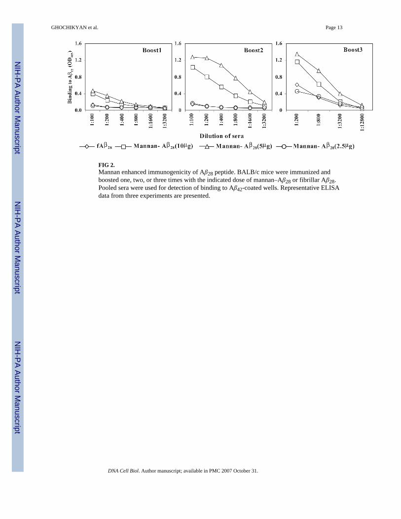
FIG 2.Mannan enhanced immunogenicity of Aβ28 peptide. BALB/c mice were immunized andboosted one, two, or three times with the indicated dose of mannan–Aβ28 or fibrillar Aβ28.Pooled sera were used for detection of binding to Aβ42-coated wells. Representative ELISAdata from three experiments are presented.
GHOCHIKYAN et al. Page 13
DNA Cell Biol. Author manuscript; available in PMC 2007 October 31.
NIH
-PA Author Manuscript
NIH
-PA Author Manuscript
NIH
-PA Author Manuscript

FIG 3.Long-lasting anti-Aβ antibody responses in mice immunized with 5 μg of Aβ28 conjugatedwith mannan (the data with similar profile were obtained with 2.5 and 10 μg mannan-Aβ28).After vaccination and three booster injections mice were rested for 2 months, and the immuneresponse was recalled with the appropriate antigen. Individual sera from animals were diluted1:250 and tested for detection of anti-Aβ42 antibodies in ELISA. The experiment was repeatedwith similar results.
GHOCHIKYAN et al. Page 14
DNA Cell Biol. Author manuscript; available in PMC 2007 October 31.
NIH
-PA Author Manuscript
NIH
-PA Author Manuscript
NIH
-PA Author Manuscript

FIG 4.(A) BALB/c mice immunized with mannan–Aβ28 (5 μg) or fibrillar Aβ28 formulated in PBSinduced Th2-polarized anti-Aβ42 antibodies of IgG1 isotype (similar results were obtained with2.5 and 10 μg mannan–Aβ28). Sera for this assay were collected before the rest period (afterthe third boost) and diluted 1:250 prior to the detection of isotypes. These results were generatedwith individual mice. (B) Immunization of B6SJL F1 mice with mannan–Aβ28 also inducedanti-Aβ42 antibodies of IgG1 isotype. (C) IgG1/IgG2a (ab) ratio for BALB/c mice immunizedwith mannan–Aβ28 or fibrillar Aβ28 and B6SJL F1 mice immunized with mannan–Aβ28.
GHOCHIKYAN et al. Page 15
DNA Cell Biol. Author manuscript; available in PMC 2007 October 31.
NIH
-PA Author Manuscript
NIH
-PA Author Manuscript
NIH
-PA Author Manuscript

FIG 5.Mannan–Aβ28 and fibrillar Aβ28-induced Th2-type cellular immune responses in BALB/cmice. (A) Immune splenocytes isolated from individual mice immunized with mannan–Aβ28or fibrillar Aβ28 and in vitro restimulated by Aβ40 peptide-induced robust T-cell proliferation.Data are presented as Stimulation Index (SI). (B) Production of Th1 (IFNγ) and Th2 (IL-4)-type cytokines, as well as pro-inflammatory TNFα by immune splenocytes isolated from miceimmunized with 5 μg of mannan–Aβ28 or fibrillar Aβ28. The ELISPOT technique was used asdescribed in Materials and Methods, and data are presented as a delta between number of spotsin activated with Aβ40 and nonactivated splenocyte cultures.
GHOCHIKYAN et al. Page 16
DNA Cell Biol. Author manuscript; available in PMC 2007 October 31.
NIH
-PA Author Manuscript
NIH
-PA Author Manuscript
NIH
-PA Author Manuscript
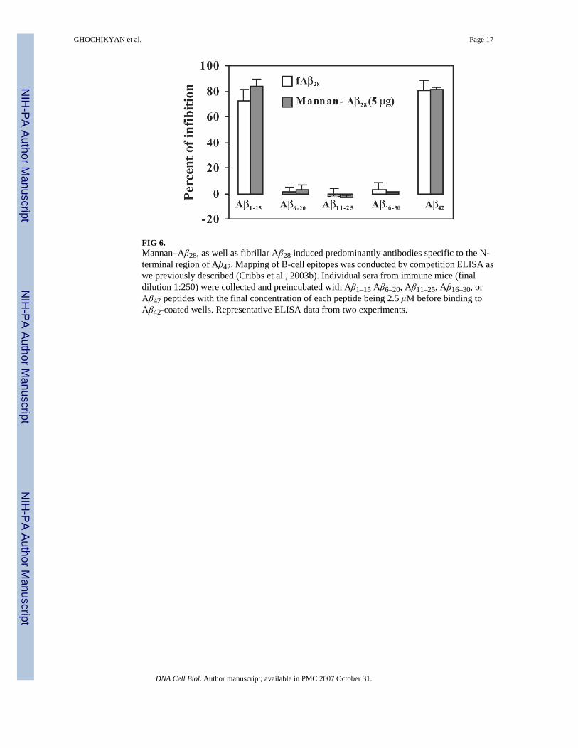
FIG 6.Mannan–Aβ28, as well as fibrillar Aβ28 induced predominantly antibodies specific to the N-terminal region of Aβ42. Mapping of B-cell epitopes was conducted by competition ELISA aswe previously described (Cribbs et al., 2003b). Individual sera from immune mice (finaldilution 1:250) were collected and preincubated with Aβ1–15 Aβ6–20, Aβ11–25, Aβ16–30, orAβ42 peptides with the final concentration of each peptide being 2.5 μM before binding toAβ42-coated wells. Representative ELISA data from two experiments.
GHOCHIKYAN et al. Page 17
DNA Cell Biol. Author manuscript; available in PMC 2007 October 31.
NIH
-PA Author Manuscript
NIH
-PA Author Manuscript
NIH
-PA Author Manuscript

FIG 7.Mannan–Aβ28 induced potentially therapeutic anti-Aβ1–15 antibodies, which are capable ofbinding to amyloid plaques in human brain tissue (A). Coincubation with Aβ28 peptide blockedthis binding (B). 6E10 antibodies were used as a positive control (C). Picture represents bindingof 1:500 diluted pooled sera to a 50-μm brain section of formalin-fixed cortical tissue from anelderly individual with neuropathological and behavioral patterns typical to severe AD.Original magnification, ×20.
GHOCHIKYAN et al. Page 18
DNA Cell Biol. Author manuscript; available in PMC 2007 October 31.
NIH
-PA Author Manuscript
NIH
-PA Author Manuscript
NIH
-PA Author Manuscript
Copyright © 2022 FDOKUMEN