Passive immunotherapy against Aβ in aged APP-transgenic mice reverses cognitive deficits and...
11
BioMed Central Page 1 of 11 (page number not for citation purposes) Journal of Neuroinflammation Open Access Research Passive immunotherapy against Aβ in aged APP-transgenic mice reverses cognitive deficits and depletes parenchymal amyloid deposits in spite of increased vascular amyloid and microhemorrhage Donna M Wilcock 1 , Amyn Rojiani 2 , Arnon Rosenthal 3 , Sangeetha Subbarao 3 , Melissa J Freeman 1 , Marcia N Gordon 1 and Dave Morgan* 1 Address: 1 Alzheimer's Research Laboratory, University of South Florida, Department of Pharmacology, 12901 Bruce B Downs Blvd, Tampa, Florida 33612, USA, 2 Alzheimer's Research Laboratory, University of South Florida, Department of Interdisciplinary Oncology, 12901 Bruce B Downs Blvd, Tampa, Florida 33612, USA and 3 Rinat Neuroscience Corp., 3155 Porter Drive, Palo Alto, California 94304, USA Email: Donna M Wilcock - [email protected]; Amyn Rojiani - [email protected]; Arnon Rosenthal - [email protected]; Sangeetha Subbarao - [email protected]; Melissa J Freeman - [email protected]; Marcia N Gordon - [email protected]; Dave Morgan* - [email protected] * Corresponding author Abstract Background: Anti-Aβ immunotherapy in transgenic mice reduces both diffuse and compact amyloid deposits, improves memory function and clears early-stage phospho-tau aggregates. As most Alzheimer disease cases occur well past midlife, the current study examined adoptive transfer of anti-Aβ antibodies to 19- and 23-month old APP-transgenic mice. Methods: We investigated the effects of weekly anti-Aβ antibody treatment on radial-arm water- maze performance, parenchymal and vascular amyloid loads, and the presence of microhemorrhage in the brain. 19-month-old mice were treated for 1, 2 or 3 months while 23-month-old mice were treated for 5 months. Only the 23-month-old mice were subject to radial-arm water-maze testing. Results: After 3 months of weekly injections, this passive immunization protocol completely reversed learning and memory deficits in these mice, a benefit that was undiminished after 5 months of treatment. Dramatic reductions of diffuse Aβ immunostaining and parenchymal Congophilic amyloid deposits were observed after five months, indicating that even well- established amyloid deposits are susceptible to immunotherapy. However, cerebral amyloid angiopathy increased substantially with immunotherapy, and some deposits were associated with microhemorrhage. Reanalysis of results collected from an earlier time-course study demonstrated that these increases in vascular deposits were dependent on the duration of immunotherapy. Conclusions: The cognitive benefits of passive immunotherapy persist in spite of the presence of vascular amyloid and small hemorrhages. These data suggest that clinical trials evaluating such treatments will require precautions to minimize potential adverse events associated with microhemorrhage. Published: 08 December 2004 Journal of Neuroinflammation 2004, 1:24 doi:10.1186/1742-2094-1-24 Received: 10 November 2004 Accepted: 08 December 2004 This article is available from: http://www.jneuroinflammation.com/content/1/1/24 © 2004 Wilcock et al; licensee BioMed Central Ltd. This is an Open Access article distributed under the terms of the Creative Commons Attribution License (http://creativecommons.org/licenses/by/2.0 ), which permits unrestricted use, distribution, and reproduction in any medium, provided the original work is properly cited.
Transcript of Passive immunotherapy against Aβ in aged APP-transgenic mice reverses cognitive deficits and...
BioMed CentralJournal of Neuroinflammation
ss
Open AcceResearchPassive immunotherapy against Aβ in aged APP-transgenic mice reverses cognitive deficits and depletes parenchymal amyloid deposits in spite of increased vascular amyloid and microhemorrhageDonna M Wilcock1, Amyn Rojiani2, Arnon Rosenthal3, Sangeetha Subbarao3, Melissa J Freeman1, Marcia N Gordon1 and Dave Morgan*1Address: 1Alzheimer's Research Laboratory, University of South Florida, Department of Pharmacology, 12901 Bruce B Downs Blvd, Tampa, Florida 33612, USA, 2Alzheimer's Research Laboratory, University of South Florida, Department of Interdisciplinary Oncology, 12901 Bruce B Downs Blvd, Tampa, Florida 33612, USA and 3Rinat Neuroscience Corp., 3155 Porter Drive, Palo Alto, California 94304, USA
Email: Donna M Wilcock - [email protected]; Amyn Rojiani - [email protected]; Arnon Rosenthal - [email protected]; Sangeetha Subbarao - [email protected]; Melissa J Freeman - [email protected]; Marcia N Gordon - [email protected]; Dave Morgan* - [email protected]
* Corresponding author
AbstractBackground: Anti-Aβ immunotherapy in transgenic mice reduces both diffuse and compactamyloid deposits, improves memory function and clears early-stage phospho-tau aggregates. Asmost Alzheimer disease cases occur well past midlife, the current study examined adoptive transferof anti-Aβ antibodies to 19- and 23-month old APP-transgenic mice.
Methods: We investigated the effects of weekly anti-Aβ antibody treatment on radial-arm water-maze performance, parenchymal and vascular amyloid loads, and the presence of microhemorrhagein the brain. 19-month-old mice were treated for 1, 2 or 3 months while 23-month-old mice weretreated for 5 months. Only the 23-month-old mice were subject to radial-arm water-maze testing.
Results: After 3 months of weekly injections, this passive immunization protocol completelyreversed learning and memory deficits in these mice, a benefit that was undiminished after 5months of treatment. Dramatic reductions of diffuse Aβ immunostaining and parenchymalCongophilic amyloid deposits were observed after five months, indicating that even well-established amyloid deposits are susceptible to immunotherapy. However, cerebral amyloidangiopathy increased substantially with immunotherapy, and some deposits were associated withmicrohemorrhage. Reanalysis of results collected from an earlier time-course study demonstratedthat these increases in vascular deposits were dependent on the duration of immunotherapy.
Conclusions: The cognitive benefits of passive immunotherapy persist in spite of the presence ofvascular amyloid and small hemorrhages. These data suggest that clinical trials evaluating suchtreatments will require precautions to minimize potential adverse events associated withmicrohemorrhage.
Published: 08 December 2004
Journal of Neuroinflammation 2004, 1:24 doi:10.1186/1742-2094-1-24
Received: 10 November 2004Accepted: 08 December 2004
This article is available from: http://www.jneuroinflammation.com/content/1/1/24
© 2004 Wilcock et al; licensee BioMed Central Ltd. This is an Open Access article distributed under the terms of the Creative Commons Attribution License (http://creativecommons.org/licenses/by/2.0), which permits unrestricted use, distribution, and reproduction in any medium, provided the original work is properly cited.
Page 1 of 11(page number not for citation purposes)

Journal of Neuroinflammation 2004, 1:24 http://www.jneuroinflammation.com/content/1/1/24
BackgroundAlzheimer's disease is characterized not only by the pres-ence of parenchymal amyloid deposits and intracellulartangles but also by the presence of amyloid deposits in thevasculature, a condition referred to as cerebral amyloidangiopathy (CAA). The CAA observed in both Alzheimer'sdisease patients [1] and some of the transgenic mousemodels [2] is primarily composed of the shorter form ofamyloid beta (Aβ), Aβ1–40, while the majority of amyloiddeposits in the parenchyma are composed of Aβ1–42,although the compact amyloid deposits also contain Aβ1–
40.
Anti-Aβ immunotherapy has been considered as a poten-tial treatment for Alzheimer's disease for some time [3,4].Active immunization with a vaccine including Aβ1–42fibrils progressed to human clinical trials where its admin-istration was suspended due to meningoencephalitits in asubset of patients [5]. To date there have been pathologyreports on two patients who participated in the trial andsubsequently died [6,7]. Both reports note that while thenumbers of parenchymal amyloid deposits appearedlower than expected in these cases, the CAA in thesepatients did not appear outside the normal range forAlzheimer's disease. In addition, one report mentionedmultiple cortical hemorrhages and the presence of hemo-siderin around the CAA vessels [7].
Given the adverse reactions to the active immunization,the irreversibility of such procedures and the variable anti-body response to vaccines in older individuals [8], passiveimmunization against the Aβ peptide emerged as an alter-native immunotherapeutic strategy. Studies in young andmiddle aged APP-transgenic mice have reported signifi-cant amyloid reductions with passive immunization [9-11]. Such treatments also demonstrate rapid improve-ments of memory function in APP-transgenic mice, some-times without detectable reductions in amyloid [12-14].Most recently, intracranial administration of anti-Aβ anti-bodies has been shown to not only remove Aβ but alsoclear, early-stage, hyperphosphorylated-tau aggregates[15]. Importantly, in the only prior study evaluatingadoptive antibody transfer in older APP-transgenic mice,Pfeifer et al. [16] reported a doubling of cerebral microhe-morrhages associated with significant reductions in amy-loid burden after administration of an N-terminal specificanti-Aβ antibody.
Materials and MethodsExperiment designMice derived from APP Tg2576 mice were obtained fromour breeding program at University of South Floridastarted in 1996 [17]. For the 5-month treatment study, 13APP-transgenic mice, aged 23 months, were assigned toone of two groups. The first group received weekly intra-
peritoneal anti-Aβ antibody injections (antibody 2286;mouse-monoclonal anti-human Aβ28–40 IgG1; Rinat Neu-rosciences, Palo Alto, CA) for a period of 5 months (n =6). The second group received weekly intraperitoneal anti-AMN antibody (2906; mouse-monoclonal anti-Drosophilaamnesiac protein IgG1; Rinat Neurosciences, Palo Alto,CA) injections for a period of 5 months (n = 7). Sevennontransgenic mice were also assigned to one of twogroups. The first group received weekly intraperitonealanti-Aβ antibody injections for a period of 5 months (n =4). The second group received weekly intraperitoneal anti-AMN antibody injections for a period of 5 months (n = 3).
For the time course study of 1-, 2- or 3-month treatment,22 APP-transgenic mice aged 19 months were assigned toone of four experimental groups, as described previously[14]. The first three groups received weekly intraperitonealanti-Aβ antibody injections for 3 months, 2 months or 1month, ending when all mice were 22 months of age. Thefourth group received weekly intraperitoneal anti-AMNantibody injections for 3 months.
Behavioral analysisFollowing 3 and 5 months of treatment, the mice from the5-month study were subjected to a two-day radial-armwater-maze paradigm. The apparatus was a 6-arm maze asdescribed previously [18]. On day one, 15 trials were runin three blocks of 5. A cohort of 4 mice were run sequen-tially for each block (i.e., each of 4 mice get trial one, thenthe same mice get trial two, etc.). After each 5-trial block,a second cohort of mice was run permitting an extendedrest period before mice were exposed to the second blockof 5 trials. The goal arm was different for each mouse in acohort to minimize odor cues. The start arm was varied foreach trial, with the goal arm remaining constant for agiven individual for both days. For the first 11 trials, theplatform was alternately visible then hidden (hidden forthe last 4 trials). On day two, the mice were run in exactlythe same manner as day one except that the platform washidden forall trials. The number of errors (incorrect armentries) was measured in a one-minute time frame. Asstandard practice, mice failing to make an arm choice in20 seconds are assigned one error, but no mice in thisstudy had to be assigned an error in this manner. Thesame individual administered the antibody treatmentsand placed mice in the radial-arm water maze. Due to thenumbers of mice in the study the researcher was unawareof treatment group identity of each mouse. Also, thedependent measures in the radial-arm water-maze task arequantitative, not evaluative, so the potential for tester biasis reduced. In order to minimize the influence of individ-ual trial variability, each mouse's errors for 3 consecutivetrials were averaged producing 5 data points for each day,which were analyzed statistically by ANOVA usingStatView (SAS Institute Inc., NC).
Page 2 of 11(page number not for citation purposes)

Journal of Neuroinflammation 2004, 1:24 http://www.jneuroinflammation.com/content/1/1/24
Tissue preparation and histologyOn the day of sacrifice mice were weighed, overdosed with100 mg/kg Nembutal (Abbott laboratories, North Chi-cago, IL), and then intracardially perfused with 25 mL of0.9% sodium chloride. Brains were rapidly removed, andthe left half of the brain was immersion fixed for 24 h infreshly prepared 4% paraformaldehyde in 100 mM KPO4(pH 7.2) for histopathology. The hemi-brains were thenincubated for 24 h in 10%, 20% and 30% sucrose sequen-tially for cyroprotection. Horizontal sections of 25 µthickness were collected using a sliding microtome andstored at 4°C in Dulbecco's phosphate-buffered salinewith sodium azide (pH 7.2) to prevent microbial growth.A series of 8 equally spaced tissue sections 600 µ apartwere randomly selected spanning the entire brain andstained using free-floating immunohistochemistry fortotal Aβ (rabbit polyclonal anti-pan Aβ; Biosource,Camarillo, CA, 1:10,000) as previously described [2,14].A second series of tissue sections 600 µm apart werestained using 0.2% Congo red in NaCl-saturated 80% eth-anol. Another set of sections were also mounted andstained for hemosiderin using 2% potassium ferrocyanidein 2% hydrochloric acid for 15 min, followed by a coun-terstain in a 1% neutral red solution for 10 min. Quanti-fication of Congo red staining and Aβimmunohistochemistry was performed using the Image-Pro Plus (Media Cybernetics, Silver Spring, MD) to ana-lyze the percent area occupied by positive stain. Oneregion of the frontal cortex and three regions of the hip-pocampus were analyzed (to ensure that there was noregional bias in the hippocampal values). The initial anal-ysis of Congo red was performed to give a total value. Asecond analysis was performed after manually editing outall of the parenchymal amyloid deposits to yield a percentarea restricted to vascular Congo red staining. To estimatethe parenchymal area of Congo red, we subtracted the vas-cular amyloid values from the total percentage. For thehemosiderin stain the numbers of Prussian blue-positivesites were counted on all sections and the average numberof sites per section calculated. Looking at the sections at alow magnification we were able to observe a qualitativedifferences between animals; however, the percent areawas so low that many fields contained no positive stain.Eight equally spaced sections were examined and thenumber of positive profiles was determined and averagedto a per-section value. To assess possible treatment-relateddifferences, the values for each treatment group were ana-lyzed by one-way ANOVA followed by Fisher's LSD meanscomparisons.
ResultsReversal of cognitive deficits by passive amyloid immunotherapyThe radial-arm water-maze task detects spatial learningand memory deficits in transgenic mouse models [18,19].
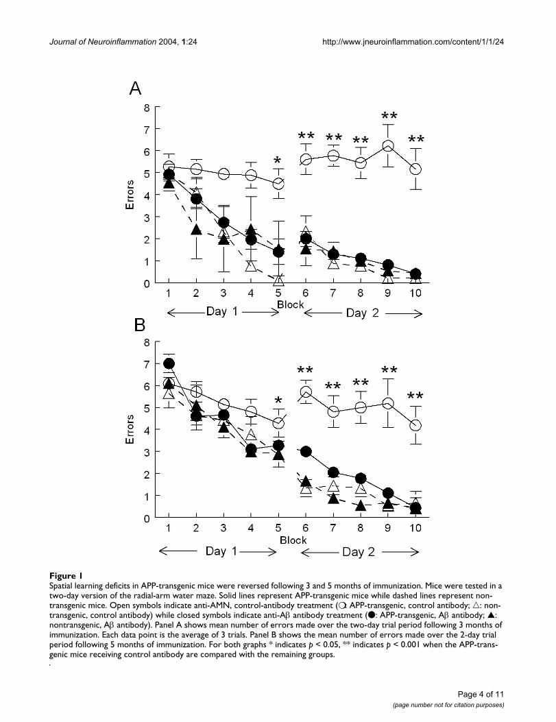
We treated 23-month-old mice for 5 months with anti-Aβantibody 2286 or control antibody 2906 (against a Dro-sophila-specific protein) and tested them for spatial navi-gation learning in a two-day version of the radial-armwater maze after 3 months of treatment and, using a newplatform location, again after 5 months of treatment. Atboth testing times we found that APP-transgenic micetreated with the control antibody failed to learn platformlocation over two days of testing and were significantlyimpaired compared to the nontransgenic mice treatedwith either antibody (Fig. 1). However, APP-transgenicmice administered the anti-Aβ antibodies demonstrated acomplete reversal of the impairment observed in the con-trol-treated APP-transgenic mice, ending day two with amean performance near 0.5 errors per trial (Fig. 1).Although learning at the later time point, when the micewere 28 months of age, may have been slightly slower forall groups, there was no impairment of the anti-Aβ anti-body-treated APP.
Passive amyloid immunotherapy clears parenchymal Aβ deposits, but increases vascular amyloidIn a prior experiment examining the effects of passive anti-Aβ immunotherapy for 1, 2 or 3 months in APP-trans-genic mice killed at 21 months of age [14], we found atime-dependent reduction of both Aβ immunostaining ofdiffuse and fibrillar deposits and Congo-red staining offibrillar amyloid deposits. In the current study we found asimilar reduction in both Aβ immunostaining (Table 1)and total Congo-red staining (Fig. 2A, left panel; p < 0.001frontal cortex and p < 0.01 hippocampus) after 5 monthsof immunotherapy. We noted that the bulk of whatremained was vascular amyloid. We then separately ana-lyzed vascular and parenchymal deposits which revealeda near 90% reduction in parenchymal deposits (p < 0.001)but a 3–4 fold elevation of vascular Congo-red staining (p< 0.0001; Fig. 2A, center and right panels, respectively).We also separately analyzed vascular and parenchymalCongo-red staining on mice from our earlier study [14],treated passively for 1, 2 or 3 months with anti-Aβ or con-trol antibody, and found a similar result. There was agraded reduction in overall Congo-red staining nearing75% as duration of antibody exposure increased (asreported previously; Fig. 2B). However, when separatedinto vascular Congo-red deposits and parenchymaldeposits, there was an antibody-exposure-time-dependentincrease in vascular deposition in both hippocampus andfrontal cortex (Fig. 2C; p < 0.05 frontal cortex and hippoc-ampus) and a corresponding nearly 90% decrease inparenchymal deposits (Fig. 2D; p < 0.001 in frontal cortexand hippocampus).
These differences were readily observed examining micro-graphs of sections from these mice. Mice treated with con-trol antibodies revealed occasional cortical vascular
Page 3 of 11(page number not for citation purposes)
Journal of Neuroinflammation 2004, 1:24 http://www.jneuroinflammation.com/content/1/1/24
Spatial learning deficits in APP-transgenic mice were reversed following 3 and 5 months of immunizationFigure 1Spatial learning deficits in APP-transgenic mice were reversed following 3 and 5 months of immunization. Mice were tested in a two-day version of the radial-arm water maze. Solid lines represent APP-transgenic mice while dashed lines represent non-transgenic mice. Open symbols indicate anti-AMN, control-antibody treatment (❍ : APP-transgenic, control antibody; �: non-transgenic, control antibody) while closed symbols indicate anti-Aβ antibody treatment (● : APP-transgenic, Aβ antibody; ▲: nontransgenic, Aβ antibody). Panel A shows mean number of errors made over the two-day trial period following 3 months of immunization. Each data point is the average of 3 trials. Panel B shows the mean number of errors made over the 2-day trial period following 5 months of immunization. For both graphs * indicates p < 0.05, ** indicates p < 0.001 when the APP-trans-genic mice receiving control antibody are compared with the remaining groups.
Page 4 of 11(page number not for citation purposes)

Journal of Neuroinflammation 2004, 1:24 http://www.jneuroinflammation.com/content/1/1/24
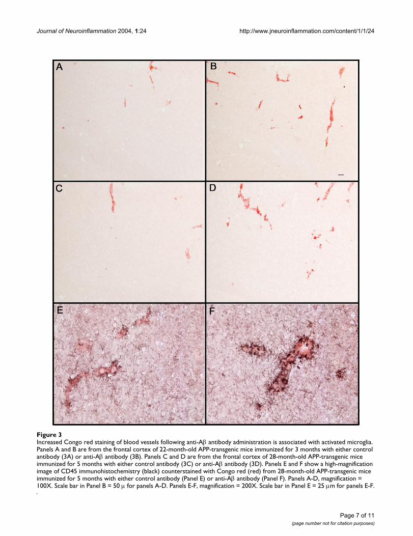
amyloid deposits (22 months, Fig. 3A, 28 months, Fig.3C), while mice administered anti-Aβ antibodies hadincreased amounts of vascular amyloid staining (3-monthtreatment, Fig 3B; 5-month treatment, Fig 3D). Those ves-sels containing amyloid following treatment with anti-Aβantibody also exhibited apparent increases in microglialactivation as measured by CD45 expression (Fig. 3F) com-pared to mice treated with control antibody (Fig. 3E).Unfortunately, the shifting numbers and sizes of vascularand parenchymal deposits caused by the antibody therapygreatly complicated measurement of microglial activationper vascular deposit area so that this apparent increase instaining intensity could not be quantified accurately.
Passive amyloid immunotherapy causes increased microhemorrhageWe used the Prussian blue histological stain to labelhemosiderin, a ferric oxide material produced in thebreakdown of hemoglobin. Extravenous blood in thebrain leads to microglial phagocytosis of the erythrocytesand breakdown of the hemoglobin within them. Theseferric oxide-containing microglia are thus markers of pasthemorrhage. In untreated, aged APP-transgenic mice weobserved very few profiles positive for Prussian-blue stain-ing in the frontal cortex (section counterstained with neu-tral red; Fig. 4A). However, following anti-Aβ antibodytreatment for 5 months we observed an increase in thenumber of Prussian-blue profiles in the frontal cortex,which were readily detectable at a low magnification inthe microscope (Fig. 4B). In the absence of anti-Aβ treat-ment, or even when treated with antibody for one month,most vessels did not stain with Prussian blue, and couldbe identified only using the red counterstain (Fig. 4C).However, even with 3 months of anti-Aβ antibody treat-ment we observed frequent vessels with associated Prus-sian-blue staining (Fig 4D). Using adjacent sectionsstained for Congo red, we confirmed that all vessels show-ing microhemorrhage contained amyloid (Figs. 4E and4F; we were unable to double-label Prussian blue-stainedsections with either Congo red or thioflavine-S). How-ever, only a minority of vessels containing amyloid dem-onstrated hemorrhage.
When we counted the number of Prussian blue-positiveprofiles in those animals receiving control antibody there
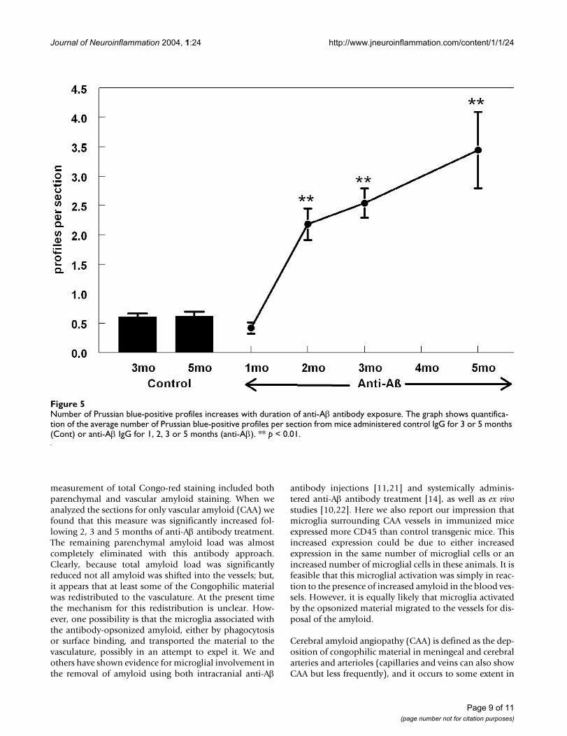
was an average of one profile per every two sections (Fig.5) and this number remained the same in both controlgroups (aged 22 or 28 months). Following treatment withanti-Aβ antibody for a period of two months we observeda striking increase in Prussian-blue staining, approxi-mately five times that observed in either the control groupor the mice immunized for one month (Fig. 5, p < 0.001).Following this initial increase in Prussian-blue staining,we observed a linear increase in staining associated withincreasing duration of anti-Aβ antibody treatment (Fig 5).Five months of anti-Aβ antibody treatment demonstrateda six-fold increase in Prussian-blue staining when com-pared the control groups (Fig. 5).
DiscussionEarlier studies with vaccines against the Aβ peptide dem-onstrated protection from the learning and memory defi-cits associated with amyloid accumulation in APP-transgenic mice [14,19]. Passive immunization protocolswith anti-Aβ antibodies also produced cognitive benefits,in some cases even in the absence of significant reductionin amyloid burden [12,13]. Our recent work found that 3months of anti-Aβ treatment of 18-month-old APP-trans-genic mice improved spontaneous alternation perform-ance on the Y-maze [14]. In the present work weconfirmed that passive anti-amyloid immunotherapy canreverse spatial learning deficits in APP-transgenic miceand that this benefit of immunotherapy is retained, evenin aged mice (26 and 28 months old at testing) with long-established amyloid pathology.
Additionally, we describe a more rapid means of testingspatial reference memory to reveal learning and memorydeficits in APP-transgenic mice. This two-day version ofthe radial arm water maze included greater spacing ofindividual trials (mice spent time in their home cage afterevery trial), combined with less spacing of aggregate trials(fifteen trials per day rather than four or five) to facilitatelearning of platform location in the nontransgenic mice,with a clear absence of learning in the age-matched trans-genic mice.
A substantial reduction in total Congophilic amyloiddeposits was observed in old APP-transgenic mice treatedwith anti-Aβ antibodies for 2 or more months. This
Table 1: Total Aβ load is significantly reduced following 5 months of anti-Aβ antibody treatment. Percent area occupied by positive immunohistochemical stain for Aβ is shown ± standard error of the mean for both the frontal cortex and hippocampus. Also shown is the percent reduction of Aβ observed following anti-Aβ antibody treatment
Region % area positive for Aβ: control treated
% area positive for Aβ: anti-Aβ treated
% reduction following anti-Aβ antibody treatment
Frontal Cortex 34.855 ± 2.265 9.681 ± 0.754 72Hippocampus 23.994 ± 0.985 8.212 ± 0.596 66
Page 5 of 11(page number not for citation purposes)

Journal of Neuroinflammation 2004, 1:24 http://www.jneuroinflammation.com/content/1/1/24
Passive immunization with anti-Aβ antibodies decreases total and parenchymal amyloid loads while increasing vascular amyloid in frontal cortex and hippocampus of APP-transgenic miceFigure 2Passive immunization with anti-Aβ antibodies decreases total and parenchymal amyloid loads while increasing vascular amyloid in frontal cortex and hippocampus of APP-transgenic mice. Panel A shows total amyloid load measured with Congo red, vascu-lar amyloid load and parenchymal amyloid load from APP-transgenic mice administered control IgG (C) or anti-Aβ IgG (Aβ) for a period of 5 months. Panels B-D show total amyloid load (Panel B), vascular amyloid load (Panel C) and parenchymal amyloid load (Panel D) from APP-transgenic mice administered control IgG for 3 months (Cont IgG) or anti-Aβ IgG for a period of 1, 2, or 3 months (Anti-Aβ IgG). For all panels, the solid bar and solid line represent values from the frontal cortex, while the open bar and dashed line represent values from the hippocampus. ** p < 0.01.
Page 6 of 11(page number not for citation purposes)
Journal of Neuroinflammation 2004, 1:24 http://www.jneuroinflammation.com/content/1/1/24
Increased Congo red staining of blood vessels following anti-Aβ antibody administration is associated with activated microgliaFigure 3Increased Congo red staining of blood vessels following anti-Aβ antibody administration is associated with activated microglia. Panels A and B are from the frontal cortex of 22-month-old APP-transgenic mice immunized for 3 months with either control antibody (3A) or anti-Aβ antibody (3B). Panels C and D are from the frontal cortex of 28-month-old APP-transgenic mice immunized for 5 months with either control antibody (3C) or anti-Aβ antibody (3D). Panels E and F show a high-magnification image of CD45 immunohistochemistry (black) counterstained with Congo red (red) from 28-month-old APP-transgenic mice immunized for 5 months with either control antibody (Panel E) or anti-Aβ antibody (Panel F). Panels A-D, magnification = 100X. Scale bar in Panel B = 50 µ for panels A-D. Panels E-F, magnification = 200X. Scale bar in Panel E = 25 µm for panels E-F.
Page 7 of 11(page number not for citation purposes)

Journal of Neuroinflammation 2004, 1:24 http://www.jneuroinflammation.com/content/1/1/24
Microhemorrhage associated with CAA following systemic administration of anti-Aβ antibodiesFigure 4Microhemorrhage associated with CAA following systemic administration of anti-Aβ antibodies. Panels A and B are low magni-fication images of the frontal cortex of APP-transgenic mice receiving either control antibodies (Panel A) or anti-Aβ antibodies (Panel B) for a period of 5 months. Panels C and D show representative images of amyloid containing vessels stained for Prus-sian blue (blue), counterstained with neutral red (red), from APP-transgenic mice receiving either control antibodies (Panel C) or anti-Aβ antibodies (Panel D) for a period of 3 months. Panel E shows a blood vessel in the frontal cortex stained for Prus-sian blue (blue), counterstained with neutral red, from an APP transgenic mouse administered anti-Aβ antibodies for 5 months. Panel F shows the same blood vessel on an adjacent section stained for Congo red, indicating that the blood vessel does in fact contain amyloid. Scale bar panel A = 120 µm for panels A-B. Scale bar panel C = 25 µm for panels C-D. Scale bar in panel F = 25 µm for panels E-F.
Page 8 of 11(page number not for citation purposes)
Journal of Neuroinflammation 2004, 1:24 http://www.jneuroinflammation.com/content/1/1/24
measurement of total Congo-red staining included bothparenchymal and vascular amyloid staining. When weanalyzed the sections for only vascular amyloid (CAA) wefound that this measure was significantly increased fol-lowing 2, 3 and 5 months of anti-Aβ antibody treatment.The remaining parenchymal amyloid load was almostcompletely eliminated with this antibody approach.Clearly, because total amyloid load was significantlyreduced not all amyloid was shifted into the vessels; but,it appears that at least some of the Congophilic materialwas redistributed to the vasculature. At the present timethe mechanism for this redistribution is unclear. How-ever, one possibility is that the microglia associated withthe antibody-opsonized amyloid, either by phagocytosisor surface binding, and transported the material to thevasculature, possibly in an attempt to expel it. We andothers have shown evidence for microglial involvement inthe removal of amyloid using both intracranial anti-Aβ
antibody injections [11,21] and systemically adminis-tered anti-Aβ antibody treatment [14], as well as ex vivostudies [10,22]. Here we also report our impression thatmicroglia surrounding CAA vessels in immunized miceexpressed more CD45 than control transgenic mice. Thisincreased expression could be due to either increasedexpression in the same number of microglial cells or anincreased number of microglial cells in these animals. It isfeasible that this microglial activation was simply in reac-tion to the presence of increased amyloid in the blood ves-sels. However, it is equally likely that microglia activatedby the opsonized material migrated to the vessels for dis-posal of the amyloid.
Cerebral amyloid angiopathy (CAA) is defined as the dep-osition of congophilic material in meningeal and cerebralarteries and arterioles (capillaries and veins can also showCAA but less frequently), and it occurs to some extent in
Number of Prussian blue-positive profiles increases with duration of anti-Aβ antibody exposureFigure 5Number of Prussian blue-positive profiles increases with duration of anti-Aβ antibody exposure. The graph shows quantifica-tion of the average number of Prussian blue-positive profiles per section from mice administered control IgG for 3 or 5 months (Cont) or anti-Aβ IgG for 1, 2, 3 or 5 months (anti-Aβ). ** p < 0.01.
Page 9 of 11(page number not for citation purposes)
Journal of Neuroinflammation 2004, 1:24 http://www.jneuroinflammation.com/content/1/1/24
nearly all Alzheimer's disease patients [23]. Severe CAA,affecting about 15% of cases, can be associated with bothinfarction and hemorrhagic injury [24,25]. It has alsobeen shown that the severity of CAA can be directly linkedto the severity of dementia in Alzheimer's disease patients[26].
In the current study we found a significantly increasednumber of microhemorrhages in the brain as detected byPrussian-blue staining, associated with the increase inCAA following passive immunization. Another transgenicmouse model of amyloid deposition, the APP23 mice,have been shown to deposit amyloid in both brain paren-chyma and blood vessels and show a CAA associatedincrease in spontaneous cerebral hemorrhages [27]. More-over, Pfeifer et al. [16] showed that these spontaneoushemorrhages were significantly increased following 5months of passive immunization of 21-month-old APP23mice using an anti-Aβ antibody with an N-terminalepitope, similar to those typically developed in activeimmunization with vaccines [4,28,29]. When young mice(6 months of age) were immunized following the sameprotocol, no hemorrhages were observed. More recently,DeMattos et al. [30] showed that passive immunizationwith an N-terminal antibody (3D6: directed againstamino acids 1–5 of Aβ) of PDAPP transgenic mice alsoresulted in significantly increased microhemorrhage. Theywere unable to detect increased microhemorrhage with amid-domain antibody (266: directed against amino acids13–28 of Aβ). Notably, antibody 266 fails to bind Aβdeposited in CAA vessels or amyloid plaques [31]. Impor-tantly, Ferrer et al. [7] noted the presence of CAA andmicrohemorrhage in the brain of one patient that partici-pated in the Aβ-vaccine trial, even though the parenchy-mal amyloid appeared lower than expected. Also, Nicoll etal. [6] noted that CAA appeared unaffected in the brain ofanother patient that participated in the Aβ-vaccine trial.
It remains to be determined whether these observationsregarding increased CAA and microhemorrhage intransgenic mice are relevant to trials of passive immuno-therapy in humans. It should be noted that, in spite ofextending the period of immunotherapy to 5 months,there was no discernable loss of the cognitive benefits ofimmunotherapy in the transgenic mice, all of whomshowed increased microhemorrhage. While the observa-tion that antibody 266 does not result in vascular leakageencourages testing of this idiotype, data from the Zurichcohort of the Aβ vaccine trial argue that brain-reactiveantibodies may be important for cognitive benefits [32].
ConclusionsOur opinion is that these results suggest that passiveimmunotherapy against Aβ should proceed with appro-priate precautions taken to minimize the risk of hemor-
rhage (e.g., by excluding patients taking anticoagulants)and instituting measures to detect such hemorrhages ifthey do occur, irrespective of the antibody specificity orproclivity for microhemorrhage in aged APP-transgenicmice.
List of abbreviationsAβ : Amyloid-beta.
APP: Amyloid precursor protein
CAA: Cerebral amyloid angiopathy.
IgG1: Immunoglobulin G type 1.
Competing interestsThe authors declare that they have no competing interests.
Authors' contributionsDMW treated the mice, performed the behavioral analy-sis, processed the tissue and performed pathological anal-yses, and drafted the manuscript. ARojiani evaluatedslides and provided expert opinion regarding CAA andmicrohemorrhage. ARosenthal and SS developed, pro-duced and purified the antibodies used in the studies. MJFperformed DNA extraction and PCR for genotyping of themice. MNG oversees the breeding colony generating micefor the studies, collected samples from the mice andassisted in editing the manuscript. DM conceived thedesign of the study, guided data interpretation andassisted in editing the manuscript.
AcknowledgementsThis work was supported by National Institutes of Aging / NIH grants AG15490 (MNG) and AG18478 (DM). DMW is the Benjamin Scholar in Alzheimer's disease research. We would like to thank Keisha Symmonds who aided in histological processing of the tissue and Nedda Wilson who was responsible for animal husbandry during the study. We would also like to thank Lori Lutz for assisting in editing the manuscript.
References1. Iwatsubo T, Odaka A, Suzuki N, Mizusawa H, Nukina N, Ihara Y: Vis-
ualization of A beta 42(43) and A beta 40 in senile plaqueswith end-specific A beta monoclonals: evidence that an ini-tially deposited species is A beta 42(43). Neuron 1994, 13:45-53.
2. Gordon MN, Holcomb LA, Jantzen PT, DiCarlo G, Wilcock D, BoyettKW, Connor K, Melachrino J, O'Callaghan JP, Morgan D: Timecourse of the development of Alzheimer-like pathology inthe doubly transgenic PS1+APP mouse. Exp Neurol 2002,173:183-195.
3. Solomon B, Koppel R, Hanan E, Katzav T: Monoclonal antibodiesinhibit in vitro fibrillar aggregation of the Alzheimer beta-amyloid peptide. Proc Natl Acad Sci USA 1996, 93:452-5.
4. Schenk D, Barbour R, Dunn W, Gordon G, Grajeda H, Guido T, HuK, Huang J, Johnson-Wood K, Khan K, Kholodenko D, Lee M, Liao Z,Lieberburg I, Motter R, Mutter L, Soriano F, Shopp G, Vasquez N,Vandevert C, Walker S, Wogulis M, Yednock T, Games D, Seubert P:Immunization with amyloid-beta attenuates Alzheimer-dis-ease-like pathology in the PDAPP mouse. Nature 1999,400:173-177.
5. Orgogozo JM, Gilman S, Dartigues JF, Laurent B, Puel M, Kirby LC,Jouanny P, Dubois B, Eisner L, Flitman S, Michel BF, Boada M, Frank
Page 10 of 11(page number not for citation purposes)
http://www.ncbi.nlm.nih.gov/entrez/query.fcgi?cmd=Retrieve&db=PubMed&dopt=Abstract&list_uids=8043280
http://www.ncbi.nlm.nih.gov/entrez/query.fcgi?cmd=Retrieve&db=PubMed&dopt=Abstract&list_uids=8043280
http://www.ncbi.nlm.nih.gov/entrez/query.fcgi?cmd=Retrieve&db=PubMed&dopt=Abstract&list_uids=8043280
http://www.ncbi.nlm.nih.gov/entrez/query.fcgi?cmd=Retrieve&db=PubMed&dopt=Abstract&list_uids=8552659
http://www.ncbi.nlm.nih.gov/entrez/query.fcgi?cmd=Retrieve&db=PubMed&dopt=Abstract&list_uids=8552659

Journal of Neuroinflammation 2004, 1:24 http://www.jneuroinflammation.com/content/1/1/24
Publish with BioMed Central and every scientist can read your work free of charge
"BioMed Central will be the most significant development for disseminating the results of biomedical research in our lifetime."
Sir Paul Nurse, Cancer Research UK
Your research papers will be:
available free of charge to the entire biomedical community
peer reviewed and published immediately upon acceptance
cited in PubMed and archived on PubMed Central
yours — you keep the copyright
Submit your manuscript here:http://www.biomedcentral.com/info/publishing_adv.asp
BioMedcentral
A, Hock C: Subacute meningoencephalitis in a subset ofpatients with AD after Abeta42 immunization. Neurology 2003,61:46-54.
6. Nicoll JA, Wilkinson D, Holmes C, Steart P, Markham H, Weller RO:Neuropathology of human Alzheimer disease after immuni-zation with amyloid-beta peptide: a case report. Nat Med2003, 9:448-452.
7. Ferrer I, Boada Rovira M, Sanchez Guerra ML, Rey MJ, Costa-Jussa F:Neuropathology and pathogenesis of encephalitis followingamyloid-beta immunization in Alzheimer's disease. BrainPathol 2004, 14:11-20.
8. Weksler ME: Immunology and the elderly: an historical per-spective for future international action. Mech Ageing Dev 1997,93:1-6.
9. DeMattos RB, Bales KR, Cummins DJ, Dodart JC, Paul SM, HoltzmanDM: Peripheral anti-Aβ antibody alters CNS and plasma Aβclearance and decreases brain Aβ burden in a mouse modelof Alzheimer's disease. Proc Natl Acad Sci USA 2001, 98:8850-8855.
10. Bard F, Cannon C, Barbour R, Burke RL, Games D, Grajeda H, GuidoT, Hu K, Huang J, Johnson-Wood K, Khan K, Kholodenko D, Lee M,Lieberberg I, Motter R, Nguyen M, Soriano F, Vasquez N, Weiss K,Welch B, Seubert P, Schenk D, Yednock T: Peripherally adminis-tered antibodies against amyloid β-peptide enter the centralnervous system and reduce pathology in a mouse model ofAlzheimer's disease. Nat Med 2000, 6:916-919.
11. Wilcock DM, DiCarlo G, Henderson D, Jackson J, Clarke K, Ugen KE,Gordon MN, Morgan : Intracranially administered anti-Aβ anti-bodies reduce β-amyloid deposition by mechanisms inde-pendent of and associated with microglial activation. JNeurosci 2003, 213:3745-3751.
12. Dodart JC, Bales KR, Gannon KS, Greene SJ, DeMattos RB, Mathis C,DeLong CA, Wu S, Wu X, Holtzman DM, Paul SM: Immunizationreverses memory deficits without reducing brain Abeta bur-den in Alzheimer's disease model. Nat Neurosci 2002, 5:452-457.
13. Kotilinek LA, Bacskai B, Westerman M, Kawarabayashi T, Younkin L,Hyman BT, Younkin S, Ashe KH: Reversible memory loss in amouse transgenic model of Alzheimer's disease. J Neurosci2002, 22:6331-6335.
14. Wilcock DM, Rojiani A, Rosenthal A, Levkowitz G, Subbarao S,Alamed J, Wilson D, Wilson N, Freeman MJ, Gordon MN, Morgan D:Passive amyloid immunotherapy clears amyloid and tran-siently activates microglia in a transgenic mouse model ofamyloid deposition. J Neurosci 2004, 24:6144-6151.
15. Oddo S, Billings L, Kesslak JP, Cribbs DH, LaFerla FM: Abeta Immu-notherapy Leads to Clearance of Early, but Not Late, Hyper-phosphorylated Tau Aggregates via the Proteasome. Neuron2004, 43:321-333.
16. Pfeifer M, Boncristiano S, Bondolfi L, Stalder A, Deller T, StaufenbielM, Mathews PM, Jucker M: Cerebral hemorrhage after passiveanti-Aβ immunotherapy. Science 2002, 298:1379.
17. Holcomb L, Gordon MN, McGowan E, Yu X, Benkovic S, Jantzen P,Wright K, Saad I, Mueller R, Morgan D, Sanders S, Zehr C, O'CampoK, Hardy J, Prada CM, Eckman C, Younkin S, Hsiao K, Duff K: Accel-erated Alzheimer-type phenotype in transgenic mice carry-ing both mutant amyloid precursor protein and presenilin 1transgenes. Nat Med 1998, 4:97-100.
18. Gordon MN, King DL, Diamond DM, Jantzen PT, Boyett KV, HopeCE, Hatcher JM, DiCarlo G, Gottschall WP, Morgan D, ArendashGW: Correlation between cognitive deficits and Abetadeposits in transgenic APP+PS1 mice. Neurobiol Aging 2001,22:377-385.
19. Morgan D, Diamond DM, Gottschall PE, Ugen KE, Dickey C, Hardy J,Duff K, Jantzen P, DiCarlo G, Wilcock D, Connor K, Hatcher J, HopeC, Gordon M, Arendash GW: A beta peptide vaccination pre-vents memory loss in an animal model of Alzheimer'sdisease. Nature 2000, 408:982-985.
20. Janus C, Pearson J, McLaurin J, Mathews PM, Jiang Y, Schmidt SD,Chishti MA, Horne P, Heslin D, French J, Mount HT, Nixon RA, Mer-cken M, Bergeron C, Fraser PE, George-Hyslop P, Westaway D: Abeta peptide immunization reduces behavioral impairmentand plaques in a model of Alzheimer's disease. Nature 2000,408:979-982.
21. Wilcock DM, Muniredddy SK, Rosenthal A, Ugen KE, Gordon MN,Morgan D: Microglial Activation Facilitates Aβ PlaqueRemoval Following Intracranial Anti-Aβ AntibodyAdministration. Neurobiol Dis 2004, 15:11-20.
22. Bard F, Barbour R, Cannon C, Carretto R, Fox M, Games D, GuidoT, Hoenow K, Hu K, Johnson-Wood K, Khan K, Kholodenko D, LeeC, Lee M, Motter R, Nguyen M, Reed A, Schenk D, Tang P, VasquezN, Seubert P, Yednock T: Epitope and isotype specificities ofantibodies to beta -amyloid peptide for protection againstAlzheimer's disease-like neuropathology. Proc Natl Acad Sci USA2003, 100:2023-2028.
23. Jellinger KA: Alzheimer disease and cerebrovascular pathol-ogy: an update. J Neural Transm 2002, 109:813-836.
24. Olichney JM, Ellis RJ, Katzman R, Sabbagh MN, Hansen L: Types ofcerebrovascular lesions associated with severe cerebralamyloid angiopathy. Ann NY Acad Sci 1997, 826:493-497.
25. Maurino J, Saposnik G, Lepera S, Rey RC, Sica RE: Multiple simulta-neous intracerbral hemorrhages: clinical features andoutcome. Arch Neurol 2001, 58:629-632.
26. Thal DR, Ghebremedhin E, Orantes M, Wiestler OD: Vascularpathology in Alzheimer disease: correlation of cerebral amy-loid angiopathy and arteriosclerosis/lipohyalinosis with cog-nitive decline. J Neuropathol Exp Neurol 2003, 62:1287-1301.
27. Winkler DT, Bondolfi L, Herzig MC, Jann L, Calhoun ME, WiederholdKH, Tolnay M, Staufenbiel M, Jucker M: Spontaneous hemor-rhagic stroke in a mouse model of cerebral amyloidangiopathy. J Neurosci 2001, 21:1619-1627.
28. Dickey CA, Morgan DG, Kudchodkar S, Weiner DB, Bai Y, Cao C,Gordon MN, Ugen KE: Duration and specificity of humoralimmune responses in mice vaccinated with the Alzheimer'sdisease-associated beta-amyloid 1–42 peptide. DNA Cell Biol2001, 20:723-729.
29. McLaurin J, Cecal R, Kierstead ME, Tian X, Phinney AL, Manea M,French JE, Lambermon MH, Darabie AA, Brown ME, Janus C, ChishtiMA, Horne P, Westaway D, Fraser PE, Mount HT, Przybylski M, StGeorge-Hyslop P: Therapeutically effective antibodies againstamyloid-beta peptide target amyloid-beta residues 4–10 andinhibit cytotoxicity and fibrillogenesis. Nat Med 2002,8:1263-1269.
30. DeMattos RB, Boone LI, Hepburn DL, Parsadanian M, Bryan MT, NessDK, Piroozi KS, Holtzman DM, Bales KR, Gitter BD, Paul SM, RackeM: In vitro and in vivo characterization of beta-amyloid anti-bodies binding to cerebral amyloid angiopathy (CAA) andthe selective exacerbation of CAA-associatedmicrohemorrhage. Neurobiol Aging 2004, 25(S2):577.
31. Racke M, Bryan MT, DeMattos RB: Binding differences betweenAbeta antibodies in CAA containing vessels from PDAPP-transgenic mice. Neurobiol Aging 2004, 25(S2):588.
32. Hock C, Konietzko U, Streffer JR, Tracy J, Signorell A, Muller-Till-manns B, Lemke U, Henke K, Moritz E, Garcia E, Wollmer MA,Umbricht D, de Quervain DJ, Hofmann M, Maddalena A, Papassoti-ropoulos A, Nitsch RM: Antibodies against beta-amyloid slowcognitive decline in Alzheimer's disease. Neuron 2003,38:547-554.
Page 11 of 11(page number not for citation purposes)
http://www.ncbi.nlm.nih.gov/entrez/query.fcgi?cmd=Retrieve&db=PubMed&dopt=Abstract&list_uids=9089565
http://www.ncbi.nlm.nih.gov/entrez/query.fcgi?cmd=Retrieve&db=PubMed&dopt=Abstract&list_uids=9089565
http://www.ncbi.nlm.nih.gov/entrez/query.fcgi?cmd=Retrieve&db=PubMed&dopt=Abstract&list_uids=9427614
http://www.ncbi.nlm.nih.gov/entrez/query.fcgi?cmd=Retrieve&db=PubMed&dopt=Abstract&list_uids=9427614
http://www.ncbi.nlm.nih.gov/entrez/query.fcgi?cmd=Retrieve&db=PubMed&dopt=Abstract&list_uids=9427614
http://www.ncbi.nlm.nih.gov/entrez/query.fcgi?cmd=Retrieve&db=PubMed&dopt=Abstract&list_uids=9329731
http://www.ncbi.nlm.nih.gov/entrez/query.fcgi?cmd=Retrieve&db=PubMed&dopt=Abstract&list_uids=9329731