A Range of Clinical Phenotypes Associated with Mutations in CRX, a Photoreceptor...
9
Am. J. Hum. Genet. 63:1307–1315, 1998 1307 A Range of Clinical Phenotypes Associated with Mutations in CRX, a Photoreceptor Transcription-Factor Gene Melanie M. Sohocki, 1 Lori S. Sullivan, 1,2 Helen A. Mintz-Hittner, 2 David Birch, 3 John R. Heckenlively, 4 Carol L. Freund, 5 Roderick R. McInnes, 5 and Stephen P. Daiger 1,2 1 Human Genetics Center, School of Public Health, and 2 Department of Ophthalmology and Visual Science, The University of Texas Health Science Center, Houston; 3 Retina Foundation of the Southwest, Dallas; 4 Jules Stein Eye Institute, University of California, Los Angeles; and 5 Program in Developmental Biology, Research Institute, Hospital for Sick Children, Toronto Summary Mutations in the retinal-expressed gene CRX (cone-rod homeobox gene) have been associated with dominant cone-rod dystrophy and with de novo Leber congenital amaurosis. However, CRX is a transcription factor for several retinal genes, including the opsins and the gene for interphotoreceptor retinoid binding protein. Because loss of CRX function could alter the expression of a number of other retinal proteins, we screened for mu- tations in the CRX gene in probands with a range of degenerative retinal diseases. Of the 294 unrelated in- dividuals screened, we identified four CRX mutations in families with clinical diagnoses of autosomal domi- nant cone-rod dystrophy, late-onset dominant retinitis pigmentosa, or dominant congenital Leber amaurosis (early-onset retinitis pigmentosa), and we identified four additional benign sequence variants. These findings im- ply that CRX mutations may be associated with a wide range of clinical phenotypes, including congenital retinal dystrophy (Leber) and progressive diseases such as cone- rod dystrophy or retinitis pigmentosa, with a wide range of onset. Introduction Inherited retinal diseases are exceptionally heterogene- ous, both genetically and phenotypically. A total of 96 genes causing inherited retinal diseases have been mapped to chromosomal sites in man, but less than half have been cloned (RetNet; Daiger et al. 1998). Not only Received April 14, 1998; accepted for publication September 12, 1998; electronically published October 16, 1998. Address for correspondence and reprints: Dr. Stephen P. Daiger, Human Genetics Center, The University of Texas Health Science Center, P.O. Box 20334, Houston, TX 77225-0334. E-mail: sdaiger @utsph.sph.uth.tmc.edu q 1998 by The American Society of Human Genetics. All rights reserved. 0002-9297/98/6305-0008$02.00 is the diagnosis of some of these diseases difficult because of overlapping phenotypes, but mutations within a single gene may cause very different phenotypes. For example, different mutations in one gene, peripherin/RDS, have been associated with several forms of retinal degenera- tion, such as cone-rod dystrophy, cone degeneration, and retinitis pigmentosa, affecting both the macula and the panretinal structures (Weleber et al. 1993; Wells et al. 1993; Keen et al. 1994; Nakazawa et al. 1994, 1996). Recently, mutations within the photoreceptor-ex- pressed gene CRX (cone-rod homeobox gene; MIM 120970) have been reported to cause dominant cone- rod dystrophy at the CORD2 locus (Freund et al. 1997; Swain et al. 1997); in addition, de novo CRX mutations have been found in isolated cases of Leber congenital amaurosis (Freund et al. 1998). CRX belongs to the orthodenticle homeobox (OTX) family of homeobox genes, members of which are involved in the develop- ment of anterior head structures and other embryonic features (Finkelstein and Boncinelli 1994). The three ex- ons of CRX encode a 299–amino acid polypeptide, which shows a high degree of sequence similarity to OTX and OTX-related homeodomain proteins: the CRX homeobox sequence has up to 88% identity with the homeobox sequences of other members of the OTX gene family. In addition to the homeodomain, CRX and the other OTX-related genes share two highly conserved peptide sequences, a 13–amino acid WSP motif and a 12–amino acid OTX tail (Furukawa et al. 1997; Swain et al. 1997; Freund et al. 1998). CRX is also expressed in the pineal gland and is a transcription factor for pin- eal-specific genes (Li et al. 1998). CRX binds specifically to conserved sequences up- stream of several photoreceptor-specific genes, including the opsins, and CRX activates transcription of inter- photoreceptor retinoid binding protein, arrestin, and b- phosphodiesterase (Chen et al. 1997; Furukawa et al. 1997). Because CRX appears to affect many retinal genes, mutations within the CRX gene may be associated with different retinal-disease phenotypes. We screened the CRX gene in a large cohort of patients with a wide range of clinical diagnoses (table 1). We
-
Upload
retinafoundation -
Category
Documents
-
view
1 -
download
0
Transcript of A Range of Clinical Phenotypes Associated with Mutations in CRX, a Photoreceptor...

Am. J. Hum. Genet. 63:1307–1315, 1998
1307
A Range of Clinical Phenotypes Associated with Mutations in CRX, aPhotoreceptor Transcription-Factor GeneMelanie M. Sohocki,1 Lori S. Sullivan,1,2 Helen A. Mintz-Hittner,2 David Birch,3John R. Heckenlively,4 Carol L. Freund,5 Roderick R. McInnes,5 and Stephen P. Daiger1,2
1Human Genetics Center, School of Public Health, and 2Department of Ophthalmology and Visual Science, The University of Texas HealthScience Center, Houston; 3Retina Foundation of the Southwest, Dallas; 4Jules Stein Eye Institute, University of California, Los Angeles; and5Program in Developmental Biology, Research Institute, Hospital for Sick Children, Toronto
Summary
Mutations in the retinal-expressed gene CRX (cone-rodhomeobox gene) have been associated with dominantcone-rod dystrophy and with de novo Leber congenitalamaurosis. However, CRX is a transcription factor forseveral retinal genes, including the opsins and the genefor interphotoreceptor retinoid binding protein. Becauseloss of CRX function could alter the expression of anumber of other retinal proteins, we screened for mu-tations in the CRX gene in probands with a range ofdegenerative retinal diseases. Of the 294 unrelated in-dividuals screened, we identified four CRX mutationsin families with clinical diagnoses of autosomal domi-nant cone-rod dystrophy, late-onset dominant retinitispigmentosa, or dominant congenital Leber amaurosis(early-onset retinitis pigmentosa), and we identified fouradditional benign sequence variants. These findings im-ply that CRX mutations may be associated with a widerange of clinical phenotypes, including congenital retinaldystrophy (Leber) and progressive diseases such as cone-rod dystrophy or retinitis pigmentosa, with a wide rangeof onset.
Introduction
Inherited retinal diseases are exceptionally heterogene-ous, both genetically and phenotypically. A total of 96genes causing inherited retinal diseases have beenmapped to chromosomal sites in man, but less than halfhave been cloned (RetNet; Daiger et al. 1998). Not only
Received April 14, 1998; accepted for publication September 12,1998; electronically published October 16, 1998.
Address for correspondence and reprints: Dr. Stephen P. Daiger,Human Genetics Center, The University of Texas Health ScienceCenter, P.O. Box 20334, Houston, TX 77225-0334. E-mail: [email protected]
q 1998 by The American Society of Human Genetics. All rights reserved.0002-9297/98/6305-0008$02.00
is the diagnosis of some of these diseases difficult becauseof overlapping phenotypes, but mutations within a singlegene may cause very different phenotypes. For example,different mutations in one gene, peripherin/RDS, havebeen associated with several forms of retinal degenera-tion, such as cone-rod dystrophy, cone degeneration, andretinitis pigmentosa, affecting both the macula and thepanretinal structures (Weleber et al. 1993; Wells et al.1993; Keen et al. 1994; Nakazawa et al. 1994, 1996).
Recently, mutations within the photoreceptor-ex-pressed gene CRX (cone-rod homeobox gene; MIM120970) have been reported to cause dominant cone-rod dystrophy at the CORD2 locus (Freund et al. 1997;Swain et al. 1997); in addition, de novo CRX mutationshave been found in isolated cases of Leber congenitalamaurosis (Freund et al. 1998). CRX belongs to theorthodenticle homeobox (OTX) family of homeoboxgenes, members of which are involved in the develop-ment of anterior head structures and other embryonicfeatures (Finkelstein and Boncinelli 1994). The three ex-ons of CRX encode a 299–amino acid polypeptide,which shows a high degree of sequence similarity toOTX and OTX-related homeodomain proteins: theCRX homeobox sequence has up to 88% identity withthe homeobox sequences of other members of the OTXgene family. In addition to the homeodomain, CRX andthe other OTX-related genes share two highly conservedpeptide sequences, a 13–amino acid WSP motif and a12–amino acid OTX tail (Furukawa et al. 1997; Swainet al. 1997; Freund et al. 1998). CRX is also expressedin the pineal gland and is a transcription factor for pin-eal-specific genes (Li et al. 1998).
CRX binds specifically to conserved sequences up-stream of several photoreceptor-specific genes, includingthe opsins, and CRX activates transcription of inter-photoreceptor retinoid binding protein, arrestin, and b-phosphodiesterase (Chen et al. 1997; Furukawa et al.1997). Because CRX appears to affect many retinalgenes, mutations within the CRX gene may be associatedwith different retinal-disease phenotypes.
We screened the CRX gene in a large cohort of patientswith a wide range of clinical diagnoses (table 1). We

1308 Am. J. Hum. Genet. 63:1307–1315, 1998
Table 1
Clinical Diagnoses of Individuals Screened for CRX Mutations, inThis Study
Clinical DiagnosisNo. of UnrelatedProbands Tested
Retinitis pigmentosa, autosomaldominant 164
Retinitis pigmentosa, autosomalrecessive 22
Retinitis pigmentosa, isolated 63Cone-rod dystrophy or degeneration,
autosomal dominant 24Rod-cone dystrophy, autosomal
dominant 6Cone degeneration, autosomal dominant 6Bardet-Biedl syndrome, autosomal
recessive 3Leber congenital amaurosis, autosomal
dominant 1Leber congenital amaurosis, isolated 1Chorioretinitis, isolated 2Usher syndrome, autosomal recessive 1Optic atrophy, autosomal dominant 1
report a new CRX mutation associated with autosomaldominant cone-rod dystrophy in one family and theE80A mutation reported by Freund et al. (1997) in asecond, unrelated family with cone-rod dystrophy. Thepresent study also demonstrates association betweenCRX mutations and a family with late-onset, autosomaldominant retinitis pigmentosa or mild, atypical, cone-rod dystrophy and a family with severe congenital cone-rod dystrophy with the clinical description “dominantLeber congenital amaurosis” (Heckenlively 1988). Clin-ical summaries of the diseases in these families are pre-sented, to emphasize the broad impact of CRX muta-tions.
Subjects, Material, and Methods
Subjects
Subjects were ascertained at one of the following sites:(1) the Anderson Vision Research Center, Retina Foun-dation of the Southwest, Dallas (108 unrelated pro-bands); (2) the Jules Stein Eye Institute, UCLA Schoolof Medicine, Los Angeles (167 unrelated probands); or(3) the Hermann Eye Center, Department of Ophthal-mology, The University of Texas Health Science Center,Houston (19 unrelated probands, including the largecone-rod dystrophy family from Texas). Informed con-sent was obtained from all subjects. For each case, clin-ical evaluation was by at least one of the coauthors and,for several cases, by two coauthors jointly. Families inwhich CRX mutations were found are shown in figures1–4; individuals from whom DNA samples were ob-
tained and tested are indicated by “DNA” to the upperright of the symbol.
DNA Isolation
DNA was isolated from peripheral blood by use ofthe Puregene DNA extraction kit (Gentra), in accor-dance with the manufacturer’s instructions.
Genotyping
Primer pairs for the chromosome 19 linkage markerswere obtained from Research Genetics. The forward-strand primer was end labeled with 32P-ATP and poly-nucleotide kinase (Promega). Amplified DNA was di-luted 2:3 (vol:vol) with a solution of 95% formamide,20 mM EDTA, 0.05% bromophenyl blue, and 0.05%xylene cyanol, and fragments were separated on 6%denaturing acrylamide gels (Promega).
SSCP Analysis
Genomic DNA samples from patients were screenedby SSCP, by use of five sets of primers (table 2). Allprimers were synthesized by a commercial source (Geno-sys Biotechnologies). PCR was performed with Ampli-Taq polymerase (Perkin Elmer). Products were radio-labeled by incorporation of 1 mCi 32P-dCTP. After aninitial denaturation step for 5 min at 957C, PCR wasconducted for 30 cycles, each with a denaturation stepof 30 s at 957C. Annealing was for 30 s at 647C forfragments 1, 2, 3a, and 3b. Annealing for fragment 3cwas for 30 s at 607C. Extension for all fragments wasfor 30 s at 727C, with a final elongation step for 10 minat 727C. The amplified products for exons 1, 2, and 3awere analyzed as described elsewhere by Freund et al.(1997). In this study, the 3b fragment of Freund et al.(1997) was amplified in two smaller independent andoverlapping fragments, 3b and 3c. The amplificationproducts of exon 3c were digested with EcoRI (Strata-gene), yielding fragments of 209 bp and 132 bp for SSCPanalysis.
For SSCP, PCR products were denatured and sepa-rated on a 0.6# MDE gel (FMC Bioproducts), either atroom temperature or at 47C. The gel was prepared in0.6# Tris-borate EDTA buffer and was subjected toautoradiography after electrophoresis.
Sequence Analysis
In general, for sequence analysis, the fragment show-ing an SSCP variant was amplified, by PCR, from thestock DNA for the patient. The fragment then wastreated with shrimp alkaline phosphatase (Amersham)and exonuclease I (Amersham), followed by sequencingwith the AmpliCycle sequencing kit (Perkin Elmer) anda primer end labeled with 33P-ATP. The PCR sequencing

Sohocki et al.: Mutations in the CRX Gene 1309
Figure 1 Family UTAD148, cone-rod dystrophy, autosomal dominant. Left, Pedigree. Blackened symbols represent affected individuals.A question mark (?) designates an individual of unknown phenotype. “DNA” to the upper right of the symbol indicates that a DNA samplewas tested. Right, Sequence of exon 2, showing the ArC substitution in one allele (indicated by arrow), at nucleotide 238.
protocol consisted of a 2-min denaturation step at 957C,followed by 30 cycles of 30 s at 957C and 30 s at 607Ceach. The sequence was separated on 6% Long Ranger(FMC) denaturing acrylamide gels.
For insertion or deletion mutations detected by PCR-product sequencing, a second amplification of the frag-ment was followed by subcloning with the PCR-ScriptAmp Cloning kit (Stratagene). The fragment then wassequenced by use of vector-specific primers, by theSeqExpress automated sequencing laboratory of Re-search Genetics. Exact sizes of deletions or insertionswere determined by comparison of the mutated sequencewith the wild-type sequence (GenBank cDNA accessionnumber AF024711).
The E80A mutation was assayed in all individualsfrom family UTAD148, by amplification of CRX exon2 followed by restriction digestion. CRX exon 2 wasamplified from patient genomic DNA, by PCR using theconditions described above, and was digested with HinfI(Stratagene) for 2 h at 377C. The enzyme was inactivatedwith a final 10-min “soak” at 807C, and digested prod-ucts were separated on a nondenaturing 2.5% agarosegel stained with ethidium bromide. Fragments of 229bp and 85 bp were observed for each normal allele, anda fragment of 314 bp was observed for each mutantallele.
Results
Of the 294 probands from unrelated families screenedfor CRX mutation (table 1), we identified four disease-associated mutations in families with inherited retinop-athies and an additional four benign variants (table 3).Of the 24 unrelated individuals from families with au-tosomal dominant cone-rod dystrophy, 2 have CRX mu-tations. Of the 164 families with autosomal dominantretinitis pigmentosa, 1 has a CRX mutation. Later anal-ysis of additional members of the retinitis pigmentosafamily suggested an alternative diagnosis of late-onset,atypical, cone-rod dystrophy. No CRX mutations wereobserved in samples from pedigrees with isolated retinitispigmentosa or recessive retinitis pigmentosa. An addi-tional CRX mutation was found in a family with au-tosomal dominant Leber congenital amaurosis. Thesemutations were not observed in any of the other un-related individuals screened, in a total of 588 chro-mosomes.
Family UTAD148, Cone-Rod Dystrophy, AutosomalDominant
Linkage of cone-rod dystrophy in a large family fromTexas, UTAD148 (fig. 1), to the CORD2 locus was dem-
1310 Am. J. Hum. Genet. 63:1307–1315, 1998
Figure 2 Family RFS014, cone-rod dystrophy, autosomal dom-inant. Left, Pedigree. Blackened symbols represent affected individuals.The symbol with a black bar represents an individual whose affectedstatus was determined by “hearsay.” “DNA” to the upper right of thesymbol indicates that a DNA sample was tested. Right, Results ofSSCP analysis of exon 3b, showing aberrantly migrating fragment inthe lane designated “m.”
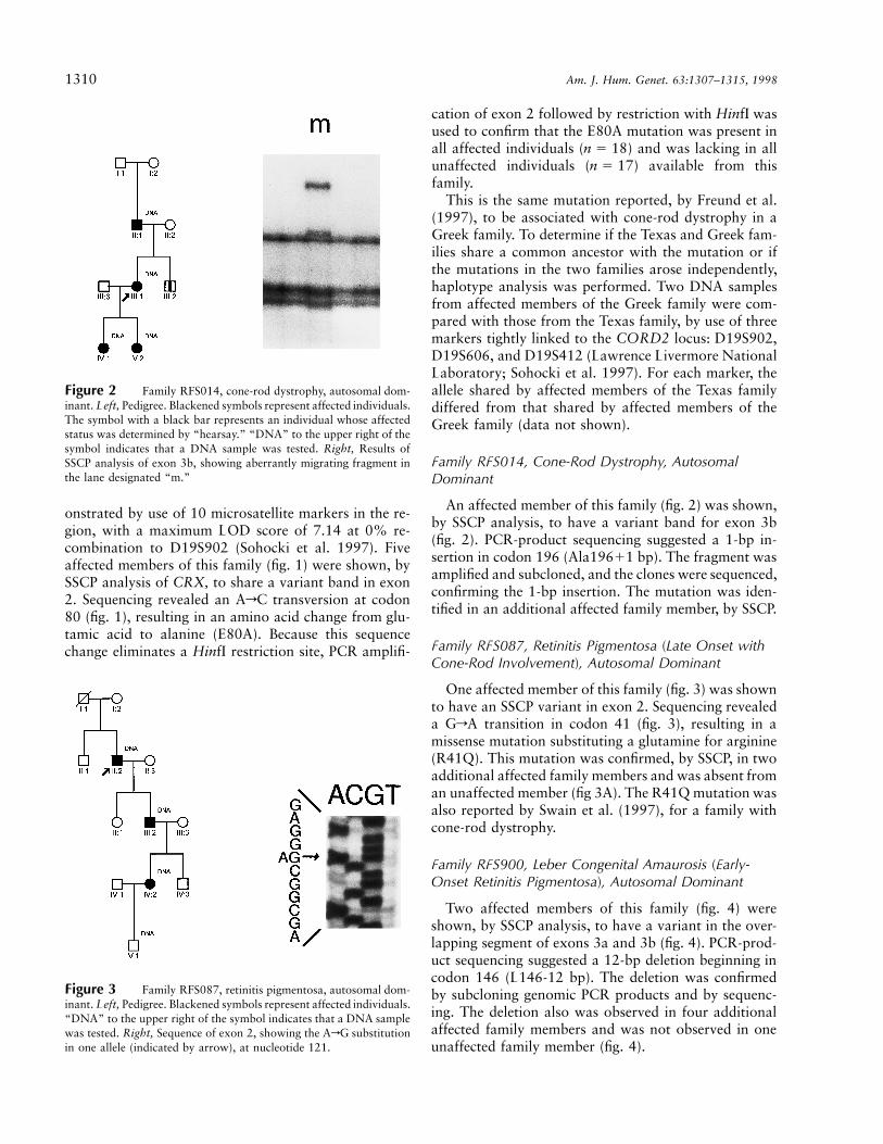
Figure 3 Family RFS087, retinitis pigmentosa, autosomal dom-inant. Left, Pedigree. Blackened symbols represent affected individuals.“DNA” to the upper right of the symbol indicates that a DNA samplewas tested. Right, Sequence of exon 2, showing the ArG substitutionin one allele (indicated by arrow), at nucleotide 121.
onstrated by use of 10 microsatellite markers in the re-gion, with a maximum LOD score of 7.14 at 0% re-combination to D19S902 (Sohocki et al. 1997). Fiveaffected members of this family (fig. 1) were shown, bySSCP analysis of CRX, to share a variant band in exon2. Sequencing revealed an ArC transversion at codon80 (fig. 1), resulting in an amino acid change from glu-tamic acid to alanine (E80A). Because this sequencechange eliminates a HinfI restriction site, PCR amplifi-
cation of exon 2 followed by restriction with HinfI wasused to confirm that the E80A mutation was present inall affected individuals ( ) and was lacking in alln 5 18unaffected individuals ( ) available from thisn 5 17family.
This is the same mutation reported, by Freund et al.(1997), to be associated with cone-rod dystrophy in aGreek family. To determine if the Texas and Greek fam-ilies share a common ancestor with the mutation or ifthe mutations in the two families arose independently,haplotype analysis was performed. Two DNA samplesfrom affected members of the Greek family were com-pared with those from the Texas family, by use of threemarkers tightly linked to the CORD2 locus: D19S902,D19S606, and D19S412 (Lawrence Livermore NationalLaboratory; Sohocki et al. 1997). For each marker, theallele shared by affected members of the Texas familydiffered from that shared by affected members of theGreek family (data not shown).
Family RFS014, Cone-Rod Dystrophy, AutosomalDominant
An affected member of this family (fig. 2) was shown,by SSCP analysis, to have a variant band for exon 3b(fig. 2). PCR-product sequencing suggested a 1-bp in-sertion in codon 196 (Ala19611 bp). The fragment wasamplified and subcloned, and the clones were sequenced,confirming the 1-bp insertion. The mutation was iden-tified in an additional affected family member, by SSCP.
Family RFS087, Retinitis Pigmentosa (Late Onset withCone-Rod Involvement), Autosomal Dominant
One affected member of this family (fig. 3) was shownto have an SSCP variant in exon 2. Sequencing revealeda GrA transition in codon 41 (fig. 3), resulting in amissense mutation substituting a glutamine for arginine(R41Q). This mutation was confirmed, by SSCP, in twoadditional affected family members and was absent froman unaffected member (fig 3A). The R41Q mutation wasalso reported by Swain et al. (1997), for a family withcone-rod dystrophy.
Family RFS900, Leber Congenital Amaurosis (Early-Onset Retinitis Pigmentosa), Autosomal Dominant
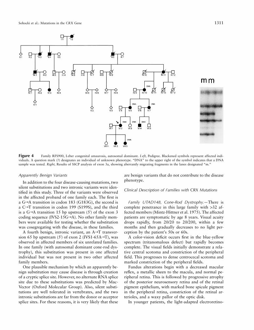
Two affected members of this family (fig. 4) wereshown, by SSCP analysis, to have a variant in the over-lapping segment of exons 3a and 3b (fig. 4). PCR-prod-uct sequencing suggested a 12-bp deletion beginning incodon 146 (L146-12 bp). The deletion was confirmedby subcloning genomic PCR products and by sequenc-ing. The deletion also was observed in four additionalaffected family members and was not observed in oneunaffected family member (fig. 4).
Sohocki et al.: Mutations in the CRX Gene 1311
Figure 4 Family RFS900, Leber congenital amaurosis, autosomal dominant. Left, Pedigree. Blackened symbols represent affected indi-viduals. A question mark (?) designates an individual of unknown phenotype. “DNA” to the upper right of the symbol indicates that a DNAsample was tested. Right, Results of SSCP analysis of exon 3a, showing aberrantly migrating fragments in the lanes designated “m.”
Apparently Benign Variants
In addition to the four disease-causing mutations, twosilent substitutions and two intronic variants were iden-tified in this study. Three of the variants were observedin the affected proband of one family each. The first isa GrA transition in codon 183 (G183G), the second isa CrT transition in codon 199 (S199S), and the thirdis a GrA transition 15 bp upstream (5′) of the exon 3coding sequence (IVS2-15GrA). No other family mem-bers were available for testing whether the substitutionwas cosegregating with the disease, in these families.
A fourth benign, intronic variant, an ArT transver-sion 65 bp upstream (5′) of exon 2 (IVS1-65ArT), wasobserved in affected members of six unrelated families.In one family (with autosomal dominant cone-rod dys-trophy), this substitution was present in one affectedindividual but was not present in two other affectedfamily members.
One plausible mechanism by which an apparently be-nign substitution may cause disease is through creationof a cryptic splice site. However, no alternate RNA splicesite due to these substitutions was predicted by Mac-Vector (Oxford Molecular Group). Also, silent substi-tutions are well tolerated in vertebrates, and the twointronic substitutions are far from the donor or acceptorsplice sites. For these reasons, it is very likely that these
are benign variants that do not contribute to the diseasephenotype.
Clinical Description of Families with CRX Mutations
Family UTAD148, Cone-Rod Dystrophy.—There iscomplete penetrance in this large family with 132 af-fected members (Mintz-Hittner et al. 1975). The affectedpatients are symptomatic by age 8 years. Visual acuitydrops rapidly, from 20/20 to 20/200, within a fewmonths and then gradually decreases to no light per-ception by the patient’s 50s or 60s.
A color-vision deficit occurs first in the blue-yellowspectrum (tritanomalous defect) but rapidly becomescomplete. The visual fields initially demonstrate a rela-tive central scotoma and constriction of the peripheralfield. This progresses to dense centrocecal scotoma andmarked constriction of the peripheral fields.
Fundus alterations begin with a decreased macularreflex, a metallic sheen to the macula, and normal pe-ripheral retina. This is followed by progressive atrophyof the posterior neurosensory retina and of the retinalpigment epithelium, with marked bone spicule pigmentin the peripheral retina, constriction of the retinal ar-terioles, and a waxy pallor of the optic disk.
In younger patients, the light-adapted electroretino-

1312 Am. J. Hum. Genet. 63:1307–1315, 1998
Table 2
PCR Primer Sets for Amplification of SSCP Fragments of CRX
Amplified Fragment(Size [in bp]) Primer Sequences
Size(s) Analyzed(bp)
Exon 1 (257) 5′-CTGCACGTCACCCCATGGTGAGTAAC-3′ 2575′-CAGAGGTCCTCCAAGAGATGAGGCC-3′
Exon 2 (314) 5′-GGATGGAATTCTTGGTCATCCCAC-3′ 178, 136a
5′-CTCTTTGTTCCGGGCAGGCCTC-3′
Exon 3a (501) 5′-CCAGCACCTCTCACCAATAAGTGTC-3′ 191, 174, 136a
5′-GGCGTAGGTCATGGCATAGG-3′
Exon 3b (294) 5′-CCTCCACAGATGTGTGTCCAGAC-3′ 2945′-TGGGAGAAAGGTAGGGGTCTAGG-3′
Exon 3c (341) 5′-GCCTCCGCTTTCTGCTCTTC-3′ 209, 132a
5′-GCCCGATGGAGAGAGATGGAGACTG-3′
a Sizes after restriction digestion.
gram (ERG) is abnormal, but the scotopic ERG and theelectro-oculogram (EOG) are normal. Gradually, thescotopic ERG and EOG become abnormal. Eventually,the ERG becomes unrecordable at all frequencies andintensities of stimulation in both light- and dark-adaptedstates, and the EOG is markedly reduced.
Family RFS014, Cone-Rod Dystrophy.—The primaryvisual disturbances in affected members of this familyare poor color vision and uncorrectable visual acuity,which become evident by age 10 years and graduallyworsen with time. At age 25 years, the proband had abest-corrected visual acuity of 20/50 OD and 20/200 OS(lower OS acuity was attributed to refractive amblyo-pia). Goldmann-Weekers dark-adapted thresholds (77below fixation) were elevated by 0.3 log units. No fielddefects were evident on screening perimetry. Full-fieldERGs revealed rod responses reduced by 50% and coneresponses reduced by 90%. At age 27 years, acuity wasfurther reduced to 20/160 OD and 20/320 OS, but nofurther decline was evident in the ERG.
The best-corrected visual acuity in the father of theproband, at age 54 years, was 20/200 OD and 20/250OS. Goldmann-Weekers dark-adapted thresholds (77 be-low fixation) were elevated by 0.2 log units. Full-fieldERGs revealed rod responses that were reduced by 85%and cone responses that were reduced by 99%. The de-ceased grandfather was thought to have had the sameproblem and was known to have had low visual acuityand color blindness prior to his death at age 59 years.
Family RFS087, Retinitis Pigmentosa (Late Onset withCone-Rod Involvement).—Affected patients in this fam-ily were asymptomatic until their 50s or 60s. At age 58years, the proband was found to have a temporal-fielddefect in each eye, associated with pigment clumping inthe nasal periphery. Visual acuity was 20/15 OD and20/20 OS. The dark-adapted threshold (77 below fixa-tion) was elevated 0.2 log units. Rod ERG amplitudeswere 60% lower than normal, while cone ERGs to 30-Hz flicker were reduced by 65%. The proband has
shown significant progression over 14 years of follow-up. On the most recent visit, at age 72 years, visual acuitywas 20/600 OD and 20/900 OS, and the dark-adaptedthreshold was elevated 0.8 log units. Both rod and coneERGs showed significant progression and were reducedby 90%.
The proband’s father died at an early age, and nothingis known about his visual status. The proband’s son wasasymptomatic at age 44 years, with visual acuity of 20/20 in both eyes. Dark-adapted thresholds were normal.Fluorescein angiography revealed slight peripheral gran-ularity of the epithelium, without narrowing of vesselsor optic-nerve pallor. Rod ERG amplitudes were 50%lower than normal, while cone ERGs to 30-Hz flickerwere reduced by 55%. Five years later, at age 49 years,he had become aware of distortions and occasional blindspots. Visual acuity was 20/20 OD and 20/25 OS, thedark-adapted threshold was still normal, and full-fieldERGs did not show significant progression.
Family RFS900, Leber Congenital Amaurosis (Early-Onset Retinitis Pigmentosa).—Affected members of thisfamily have severe visual disturbances in infancy, ac-companied by nystagmus; some family members are se-verely affected from birth, whereas other affected mem-bers have partial vision into midlife (Heckenlively 1988).The proband (IV:5 in fig. 4) was seen at age 27 years,with best-corrected acuity of 20/300 OD and 20/80 OS.His rod ERG was undetectable, and his cone ERG wasbarely detectable (0.1 mV). His Humphrey field with spotsize 3 had an equivalent diameter of 67. His fundusshowed attenuated vessels, clumped pigment, and retinalpigment epithelium atrophy, including the macula. Atage 11 years, his acuity was 20/200 OD and 20/60 OS.
His mother (III:3 in fig. 4) was seen at age 46 years.She retains only light perception in each eye. Her rodand cone ERG responses are undetectable. She was un-able to see the most intense Goldmann-Weekers test tar-get (elevated 16 log units) and was unable to performa visual field. She showed severe vessel attenuation, RPE
Sohocki et al.: Mutations in the CRX Gene 1313
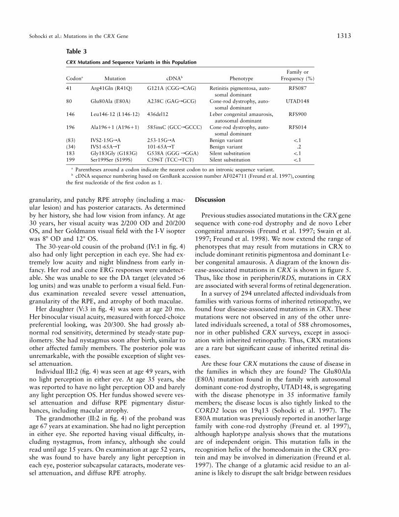
Table 3
CRX Mutations and Sequence Variants in this Population
Codona Mutation cDNAb PhenotypeFamily or
Frequency (%)
41 Arg41Gln (R41Q) G121A (CGGrCAG) Retinitis pigmentosa, auto-somal dominant
RFS087
80 Glu80Ala (E80A) A238C (GAGrGCG) Cone-rod dystrophy, auto-somal dominant
UTAD148
146 Leu146-12 (L146-12) 436del12 Leber congenital amaurosis,autosomal dominant
RFS900
196 Ala19611 (A19611) 585insC (GCCrGCCC) Cone-rod dystrophy, auto-somal dominant
RFS014
(83) IVS2-15GrA 253-15GrA Benign variant !.1(34) IVS1-65ArT 101-65ArT Benign variant .2183 Gly183Gly (G183G) G538A (GGG rGGA) Silent substitution !.1199 Ser199Ser (S199S) C596T (TCCrTCT) Silent substitution !.1
a Parentheses around a codon indicate the nearest codon to an intronic sequence variant.b cDNA sequence numbering based on GenBank accession number AF024711 (Freund et al. 1997), counting
the first nucleotide of the first codon as 1.
granularity, and patchy RPE atrophy (including a mac-ular lesion) and has posterior cataracts. As determinedby her history, she had low vision from infancy. At age30 years, her visual acuity was 2/200 OD and 20/200OS, and her Goldmann visual field with the I-V isopterwas 87 OD and 127 OS.
The 30-year-old cousin of the proband (IV:1 in fig. 4)also had only light perception in each eye. She had ex-tremely low acuity and night blindness from early in-fancy. Her rod and cone ERG responses were undetect-able. She was unable to see the DA target (elevated 16log units) and was unable to perform a visual field. Fun-dus examination revealed severe vessel attenuation,granularity of the RPE, and atrophy of both maculae.
Her daughter (V:3 in fig. 4) was seen at age 20 mo.Her binocular visual acuity, measured with forced-choicepreferential looking, was 20/300. She had grossly ab-normal rod sensitivity, determined by steady-state pup-ilometry. She had nystagmus soon after birth, similar toother affected family members. The posterior pole wasunremarkable, with the possible exception of slight ves-sel attenuation.
Individual III:2 (fig. 4) was seen at age 49 years, withno light perception in either eye. At age 35 years, shewas reported to have no light perception OD and barelyany light perception OS. Her fundus showed severe ves-sel attenuation and diffuse RPE pigmentary distur-bances, including macular atrophy.
The grandmother (II:2 in fig. 4) of the proband wasage 67 years at examination. She had no light perceptionin either eye. She reported having visual difficulty, in-cluding nystagmus, from infancy, although she couldread until age 15 years. On examination at age 52 years,she was found to have barely any light perception ineach eye, posterior subcapsular cataracts, moderate ves-sel attenuation, and diffuse RPE atrophy.
Discussion
Previous studies associated mutations in the CRX genesequence with cone-rod dystrophy and de novo Lebercongenital amaurosis (Freund et al. 1997; Swain et al.1997; Freund et al. 1998). We now extend the range ofphenotypes that may result from mutations in CRX toinclude dominant retinitis pigmentosa and dominant Le-ber congenital amaurosis. A diagram of the known dis-ease-associated mutations in CRX is shown in figure 5.Thus, like those in peripherin/RDS, mutations in CRXare associated with several forms of retinal degeneration.
In a survey of 294 unrelated affected individuals fromfamilies with various forms of inherited retinopathy, wefound four disease-associated mutations in CRX. Thesemutations were not observed in any of the other unre-lated individuals screened, a total of 588 chromosomes,nor in other published CRX surveys, except in associ-ation with inherited retinopathy. Thus, CRX mutationsare a rare but significant cause of inherited retinal dis-eases.
Are these four CRX mutations the cause of disease inthe families in which they are found? The Glu80Ala(E80A) mutation found in the family with autosomaldominant cone-rod dystrophy, UTAD148, is segregatingwith the disease phenotype in 35 informative familymembers; the disease locus is also tightly linked to theCORD2 locus on 19q13 (Sohocki et al. 1997). TheE80A mutation was previously reported in another largefamily with cone-rod dystrophy (Freund et. al 1997),although haplotype analysis shows that the mutationsare of independent origin. This mutation falls in therecognition helix of the homeodomain in the CRX pro-tein and may be involved in dimerization (Freund et al.1997). The change of a glutamic acid residue to an al-anine is likely to disrupt the salt bridge between residues
1314 Am. J. Hum. Genet. 63:1307–1315, 1998
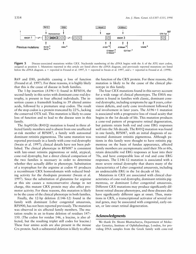
Figure 5 Disease-associated mutations within CRX. Nucleotide numbering of the cDNA begins with the A of the ATG start codon,assigned as position 1. Mutations reported in this article are listed above the cDNA diagram, and previously reported mutations are listedbelow the cDNA diagram. a 5 reported in Freund et al. 1997, b 5 reported in Swain et al. 1997, and c 5 reported in Freund et al. (1998).
R69 and E80, probably causing a loss of function(Freund et al. 1997). For these reasons, it is highly likelythat this is the cause of disease in both families.
The 1-bp insertion (A19611) found in RFS014, thesecond family in this series with dominant cone-rod dys-trophy, is present in four affected individuals. The in-sertion causes a frameshift leading to 39 altered aminoacids, followed by a premature stop codon. The resultof the stop codon is a protein truncated by 22%, lackingthe conserved OTX tail. This mutation is likely to causeloss of function and to lead to the disease seen in thisfamily.
The Arg41Gln (R41Q) mutation is found in three af-fected family members and is absent from one unaffectedat-risk member of RFS087, a family with autosomaldominant retinitis pigmentosa. The same mutation wasreported previously in a family with cone-rod dystrophy(Swain et al. 1997); clinical details have not been pub-lished. The clinical phenotype in RFS087 is consistentwith late-onset retinitis pigmentosa or mild, atypical,cone-rod dystrophy, but a direct clinical comparison ofthe two families is necessary in order to determinewhether they actually differ in phenotype. Substitutionof a tryptophan for the arginine at codon 41 producesa recombinant CRX homeodomain with reduced bind-ing activity for the rhodopsin promoter (Swain et al.1997). Since the substitution of glutamine for arginineat this site causes a nonconservative change in netcharge, this mutant CRX protein may also affect pro-motor activity. For these reasons, this mutation is likelyto be the cause of the clinical phenotype in both families.
Finally, the 12-bp deletion (L146-12) found in thefamily with dominant Leber congenital amaurosis,RFS900, has not been reported previously. The mutationwas observed in six affected family members. The mu-tation results in an in-frame deletion of residues 147–150. (The codon for residue 146, a leucine, is also af-fected, but the resulting triplet still codes for leucine.)These four amino acids are also present in the mouseCrx protein. Such a substantial deletion is likely to affect
the function of the CRX protein. For these reasons, thismutation is likely to be the cause of the clinical phe-notype in this family.
The four CRX mutations found in this survey accountfor a wide range of clinical phenotypes. The E80A mu-tation is found in families with classic, dominant cone-rod dystrophy, including symptoms by age 8 years, color-vision deficits, and early cone involvement followed byrod involvement in later years. The A19611 mutationis associated with a progressive loss of visual acuity thatbegins in the 1st decade of life. This mutation producesa cone-rod pattern of progressive retinal degeneration,but patients retain both rod and cone ERG responseswell into the 5th decade. The R41Q mutation was foundin one family, RFS087, with an initial diagnosis of au-tosomal dominant retinitis pigmentosa. Although pa-tients in this family were thought to have retinitis pig-mentosa on the basis of fundus appearance, affectedfamily members are asymptomatic until their 50s or 60s,retain detectable rod ERG responses at least into their70s, and have comparable loss of rod and cone ERGresponses. The L146-12 mutation is associated with amore severe retinal dystrophy that shares many of thecharacteristics of Leber congenital amaurosis, includingan undetectable ERG in the 1st decade of life.
Mutations in CRX are associated with clinical char-acteristics of cone-rod dystrophy, dominant retinitis pig-mentosa, or dominant Leber congenital amaurosis.Different CRX mutations may produce significantly dif-ferent retinal disease phenotypes, and these diseases alsohave significantly different ages at onset. Thus, muta-tions in CRX, a transcriptional activator of several ret-inal genes, may be associated with congenital, early-on-set, or late-onset retinal degeneration.
Acknowledgments
We thank Dr. Shomi Bhattacharya, Department of Molec-ular Genetics, Institute of Ophthalmology, London, for pro-viding DNA samples from the Greek family with cone-rod

Sohocki et al.: Mutations in the CRX Gene 1315
dystrophy. This work was supported by the Foundation Fight-ing Blindness and the George Gund Foundation, Research toPrevent Blindness, the Farish Fund and the M. D. AndersonFoundation, National Institutes of Health grants EY07412 andEY05235, Institutional National Research Service AwardEY07024 from the National Eye Institute–National Institutesof Health, and grants from the Medical Research Council ofCanada and the Retinitis Pigmentosa Foundation of Canada.R.R.M. is an International Research Scholar of the HowardHughes Medical Institute.
Electronic-Database Information
Accession numbers and URLs for data in this article are asfollows:
GenBank, http://www.ncbi.nlm.nih/Web/Genbank/ (for CRXcDNA sequence [AF024711])
Lawrence Livermore National Laboratory, http://www.llnl.gov/ (for human chromosome 19 sequences)
Online Mendelian Inheritance in Man (OMIM), http://www.ncbi.nlm.gov/Omim (for CRX [MIM 120970]
Retinal Information Network (RetNet), http://www.sph.uth.tmc.edu/RetNet (for cloned and/or mapped genes causinginherited retinal degeneration in humans)
References
Chen S, Wang QL, Nie Z, Sun H, Lennon G, Copeland NG,Gilbert DJ, et al (1997) Crx, a novel otx-like paired-hom-eodomain protein, binds to and transactivates photoreceptorcell-specific genes. Neuron 19:1017–1030
Daiger SP, Rossiter BF, Greenberg J, Christoffels A, Hide W(1998) Data services and software for identifying genes andmutations causing retinal degeneration. Invest OphthalmolVis Sci Suppl 39:S295
Finkelstein R, Boncinelli E (1994) From fly head to mammalianforebrain: the story of otd and OTX. Trends Genet 10:310–315
Freund CL, Gregory-Evans CY, Furukawa T, Papaioannou M,Looser J, Ploder L, Bellingham J, et al (1997) Cone-roddystrophy due to mutations in a novel photoreceptor-specifichomeobox gene (CRX) essential for maintenance of the pho-toreceptor. Cell 91:543–553
Freund CL, Wang QL, Chen S, Muskat BL, Sheffield VC, Ja-cobson SG, McInnes RR, et al (1998) De novo mutations
in the CRX homeobox gene associated with Leber congenitalamaurosis. Nat Genet 18:311–312
Furukawa T, Morrow EM, Cepko CL (1997) Crx, a novel otx-like homeobox gene, shows photoreceptor-specific expres-sion and regulates photoreceptor differentiation. Cell 91:531–541
Heckenlively JR (1988) Autosomal dominant Leber’s amau-rosis congenita. In: Heckenlively JR (ed) Retinitis pigmen-tosa. Lippincott, Philadelphia, pp 146–149
Keen TJ, Inglehearn CF, Kim R, Bird AC, Bhattacharya S(1994) Retinal pattern dystrophy associated with 4 bp in-sertion at codon 140 in the RDS-peripherin gene. Hum MolGenet 3:367–368
Li X, Chen S, Wang Q, Zack DJ, Snyder SH (1998) A pinealregulatory element (PIRE) mediates transactivation by thepineal/retina-specific transcription factor CRX. Proc NatlAcad Sci USA 95:1876–1881
Hittner HM, Murphree AL, Garcia CA, Justice J Jr, ChokshiDB (1975) Dominant cone-rod dystrophy. Doc Ophthalmol39:29–52
Nakazawa M, Kikawa E, Chida Y, Tamai M (1994)Asn244His mutation of the peripherin/RDS gene causingautosomal dominant cone-rod degeneration. Hum Mol Ge-net 3:1195–1196
Nakazawa M, Kikawa E, Chida Y, Wada Y, Shiono T, TamaiM (1996) Autosomal cone-rod retinal dystrophy associatedwith mutations in codon 244 (Asn244His) and codon 184(Tyr184Ser) of the peripherin/RDS gene. Arch Ophthalmol114:72–78
Sohocki MM, Sullivan LS, Mintz-Hittner HA, Daiger SP(1997) Identification of candidate genes involved in the au-tosomal dominant cone rod dystrophy locus (CORD2)which maps to 19q13.1. Am J Hum Genet 61:A295
Swain PK, Chen S, Wang Q, Affigato LM, Coats CL, BradyKD, Fishman GA, et al (1997) Mutations in the cone-rodhomeobox gene are associated with the cone-rod dystrophyphotoreceptor degeneration. Neuron 19:1329–1336
Weleber RG, Carr RE, Murphet WH, Sheffield VC, Stone EM(1993) Phenotypic variation including retinitis pigmentosa,pattern dystrophy, and fundus flavimaculatus in a singlefamily with a deletion of codon 153 or 154 of the peripherin/RDS gene. Arch Ophthalmol 111:1531–1542
Wells J, Wroblewski J, Keen J, Inglehearn C, Jubb C, EcksteinA, Jay M, et al (1993) Mutations in the human retinal de-generation slow (rds) gene can cause either retinitis pig-mentosa or macular dystrophy. Nat Genet 3:213–218