Dopamine modulates diurnal and circadian rhythms of protein phosphorylation in photoreceptor cells...
10
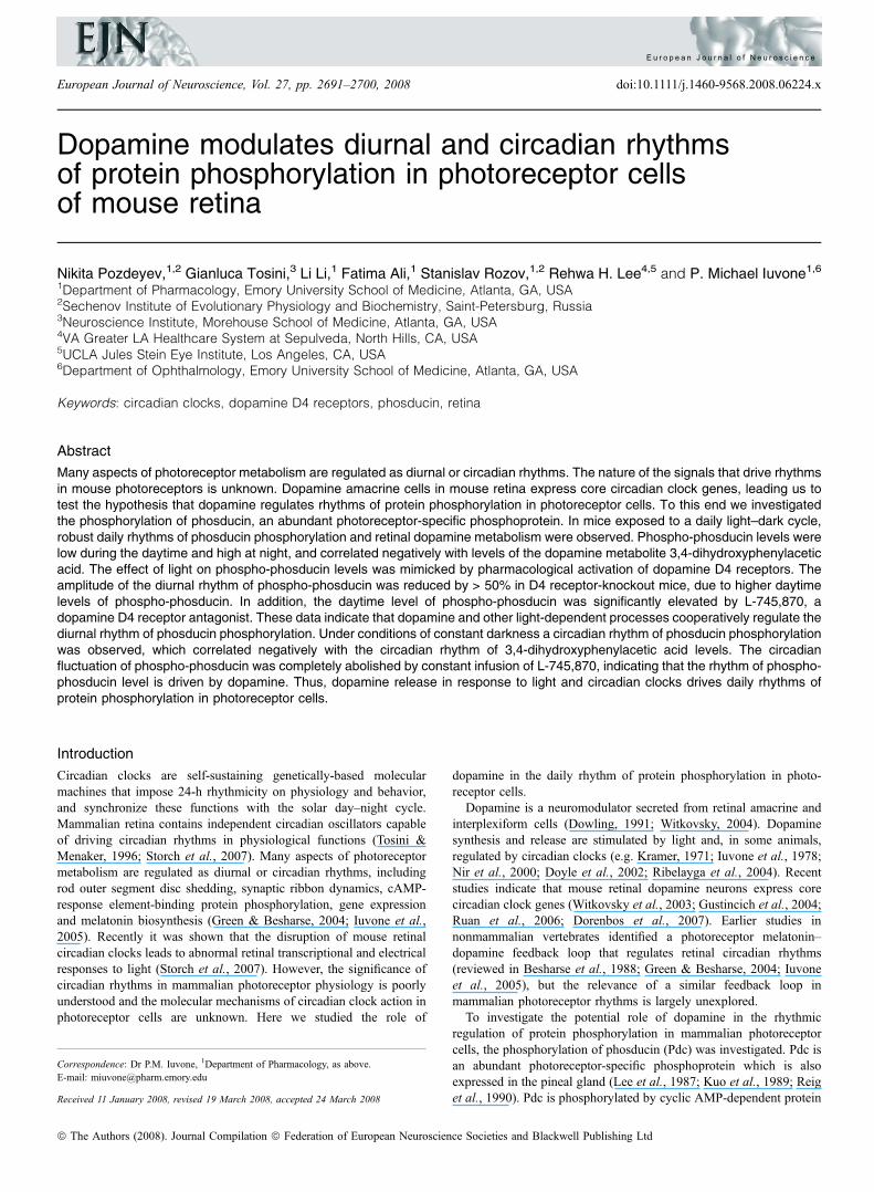
Dopamine modulates diurnal and circadian rhythms of protein phosphorylation in photoreceptor cells of mouse retina Nikita Pozdeyev, 1,2 Gianluca Tosini, 3 Li Li, 1 Fatima Ali, 1 Stanislav Rozov, 1,2 Rehwa H. Lee 4,5 and P. Michael Iuvone 1,6 1 Department of Pharmacology, Emory University School of Medicine, Atlanta, GA, USA 2 Sechenov Institute of Evolutionary Physiology and Biochemistry, Saint-Petersburg, Russia 3 Neuroscience Institute, Morehouse School of Medicine, Atlanta, GA, USA 4 VA Greater LA Healthcare System at Sepulveda, North Hills, CA, USA 5 UCLA Jules Stein Eye Institute, Los Angeles, CA, USA 6 Department of Ophthalmology, Emory University School of Medicine, Atlanta, GA, USA Keywords: circadian clocks, dopamine D4 receptors, phosducin, retina Abstract Many aspects of photoreceptor metabolism are regulated as diurnal or circadian rhythms. The nature of the signals that drive rhythms in mouse photoreceptors is unknown. Dopamine amacrine cells in mouse retina express core circadian clock genes, leading us to test the hypothesis that dopamine regulates rhythms of protein phosphorylation in photoreceptor cells. To this end we investigated the phosphorylation of phosducin, an abundant photoreceptor-specific phosphoprotein. In mice exposed to a daily light–dark cycle, robust daily rhythms of phosducin phosphorylation and retinal dopamine metabolism were observed. Phospho-phosducin levels were low during the daytime and high at night, and correlated negatively with levels of the dopamine metabolite 3,4-dihydroxyphenylacetic acid. The effect of light on phospho-phosducin levels was mimicked by pharmacological activation of dopamine D4 receptors. The amplitude of the diurnal rhythm of phospho-phosducin was reduced by > 50% in D4 receptor-knockout mice, due to higher daytime levels of phospho-phosducin. In addition, the daytime level of phospho-phosducin was significantly elevated by L-745,870, a dopamine D4 receptor antagonist. These data indicate that dopamine and other light-dependent processes cooperatively regulate the diurnal rhythm of phosducin phosphorylation. Under conditions of constant darkness a circadian rhythm of phosducin phosphorylation was observed, which correlated negatively with the circadian rhythm of 3,4-dihydroxyphenylacetic acid levels. The circadian fluctuation of phospho-phosducin was completely abolished by constant infusion of L-745,870, indicating that the rhythm of phospho- phosducin level is driven by dopamine. Thus, dopamine release in response to light and circadian clocks drives daily rhythms of protein phosphorylation in photoreceptor cells. Introduction Circadian clocks are self-sustaining genetically-based molecular machines that impose 24-h rhythmicity on physiology and behavior, and synchronize these functions with the solar day–night cycle. Mammalian retina contains independent circadian oscillators capable of driving circadian rhythms in physiological functions (Tosini & Menaker, 1996; Storch et al., 2007). Many aspects of photoreceptor metabolism are regulated as diurnal or circadian rhythms, including rod outer segment disc shedding, synaptic ribbon dynamics, cAMP- response element-binding protein phosphorylation, gene expression and melatonin biosynthesis (Green & Besharse, 2004; Iuvone et al., 2005). Recently it was shown that the disruption of mouse retinal circadian clocks leads to abnormal retinal transcriptional and electrical responses to light (Storch et al., 2007). However, the significance of circadian rhythms in mammalian photoreceptor physiology is poorly understood and the molecular mechanisms of circadian clock action in photoreceptor cells are unknown. Here we studied the role of dopamine in the daily rhythm of protein phosphorylation in photo- receptor cells. Dopamine is a neuromodulator secreted from retinal amacrine and interplexiform cells (Dowling, 1991; Witkovsky, 2004). Dopamine synthesis and release are stimulated by light and, in some animals, regulated by circadian clocks (e.g. Kramer, 1971; Iuvone et al., 1978; Nir et al., 2000; Doyle et al., 2002; Ribelayga et al., 2004). Recent studies indicate that mouse retinal dopamine neurons express core circadian clock genes (Witkovsky et al., 2003; Gustincich et al., 2004; Ruan et al., 2006; Dorenbos et al., 2007). Earlier studies in nonmammalian vertebrates identified a photoreceptor melatonin– dopamine feedback loop that regulates retinal circadian rhythms (reviewed in Besharse et al., 1988; Green & Besharse, 2004; Iuvone et al., 2005), but the relevance of a similar feedback loop in mammalian photoreceptor rhythms is largely unexplored. To investigate the potential role of dopamine in the rhythmic regulation of protein phosphorylation in mammalian photoreceptor cells, the phosphorylation of phosducin (Pdc) was investigated. Pdc is an abundant photoreceptor-specific phosphoprotein which is also expressed in the pineal gland (Lee et al., 1987; Kuo et al., 1989; Reig et al., 1990). Pdc is phosphorylated by cyclic AMP-dependent protein Correspondence: Dr P.M. Iuvone, 1 Department of Pharmacology, as above. E-mail: [email protected] Received 11 January 2008, revised 19 March 2008, accepted 24 March 2008 European Journal of Neuroscience, Vol. 27, pp. 2691–2700, 2008 doi:10.1111/j.1460-9568.2008.06224.x ª The Authors (2008). Journal Compilation ª Federation of European Neuroscience Societies and Blackwell Publishing Ltd European Journal of Neuroscience
-
Upload
independent -
Category
Documents
-
view
1 -
download
0
Transcript of Dopamine modulates diurnal and circadian rhythms of protein phosphorylation in photoreceptor cells...
Dopamine modulates diurnal and circadian rhythmsof protein phosphorylation in photoreceptor cellsof mouse retina
Nikita Pozdeyev,1,2 Gianluca Tosini,3 Li Li,1 Fatima Ali,1 Stanislav Rozov,1,2 Rehwa H. Lee4,5 and P. Michael Iuvone1,6
1Department of Pharmacology, Emory University School of Medicine, Atlanta, GA, USA2Sechenov Institute of Evolutionary Physiology and Biochemistry, Saint-Petersburg, Russia3Neuroscience Institute, Morehouse School of Medicine, Atlanta, GA, USA4VA Greater LA Healthcare System at Sepulveda, North Hills, CA, USA5UCLA Jules Stein Eye Institute, Los Angeles, CA, USA6Department of Ophthalmology, Emory University School of Medicine, Atlanta, GA, USA
Keywords: circadian clocks, dopamine D4 receptors, phosducin, retina
Abstract
Many aspects of photoreceptor metabolism are regulated as diurnal or circadian rhythms. The nature of the signals that drive rhythmsin mouse photoreceptors is unknown. Dopamine amacrine cells in mouse retina express core circadian clock genes, leading us totest the hypothesis that dopamine regulates rhythms of protein phosphorylation in photoreceptor cells. To this end we investigatedthe phosphorylation of phosducin, an abundant photoreceptor-specific phosphoprotein. In mice exposed to a daily light–dark cycle,robust daily rhythms of phosducin phosphorylation and retinal dopamine metabolism were observed. Phospho-phosducin levels werelow during the daytime and high at night, and correlated negatively with levels of the dopamine metabolite 3,4-dihydroxyphenylaceticacid. The effect of light on phospho-phosducin levels was mimicked by pharmacological activation of dopamine D4 receptors. Theamplitude of the diurnal rhythm of phospho-phosducin was reduced by > 50% in D4 receptor-knockout mice, due to higher daytimelevels of phospho-phosducin. In addition, the daytime level of phospho-phosducin was significantly elevated by L-745,870, adopamine D4 receptor antagonist. These data indicate that dopamine and other light-dependent processes cooperatively regulate thediurnal rhythm of phosducin phosphorylation. Under conditions of constant darkness a circadian rhythm of phosducin phosphorylationwas observed, which correlated negatively with the circadian rhythm of 3,4-dihydroxyphenylacetic acid levels. The circadianfluctuation of phospho-phosducin was completely abolished by constant infusion of L-745,870, indicating that the rhythm of phospho-phosducin level is driven by dopamine. Thus, dopamine release in response to light and circadian clocks drives daily rhythms ofprotein phosphorylation in photoreceptor cells.
Introduction
Circadian clocks are self-sustaining genetically-based molecularmachines that impose 24-h rhythmicity on physiology and behavior,and synchronize these functions with the solar day–night cycle.Mammalian retina contains independent circadian oscillators capableof driving circadian rhythms in physiological functions (Tosini &Menaker, 1996; Storch et al., 2007). Many aspects of photoreceptormetabolism are regulated as diurnal or circadian rhythms, includingrod outer segment disc shedding, synaptic ribbon dynamics, cAMP-response element-binding protein phosphorylation, gene expressionand melatonin biosynthesis (Green & Besharse, 2004; Iuvone et al.,2005). Recently it was shown that the disruption of mouse retinalcircadian clocks leads to abnormal retinal transcriptional and electricalresponses to light (Storch et al., 2007). However, the significance ofcircadian rhythms in mammalian photoreceptor physiology is poorlyunderstood and the molecular mechanisms of circadian clock action inphotoreceptor cells are unknown. Here we studied the role of
dopamine in the daily rhythm of protein phosphorylation in photo-receptor cells.Dopamine is a neuromodulator secreted from retinal amacrine and
interplexiform cells (Dowling, 1991; Witkovsky, 2004). Dopaminesynthesis and release are stimulated by light and, in some animals,regulated by circadian clocks (e.g. Kramer, 1971; Iuvone et al., 1978;Nir et al., 2000; Doyle et al., 2002; Ribelayga et al., 2004). Recentstudies indicate that mouse retinal dopamine neurons express corecircadian clock genes (Witkovsky et al., 2003; Gustincich et al., 2004;Ruan et al., 2006; Dorenbos et al., 2007). Earlier studies innonmammalian vertebrates identified a photoreceptor melatonin–dopamine feedback loop that regulates retinal circadian rhythms(reviewed in Besharse et al., 1988; Green & Besharse, 2004; Iuvoneet al., 2005), but the relevance of a similar feedback loop inmammalian photoreceptor rhythms is largely unexplored.To investigate the potential role of dopamine in the rhythmic
regulation of protein phosphorylation in mammalian photoreceptorcells, the phosphorylation of phosducin (Pdc) was investigated. Pdc isan abundant photoreceptor-specific phosphoprotein which is alsoexpressed in the pineal gland (Lee et al., 1987; Kuo et al., 1989; Reiget al., 1990). Pdc is phosphorylated by cyclic AMP-dependent protein
Correspondence: Dr P.M. Iuvone, 1Department of Pharmacology, as above.E-mail: [email protected]
Received 11 January 2008, revised 19 March 2008, accepted 24 March 2008
European Journal of Neuroscience, Vol. 27, pp. 2691–2700, 2008 doi:10.1111/j.1460-9568.2008.06224.x
ª The Authors (2008). Journal Compilation ª Federation of European Neuroscience Societies and Blackwell Publishing Ltd
E u r o p e a n J o u r n a l o f N e u r o s c i e n c e

kinase A (PKA; Lee et al., 1990a; Pagh-Roehl et al., 1995; Wilkinset al., 1996) and Ca2+ ⁄ calmodulin-dependent protein kinase II(CaMKII; Lee et al., 1990b; Thulin et al., 2001) at two distinctserine residues. Light promotes dephosphorylation of Pdc and itsconsequent binding to transducin bc subunits (Gtbc; Lee et al., 1987;Yoshida et al., 1994; Thulin et al., 2001; Sokolov et al., 2004). Pdcthus represents an excellent model protein for studying photoreceptor-specific phosphorylation by PKA and CaMKII. Proposed functions ofPdc include transducin translocation within photoreceptor cell com-partments (Sokolov et al., 2004), protection of Gtbc from ubiquiti-nation and proteasomal degradation (Zhu & Craft, 1998; Obin et al.,2002), and transcriptional regulation (Zhu & Craft, 2000a,b). In thisstudy, we found that dopamine contributed to diurnal and circadianrhythms of the phosphorylation state of the protein in mousephotoreceptors, providing direct evidence that dopamine plays a rolein regulation of photoreceptor rhythms in mammals.
Materials and methods
Animals
Three strains of mice were used in this study: wild-type C57Bl ⁄ 6Jmice purchased from The Jackson Laboratory (Bar Harbor, ME,USA), congenic mice lacking dopamine D4 receptors on a C57Bl ⁄ 6Jbackground (Drd4) ⁄ ) mice; Rubinstein et al., 1997) and C3H ⁄ f + ⁄ +
mice, a C3H substrain that is unaffected by the retinal degeneration(rd1) mutation (Tosini & Menaker, 1998). Animals were kept in a 12-hlight–12-h dark cycle, with lights on from zeitgeber time (ZT)0 toZT12. Animals were killed by cervical dislocation. All manipulationson mice and tissues under conditions of ‘darkness’ were performedunder dim red light (No. 92 filter; Eastman Kodak, Rochester, NY,USA). For circadian rhythm experiments mice were kept in totaldarkness (24 h ⁄ day) for 2–3 days before dissection.All experimental protocols met the guidelines of the National
Institutes of Health Guide for the Care and Use of Laboratory Animalsand were approved by the Institutional Animal Care and UseCommittee of Emory University.
Characterization of a-Ser 73-Pdc antibody AN519
Rabbit polyclonal antibody AN519 was generated against a keyholelimpet hemacyanin-conjugated phosphopeptide, FSRKMpS73VQEY-ELIHKC, which corresponds to amino acid residues 68–82 of bovinePdc (GenBank accession number P19632). The antibody wascharacterized with the following experiments. The time course ofPdc phosphorylation was assessed by autoradiography and by Westernblotting with AN519 in Fig. 1A. The Pdc ⁄ Gtbc complex, purifiedfrom bovine retinas (Lee et al., 1987), was phosphorylated by thecatalytic subunit of PKA in the presence of 32P-c-ATP, as describedpreviously (Lee et al., 1990a; Chen & Lee, 1997). The time course ofphosphate incorporation was monitored by quenching an aliquot ofreaction mixture at 0, 5, 10, 20 and 60 min after the start of thereaction and by subjecting the samples to SDS-PAGE and electrob-lotting to poly vinylidine difluoride (PVDF) membranes (Millipore,Billerica, MA, USA). One membrane which contained 1.5 lg Pdc perlane was stained with Coomassie Brilliant Blue (CBB), and 32P-Pdcwas detected by autoradiography. A parallel membrane whichcontained 0.04 lg Pdc per lane was subjected to Western blot analysiswith AN519 (1 : 10 000 dilution) and the bound antibodies weredetected by chemiluminescence (West-Pico kit; Pierce, Rockford, IL,USA). The intensities of CBB staining, X-ray film and chemilumi-nescence were captured with a CCD camera (AlphaInnotech
ChemImager 5500; San Leandro, CA, USA). Western blot analysisshowed a time-dependent increase in anti-pS73-Pdc immunoreactivitythat parallels incorporation of 32P into Pdc. The specificity of AN519for the pS73 site of bovine Pdc (Fig. 1B) was established byincubating Pdc ⁄ Gtbc samples for 3 h with high concentrations ofPKA with or without 32P-c-ATP to prepare phosphorylated 32P-labeldPdc and nonphosphorylated Pdc samples. Following SDS-PAGE andCBB staining, the amounts of Pdc were determined using BSA asstandards; the stoichiometry of Pdc phosphorylation (1 : 1) wasestablished by scintillation counting of the excised 32P-labeled Pdcband, as described (Lee et al., 1990a; Chen & Lee, 1997). To evaluatethe specificity of AN519, a series of phosphorylated Pdc (Pdc-pS73)and nonphosphorylated Pdc samples containing 1–100 ng of Pdc weresubjected to Western blot analysis with AN519 and the immunore-activity was detected and quantified using an AlphaInnotech Chem-Imager 5500. Based on the slope of dose–response curves, the affinityfor pS73-Pdc was at least 50 times higher than for nonphosphorylatedPdc. Phosphorylation of Pdc in mouse retinal homogenate (Fig. 1C):two frozen adult C57Bl ⁄ 6 mouse retinas were homogenized in 500 lLof 10 mm Tris–HCl, pH 7.4, containing 2 mm dithiothreitol. Theprotein concentration of retinal homogenate was determined by CBBbinding using BSA as the standard (Bio-Rad binding assay; Hercules,CA, USA). Two aliquots of retinal homogenate (100 lg each) wereincubated at 30�C for 20 min with 0.1 mm ATP and 10 mm MgCl2,with (lanes 1, 3 and 5) or without (lanes 2 and 4) 0.1 mm cyclic AMP.At the end of incubation, multiple sets of retinal samples (20 lg perlane) were separated, in parallel, by SDS-PAGE. After excising aportion of the SDS gel containing one set of sample for CBB staining(lane 1), the remaining gel was electroblotted for Western blot analysiswith AN519 (1 : 10 000 dilution; lanes 2 and 3) and anti-Pdc-pan(1 : 50 000 dilution; Lee et al., 1988) (lanes 4 and 5), respectively.
Fig. 1. Characterization of antibodyAN519 (anti-Pdc-Ser73). (A) Time course ofpurified bovine Pdc phosphorylation by PKA, analyzed with autoradiographyand Western blotting. (B) Specificity of AN519 for phosphorylated bovine Pdc.Increasing amounts of phosphorylated (Pdc-pSer73) or nonphosphorylated (Pdc)bovine Pdc were subjected to Western blot analysis with AN519. (C)Phosphorylation of Pdc in mouse retinal homogenate. Mouse retinal extractwas incubated with ATP–Mg, okadaic acid, IBMX with (lanes 1, 3 and 5) orwithout (lanes 2 and 4) cAMP followed by SDS–PAGE. The proteins werevisualized either with Coomassie brilliant blue (CBB) staining or Western blotanalysis with AN519 or anti-Pdc-pan. See Materials and methods for details.
2692 N. Pozdeyev et al.
ª The Authors (2008). Journal Compilation ª Federation of European Neuroscience Societies and Blackwell Publishing LtdEuropean Journal of Neuroscience, 27, 2691–2700

The equal intensities in the immunoreactivity of anti-Pdc-pan (lanes 4and 5) established the presence of the same levels of Pdc in allsamples. The detection of a single anti-Pdc-pS73 band which co-migrated with Pdc and showed increasing immunoreactivity bycyclic AMP-activated PKA phosphorylation established that AN519recognized and was monospecific for the corresponding PKAphosphorylation site of mouse Pdc, which is Ser71 in the mousesequence.
Measurement of Ser 71- and Ser 54 Pdc phosphorylationby immunoblotting
Retinas were homogenized in 100 lL of 10 mm HEPES buffer, pH7.5, containing EDTA, 10 mm; dithiothreitol, 1 mm; microcystin LR,1 lm (Sigma, St Louis, MO, USA); and phenylmethylsulfonylfluoride, 0.2 mm. This solution inhibits Pdc phosphorylation anddephosphorylation (Lee et al., 2004). After centrifugation at 15000 gfor 10 min, protein concentration was measured in supernatantfractions (Lowry et al., 1951) using bovine serum albumin asstandard. Protein (20 lg) was denatured by sonication for 10 min andseparated on 10% Bis–Tris Criterion XT precast gel (Bio-RadLaboratories, Hercules, CA, USA). After semi-dry transfer of proteinsto PVDF membrane, total Pdc, pSer54-Pdc and pSer71-Pdc weredetected by rabbit polyclonal antibodies: anti-Pdc-pan (1 : 20 000;Lee et al., 1988), which recognizes both unphosphorylated andphospho-Pdc (Chen & Lee, 1997), Pdc54p (1 : 500), which specif-ically recognizes mouse pSer54-Pdc (Song et al., 2007), and AN519(1 : 5000), which specifically recognizes mouse pSer71-Pdc (Fig. 1).
Protein bands were detected using ECLTM Western blottingdetection reagents (Amersham Biosciences, Buckinghamshire, UK).Band densities were quantified using Kodak Molecular Imagingsoftware (Kodak, Rochester, NY, USA). Phospho-Pdc ⁄ Pdc ratios werenormalized to the average ratio of the control group (vehicle injectedand ⁄ or wild-type animals), which was assigned a value of 1.
Retina incubation in vitro
To test the effect of quinpirole on Pdc phosphorylation in vitro, mouseretinas were incubated as described elsewhere (Nir et al., 2002) withminor modifications. Retinas were isolated from dark-adapted miceand placed in ice-cold Earle’s saline in an atmosphere of 5% CO2 and95% O2. After isolation, individual retinas were incubated for 15 minat 37�C in 0.7 mL of Earle’s saline with or without quinpirole(10 lm). Following incubation, retinas were frozen on dry ice andanalyzed for phosphorylated and total Pdc.
Immunoprecipitation of Pdc ⁄ Gtbc complexes
Retinas were homogenized in 200 lL of immunoprecipitation buffer(IP buffer) containing Tris–HCl, 50 mm; NaCl, 150 mm; NP-40, 1%;phenylmethylsulfonyl fluoride, 0.1 mm; microcystin LR, 10 lm;phosphatase inhibitor mixture, 1%; and protease inhibitor mixture, 1and 25 lL ⁄ retina (both mixtures were obtained from Sigma). Thesupernatant was pre-cleared with 20 lL of protein G Sepharose 4 FastFlow (Amersham Biosciences, Uppsala, Sweden) for 1 h at +4�C.Pdc ⁄ Gtb complex was immunoprecipitated overnight with 50 lL anti-Gtb antibody (Gb1, C-16; Santa Cruz Biotechnologies, Santa Cruz,CA, USA), which binds to an N-terminal epitope of Gtb and does notinterfere with Pdc ⁄ Gtb interactions based on the known structure ofthe Pdc ⁄ Gtbc complex. (Gaudet et al., 1996, 1999), or an equalamount of nonimmune rabbit IgG as a control. The antibody–protein
complexes were bound to 50 lL of protein G Sepharose 4 Fast Flowbeads during 2 h incubation at +4�C and subjected to immunoblottinganalysis after multiple washes with immunoprecipitation buffer.Bovine Pdc ⁄ Gtbc complex was purified as described before (Leeet al., 1987) and serial dilutions were loaded together with immuno-precipitate to serve as a standard.
Dopamine and 3,4-dihydroxyphenylacetic acid (DOPAC)analysis by high-performance liquid chromatographywith coulometric detection
Levels of dopamine and DOPAC in mouse retina were determined byion-pair reversed-phase high-performance liquid chromatography(HPLC) with coulometric detection (guard cell at 0.6 V and coulo-metric analytical cell at 0.3 V) using a modification of the method ofNir et al. (2000). Retinas were homogenized in 100 lL of 0.2 n
HClO4 containing 0.01% of sodium metabisulfite and 25 ng ⁄ mL ofinternal standard 3,4-dihydroxybenzylamine hydrobromide. Aftercentrifugation at 15 000 g for 10 min, an 80-lL aliquot of supernatantwas analyzed. The separation was performed on an Ultrasphere ODS250 · 4.6 mm column, 5 lm (Beckman Coulter, Fullerton, CA, USA)with a mobile phase containing sodium phosphate, 0.1 m; EDTA,0.1 mm; sodium octyl sulfate, 0.35 mm; and acetonitrile, 5.5%(vol ⁄ vol), pH 2.7. External standards of dopamine and DOPAC wereanalyzed in each experiment.
Chronic delivery of the dopamine D4 receptor antagonistL-745,870 via surgically implanted osmotic pumps
Osmotic pumps, model 1003D (ALZET Osmotic Pumps, Cupertino,CA, USA), were loaded with L-745,870 solution [2 mg ⁄ mL dissolvedin 45% (2-hydroxypropyl)-b-cyclodextrin] or vehicle and asepticallyimplanted on the backs of the C3H ⁄ f+ ⁄ + mice, subcutaneously slightlyposterior to the scapulae. After recovery from anesthesia, mice wereplaced in constant darkness and retinas were dissected on the secondday at circadian time (CT)6 and CT18. Retinas of right eyes were usedfor Pdc phosphorylation measurements by immunoblotting; L-745,870content was estimated in the contralateral retinas as an index ofosmotic pump performance.L-745,870 was measured by HPLC with coulometric detection
using the same column and HPLC system as for dopamine andDOPAC. Samples were prepared by precipitation of retinal proteins in100 lL of 0.2 n HClO4. KH2PO4 (0.05 m) with 30% (vol ⁄ vol) ofacetonitrile was used as the mobile phase. The potential of the workingelectrode was set to 0.65 V, which was found to be optimal based on ahydrodynamic voltammogramm constructed for L-745,870. Thedetection limit of the method was 20 pg (signal-to-noise ratio 3 : 1).
Chemicals
Quinpirole was purchased from Tocris Bioscience (Ellisville, MO,USA). PD168077, L-745,870 and HPLC standards for dopamine andDOPAC were obtained from Sigma Chemical Co. For i.p. injections,quinpirole and PD168077 were dissolved in water and L-745,870 in45% (2-hydroxypropyl)-b-cyclodextrin.
Statistics
Data are expressed as mean ± SEM. Comparisons among groups weremade with one- or two-way anova with Student–Newman–Keuls
Circadian photoreceptor metabolism and dopamine 2693
ª The Authors (2008). Journal Compilation ª Federation of European Neuroscience Societies and Blackwell Publishing LtdEuropean Journal of Neuroscience, 27, 2691–2700
multiple comparison test. Comparisons of only two groups wereperformed using a two-tailed Student’s t-test.
Results
Diurnal rhythms of Pdc phosphorylation state and dopaminemetabolism
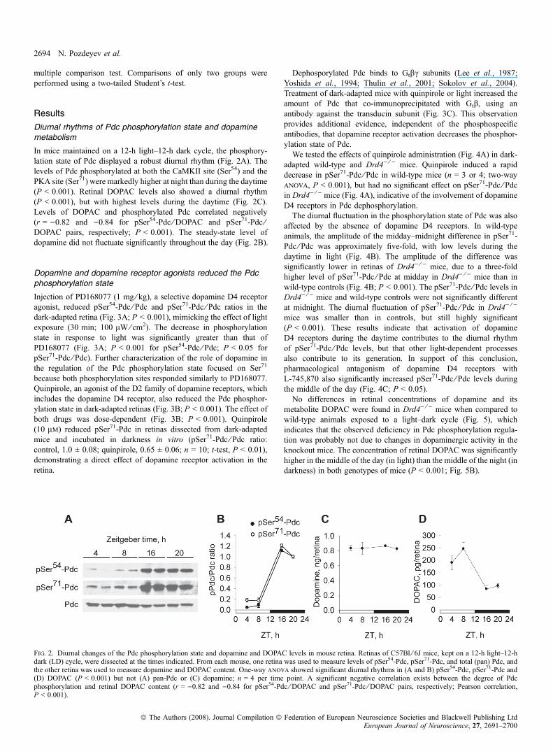
In mice maintained on a 12-h light–12-h dark cycle, the phosphory-lation state of Pdc displayed a robust diurnal rhythm (Fig. 2A). Thelevels of Pdc phosphorylated at both the CaMKII site (Ser54) and thePKA site (Ser71) were markedly higher at night than during the daytime(P < 0.001). Retinal DOPAC levels also showed a diurnal rhythm(P < 0.001), but with highest levels during the daytime (Fig. 2C).Levels of DOPAC and phosphorylated Pdc correlated negatively(r = )0.82 and )0.84 for pSer54-Pdc ⁄ DOPAC and pSer71-Pdc ⁄DOPAC pairs, respectively; P < 0.001). The steady-state level ofdopamine did not fluctuate significantly throughout the day (Fig. 2B).
Dopamine and dopamine receptor agonists reduced the Pdcphosphorylation state
Injection of PD168077 (1 mg ⁄ kg), a selective dopamine D4 receptoragonist, reduced pSer54-Pdc ⁄ Pdc and pSer71-Pdc ⁄ Pdc ratios in thedark-adapted retina (Fig. 3A; P < 0.001), mimicking the effect of lightexposure (30 min; 100 lW ⁄ cm2). The decrease in phosphorylationstate in response to light was significantly greater than that ofPD168077 (Fig. 3A; P < 0.001 for pSer54-Pdc ⁄ Pdc; P < 0.05 forpSer71-Pdc ⁄ Pdc). Further characterization of the role of dopamine inthe regulation of the Pdc phosphorylation state focused on Ser71
because both phosphorylation sites responded similarly to PD168077.Quinpirole, an agonist of the D2 family of dopamine receptors, whichincludes the dopamine D4 receptor, also reduced the Pdc phosphor-ylation state in dark-adapted retinas (Fig. 3B; P < 0.001). The effect ofboth drugs was dose-dependent (Fig. 3B; P < 0.001). Quinpirole(10 lm) reduced pSer71-Pdc in retinas dissected from dark-adaptedmice and incubated in darkness in vitro (pSer71-Pdc ⁄ Pdc ratio:control, 1.0 ± 0.08; quinpirole, 0.65 ± 0.06; n = 10; t-test, P < 0.01),demonstrating a direct effect of dopamine receptor activation in theretina.
Dephosporylated Pdc binds to Gtbc subunits (Lee et al., 1987;Yoshida et al., 1994; Thulin et al., 2001; Sokolov et al., 2004).Treatment of dark-adapted mice with quinpirole or light increased theamount of Pdc that co-immunoprecipitated with Gtb, using anantibody against the transducin subunit (Fig. 3C). This observationprovides additional evidence, independent of the phosphospecificantibodies, that dopamine receptor activation decreases the phosphor-ylation state of Pdc.We tested the effects of quinpirole administration (Fig. 4A) in dark-
adapted wild-type and Drd4) ⁄ ) mice. Quinpirole induced a rapiddecrease in pSer71-Pdc ⁄ Pdc in wild-type mice (n = 3 or 4; two-wayanova, P < 0.001), but had no significant effect on pSer71-Pdc ⁄ Pdcin Drd4) ⁄ ) mice (Fig. 4A), indicative of the involvement of dopamineD4 receptors in Pdc dephosphorylation.The diurnal fluctuation in the phosphorylation state of Pdc was also
affected by the absence of dopamine D4 receptors. In wild-typeanimals, the amplitude of the midday–midnight difference in pSer71-Pdc ⁄ Pdc was approximately five-fold, with low levels during thedaytime in light (Fig. 4B). The amplitude of the difference wassignificantly lower in retinas of Drd4) ⁄ ) mice, due to a three-foldhigher level of pSer71-Pdc ⁄ Pdc at midday in Drd4) ⁄ ) mice than inwild-type controls (Fig. 4B; P < 0.001). The pSer71-Pdc ⁄ Pdc levels inDrd4) ⁄ ) mice and wild-type controls were not significantly differentat midnight. The diurnal fluctuation of pSer71-Pdc ⁄ Pdc in Drd4) ⁄ )
mice was smaller than in controls, but still highly significant(P < 0.001). These results indicate that activation of dopamineD4 receptors during the daytime contributes to the diurnal rhythmof pSer71-Pdc ⁄ Pdc levels, but that other light-dependent processesalso contribute to its generation. In support of this conclusion,pharmacological antagonism of dopamine D4 receptors withL-745,870 also significantly increased pSer71-Pdc ⁄ Pdc levels duringthe middle of the day (Fig. 4C; P < 0.05).No differences in retinal concentrations of dopamine and its
metabolite DOPAC were found in Drd4) ⁄ ) mice when compared towild-type animals exposed to a light–dark cycle (Fig. 5), whichindicates that the observed deficiency in Pdc phosphorylation regula-tion was probably not due to changes in dopaminergic activity in theknockout mice. The concentration of retinal DOPAC was significantlyhigher in the middle of the day (in light) than the middle of the night (indarkness) in both genotypes of mice (P < 0.001; Fig. 5B).
Fig. 2. Diurnal changes of the Pdc phosphorylation state and dopamine and DOPAC levels in mouse retina. Retinas of C57Bl ⁄ 6J mice, kept on a 12-h light–12-hdark (LD) cycle, were dissected at the times indicated. From each mouse, one retina was used to measure levels of pSer54-Pdc, pSer71-Pdc, and total (pan) Pdc, andthe other retina was used to measure dopamine and DOPAC content. One-way anova showed significant diurnal rhythms in (A and B) pSer54-Pdc, pSer71-Pdc and(D) DOPAC (P < 0.001) but not (A) pan-Pdc or (C) dopamine; n = 4 per time point. A significant negative correlation exists between the degree of Pdcphosphorylation and retinal DOPAC content (r = )0.82 and )0.84 for pSer54-Pdc ⁄ DOPAC and pSer71-Pdc ⁄ DOPAC pairs, respectively; Pearson correlation,P < 0.001).
2694 N. Pozdeyev et al.
ª The Authors (2008). Journal Compilation ª Federation of European Neuroscience Societies and Blackwell Publishing LtdEuropean Journal of Neuroscience, 27, 2691–2700

Circadian rhythms of pSer 71-Pdc ⁄ Pdc, dopamine and DOPACin the retinas of C57Bl ⁄ 6J and C3H ⁄ f + ⁄ + mice
The circadian regulation of Pdc phosphorylation was examined in twostrains of mice: the C3H ⁄ f + ⁄ + strain, in which retinal dopamine issubject to both light-driven and circadian regulation, and theC57Bl ⁄ 6J strain, in which retinal dopamine is regulated only by light(Doyle et al., 2002). We confirmed that the levels of retinal dopamineand its primary metabolite DOPAC in C57Bl ⁄ 6J mice were notrhythmic in constant (24 h ⁄ day) darkness (Fig. 6C and D; P > 0.05).In contrast, retinas of C3H ⁄ f + ⁄ + mice displayed a robust rhythm inDOPAC levels (Fig. 6H, P < 0.001), and a less prominent rhythm indopamine concentration (Fig. 6G, P < 0.05). Accordingly, a circadianrhythm of pSer71-Pdc ⁄ Pdc was found in C3H ⁄ f + ⁄ + mice (Fig. 6E andF, P < 0.001) but not in C57Bl ⁄ 6J animals (Fig. 6A and B).Significant interactions between factors ‘circadian time’ and ‘geno-type’ (two-factor anova, P < 0.05 for pSer71-Pdc ⁄ Pdc ratio and
dopamine, P < 0.001 for DOPAC) indicate that retinal circadianrhythms in Pdc, dopamine and DOPAC are dependent on genotype.A significant negative correlation between retinal DOPAC concentra-tion and pSer71-Pdc ⁄ Pdc (r = )0.77, P < 0.001) suggests that thecircadian rhythm in Pdc phosphorylation is regulated by retinaldopamine.To further test the hypothesis that the circadian rhythms of Pdc
phosphorylation state were driven by dopamine, the effect of constantinfusion of the dopamine D4 receptor antagonist, L-745,870, wasexamined (Fig. 7). In C3H ⁄ f + ⁄ + mice implanted with osmotic mini-pumps containing vehicle, a significant circadian fluctuation in thedegree of both Ser54- and Ser71-Pdc phosphorylation was observed onthe second day of constant darkness (P < 0.001), with highest levels atnight. In contrast, no day–night difference in the phosphorylation stateof either Ser54 or Ser71 was detected in C3H ⁄ f + ⁄ + mice implantedwith mini-pumps releasing L-745,870. Retinas in drug-treated micehad high levels of phosphorylation during the day and night.
Fig. 3. Agonists of dopamine D4 receptors induced dephosphorylation of Pdc in the retina and promoted Pdc ⁄ Gtb interaction. C57Bl ⁄ 6J mice, which had beendark-adapted for 14 h beginning at ZT12, were injected intraperitoneally (i.p.) with (A and B) PD168077 and (B and C) quinpirole and vehicles, or (A and C) wereexposed to light for 30 min (100 lW ⁄ cm2). Retinas were dissected 30 min after injection or the beginning of light exposure. (A) PD168077 (1 mg ⁄ kg of bodyweight) caused dephosphorylation of both Ser54 and Ser71 of Pdc mimicking the effect of light (n = 3; anova P < 0.001). (B) The effects of PD168077 andquinpirole on Ser71-Pdc were dose-dependent (n = 4; anova P < 0.001 for both drugs) (C) Proteins from dark-adapted (D), dark-adapted quinpirole-treated (Q;1 mg ⁄ kg body weight for 30 min) or light-treated (L; 30 min, 100 lW ⁄ cm2) retinas were subjected to immunoprecipitation with anti-Gtb antibody (Gb1, C-16) ornonimmune rabbit IgG. Precipitated proteins were analyzed by immunoblotting (anti-Pdc-pan, 1 : 20 000 and Gb1, 1 : 1000 for Pdc and Gtb, respectively). Resultsshown are representative of three independent experiments. Quinpirole and light promoted the interaction of Pdc with Gtb.
Circadian photoreceptor metabolism and dopamine 2695
ª The Authors (2008). Journal Compilation ª Federation of European Neuroscience Societies and Blackwell Publishing LtdEuropean Journal of Neuroscience, 27, 2691–2700

Discussion
Our data indicate that dopamine, released from the inner retinal neuronsin response to light and ⁄ or circadian clocks and acting on dopamineD4 receptors, regulates rhythms of protein phosphorylation in mam-malian photoreceptor cells. The data show that the photoreceptor-specific protein Pdc undergoes diurnal and circadian fluctuations in thedegree of phosphorylation of its PKA (Ser71) and CaMKII (Ser54)consensus sites. The involvement of dopamine in the generation ofthese rhythms is supported by several lines of evidence. The level ofPdc phosphorylation is lowest during the daytime in light, whendopamine synthesis and metabolism are highest (e.g. Kramer, 1971;Iuvone et al., 1978; Nir et al., 2000; Doyle et al., 2002; Ribelaygaet al., 2004). Pharmacological activation of retinal dopamine D4receptors results in dephosphorylation of Pdc and promotes Pdcbinding to Gtbc. Genetic ablation of the gene encoding dopamine D4
receptors or pharmacological blockade of D4 receptors increases thelevel of phosphorylated Pdc during the daytime, reducing theamplitude of the diurnal fluctuation. A circadian rhythm of Pdcphosphorylation state is observed in a mouse strain (C3H ⁄ f + ⁄ +) thatalso expresses a circadian rhythm in dopamine metabolism, but not in astrain that does not (C57Bl ⁄ 6J). Lastly, the circadian fluctuation in Pdcphosphorylation in photoreceptors of C3H ⁄ f + ⁄ + mice is completelyeliminated by dopamine D4 receptor antagonism.The partial reduction in amplitude of the diurnal rhythm of the Pdc
phosphorylation state in Drd4) ⁄ ) mice suggests that dopamine andother light-dependent processes within the photoreceptors act coop-eratively to generate the rhythm in intact mice. The effect of light onthe Pdc phosphorylation state may be mediated by activation andtranslocation of phosphoprotein phosphatase (Brown et al., 2002).Reductions in protein kinase activities may also contribute to the lightand dopamine responses. The observation that both the PKA- andCaMKII phosphorylation sites are regulated similarly by light anddopamine D4 receptor activation suggest that both kinases may beinvolved. Light-induced reductions in PKA activity occur subsequentto decreased cAMP, and light and dopamine D4 receptor activationappear to reduce the same pool of cAMP in mouse photoreceptor cells(Cohen & Blazynski, 1990; Cohen et al., 1992), but by differentmechanisms (Nir et al., 2002). A reduction in CaMKII in photore-ceptors may occur subsequent to light- or dopamine-induced decreasesin intracellular Ca2+. Light rapidly decreases intracellular Ca2+ levelsby closing cGMP-gated and voltage-gated channels (Krizaj &Copenhagen, 2002). Activation of dopamine receptors also decreasesintracellular Ca2+ in photoreceptors (Thoreson et al., 2002; Ivanovaet al., 2008), but the mechanisms involved are less well established.Circadian rhythms in retinal dopamine synthesis and metabolism
have been observed in rats, birds, fish, amphibians and some strains ofmice (Wirz-Justice et al., 1984; Adachi et al., 1998; Pozdeyev &Lavrikova, 2000; Doyle et al., 2002; Ribelayga et al., 2004; Bartell
Fig. 5. Diurnal changes of retinal dopamine and DOPAC in Drd4+ ⁄ + andDrd4) ⁄ ) mice. Retinas from wild-type and mutant mice were dissected at ZT6during the daytime (in light) and at ZT18 at night, and dopamine (A) andDOPAC (B) levels were measured by HPLC with electrochemical detection;n = 5 per group. No significant differences in dopamine or DOPAC levelsbetween genotypes were found (two-way anova , P > 0.05).
Fig. 4. Role of dopamine D4 receptors in the regulation of Pdc Ser71 phosphorylation. Pdc Ser71 phosphorylation was studied in dark-adapted wild-type (Drd4+ ⁄ +)and dopamine D4 receptor-knockout (Drd4) ⁄ )) mice on a C57Bl ⁄ 6 background. (A) Mice were injected i.p. with quinpirole (1 mg ⁄ kg body weight) and retinaswere dissected in darkness 15, 30 or 60 min after drug administration. Two-way anova indicated significant effects of quinpirole (P < 0.001), genotype(P < 0.001), and a significant interaction of quinpirole and genotype (P = 0.011); n = 4 per group. (B) pSer71-Pdc ⁄ Pdc was measured in Drd4+ ⁄ + and Drd4) ⁄ )
mice, maintained on a regular 12-h light–12-h dark schedule, at ZT6 (middle of the day) and ZT18 (middle of the night). Both time of day (P < 0.001) and genotype(P = 0.02) significantly affected phosphorylation of Pdc Ser71, and a significant interaction of time and genotype was observed (P = 0.005); n = 4 per group. (C) Thedopamine D4 receptor antagonist L-745,870 (1 mg ⁄ kg body weight) increases the amount of pSer71-Pdc during the daytime (ZT6). n = 4 per group, t-test,*P < 0.05). Representative immunoblots and densitometry data are shown for each experiment.
2696 N. Pozdeyev et al.
ª The Authors (2008). Journal Compilation ª Federation of European Neuroscience Societies and Blackwell Publishing LtdEuropean Journal of Neuroscience, 27, 2691–2700

et al., 2007). A circadian rhythm in Pdc was observed in C3H ⁄ f + ⁄ +
mice, which show robust circadian rhythms of dopamine metabolism.The highly significant negative correlation between retinal DOPAClevels and pSer71-Pdc ⁄ Pdc, coupled with our data showing regulationof Pdc phosphorylation by dopamine D4 receptors and the abolition ofthe circadian rhythm of Pdc phosphorylation state by a D4 receptorantagonist, strongly supports the conclusion that clocks generate acircadian rhythm in Pdc phosphorylation through rhythmic changes indopaminergic neuronal activity.
Recent findings support the hypothesis that dopaminergic amacrinecells contain an autonomous circadian clock that drives dopaminerelease andmetabolism (Witkovsky et al., 2003; Gustincich et al., 2004;Ruan et al., 2006; Dorenbos et al., 2007). However, it must be noted thatretinal dopamine content and metabolism are circadian in mice thatrhythmically synthesize melatonin, including C3H ⁄ f + ⁄ + mice, but notin mice that are genetically incapable of synthesizing melatonin (Tosini&Menaker, 1998;Nir et al., 2000; Doyle et al., 2002). InC57Bl ⁄ 6mice,which do not synthesize melatonin, a circadian rhythm of dopamine
Fig. 6. Circadian rhythms of pSer71-Pdc ⁄ Pdc, dopamine, and DOPAC in the retinas of C57Bl ⁄ 6J and C3H ⁄ f + ⁄ + mice. Retinal concentrations of pSer71-Pdc, totalPdc, dopamine and the dopamine metabolite DOPAC were measured in two strains of mice, (A–D) C57Bl ⁄ 6J and (E–H) C3H ⁄ f + ⁄ +, on the second or third day ofconstant (24h ⁄ day) darkness. Two-factor anova shows a significant effect of ‘circadian time’ for C3H ⁄ f + ⁄ + mice but not C57Bl ⁄ 6J mice for pSer71-Pdc ⁄ Pdc(n = 4 or 5; P < 0.001), dopamine (n = 5; P < 0.05) and DOPAC (n = 5; P < 0.001), and significant interactions between factors circadian time and genotype(P < 0.05 for pSer71-Pdc ⁄ Pdc ratio and dopamine, P < 0.001 for DOPAC). A significant negative correlation existed between retinal DOPAC content and pSer71-Pdc ⁄ Pdc ratio (Pearson correlation coefficient r = )0.77, P < 0.001).
Fig. 7. Abolition of the circadian rhythm of Pdc phosphorylation state by the dopamine D4 receptor antagonist L-745,870. C3H ⁄ f + ⁄ + mice were implanted withosmotic pumps releasing L-745,870 or vehicle and transferred to constant darkness. On the second day of constant darkness, retinas were dissected in the middle ofsubjective day (CT6) and the middle of subjective night (CT18). From each mouse, one retina was used to measure levels of pSer54-Pdc, pSer71-Pdc and total (pan)Pdc, and the other retina to measure the content of L-745,870. The levels of L-745,870 was 156 ± 26 and 129 ± 21 pg per retina at 06.00 and 18.00 h, respectively(n = 5, t-test, P = 0.47) in drug-treated mice. The degree of phosphorylation of both Ser54 and Ser71 exhibited circadian rhythms in vehicle-treated mice (two-wayanova , Student–Newman–Keuls test, P < 0.001, n = 5) that were eliminated by the dopamine D4 receptor antagonist (P > 0.05).
Circadian photoreceptor metabolism and dopamine 2697
ª The Authors (2008). Journal Compilation ª Federation of European Neuroscience Societies and Blackwell Publishing LtdEuropean Journal of Neuroscience, 27, 2691–2700
metabolism can be induced by daily injections of melatonin (Doyleet al., 2002). Similarly, the circadian rhythms of dopamine release infish, lizard and bird retinas appear to be dependent onmelatonin receptoractivation (Adachi et al., 1998; Ribelayga et al., 2004; Bartell et al.,2007). It is tempting to speculate that melatonin released fromphotoreceptor cells is required to entrain the circadian oscillators indopamine amacrine cells to the daily light–dark cycle. Our new datasuggest that melatonin in the mouse retina may contribute to circadianrhythms in photoreceptors, including rhythms of protein phosphoryla-tion, by regulating dopamine release, and indicate that the retinaldopamine–melatonin feedback loop previously described in nonmam-malian vertebrates (reviewed in Besharse et al., 1988; Green &Besharse, 2004; Iuvone et al., 2005) plays an important role in thecircadian organization of mammalian retina.
In rat retina, a circadian oscillator that drives melatonin synthesisrhythms is localized to photoreceptor cells, which also expresstranscripts of the core circadian clock genes Per1, Per3, Cry1, Cry2,Clock, Bmal1, Rev-erba and Rora (Tosini et al., 2007), albeit at lowerlevels than in the inner retina. However, in mouse retina, the fullcomplement of core clock gene transcripts was not detected by single-cell reverse transcriptase–polymerase chain reaction (RT-PCR; Ruanet al., 2006; Dorenbos et al., 2007). It remains to be determinedwhether this represents a fundamental difference in circadian organi-zation of mouse and rat retina or whether clock genes in mousephotoreceptors are expressed at levels below the detection limit ofsingle-cell RT-PCR.Our present study and earlier published data provide the basis for
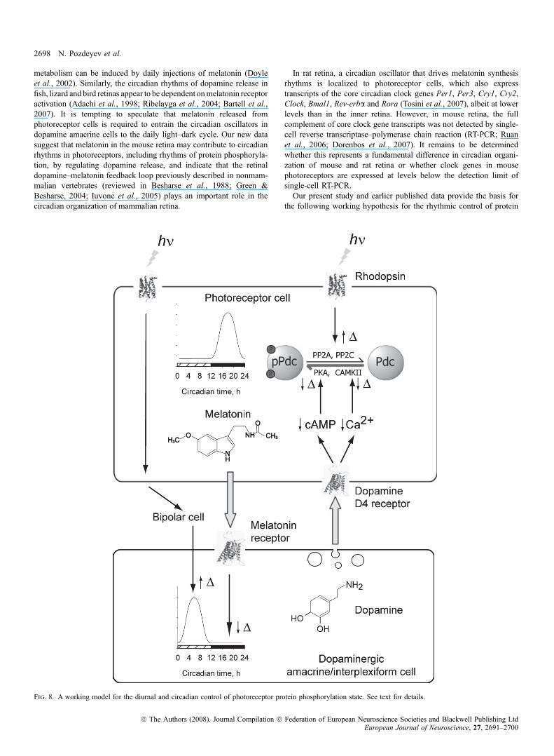
the following working hypothesis for the rhythmic control of protein
Fig. 8. A working model for the diurnal and circadian control of photoreceptor protein phosphorylation state. See text for details.
2698 N. Pozdeyev et al.
ª The Authors (2008). Journal Compilation ª Federation of European Neuroscience Societies and Blackwell Publishing LtdEuropean Journal of Neuroscience, 27, 2691–2700

phosphorylation in photoreceptor cells (Fig. 8). In darkness, cyclicGMP levels in photoreceptors are high, leading to Na+ and Ca2+
influx through cyclic nucleotide-gated cation channels, depolarizationof the plasma membrane and activation of voltage-gated Ca2+
channels (reviewed in Iuvone et al., 2005). The increase ofintracellular Ca2+ activates CaMKII and stimulates cyclic AMPformation, activating PKA, resulting in increased protein phosphor-ylation by these kinases. Activation of PKA also stimulates nocturnalmelatonin formation in a circadian clock-dependent manner (Fuku-hara et al., 2004). Melatonin acts on dopamine neurons to suppressdopamine synthesis and release at night (Dubocovich, 1983; Adachiet al., 1998; Ribelayga et al., 2004; Bartell et al., 2007) and,possibly, to entrain the dopaminergic circadian clock. Light exposureduring the daytime reverses these effects. Light decreases cyclicGMP, Ca2+ and cyclic AMP in photoreceptor cells, reducing proteinkinase activity. These effects, combined with increased phosphopro-tein phosphatase activity (Brown et al., 2002), result in a decrease inthe phosphorylation state of the kinase substrates. In addition, lightexposure, acting via photoreceptors and retinal circuitry, stimulatesdopamine release, with dopamine acting on D4 receptors to furtherreduce Ca2+ and cyclic AMP levels, and the activities of therespective protein kinases. The effects of light and darkness onphotoreceptors, combined with the effects of dopamine, drive therobust diurnal rhythms of phosphorylation of PKA and CaMKIIsubstrates in photoreceptor cells. We propose that in animal speciescapable of circadian synthesis of melatonin and containing circadianoscillators in the dopamine cells, the day–night rhythm in photore-ceptor protein phosphorylation state persists in constant darkness,albeit with lower amplitude, due to the reciprocal circadian release ofmelatonin and dopamine, with activation of photoreceptor dopamineD4 receptors during the subjective day.
In conclusion, our data demonstrate a role for dopamine in theregulation of diurnal and circadian rhythms of protein phosphory-lation state in photoreceptor cells. The role of dopamine in otheraspects of circadian photoreceptor metabolism remains to beinvestigated.
Acknowledgements
The authors are grateful to Amy Visser and Holly Zhou for assistancegenotyping the mice, to David Grandy and Malcolm Low (Oregon HealthSciences University) for providing the Drd4) ⁄ ) breeder mice, and MaximSokolov (West Virginia University) for providing the pSer54-Pdc antibody. Thisstudy was supported by NIH grants EY004864 and EY014764 (P.M.I.),NS43459 (G.T.), and a VA Merit Award (R.H.L.). N.P. and S.R. were supportedby a grant RUB1-2637 from the US Civilian Research & DevelopmentFoundation.
Abbreviations
CaMKII, Ca2+ ⁄ calmodulin-dependent protein kinase II; CT, circadian time;DOPAC, 3,4-dihydroxyphenylacetic acid; Gtbc, transducin bc subunits; HPLC,high-performance liquid chromatography; Pdc, phosducin; PKA, cyclic AMP-dependent protein kinase; ZT, zeitgeber time.
References
Adachi, A., Nogi, T. & Ebihara, S. (1998) Phase-relationship and mutual effectsbetween circadian rhythms of ocular melatonin and dopamine in the pigeon.Brain Res., 792, 361–369.
Bartell, P.A., Miranda-Anaya, M., McIvor, W. & Menaker, M. (2007)Interactions between dopamine and melatonin organize circadian rhythmicityin the retina of the green iguana. J. Biol. Rhythms, 22, 515–523.
Besharse, J.C., Iuvone, P.M. & Pierce, M.E. (1988) Regulation of rhythmicphotoreceptor metabolism: a role for post-receptoral neurons. Prog. Retin.Res., 7, 21–61.
Brown, B.M., Carlson, B.L., Zhu, X., Lolley, R.N. & Craft, C.M. (2002) Light-driven translocation of the protein phosphatase 2A complex regulateslight ⁄ dark dephosphorylation of phosducin and rhodopsin. Biochemistry, 41,13526–13538.
Chen, F. & Lee, R.H. (1997) Phosducin and betagamma–transducin interactionI: effects of post-translational modifications. Biochem. Biophys. Res.Commun., 233, 370–374.
Cohen, A.I. & Blazynski, C. (1990) Dopamine and its agonists reduce a light-sensitive pool of cyclic AMP in mouse photoreceptors. Vis. Neurosci., 4,43–52.
Cohen, A.I., Todd, R.D., Harmon, S. & O’Malley, K.L. (1992) Photoreceptorsof mouse retinas possess D4 receptors coupled to adenylate cyclase. Proc.Natl Acad. Sci. USA, 89, 12093–12097.
Dorenbos, R., Contini, M., Hirasawa, H., Gustincich, S. & Raviola, E. (2007)Expression of circadian clock genes in retinal dopaminergic cells. Vis.Neurosci., 24, 573–580.
Dowling, J.E. (1991) Retinal neuromodulation: the role of dopamine. Vis.Neurosci., 7, 87–97.
Doyle, S.E., Grace, M.S., McIvor, W. & Menaker, M. (2002) Circadianrhythms of dopamine in mouse retina: the role of melatonin. Vis. Neurosci.,19, 593–601.
Dubocovich, M.L. (1983) Melatonin is a potent modulator of dopamine releasein the retina. Nature, 306, 782–784.
Fukuhara, C., Liu, C., Ivanova, T.N., Chan, G.C., Storm, D.R., Iuvone, P.M. &Tosini, G. (2004) Gating of the cAMP signaling cascade and melatoninsynthesis by the circadian clock in mammalian retina. J. Neurosci., 24,1803–1811.
Gaudet, R., Bohm, A. & Sigler, P.B. (1996) Crystal structure at 2.4 angstromsresolution of the complex of transducin betagamma and its regulator,phosducin. Cell, 87, 577–588.
Gaudet, R., Savage, J.R., McLaughlin, J.N., Willardson, B.M. & Sigler, P.B.(1999) A molecular mechanism for the phosphorylation-dependent regu-lation of heterotrimeric G proteins by phosducin. Mol. Cell, 3, 649–660.
Green, C.B. & Besharse, J.C. (2004) Retinal circadian clocks and control ofretinal physiology. J. Biol. Rhythms, 19, 91–102.
Gustincich, S., Contini, M., Gariboldi, M., Puopolo, M., Kadota, K., Bono, H.,LeMieux, J., Walsh, P., Carninci, P., Hayashizaki, Y., Okazaki, Y. & Raviola,E. (2004) Gene discovery in genetically labeled single dopaminergic neuronsof the retina. Proc. Natl Acad. Sci. USA, 101, 5069–5074.
Iuvone, P.M., Galli, C.L., Garrison-Gund, C.K. & Neff, N.H. (1978) Lightstimulates tyrosine hydroxylase activity and dopamine synthesis in retinalamacrine neurons. Science, 202, 901–902.
Iuvone, P.M., Tosini, G., Pozdeyev, N., Haque, R., Klein, D.C. & Chaurasia,S.S. (2005) Circadian clocks, clock networks, arylalkylamine N-acetyl-transferase, and melatonin in the retina. Prog. Retin. Eye Res., 24, 433–456.
Ivanova, T.N., Alonso-Gomez, A.L. & Iuvone, P.M. (2008) Dopamine D4receptors regulate intracellular calcium concentration in cultured conephotoreceptor cells: relationship to dopamine receptor-mediated inhibition ofcAMP formation. Brain Res., 1207, 111–119.
Kramer, S.G. (1971) Dopamine: a retinal neurotransmitter. I. Retinal uptake,storage, and light-stimulated release of H3-dopamine in vivo. InvestOphthalmol., 10, 438–452.
Krizaj, D. & Copenhagen, D.R. (2002) Calcium regulation in photoreceptors.Front Biosci., 7, d2023–d2044.
Kuo, C.H., Akiyama, M. & Miki, N. (1989) Isolation of a novel retina-specificclone (MEKA cDNA) encoding a photoreceptor soluble protein. Brain Res.Mol. Brain Res., 6, 1–10.
Lee, R.H., Lieberman, B.S. & Lolley, R.N. (1987) A novel complex frombovine visual cells of a 33 000-dalton phosphoprotein with beta- andgamma-transducin: purification and subunit structure. Biochemistry, 26,3983–3990.
Lee, R.H., Whelan, J.P., Lolley, R.N. & McGinnis, J.F. (1988) Thephotoreceptor-specific 33 kDa phosphoprotein of mammalian retina: gener-ation of monospecific antibodies and localization by immunocytochemistry.Exp. Eye Res., 46, 829–840.
Lee, R.H., Brown, B.M. & Lolley, R.N. (1990a) Protein kinase A phospho-rylates retinal phosducin on serine 73 in situ. J. Biol. Chem., 265, 15860–15866.
Lee, R.H., Fowler, A., McGinnis, J.F., Lolley, R.N. & Craft, C.M. (1990b)Amino acid and cDNA sequence of bovine phosducin, a soluble
Circadian photoreceptor metabolism and dopamine 2699
ª The Authors (2008). Journal Compilation ª Federation of European Neuroscience Societies and Blackwell Publishing LtdEuropean Journal of Neuroscience, 27, 2691–2700

phosphoprotein from photoreceptor cells. J. Biol. Chem., 265, 15867–15873.
Lee, B.Y., Thulin, C.D. & Willardson, B.M. (2004) Site-specific phosphory-lation of phosducin in intact retina. Dynamics of phosphorylation andeffects on G protein beta gamma dimer binding. J. Biol. Chem., 279, 54008–54017.
Lowry, O.H., Rosebrough, N.J., Farr, A.L. & Randall, R.J. (1951) Proteinmeasurement with the folin phenol reagent. J. Biol. Chem., 193, 265–275.
Nir, I., Haque, R. & Iuvone, P.M. (2000) Diurnal metabolism of dopamine inthe mouse retina. Brain Res., 870, 118–125.
Nir, I., Harrison, J.M., Haque, R., Low, M.J., Grandy, D.K., Rubinstein, M. &Iuvone, P.M. (2002) Dysfunctional light-evoked regulation of cAMP inphotoreceptors and abnormal retinal adaptation in mice lacking dopamine D4receptors. J. Neurosci., 22, 2063–2073.
Obin, M., Lee, B.Y., Meinke, G., Bohm, A., Lee, R.H., Gaudet, R., Hopp, J.A.,Arshavsky, V.Y., Willardson, B.M. & Taylor, A. (2002) Ubiquitylation of thetransducin betagamma subunit complex. Regulation by phosducin. J. Biol.Chem., 277, 44566–44575.
Pagh-Roehl, K., Lin, D., Su, L. & Burnside, B. (1995) Phosducin and PP33 arein vivo targets of PKA and type 1 or 2A phosphatases, regulators ofcell elongation in teleost rod inner-outer segments. J. Neurosci., 15, 6475–6488.
Pozdeyev, N.V. & Lavrikova, E.V. (2000) Diurnal changes of tyrosine,dopamine, and dopamine metabolites content in the retina of rats maintainedat different lighting conditions. J. Mol. Neurosci., 15, 1–9.
Reig, J.A., Yu, L. & Klein, D.C. (1990) Pineal transduction. Adrenergic –cyclic AMP-dependent phosphorylation of cytoplasmic 33-kDa protein(MEKA) which binds beta gamma-complex of transducin. J. Biol. Chem.,265, 5816–5824.
Ribelayga, C., Wang, Y. & Mangel, S.C. (2004) A circadian clock in the fishretina regulates dopamine release via activation of melatonin receptors.J. Physiol., 554, 467–482.
Ruan, G.X., Zhang, D.Q., Zhou, T., Yamazaki, S. & McMahon, D.G. (2006)Circadian organization of the mammalian retina. Proc. Natl Acad. Sci. USA,103, 9703–9708.
Rubinstein, M., Phillips, T.J., Bunzow, J.R., Falzone, T.L., Dziewczapolski, G.,Zhang, G., Fang, Y., Larson, J.L., McDougall, J.A., Chester, J.A., Saez, C.,Pugsley, T.A., Gershanik, O., Low, M.J. & Grandy, D.K. (1997) Micelacking dopamine D4 receptors are supersensitive to ethanol, cocaine, andmethamphetamine. Cell, 90, 991–1001.
Sokolov, M., Strissel, K.J., Leskov, I.B., Michaud, N.A., Govardovskii, V.I. &Arshavsky, V.Y. (2004) Phosducin facilitates light-driven transducintranslocation in rod photoreceptors. Evidence from the phosducin knockoutmouse. J. Biol. Chem., 279, 19149–19156.
Song, H., Belcastro, M., Young, E.J. & Sokolov, M. (2007) Compartment-specific phosphorylation of phosducin in rods underlies adaptation to variouslevels of illumination. J. Biol. Chem., 282, 23613–23621.
Storch, K.F., Paz, C., Signorovitch, J., Raviola, E., Pawlyk, B., Li, T. & Weitz,C.J. (2007) Intrinsic circadian clock of the mammalian retina: importance forretinal processing of visual information. Cell, 130, 730–741.
Thoreson, W.B., Stella, S.L. Jr, Bryson, E.I., Clements, J. & Witkovsky, P.(2002) D2-like dopamine receptors promote interactions between calciumand chloride channels that diminish rod synaptic transfer in the salamanderretina. Vis. Neurosci., 19, 235–247.
Thulin, C.D., Savage, J.R., McLaughlin, J.N., Truscott, S.M., Old, W.M., Ahn,N.G., Resing, K.A., Hamm, H.E., Bitensky, M.W. & Willardson, B.M.(2001) Modulation of the G protein regulator phosducin by Ca2+ ⁄ calmod-ulin-dependent protein kinase II phosphorylation and 14-3-3 protein binding.J. Biol. Chem., 276, 23805–23815.
Tosini, G. & Menaker, M. (1996) Circadian rhythms in cultured mammalianretina. Science, 272, 419–421.
Tosini, G. & Menaker, M. (1998) The clock in the mouse retina: melatoninsynthesis and photoreceptor degeneration. Brain Res., 789, 221–228.
Tosini, G., Davidson, A.J., Fukuhara, C., Kasamatsu, M. & Castanon-Cervantes, O. (2007) Localization of a circadian clock in mammalianphotoreceptors. FASEB J., 21, 3866–3871.
Wilkins, J.F., Bitensky, M.W. & Willardson, B.M. (1996) Regulation of thekinetics of phosducin phosphorylation in retinal rods. J. Biol. Chem., 271,19232–19237.
Wirz-Justice, A., Da Prada, M. & Reme, C. (1984) Circadian rhythm in ratretinal dopamine. Neurosci. Lett., 45, 21–25.
Witkovsky, P. (2004) Dopamine and retinal function. Doc. Ophthalmol., 108,17–40.
Witkovsky, P., Veisenberger, E., LeSauter, J., Yan, L., Johnson, M., Zhang,D.Q., McMahon, D. & Silver, R. (2003) Cellular location and circadianrhythm of expression of the biological clock gene period 1 in the mouseretina. J. Neurosci., 23, 7670–7676.
Yoshida, T., Willardson, B.M., Wilkins, J.F., Jensen, G.J., Thornton, B.D. &Bitensky, M.W. (1994) The phosphorylation state of phosducin determines itsability to block transducin subunit interactions and inhibit transducin bindingto activated rhodopsin. J. Biol. Chem., 269, 24050–24057.
Zhu, X. & Craft, C.M. (1998) Interaction of phosducin and phosducin isoformswith a 26S proteasomal subunit, SUG1. Mol. Vis., 4, 13.
Zhu, X. & Craft, C.M. (2000a) Modulation of CRX transactivation activity byphosducin isoforms. Mol. Cell. Biol., 20, 5216–5226.
Zhu, X. & Craft, C.M. (2000b) The carboxyl terminal domain of phosducinfunctions as a transcriptional activator. Biochem. Biophys. Res. Commun.,270, 504–509.
2700 N. Pozdeyev et al.
ª The Authors (2008). Journal Compilation ª Federation of European Neuroscience Societies and Blackwell Publishing LtdEuropean Journal of Neuroscience, 27, 2691–2700